Ultrasound for DVT
Ultrasound for DVT
Ultrasound for DVT

You also want an ePaper? Increase the reach of your titles
YUMPU automatically turns print PDFs into web optimized ePapers that Google loves.
Emergency USG Workshop<br />
USG <strong>for</strong> LL <strong>DVT</strong><br />
Dr. Mok Ka Leung<br />
Associate Consultant<br />
AED Ruttonjee Hospital
Deep Vein Thrombosis<br />
Thrombosis in the deep veins, esp in the<br />
lower limbs<br />
0.1% annual incidence in white<br />
population*<br />
Frequent DDx to be ruled out in<br />
Emergency care settings<br />
*White RH. The epidemiology of venous thromboembolism. Ciruclation 2003; 107<br />
(supp 1): I-4-I-8.
Deep Vein Thrombosis<br />
If missed, may lead to<br />
fatal PE or chronic<br />
venous insufficiency<br />
Clinical signs are<br />
neither sensitive nor<br />
specific [50% <strong>DVT</strong> missed,<br />
30% misdiagnosed as <strong>DVT</strong>]<br />
Kakkar V. Circulation 1975; 51:8-19.<br />
Hillner B et al. Arch Intern Med 1992;<br />
52:165-75
Deep Vein Thrombosis<br />
Gold standard=contrast<br />
venography<br />
Rarely per<strong>for</strong>med because:<br />
Invasive, expensive<br />
Time consuming not 24<br />
hourly a/v in most<br />
institutions<br />
Complications: anaphylaxis,<br />
inducing <strong>DVT</strong>, extravasation<br />
of contrast<br />
Non-diagnostic: around 1%
Modern Imaging Modality<br />
CT venography/MR<br />
venography<br />
<br />
<br />
<br />
<br />
<br />
<br />
Expensive<br />
Required contrast media [CT]<br />
Not always a/v<br />
High sensitivity<br />
Calf vein not always included<br />
Can readily identify<br />
thrombosis in IVC and iliac<br />
veins<br />
A clot is seen in the IVC in<br />
Contrast venography
Compression USG<br />
Compression USG<br />
<br />
<br />
<br />
<br />
<br />
<br />
Advocated <strong>for</strong> >20 years<br />
Now recognized as the primary diagnostic modality<br />
+ clinical probability + D-dimerD<br />
Noninvasive, readily a/v<br />
Cheap, repeatable<br />
Portable<br />
Emergency Physicians can accurately detect <strong>DVT</strong><br />
on focused exam.<br />
Blavias M, Lambert MJ, Harwood RA et al. Lower-extremity Doppler <strong>for</strong> <strong>DVT</strong>can<br />
emergency physicians be accurate and fast: Acad Emerg Med 2000;<br />
7:120-126.
Anatomy of LL venous system<br />
•Most believe only<br />
proximal <strong>DVT</strong> needs<br />
treatment.<br />
•But 20% of calf <strong>DVT</strong><br />
may propagate to the<br />
more proximal veins † .<br />
† Pezzullo JA, Perkins AB, Cronan JJ: symptomatic<br />
<strong>DVT</strong>: Dx with limited compression USG. Radiology<br />
1996; 196:67-70<br />
70<br />
Scarvelis D, PS Wells. Dx and Tx of <strong>DVT</strong>. Canad<br />
Med Ass L. 2006; 175(9): 1087-92.
USG Protocols<br />
2-point compression USG [2CUS[<br />
2CUS]<br />
<br />
<br />
Scan only the groin and popliteal<br />
fossa<br />
i.e. CFV and proximal SFV, and PoPV<br />
Complete compression USG<br />
[CCUS]<br />
<br />
<br />
From groin to calf, every cm<br />
i.e. CFV to paired calf veins<br />
Extended compression USG<br />
[ECUS]<br />
<br />
<br />
From groin to thigh then to popliteal<br />
fossa<br />
i.e. CFV, SFV, PoPV till trifurcation
CCUS<br />
<br />
<br />
<br />
<br />
Time consuming<br />
USG Protocols<br />
• 5.5 mins <strong>for</strong> targeted scan Vs 37 mins <strong>for</strong> CCUS +<br />
Difficult to identify the calf veins<br />
Normal variants occur<br />
3 month failure rate=0.3-05%*.<br />
05%*.<br />
+Poppiti R et al. J Vasc Surg. 1995; 22: 553-57.<br />
*Elias et al. Thromb Haemost 2003; 89: 221-227.<br />
Schellong et al. Thromb Haemost 2003; 89: 228-234
2CUS/ECUS<br />
USG Protocols<br />
Easy & fast to per<strong>for</strong>m- iliofemoral & popliteal<br />
regions are superficial<br />
Repeat if 1st USG –ve<br />
in high risk case<br />
EP are competent in per<strong>for</strong>ming 2-CUS2<br />
+<br />
• Kappa=0.9, 98% agreement with vascular<br />
sonorgraphers<br />
• Median time=3 min 28 sec<br />
3-month failure rate was 0.7 to 2%*<br />
+Blaivas M et al. Acad Emerg Med 2000 7(2): 120-126<br />
*Ginsberg JS. N Eng J Med 1996; 335:1816-1828<br />
*Cogo A et al. Thromb Hemost 1995; 73:1098
Hardware preparation<br />
High frequency linear transducer<br />
5-10MHz<br />
Mainly B mode imaging<br />
Colour flow / Pulse wave Doppler<br />
application optional<br />
Adequate transonic Gel<br />
Adjust TGC and Depth when scanning<br />
mid-thigh level [veins go deeper]
Image Acquisition<br />
Patient in supine position with slightly<br />
externally rotation and flexion of hip<br />
Transverse scan<br />
Start just below the inguinal ligament
Image Acquisition<br />
Identify the Common<br />
Femoral Vein [CFV] in cross<br />
section<br />
It comes close with the<br />
Common Femoral Artery<br />
[CFA].<br />
Apply firm and direct<br />
pressure to assess the<br />
compressibility of the CFV<br />
Then go distal 1cm by 1cm<br />
to assess the sapheno-<br />
femoral junction
Image Acquisition<br />
Further distal movement of<br />
probe to notice splitting of CFV<br />
into Superficial Femoral Vein<br />
[SFV] and Deep Femoral Vein<br />
[DFV].<br />
SFV is actually part of the<br />
deep vein system!<br />
SFV goes medial and deep<br />
into muscle layer and turns<br />
posteriorly into the popliteal<br />
fossa in the adductor canal.<br />
Need to use bimannual<br />
technique to compress the<br />
SFV at this point.
Image Acquisition<br />
Put the transducer to the<br />
Popliteal fossa right behind<br />
the knee to assess the<br />
Popliteal vein [PopV[<br />
PopV]<br />
PoPV is above the<br />
popliteal artery at this point.<br />
Assess the compressibility<br />
at least on the proximal<br />
2cm of the vein and just<br />
distal to the trifurcation<br />
where the veins split into<br />
anterior and posterior tibal<br />
viens and peroneal vein.
Compressibility<br />
Apple firm and direct pressure in transverse<br />
scan<br />
Probe must be perpendicular to the vein.<br />
Normal compressibility<br />
<br />
<br />
Pitfalls<br />
<br />
<br />
<br />
Complete collapse of the vein as shown in the<br />
ultrasound monitor.<br />
Adequate pressure=Pressure<br />
just enough to de<strong>for</strong>m<br />
the correspondent arteries<br />
Pressure at wrong angles and vectors<br />
Difficult areas: abductor canal<br />
Longitudinal scansslip<br />
slip away from the vein!
<strong>DVT</strong> features<br />
Acute <strong>DVT</strong>:<br />
<br />
<br />
<br />
Fresh clots may not visible,<br />
depending on the<br />
echogenicity.<br />
Only complete<br />
compressibilityrule<br />
rule out<br />
<strong>DVT</strong><br />
Avoid excessive<br />
compression to prevent<br />
dislodgement of the clot<br />
risk of PE!<br />
An echogenic clot at the CFV
<strong>DVT</strong> features<br />
Left SFV and Pop V failed to compress ie. <strong>DVT</strong> +ve. Also absence<br />
of colour flow doppler signs in these 2 viens
<strong>DVT</strong> features<br />
Dilated left CFV and Pop V with echogenic intramural clots
<strong>DVT</strong> features<br />
Chronic <strong>DVT</strong>:<br />
Clots begin to organize within 5-5<br />
10 days.<br />
Aging thrombi recannulize<br />
centrally and blood flow can be<br />
possible<br />
Complete compression is still not<br />
possible but near-complete<br />
collapse occur!<br />
Appears as thickened venous<br />
wall<br />
Longitudinal scan with colour<br />
flow doppler can help.
Doppler Assessment<br />
Not an essential examination <strong>for</strong><br />
identifying <strong>DVT</strong><br />
But provide more info about the blood flow<br />
within the vessels ie. . Venous obstruction<br />
Useful in anatomical areas where<br />
compression is not possible e.g. Iliac veins<br />
Colour flow and Pulse-wave<br />
doppler<br />
studies
Colour Flow Doppler<br />
Low PRF <strong>for</strong> venous study<br />
Aid to differentiate the veins from arteries<br />
Colour should be able fill up the vein<br />
completely. If not clots may be present.
Colour Flow Doppler<br />
FA<br />
CFV<br />
SFV<br />
Absence of flow in the CFV and SFV with echogenic clots
Pulse-wave Doppler<br />
Appropriate sampling size<br />
Avoid angle >60 o to prevent<br />
inaccurate measurement of<br />
doppler flow<br />
Normal phasic change occurs<br />
during respiration in the<br />
proximal veins e.g. CFV, SFV<br />
Augmentation >100% of flow<br />
by squeezing the calf or<br />
flexing the ankle
Sensitivity and Specificity<br />
However,<br />
compression only<br />
USG not 100%<br />
sensitive and specific.<br />
Sensitivity falls when<br />
scanning calf veins:<br />
30-70%<br />
†<br />
†Birdwell, Raskob GE, Whitsett TL, et al. The clinical validity of normal compression USG in<br />
outpatients suspected of having <strong>DVT</strong>. Ann Intern Med 1998; 128:1-7.<br />
Lensing AW, Prandoni P, Brandjes D et al. Detection of <strong>DVT</strong> by real time B mode USG. N Engl J<br />
med 1989; 320: 342-5.<br />
Kearon C, Ginsberg JS, Hirsh J. The role of venous UGS in the Dx of suspected <strong>DVT</strong> and PE. Ann<br />
Intern Med 1998; 129:1044-9.
Sensitivity and Specificity<br />
A recent Meta-analysis analysis by Goodacre et al<br />
<br />
<br />
<br />
<br />
100 studies comparing all USG techniques Vs<br />
venography<br />
Sensitivity: 94.2% <strong>for</strong> proximal <strong>DVT</strong> and<br />
63.5% <strong>for</strong> distal <strong>DVT</strong><br />
Specificity: 93.8% overall<br />
With duplex:<br />
• sensitivity 96% <strong>for</strong> proximal <strong>DVT</strong> and 71% <strong>for</strong> calf<br />
<strong>DVT</strong><br />
• Specificity 94% overall<br />
Goodacre S et al Health Technol Assess 2006; 10:168, iii-iv.
Pitfalls<br />
Anatomical variation e.g. Double pop veins.<br />
<br />
<br />
32.5% have multiple SFV †<br />
42% have more than 1 POP veins in popliteal<br />
fossa; ; 5% true duplications †<br />
Technical aspect [Doppler may help]<br />
<br />
<br />
Groin Lymph Nodes: mistaken as a<br />
thrombosed vein<br />
Mistaken an artery as a vein with failed<br />
compression<br />
† Quinlan DJ, AAlikhan R, Gishen P Sidhu PS. Variations in Lower limb venous<br />
anatomy: implications <strong>for</strong> USG diagnosis of <strong>DVT</strong>. Rdaiology 2003; 228:443-448.
Limitation of USG<br />
Acute on chronic <strong>DVT</strong><br />
<br />
<br />
Not accurate to differentiate old/new clots<br />
Need venography<br />
Physical obstacles<br />
<br />
<br />
<br />
POP/Cast insitu<br />
Surgical emphysema/open laceration wound<br />
Iliac viens/IVC: relies on doppler flow<br />
assessment because compression is<br />
impossible
Well Score
Diagnostic Strategy<br />
http://www.icis.org
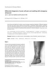
Case Study I<br />
67/M, ADL independent<br />
Good past health<br />
Gradual onset of left leg swelling <strong>for</strong> 1<br />
week<br />
Mild aching pain<br />
No history of injury<br />
No recent air travel, operation nor any<br />
drugs
Physical findings:<br />
<br />
<br />
<br />
<br />
<br />
<br />
Case Study I<br />
Afebrile and stable<br />
haemodynamics<br />
Swollen left LL from groin<br />
to calf<br />
Edematous<br />
Slightly engorged<br />
superficial viens+<br />
Palpable left groin LNs+<br />
significantly enlarged left<br />
calf circumference:<br />
L:R=39cm: 35cm
Case Study I<br />
Left dorsalis pedis strong<br />
Chest clear, Heart sounds normal, No<br />
mass felt in abdomen.<br />
CXR: rotated film, old left 8th rib<br />
fracture<br />
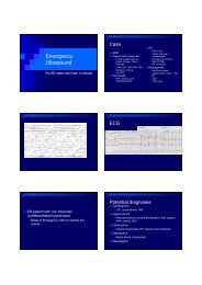
ECG: NSR 70/min, T inversion II III<br />
aVF
Case Study I<br />
Pretest Probability: high<br />
Wells score: at least 4<br />
Doppler USG in AED:<br />
<br />
<br />
<br />
Non-compressible CFV and Pop V,<br />
with echogenic intramural clots in CFV and<br />
Pop V<br />
loss of normal phasic venous blood flow and<br />
augmentation.<br />
<strong>DVT</strong> confirmed!
Case Study I<br />
SFA<br />
POP V<br />
CFV<br />
DFA<br />
Extensive <strong>DVT</strong> from CFV to POPV evidenced by non-compressibility and<br />
echogenic intramural clots.
Case Study II<br />
52/F Philipino maid, ADL independent<br />
Known RA, good control be<strong>for</strong>e. But defualted<br />
FU in Rheumatology clinic<br />
Previously on MTX, prednisolone 7.5mg QD, but<br />
ran out of medicine <strong>for</strong> more than 1 month<br />
already<br />
Sudden onset of left calf swelling <strong>for</strong> 5 days<br />
Pain on walking, no history of trauma<br />
Post menopausal, not on any HRT<br />
No recent air travel or pelvic operation
Physical Findings<br />
<br />
<br />
<br />
<br />
<br />
<br />
<br />
<br />
Case Study II<br />
Afebrile, , stable haemodynamics<br />
Mild swelling over the left calf with tenderness.<br />
Tinge of bruise over the left calf area<br />
Edema+/-<br />
Left knee not inflammed and ROM sat<br />
No cellulitis or LN palpable.<br />
Calf circumference L:R=38cm:36cm<br />
No engorged superficial veins
Case Study II<br />
Pretest Probability: low<br />
Wells score: 0<br />
Doppler USG in AED showed:<br />
<br />
<br />
<br />
<br />
Normal size and compressibility of CFV and Pop V<br />
No intramural clots seen<br />
Normal phasic venous flow and augmentation seen<br />
Baker cyst was found with extension of<br />
hypoechogenic fluid tracking between gastroc and<br />
soleus muscle. pointed configuration of the lower<br />
border +.<br />
Diagnosis: Ruptured left Baker’s s cyst, <strong>DVT</strong><br />
excluded
Case Study II
Common DDx<br />
Cellulitis/subcutaneous abscess<br />
Superficial thrombophlebitis<br />
Calf muscle tear: medical head of<br />
gastronemicus<br />
Ruptured Baker’s s cyst
Cellulitis
Superficial thrombophlebitis<br />
Non-compressibility and echogenic<br />
intramural clots of the superficial veins
Gastrocnemius tear<br />
Tear of left medial gastrocnemius<br />
muscle with <strong>for</strong>mation of hypechoic<br />
collection of fluid between<br />
gastrocnemius and soleus muscles<br />
Normal gastrocnemius <strong>for</strong> comparison
Ruptured Baker’s s cyst<br />
Appearance of Baker’s cyst in<br />
MRI<br />
USG appearance of Baker’s cyst with<br />
sign of rutpure and dissection into the<br />
calf
THANK YOU<br />
Any Questions?