Ultrasound for DVT
Ultrasound for DVT
Ultrasound for DVT

Create successful ePaper yourself
Turn your PDF publications into a flip-book with our unique Google optimized e-Paper software.
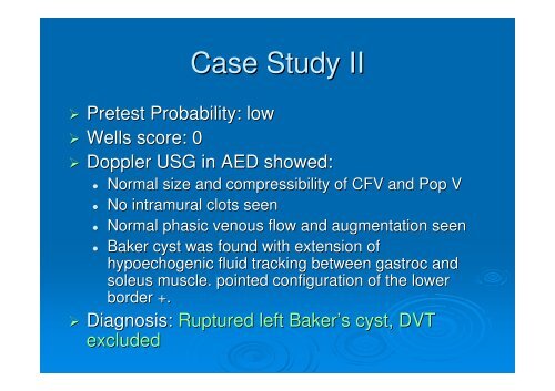
Case Study II<br />
Pretest Probability: low<br />
Wells score: 0<br />
Doppler USG in AED showed:<br />
<br />
<br />
<br />
<br />
Normal size and compressibility of CFV and Pop V<br />
No intramural clots seen<br />
Normal phasic venous flow and augmentation seen<br />
Baker cyst was found with extension of<br />
hypoechogenic fluid tracking between gastroc and<br />
soleus muscle. pointed configuration of the lower<br />
border +.<br />
Diagnosis: Ruptured left Baker’s s cyst, <strong>DVT</strong><br />
excluded