Abstracts of the Scientific Posters, 2013 AACC Annual Meeting ...
Abstracts of the Scientific Posters, 2013 AACC Annual Meeting ...
Abstracts of the Scientific Posters, 2013 AACC Annual Meeting ...
You also want an ePaper? Increase the reach of your titles
YUMPU automatically turns print PDFs into web optimized ePapers that Google loves.
Cancer/Tumor Markers<br />
Tuesday, July 30, 9:30 am – 5:00 pm<br />
as identified by MARS. The importance <strong>of</strong> glycerol and ethanolamine in <strong>the</strong> HCC<br />
MARS metabolite panel points to a potential cancer-related variation in fatty acid<br />
metabolism.<br />
released using <strong>the</strong> on-membrane deglycosylation method and labeled and analyzed by<br />
fluorophore-assisted carbohydrate electrophoresis using a capillary electrophoresisbased<br />
ABI3130 sequencer.<br />
A-13<br />
Complete mitochondrial genome sequencing reveals association with<br />
acute myeloid leukemia<br />
J. Zhou, W. Wang, Y. Ye, X. Lu, B. Ying, L. Wang. West China Hospital,<br />
Sichuan University, Chengdu, China<br />
Background: To explore mitochondrial DNA variations in acute myeloid leukemia<br />
(AML) patients.<br />
Methods: Blood or bone marrow samples <strong>of</strong> 47 AML patients (20 M1 and 27 M2)<br />
who met with WHO diagnostic criteria and 40 age- and sex-matched healthy controls<br />
were collected. The whole mitochondrial genome was directly sequenced in 24<br />
overlapping fragments, <strong>the</strong>n spliced by DNAstar s<strong>of</strong>tware, and eventually compared<br />
with Cambridge reference sequence (CRS) using CodonCode Aligner s<strong>of</strong>tware.<br />
Results: A total <strong>of</strong> 639 variations were found, twenty eight <strong>of</strong> which have not been<br />
reported at www.mitomap.org and mtDB (www.genpat.uu.se/mtDB/). 224 variations<br />
were found only in patients, two <strong>of</strong> which were statistical significant, <strong>the</strong>y were<br />
T14200C (6/47, P=0.029) in ND6 region and C14929A (14/47, P=0.000) in CYTB<br />
region. We <strong>the</strong>n compared <strong>the</strong> frequencies <strong>of</strong> <strong>the</strong> rest variations between case and<br />
control groups, C6455T in COI region and T16297C in D-loop region were found<br />
to be associated with <strong>the</strong> risk <strong>of</strong> AML. People carrying C6455T mutation had a 5.8-<br />
fold risk <strong>of</strong> developing AML (95%CI: 1.203-28.0, P=0.016), and people carrying<br />
T16297C mutation had a 4.5-fold risk (95%CI: 0.911-22.21, P=0.048). Fur<strong>the</strong>rmore,<br />
we analyzed <strong>the</strong> relationship between <strong>the</strong> above four variations and clinical features,<br />
and found that C6455T, T16297C and C14929A occurred more frequently in M1<br />
patients than in M2 patients, and patients with <strong>the</strong>se variations had higher percentage<br />
<strong>of</strong> myeloblasts in <strong>the</strong> bone marrow and higher percentage <strong>of</strong> abnormal cells in blood,<br />
While T14200C showed <strong>the</strong> opposite results.<br />
Conclusion: C6455T, T16297C, C14929A and T14200C might be used as potential<br />
biomarkers for M1/M2 patients. Fur<strong>the</strong>r studies are needed to verify <strong>the</strong>se findings in<br />
ano<strong>the</strong>r large population, and <strong>the</strong> roles <strong>of</strong> <strong>the</strong> variations play in <strong>the</strong> pathogenesis <strong>of</strong><br />
AML remains to be explored.<br />
Table 1 Characteristics <strong>of</strong> <strong>the</strong> four positive-association variations from screening <strong>the</strong><br />
whole mitochondrial genome<br />
Position Base Case Control P-<br />
change N % N %<br />
OR(95%CI) Region Codon Amino Change<br />
value<br />
6455 C-T 11(4) # 23.40% 2 5.00% 0.016 5.806(1.203-28.0) COI 184 Phe-> Phe<br />
14200 T-C 6(5) # 12.77% 0 0.00% 0.029 / ND6 158 Trp -> Trp<br />
14929 C-A 14(14) # 29.79% 0 0.00% 0.000 / CYTB 61 Thr -> Thr<br />
16297 T-C 9(2) # 19.15% 2 5.00% 0.048 4.5(0.911-22.21) D-loop<br />
#<br />
The number inside <strong>the</strong> paren<strong>the</strong>ses presented <strong>the</strong> frequencies <strong>of</strong> heterozygote<br />
A-14<br />
Prostate proteins glycosylation pr<strong>of</strong>ile and its potential as a diagnostic<br />
biomarker for prostate cancer<br />
T. Vermassen 1 , N. Lumen 2 , C. Van Praet 2 , D. Vanderschaeghe 3 , N.<br />
Callewaert 3 , P. Hoebeke 2 , S. Van Belle 1 , S. Rottey 1 , J. R. Delanghe 4 .<br />
1<br />
Department <strong>of</strong> Medical Oncology, University Hospital Ghent, Ghent,<br />
Belgium, 2 Department <strong>of</strong> Urology, University Hospital Ghent, Ghent,<br />
Belgium, 3 VIB Department <strong>of</strong> Molecular Biomedical Research - Ghent<br />
University, Ghent, Belgium, 4 Department <strong>of</strong> Clinical Chemistry, University<br />
Hospital Ghent, Ghent, Belgium<br />
Introduction: Serum Prostate Specific Antigen (sPSA) is widely used for screening<br />
and early diagnosis <strong>of</strong> prostate cancer (PCa). This analysis is associated with<br />
considerable sensitivity and specificity problems especially in <strong>the</strong> diagnostic gray<br />
zone (sPSA between 4 and 10ng/ml). In urine, PSA is only detected in its free form<br />
with concentrations largely exceeding <strong>the</strong> values observed in serum or plasma.<br />
Because <strong>of</strong> aberrant glycosylation changes in tumorogenesis, we explored <strong>the</strong> use <strong>of</strong><br />
prostate proteins and its glycosylation pr<strong>of</strong>ile as a new biomarker for PCa.<br />
Materials and Methods: We determined standard biochemical markers (total<br />
urinary protein, albumin in urine, gamma-GT in urine, urinary total PSA, urinary<br />
free PSA and sPSA) in healthy volunteers (HV; n = 16), patients with BPH (n =<br />
39), PCa patients (n = 29) and prostatitis (n = 14). Urinary protein N-glycans were<br />
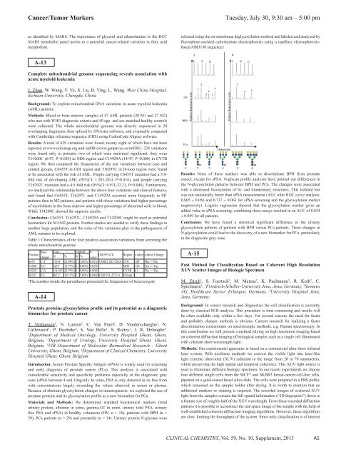
Results: None <strong>of</strong> <strong>the</strong>se markers was able to discriminate BPH from prostate<br />
cancer, except for sPSA. N-glycan pr<strong>of</strong>ile analyses have pointed out differences in<br />
<strong>the</strong> N-glycosylation patterns between BPH and PCa. The changes were associated<br />
with a decreased fucosylation <strong>of</strong> bi- and triantennary structures. This isolated test<br />
was not statistically better than sPSA measurement (AUC after ROC curve analysis:<br />
0.805 ± 0.056 and 0.737 ± 0.063 for sPSA screening and <strong>the</strong> glycosylation marker<br />
respectively). Logistic regression showed that <strong>the</strong> glycosylation marker gives an<br />
added value to sPSA screening: combining <strong>the</strong>se assays resulted in an AUC <strong>of</strong> 0.854<br />
± 0.049 for all patients.<br />
Conclusion: We have found a statistical significant difference in <strong>the</strong> urinary<br />
glycosylation patterns <strong>of</strong> patients with BPH versus PCa patients. These changes in<br />
N-glycosylation could lead to <strong>the</strong> discovery <strong>of</strong> a new biomarker for PCa, particularly<br />
in <strong>the</strong> diagnostic gray zone.<br />
A-15<br />
Fast Method for Classification Based on Coherent High Resolution<br />
XUV Scatter Images <strong>of</strong> Biologic Specimen<br />
M. Zürch 1 , S. Foertsch 2 , M. Matzas 2 , K. Pachmann 3 , R. Kuth 2 , C.<br />
Spielmann 1 . 1 Friedrich-Schiller-University Jena, Jena, Germany, 2 Siemens<br />
AG, Healthcare Sector, Erlangen, Germany, 3 University Hospital Jena,<br />
Jena, Germany<br />
Background: In cancer research and diagnostics <strong>the</strong> cell classification is currently<br />
done by classical PCR analysis. This procedure is time consuming and results will<br />
be <strong>of</strong>ten available only within a few days. For several reasons <strong>the</strong> need for faster<br />
and probably cheaper methods is obvious. Current research for realizing a faster<br />
discrimination concentrates on spectroscopic methods, e.g. Raman spectroscopy. In<br />
this contribution we will present a method relying on high resolution imaging based<br />
on coherent diffraction imaging <strong>of</strong> biological samples such as a single cell illuminated<br />
with coherent short wavelength light.<br />
Methods: Our experimental apparatus is based on a commercial ultra-short infrared<br />
laser system. With nonlinear methods we convert <strong>the</strong> visible light into laser-like<br />
light extreme ultraviolet (XUV) radiation in <strong>the</strong> range from 20 to 70 nanometers,<br />
whilst preserving <strong>the</strong> high spatial and temporal coherence. This XUV light source is<br />
used to illuminate different biologic specimen. In our recent experiments we choose<br />
four different single cells from <strong>the</strong> MCF7 and SKBR3 breast-cancer-cell-line cells,<br />
pipetted on a gold-coated fused silica slide. The cells were prepared in a PBS puffer,<br />
which remained on <strong>the</strong> sample holder after drying. It is worth to mention that no<br />
additional markers or staining is required. The recorded images <strong>of</strong> scattered XUV<br />
light from <strong>the</strong> samples contain <strong>the</strong> full spatial information (“2D fingerprint”) down to<br />
a feature size <strong>of</strong> roughly half <strong>of</strong> <strong>the</strong> XUV wavelength. From <strong>the</strong>se recorded diffraction<br />
patterns it is possible to reconstruct <strong>the</strong> real space image <strong>of</strong> <strong>the</strong> sample with <strong>the</strong> help <strong>of</strong><br />
well-established coherent diffraction imaging algorithms. However, <strong>the</strong>se algorithms<br />
are slow, limiting <strong>the</strong> throughput <strong>of</strong> <strong>the</strong> system. Since only classification is <strong>of</strong> interest<br />
CLINICAL CHEMISTRY, Vol. 59, No. 10, Supplement, <strong>2013</strong><br />
A5


