2011 (SBTE) 25th Annual Meeting Proceedings - International ...
2011 (SBTE) 25th Annual Meeting Proceedings - International ...
2011 (SBTE) 25th Annual Meeting Proceedings - International ...

You also want an ePaper? Increase the reach of your titles
YUMPU automatically turns print PDFs into web optimized ePapers that Google loves.
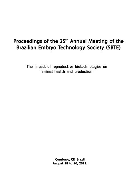
<strong>Proceedings</strong> of the 25 th <strong>Annual</strong> <strong>Meeting</strong> of the<br />
Brazilian Embryo Technolo<br />
echnology Societ<br />
ciety (<strong>SBTE</strong>)<br />
The impact of reproductive biotechnologies on<br />
animal health and production<br />
Cumbuc<br />
umbuco, CE, Brazil<br />
August 18 to 20, <strong>2011</strong>.
Acta Scientiae Veterinariae. 39 (Suppl 1): <strong>2011</strong>.<br />
From the President<br />
Dear colleagues,<br />
Welcome to Cumbuco! It is with a great pleasure that we bring, for the second time in the <strong>SBTE</strong>’s<br />
history, our <strong>Annual</strong> <strong>Meeting</strong> to Ceará State. The called “Land of Light” is one of the most beautiful areas in<br />
the Northeast of Brazil and has magnificent beaches like this in Cumbuco. There could not be a more appropriate<br />
stage for a scientific meeting and a very special moment for the professionals in the embryo technology.<br />
This year, we decided to bring to our scientific program, in addition to technologies already in use,<br />
emerging issues in reproductive technology. Also, with great satisfaction, <strong>SBTE</strong> brings the first meeting of the<br />
Innovation Network on Animal Reproduction (EMBRAPA), which presents and discusses important issues to<br />
the development of our activity. The <strong>Annual</strong> <strong>Meeting</strong>, as usual, has a high level scientific program. We hope<br />
you all enjoy.<br />
Trusting in the relevance of our meeting to the scientific progress and professional qualification, we<br />
received the institutional support from CNPq and CAPES, traditional <strong>SBTE</strong>’s partners, and now from CRMV-<br />
CE, additionally. I also thank the support of condominium of companies, which demonstrate the importance<br />
of the interaction between science and commercial activity.<br />
At the end of our administration (2010-<strong>2011</strong>), I would like to thank all members of the current board:<br />
Pietro Baruselli, Alexsandra Pereira, Rodrigo Alonso, Dárcio Teixeira, Guilherme Nogueira, Marcelo Bertolini<br />
e Luciana Melo. These people faced the great challenge that was to continue the excellent job made by the<br />
previous administrations, seeking to contribute to the advancement of our society. In the spirit of renewal, we<br />
wish success to the new board, which can count on our full support.<br />
Vicente José de Figueirêdo Freitas<br />
<strong>SBTE</strong> President - 2010-<strong>2011</strong><br />
II
Acta Scientiae Veterinariae. 39 (Suppl 1): <strong>2011</strong>.<br />
From the Scientific Committe<br />
Greetings!<br />
It has been a great pleasure to serve you and our society at the scientific committee for the past two<br />
years.With the combined efforts of intensive and productive collaboration among ourselves, along with the<br />
tireless support from our associates, post-docs and graduate and undergraduate students, we have strived to<br />
meet our commitment to you, and to bring you the most excellent scientific events and best opportunities for<br />
technical advancement as possible during our annual meetings.<br />
Our Brazilian society has grown and matured into a well respected organization worldwide. Our<br />
annual meeting has attained international proportions, and we are pleased to have many Latin-American<br />
colleagues as faithful <strong>SBTE</strong> members and regular attendees in our audience, as well as the presence of<br />
participants from many countries throughout North America and Europe. Carrying out annual meetings at<br />
an international scale has been a tradition for <strong>SBTE</strong>, with the continuous presence of distinguished invited<br />
speakers from Brazil and abroad, who come to us to address issues that are of broad relevance to our country<br />
and economy. The scientific contents of our annual meetings have been directed towards promoting progress<br />
and development to every corner where we may exert influence as professionals and/or members of academia<br />
and research institutes. To keep up with the tradition, we have carefully analyzed the topics and themes<br />
discussed at our past events over the years, with the goal of maintaining those ideas that have been greatly<br />
accepted and anticipated by the public. In addition, by envisioning the present and future of our society, we<br />
attempted to uncover fields and areas of interest that have been less frequently addressed at the meetings. In<br />
doing so, we hope to maintain aspects of the program that have been successful in our scientific discussions<br />
over the years, as well as to expand the scope of the scientific program by including advanced topics that<br />
inexorably have relevance in what we all do, either directly or indirectly, currently or in the near future to us<br />
all.<br />
We must all be aware that the scientific contents of our meetings cannot possibly cover all aspects in<br />
animal reproduction at once. However, as <strong>SBTE</strong> looks forward into long-term planning, all areas and themes N<br />
are envisioned to be covered over time. That is the reason we try to have a healthy, productive and transparent<br />
transition between administrations. Also, as <strong>SBTE</strong> is characterized by such wide diversity in terms of<br />
backgrounds, interests, and activities, the programs in the past two annual meetings (24 th and 25 th ) have been<br />
organized in sections blocked as common subjects so that you could focus your attention on specific sections.<br />
This design also allows you to use the time during sections of lower personal interest to carry out other<br />
activities related or unrelated to our event, such as for leisure and entertainment. The simultaneous preconference<br />
symposia offer you a choice of six themes that range from in-depth scientifically oriented topics to<br />
very practical and applied subjects. Following a similar pattern, the main conferences are organized in<br />
sessions covering either basic scientific topics or practical aspects, centered on the application of current<br />
biotechnology of reproduction in an array of animal species, with the bovine species as the main axis.<br />
On behalf of the scientific committee, we would like to take this opportunity to thank: (a) the members<br />
of the Executive Board, for their trust in our ability to carry out our tasks at the Scientific Committee, and for<br />
the organization of this terrific event as a whole; (b) the invited speakers, for their great insights, outstanding<br />
knowledge, and high quality of the scientific material shared with us, along with their kind patience and<br />
willingness to work hard to meet our short deadlines; (c) the Chairs of the pre-conference symposia, for<br />
organizing such exceptional sessions; (d) each member of the Scientific Committee, Section Editors, and<br />
abstract reviewers, for their tremendous efforts to help us in preparing the proceedings in time; (e) the individuals<br />
who worked relentlessly on the never-ending task of formatting and editing the proceedings, along with the<br />
support from the Editor and staff at the Journal Acta Scientiae Veterinariae; (f) the Section Chairs at the main<br />
event, for volunteering to host our invited speakers; and (g) the <strong>SBTE</strong> staff, composed of the <strong>SBTE</strong> secretary<br />
and our own students and scholars, who tirelessly helped us all during the events.<br />
Finally, your participation in the scientific program has been the very soul of our conferences. In both<br />
annual meetings (24 th and 25 th ), close to 500 abstracts have been accepted and presented at the meeting as<br />
III
Acta Scientiae Veterinariae. 39 (Suppl 1): <strong>2011</strong>.<br />
posters. Also, we had more than 60 initial abstract entries for the <strong>SBTE</strong> Student Competition Award, and<br />
numerous entries for the other three important categories of awards given by <strong>SBTE</strong>. This has been a true<br />
demonstration of your engagement to our society and trust in our efforts. For those important reasons, we<br />
thank YOU, for your faithful support to our society and for coming to our most important venue seeking novel<br />
knowledge, social and scientific interactions, open-minded discussions, and fruitful collaborations during<br />
our meeting. Again, it was truly a privilege to serve you and our society at both the 25 th anniversary (2010)<br />
and the 25 th annual meeting (<strong>2011</strong>) of the Brazilian Embryo Technology Society.<br />
I hope you enjoy this meeting. We look forward to an exciting experience shared by all!<br />
Marcelo Bertolini<br />
Chair of the Scientific Committee - 2010-<strong>2011</strong><br />
www.ufrgs.br/actavet<br />
Acta Scientiae Veterinariae. 39(Suppl 1)<br />
<strong>2011</strong><br />
IV
Acta Scientiae Veterinariae. 39 (Suppl 1): <strong>2011</strong>.<br />
25 th ANNUAL AL MEETING OF THE BRAZILIAN EMBRYO TECHNOLOGY OGY SOCIETY (<strong>SBTE</strong>)<br />
August 18 to 20, <strong>2011</strong> - Cumbuco, CE, Brazil<br />
Executive Board<br />
Vicente José de Figueirêdo Freitas,<br />
President<br />
Pietro Sampaio Baruselli, Vice-<br />
President<br />
Dárcio Ítalo Alves Teixeira, 1 st Treasurer<br />
Guilherme de Paula Nogueira, 2 nd<br />
Treasurer<br />
Alexsandra Fernandes Pereira, 1 st Secretary<br />
Rodrigo Vitório Alonso, 2 nd Secretary<br />
Luciana Magalhães Melo, Director of<br />
Communications<br />
Marcelo Bertolini, Scientific Committee<br />
Scientific Committee<br />
Marcelo Bertolini, Chair<br />
Carlos Eduardo Ambrósio<br />
Dárcio Ítalo Alves Teixeira<br />
Fabiana Forell<br />
Fernanda da Cruz Landin-Alvarenga<br />
Fidel Ovídio Castro<br />
Flávio Vieira Meirelles<br />
João Henrique Moreira Viana<br />
Jorge Luis Zambrano Varón<br />
José Luiz Rodrigues<br />
José Buratini Júnior<br />
Mário Binelli<br />
Matthew B. Wheeler<br />
Ricardo C. Chebel<br />
Section Editors<br />
Marcelo Bertolini, Editor-in-Chief<br />
Carlos Eduardo Ambrósio<br />
Fernanda da Cruz Landin-Alvarenga<br />
Flávio Vieira Meirelles<br />
João Henrique Moreira Viana<br />
José Buratini Jr.<br />
Mário Binelli<br />
Ricardo C. Chebel<br />
Manuscripts and abstract<br />
reviewers<br />
Alejandro Gibbons<br />
Alexandre Henryli de Souza<br />
Andre Penido de Oliveira<br />
Anibal Ballarotti do Nascimento<br />
Anna Carolina Denicol<br />
Anthony Cesar Souza Castilho<br />
Antônio Chaves de Assis Neto<br />
Barbara Loureiro<br />
Bruno Campos Carvalho<br />
Carlos Antônio Carvalho Fernandes<br />
Carlos Eduardo Ambrósio<br />
Carlos Jose Hoff de Souza<br />
Celina Furlanetto Mançanares<br />
Christina Ramires Ferreira<br />
Ciro Moraes Barros<br />
Claudia Lima Verde Leal<br />
Claudia Maria Bertan Membrive<br />
Daniele dos Santos Martins<br />
Dárcio Ítalo Alves Teixeira<br />
Denise Lopes<br />
Eduardo Kenji N. Arashiro<br />
Eduardo Ramos de Oliveira<br />
Erica Zimberknopf<br />
Fabiana Bressan<br />
Fabiana Forell<br />
Fabiola Freitas Paula Lopes<br />
Felipe Perecin<br />
Felipe Zandonadi<br />
Fernanda da Cruz Landin-Alvarenga<br />
Fernando Silveira Mesquita<br />
Flavia Lombardi Lopes<br />
Flávio Vieira Meirelles<br />
Frederico Ozanam Papa<br />
Gisele Zoccal Mingoti<br />
Guilherme de Paula Nogueira<br />
Hymerson Costa Azevedo<br />
Ian Martin<br />
Ines Cristina Giometti<br />
Jeferson Ferreira da Fonseca<br />
João Henrique Moreira Viana<br />
Jorge Luis Zambrano Varón<br />
José Antonio Dell’Aqua Jr.<br />
José Buratini Junior<br />
José Carlos Ferrugem Moraes<br />
Jose Luiz Moraes Vasconcelos<br />
José Luiz Rodrigues<br />
José Nélio S. Sales<br />
José Roberto Viana Silva<br />
Juliana Lopes Almeida<br />
Klibs N. Galvao<br />
Ligia Garcia Mesquita<br />
Ligia Pegoraro<br />
Lilian de Jesus Oliveira<br />
Lilian Tamy Iguma<br />
Lúcia Daniel Machado da Silva<br />
Luciana Relly Bertolini<br />
Luciano Andrade Silva<br />
Luiz Gustavo Bruno Siqueira<br />
Luiz Sergio Almeida Camargo<br />
Manoel Francisco de Sá Filho<br />
Mara Iolanda Batistella Rubin<br />
Marcelo Bertolini<br />
Marcelo Fabio Gouveia Nogueira<br />
Marcelo Marcondes Seneda<br />
Marcelo Rezende Luz<br />
Marco Aurélio Carneiro Meira Bergamaschi<br />
Marcos R Chiaratti<br />
Margot Alves Nunes Dode<br />
Maria Angelica Miglino<br />
Mário Binelli<br />
Maurício Machaim Franco<br />
Mayra Elena Ortiz D‘Avila Assumpção<br />
Michele Munk Pereira<br />
Miller Pereira Palhão<br />
Milton Passipieri<br />
Paula de Carvalho Papa<br />
Paulo Bayard Dias Gonçalves<br />
Rafael Augusto Satrapa<br />
Raquel Varella Serapião<br />
Reno Roldi de Araujo<br />
V<br />
Ricardo C. Chebel<br />
Roberto Sartori Filho<br />
Robson Fortes Giglio<br />
Rogério Ferreira<br />
Ronaldo Luis Aoki Cerri<br />
Rui Machado<br />
Sabine Wohlres Viana<br />
Sony Dimas Bicudo<br />
Vilceu Bordignon<br />
<strong>Proceedings</strong> formatting and<br />
editing<br />
N<br />
Molecular and Developmental Biology Lab<br />
University of Fortaleza (UNIFOR)<br />
Marcelo Bertolini, Editor-in-Chief<br />
Juliana Lopes Almeida, Coordinator<br />
Luciana Relly Bertolini<br />
Ana Karoline Freire da Rocha<br />
Cristiano Feltrin<br />
Felipe de Jesus Moraes Júnior<br />
Igor de Sá Carneiro<br />
Kaio César Simiano Tavares<br />
Leonardo Tondello Martins<br />
Luís Fernando Schütz<br />
Maurício Barbosa Salviano<br />
Saul Gaudêncio Neto<br />
Victor Hugo Vieira Rodrigues<br />
Chairs of the Pre-Conference<br />
Symposia<br />
Carlos Eduardo Ambrósio<br />
Cezinande de Meira<br />
Jéferson Fonseca<br />
José Buratini Jr.<br />
Maurício Machain Franco<br />
Pietro Sampaio Baruselli<br />
Acta Scien<br />
enti<br />
tiae Veterin<br />
eterinari<br />
ariae<br />
ae<br />
Laerte Ferreiro, Editor<br />
Itabajara da Silva Vaz Jr<br />
Bruna Crespo Barbosa
CONTENTS<br />
ISSN 1679-9216 (Online)<br />
Acta Scientiae Veterinariae. 39 (Suppl 1): s1-s467<br />
<strong>2011</strong><br />
PRE-CONFERENCE SYMPOSIA<br />
<strong>SBTE</strong> <strong>2011</strong><br />
SYMPOSIUM 1: STRATEGIES TO OPTIMIZE FERTILITY AND GENETIC IMPROVEMENT IN CATTLE<br />
What is producing the dramatic improvement in reproductive efficiency in U.S. dairy herds from 2000 until now? ....... s1<br />
Milo Wiltbank, Alexandre H. Souza, Jerry N. Guenther, Mary M. Herlihy & Roberto Sartori .<br />
How FTAI and FTET impact reproductive efficiency of Brazilian dairy herds ................................................................... s3<br />
Carlos Alberto Rodrigues, Roberta Machado Ferreira, Lais Mendes Vieira, Andressa L. Ranieri, Péricles R.L. Silva & Pietro<br />
Sampaio Baruselli<br />
Fixed time artificial insemination and embryo transfer programs in Brazil ..................................................................... s15<br />
Luis Fernando Nasser, Luciano Penteado, Carlos R. Rezende, Manoel F. Sá Filho & Pietro Sampaio Baruselli<br />
Implementation of DNA markers to produce genomically-enhanced EPDs in Nellore cattle ........................................ s23<br />
Raysildo Barbosa Lôbo, Donald Nkrumah, Daniela do Amaral Grossi, Priscila Sales de Barros, Pablo Paiva, Luiz Antônio<br />
Framatino Bezerra, Henrique Nunes de Oliveira & Marcos Vinícius Barbosa da Silva<br />
SYMPOSIUM 2: ADVANCES IN EMBRYO PRODUCTION AND EMBRYO TRANSFER TECHNOLOGY IN SMALL RUMINANTS<br />
State-of-the-art in the superovulation of ewes ................................................................................................................ s29<br />
Maria Emilia Franco Oliveira<br />
State-of-the-art in the transcervical embryo collection in goats and sheep ................................................................... s37<br />
Alberto Lopes Gusmão<br />
In vitro culture of goat preantral follicles ......................................................................................................................... s43<br />
José Ricardo de Figueiredo, Ana Paula Ribeiro Rodrigues & Valdevane Rocha Araújo<br />
Use of embryo transfer for the preservation of small ruminants in risk of extinction ...................................................... s45<br />
Edilson Soares Lopes Júnior & Vicente José de Figueirêdo Freitas<br />
SYMPOSIUM 3: THE INTERFACE BETWEEN HUMAN AND ANIMAL REPRODUCTION<br />
State-of-the-art in human assisted reproduction ............................................................................................................. s47<br />
Eduardo Leme Alves da Motta, Marcilio Nichi & Paulo Cesar Serafini<br />
The equine model to study the influence of obesity and insulin resistance in human ovarian function ........................ s57<br />
Eduardo Leite Gastal, Melba de Oliveira Gastal, Áurea Wischral & Jeremy Davis Schimidt<br />
Using animals to develop assisted human reproduction ............................................................................................... s71<br />
Carlos Gilberto Almodin<br />
The role of veterinarians in human in vitro embryo production ...................................................................................... s73<br />
Daniela Paes de Almeida Ferreira Braga & Edson Borges Jr.<br />
SYMPOSIUM 4: STEM CELLS AND GAMETE AND EMBRYO EPIGENETICS<br />
Veterinary applications in regenerative medicine; development of induced pluripotential stem cells (iPSC) in dogs .. s81<br />
Sehwon Koh, Steve Bischoff, Shengdar Tsai & Jorge A. Piedrahita<br />
Reprogramming somatic cells: pluripotency through gene induction and nuclear transfer ........................................... s83<br />
Fabiana Fernandes Bressan, Felipe Perecin, Juliano Rodrigues Sangalli & Flávio Vieira Meirelles<br />
Fetal membranes stem cells aplication in small animals ............................................................................................... s97<br />
Carlos Eduardo Ambrósio, Cristiane Valverde Wenceslau, José Luiz Nogueira, Dilayla Kelly de Abreu, Elaine Aparecida<br />
Fernandes Rodrigues, Thais Borges Lessa, Daniele dos Santos Martins, Luciana Relly Bertolini & Maria Angelica Miglino<br />
Reprogramming of genomic imprints by in vitro culture and cloning procedures in cattle .......................................... s103<br />
Lawrence Charles Smith, João Suzuki Jr., Felipe Perecin & Flávio Vieira Meirelles<br />
WORKSHOP 5: THE USE OF DOPPLER ULTRASONOGRAPHY ON EQUINE REPRODUCTION<br />
Doppler ultrasonography principles and methods of evaluation of the reproductive tract in mares ........................... s105<br />
Jair Camargo Ferreira, Fernanda Saules Ignácio & Cezinande de Meira<br />
VII
Acta Scientiae Veterinariae. 39 (Suppl 1): <strong>2011</strong>.<br />
Mare’s folliculogenesis: assessment of ovarian and perifollicular vascular perfusion by Doppler ultrasound ........... s113<br />
Renata Cristina Uliani, Luciano Andrade Silva & Marco Antonio Alvarenga<br />
Uterine and luteal hemodynamic evaluation of the non pregnant mare ...................................................................... s117<br />
Fernanda Saules Ignácio, Jair Camargo Ferreira & Cezinande de Meira<br />
Local effect of the conceptus on uterine vascular perfusion and remodeling during early pregnancy in mares - new<br />
findings by Doppler ultrasonography .................................................................................................................... s123<br />
Luciano Andrade Silva<br />
WORKSHOP 6: RESEARCH NETWORK IN ANIMAL REPRODUCTION<br />
The innovation network in animal reproduction: EMBRAPA’s experience in organizing a research project on<br />
reproductive biotechnology ................................................................................................................................... s135<br />
Maurício Machain Franco, Aiesca Pellegrin, Ricardo Alamino Figueiredo, Alexandre Rossetto Garcia, Luiz Sergio Almeida<br />
Camargo, Hymerson Costa Azevedo & João Henrique Moreira Viana<br />
MAIN CONFERENCE<br />
<strong>SBTE</strong> <strong>2011</strong><br />
The year 2009 worldwide statistics of embryo transfer in domestic farm animals summary of the <strong>International</strong> Embryo<br />
Transfer (IETS) Data Retrieval Committee Report ................................................................................................ s139<br />
Brad Stroud & Gabriel A. Bó<br />
Temporal historical observations, rapidly expanding technological tools, and integration of scientific disciplines to<br />
enhance reproductive performance of lactating dairy cows: the foster mothers of the human race .................... s147<br />
William W. Thatcher<br />
SESSION 1: THE FEMALE EFFECT ON ANIMAL REPRODUCTION<br />
Mechanisms involved in selection of a single dominant follicle and regression of the corpus luteum: Does a common<br />
differentiating cell type, the granulosa/large luteal cell, underlie these two disparate physiological events? .... s171<br />
Milo Wiltbank, Brian W. Kirkpatrick, Michele R. Bastos, Paulo D. Carvalho, Gulnaz Yilmazbas-Mecitaglu, Chris A. Burke,<br />
Alexandre H. Souza & Roberto Sartori<br />
Deciphering early sensor and driver properties of the endometrium: contribution of the uterus to pregnancy outcome . s173<br />
Olivier Sandra<br />
Use of applied reproductive technologies (FTAI, FTET) to improve the reproductive efficiency in dairy cattle ........... s183<br />
Ricardo C. Chebel<br />
SESSION 2: DEVELOPMENTAL BIOLOGY: FROM NORMAL TO ABNORMAL<br />
From hatching into fetal life in the pig ............................................................................................................................ s203<br />
Poul Hyttel, Kristian M. Kamstrup & Sara Hyldig<br />
Abnormalities in bovine conceptus development during the embryonic phase after in vitro fertilization (IVF) and cloning<br />
by nuclear transfer (NT) ......................................................................................................................................... s223<br />
Antônio Chaves de Assis Neto, Álvaro Riveiros Galdos, Ana Carolina Furlanetto Mançanares, Míryan Lança Vilia Alberto,<br />
Carlos Eduardo Ambrósio, Phelipe Oliveira Favaron, Flavio Vieira Meirelles & Maria Angelica Miglino<br />
The placenta of bovine clones ...................................................................................................................................... s227<br />
Pascale Chavatte-Palmer, Rita Sau Fong Lee, Sylvaine Camous, Nicolas Le Cleac’h, Hélène Jammes & Michel Guillomot<br />
Clinical disorders observed during the first 30 days of life of cloned Zebu calf ........................................................... s243<br />
Eduardo Harry Birgel Junior, Flávio Vieira Meirelles, Eliza Rossi Komninou, Mariana Tikuma Nunes, Fábio Celidonio<br />
Pogliani, Paulo Fantinato Neto, Melina Marie Yasuoka, José Rodrigo V. Pimentel, Flávia Saldanha Kubrusly & Maria Angélica<br />
Miglino<br />
SESSION 3: ADVANCED REPRODUCTIVE AND SUPPORTIVE TECHNOLOGIES<br />
Reproductive technologies and epigenetics their implications for genomic selection in cattle ................................... s253<br />
Patrice Humblot<br />
Application of functional genomic approaches to the study of placental and fetal function ......................................... s263<br />
Jorge A. Piedrahita<br />
VIII
Acta Scientiae Veterinariae. 39 (Suppl 1): <strong>2011</strong>.<br />
Derivation and potential applications of pluripotent stem cells for regenerative medicine in horses .......................... s273<br />
Olivia Eilers Smith, Bruce Douglas Murphy & Lawrence Charles Smith<br />
Recent advances in micromanipulation and transgenesis in domestic mammals ...................................................... s285<br />
Daniel Salamone, Romina Bevacqua, Federico Pereyra Bonnet, Andrés Gambini, Natalia Canel, M. Inés Hiriart, Gabriel<br />
Vichera, Lucia Moro & Javier Jarazo<br />
Genetic engineering of livestock to improve human health: The human lysozyme transgenic goat model ................ s295<br />
Elizabeth Ann Maga & James Donald Murray<br />
SESSION 4: MULTI-SPECIES<br />
Use of Color-Doppler Ultrasonography to Monitor Follicle Dynamics in Horses ......................................................... s301<br />
Eduardo Leite Gastal<br />
Biotechnology of reproduction in the canine species: where do we go? ..................................................................... s303<br />
John Verstegen, Karine Verstegen-Onclin & Karine Reynaud<br />
Increasing ovulation quota: more than a matter of energy ............................................................................................ s305<br />
Carlos G. Gutierrez, Silene Ferraro, Victor Martinez, Adriana Saharrea, Clarisa Cortez, Arantzatzu Lassala, Héctor Basurto &<br />
Joel Hernandez<br />
Ovum pick-up and in vitro embryo production in buffalo species: an update ............................................................... s317<br />
Bianca Gasparrini<br />
ABSTRACTS<br />
<strong>SBTE</strong> <strong>2011</strong><br />
STUDENT COMPETITION<br />
A001 Changes on Oocyte Quality of Repeat Breeder Holstein Cows May Explain Their Reduced Fertility ............... s337<br />
R.M. Ferreira, M.R. Chiaratti, H. Ayres, M.L. Ferraz, C.A. Rodrigues, et al.<br />
A002 Effect of Zona Pellucida Deficiency on Early Development of Bovine Embryos ................................................. s337<br />
A.E. Velasquez, J.R. Manriquez, F.O. Castro, L. Rodriguez<br />
A003 Effect of Ethanol Extract of Azadirachta indica in the Synchronization of the Cell Cycle of Bovine Fibroblasts s338<br />
N.C. Rabelo, C. Capobiango, R. Quintão, A.P. Moreira, S.G. Brito, et al.<br />
A004 Influence of High or Low Intake of Dry Matter/Energy on In Vitro Production of Bovine Embryos ...................... s338<br />
A.B. Prata, R.S. Surjus, M. Borsato, M.C.M. Silveira, M.C.C. Mattos, et al.<br />
A005 Oocyte Recovery in Queens after Contraceptive Treatment with Deslorelin Acetate (Suprelorin) .................... s339<br />
C.L. Ackermann, E. Trevisol, R. Volpato, A.A.P. Derussi, C.R.F. Guatolini, et al.<br />
A006 The Methylation Patterns of the IGF2 And IGF2r Genes in Bovine Spermatozoa are not Affected by Flow<br />
Cytometry Sex Sortin ............................................................................................................................................. s339<br />
J.O. Carvalho Neto, V.A. Michalczechen-Lacerda, R. Sartori, F.C. Rodrigues4; O. Bravim, et al.<br />
MALE REPRODUCTIVE PHYSIOLOGY AND SEMEN TECHNOLOGY<br />
A007 High Fat Diet and Genetic Dyslipidemia Impaired Testicle Morphometry in Nice: Preliminary Results ............. s340<br />
P.A.B.Cordeiro, A.C.S. Figueiredo, M.P. Palhão, I.C.S. Loyola, M.M.Gioso, et al.<br />
A008 Recommended Antibiotic for Ram Semen Cryopreservation Extender ............................................................. s340<br />
E.M. Madeida, K.L. Goularte, J. Pradieé, I. Bianchi, F.P.L. Leite, et al.<br />
A009 Qualitative Analysis of the Established Spermatogenesis in Cavia Porcellus (Linnaeus 1758) ....................... s341<br />
A. Gradela, A.K.R. Nunes, L.F.T. Martins, J.M. Santos, B.B. Gouveia, et al.<br />
A010 Glycerol and Dimethylformamide Cryopretectans Association for the Ovine Semen Cryopreservation ........... s341<br />
R.F. Bittencourt, A.L. Ribeiro Filho, G.F.O. Menezes, R.C. Andrade, L.O. Mascarenhas, et al.<br />
A011 Echogenicity Evaluation of the Testicular Parenchyma in Prepubertal Ovines .................................................. s342<br />
P.P.M. Teixeira, D.J. Cardilli, L.C. Padilha, C.C. D´Amato, M.E.F. Oliveira, et al.<br />
A012 Evaluation of Protamine and Transition Protein Gene Expression in Bovine Sperm Cells and Testicles .......... s342<br />
R. Simões, M.P. Milazzotto, F.R.O. Barros, M.A.M.M. Ferraz, M. Nichi, et al.<br />
A013 Age at Puberty in Boar Submitted to Porcine Somatotropin (PST) Treatment ..................................................... s343<br />
C. Pizoni, C.F. Demarco, I.M. Cavazini, V.R. Rabassa, A. Schneider, et al.<br />
IX
Acta Scientiae Veterinariae. 39 (Suppl 1): <strong>2011</strong>.<br />
A015 Evaluation of Different Extenders in the Cryopreservation of Bovine Spermatozoa From Cauda Epididymides<br />
Stored for 24 hours ................................................................................................................................................ s344<br />
I.C. Giometti, P.M. Papa, F.O. Papa, L.A. Oliveira<br />
A016 Evaluation of Two Different Percoll Gradients for Bovine Sperm Selection ........................................................ s344<br />
R.C. Nishimura, J.O. Carvalho Neto, M.A.N. Dode<br />
A017 Action of Nitric Oxide Routes CGMP in In Vitro Capacitation of Cryopreserved Bovine Spermatozoa .............. s345<br />
A.C.M.S. Leal, M.C.Caldas-Bussiere, C.S.P. Carvalho, C.R. Quirino, P.A.P.M. Silva<br />
A018 Testis Size, Semen Criteria and Seminal Plasma Proteins of Rams Subjected to Scrotal Insulation ................ s345<br />
A.A. Moura, D.R. Rocha, C.E.A. Souza, A.A. Araújo, M.F.Van Tilburg, et al.<br />
A019 Glass Wool Colunm vs Mini-Percoll® Gradient in Bovine Sperm Separation – Preliminary Results ................ s346<br />
S.M.P. Souza, G.M. Mican, J.A. Narváez, R.S. Fontes, C.R. Quirino<br />
A020 Comparison of Bi and Tridimensional Culture System of Equine Testicle Samples ........................................... s346<br />
I. Martin, L. Maia, C.C. Macedo, B.Vita, G.A. Monteiro, et al.<br />
A021 Efficiency Comparison of Media for the Equine Sperm Capacitation through Flow Cytometry Analysis ........... s347<br />
T.P. Gardes, R.P. Arruda, D.F. Silva, R.N.R. Cardoso, J. Nascimento, et al.<br />
A022 Comparison of Different Methods for Evaluation of Morphology, Plasm Membrane and Acrosome Integrity of<br />
Cryopreserved Bovine Semen Imported ............................................................................................................... s347<br />
E.R. Cunha, C.G. Silva, A.M. Cunha, L.O.F. Rezende, G.H.L. Martins, et al.<br />
A023 Correlation Among Osmotic Test and Different Procedures of Post-Thaw Sperm Viability Evaluation .............. s348<br />
G.F.O. Menezes, R.F. Bittencourt, L.R.A. Santos, R.C. Andrade, L.O. Mascarenhas, et al.<br />
A024 Canine Semen Cryopreservation: Identification of Critical Points ....................................................................... s348<br />
C.F. Lucio, F.M. Regazzi, L.G. Silva, D.S.R. Angrimani, M. Nichi, et al.<br />
A025 Detection of Pathogens in Bovine Semen Using Fluorescent Multiplex PCR .................................................... s349<br />
F.E.F. Dias, T.V. Cavalcante, A.K.F. Lima, C.M. Nunes, J.F. Sousa, et al.<br />
A026 Different Protocols of Thawing Ram Semen Cryopreserved in Extendes Containing Low-DensityLipoprotein s349<br />
L.C.O. Moura, M.C. Silva, A.C.D. Oliveira, M.M.Neves, P.P.N. Snoeck, et al.<br />
A027 Effect of Porcine Somatotropin (PST) on Testicular Morphological and Functional Characteristics from<br />
Prepubertal Boars ................................................................................................................................................. s350<br />
D. Perazzoli, D.F. Frigotto, C.M. Pereira, V.R. Rabassa, I. Bianchi, et al.<br />
A028 Effect of Different Concentrations of Reduced Glutathione (GSH) for Cryopreservation of Canine Sperm ....... s350<br />
C.I. Vannuchi, C.F. Lucio, L.G. Silva, F.M. Regazzi, D.S.R. Agrimani, et al.<br />
A029 Effects of Selenium and Chromium on Seminal Quality of Buffaloes Supplemented with Byproducts of<br />
Amazonian Agroindustry ....................................................................................................................................... s351<br />
A.X. Santos, A.R. Garcia, C. Faturi, B.S. Nanhúm, J.B. Lourenço Junior, et al.<br />
A030 Efficiency of the Capacitation Media on Motility Maintenance and Induction of Equine Spermatozoa<br />
Hyperactivation ...................................................................................................................................................... s351<br />
D.F. Silva, R.P.Arruda, T.P. Gardes, R.N.R. Cardoso, J. Nascimento, et al.<br />
A031 Improvement of Sperm and Seminal Quality in Nelore vs Canchim Bulls after Inclusion in a Feedlot System . s352<br />
M.M. Guardieiro, J.O. Carvalho Neto, F.L.M. Silva, M.C.M. Silveira, T.N. Rodrigues, et al.<br />
A032 Influence of Sexing on Bovine Sperm Nanoroughness Measured by Atomic Force Microscopy ...................... s352<br />
S.R. Moita, J.O. Carvalho Neto, M.A.N. Dode, L.P.Silva<br />
A033 The Influence of Culture Medium on Oviduct Cells Sperm Viability in Cattle ..................................................... s353<br />
P.S. Valleriote, H.F. Gomes, C.S.P. Carvalho, F.P. Carvalho, A.J.B. Dias<br />
A034 Influence of Period of the Year in Seminal Quality of Captive Collared Pecaries (Pecari tajacu) Raised in the State<br />
of Para ................................................................................................................................................................... s353<br />
P.R. Kahwage, A.R. Garcia, D.A.A. Guimarães, O.M. Ohashi, N.I. Albuquerque, et al.<br />
A035 Influence of Storage Container (Cylindrical and Flats) on Semen Equine Cryopreserved ................................ s354<br />
B. Fagundes, J.A. Narváez, M.F. Van Tilburg, M.A. Barreto, W. Martins, et al.<br />
A036 Improving the Quality of RNA from Bovine Semen for Use in PCR Q-Real Time ............................................... s354<br />
V.T.F. Cipriano, A. Mioranza, A. Renzi, E.S. Ramos, R.B. Lôbo<br />
A037 Experimental Model to Preserve Wild Canine Sperm Through the Freeze-Drying Process .............................. s355<br />
L.C.O. Magalhães, C.M.M. Oña, M.J. Sudano, L.F. Crocomo, D.M. Paschoal, et al.<br />
X
Acta Scientiae Veterinariae. 39 (Suppl 1): <strong>2011</strong>.<br />
A038 Spermatic Profile of Ejaculated and Epididymal Semen from Dogs Epididymides after Cooling ...................... s355<br />
D.S.R. Angrimani, C.F. Lucio, G.A.L. Veiga, A. Dalmazzo, M. Nichi, et al.<br />
A039 Lyophilized Seminal Plasma Impoves the Motility of Frozen Ram Semen and Increases the Cleavage in<br />
Heterologous IVF ................................................................................................................................................... s356<br />
R. Casali, F.C. Zago, M. Federle, J.N. Drum, M. Sponchiado, et al.<br />
A040 Use of the Powdered Coconut Water as an Alternative Extender for the Cryopreservation of Collared Peccaries<br />
(Tayassu tajacu) Semen ........................................................................................................................................ s356<br />
M.A. Silva, L.B. Campos, J.A.B. Bezerra, G.L. Lima, G.C.X. Peixoto, et al.<br />
FOLLICULOGENESIS, OOGENESIS AND SUPEROVULATION<br />
A041 Addition of Insulin to the In Vitro Culture Medium Promotes Survival and Development of Follicles Preantral<br />
Goats...................................................................................................................................................................... s357<br />
R.N Chaves, A.M.C.V. Alves, L.R. Faustino, C.A.P. Lopes, K.P.L. Oliveira, et al.<br />
A042 Analysis of Reference Genes and Expression of mRNAs for the Bmp System in Goat Ovarian Follicles Grown In<br />
Vitro and In Vivo .................................................................................................................................................... s357<br />
J.J.N.Costa, M.J. Passos, G.L. Vasconcelos, A.M.L.R. Portela, A.W.B. Silva, et al.<br />
A043 Apoptosis in Fetal Bovine and Buffaloes Ovaries by Tunel Method .................................................................... s358<br />
T.V.G. Silva, S.S.D. Santos, B.B. Silva, B.L.G. Bittencourt, R.V. Sampaio, et al.<br />
A044 Evaluation of a FSH Protocol for Transvaginal Follicular Aspiration ................................................................... s358<br />
T.C.C. Sovernigo, É.S. Santos, R. Zanin, P.R. Adona, S. Guemera, et al.<br />
A045 Comparison Between Luteal Phase Period and the Number of Follicular Waves in Bos Taurus, Bos Indicus and<br />
Bubalus Bubalis Heifers Maintained on the Same Nutritional and Environmental Status .................................. s359<br />
J.M. Baldrighi, M.F.S. Filho, J.A. Visintin, P.S. Baruselli & M.E. Assumpção<br />
A046 Behavior of a New Polymorphism on Gdf-9 Gene (Fecgsi) in Ewes of Santa Inês Breed in Front of Superovulation<br />
Protocols ................................................................................................................................................................ s359<br />
B.D.M. Silva, T.A.S.N. Silva & J.P. Neves<br />
A047 Canine Preantral Follicles Culture at Different Concentrations of Insulin in Presence of Follicle-Stimulating<br />
Hormone (FSH) ..................................................................................................................................................... s360<br />
M.K. Brasil, G.M. Silva, A.B.G. Duarte, V.R. Araújo, A.K.F. Lima, et al.<br />
A048 Follicular Dynamics in Nelore Donors Superstimulated with eCG Administered in a Single Dose or Fractioned<br />
s360<br />
L.U. Gimenes, G.F.C. Grabert, M.G.Lourenço, J. Tojal, A.M.S. Chanquetti, et al.<br />
A049 The Effect of Ecg in Multiple Ovulation Protocols for High Production Holstein Cows Embryo Donors ............. s361<br />
C.AL. Rodrigues, L.M. Vieira, A.L. Ranieri, P.R.L. Silva, H. Ayres, et al.<br />
A050 Effect of Activin-a on The Development of Bovine Secondary Follicles In Vitro Cultured .................................. s361<br />
J.R.S. Passos, A.W.B. Silva, G.L. Vasconcelos, M.J. Passos, J.J.N. Costa, et al.<br />
A051 Effect of Low Level Laser Irradiation on Mitochondrial Membrane Potential and Cycle of Bovine Cumullus Cells s362<br />
C.A. Soares, K. Annes, T.R. Dreyer, H.S. Martinho, S.S. Nicolau, et al.<br />
A052 Effects of Ovarian Ortotopic Autotransplantation in Reproductive Behavior in Goats (Capra hircus) ................ s362<br />
N.A. Carmo, S.A. Marcondes, F.A. Santos, G.H.F. Moura, T.L. Nunes, et al.<br />
A053 Effect of Ecg on Ovulation and Conception Rates in Red Sindhi Cows (Bos taurus indicus) Treated with Two<br />
Synchronization of Ovulation Protocols ................................................................................................................ s363<br />
R.R.C. Mello, J.E. Ferreira, A.P.T.B. Silva, L.M. Mascarenhas, B.J.F. Silva, et al.<br />
A054 Timing Effect of Insemination Using Sex-Sorted Sperm in Embryo Production with Nelore (Bos indicus)<br />
Superovulated Donors .......................................................................................................................................... s363<br />
J.G. Soares, N.A.T. Carvalho, P.S. Baruselli, C.M. Martins, M. Nichi, et al.<br />
A055 Efficiency of Different Treatments of Ovarian Cysts in Dairy Cows ..................................................................... s364<br />
T.A. Miyauchi, C.A.C. Fernandes, M.R.P. Portinari, M.M. Gioso, B.F.L. Alves, et al.<br />
A056 Stability of Reference Genes and Levels of mRNA for The IGF System in Bovine Ovarian Follicles Grown In Vitro<br />
and In Vivo ............................................................................................................................................................. s364<br />
G.L. Vasconcelos, A.W.B. Silva, J.J.N. Costa, M.J. Passos, R.O.D.S. Rossi, et al.<br />
A057 Expression of mRNA Encoding Fgfr1b and Fgfr2b in Bovine Fetal Ovaries during Gestation .......................... s365<br />
R.B.Silva, E.S. Caixeta, C. Price & J.B. Junior<br />
XI
Acta Scientiae Veterinariae. 39 (Suppl 1): <strong>2011</strong>.<br />
A058 Leukemia Inhibitory Factor Stimulates the In Vitro Development of Sheep Preantral Follicles and the Production<br />
Of Embryos ............................................................................................................................................................ s365<br />
V.B. Luz, T.F.P. Silva, V.R. Araújo, A.B.G. Duarte, J.J.H. Celestino, et al.<br />
A059 Effects of FSH and Growth Differentiation Factor -9 (GDF-9) on In Vitro Growth of Bovine Secondary Follicless366<br />
R.P. Ribeiro, G.L. Vasconcelos, A.W.B. Silva, M.J. Passos, F.T.G. Bezerra, et al.<br />
A060 Influence of Protocols for Superovulation on Induced Stress of Brown Brocket Deer (Mazama Gouazoubira) s366<br />
E.S. Zanetti & J.M.B. Duarte<br />
A061 Steady-State Levels of Vasoactive Intestinal Peptide mRNA in Goat Ovaries and Its Effect on the In Vitro<br />
Development of Isolated Preantral Follicles ......................................................................................................... s367<br />
J.B. Bruno, V.R. Araújo, J.J.H. Celestino, M.V.A. Saraiva, R.M.P. Rocha, et al.<br />
A062 Standard of Emergency Follicular Waves in Long Protocols (With or Without CIDR Replacement) and<br />
Seasonality Effect in Santa Ines Ewes .................................................................................................................. s367<br />
M.E.F. Oliveira, C.C. D’amato, H. Ayres, L.G. Oliveira, P.P.M. Teixeira, et al.<br />
A063 Behavioral Parameters of Dairy Goats Subjected to Synchronization with PGF2a ............................................ s368<br />
A.F. Silva, L.V. Esteves, F. Zandonadi, R.C. Cruz & J.F. Fonseca<br />
A064 Expression Profile of Candidate Genes for the Acquisition of Competence during Oocyte Growth in Bovine .. s368<br />
I.R. Bessa, R.C. Nishimura, F. Paulini, M.M. Franco & M.A.N. Dode<br />
A065 Antral and Preantral Follicular Population in Nelore Ovaries with High and Low Antral Follicular Counting:<br />
Preliminary Results ............................................................................................................................................... s369<br />
G.M.G. Santos, K.C. Silva-Santos, C. L. Schneider, L.S. Siloto, F. Morotti, et al.<br />
A066 Follicular Proportion and Population of Rats Submitted to Different Procedures of Ovarian Autologous<br />
Transplantation ...................................................................................................................................................... s369<br />
M.F. Macedo, M.B. Bezerra, N.A. Carmo, F.F.P.C. Barros & W.R.R. Vicente<br />
A067 First Successful Estrous Synchronization Using a Prostaglandin Analogue in Collared Peccaries (Tayassu<br />
Tajacu) ................................................................................................................................................................... s370<br />
A.R. Silva, K.M. Maia, M.A. Silva, G.C.X. Peixoto, J.A.B. Bezerra, et al.<br />
A068 Cellular Proliferation of Poliovular Follicles in Adult Nelore Ovaries .................................................................. s370<br />
K.C. Silva-Santos, G.M.G. Santos, R.L. Oliveira, L.S. Siloto, A.P.F.R.L. Bracarense, et al.<br />
A069 Superovulatory Response and Pregancy Rates of Holstein Cows In Vivo Embryo Production Within Different<br />
Catagories During Summer and Winter ................................................................................................................ s371<br />
L.M. Vieira, C.A. Rodrigues, P.R.L. Silva, J.N.S. Sales, M.F.S. Filho, et al.<br />
A070 Superovulation of Nelore Heifers Using Single Injection of Folicle Stimulant Hormone Carried Into Polimeric<br />
Matrix ..................................................................................................................................................................... s371<br />
E.M. Mogollon-Waltero, H.J. Narvaez, R. Costa, C.R. Quirino & R.S. Fontes<br />
A071 Energy Supplementation in Santa Inês Sheep ................................................................................................... s372<br />
S.S. Venturi, I.R. Soares, A.F. Silva, E.C. Cardoso, A.R. Rodrigues, et al.<br />
A072 Variation on Vascularization of Follicles Induced to Ovulate with HCG and GnRH Evaluated by Doppler<br />
Ultrasonography - Preliminary Results ................................................................................................................. s372<br />
R.C. Uliani, L A Silva & M.A. Alvarenga.<br />
A073 Viability of Canine Preantral Follicles Cultured In Vitro after Conservation in an Oocyte Transporter ............... s373<br />
R.J.S. Gonçalves, V.R.P. Barros, A.P. Almeida, A.B.G.Duarte, V.R. Araújo, et al.<br />
A074 Ascorbic Acid Improves the Survival and In Vitro Growth of Isolated Caprine Preantral Follicles ..................... s373<br />
G.M. Silva, C.H. Lobo, V.R. Araújo, A.B.G. Duarte, A.A. Moura, et al.<br />
FTAI, FTET AND AI<br />
A075 Follicular and Reproductive Traits in Short-Term Progesterone and PGF2alpha Based Estrous- Synchronization<br />
Protocol in Sheep .................................................................................................................................................. s374<br />
R.V. Allende, F.R. Saravia, E.N. Lara, A.F. Leiva; T. Díaz, et al.<br />
A076 Effect of CIDR® Reutilization in a Progesterone-PGF2alpha Based Estrous-Synchronization Protocol on Estrous<br />
Presentation and Ovulatory Performance in Sheep .............................................................................................. s374<br />
F.R. Saravia, R.V. Allende, E.N. Lara, A.F. Leiva, T. Diaz, et al.<br />
XII
Acta Scientiae Veterinariae. 39 (Suppl 1): <strong>2011</strong>.<br />
A077 Effect of Estradiol Benzoate at CIDR Insertion on Ovarian Traits after Short-Term Progesterone and PGF2alpha<br />
Based Estrous-Synchronization Protocol in Sheep .............................................................................................. s375<br />
J.F. Cox, E.N. Lara, A.F. Leiva, H. Muñoz, R.V. Allende, et al.<br />
A078 TAI Increases the Reproductive Performance of Lactating Primiparous Acyclic Nelore Cows .......................... s375<br />
R.R. Cunha, M.M. Gioso, R.J. Carvalho, C.A.C. Fernandes, M.P. Palhão, et al.<br />
A079 Occurrence of Estrus after Synchronization Improve the Use of Sex Semen in Timed Artificial Insemiantion<br />
Protocol .................................................................................................................................................................. s376<br />
P.S. Baruselli, E.K. Abe, R.V. Sala, M. Nichi, E.P. Campos Filho, et al.<br />
A080 The Use of Bioabortogen® and Bioleptogen® Vaccines may Improve Reproductive Efficiency of Beef Cows . s376<br />
M. Sivieri, R.M. Ferreira, D.P. Borges, T.S. Antonio, R.L. Gonçalves, et al.<br />
A081 Evaluation of Artificial Insemination in Girolando Heifers with Sexed Semen ................................................... s377<br />
M.F. Mendanha, S. Ferrari & D.S. Silva<br />
A082 Evaluation of Different Doses of eCG in the TAI Conception Rate in Cycling Nelore Heifers Synchronized with<br />
Intravaginal Progesterone Device Previously Used For 9 Days .......................................................................... s377<br />
E.R. Carvalho, A.D.P. Rodrigues, T. Martins, H.B. Graff, R.F.G. Peres, et al.<br />
A083 Evaluation of the Effects of Heat Stress on Reproductive Performance of Nelore Heifers during the Breeding<br />
Season................................................................................................................................................................... s378<br />
M. Maturana Filho, N.A. Teixeira, S.C. Scolari, A. Kehrle, P.H.P. Miguez, et al.<br />
A084 The Characteristics of the Folicular Dynamics Over the Diameter of the Corpus Luteum in Fixed Time Embryo<br />
Transfer Recipients ................................................................................................................................................ s378<br />
A.L. Ribeiro Filho, B.H.A. AndradeT.P. Coutinho, L.M.C. Feitosa, M.V.G. Loiola, et al.<br />
A085 Follicular Features in Murrah, Mediterranean and Crossbred Buffalo Heifers Submitted to Exogenous Control of<br />
Ovulation................................................................................................................................................................ s379<br />
A.R. Garcia, G.R. Silva, B.S. Nahúm, J.S. Pessoa, A.A. Gonçalves, et al.<br />
A086 Comparative Pregnancy Rates of High-Producing Holstein Cows Synchronized for FTAI with Progesterone<br />
Estradiol-Based Protocol or Double...................................................................................................................... s379<br />
A.L. Ranieri, C.A. Rodrigues, L.M. Vieira, P.R.L. Silva, G.B. Gouvea, et al.<br />
A087 Comparison of the Conception Rates of Nelore Lactating Cows Submitted to FTAI and Treated with Porcine<br />
Pituitary Extract and Equine Chorionic Gonadotropin .......................................................................................... s380<br />
S.M. González, J.T. Campos, W. Blaschi, T.R.R. Barreiros & M.M. Seneda<br />
A088 Comparison of Rates of Conception and Pregnancy Losses in Different Years in Nelore Cows under FTAI<br />
Vaccinated or not Against BVDV and BoHV-1....................................................................................................... s380<br />
T.R.R. Barreiros, A.A.O. Friedel, R.A.F. Stellato, S.M. González, W. Blaschi, et al.<br />
A089 Conception Rates in Postpartum Nelore Cows Treated with eCG Before and/or after Fixed-Time Artificial<br />
Insemination .......................................................................................................................................................... s381<br />
A.P. Lemes, A.D.P. Rodrigues, R.F.G. Peres, H.B. Graff, E.R. Carvalho, et al.<br />
A090 Intracornual Insemination with Sex Sorted Sperm does not affect the Pregnancy per AI after Timed Artificial<br />
Insemination .......................................................................................................................................................... s381<br />
R.V. Sala, M.F. Sá Filho, R.W. Girotto, M. Nichi, E.P. Campos Filho, et al.<br />
A091 Effect of New and Reused Vaginal Inserts on Plasma Progesterone Profile and Follicular Size at the End of<br />
Progesterone and Prostaglandin F2alpha Estrous Synchronization Treatment in Beef Cows ............................ s382<br />
E.N. Lara, A.F. Leiva, C. Baeza, A. Rodríguez, P. Rojas, et al.<br />
A092 Dose Effect of Estradiol Benzoate Associated with Progesterone on the Synchronization of Follicular Wave<br />
Emergence in Bos Indicus and Bos Taurus Cows ................................................................................................ s382<br />
M.R. Bastos, R.S. Surjus, A.B. Prata, M.A.P. Meschiatti, M. Borsato, et al.<br />
A093 Effect of Estrous Detection on Pregnancy Rate in Multiparous Nelore (Bos Indicus) Cows Submitted to a Timed AI<br />
in the Brazilian South Pantanal ............................................................................................................................. s383<br />
J.C. Borges, M.R. Silva, I.C. Canavari, R.R. Raineri, J.F. Massoneto, et al.<br />
A094 Effect of hCG and/or CIDR in Lactating Dairy Cattle on Circulating Progesterone and Conception Rates ....... s383<br />
A.B. Nascimento, A.H. Souza, M. Herlihy, A. Keskin, F.P. Dalla Costa, et al.<br />
A095 Effect of the Ovulatory Follicle Diameter on the Pregnancy Rate of Lactating Cows Fixed Time Inseminated with<br />
Low Progesterone Protocols ................................................................................................................................. s384<br />
L.F.M. Pfeifer, S.C.B.S. Leal, A. Schneider, E. Schmitt & M.N. Corrêa<br />
XIII
Acta Scientiae Veterinariae. 39 (Suppl 1): <strong>2011</strong>.<br />
A096 Effect of Synchronization Follicular Wave Method on Bovine Superovulatory Response ................................. s384<br />
J.E. Ferreira, R.R.C. Mello, A.P.T.B. Silva, L.M. Mascarenhas, B.J.F. Silva, et al.<br />
A097 Effect of Treatment with GnRH Analogue at the Moment of Insemination on Pregnancy Rate in Primiparous<br />
Nelore (Bos Indicus) Cows Submitted to a Timed AI in the Brazilian South Pantanal ......................................... s385<br />
M.R. Silva, J.F. Massoneto, D.B. Marinho, I.C. Canavari, R.R. Raineri, et al.<br />
A098 Efficiency of Different Stimuli Gonadotropin in Resynchronization Nellore Lactating Cows Submitted to FTAI s385<br />
J.T. Campos, S.M. González, W. Blaschi, T.R.R. Barreiros & M.M. Seneda<br />
A099 Eficiency of Timed Artificial Insemination Protocol for Holstein Cows Reared under Semiarid Condition ........ s386<br />
S.I. Guido, A.S. Santos Filho, L.C. Pereira, J.C.O. Andrade, F.C.L. Guido, et al.<br />
A100 Luteolytic Effectiveness of Prostaglandina Tortuga® in Bovine Cattle ............................................................... s386<br />
M.S. Baruselli, R.M. Ferreira, M.F. Sá Filho, L.F.M. Tamassia, C.A. Nascimento, et al.<br />
A101 Establishment of a Synchronization of Ovulation Protocol for Fixed Time Embryo Transfer Applied on Dairy Cattle<br />
Genetic Improvement Program of Acre State ........................................................................................................ s387<br />
R. Satrapa, J.V.A. Diniz, F.L. Dantas, R.R. Marcelino, N.L. Vasconcelos, et al.<br />
A102 Use of the Estradiol Benzoate to Induction Follicular Regression in Mares Treated with Intravaginal Progesterone<br />
Device .................................................................................................................................................................... s387<br />
C. Meira, J.C. Ferreira & A.I.N. Boff<br />
A103 Influence of the Uterine Lavage to Endometrial Cytology on the Bovine Fertility ............................................... s388<br />
H.E. Thomé, R.P. Arruda, B.M.M. Oliveira, M. Maturana Filho, G.C. Oliveira, et al.<br />
A104 Artificial Insemination in Pheasants Lodged in Individual Cages ....................................................................... s388<br />
O.A. Resende, J. Almeida & E.P. Ponte<br />
A105 Early Embryo Mortality in Beef Cows after Fixed Time Artificial Insemination (FTAI) and Hormonal Treatmentss389<br />
R. Machado, M.J. Sudano, M.A. Bergamaschi, G.P. Nogueira, C.M.B. Membrive, et al.<br />
A106 Modified Method of Artificial Insemination with Cervical Retraction in Dorper Ewes ......................................... s389<br />
M.F. Cordeiro, T.V.C. Nascimento, C.H.S.C. Barros, L.C. Magalhães, A.P.O. Monte, et al.<br />
A107 Ovarian Parameters of Dairy Goats Undergoing Synchronization with PGF2α Associated or Not with the Use of<br />
hCG in the Beginning of Estrus ............................................................................................................................. s390<br />
L.V. Esteves, F. Zandonadi, A.F. Silva, S.S. Venturi & J.F. Fonseca<br />
A108 The Protocol for FTAI with Estradiol Cypionate does not Require Additional GnRH and Allows Insemination in<br />
Two Periods ........................................................................................................................................................... s390<br />
F.P. Torres, R.M. Ferreira, F.A. Lima, M.B. Veras, F.R.F. Silva, et al.<br />
A109 Synchronization and Resynchronization Using a Norgestomet Ear Implant in Nelore Heifers Timed Artificial<br />
Inseminated ........................................................................................................................................................... s391<br />
M.F. Sá Filho, R.A.I. Oliveira, R.V. Sala, G.G. Macedo, R.W. Girotto, et al.<br />
A110 Synchronization of Ovulation in Water-Buffalo Heifers with Estradiol Benzoate or Gonadorelin ...................... s391<br />
M. Nichi, N.A.T. Carvalho, J.G. Soares, D.C. Souza & P.S. Baruselli<br />
A111 Synchronization and Induction of Fertile Estrus of Paca (Cuniculus Paca L.) .................................................... s392<br />
V.M.F. Ribeiro, R. Satrapa, J.V.A. Diniz, R.A. Satrapa & R. Rumpf<br />
A112 Supplementation of Nelore Cows with Rumen-Protected Fat Before and/or After TAI ....................................... s392<br />
I.M. Sokoloski, M.M. Guardieiro, A.B. Prata, L.H.D. Carrijo, G.B. Mourão, et al.<br />
A113 Pregnancy Rates of Different Protocols for Fixed Time Transfer of In Vitro Produced Bovine Embryos ............ s393<br />
T.P. Coutinho, Y.F. Watanabe, L.M.C. Feitosa, M.V.G. Loiola, B.H.A. Andrade, et al.<br />
A114 Pregnancy Rate and Embryonic/Fetal Loss After eCG Administration Given 5 or 10 Days After Insemination in<br />
Cycling Ewes ......................................................................................................................................................... s393<br />
C. García-Pintos, P.C. Santos-Neto, A. Menchaca<br />
A115 Conception Rates in Beef Cattle Given a New or Previously Used Progesterone Device in Fixed-Time Artificial<br />
Insemination .......................................................................................................................................................... s394<br />
A. Kehrle, M. Pereira Neto, M. Maturana Filho, P.H.P. Miguez, G.C. Gomes, et al.<br />
A116 Use of DIB® and Crestar® to Synchronize Ovulation and FTAI in Buffalo Heifers during the Non-Breeding<br />
Season................................................................................................................................................................... s394<br />
N.A.T. Carvalho, J.G. Soares, E.L. Reis & P.S. Baruselli<br />
XIV
Acta Scientiae Veterinariae. 39 (Suppl 1): <strong>2011</strong>.<br />
A117 Reproductive Ultrasonography for Use of High Cost Semen in FTAI Programs ................................................ s395<br />
J.R.S. Torres Júnior, H.M.V.B. Aguiar & P.H. Cavalcanti<br />
A118 Pregnancy Rate Monthly Variation of the Nellore Cows Submitted to the FTAI in Bolivian Farm ....................... s395<br />
E. Nogueira, M.R. De Paula, H.R. Marques Junior & A.S. Silva<br />
OPU-IVP AND ET<br />
A119 Ultrasound-Guided Follicle Aspiration Do Not Reduce Blood Flow in the Follicular Wall - Preliminary Results ... s396<br />
A.M. Ghetti, F. Zandonadi, l.G.B. Siqueira, l.S.A. Camargo, E.K.N. Arashiro, et al.<br />
A120 Evaluation of Oocytes Recovered from Cows Undergoing Immunosuppressive Protocol with Dexamethasone .. s396<br />
A.P. Oliveira, P.M.P. Nascimento, J.M. Bicalho, G.G. Carvalho, R.S. Diniz, et al.<br />
A121 Evaluation of a Medium Based on Coconut Water (Cocos Nucifera) To In Vitro Embryo Production of Bovine s397<br />
M.S. Cordeiro, P.C.A. Ramos, N.N. Costa, T.V.G. Silva, P.P.B. Santana, et al.<br />
A122 Evaluation of a New Method for Identifying Endometritis in Anestrous Mares through Ultrassonography ........ s397<br />
G.M.Greco, L.M.P. Maia, M.M.C. Chaves & M.A. Alvarenga<br />
A123 Hemodinamic Evaluation of the Uterus in Mares Used as Recipient: Partial Results ........................................ s398<br />
F.S. Ignácio, J.C. Ferreira, R.P. Melo, N.C. Abreu & G.H.M. Araújo<br />
A124 Comparison of the Pregnancy Rate of Embryos Coming of Ovulation in Mares Single or Double Race Polo<br />
Argentino ............................................................................................................................................................... s398<br />
T.D. Reis, M.M. Higuti, J.T. Paula, S.M. González, E. Bernardi, et al.<br />
A125 Comparison between the Number of Aspiraded Follicles by Videolaparoscopy and the Oocyte Recovery in<br />
Dorper and Santa Inês Ewes ................................................................................................................................ s399<br />
A.S. Rodrigues, J.S. Chacon, A.L. Ribeiro Filho, B.H.A. Andrade, M.V.G. Loiola, et al.<br />
A126 Correlation Of Kinetics Of Development And Sex Of In Vitro Produced Bovine Embryos ................................. s399<br />
A.R. Pupulim, Á.R. Buzzo, J. Mazucheli, F.V. Meirelles, R.Z. Puelker, et al.<br />
A127 Different Volumes and Centrifugations Time of Percoll Gradient Do Not Affect the Sperm Quality and Embryo<br />
Development of In Vitro Produced Bovine Embryos ............................................................................................ s400<br />
L.L. Vianna, J. Pradieé, E.C.S. Santos, A.O. Gonçalves, L. Anghinoni, et al.<br />
A128 Cumulative Effect of Bovine Recombinant Somatotropin (Rbst) on the In Vitro Production of Embryos from<br />
Lactating Guzolando Females .............................................................................................................................. s400<br />
F.J. Moraes Junior, L.F. Schütz, C. Feltrin, L.T. Martins, S. Gaudencio Neto, et al.<br />
A129 Effect of High Energy Diet on Metabolic, Endocrine and Reproductive Parameters on Bos Indicus and Bos<br />
Taurus Cows .......................................................................................................................................................... s401<br />
J.N.S. Sales, L.T. Iguma, C.C.R. Quintão, M.A.S. Gama, C. Freitas, et al.<br />
A130 Effect of the Sperm Selection with Isolate ® or Percoll ® on Sperm Quality and In Vitro Bovine Embryo Development<br />
for Frozen-Thawed Sexed and Non-Sexed Semen .............................................................................................. s401<br />
P.R. Villamil, M.E.G. Gomez, M.F. Taranco, M. Caccia & G. Bó<br />
A131 The Effect of Follicle Wave Synchronization and eCG Treatment on Cumulus Oocyte Complex Recovery Rates<br />
and Quality in Brangus and Angus Donors .......................................................................................................... s402<br />
F.L. Ongaratto, P.R. Villamil, A. Tribulo & G. Bó<br />
A132 Effect of FSH Incorporated in a Polimeric Matrix of Pluronic F-127® about the Quality of Oocytes Collected by<br />
Follicular Aspiration in Zebu Heifers ..................................................................................................................... s402<br />
H.J. Narvaez, R.S. Fontes, G.M. Mican & C.R. Quirino<br />
A133 Effect of Corpus Luteum on the Recovery Rate and Number of Oocytes in Crossbred Zebu Heifers ............... s403<br />
B.P. Carvalho, F.Q. Costa, F.B. Rosa, A.M. Arrais, R.C. Maia, et al.<br />
A134 Effect of Stage of Development on the Sex Ratio of Pregnancies Originated from In Vitro Produced Embryos s403<br />
L.M. Gouvêa<br />
A135 Effect of the Method for the Retrieval of Cumulus-Oocyte Complexes on the In Vitro Production Efficiency of<br />
Sheep Embryos ..................................................................................................................................................... s404<br />
M.B. Salviano, C. Feltrin, J.L. Almeida, K.C.S. Tavares, F.J. Moraes Junior, et al.<br />
A136 Efficiency of OPU-IVP-Et of Fresh and Vitrified Embryos in Buffalo .................................................................... s404<br />
W.P. Saliba, R.M. Drumond, H.X. Bayão, M.T. Alvim, P.S. Baruselli, et al.<br />
XV
Acta Scientiae Veterinariae. 39 (Suppl 1): <strong>2011</strong>.<br />
A137 Efficacy of Short Protocols with CIDR® on Estrus Synchronization of Embryos Recipients Ewes .................... s405<br />
F.S. D´Angieri, J.C.B. Silva, W.K. Okabe, M. Maturana Filho & A.S. Traldi<br />
A138 Fertility of Recipients Transferred with Embryos in Oviduct by Two Days of Santa Inês and Dorper Sheep In Vitro<br />
Produced ............................................................................................................................................................... s405<br />
E.A.B. Araujo, S.N. Oliveira, A.C. Basso, J.S. Chacon, M. Chalhoub, et al.<br />
A139 Apoptosis in Fresh and Vitrified In Vitro and In Vivo Produced Bovine Embryos ............................................... s406<br />
M.J. Sudano, D.M. Paschoal, T.S. Rascado, M.D. Guastali, R.R.D. Maziero, et al.<br />
A140 Influence of Lineage of Oocyte Donor on the Yield and Morphology of Oocytes Recovered By Ultrasound-<br />
Guided Follicular Aspiration in Nellore Cows ....................................................................................................... s406<br />
A. Martins Júnior, R.S. Calegari, D.M. Paschoal, D.G. Souza & M.J. Sudano<br />
A141 Influence of the Sperm Pre-Incubation on Cleavage Rates, Blastocyst Production and Sex Distribution of In Vitro<br />
Produced Bovine Embryos .................................................................................................................................... s407<br />
A.G. Curcio, R.S. Fontes, S.R. Leal, C.R. Quirino, G.M. Mican, et al.<br />
A142 Influence of Semen on the Pregnancy Rate in Mares under Embryo Transfer in the Northeast of the State of Para ........ s407<br />
S.T. Rolim Filho, F.O. Barbosa Filho, H.F.L. Ribeiro, E.Y.E. Mesquita, K.C. Sousa, et al.<br />
A143 IGF-I (Insulin-Like Growth Factor I) Increases In Vitro Production, but Does Not Protect Embryos from Deleterious<br />
Effect of Heat Stress in Nelore (Bos indicus) and Holstein (Bos taurus) Breeds ................................................. s408<br />
A.C.S. Castilho, R.A. Satrapa, E.M. Razza, C.F. Silva, R. Simões, et al.<br />
A144 Treated Oocytes with Retinoids and with Retinoids and Growth Factor Improve the In Vitro Embryos Production<br />
and Inhibit the Apoptosis? ..................................................................................................................................... s408<br />
R.M. Chaves, J.C. Conceição, M.A.V. Silva, E.R. Santos Júnior, L.M. Freitas Neto, et al.<br />
A145 Gene Expression Profile of IGF System Members on In Vitro Produced Bovine Blastocysts: Comparison between<br />
Nelore (Bos Indicus) and Holstein (Bos taurus) ................................................................................................... s409<br />
R.A. Satrapa, A.C.S. Castilho, R. Simões, E.M. Razza, C.F. Silva, et al.<br />
A146 Potential of the Gyr (Bos taurus indicus) and Holstein (Bos taurus taurus) Donors for Embryo Production ...... s409<br />
J.H.M. Viana, T.M. Miyauchi, T.A. Miyauchi, E.R. Oliveira, J.A.D. Garcia, et al.<br />
A147 Preservation of Intrafollicular Oocytes at 4°C ...................................................................................................... s410<br />
R.S. Ferro & O.S. Garcia<br />
A148 Production of Ovine Oocytes from Slaughterhouse Ovaries Using Two Collection Procedures ........................ s410<br />
P.M. Rafaelli, P.Z.D. Pumará, P. Ortiz & J.I. Ernesto<br />
A149 In Vitro Embryo Production (IVEP) after the Laparoscopic Oocyte Recovery (LOR) In Canindé Goats ............ s411<br />
A.F. Pereira, R.R. Moura, R.I.T.P. Batista, J.M.G. Souza, A.S. Alcântara Neto, et al.<br />
A150 In Vitro Embryo Production Using Frozen Semen Previously Infected with Bovine Herpesvirus Type 5 ........... s411<br />
D.G. Souza, A. Martins Júnior, R.S. Calegari, J.O. Caldeira, C. Silva-Frade, et al.<br />
A151 In Vitro Production of Ovine Embryos of Dorper and Santa Ines Breed Aspirated by Videolaparoscopy and<br />
Transported over Long Distances ......................................................................................................................... s412<br />
M. Chalhoub, A.C. Basso, J.S. Chacon, A.L. Ribeiro Filho, M.V.G. Loiola, et al.<br />
A152 In Vivo Embryo Production in Cows Superovulated 1 or 2 Days after OPU ....................................................... s412<br />
R.S. Surjus, A.B. Prata, M. Borsato, F.C.S.Z. Mattos, M.C.M. Silveira, et al.<br />
A153 Hormonal Protocols for Oocyte Retrieval and In Vitro Production of Embryos in Bovine Donors ...................... s413<br />
T.M. Miyauchi, T.A. Miyauchi, M.P. Palhão, A.S. Camargos, C.A.C. Fernandes, et al.<br />
A154 Technical Report: Ovun Pick Up by Single for Recovery of Oocytes in Mares in Transition Period .................... s413<br />
A.G. Rigo, B.V. Sanches, J.H.F. Pontes, L.L. Moino, R.M. Untura, et al.<br />
A155 Ovulatory Response of Cycling Mares Stimulated with an Association of pFSH and EPE ................................ s414<br />
N.C. Abreu, C. Meira, F.S. Ignácio & G.H.M. Araújo<br />
A156 Pregnancy Rate in Equine Embryo Recipients Treated with Flunixin Meglumine ............................................. s414<br />
J.P. Oliveira, H.L. Resende & J.C.F, Jacob<br />
A157 Pregnancy Rate in Mangalarga Marchador Recipients at Different Ages with or without Progesterone<br />
Supplementation ................................................................................................................................................... s415<br />
S.O. Paiva, C.G.M. Oliveira, P.C. Almeida, M.A.F. Sá, R.G.C. Junqueira, et al.<br />
XVI
Acta Scientiae Veterinariae. 39 (Suppl 1): <strong>2011</strong>.<br />
A158 Conception Rates of In Vitro Produced Bovine Embryos with Conventional and Thawed-Sexed Sperm of Nellore<br />
Bulls ....................................................................................................................................................................... s415<br />
J.C.B. Silva, W.K. Okabe, M.C.C. Mattos, B.V. Sanches, J.H.F. Pontes, et al.<br />
A159 Use of Retinoids to Inhibit the In Vitro Apoptosis of Caprine Oocytes and Embryos ......................................... s416<br />
J.C. Conceição, R.M. Chaves, M.A.V. Silva, E.R. Santos Júnior, L.M. Freitas Neto, et al.<br />
A160 FSH in Protocols for Follicular Wave Synchronization and Embryo Production in Taurin and Zebu Donnors .. s416<br />
C.A.C. Fernandes, A.C.S. Figueiredo, B.F.L. Alves, E.R. Oliveira, T.C. Rossi, et al.<br />
EMBRYOLOGY, BIOLOGY OF DEVELOPMENT AND PHYSIOLOGY OF REPRODUCTION<br />
A161 Use of b-Mercaptoethanol for In Vitro Maturation and Culture of Ovine Embryos .............................................. s417<br />
J. Pradieé, L.L. Vianna, A.O. Gonçalves, E.C.S. Santos, R.G. Mondadori, et al.<br />
A162 Addition of Nerve Growth Factor on Maturation and Development Media for In Vitro Production of Sheep Embryos ...... s417<br />
M. Vilariño, M. Crispo, P.C. Santos Neto, R. Wijma, N. Barrera, et al.<br />
A163 Physiological and Endocrine Changes of Bovine Corpus Luteum after Challenges with Sub-Dose of Cloprotenol<br />
Sodium................................................................................................................................................................... s418<br />
E. Trevisol, W.C. Marques Filho, F.C. Destro, J.C. Ferreira, A.S. Camargos, et al.<br />
A164 Changes in Blood Chemistry Profiles and Association with Birthweights in IVF-Derived Newborn Calves ..... s418<br />
L.F. Schütz, F.C. Zago, L.H.Aguiar, P.C. Santos Neto, J. Machado Júnior, et al.<br />
A165 Breeding Season Anticipation in Mares Submitted to a Progesterone Intravaginal Implant or Artificial Lighting ... s419<br />
R.P. Melo, F.S. Ignácio, J.C. Ferreira, J.N.P. Puoli Filho & C. Meira<br />
A166 Evaluation of Hypothalamic-Pituitary Responsiveness during the Postpartum Nellore Cows .......................... s419<br />
C.M. Barros, J.R. Cury, R.A. Satrapa, M. Pegorer, L.A. Trinca, et al.<br />
A167 Immunohistochemical Evaluation of Collagen I and III in Cervix and Uterus from Bitches with Open or Closed<br />
Pyometra................................................................................................................................................................ s420<br />
R. Volpato, I. Martin, C.L. Ackermann, M.H. Tsunemi, R.L. Amorim, et al.<br />
A168 Ultrasonographic Evaluation of the Criollo Cloned Equine Placenta ................................................................. s420<br />
A. Gambini, J. Jarazo, F. Karlanian, A. Stefano, C. Bergadá, et al.<br />
A169 Chimeric Mouse Blastocysts Reconstructed by Inner Cell Mass and Trophectoderm Aggregation .................. s421<br />
I.P. Emanuelli, C.P. Godói, B.C.S. Campanha, P.D. Moço, B. Cazari, et al.<br />
A170 Characterization of Secondary Corpora Lutea Formation in Non-Cyclic and Cyclic Embryo Recipient Mares<br />
(Partial Results) ..................................................................................................................................................... s421<br />
E.S.M. Silva, S.F. Frade & C. Meira<br />
A171 Morphological Characterization of the Placenta of Curraleiro/Pé-Duro Cows ................................................... s422<br />
H.C.A. Teixeira, P.L.G. Souto, E.A. Barbosa, N.H. Moreira, A.S. Mariante, et al.<br />
A172 Sodium Cloprostenol Treatment and Postpartum Uterine Involution in Dairy Goats - Preliminary Results ....... s422<br />
L.B. Rizzoni, J.A.D. Garcia, M.P. Palhão, T.A. Miyauchi, S.D.A. Ribeiro, et al.<br />
A173 Sodium Cloprostenol and Postpartum Treatments of Dairy Cows ...................................................................... s423<br />
R.J. Carvalho, M.P. Palhão, M.M. Gioso, B.F.L. Alves, E.R. Oliveira, et al.<br />
A174 Plasma Leptin Concentration in Nelore Heifers Supplemented with Protected Fat or Carbohydrate Excess ... s423<br />
G.P. Nogueira, R.S. Cipriano, M.C.V. Miguel, H.F. Costa, L.M. Pavanello, et al.<br />
A175 Progesterone Concentration during Estrous Cycle in Two Seasonal Periods in Jennies .................................. s424<br />
J.V. Oliveira, E.S.M. Silva & C. Meira<br />
A176 Culture of Inner Cell Mass Cells Obtained from Bovine Embryos Produced In Vitro and In Vivo in the Presence of<br />
Leukemia Inhibitory Factor (LIF) ............................................................................................................................ s424<br />
M.D. Guastali, M.J. Sudano, D.M. Paschoal, T.S. Rascado, L.F. Crocomo, et al.<br />
A177 Reproductive Performance of Two Wistar Rats Generations Supplemented with the Omega-3 Fatty Acid ....... s425<br />
C.B. Jacometo, S. Halfen, F. Bado, F.T. Rosa, E. Schmitt, et al.<br />
A178 The Onset of Puberty in Ewe Lambs After Biostimulation and Medroxyprogesterone Acetate or Long-Acting<br />
Progesterone ......................................................................................................................................................... s425<br />
C.D.M. Toma, S.D. Bicudo, H.S. Toma, C.E.A. Biscarde, T.M. Oliveira, et al.<br />
XVII
Acta Scientiae Veterinariae. 39 (Suppl 1): <strong>2011</strong>.<br />
A179 Dimensions, Steroidogenesis and Vascularization of the Bovine Corpus Luteum in Response to Manipulation of<br />
the Pre-Ovulatory Follicle Growth ......................................................................................................................... s426<br />
F.S. Mesquita, M.R. França, S.C. Scolari, V. Pavanello Jr., F.F. Bressan, et al.<br />
A180 Effect of Dexamethasone and Triiodothyronine on In Vitro Bovine Embryo ....................................................... s426<br />
N.N. Costa, M.S. Cordeiro, T.V.G. Silva, P.D.P.B. Santana, R.V. Sampaio, et al.<br />
A181 Effect of Cell Quantity in Obtaining Mice Chimeras by Demi-Blastocysts Aggregation ...................................... s427<br />
C.P. Godói, P.D. Moço, B. Cazari, P.T. Mihara, P.V. Silva, et al.<br />
A182 Effect of Different Media on Bone Morphogenetic Protein 15 (BMP-15) and Growth Differentiation Factor 9 (GDF-<br />
9) mRNA Expression During Oocyte In Vitro Maturation (IVM) ............................................................................. s427<br />
P.F. Lima, M.F. Machado, E.S. Caixeta, F.M. Dalanezi, C. Price, et al.<br />
A183 Effect of Dry and Wet Seasons on the Ability of Oocytes Developing and In Vitro Production of Embryos in Sheep ....... s428<br />
M.T.C. Gonçalves, F.Q.G. Bezerra, R.M. Chaves, C.R. Aguiar Filho, F.F. Paula-Lopes, et al.<br />
A184 Uterine Hemodynamics Changes After Semen Infusion in Mares ..................................................................... s428<br />
J.C. Ferreira, F.S. Ignácio & C. Meira<br />
A185 Effects of Oxygen Tension and Glucose Concentration During In Vitro Maturation of Bovine Oocyte on Oocyte<br />
Quality and Maturation and Embryonic Development .......................................................................................... s429<br />
G.Z. Mingoti, F. Filion, L.C. Smith & P. Vincent<br />
A186 Florfenicol and Sodium Cloprostenol for the Treatment of Retained Placenta in Dairy Cows ........................... s429<br />
A. Pereira, M.P. Palhão, B.F.L. Alves, E.R. Oliveira, T.M. Miyauchi, et al.<br />
A187 Estrogen Sources in Female Sirio Hamsters (Mesocricetus auratus) After Ovarietomy ..................................... s430<br />
K. Annes, M.O.M. Chelini, N.L. Souza, S.R.G. Cortopassi & M.P. Milazzotto<br />
A188 Immunolocalization of the Epidermal Growth Factor (EGF), Transforming Growth Factor- Alpha (TGFA) and EGF<br />
Receptor (EGFR) in the Ovary of the Bitch ............................................................................................................ s430<br />
D.J. Cardilli, J.F. Pérez-Gutiérrez, K.S. Oliveira, C.F. João, F.A. Voorwald, et al.<br />
A189 Induction of Parturition with Aglepristone in Ewes .............................................................................................. s431<br />
F.M. Regazzi, L.G. Silva, G.A.L. Veiga, C.F. Lucio, G.C. Oliveira, et al.<br />
A190 Evidence of the Nitric Oxide Importance to In Vitro Development of Bovine Embryos ...................................... s431<br />
P.D.P.B. Santana, T.V.G. Silva, B.B. Silva, N.N. Costa, D.C.N. Santos, et al.<br />
A191 Influence of Follicle Stimulating Hormone on Genes Involved in the Regulation of Maturation after Culture of<br />
Bovine Cumulus-Oocyte Complexes .................................................................................................................... s432<br />
P.R.L. Pires, K.R.L. Schwarz, L.G. Mesquita, M.R. Chiaratti, L.V.F. Pessôa, et al.<br />
A192 Influence of the Bull and the Grade of Morphological Quality of Cumulus-Oocyte Complexes In Vitro Bovine<br />
Embryonic Developmental .................................................................................................................................... s432<br />
R.C. Lemes, F.P. Elias, A. Renzi, R.A. Vila, C.C. Paz, et al.<br />
A193 Bovine Embryo Modulates Gene Expression of Uterine Growth Factors ........................................................... s433<br />
A.D.B. Lira, A.E. Silva Júnior, L.A. Fátima, L.O. Barros, P.C. Papa, et al.<br />
A194 New Medium for the Use of Brilliant Cresyl Blue in Oocyte Selection for In Vitro Swine Embryos Production -<br />
Preliminary Data .................................................................................................................................................... s433<br />
E.C.S. Santos, J. Pradieé, A.O. Gonçalves, R.G. Mondadori, T. Lucia Jr, et al.<br />
A195 The WNT Signaling-Antagonist DKK1 does Not Impair Development of Bovine Embryos to the Blastocyst Stage s434<br />
K.B. Dobbs, A.C. Denicol & P.J. Hansen<br />
A196 The Effect of Insulin-Like Growth Factor-I on Mitochondrial Activity of ovine Oocytes Exposed to Heat Shock . s434<br />
J. Ispada, R.S. Lima, P.H.B. Risolia, M.E. Assumpção, J.A. Visintin, et al.<br />
A197 The Role of Insulin-Like Growth Factor-I on Developmental Competence of Bovine Oocytes Exposed to Heat<br />
Shock ..................................................................................................................................................................... s435<br />
P.H.B.Risolia, R.S. Lima, J. Ispada, M.E. Assumpção, J.A. Visintin, et al.<br />
A198 Expression Profile of Histone Deacetylase 2 (HDAC2) During Development of Bovine Embryos Produced In Vitro ..... s435<br />
A.R. Ferreira, G.M. Machado, I.R. Bessa, R.C. Nishimura, R. Sartori, et al.<br />
A199 Leptin Gene Polymorphisms and their Associations with Puberty in Nellore Cattle .......................................... s436<br />
L.L. Coutinho, G. Gasparin, F. Savalio, G.B. Mourão, J.B. Ferraz, et al.<br />
XVIII
Acta Scientiae Veterinariae. 39 (Suppl 1): <strong>2011</strong>.<br />
A200 Production of Bovine Embryonic Chimeras by Aggregation of Diploid (Bos taurus) and Tetraploid Embryos (Bos<br />
indicus) .................................................................................................................................................................. s436<br />
E.M. Razza, R.A. Satrapa, I.P. Emanuelli, C.M. Barros & M.F.G. Nogueira<br />
A201 Embryo Production by Parthenogenetic Activation and IVF in Capuchin Monkeys ........................................... s437<br />
S.F.S. Domingues, J.S. Lima, D.L. Leão, A.B. Brito, R.V. Sampaio, et al.<br />
A202 Selection for Post Weaning does Not Change Age at Puberty in Heifers ........................................................... s437<br />
J.M. Garcia, F.M. Monteiro & A.T. Arevalo<br />
A203 Supplementation of Media for In Vitro Maturation of Oocytes of Rabbits ........................................................... s438<br />
T.M. Gonçalves, J.R. Sangalli, M.R. Chiaratti & F.V. Meirelles<br />
A204 Use of C57BL/6/EGFP Mouse Testicular Cells to Validate the Technique of Microinjection in Embryonic Chimera<br />
Production ............................................................................................................................................................. s438<br />
D.M. Souza, H. Fernandes, P.V. Silva, B. Cazari, P.D. Moço, et al.<br />
A205 Alterations in Methylation Profiles of Lysine Residues 4 and 27 of Histone H3 in Bovine Blastocysts Following<br />
Cryopreservation ................................................................................................................................................... s439<br />
D.R. Arnold, B.M.C. Faccio, R. Vantini, C.V. Rocha Jr, C.A.P. Corrêa, et al.<br />
CLONING, TRANSGENESIS AND STEM CELLS<br />
A206 Mesenchymal Stem Cells Isolated from Subcutaneous Fat of a Mare Display Chondrogenic, Osteogenic and<br />
Adipogenic Potential .............................................................................................................................................. s439<br />
A.T. Arevalo, J.C. Salazar, L. Rodriguez & F.O. Castro<br />
A207 Cloned Embryos from Two Cell Types of Endangered Pig Breeds ..................................................................... s440<br />
L.U. Ohlweiler; J.C. Mezzalira, D.S. Brum, J. Cristani, A.N. Moraes, et al.<br />
A208 Establishment of a hG-CSF Transgenic Goat Line Originated from a Male Founder and Detection of Age at<br />
Puberty in F1.......................................................................................................................................................... s440<br />
R.I.T.P. Batista, J.M.G. Souza, C.R.G.S. Oliveira, T.M. Almeida, M.P. Vieira, et al.<br />
A209 In Vitro Culture and Osteogenic Differentiation of Cryopreserved Canine Adipose-Derived Stem Cell ........... s441<br />
I. Arruda, C.C. Macedo, A.J. Listoni, M.D. Guastali, M. Pardo, et al.<br />
A210 Effect of Fibroblast Cell Passage on Production of Gyr Embryos by Nuclear Transfer Technique ..................... s441<br />
C.C.R. Quintão, M.M. Pereira, N.C. Rabelo, J.R.T. Alonso, L.T. Iguma, et al.<br />
A211 Effect of Trichostain a on Production of Bovine Embryos Produced by Somatic Cell Nuclear Transfer and<br />
Subsequent Gestations ......................................................................................................................................... s442<br />
L.S.A. Camargo, C.C.R. Quintão, A.C. Denicol; L.T. Iguma; B.C. Caravalho, et al.<br />
A212 Efficiency of Nuclear Transfer in Equine: Direct Injection of the Donor Cell Nucleus into the Recipient Cytoplasts s442<br />
C.B. Fernandes, C.G.P. Gonçalves, L.R. Martins & F.C.L. Alvarenga<br />
A213 New Strategy to Induce Transgenesis in In Vitro Fertilization (IVF) Bovine Embryos ......................................... s443<br />
N.G. Canel, R.J. Bevacqua, M.I. Hiriart & D. Salamone<br />
A214 Expression of Xist, G6pd and Hspa1a Genes in Bovine Blastocysts Reconstituted by NT from Recipient Oocytes<br />
Produced by Chemically Assisted Enucleation .................................................................................................... s443<br />
N.Z. Saraiva, C.S. Oliveira, T.A.D. Tetzner, M.R. Lima, S.C. Méo & J.M. Garcia<br />
A215 Development of Bovine Clone Embryos Using Fibroblasts Cultured in SOF Supplemented with Conjugated<br />
Linoleic Acid (C-9,T-11) ......................................................................................................................................... s444<br />
N. Klein, L.U. Ohlweiler, F.G. Leivas, D.S. Brum, M.G.L.Pinto, et al.<br />
A216 Pregnancy of Cloned Embryos from a Freemartin Cow of an Endangered Breed ............................................. s444<br />
J.C. Mezzalira, M.G.L. Pinto, F.C. Zago, A.M. Silva, N. Albino, et al.<br />
A217 Age Influence of Cytoplst Donor in Bovine Clone Production by Nuclear Transfer ............................................ s445<br />
E. Siqueira Filho, T.O. Diesel, E. Ramiro Júnior, A.A. Fidelis & R. Rumpf<br />
A218 Isolation and Immunophenotyping of Multipotent Mesenchimal Stem Cells from Bovine Embryos Yolk Sac .... s445<br />
C.A.F. Mançanares, L.J. Oliveira, R.V. Sampaio, R.S.N. Barreto, A.C.F. Mançanares, et al.<br />
A219 The Expression of Pluripotence Markers in Donor Cells Increased Cloning Efficiency in Bovine ..................... s446<br />
J.R. Manriquez, A.E.Velasquez; F.O. Castro & L. Rodriguez<br />
XIX
Acta Scientiae Veterinariae. 39 (Suppl 1): <strong>2011</strong>.<br />
A220 In Vitro Culture of Bovine Embryos in Murine Es Cell Conditioned Media Negatively Affects Expression of<br />
Pluripotency Related Markers Oct4, Sox2 and SSEA1 ........................................................................................ s446<br />
C.S. Oliveira, M.M. Souza, N.Z. Saraiva, T.A.D. Tetzner, M.R. de Lima, et al.<br />
A221 Technical Report: First Pregnancy of a Equine Clone in Brazil ........................................................................... s447<br />
B.V. Sanches; A.G. Rigo, J.H.F. Pontes; R.M. Untura, L.L. Moino, et al.<br />
A222 Toxicity of Carbon Nanotubes in Bovine Fibroblast Cells ................................................................................... s447<br />
M.M. Pereira, C.C.R. Quintão, H.M. Brandão, N.B.R. Raposo, N. Rabelo, et al.<br />
A223 Nuclear Transfer with Apoptotic Bovine Fibroblasts: Can Programmed Cell Death Be Reprogrammed? ......... s448<br />
M.S. Miranda; F.F. Bressan; T.H.C. de Bem, G.K. Merighe4, W.A. King, et al.<br />
A224 Use of Embryo Transcervical Collection in a Founder Transgenic Goat for Human Granulocyte Colony-<br />
Stimulating Factor (Hg-CSF) .................................................................................................................................. s448<br />
R.R. Moura, J.M.G. de Souza, T.M. de Almeida, M.P. Vieira, C.H, S. de Melo, et al.<br />
A225 Success on Isolation, Immunophenotypical Characterization and Differentiation in Osteogenic, Adipogenic and<br />
Chondrogenic Lineages of Mesenchymal Stem Cells (MSCS) From the Equine Amniotic Liquid ...................... s449<br />
B. de Vita, I. Martin, L.L. Campos, A. J. Listoni, N.P.P. Freitas, et al.<br />
SUPPORTIVE BIOTECHNOLOGIES: CRYOPRESERVATION AND CRYOBIOLOGY, IMAGE ANALYSIS AND DIAGNOSIS, MOLECULAR BIOLOGY AND “OMICS”<br />
A226 Altered Expression of Genes Related to Glucose Metabolism in Bovine Oocytes Maturated In Vitro or In Vivo s449<br />
S. Wohlres-Viana, M.M. Pereira, J.N.S. Sales, A.J.R. Camargo, C.C.R. Quintão, et al.<br />
A227 Comparative Analysis of Membrane Integrity Methods of Ovine Cryopreserved Semen .................................. s450<br />
A.M. Arrais, C.S.P. Carvalho, B.P. Carvalho, F.Q. Costa, E.M. Mogollon-Waltero, et al.<br />
A228 Peptide Fingerprinting Analysis of Bovine Embryos of Nelore (Bos indicus) and Red Angus (Bos taurus) Races<br />
Obtained Through In Vitro Fertilization ................................................................................................................. s450<br />
R.C. Rochetti, Y.F. Watanabe, F. Perecin, J. R. Sangalli, F.V. Meirelles, et al.<br />
A229 Proteomic Analysis of Primitive Gut in Bovine Embryos ..................................................................................... s451<br />
A.C.F. Mançanares, L.J. Oliveira, E. Zimberknopf, F. Perecin, G.H.M.F. Souza, et al.<br />
A230 Evaluation of Different Times of Equilibrium on Intensive Progressive Motility and Sperm Pathologies of<br />
Cryopreserved Ram Semen .................................................................................................................................. s451<br />
R.T. Pereira & O. L. Garszareck<br />
A231 Endometrial Biopsy in Mares in Heat Repeater: Immunohistochemical Analysis of T Lymphocytes and<br />
Macrophages ......................................................................................................................................................... s452<br />
F.B.F. Silva, J.F.E. Moreira, J.S. Leite & A.M.R. Ferreira<br />
A232 Micro-FTIR Biochemical Characterization of Bull Spermatozoa Irradiated by Low Power Laser ...................... s452<br />
T.R. Dreyer, A.F.Siqueira, T.D. Magrini, M.E. Assumpção, H.S. Martinho, et al.<br />
A233 Comparison of mRNA Expression of Stat3 and Akp2 in Bovine Embryos with 7 and 11 Days after In Vitro<br />
Fertilization ............................................................................................................................................................ s453<br />
T.S. Rascado, M.D. Guastali, L. E. Vergara, R.R.D. Maziero, M. J. Sudano, et al.<br />
A234 Correlation Between Stallion Fertility and DNA Fragmentation of Frozen Sperm Analysed by The Acridina Orange<br />
Test ......................................................................................................................................................................... s453<br />
C.P.F. Dell´aqua, J.A. Dell ´aqua Junior, M. A. Alvarenga & F.O. Papa<br />
A235 Cryopreservation of Somatic Cells in 0.5 Ml Or 0.25 Ml Straws and Propylene Glycol or Ethylene Glycol ....... s454<br />
M. Urio, C.R.A. Duarte, R.C. Fleith, M.L. Munhoz, F.C. Zago, et al.<br />
A236 Ovarian Tissue Cryopreservation from Bitches: Morphologic Evaluation of Pre-Antrals Follicles ..................... s454<br />
J.L.J.P. Rôlo, M.S. Araújo, F. Paulini & C.M. Lucci<br />
A237 Cryotolerance of Bovine In Vitro-Produced Embryos Subjected to Conjugated Linoleic Acid .......................... s455<br />
L.S.R. Marinho, J.C. Mezzalira, L.U. Ohlweiler, F. Forell, D.E. Oliveira, et al.<br />
A238 BVDV Detection by a Polymerase Chain Reaction-Based Assay in Bovine Follicular Fluid ............................. s455<br />
A.G. Galuppo, M.N. Weber, R.F. Budaszewski, A.O. Corbellini, L.S. Marques, et al.<br />
A239 Echogenicity of Testes of Buffalo Crossbreds Created in the State Of Para, Amazon, Brazil ............................. s456<br />
H.D.M. Ayala, S.T.R. Filho, H.F.L. Ribeiro, K.B.Nunes, E.Y.E. Mesquita, et al.<br />
XX
Acta Scientiae Veterinariae. 39 (Suppl 1): <strong>2011</strong>.<br />
A240 Effect of Different Cryoprotectant Solutions in the Bovine Spermatogonial Cell Viability Submitted to Two Cooling<br />
Curves ................................................................................................................................................................... s456<br />
A.M. Cunha, H.C.Bessler, E.R.Cunha, C.G. Silva, G.H.L. Martins, et al.<br />
A241 Effect of Antioxidant Substances in Cryopreserved Semen from Buffalos (Bubalus bubalis) ........................... s457<br />
A.A. Gonçalves, S.R.S. Castro, A.R. Garcia, A.X. Santos, G.R. Silva, et al.<br />
A242 Bull Effect in the Embryo Survival Produced In Vitro after Submission to Slow Freezing .................................. s457<br />
M.B. Fernandes, A. Mioranza, V.T.F. CiprianO, R.A. Vila, M.A. Galerani, et al.<br />
A243 Effect of Polyunsaturated Fatty Acids during In Vitro Culture of Bovine Embryos on the Success of Vitrification -<br />
Preliminary Results ............................................................................................................................................... s458<br />
N.A.S. Rocha, B.C.S. Leão, G.Z. Mingoti & M.F. Accorsi<br />
A244 Effects of Polyunsaturated Fatty Acids Omega 3 and 6 during In Vitro Maturation and Culture on Bovine Embryo<br />
Development and Cryotolerance .......................................................................................................................... s458<br />
B.C.S. Leão, N.A.S. Rocha, M.F.Accorsi, G.Z. Mingoti<br />
A245 Study of Folicular Vascularization Using Tridimensional Images: A New Approach ........................................... s459<br />
E.K.N Arashiro, M.P. Palhão, S. Wohlres-Viana, L.G.B. Siqueira, M.R.J.M. Henry, et al.<br />
A246 Qualitative Study of the Interaction of BSPS (Binder Of Sperm Proteins) With The Sperm Cells in Ruminants s459<br />
M.F.V. Tilburg, Í.C. Lima, V.G. Cadavid, A.A. Araújo, D.R. Rocha, et al.<br />
A247 Gene Expression of Embryos In Vitro Produced With Sorted Sperm by Density Gradient Centrifugation and Flow<br />
Cytometry ............................................................................................................................................................... s460<br />
A.C. Lucio<br />
A248 DNA Extraction by Alkaline Lysis for Molecular Diagnosis: A Fast, Simple and Efficient Method for the PCR-Based<br />
Sexing of In Vitro-Produced Sheep Embryos ....................................................................................................... s460<br />
K.C.S. Tavares, C. Feltrin, I.S. Carneiro, D.B. Riosa. R.K.Costa, et al.<br />
A249 Multiparous Buffaloes Submited to Exogenous Melatonin in Long Photoperiod ............................................... s461<br />
A.O. Sousa<br />
A250 Equine Epididymal Semen Fertility after the Addition Of Seminal Plasma ......................................................... s461<br />
C.M.M. Oña, L.C.O. Magalhães, F.O.Papa, F.C.L. Alvarenga, I. Martin, et al.<br />
A251 Embryo Genotyping from Biopsied Bovine Embryos Using A 50 K Snp Chip .................................................... s462<br />
D. Bourhis, E. Mullaart, P. Humblot, H. Knijn, B. Guienne, et al.<br />
A252 Acrosome Integrity of Ram Spermatozoa Submitted to Cryopreservation: Dynamics Related to Plasmatic<br />
Membrane Cryoresistence .................................................................................................................................... s462<br />
H.C. Azevedo, S.D. Bicudo, M.S. Maia, D.B. Sousa, L. Rodello, et al.<br />
A253 In Vitro Maturation of Oocytes Vitrified Immature in Different Conditions ........................................................... s463<br />
F. Forell, N. Albino, J.M. Junior, F.C.Zago & A. Mezzalira<br />
A254 DNA Methylation Pattern of the Igf2 and Xist Genes in Oocytes Obtained of the Preantral Follicles from Nellore<br />
Cows (Bos taurus indicus) ..................................................................................................................................... s463<br />
L.F.S. Gomes, I.R. Bessa, F.C. Rodrigues, P.J.S. Rua, O. Bravim, et al.<br />
A255 Ultrasonographic Profile of Embryonic/Fetal Growth of Transgenic Goats for hG-CSF ...................................... s464<br />
D.Í.A. Teixeira, C.R.G.S. Oliveira, C.H.S. Melo, A.C.A.T. Filho, R.I.T.P. Batista, et al.<br />
A256 Proteome-Based Insights on Oocyte Competence from a Retrospective Analysis of Bovine Follicular Fluid ... s464<br />
F. Perecin, L.J. Oliveira, J.R.Sangalli, T.H.C. De BEM & G.H.M.F. Souza<br />
A257 Relative Abundance of Maternal Transcripts in Oocytes with and Without Polar Body after In Vitro Maturation s465<br />
L.T. Iguma, M. M. Pereira, S. Wohlres-Viana, J.N.S. Sales, C.C.R. Quintão, et al.<br />
A258 Relationship between Acrossome Reaction, Post-Thaw Sperm Evaluation and Total Protein in Moxotó Goat<br />
Semen ................................................................................................................................................................... s465<br />
R.V. Valle, Â.M.X. Eloy, N. M. M. Silva, F.W.V. Silva & J.R. Furtado<br />
A259 Moura Pig Sperm Viability after Cryopreservation .............................................................................................. s466<br />
M.G. Marques, A. Dahmer, V. H. Grings & E.A. P. Figueiredo<br />
A260 Vitrification of In Vitro Produced Bovine Embryos after Chemical Lipolysis ....................................................... s466<br />
D.M. Paschoal, M.J. Sudano, T. S. Rascado, L.C.O. Magalhães, L.F.Crocomo, et al.<br />
A261 Vitrification of In Vitro Produced Bovine Embryos Supplemented With Triiodothyronine (T3) and Thyroxine (T4) . s467<br />
M.C.C. Mattos, E.R. Siqueira, F.S.P. Rochelle, A.C.F. Pereira, B.V. Sanches, et al.<br />
XXI
A262 Bovine Oocyte Vitrification: Effects on Fertilization Rates ................................................................................... s467<br />
J.F.W. Sprícigo, R. Rumpf & M.A.N. Dode<br />
www.ufrgs.br/actavet<br />
Acta Scientiae Veterinariae. 39(Suppl 1): s1-s467.<br />
<strong>2011</strong><br />
XXII
R.C. Uliani, L.A. Silv<br />
ilva,<br />
M.A. Alv<br />
lvar<br />
arenga.<br />
<strong>2011</strong>. Mare’s Folliculogenesis: Assessment of ovarian and perifollicular<br />
vascular perfusion by Doppler ultrasound. Acta Scientiae Veterinariae. 39(Suppl 1): s113 - s116.<br />
Acta Scientiae Veterinariae, <strong>2011</strong>. 39(Suppl 1): s1.<br />
ISSN 1679-9216 (Online)<br />
What is producing the dramatic improvement in reproductive efficiency in U.S.<br />
dairy herds from 2000 until now?<br />
Milo C. Wiltbank 1 , Alexandre H. Souza 1 , Jerry N. Guenther 1 , Mary M. Herlihy 1 & Roberto Sartori 2<br />
ABSTRACT<br />
Background: Efficient reproduction is essential for profitability on dairy farms. Nevertheless, reproductive efficiency is not<br />
optimal on many dairies in the United States (US) and Brazil.<br />
Review: Daughter pregnancy rate has been used to monitor changes in reproductive efficiency in US dairy herds using data on<br />
days open collected by the US Department of Agriculture. Between 1977 and 2000, this value decreased over 25%, from 27.78<br />
to 20.97, for an average decrease of about 1% per year. The decline in reproductive efficiency was due to declines in both<br />
fertility (pregnancies per AI; P/AI) and heat detection rate (percentage of eligible cows that receive AI every 21 days).<br />
Surprisingly, there has now been a sustained increase in daughter pregnancy rate from 20.97 to 25.48 between 2000 and 2008,<br />
representing an increase at about twice the rate (2%/year) as the previous decline. The increase in reproductive efficiency is<br />
due to improvements in heat detection rate without any consistent change in P/AI. Between 1998 and 2008, there was a<br />
dramatic change in the US dairy industry from less than 10% (1998) of dairy cows managed on dairies that used timed AI (TAI)<br />
to more than 50% (58%) of cows on dairies that use TAI by 2008. The improvements in reproductive efficiency were observed<br />
in herds using TAI and not in herds that were not synchronized. The P/AI are similar on herds that use TAI, compared to herds<br />
that breed after detection of estrus. Protocols, such as Ovsynch, that allow TAI but do not increase P/AI are termed “service rate<br />
programs” because they only improve the heat detection rate without a change in P/AI. An understanding of the physiology<br />
of dairy cows has allowed the development of TAI protocols that consistently improve P/AI, compared to AI after detection of<br />
N<br />
estrus. However, these “fertility programs” are just beginning to be effectively utilized on most commercial US dairies, leaving<br />
the possibility for further improvements in reproductive efficiency in US dairy herds during the next few years. Further, the TAI<br />
programs utilized in the US generally have a lack of an effect of milk production on either heat detection rate or P/AI.<br />
Conclusion: It is now possible to achieve high milk production and exceptional reproduction by effectively utilizing current<br />
nutritional, genetic, and reproductive management programs.<br />
Keywords: fertility, reproductive management, dairy cattle.<br />
1<br />
Department of Dairy Science, University of Wisconsin-Madison, USA. 2 Department of Animal Science, Superior School of Agriculture “Luiz<br />
de Queiroz” (ESALQ), University of São Paulo, Piracicaba, SP, Brazil. CORRESPONDENCE: M.C. Wiltbank [wiltbank@wisc.edu]. 1675<br />
Observatory Drive, Madison, WI 53706-1284, USA.<br />
s1
C.A. Rodr<br />
drigues<br />
igues, R.M. Fer<br />
erreir<br />
eira,<br />
L.M. Vieir<br />
ieira,<br />
et al. <strong>2011</strong>. How FTAI and FTET Impact Reproductive Efficiency of Brazilian Dairy<br />
Herds. Acta Scientiae Veterinariae. 39(Suppl 1): s3 - s13.<br />
Acta Scientiae Veterinariae, <strong>2011</strong>. 39(Suppl 1): s3- s13.<br />
ISSN 1679-9216 (Online)<br />
How FTAI and FTET Impact t Repr<br />
eproduc<br />
ductiv<br />
tive Efficienc<br />
fficiency of Brazilian Dair<br />
airy Her<br />
erds<br />
Carlos Alberto Rodrigues 1 , Roberta Machado Ferreira 2 , Lais Mendes Vieira 2 , Andressa L. Ranieri 3 ,<br />
Péricles R.L. Silva 1 & Pietro Sampaio Baruselli 2<br />
ABSTRACT<br />
Background: Throughout dairy cows evolution, milk production was always the key point to select the superior animal.<br />
Currently, several evidences has shown that high milk production have intensively contributed to the decline of dairy cattle<br />
fertility. Beyond milk production, dairy cows have their reproductive performance impaired by another factors, heat stress and<br />
repeat-breeding. Methods like fixed time artificial insemination and embryo transfer were developed to minimize the effects of<br />
these factors, and improve dairy herds profitability. This review aims to show some key-point experiments conducted to improve<br />
the efficiency of the self-appointed protocols for artificial insemination and embryo transfer in Brazil, overcoming several<br />
reproductive problems. Our goal is to develop cheap and easy self-appointed programs that facilitate animal handling and<br />
maximize their reproductive outcomes all over the year.<br />
Review: Failure in estrus detection is the mainly limiting factor for the use of artificial insemination in high-production dairy herd.<br />
An excellent alternative to overcome the need of estrus detection is fixed time artificial insemination. Many protocols with and<br />
without the use of estradiol have been developed to that end. Among the protocols for fixed time artificial insemination without<br />
estradiol, DoubleOvsynch has been extensively used recently in American dairy herds. In Brazil, similar pregnancy rate was<br />
obtained compared to progesterone-estradiol based protocols for fixed time artificial insemination. Particularities of progesteroneestradiol<br />
based protocols as (1) new progesterone device or devices previously used for eight days; (2) different doses of eCG;<br />
and (3) the use of estradiol cypionate for fixed time artificial insemination have been studied in Brazil. The use of self-appointed<br />
artificial insemination also enabled the reduction of the interval calving-conception compared to cows inseminated following the N<br />
standing estrus. Regarding the low fertility of repeat breeders and the effect of heat stress at early pregnancy, other methods like<br />
embryo transfer became important tools to enhance reproductive efficiency of Brazilian dairy herds. Protocols were also developed<br />
to allow fixed time embryo transfer, eliminating the need of estrus detection and improving the reproductive efficiency of lactating<br />
recipients. As well as described for fixed time artificial insemination treatments, there is a large variety of hormone combination<br />
for fixed time embryo transfer (with and without estradiol). An experiment conducted in Brazil demonstrated that protocols for<br />
fixed time embryo transfer without estradiol can be as good as with estradiol to synchronize high-producing Holstein recipients,<br />
essentially during summer. Particularities related to embryos cryopreservation, synchronization of the estrus cycle of donors<br />
and recipients and the site of embryo release into the uterine horn were also investigated. Greater conception rates were achieved<br />
when fresh embryos were transferred compared to frozen-thawed ones. Also, the tight synchronization between donor and<br />
recipient (same day of estrus) resulted more pregnancies than when recipients were one day later or in advantage in relation to<br />
donors. Moreover, the site of embryo release into the uterine horn (ipsilateral to the corpus luteum) had no effect on pregnancy<br />
rates after in vivo produced embryo transfer.<br />
Conclusion: Both fixed time artificial insemination and fixed time embryo transfer are important tools to improve reproductive<br />
efficiency of high-producing dairy cows. These biotechnologies help bypassing some of the greatest challenges of dairy cattle<br />
reproduction: the difficulties of estrus detection, and the low fertility associated to heat stress and repeat breeding.<br />
Keywords: FTAI, FTET, heat stress, repeat breeders, reproductive efficiency.<br />
Descritores: IATF, TETF, estresse térmico, repetidoras de serviço, eficiência reprodutiva.<br />
1<br />
Clínica SAMVET de São Carlos, São Carlos, SP, Brazil. 2 Departamento de Reprodução Animal da Faculdade de Medicina Veterinária e<br />
Zootecnia (FMVZ) da Universidade de São Paulo (USP), São Paulo, SP, Brazil. 3 Departamento de Reprodução Animal e Medicina Veterinária<br />
Preventiva da Faculdade de Ciências Agrárias e Veterinária (FCAV) da Universidade Estadual Paulista (UNESP), Jaboticabal, SP, Brazil.<br />
CORRESPONDENCE: C.A. Rodrigues [carlos.samvet@terra.com.br/barusell@usp.br]. Clínica SAMVET de São Carlos, Avenida Getúlio<br />
Vargas, n. 300, Jardim São Paulo. CEP 13570-390, São Carlos, SP, Brazil<br />
s3
C.A. Rodr<br />
drigues<br />
igues, R.M. Fer<br />
erreir<br />
eira,<br />
L.M. Vieir<br />
ieira,<br />
et al. <strong>2011</strong>. How FTAI and FTET Impact Reproductive Efficiency of Brazilian Dairy<br />
Herds. Acta Scientiae Veterinariae. 39(Suppl 1): s3 - s13.<br />
I. INTRODUCTION<br />
II. THE USE OF FTAI IN HIGH PRODUCTION HOLSTEIN<br />
CATTLE<br />
2.1 Protocols for FTAI without estradiol<br />
2.2 Protocols for FTAI with estradiol<br />
III. THE USE OF ET IN HIGH-PRODUCING HOLSTEIN<br />
CATTLE<br />
3.1 Benefits of applying embryo technology in Holstein cattle<br />
raised in tropical condition<br />
3.2 Advantages of using FTET<br />
3.3 FTET protocols with or without E2<br />
3.4 Particularities of ET process<br />
IV. CONCLUSION<br />
I. INTRODUCTION<br />
During the last decades, the selection of<br />
genetically superior cows for milk production has been<br />
correlated to the lower fertility of dairy herds. However,<br />
the decline in reproductive efficiency has become<br />
alarming [10,11,21]. Several evidences lead to a role of<br />
high milk production and heat stress (HS) in contributing<br />
with changes in reproductive physiology that may underlie<br />
the decline of cows profitability [10,27].<br />
Aiming to avoid the need for estrus detection<br />
and enhance pregnancy rates, different protocols for<br />
synchronization of follicular wave emergence and<br />
ovulation with self-appointed managements have been<br />
developed. Mainly, the hormonal treatments are based<br />
on a combination of GnRH/PGF 2α<br />
/GnRH – Ovsynch<br />
[14] or association of progesterone (P4) releasing devices<br />
and estradiol (E2) [3]. Currently, there are numerous<br />
protocols for fixed time artificial insemination (FTAI) and<br />
embryo transfer (FTET) with different combination.<br />
Based on the knowledge that oocytes and<br />
embryos at early stages of development are extremely<br />
sensitive to HS [5,15,7], embryo transfer (ET) was<br />
employed as a potential tool to improve summer fertility<br />
by bypassing the effects of HS on early embryonic<br />
development [1,9]. Moreover, FTET can be successfully<br />
employed to bypass the conception failure observed in<br />
repeat breeding (RB) cows, especially during HS<br />
[reviewed by 6, 17].<br />
This review aims to show some key-point<br />
experiments conducted to improve the efficiency of FTAI<br />
and FTET protocols in Brazil, overcoming several<br />
reproductive problems. Our goal is to develop cheap and<br />
easy self-appointed programs that facilitate animal<br />
handling and maximize their reproductive outcomes all<br />
over the year.<br />
II. THE USE OF FTAI IN HIGH PRODUCTION HOLSTEIN<br />
CATTLE<br />
The main limiting factors for the massive use<br />
of artificial insemination (AI) are the failures and<br />
difficulties to perform efficient estrus detection. Even<br />
in well-organized farms the rate of estrus detection<br />
barely achieves 50% [26], which in turn result in few<br />
number of AI. When FTAI is used the service rate<br />
achieves 100%, ending up with a larger number of<br />
cows been inseminated, increasing the percentage of<br />
P/AI. Protocols currently used combine GnRH and<br />
PGF 2α<br />
or P4 and E2. The choice for one or another<br />
mainly relies on the availability of the drugs, the<br />
permission to use them (federal laws) and their cost.<br />
2.1 Protocols for FTAI without estradiol<br />
A protocol developed to synchronize both<br />
cyclic and anovular cows was the Double-Ovsynch<br />
(DO) [23]. It consists of a pre-synchronization<br />
method using an Ovsynch 7d prior to the Ovsynchtimed<br />
AI protocol. In addition to treatment of anovular<br />
cows, it seems likely that DO more tightly<br />
synchronized the stage of the estrous cycle at<br />
initiation of Ovsynch compared to Presynch,<br />
increasing fertility to the FTAI [23]. Recently, DO<br />
was compared to a P4-E2 based protocol [P4 device<br />
and estradiol benzoate (EB) - 8d - device removal,<br />
eCG, PGF 2α<br />
and EC - 56h – GnRH and AI]<br />
commercially used in Brazilian Holstein cows<br />
(Ranieri & Baruselli, unpublished data). Both<br />
protocols resulted similar 30 (P = 0.15) and 60d<br />
pregnancy rate (P = 0.20) and pregnancy loss (P =<br />
0.83; Figure 1). Also, when cows had a corpus luteum<br />
(CL) at the beginning of the treatment greater 30 and<br />
60d pregnancy rates were observed, but pregnancy<br />
loss was similar (Figure 2).<br />
In conclusion, the P4-E2 based protocols require<br />
lesser managements (3 vs 7) and have shorter duration<br />
(10 vs 25d) than DO with similar 30 and 60d P/AI<br />
(although it reached 12.5 and 9.6 points over DO,<br />
respectively).<br />
2.2. Protocols for FTAI with estradiol<br />
Currently, numerous P4-E2 based protocols for<br />
FTAI [2,12] with different combinations of hormones,<br />
doses and duration are available in Brazil. The possibility<br />
of using P4 devices previously used for eight days and<br />
the benefits of enlarging the proestrus period [by giving<br />
estradiol cypionate (EC) at device withdrawal] during<br />
s4
C.A. Rodr<br />
drigues<br />
igues, R.M. Fer<br />
erreir<br />
eira,<br />
L.M. Vieir<br />
ieira,<br />
et al. <strong>2011</strong>. How FTAI and FTET Impact Reproductive Efficiency of Brazilian Dairy<br />
Herds. Acta Scientiae Veterinariae. 39(Suppl 1): s3 - s13.<br />
Figure 1. Percentage of 30 and 60 d pregnancy and pregnancy loss of high-producing Holstein<br />
cows treated with different protocols for FTAI. P = 0.15, 0.20 and 0.83, respectively (Adapted<br />
from Ranieri & Baruselli, unpublished data).<br />
N<br />
Figure 2. Percentage of 30 and 60 d pregnancy and pregnancy loss of high-producing<br />
Holstein cows with or without a CL at the begging of the protocol for FTAI.* P = 0.01.<br />
Adapted from Souza et al.[23].<br />
protocols for FTAI was evaluated [22]. Neither the<br />
type of device (new or previously used) nor the<br />
administration of EC affected P/AI of cows receiving<br />
GnRH as ovulatory stimulus. Thus, the device<br />
previously used can be efficiently associated with EB<br />
in protocols for FTAI. Also, there was no additional<br />
effect of using EC to enlarge the proestrus duration<br />
when cows are treated with GnRH prior to AI (Figure<br />
3). However, cows with BCS < 2.75 EC improved<br />
P/AI compared to GnRH (21.7% vs. 6.1%; P <<br />
0.05)[22].<br />
Another goal was to verify if additional eCG<br />
(400IU) at device removal could improve P/AI. Also,<br />
the type of ovulatory stimulus (GnRH or EC) was<br />
s5
C.A. Rodr<br />
drigues<br />
igues, R.M. Fer<br />
erreir<br />
eira,<br />
L.M. Vieir<br />
ieira,<br />
et al. <strong>2011</strong>. How FTAI and FTET Impact Reproductive Efficiency of Brazilian Dairy<br />
Herds. Acta Scientiae Veterinariae. 39(Suppl 1): s3 - s13.<br />
evaluated [22,24]. The average diameter of the<br />
ovulatory follicle (13.9 mm) and time to the LH peak<br />
(average 43.6 h after device removal) was unaffected<br />
by the type of ovulatory stimulus or the use of eCG.<br />
However, greater variability in time to the LH peak<br />
was found when cows where treated with EC than<br />
GnRH (S.E.M. 2.6 vs 1.8). Overall, time to ovulation<br />
averaged 73.2 h after device removal. The size of<br />
the CL, P4 concentrations in the subsequent diestrus,<br />
and P/AI (Figure 4) were unaffected by the use of<br />
eCG or type of ovulatory stimulus. However, thinner<br />
cows (body condition score below 2.75) receiving<br />
eCG had greater P/AI than those not treated with eCG<br />
(Figure 5).<br />
Another study was done to reevaluate the<br />
need of using eCG (at device removal) and the benefit<br />
Figure 3. P/AI for Holstein cows treated for FTAI with new progesterone device<br />
or device previously used for 8 days, and receiving or not estradiol cypionate at<br />
device removal. P > 0.05. Adapted from Souza et al. [23].<br />
Figure 4. P/AI for Holstein cows treated or not with eCG, and receiving or not<br />
estradiol cypionate at device removal for FTAI. P > 0.05. Adapted from Souza et<br />
al. [23].<br />
s6
C.A. Rodr<br />
drigues<br />
igues, R.M. Fer<br />
erreir<br />
eira,<br />
L.M. Vieir<br />
ieira,<br />
et al. <strong>2011</strong>. How FTAI and FTET Impact Reproductive Efficiency of Brazilian Dairy<br />
Herds. Acta Scientiae Veterinariae. 39(Suppl 1): s3 - s13.<br />
Figure 5. P/AI for Holstein cows receiving or not eCG for FTAI and<br />
with BCS < 2.75 or = 2.75. P < 0.05. Adapted from Souza et al. [23].<br />
of increasing its dose (0, 400 or 600 IU) in protocols<br />
for FTAI. Although follicular growth rate tended to<br />
increase in cows treated with eCG (no eCG = 2.5 ±<br />
0.5 a vs 400IU eCG = 3.8 ± 0.6 b and 600IU eCG = 3.5<br />
± 0.5 b ; P = 0.06), follicular diameter, ovulation rate,<br />
time to ovulation, size of the CL and P4 concentration<br />
were not affected by eCG or its dose (unpublished<br />
data). P/AI was also similar among groups (Figure<br />
6), suggesting that eCG might not be essential to FTAI<br />
protocols in high-producing confined Holstein cows<br />
in Brazil.<br />
Recently, the interval calving-AI and calvingconception<br />
and P/AI at first, second, third services, and<br />
150 DIM were compared among FTAI and AI 12h after<br />
estrus detection [25]. Similar P/AI after first, second and<br />
third AI and at 150 DIM were achieved. However,<br />
pregnancy loss was greater with FTAI compared to<br />
AI following estrus (Figure 7). The interval calving-<br />
AI and calving-conception were reduced in FTAI<br />
cows, because they started the protocol around 57<br />
DIM regardless the observation of estrus, anticipating<br />
the first and second AI postpartum with similar P/AI<br />
(Figure 8). Thus, although FTAI protocols initiated<br />
around 60 DIM decreased the interval calvingconception,<br />
the percentage of pregnant cows at 150<br />
DIM was similar among cows subjected to FTAI and<br />
AI after estrus detection.<br />
N<br />
III. THE USE OF ET IN HIGH-PRODUCING HOLSTEIN<br />
CATTLE<br />
The main advantages of implementing the ET<br />
is (1) to accelerate the herd genetic gain by producing<br />
more calves per selected donors, (2) to increase<br />
reproductive efficiency of RB cows, and (3) to reduce<br />
the deleterious effects of HS.<br />
Figure 6. P/AI for high-producing Holstein cows receiving 0, 400 or<br />
600 IU of eCG for FTAI. P > 0.05. Adapted from Ferreira & Baruselli,<br />
unpublished data.<br />
s7
C.A. Rodr<br />
drigues<br />
igues, R.M. Fer<br />
erreir<br />
eira,<br />
L.M. Vieir<br />
ieira,<br />
et al. <strong>2011</strong>. How FTAI and FTET Impact Reproductive Efficiency of Brazilian Dairy<br />
Herds. Acta Scientiae Veterinariae. 39(Suppl 1): s3 - s13.<br />
Figure 7. P/AI at first, second and third services and at 150 days in milk<br />
(DIM), and pregnancy loss (first to third services) of Holstein cows<br />
inseminated 12 h following estrus detection or treated for FTAI. P > 0.05.<br />
Adapted from Teixeira [25].<br />
Figure 8. Interval calving-insemination and calving-conception of Holstein cows<br />
inseminated 12 h following estrus detection or treated for FTAI. *P = 0.0001; **P<br />
= 0.02. Adapted from Teixeira [25].<br />
3.1 Benefits of applying embryo technology in Holstein<br />
cattle raised in tropical condition<br />
It is already well established that HS strongly<br />
impairs Holstein cows fertility, decreasing the herd<br />
reproductive efficiency [9]. Oocytes and embryos at early<br />
stages of development are extremely sensitive to heat<br />
stress [7,9]. A strategy that has been used to improve<br />
fertility during HS is ET.<br />
In attempt to elucidate the reproductive performance<br />
of Brazilian high-producing Holstein cows after<br />
AI or ET, a retrospective study was conducted [16].<br />
Cows receiving an embryo had higher P/AI (41.9%) than<br />
s8
C.A. Rodr<br />
drigues<br />
igues, R.M. Fer<br />
erreir<br />
eira,<br />
L.M. Vieir<br />
ieira,<br />
et al. <strong>2011</strong>. How FTAI and FTET Impact Reproductive Efficiency of Brazilian Dairy<br />
Herds. Acta Scientiae Veterinariae. 39(Suppl 1): s3 - s13.<br />
those inseminated (28.5%) throughout the year (Figure<br />
9). Similar results were observed when RB were<br />
evaluated [17]; Figure 9. Therefore, ET came out as a<br />
valuable alternative to obtain satisfactory P/AI throughout<br />
the year, especially during HS and in RB.<br />
Although ET enhances P/AI compared to AI,<br />
Freitas et al. [2010] demonstrated that pregnancy loss<br />
between 30 and 60 d pregnancy was higher when<br />
cows received embryos (20.5%) than AI (17.3%; P =<br />
0.001). RB had similar (P = 0.39) embryonic losses<br />
compared to non RB after AI (17.1 vs 17.9%) or ET<br />
(19.4 vs 20.7%). Overall, HS increased (P = 0.001)<br />
pregnancy loss regardless the use of AI (16.1 vs<br />
19.1%) or ET (18.4 vs 23.1%).<br />
3.2 Advantages of using FTET<br />
As for FTAI, an alternative to avoid the need<br />
of estrus detection to transfer embryos is the use of<br />
FTET [2,4]. Rodrigues et al. [2009] compared the<br />
reproductive efficiency of high-producing RB<br />
recipients receiving embryo at fixed time or 7 d after<br />
estrus detection. The protocol (P4 implant and E2 -<br />
8d - implant removal, eCG and PGF2α - 9d – ET and<br />
GnRH) increased transferred-treated and pregnancy<br />
rates. When cows had a CL at the beginning of the<br />
treatment transferred-treated rate was enhance,<br />
however similar pregnancy was obtained compared<br />
to cows that started the protocol without a CL (Figure<br />
10). Thus, FTET increased the number of recipients<br />
suitable for ET compared to estrus detection, and<br />
allow the use of cows regardless the presence of a<br />
CL at the begging of the protocol, with same<br />
efficiency.<br />
3.3 FTET protocols with or without E2<br />
High-producing Holstein cows submitted to<br />
FTET using or not E2, and with or without eCG at<br />
device removal had similar 25 and 42 d pregnancy<br />
rates during winter. However, during summer the<br />
recipients treated with GnRH (without E2) had greater<br />
25 d pregnancy rates. But protocols with E2 seem to<br />
be more efficient during winter (Figure 11;<br />
unpublished data). Similarly to FTAI, the use of eCG<br />
had no effect in RB recipients subjected to FTET (Figure<br />
12). In conclusion, FTET without E2 can be an<br />
important strategy (need more studies) during HS in<br />
high-producing Holstein recipients<br />
3.4 Particularities of ET process<br />
A retrospective study with Holstein cows<br />
raised in tropical regions showed that greater<br />
conception rates were achieved when fresh embryos<br />
were transferred (43.9%; n = 2,634) compared to<br />
frozen-thawed ones (39.5%; n = 2,237) [18]. Further,<br />
the tight synchronization between donor and recipient<br />
N<br />
(same day of estrus) showed higher efficacy (43.6%;<br />
n = 2,648) than when recipients were one day later<br />
(41,5%; n = 1,259) or advantage (37.6%; n = 964) in<br />
Figure 9. Monthly conception rate of high-producing Holstein cows after AI or ET over six years: (A) overall (all services), AI = 18,568 and<br />
ET = 4,871 and (B) repeat-breeder cows (= 3 services), AI = 5,693 and ET = 3,858. Adapted from Rodrigues et al. [16,17].<br />
s9
C.A. Rodr<br />
drigues<br />
igues, R.M. Fer<br />
erreir<br />
eira,<br />
L.M. Vieir<br />
ieira,<br />
et al. <strong>2011</strong>. How FTAI and FTET Impact Reproductive Efficiency of Brazilian Dairy<br />
Herds. Acta Scientiae Veterinariae. 39(Suppl 1): s3 - s13.<br />
Figure 10. Transferred-to-treated and 60 d pregnancy rates of recipient Holstein cows receiving an<br />
embryo at fixed time (FTET) or following estrus detection. *P < 0.0001; ** P = 0.003; ***P = 0.001.<br />
Adapted from Rodrigues et al. [20].<br />
Figure 11. In sequence, 25 and 42 d pregnancy rate of recipient Holstein cows receiving an embryo<br />
following a FTET protocol using GnRH or estradiol. Values within treatment (ab) and within<br />
seasons (*) differ (P < 0.05). Adapted from Rodrigues et al. [20].<br />
s10
C.A. Rodr<br />
drigues<br />
igues, R.M. Fer<br />
erreir<br />
eira,<br />
L.M. Vieir<br />
ieira,<br />
et al. <strong>2011</strong>. How FTAI and FTET Impact Reproductive Efficiency of Brazilian Dairy<br />
Herds. Acta Scientiae Veterinariae. 39(Suppl 1): s3 - s13.<br />
Figure 12. Reproductive parameters of recipient Holstein cows receiving an embryo<br />
following a FTET protocol using or not eCG. P > 0.05. Adapted from Rodrigues<br />
et al. 2010.<br />
relation to donors, especially when cryopreserved<br />
embryos were used [19]. These results suggest that,<br />
in Holstein cows raised in tropical climates, the use<br />
of fresh embryos produce greater conception rates<br />
than frozen embryos. In general, the difference in<br />
conception rate between fresh and frozen embryos seems<br />
to be greater in later stages of embryo development.<br />
Another study investigated the effect of the site<br />
of embryo release (proximal, median or distal) into<br />
the uterine horn ipsilateral to the ovary with the CL<br />
on pregnancy rates after fresh and frozen-thawed<br />
embryo transfer [13]. It was verified that the site of<br />
embryo release did not influence (P = 0.84) 27 and<br />
50d pregnancy rates in recipients receiving fresh or<br />
frozen-thawed embryos. Also, pregnancy loss was<br />
similar among sites, regardless the type of embryo<br />
(Figure 13).<br />
In conclusion, results are suggestive that fresh<br />
N<br />
or frozen-thawed embryos can be transferred to any<br />
site of the uterine horn adjacent to the ovary<br />
Figure 13. Conception rate and pregnancy loss of recipient Holstein cows receiving<br />
fresh or frozen-thawed embryo following a FTET. Embryos were released in<br />
different sites (proximal, median or distal) of the uterine horn adjacent to the<br />
corpus luteum. P > 0.05. Adapted from Pieroni et al. [13].<br />
s11
C.A. Rodr<br />
drigues<br />
igues, R.M. Fer<br />
erreir<br />
eira,<br />
L.M. Vieir<br />
ieira,<br />
et al. <strong>2011</strong>. How FTAI and FTET Impact Reproductive Efficiency of Brazilian Dairy<br />
Herds. Acta Scientiae Veterinariae. 39(Suppl 1): s3 - s13.<br />
containing the CL, without interfering in pregnancy<br />
rates.<br />
IV. CONCLUSION<br />
Both FTAI and FTET are important tools to<br />
improve reproductive efficiency of high-producing dairy<br />
cows. These biotechnologies help bypassing some of the<br />
greatest challenges of dairy cattle reproduction: the<br />
difficulties of estrus detection, and the low fertility<br />
associated to HS and RB.<br />
Unpublished data. Ferreira & Baruselli (robertinhavet@yahoo.com.br);<br />
Ranieri & Baruselli (barusell@usp.br)<br />
- abstract sent to this meeting.<br />
REFERENCES<br />
1 Ambrose J.D., Drost M., Monson R.L., Rutledge J.J., Leibfried- Rutledge M.L., Thatcher M-J., Kassa T., Binelli M.,<br />
Hansen P.J., Chenoweth P.J. & Thatcher W.W. 1999. Efficacy of timed embryo transfer with fresh and frozen in vitro<br />
produced embryos to increase pregnancy rates in heat-stressed dairy cattle. Journal of Dairy Science.82:2369–2376.<br />
2 Baruselli P.S., Ferreira R.M., Filho M.F., Nasser L.F., Rodrigues C.A. & Bó G.A. 2010. Bovine embryo transfer recipient<br />
synchronisation and management in tropical environments. Reproduction Fertililty and Development. 22(1): 67-74.<br />
3 Bó G.A., Adams G.P., Caccia M., Martinez M., Pierson R.A. & Mapletoft, R.J. 1995. Ovarian follicular wave emergence after<br />
treatment with progestogen and estradiol in cattle. Animal Reproduction Science 39:193-204.<br />
4 Bó G.A., Baruselli P.S., Moreno D., Cutaia L., Caccia M., Tríbulo R., Tríbulo H. & Mapletoft R.J. 2002. The control of follicular<br />
wave development for self-pointed embryo transfer programs in cattle. Theriogenology. 57(1):53-72.<br />
5 Ealy A.D., Drost M. & Hansen P.J. 1993. Developmental changes in embryonic resistance to adverse effects of maternal heat<br />
stress in cows. Journal of Dairy Science.76(10): 2899–2905.<br />
6 Ferreira R.M., Ayres H., Chiaratti M.R., Rodrigues C.A., Freitas B.G., Meirelles F.V. & Baruselli P.S. 2010. Estresse térmico<br />
e produção embrionária em vacas de leite de alta produção. In: <strong>Annual</strong> meeting Brazilian embryo technology society<br />
(Porto de Galinhas, Brasil). Acta Scientiae Veterinariae. 38: 49-58.<br />
7 Ferreira R.M., Ayres H., Chiaratti M.R., Ferraz M.L., Araújo A.B., Rodrigues C.A., Watanabe Y.F., Vireque A.A., Joaquim<br />
D.C., Smith L.C., Meirelles F.V. & Baruselli P.S. <strong>2011</strong>. The low fertility of repeat-breeder cows during summer heat stress<br />
is related to a low oocyte competence to develop into blastocysts. Journal of Dairy Science. 94(5): 2383-2392.<br />
8 Freitas B.G., Sales J.N.S., Teixeira A.A., Ferreira R.M., Ayres H., Ranieri A.L., Rodrigues C.A. & Baruselli P.S. 2010.<br />
Pregnancy loss (between 30 and 60 days) following artificial insemination or embryo transfer in high production Holstein<br />
cows. In: <strong>Annual</strong> meeting Brazilian embryo technology society (Porto de Galinhas, Brasil). Acta Scientiae Veterinariae.<br />
38: 393.<br />
9 Hansen P.J., Drost M., Rivera R.M., Paula-Lopes F.F., Al-Katanani Y.M., Krininger C.E. & Chase C.C.Jr. 2001 Adverse<br />
impact of heat stress on embryo production: Causes and strategies for mitigation. Theriogenology. 55(1): 91–103<br />
10 Lopez H., Satter L.D. & Wiltbank M.C. 2004. Relationship between level of milk production and estrous behavior of<br />
lactating dairy cows. Animal Reproduction Science. 81(3-4): 209-223.<br />
11 Lucy M.C. 2001. Reproductive loss in high-producing dairy cattle: where will it end? Journal of Dairy Science. 84(6): 1277–<br />
1293.<br />
12 Mapletoft R.J., Steward K.B. & Adams G.P. 2002. Recent advances in the superovulation in cattle. Reproduction Nutrition<br />
Development. 42(6): 601-611.<br />
13 Pieroni J.S.P., Rodrigues C.A., Teixeira A.A., Mancilha R.F., Oliveira M.E.F., Ferreira R.M. & Franceschini P.H. 2008.<br />
Effect of the site of embryo release into the uterire horn on pregnancy rates after fresh and frozen-thawed in vivo produced<br />
embryo transfer. In: <strong>Annual</strong> meeting Brazilian embryo technology society (Guarujá, Brasil). Acta Scientiae Veterinariae.<br />
36: 579.<br />
14 Pursley J.R., Wiltbank M.C., Stevenson J.S., Ottobre J.S., Garverick H.A. & Anderson L.L.1997. Pregnancy rates per<br />
artificial insemination for cows and heifers inseminated at a synchronized ovulation or synchronized estrus. Journal of Dairy<br />
Science. 80(2): 295-300.<br />
15 Ray D.E., Halbach T.J. & Armstrong D.V., 1993. Season and lactation number effects on milk production and reproduction<br />
in dairy cattle in Arizona. Journal of Dairy Science. 75(11): 2976-2983.<br />
s12
C.A. Rodr<br />
drigues<br />
igues, R.M. Fer<br />
erreir<br />
eira,<br />
L.M. Vieir<br />
ieira,<br />
et al. <strong>2011</strong>. How FTAI and FTET Impact Reproductive Efficiency of Brazilian Dairy<br />
Herds. Acta Scientiae Veterinariae. 39(Suppl 1): s3 - s13.<br />
16 Rodrigues C.A., Ayres H., Ferreira R.M., Teixeira A.A., Mancilha R.F., Oliveira M.E.F., Souza A.H. & Baruselli P.S.<br />
2007. Comparison of pregnancy rates after artificial insemination or embryo transfer in highproducing repeat breeder<br />
Holstein cows. In: <strong>Annual</strong> meeting Brazilian embryo technology society (Costa do Sauípe, Brasil). Acta Scientiae<br />
Veterinariae. 35: 1255.<br />
17 Rodrigues C.A., Ayres H., Ferreira R.M., Teixeira A.A., Mancilha R.F., Oliveira M.E.F., Souza A.H. & Baruselli P.S.<br />
2007. Comparison of pregnancy rates after artificial insemination or embryo transfer in high-producing repeat breeder<br />
Holstein cows. In: <strong>Annual</strong> meeting Brazilian embryo technology society (Costa do Sauípe, Brasil). Acta Scientiae<br />
Veterinariae. 35: 1255.<br />
18 Rodrigues C.A., Ayres H., Ferreira R.M., Teixeira A.A., Mancilha R.F., Oliveira M.E.F., Souza A.H. & Baruselli P.S.<br />
2007. Conception rate in fresh and cryopreserved embryos transfered in high producing holstein cows. In: <strong>Annual</strong> meeting<br />
Brazilian embryo technology society (Costa do Sauípe, Brasil). Acta Scientiae Veterinariae. 35: 1256.<br />
19 Rodrigues C.A., Ayres H., Ferreira R.M., Teixeira A.A., Mancilha R.F., Oliveira M.E.F., Souza A.H. & Baruselli P.S.<br />
2007. Effect of the synchrony between donor and recipient in conception rates after transfer of fresh or frozen embryos in<br />
high producing holstein cows. In: <strong>Annual</strong> meeting Brazilian embryo technology society (Costa do Sauípe, Brasil). Acta<br />
Scientiae Veterinariae. 35: 1257.<br />
20 Rodrigues C.A., Teixeira A.A., Ferreira R.M., Ayres H., Mancilha R.F., Souza A.H. & Baruselli P.S. 2010. Effect of fixedtime<br />
embryo transfer on reproductive efficiency in high-producing repeat-breeder Holstein cows. Animal Reproduction<br />
Science. 118(2-4): 110-117.<br />
21 Sartori R., Sartor-Bergfelt R., Mertens S.A., Guenther J.N., Parrish J.J. & Wiltbank M.C. 2002. Fertilization and early<br />
embryonic development in heifers and lactating cows in summer and lactating and dry cows in winter. Journal of Dairy<br />
Science.85(11): 2803-12.<br />
22 Souza A.H. 2008. Inseminação artificial em tempo fixo em vacas Holandesas de alta produção.152p. São Paulo, SP.<br />
Dissertation (PhD in Animal Science) - Programa de pós-graduação em Reprodução animal, Universidade de São Paulo.<br />
23 Souza A.H., Ayres H., Ferreira R.M. & Wiltbank M.C. 2008. A new presynchronization system (Double-Ovsynch) increases<br />
fertility at first postpartum timed AI in lactating dairy cows. Theriogenology.70(2): 208 -215.<br />
24 Souza A.H, Viechnieski S., Lima F. A., Silva F. F., Araújo R., Bó G. A., Wiltbank M.C. & Baruselli P.S. 2009. Effects of<br />
equine chorionic gonadotropin and type of ovulatory stimulus in a timed-AI protocol on reproductive responses in dairy<br />
N<br />
cows. Theriogenology. 72(1): 10-21.<br />
25 Teixeira A.A. 2010. Impacto da inseminação artificial em tempo fixo na eficiência reprodutiva de vacas de leite de alta<br />
produção. 69p. São Paulo, SP. Dissertation (Master in Animal Science) - Programa de pós-graduação em Reprodução animal,<br />
Universidade de São Paulo.<br />
26 Van Eerdenburg F.J., Karthaus D., Taverne M.A., Merics I. & Szenci O. 2002. The relationship between estrous<br />
behavioral score and time of ovulation in dairy cattle. Journal of Dairy Science. 85(5): 1150-1156.<br />
27 Wiltbank M.C., Lopez H., Sartori R., Sangsritavong S. & Gümen A. 2006. Changes in reproductive physiology of lactating<br />
dairy cows due to elevated steroid metabolism. Theriogenology. 65(1): 17-19.<br />
www.ufrgs.br/actavet<br />
39(Suppl 1)<br />
s13
L.F. Nasser<br />
asser, L. Pen<br />
enteado<br />
eado, C.R. Rezende<br />
ende, M.F. Sá Filho & P.S.<br />
Bar<br />
aruselli.<br />
<strong>2011</strong>. Fixed time Artificial Insemination and Embryo<br />
Transfer Programs in Brazil. Acta Scientiae Veterinariae. 39(Suppl 1): s15 - s22.<br />
Acta Scientiae Veterinariae, <strong>2011</strong>. 39(Suppl 1): s15 - s22.<br />
ISSN 1679-9216 (Online)<br />
Fix<br />
ixed time Artificial Insemination and Embryo Transf<br />
ansfer Progr<br />
grams in Brazil<br />
Luis Fer<br />
ernando Nasser<br />
1,2 , Luciano Pen<br />
enteado<br />
eado 1,2 , Car<br />
arlos R. Rezende<br />
ende 1 , Mano<br />
anoel F. Sá Filho<br />
3 & Pietr<br />
ietro S.<br />
Baruselli 3<br />
ABSTRACT<br />
Background: Currently, fixed-time protocols for either artificial insemination or embryo transfer can be routinely applied in<br />
the reproductive programs on commercial farms. The control of the follicular wave emergency and the induction of ovulation<br />
on a pre determined time without the need for heat detection facilitate the application of such biotechnologies on a large scale<br />
basis increasing the reproductive and productive efficiency. This article will discuss the development of reproductive programs<br />
that became practical to apply on Brazilian commercial farms as well as the factors that affect its efficiency.<br />
Review: Nowadays, Brazil is the world leader on bovine meat market and also in commercial application of biotechnology,<br />
such as fixed-time artificial insemination (FTAI) and fixed-time embryo transfer (TETF). In Brazil, there are several hormones<br />
commercially available that can be used for manipulation of follicular wave dynamics and induction of ovulation. The<br />
evolution of such protocols were also driven towards the decrease on the amount of time that the animals should came to the<br />
curral for hormones treatments in order to make it practical to be use on a large scale basis. Among the commercial established<br />
synchronization protocols, one of the most commonly used is with the insertion of a progesterone (P4) device associated with<br />
an injection of Estradiol Benzoate (EB) at the beginning of the synchronization protocol to induce an emergence of the new<br />
follicular wave. Eight days later, the P4 device is removed associated with the intramuscular administration of prostaglandin,<br />
equine chorionic gonadotropin (eCG) and Estradiol Cipionate (ECP). In spite of satisfactory follicular manipulation and<br />
precisely synchronization of the time of ovulation there are several factors that can affect the efficiency of FTAI or TETF<br />
programs. The FTAI pregnancy rate was influenced by the farm as well as by the body condition scores that the cows presented N<br />
at the beginning of the synchronization protocol. Other important factors that alter the programs results were the bull used and<br />
personal performance during artificial insemination. In general, treatments used for FTET are very similar to those applied for<br />
FTAI. In the FTET protocols, the main objective is to increase follicular growth and the diameter of the dominant follicle in<br />
order to increase P4 concentrations of the subsequent cycle. Some of the strategies used during the evolution of FTET protocol<br />
were to superstimulate the growing follicles of the induced wave after the injection of EB at the beginning of synchronization<br />
protocol with eCG or to decrease P4 blood concentration increasing LH pulse frequency promoting an increase on the growth<br />
of the dominant follicle during synchronization treatment by advancing the PGF treatment. The final result is to have a large<br />
pre ovulatory follicle or a pool of follicles in order to produce a single large or multiples functional CL at the time of embryo<br />
transfer. The FTET pregnancy rate was influenced by the diameter of the single CL; by recipient superovulation response by<br />
the time of year being lower during the months of autumn and winter in relation to the months of spring and summer. Besides,<br />
pregnancy rate at 30 days was also affected by the age of the embryo. Additionally, the correct application of cited biotechnologies<br />
enhances reproductive efficiency of livestock bringing sustainable and economic return, increasing the viability of the<br />
activity.<br />
Conclusion: Therefore, the standardization of the procedures is necessary for the commercial application of FTAI and FTET in<br />
Brazil being fundamental for obtaining expressive results, so that Brazil could also export such technologies for countries<br />
under the same management production system.<br />
Keywords: cattle, estrus synchronization, embryo, artificial insemination, biotechnology.<br />
1<br />
Firmasa Tecnologia para Pecuária, Campo Grande, MS, Brasil, 2 Born Animal Biotechnology Cuidad del Saber, Panama. 3 Departamento de<br />
Reprodução Animal (VRA), Universidade de São Paulo, São Paulo SP. CORRESPONDENCE: L.F. Nasser [nasser@firmasa.com.br/<br />
nasser@born.com.pa]. Rua Antonio Maria Coelho M.3443. Bairro V Lia. CEP 79020-210. Campo Grande, MS, Brazil.<br />
s15
L.F. Nasser<br />
asser, L. Pen<br />
enteado<br />
eado, C.R. Rezende<br />
ende, M.F. Sá Filho & P.S.<br />
Bar<br />
aruselli.<br />
<strong>2011</strong>. Fixed time Artificial Insemination and<br />
Embryo Transfer Programs in Brazil. Acta Scientiae Veterinariae. 39(Suppl 1): s15 - s22.<br />
I. INTRODUCTION<br />
II. THE EVOLUTION OF FTAI PROTOCOLS IN BRAZIL<br />
III. FACTORS THAT AFFECT THE EFFICIENCY OF<br />
FTAI PROGRAMS<br />
IV. THE EVOLUTION OF FTET PROTOCOLS IN<br />
BRAZIL<br />
V. FACTORS THAT AFFECT THE FTET PROGRAMS<br />
VI. CONCLUSIONS<br />
I. INTRODUCTION<br />
The animal protein production system is under<br />
constant pressure by the great demand for these<br />
products in a growing world market. Particularly in<br />
the case of beef, the decrease of available areas implies<br />
the need for greater production efficiency and<br />
therefore the use of reproductive biotechnology is<br />
the tool to achieve these goals.<br />
Nowadays, Brazil is the world leader on<br />
bovine meat market, in addition the country find itself<br />
in a prominent place in scientific development and<br />
commercial application of biotechnology, such as<br />
fixed-time artificial insemination (FTAI) and in vitro<br />
embryo production (IVP). Once these techniques are<br />
inserted into the herd reproductive management, it<br />
resulted on an increased of reproductive efficiency<br />
and a genetic improvement on its livestock, projecting<br />
the country as one of the largest beef producer and<br />
meat supplier for the coming years.<br />
The dimensions and the national productive<br />
system feature require the development of programs<br />
for FTAI and FTET that can be practical and easy to<br />
apply on a large scale basis decreasing the amount<br />
of hormonal treatments and cattle handling required<br />
for such protocols.<br />
The objective of this article will be discuss<br />
the main factors that can affect the FTAI and FTET<br />
programs, as well as the scientific lines of research<br />
commercially applied in Brazil.<br />
II. THE EVOLUTION OF THE FTAI PROTOCOLS IN<br />
BRAZIL<br />
In Brazil, there are several hormones<br />
commercially available that can be used for manipulation<br />
of follicular wave dynamics and induction<br />
of ovulation enabling large-scale employment of<br />
FTAI programs. The protocols are well established<br />
and without doubt one of the most commonly used<br />
is with the insertion of a device containing<br />
progesterone (P4) associated with an injection of 2<br />
mg of Estradiol Benzoate (EB) at the beginning of<br />
the synchronization protocol (D 0) to induce a new<br />
wave of follicular growth. Eight days later (D 8) along<br />
with the withdrawal of the device, an injection of<br />
prostaglandin (PGF) (to ensure luteolysis) and 400<br />
IU of the hormone equine chorionic gonadotropin<br />
(eCG) is performed along with 0.5 mg of Estradiol<br />
Cipionate for an induction of ovulation [1,4]. The<br />
benefits of the application of eCG at P4 device<br />
removal made it possible to obtain an average<br />
pregnancy rate around 50% [2,8] that drove the<br />
implementation of this biotechnology throughout the<br />
national herds.<br />
III. FACTORS THAT AFFECT THE EFFICIENCY OF FTAI<br />
PROGRAMS<br />
In spite of satisfactory follicular manipulation<br />
and precisely synchronization of the time of ovulation<br />
there are several factors that can affect the efficiency<br />
of FTAI programs. In a retrospective field study<br />
performed with information originated from Firmasa<br />
and Geraembryo, companies specialized in applied<br />
biotechnology; an average of 32.000 FTAI was<br />
analyzed in order to identify the factors affecting its<br />
efficiency when applied on a large scale. The FTAI<br />
pregnancy rate was influenced by the farm (P < 0.01;<br />
Figure 1) as well as by the body condition scores that<br />
the cows presented at the beginning of the<br />
synchronization protocol (P < 0.01; Figure 2). Other<br />
important factors that alter the programs results were<br />
the bull used (Figure 3) and personal performance<br />
during artificial insemination (Figures 4 and 5). An<br />
interesting point was that only 50% of the bulls<br />
achieved results above 50% of pregnancy rate. These<br />
results corroborate with those observed by other<br />
authors [8]. The knowledge of the factors that affect<br />
commercial FTAI programs in Brazil has fundamental<br />
importance for increasing reproductive and<br />
productive efficiency of the national herd. Currently,<br />
the FTAI was inserted into the routine management<br />
of commercial properties serving as example for other<br />
countries that rely on productive systems similar to<br />
those used in Brazil.<br />
IV. EVOLUTION OF FTET PROTOCOLS IN BRAZIL<br />
In general, treatments used for FTET are very<br />
similar to those applied for FTAI. In the FTET<br />
protocols, the main objective is to increase follicular<br />
s16
L.F. Nasser<br />
asser, L. Pen<br />
enteado<br />
eado, C.R. Rezende<br />
ende, M.F. Sá Filho & P.S.<br />
Bar<br />
aruselli.<br />
<strong>2011</strong>. Fixed time Artificial Insemination and Embryo<br />
Transfer Programs in Brazil. Acta Scientiae Veterinariae. 39(Suppl 1): s15 - s22.<br />
Figure 1. Effect of farm on the pregnancy per fixed time artificial insemination (FTAI; n=32.213<br />
FTAIs performed on 24 commercial farms). Proportion of farms that achieved low (= 40 %; n= 5),<br />
average (entre 40 a 50 %; n= 12) or high (= 50 %; n= 7) pregnancy results after FTAI.<br />
N<br />
Figure 2. Effect of body condition score (1 to 5 scale) at the first day of the synchronization<br />
protocol on the pregnancy per fixed time artificial insemination (FTAI; n=27.443 FTAIs).<br />
Figure 3. Effect of bull on the pregnancy per fixed time artificial insemination (FTAI; n=32.213 FTAIs<br />
performed on 24 commercial farms). Proportion of bull that promoted low (= 40 %; n= 13), average<br />
(entre 40 a 50 %; n= 23) or high (= 50 %; n= 37) pregnancy results after FTAI.<br />
s17
L.F. Nasser<br />
asser, L. Pen<br />
enteado<br />
eado, C.R. Rezende<br />
ende, M.F. Sá Filho & P.S.<br />
Bar<br />
aruselli.<br />
<strong>2011</strong>. Fixed time Artificial Insemination and<br />
Embryo Transfer Programs in Brazil. Acta Scientiae Veterinariae. 39(Suppl 1): s15 - s22.<br />
Figure 4. Pregnancy per fixed time artificial insemination (FTAI) according the AI technician (n=2.873<br />
FTAI performed on a single commercial farm).<br />
Figure 5. Effect of AI technician on the pregnancy per fixed time artificial insemination (FTAI; n=31.906<br />
FTAIs performed by 47 different AI technicians). Proportion of AI technician that achieved low (= 40 %;<br />
n= 12), average (entre 40 a 50 %; n= 25) or high (= 50 %; n= 10) pregnancy per AI.<br />
growth and the diameter of the dominant follicle in<br />
order to increase P4 concentrations of the subsequent<br />
cycle.<br />
Firstly, studies were done in order to compare<br />
the effect of PGF treatment on day 5 of the<br />
synchronization protocol with the traditional luteolytic<br />
treatment at the time of P4 device removal. The<br />
intention of advancing the PGF treatment is to<br />
decrease P4 blood concentration increasing LH pulse<br />
frequency promoting an increase on the growth<br />
of the dominant follicle during synchronization<br />
treatment [4].<br />
Another strategy used to improve the results<br />
of the FTET was the inclusion of hormone equine<br />
chorionic gonadotrophin (eCG) on day 5 of the<br />
protocol [3]. In an initial study carried out in Argentina<br />
with crossbred recipients [4], eCG treatment<br />
increased the corpus luteum (CL) diameter when<br />
compared to control group (18.5 ± 17.7 ± 0.4 vs 0.4<br />
mm, respectively, P < 0.05), as well as the conception<br />
rate [76/132 (57.6) v. 53/127 (41.7%), respectively,<br />
P < 0.05]. Although previous studies demonstrate the<br />
effectiveness of the protocol on achieving high rates<br />
of utilization and satisfactory conception rates, the<br />
recipients had to be brought to the curral at least five<br />
times (four times for drugs administration and one<br />
for embryo transfer). In this sense, with the aim of<br />
reducing the number of managements required to<br />
achieve the FTET, studies were conducted to simplify<br />
synchronization protocol [1].<br />
s18
L.F. Nasser<br />
asser, L. Pen<br />
enteado<br />
eado, C.R. Rezende<br />
ende, M.F. Sá Filho & P.S.<br />
Bar<br />
aruselli.<br />
<strong>2011</strong>. Fixed time Artificial Insemination and Embryo<br />
Transfer Programs in Brazil. Acta Scientiae Veterinariae. 39(Suppl 1): s15 - s22.<br />
In a first moment, it was studied the effect of<br />
the change of time of administration of eCG and PGF<br />
usually done on day 5 to day 8, coinciding with the<br />
removal of the P4 device in order to try to avoid the<br />
treatment on day 5. In addition, it was further<br />
investigated the effect of different doses of eCG (400<br />
IU 500 IU and 600 IU) on pregnancy rate on Bos<br />
indicus x Bos taurus recipients which received an in<br />
vitro embryo produced [7]. The results showed that<br />
the dose of eCG does not interfere with the<br />
effectiveness of the Protocol. However, administration<br />
of eCG on day 8 of the Protocol has reduced the<br />
pregnancy rate when compared to administration on<br />
day 5. Similar results were reported by Nasser et al.<br />
[5], who demonstrated that recipients receiving eCG<br />
on day 5 achieved higher pregnancy rates than those<br />
treated at day 8 of the protocol [(47.0% (71/151) and<br />
40.7% (61/150), respectively].<br />
It was also demonstrated by Nasser et al. [5]<br />
that the administration of 2 mg EB without 50 mg of<br />
progesterone at the beginning of synchronization<br />
protocol did not reduce the FTET protocol efficiency.<br />
Treatment with BE + P4 presented the same<br />
pregnancy rate than the treatments with only EB<br />
[(45.3% (68/150) to EB and 42.4% (64/151) EB +<br />
P4], eliminating the need for an application of<br />
injectable progesterone at the beginning of<br />
synchronization protocols for FTET.<br />
These studies have demonstrated that<br />
changing the day of administration of eCG (5 th to 8 th<br />
after the beginning of the synchronization protocol)<br />
had a negative influence on the results of FTET<br />
Protocol. This decrease can be related to the<br />
suppression of follicular growth between days 5 and<br />
8. Since the majority of recipients used in the<br />
experiment have a CL at the moment of P4 device<br />
insertion, the delay in the administration of PGF for<br />
day 8 of the protocol may have exposed the recipients<br />
to high circulating concentrations of P4 (CL more P4<br />
device), which may have reduced the LH pulse<br />
frequency [9]. This factor, associated with the absence<br />
of eCG treatment to stimulate follicular growth at the<br />
beginning of the follicular wave, compromised the<br />
dominant follicle growth.<br />
An alternative to reduce P4 concentrations<br />
during synchronization treatment with intravaginal<br />
P4 device without increasing the number cattle of<br />
management is to administrate PGF at the moment<br />
of P4 device insertion (D0). In a study using Bos<br />
indicus x Bos taurus cross heifers, heifers receive a<br />
half dose of PGF at the P4 device insertion and another<br />
half dose at P4 device removal or a single dose of<br />
PGF on P4 device removal [6]. All recipients received<br />
1 mg EC and 400 IU of eCG on P4 device removal<br />
(Day 8). Recipients treated with PGF on day 0 and 8<br />
have larger diameter of the dominant follicle (14.7 ±<br />
0.4 vs. 12.6 ± 0.5 mm, P < 0.05) and the CL on the<br />
day of embryo transfer (19.3 ± 0.5 vs. 18.1 ± 0.4<br />
mm) and presented higher pregnancy rate [50.5%<br />
(50/99) vs. 39.4% (39/99), respectively] than those<br />
receiving PGF treatment only on Day 8, demonstrating<br />
the possibility of a protocol with two managements<br />
for FTET programs.<br />
V. FACTORS THAT AFFECT THE EFFICIENCY OF FTET<br />
PROGRAMS<br />
Similar to previous described for FTAI<br />
programs, there are several factors that can influence<br />
the FTET results when applied in commercial farms.<br />
Recently we evaluate the possible factors that could<br />
influence the conception and embryo survival of the<br />
recipients receiving an in vitro produced embryo at<br />
fixed time. In this study, 10,195 embryo recipients<br />
Bos taurus x Bos indicus during the years 2004-2007 N<br />
were evaluated. In all FTET only females with CL<br />
and absence of uterine abnormalities were selected<br />
to synchronization. All females were fixed-time<br />
embryo transferred after synchronization of ovulation<br />
using intravaginal P4 device and EB at the beginning<br />
of treatment. The time of P4 device withdraw, a PGF<br />
injection were administered along with eCG (400 IU)<br />
and ECP (0.5 mg). All recipients received an in vitro<br />
produced embryo and were evaluated by<br />
ultrasonography at the time of embryo transfer to<br />
verify the presence of CL, diameter (16, 18, 20 or 22<br />
mm; Figure 6) and also to quantify the number of<br />
CL. Pregnancy diagnoses were done by ultrasound<br />
at 30 and 60 days after transfer, where it was also<br />
evaluated the occurrence of pregnancy loss.<br />
The conception rate varied depending on the<br />
time of year. The conception rate was lower during<br />
the months of autumn and winter (41.1%; 448/1090)<br />
in relation to the months of spring and summer<br />
(48.1%; 1760/3658). These results may be related to<br />
the dry climate and/or lower availability/quality of<br />
pasture supply typically observed during these<br />
periods in tropical regions (Figure 7).<br />
s19
L.F. Nasser<br />
asser, L. Pen<br />
enteado<br />
eado, C.R. Rezende<br />
ende, M.F. Sá Filho & P.S.<br />
Bar<br />
aruselli.<br />
<strong>2011</strong>. Fixed time Artificial Insemination and<br />
Embryo Transfer Programs in Brazil. Acta Scientiae Veterinariae. 39(Suppl 1): s15 - s22.<br />
Figura. 6. Pregnant-to-transferred rate according to the diameter of the corpus luteum (CL) in Bos indicus x Bos<br />
taurus recipients (n=8,034) receiving in vitro-produced embryos (P < 0.0001).<br />
Figure 7. Conception rates in bovine embryo recipients receiving an in vitro produced embryo according the<br />
month of the year (n=4748 FTET).<br />
Besides, pregnancy rate at 30 days was also<br />
affected by the diameter of the single CL [Figure 5;<br />
P < 0.0001]; by recipient superovulation response<br />
[superovulated=51.3 (328/639) or nonsuperovulated=45.8%<br />
(1880/4109); P = 0.01; Figure<br />
8] and by the age of the embryo [6 days=55.0% (132/<br />
240) a ; 7 days=46.5% (2046/4404) b ; 8 days=28.9%<br />
(30/104) b ; P < 0.0001].<br />
The average gestational loss rate (between 30<br />
to 60 days) was 11.7% (258/2208), and was not being<br />
influenced by the diameter of the single CL or by<br />
recipient superestimulation, by season or by stage of<br />
embryonic development. However, the gestational<br />
loss was influenced by the parity of the recipient.<br />
Heifers have higher rate of gestational loss (14.5%,<br />
42/269) than cows with calves (9.2%; 27/294; P =<br />
0.05; Figure 9).<br />
VI. CONCLUSIONS<br />
Currently, it is possible to manipulate the<br />
follicular dynamics and luteal phase, abolishing the<br />
need of the heat detection for FTAI or FTET. The<br />
s20
L.F. Nasser<br />
asser, L. Pen<br />
enteado<br />
eado, C.R. Rezende<br />
ende, M.F. Sá Filho & P.S.<br />
Bar<br />
aruselli.<br />
<strong>2011</strong>. Fixed time Artificial Insemination and Embryo<br />
Transfer Programs in Brazil. Acta Scientiae Veterinariae. 39(Suppl 1): s15 - s22.<br />
Figura 8. Conception rates after embryo transfer according to the number of corpus luteum (CL) at fixed<br />
time embryo transfer [>1CL (n= 639) vs 1 CL (n=4109); P = 0.01].<br />
N<br />
Figure 9. Conception rates at 30, at 60 and pregnancy loss occurred from 30 to 60 days in bovine in vitro<br />
embryo recipients after fixed time embryo transfers according the animal category (lactating cows or<br />
cyclic heifers). [Cyclic heifers (n=611) and Lactating cows (n=587)].<br />
correct application of such biotechnologies enhances<br />
reproductive efficiency of livestock bringing sustainable<br />
and economic return, increasing the viability<br />
of the activity. It is important to mention that other<br />
factors such as nutrition and sanitary management as<br />
well as availability of personal qualification or training<br />
can influence significantly on the successful use of<br />
these biotechnologies.<br />
Therefore, the standardization of the<br />
procedures necessary for the commercial application<br />
of FTAI and FTET in Brazil are fundamental for<br />
obtaining expressive results. This reality has placed<br />
the country in a prominent position as an exporter of<br />
biotechnologies for other countries that use similar<br />
production systems.<br />
REFERENCES<br />
1 Baruselli P.S., Ferreira R.M., Sá Filho M.F., Nasser L.F.T., Rodrigues C.A. & Bó G.A. 2010. Bovine embryo transfer<br />
recipient synchronization and management in tropical environments. Reproduction, Fertility and Development. 22: 67-<br />
74.<br />
s21
L.F. Nasser<br />
asser, L. Pen<br />
enteado<br />
eado, C.R. Rezende<br />
ende, M.F. Sá Filho & P.S.<br />
Bar<br />
aruselli.<br />
<strong>2011</strong>. Fixed time Artificial Insemination and<br />
Embryo Transfer Programs in Brazil. Acta Scientiae Veterinariae. 39(Suppl 1): s15 - s22.<br />
2 Baruselli P.S., Reis E.L., Marques M.O., Nasser L.F. & Bo GA. 2004. The use of hormonal treatments to improve reproductive<br />
performance of anestrous beef cattle in tropical climates. Animal Reproduction Science. 82: 479-486.<br />
3 Baruselli P.S., Marques M.O., Madureira E.H., Costa Neto W.P., Grandinetti R.R. & Bó GA. 2001. Increased pregnancy<br />
rates in embryo recipients treated with CIDR-B devices and eCG. Theriogenology. 55: 157.<br />
4 Bó G.A., Baruselli P.S., Moreno D., Cutaia L., Caccia M., Tríbulo R., Tríbulo H. & Mapletoft R.J. 2002. The control of<br />
follicular wave development for self-appointed embryo transfer programs in cattle. Theriogenology. 57: 53-72.<br />
5 Nasser L.F., Reis E.L., Oliveira M.A., Bó GA. & Baruselli PS. 2004. Comparison of four synchronization protocols for<br />
fixed-time bovine embryo transfer in Bos indicus X Bos taurus recipients. Theriogenology. 62: 1577–84.<br />
6 Reis P.O., Martins C.M., Ferreira R.M., Ayres H., Sales J.N.S., Crepaldi G.A. & Baruselli P.S. 2008. Effect of the<br />
administration of PGF at the beginning of the protocol for synchronization of ovulation on pregnancy rate in crossbred<br />
(Bos taurus x Bos indicus) heifers submitted to FTET. In: XXII Reunião Anual da Sociedade Brasileira de Tecnologia de<br />
Embriões. Acta Scientiae Veterinariae. 36: 637.<br />
7 Reis E.L., Nasser L.F.T., Menegatti J.A., Resende L.F., Mantovani A.P. & Baruselli P.S. 2004. Effect of time and dose of<br />
eCG treatment in Bos indicus x Bos taurus recipients treated with progesterone for timed embryo transfer. In: <strong>International</strong><br />
Congress on Animal Reproduction. 395 [abstract].<br />
8 Sá Filho O.J., Meneghetti M., Peres R., Lamb G. & Vasconcelos J.L.M. 2009. Fixed-time artificial insemination with<br />
estradiol and progesterone for Bos indicus cows II: Strategies and factors affecting fertility. Theriogenology. 72: 210-218.<br />
9 Savio J.D., Thatcher W.W., Morris G.R., Entwistle K., Drost M. & Mattiacci M.R. 1993. Effects of induction of low plasma<br />
progesterone concentrations with a progesterone-releasing device on follicular turnover and fertility in cattle. Journal of<br />
Reproduction and Fertility. 98: 77-84.<br />
www.ufrgs.br/actavet<br />
39(Suppl 1)<br />
s22
R.B. Lôbo, D. Nkruman,<br />
D.A.<br />
Grossi,<br />
P.S Bar<br />
arros<br />
os, L.A.F. Bezer<br />
erra,<br />
et al. <strong>2011</strong>. Implementation of DNA Markers to<br />
Produce Genomically – Enhanced EPDs in Nellore Cattle. Acta Scientiae Veterinariae. 39(Suppl 1): s23 - s27.<br />
Acta Scientiae Veterinariae, <strong>2011</strong>. 39(Suppl 1): s23 - s27.<br />
ISSN 1679-9216 (Online)<br />
Implementation of DNA Markers to Produce Genomically - Enhanced EPDs in<br />
Nellore Cattle<br />
Raysildo Barbosa Lôbo 1 , Donald Nkrumah 2 , Daniela do Amaral Grossi 3 , Priscila Sales de Barros 4<br />
Pablo Paiva 5 , Luiz Antônio Framatino Bezerra 6 , Henrique Nunes de Oliveira 7 & Marcos Vinícius<br />
Barbosa da Silva 8<br />
ABSTRACT<br />
Background: The sequencing and publication of the cattle genome and the identification of single nucleotide polymorphism<br />
(SNP) molecular markers have provided new tools for animal genetic evaluation and genomic-enhanced selection. These new<br />
tools aim to increase the accuracy and scope of selection while decreasing generation interval. The objective of this study was<br />
to evaluate the enhancement of accuracy caused by the use of genomic information (Clarifide® - Pfizer) on genetic evaluation<br />
of Brazilian Nellore cattle.<br />
Review: The application of genome-wide association studies (GWAS) is recognized as one of the most practical approaches to<br />
modern genetic improvement. Genomic selection is perhaps most suited to the improvement of traits with low heritability in<br />
zebu cattle. The primary interest in livestock genomics has been to estimate the effects of all the markers on the chip, conduct<br />
cross-validation to determine accuracy, and apply the resulting information in GWAS either alone [9] or in combination with<br />
bull test and pedigree-based genetic evaluation data. The cost of SNP50K genotyping however limits the commercial application<br />
of GWAS based on all the SNPs on the chip. However, reasonable predictability and accuracy can be achieved in GWAS by<br />
using an assay that contains an optimally selected predictive subset of markers, as opposed to all the SNPs on the chip. The best<br />
way to integrate genomic information into genetic improvement programs is to have it included in traditional genetic N<br />
evaluations. This approach combines traditional expected progeny differences based on phenotype and pedigree with the<br />
genomic breeding values based on the markers. Including the different sources of information into a multiple trait genetic<br />
evaluation model, for within breed dairy cattle selection, is working with excellent results. However, given the wide genetic<br />
diversity of zebu breeds, the high-density panel used for genomic selection in dairy cattle (Ilumina Bovine SNP50 array)<br />
appears insufficient for across-breed genomic predictions and selection in beef cattle. Today there is only one breed-specific<br />
targeted SNP panel and genomic predictions developed using animals across the entire population of the Nellore breed<br />
(www.pfizersaudeanimal.com), which enables genomically - enhanced selection. Genomic profiles are a way to enhance our<br />
current selection tools to achieve more accurate predictions for younger animals.<br />
Material and Methods: We analyzed the age at first calving (AFC), accumulated productivity (ACP), stayability (STAY) and<br />
heifer pregnancy at 30 months (HP30) in Nellore cattle fitting two different animal models; 1) a traditional single trait model,<br />
and 2) a two-trait model where the genomic breeding value or molecular value prediction (MVP) was included as a correlated<br />
trait. All mixed model analyses were performed using the statistical software ASREML 3.0.<br />
Results: Genetic correlation estimates between AFC, ACP, STAY, HP30 and respective MVPs ranged from 0.29 to 0.46. Results<br />
also showed an increase of 56%, 36%, 62% and 19% in estimated accuracy of AFC, ACP, STAY and HP30 when MVP<br />
information was included in the animal model.<br />
Conclusion: Depending upon the trait, integration of MVP information into genetic evaluation resulted in increased accuracy<br />
of 19% to 62% as compared to accuracy from traditional genetic evaluation. GE-EPD will be an effective tool to enable faster<br />
genetic improvement through more dependable selection of young animals.<br />
Keywords: DNA Markers, Genomic Enhanced EPDs, Genetic Evaluation, Genomic Selection, Nellore Cattle, SNP.<br />
1<br />
Universidade de São Paulo (USP), Ribeirão Preto, SP e Associação Nacional de Criadores e Pesquisadores (ANCP). 2 Pfizer Animal Genetics<br />
- Kalamazoo, MI, USA. 3 Associação Nacional de Criadores e Pesquisadores (ANCP) – Ribeirão Preto, SP, Brazil. 4 Pfizer Animal Genetics, São<br />
Paulo, SP, Brazil. 5 Pfizer Saúde Animal, São Paulo, SP, Brazil. 6 Universidade de São Paulo (USP), Ribeirão Preto, SP, Brazil. 7 Universidade<br />
Estadual Paulista (UNESP), Jaboticabal, SP, Brazil. 8 Empresa Brasileira de Pesquisa Agropecuária (EMBRAPA),Juiz de Fora, MG, Brazil.<br />
CORRESPONDENCE: R.B. LÔBO [raysildo@ancp.org.br - FAX: +55 (16) 3877-3260]. Associação Nacional de Criadores e Pesquisadores<br />
(ANCP), Rua João Godoy, 463 - Jardim América, CEP: 14020-230. Ribeirão Preto, SP, Brazil.<br />
s23
R.B. Lôbo, D. Nkr uman, D.A.<br />
Grossi,<br />
P.S Bar<br />
arros<br />
os, L.A.F. Bezer<br />
erra,<br />
et al. <strong>2011</strong>. Implementation of DNA Markers to<br />
Produce Genomically – Enhanced EPDs in Nellore Cattle. Acta Scientiae Veterinariae. 39(Suppl 1): s23 - s27.<br />
I. INTRODUCTION<br />
II. MATERIAL AND METHODS<br />
III. RESULTS<br />
IV. DISCUSSION<br />
V. CONCLUSIONS<br />
I. INTRODUCTION<br />
Genetic evaluation programs have<br />
significantly increased the productivity of animals and<br />
the quality as well as yield of beef products throughout<br />
the world. In Brazil, there was a significant positive<br />
genetic trend in traits of interest in beef cattle [10].<br />
However, this genetic progress can be maximized if<br />
the best animals are identified early in life and more<br />
aggressively propagated.<br />
After the discovery of single nucleotide<br />
polymorphism (SNP) molecular markers and the<br />
genome cattle publication, new approaches have been<br />
proposed for genetic evaluation in order to increase<br />
the accuracy of estimated breeding values and<br />
decrease the time needed for dependable evaluation<br />
of the animals (i.e. decrease generation interval and<br />
increase the genetic progress). The process of using<br />
genomic information to assist in animal selection is<br />
called genomic-enhanced selection.<br />
The genomic-enhanced selection in dairy<br />
cattle is working with excellent results. However,<br />
given the wide genetic diversity of the zebu breeds,<br />
and considering the influence of Taurus breeds, the<br />
panel used for genomic selection in dairy cattle (Ilumina<br />
Bovine SNP50 array) appears insufficient for<br />
across-breed genomic predictions and selection in<br />
zebu cattle [10]. Despite being less informative for<br />
zebu breeds, recent efforts have proven that it is<br />
possible to use the SNP50K for effective genomicenhanced<br />
predictions and selection. Therefore a breed<br />
specific targeted SNP panel and genomic predictions<br />
was developed by Pfizer which enables genomicallyenhanced<br />
selection on Nellore cattle.<br />
In the present study, we proposed an approach<br />
to evaluate the improvement in accuracy from<br />
integration of genomic information (Clarifide® -<br />
Pfizer) into the genetic evaluation of Brazilian Nellore<br />
cattle.<br />
II. MATERIALS AND METHODS<br />
Phenotypic data were collected from Nellore<br />
animals, belonging to farms participating in the<br />
Nellore Brazil Genetic Evaluation Program,<br />
coordinated by the National Association of Breeders<br />
and Researchers (“Associação Nacional de Criadores<br />
e Pesquisadores” - ANCP). Traits included in these<br />
analyses were: age at first calving (AFC), accumulated<br />
productivity (ACP), stayability (STAY), and heifer<br />
pregnancy at 30 months (HP30). Molecular value<br />
predictions (MVP) for each trait (MVP AFC<br />
, MVP ACP<br />
,<br />
MVP STAY<br />
, MVP HP30<br />
) were determined from Clarifide<br />
prediction equations (Clarifide is a registered<br />
trademark of Pfizer Animal Health) specifically<br />
developed for Nellore cattle.<br />
AFC is a measure of the age of entry of heifers<br />
into the beef cattle production system. This is an easily<br />
measured trait that can be used as a selection criterion<br />
for earlier expressed reproductive performance. ACP<br />
is an index that evaluates female productivity,<br />
considering progeny weight at weaning and number<br />
of offspring produced. The ACP depends directly on<br />
age at first calving, the calving intervals, and on the<br />
duration of time the cow remains in the herd. ACP<br />
expresses the cow’s ability to conceive and give birth<br />
regularly, to begin production early in life, and to<br />
wean heavier calves [6]. STAY is a trait that has a<br />
large impact on the costs of beef production because<br />
it is directly related to the cow’s ability to produce a<br />
number of calves over a given period of time [1],<br />
and the need for resources to be used for producing<br />
replacement females. HP30 quantifies the probability<br />
of successful conception and calving by 30 months<br />
of age.<br />
The best way to implement the genomic<br />
information into breeding programs is to simply<br />
integrate the genomic predictions into traditional<br />
genetic evaluation. Using this approach, traditional<br />
expected progeny differences based on phenotypic<br />
and pedigree information is combined with genomic<br />
predictions based on markers. Another approach is<br />
to include the two sources of information into a<br />
multiple trait genetic evaluation model [10].<br />
We analyzed reproductive traits in Nellore<br />
cattle with two different animal models. The<br />
traditional single trait model and a two-trait model<br />
where the MVP was fit as a correlated trait were<br />
analyzed. The linear mixed model used to estimate<br />
MVP genetic variances and covariances, breeding<br />
values and respective accuracy is described in [8].<br />
All mixed models analyses were performed using the<br />
s24
R.B. Lôbo, D. Nkruman,<br />
D.A.<br />
Grossi,<br />
P.S Bar<br />
arros<br />
os, L.A.F. Bezer<br />
erra,<br />
et al. <strong>2011</strong>. Implementation of DNA Markers to<br />
Produce Genomically – Enhanced EPDs in Nellore Cattle. Acta Scientiae Veterinariae. 39(Suppl 1): s23 - s27.<br />
statistical software ASREML 3.0 [3] fitting an animal<br />
model similar to those used in the Nellore Brazil<br />
Genetic Evaluation to all traits analyzed. MVP models<br />
included just the overall mean. We analyzed 813<br />
animals with MVP for all traits evaluated. The data<br />
available for each trait are described on Table 1.<br />
Estimated accuracies of EPD from analyses<br />
with and without MVP information were compared<br />
to calculate the increase in accuracy caused by the<br />
inclusion of MVP in the genetic evaluation of animals<br />
for these traits.<br />
III. RESULTS<br />
Mean values, standard deviations, minimums<br />
and maximums for AFC, ACP, STAY, HP30 and<br />
respective MVPs are provided in Table 2.<br />
The estimated heritabilities for AFC, ACP,<br />
STAY and HP30 were 0.11, 0.20, 0.12 and 0.24,<br />
respectively and for MVP AFC<br />
, MVP ACP<br />
, MVP STAY<br />
and<br />
MVP HP30<br />
the heritabilities ranged from 0.95 to 0.98.<br />
Genetic correlation estimates between AFC, ACP,<br />
STAY, HP30 and respective MVPs ranged from 0.29<br />
to 0.46.<br />
The difference between the average GE-EPD<br />
and the average of traditional EPD ranged from 1 to<br />
11%. This variation occurs because the MVP is new<br />
information that can move up or down the traditional<br />
EPD. However, the GE-EPD estimates are in average<br />
more accurate than traditional EPD (Table 3).<br />
Table 3. shows the accuracy increase for all<br />
traits when the MVP information was included in the<br />
genetic evaluation.<br />
Table 1. Description of data used to estimate the variance<br />
components and genomic-enhanced expected progeny<br />
difference for age at first calving (AFC), accumulated<br />
productivity (ACP), stayability (STAY) and heifer<br />
pregnancy at 30 months (HP30).<br />
Trait N Phenotype MVP<br />
AFC 18462 18457 813<br />
ACO 10325 9837 813<br />
N<br />
STAY 17218 16694 813<br />
HP3O 4014 3760 813<br />
Table 2. Means, standard deviations (SD), and minimum (Min) and maximum<br />
(Max) values for age at first calving (AFC), accumulated productivity (ACP),<br />
stayability (STAY) and heifer pregnancy at 30 months (HP30) and respective<br />
molecular value predictions (MVP).<br />
Trait N Min Max Mean+SD<br />
AFC (months)<br />
18457 21.00 49.00 35.80±5.59<br />
MVP AFC(months)<br />
813 -2.21 1.28 -0.33±0.55<br />
ACP (kilograms)<br />
9837 55.00 264.00 150.99±29.87<br />
MVP ACP(kilograms)<br />
813 -5.48 12.01 1.51±3.03<br />
STAY (probability)<br />
16694 0.00 1.00 0.43±0.50<br />
MVP STAY(probability)<br />
813 46.96 63.74 53.40±3.00<br />
HP30 (probability)<br />
3760 0.00 1.00 0.26±0.44<br />
MVP HP30(probability)<br />
813 43.81 58.68 49.42±2.59<br />
s25
R.B. Lôbo, D. Nkr uman, D.A.<br />
Grossi,<br />
P.S Bar<br />
arros<br />
os, L.A.F. Bezer<br />
erra,<br />
et al. <strong>2011</strong>. Implementation of DNA Markers to<br />
Produce Genomically – Enhanced EPDs in Nellore Cattle. Acta Scientiae Veterinariae. 39(Suppl 1): s23 - s27.<br />
Table 3. Mean accuracy of expected progeny difference estimated in the analysis without<br />
using Clarifide predictions (without MVP) and considering the molecular value predictions<br />
(with MVP) in the model for age at first calving (AFC), accumulated productivity<br />
(ACP), stayability (STAY) and heifer pregnancy at 30 months (HP30).<br />
Trait<br />
Accuracy Mean a<br />
Increase<br />
Without MVP With MVP Difference (%) b<br />
AFC 0.16 0.25 0.09 56<br />
ACP 0.14 0.19 0.05 36<br />
STAY 0.13 0.21 0.08 62<br />
HP30 0.16 0.19 0.03 19<br />
a<br />
Beef Improvement Federation (BIF) accuracy; b Increase observed by inclusion of predicted<br />
molecular value (MVP) in animal genetic evaluation.<br />
IV. DISCUSSION<br />
The observed means are similar to those<br />
reported for the Nellore breed [7], showing that the<br />
data file analyzed is representative of the breed in<br />
Brazil.<br />
Estimated heritabilities are consistent with<br />
those previously reported to Nellore cattle [5,14],<br />
except for HP30 where [2,12] reported large<br />
heritabilities. However, the data used was smaller than<br />
those considered by [2,12]. In despite of the large<br />
MVP heritabilities estimates, it was expected since<br />
MVPs are the sum of SNP additive genetic effects<br />
present on Clarifide® panel and it should have a<br />
smaller environment variance component.<br />
Considering that heritabilities account for<br />
correlation between estimated breeding values and<br />
phenotypes and that the MVP genetic correlation with<br />
the phenotype (from 0.29 to 0.46) are higher than<br />
the estimated heritabilities (from 0.11 to 0.24), we<br />
can summarize that GE-EPD are more correlated with<br />
phenotype than traditional EPD. It will result in greater<br />
response to selection consequently more genetic gain<br />
per generation when used GE-EPD as a selection<br />
criterion.<br />
The increase in accuracy observed in this<br />
research (Table 3) are similar with those reported by<br />
[8] in carcass marbling trait of Angus cattle that found<br />
an accuracy increase ranging from 36 to 85%. It<br />
should be recognized from this results that the use of<br />
Clarifide® information can provide more accurate<br />
EPDs for these traits.<br />
V. CONCLUSIONS<br />
Depending upon the trait, integration of MVP<br />
information into genetic evaluation resulted in<br />
increased accuracy of 19% to 62% as compared to<br />
accuracy from traditional genetic evaluation. GE-EPD<br />
will be an effective tool to enable faster genetic<br />
improvement through more dependable selection of<br />
young animals.<br />
Acknowledgements. The project for discovery and<br />
validation of molecular markers (Clarifide®) was developed<br />
by Brazilian and abroad researchers and Pfizer Animal<br />
Genetics using Nellore cattle from Brazilian breeders. We<br />
thank National Association of Breeders and Researchers<br />
(ANCP), Pfizer and breeders for providing the data used in<br />
this study, CTAG for analysis support.<br />
REFERENCES<br />
1 Buzanskas M.E., Grossi D.A., Baldi F., Barrozo D., Silva L.O.C., Torres Júnior R.A.A., Munari D.P. & Alencar M.M.<br />
2010. Genetic associations between stayability and reproductive and growth traits in Canchim beef cattle. Livestock<br />
Science. 132: 107-112.<br />
s26
R.B. Lôbo, D. Nkruman,<br />
D.A.<br />
Grossi,<br />
P.S Bar<br />
arros<br />
os, L.A.F. Bezer<br />
erra,<br />
et al. <strong>2011</strong>. Implementation of DNA Markers to<br />
Produce Genomically – Enhanced EPDs in Nellore Cattle. Acta Scientiae Veterinariae. 39(Suppl 1): s23 - s27.<br />
2 Eler J.P., Silva J.A., Ferraz J.B., Dias F., Oliveira H.N., Evans J.L. & Golden B.L. 2002.Genetic evaluation of the<br />
probability at 14 months for Nellore heifers. Journal of Animal Science. 80(4): 951-954.<br />
3 Gilmour A.R., Gogel B.J., Cullis B.R. & Thompson R. 2009. ASReml User Guide Release 3.0 VSN <strong>International</strong> Ltd, Hemel<br />
Hempstead, HP1 1ES, UK. [Available at: ].<br />
4 Goddard M.E., & Hayes B.J. 2007. Genomic selection. Journal of Animal Breeding and Genetics. 124(6): 323-330.<br />
5 Grossi D.A., Frizzas O.G., Paz C.C.P., Bezerra L.A.F., Lôbo R.B., Oliveira J.A. & Munari D.P. 2008. Genetic associations<br />
between accumulated productivity, and reproductive and growth traits in Nelore cattle. Livestock Science. 117: 139-146.<br />
6 Lôbo R.B., Bezerra L.A.F., Oliveira H.N., Garnero A.V., Schwengber E.B. & Marcondes C.R. 2000. Avaliação genética de<br />
animais jovens, touros e matrizes: Sumário ANCP. Ribeirão Preto, SP, Brasil. p.90.<br />
7 Lôbo R.B., Bezerra L.A.F., Vozzi P.A., Magnabosco C.U., Albuquerque L.G., Sainz R.D., Bergamann J.A.G., Faria C.U.<br />
& Oliveira H.N. <strong>2011</strong>. Avaliação genética de touros das raças Nelore, Guzerá, Brahman e Tabapuã: Sumário ANCP<br />
(Ribeirão Preto, Brasil) p.136.<br />
8 MacNeil, M.D., Nkrumah J.D., Woodward B.W. & Northcutt S.L. 2010. Genetic evaluation of Angus cattle for carcass<br />
marbling using ultrasound and genomic indicators. Journal of Animal Science. 88 (2):517-22.<br />
9 Meuwissen T.H., Hayes B.J. & Goddard M. E. 2001. Prediction of total genetic value using genome-wide dense marker<br />
maps. Genetics. 157(4): 1819-1829.<br />
10 Miller S. 2010. Genetic improvement of beef cattle through opportunities in genomics. Revista Brasileira de Zootecnia. 39<br />
(Supp l) 1: 247-255.<br />
11 Northcutt S.L. 2010. Implementation and deployment of genomically enhanced EPDS: challenges and opportunities. In:<br />
<strong>Proceedings</strong> of the Beef improvement federation: Research Symposium & <strong>Annual</strong> <strong>Meeting</strong> (Columbia, Missouri). [Fonte:<br />
].<br />
12 Shiotsuki L., Silva J.A.II V. & Albuquerque L.G. 2009. Associação genética da prenhez aos 16 meses com o peso à<br />
desmama e o ganho de peso em animais da raça Nelore. Revista Brasileira de Zootecnia. 38: 1211-1217.<br />
13 Van Mellis M.H., Eler J.P., Oliveira H.N., Rosa G.J.M., Silva J.A.II V., Ferraz J.B.S. & Pereira E. 2007. Study of<br />
stayability in Nellore cows using a threshold model. Journal of Animal Science. 85(7): 1780-1786.<br />
14 VanRaden P.M., Van Tassel C.P., Wiggans G.R., Sonstegard T.S., Schnabel R.D., Taylor J.F. & Schenkel F.S. 2009.<br />
Invited Review: Reliability of genomic predictions for North American Holstein bulls. Journal of Dairy Science. 92(1): 16<br />
N<br />
-24.<br />
www.ufrgs.br/actavet<br />
39(Suppl 1)<br />
s27
M.E.F<br />
.F. Oliv<br />
liveir<br />
eira.<br />
<strong>2011</strong>. Estado da arte da superovulação em ovelhas. aaaaaa aaaaaaaaaaaa<br />
aaaaaaaaaaaa Acta Scientiae Veterinariae. 39(Suppl 1): s29 - s35.<br />
Acta Scientiae Veterinariae, <strong>2011</strong>. 39(Suppl1): s29 - s35.<br />
ISSN 1679-9216 (Online)<br />
State-of-the-art in the superovulation of ewes<br />
Maria Emilia ia Franco Oliveira<br />
RESUMO<br />
Background: Os programas de múltipla ovulação e transferência de embriões (MOTE) em ovinos têm sido implantados com<br />
sucesso em todo mundo. O impacto é evidente nos programas de melhoramento genético, zootécnicos e sanitários, bem como,<br />
no resgate e conservação de raças ameaçadas de extinção e no apoio a outras biotécnicas relacionadas. A simplificação da<br />
técnica, incremento da eficiência dos resultados, assim como, o aumento no número de técnicos capacitados, pode acelerar<br />
ainda mais este desenvolvimento. De suas etapas, os protocolos superovulatórios são os responsáveis por induzir ampla<br />
variabilidade das respostas ovulatórias e produção de embriões. Tal efeito é, indubitavelmente, o principal limitante da MOTE<br />
nesta espécie.<br />
Revisão: Tradicionalmente os protocolos utilizados consideram fundamentalmente a duração do ciclo estral, e não o fenômeno<br />
biológico da dinâmica folicular, desconhecendo-se as condições prejudiciais ao desenvolvimento folicular adequado e<br />
produção de oócitos/embriões de qualidade. Alguns pontos críticos têm sido apontados como potenciais responsáveis pelos<br />
efeitos negativos, destacando-se: (i) o perfil de progesterona induzido pelos dispositivos utilizados no tratamento; (ii) a<br />
condição folicular presente ao início do protocolo superestimulatório e; (iii) a deficiência ou inexistência do pico préovulatório<br />
de LH após tratamento com gonadotrofinas. As respostas são acompanhadas pela ampla variação nas taxas de<br />
ovulação e fecundação dos oócitos, bem como, no número e qualidade dos embriões recuperados. Neste contexto, diversos<br />
estudos têm buscado desenvolver novas estratégias para o controle da dinâmica folicular. Acredita-se que há um efeito<br />
prejudicial da dominância folicular na resposta superovulatória em pequenos ruminantes. Nos tratamentos tradicionais, 70-<br />
85% das doadoras apresentam grandes folículos dominantes ao início dos tratamentos com FSH. Considerando Na<br />
imprevisibilidade do dia da emergência de cada onda folicular em ovinos, a verdadeira questão é como sincronizá-la. O<br />
emprego da pré-sincronização do estro e superestimulação da primeira onda emergente têm promovido maior eficiência. Outra<br />
estratégia, comumente empregada em bovinos, é a indução de uma nova onda folicular pelo emprego de estrógenos associado<br />
ao progestágeno, entretanto, sua eficiência em ovinos ainda é incipiente. Paralelamente, a indução do pico de LH ao final do<br />
tratamento superestimulatório tem sido investigada quanto aos benefícios em promover incremento da taxa ovulatória e<br />
número de embriões viáveis, bem como, melhoria da sincronia entre as ovulações visando aumentar a taxa de fecundação dos<br />
oócitos. A regressão luteal precoce é outra problemática que afeta os resultados dos programas de MOTE em pequenos<br />
ruminantes. Este fenômeno parece estar associado a elevadas concentrações plasmáticas de estrógenos durante a fase luteal<br />
inicial, resultando em decréscimo na resposta superovulatória e diminuição do número e qualidade dos embriões. A administração<br />
de progesterona exógena, agentes anti-luteolíticos ou luteotróficos pode prevenir ou reduzir os efeitos deletérios da<br />
regressão luteal precoce.<br />
Conclusão: A indústria da múltipla ovulação e transferência de embrião tem se tornado um negócio de escala internacional.<br />
As inúmeras vantagens relacionadas à biotécnica, a crescente exigência mundial por produção de alimentos seguros e sustentáveis<br />
têm demandado o incremento da eficiência reprodutiva e produtiva dos animais. Neste contexto, esta revisão abordará<br />
o estado da arte da superovulação em ovinos, bem como, as perspectivas voltadas a melhoria de seus resultados e dos<br />
programas de MOTE.<br />
Descritores: múltipla ovulação, produção de embrião, ovino.<br />
Universidade Estadual Paulista “Julio de Mesquita Filho”, Faculdade de Ciências Agrárias e Veterinárias (UNESP/FCAV - Jaboticabal).<br />
CORRESPONDÊNCIA: M.E.F. Oliveira [m_emiliafraoli@yahoo.com.br - TEL: +55 (16) 3209-2633]. Faculdade de Ciências Agrárias e<br />
Veterinárias (UNESP/FCAV - Jaboticabal). Via de acesso Prof. Paulo Donato Castellane. CEP 14.884-900 Jaboticabal, SP, Brasil.<br />
s29
M.E.F<br />
.F. Oliv<br />
liveir<br />
eira.<br />
<strong>2011</strong>. Estado da arte da superovulação em ovelhas. aaaaaa aaaaaaaaaaaa<br />
aaaaaaaaaaaa Acta Scientiae Veterinariae. 39(Suppl 1): s29 - s35.<br />
I. INTRODUÇÃO<br />
II. PROTOCOLOS SUPEROVULATÓRIOS<br />
2.1 Superestimulação gonadotrófica<br />
2.2 Protocolos base para superovulação<br />
2.3 Indução da ovulação<br />
2.4 Controle da regressão luteal precoce<br />
III. CONCLUSÕES<br />
I. INTRODUÇÃO<br />
O processo superovulatório tem como princípio<br />
o fornecimento de preparações hormonais que<br />
estimulam o crescimento e, subsequente, ovulação<br />
de uma série de folículos. Tal procedimento dá início<br />
ao programa de múltipla ovulação e transferência de<br />
embriões (MOTE); biotecnologia que alavanca grandes<br />
avanços na multiplicação dos rebanhos, programas<br />
de melhoramento genético, trâmites comerciais<br />
(nacional ou internacionalmente) e conservação de<br />
germoplasmas animais em todo mundo.<br />
Os registros da movimentação mundial na<br />
área de transferência de embriões em pequenos ruminantes<br />
[14], embora instável e, de certo modo,<br />
imprecisa, podem demonstrar o sucesso desta prática.<br />
Entretanto, há grande demanda por melhorar a<br />
eficiência em resposta aos tratamentos superovulatórios<br />
e obter uma consistente produção de embriões<br />
viáveis.<br />
Dentre uma série de fatores intrínsecos e<br />
extrínsecos aos animais, a alta variabilidade das respostas<br />
à estimulação ovariana é destacadamente o<br />
maior desafio frente ao aumento da eficiência dos<br />
programas de MOTE em ovinos. O mesmo comportamento<br />
é registrado em outras espécies, a exemplo<br />
dos bovinos. Acredita-se que esta heterogeneidade<br />
nos resultados estimulatórios, em ovinos, decorra do<br />
uso de protocolos que consideram fundamentalmente<br />
a duração do ciclo estral, e não o fenômeno biológico<br />
da dinâmica folicular. Alguns pontos críticos têm<br />
sido apontados como potenciais responsáveis pelos<br />
efeitos negativos, destacando-se: (i) o perfil de<br />
progesterona induzido pelos dispositivos utilizados<br />
no tratamento [6]; (ii) a condição folicular presente<br />
ao início do protocolo superestimulatório [19,39,40]<br />
e; (iii) a deficiência ou inexistência do pico préovulatório<br />
de LH após tratamento com gonadotrofinas<br />
[18]. As respostas são acompanhadas pela ampla<br />
variação nas taxas de ovulação e fecundação dos<br />
oócitos, bem como, no número e qualidade dos embriões<br />
recuperados. Neste contexto, esta revisão abordará<br />
o estado da arte da superovulação em ovinos e<br />
as perspectivas voltadas a melhoria de seus resultados.<br />
II. PROTOCOLOS SUPEROVULATÓRIOS<br />
2.1 Superestimulação gonadotrófica<br />
Durante o intervalo interovulatório (i.e. ciclo<br />
estral) nas ovelhas, há tipicamente três ou quatro<br />
ondas de crescimento folicular. A emergência de cada<br />
onda é primariamente controlada pelo incremento das<br />
concentrações do hormônio folículo estimulante<br />
(FSH). Com o desenvolvimento desse pool de folículos,<br />
as concentrações do FSH diminuem pelo estabelecimento<br />
da dominância [3]. Em outras palavras,<br />
um a quatro folículos podem alcançar a diâmetro<br />
ovulatório em um ciclo normal [12], dentre outros<br />
aspectos, privando os demais folículos (i.e. subordinados)<br />
do FSH [17]. Com base neste princípio, o<br />
tratamento superovulatório é realizado mediante<br />
aporte de altas doses de gonadotrofinas exógenas,<br />
como FSH, gonadotrofina coriônica equina (eCG) e<br />
gonadotrofina menopausal humana (hMG). Em resposta<br />
a este procedimento há incremento da taxa<br />
ovulatória e, consequentemente, do número de embriões<br />
obtidos por fêmea tratada.<br />
Diversas preparações hormonais estão disponíveis<br />
comercialmente. O número e a frequência de<br />
administrações, bem como, a dose empregada pode<br />
variar de acordo com o protocolo. Como relato histórico,<br />
os tratamentos estimulatórios iniciaram-se com<br />
o uso da eCG em dose única, normalmente, de 1000-<br />
1500 UI administrada 48 h antes da retirada do<br />
progestágeno [36]. Atualmente, a eCG não é utilizada<br />
isoladamente em protocolos de superovulação por<br />
promover alta taxa de folículos anovulatórios, que<br />
se luteinizam; efeito dependente da dose, momento<br />
de aplicação e vida média do preparado comercial<br />
[26]. A produção de anticorpo anti-eCG, observada<br />
em fêmeas que recebem o tratamento repetidas vezes,<br />
tem ação biológica de inibir a atividade<br />
estimulatória da gonadotrofina administrada, levando<br />
a um atraso ou ausência da ovulação, podendo<br />
afetar a fertilidade dos animais [37].<br />
Grande melhoria na eficiência dos programas<br />
de superovulação para produção in vivo de embriões<br />
tem sido observada nas últimas três décadas pela<br />
substituição da eCG por preparados de FSH de origem<br />
porcina, ovina ou caprina. Com este hormônio<br />
s30
M.E.F<br />
.F. Oliv<br />
liveir<br />
eira.<br />
<strong>2011</strong>. Estado da arte da superovulação em ovelhas. aaaaaa aaaaaaaaaaaa<br />
aaaaaaaaaaaa Acta Scientiae Veterinariae. 39(Suppl 1): s29 - s35.<br />
se obtêm taxas de ovulações superiores e menor incidência<br />
de folículos anovulatórios, além de resposta<br />
individual mais uniforme quando comparado aos tratamentos<br />
com eCG [1]. No entanto, para estimular<br />
adequadamente os folículos é necessário administrálo<br />
a intervalo de 12 h, devido suas concentrações na<br />
circulação periférica decrescerem a concentrações<br />
basais em 10 h [11]. Tal fato gera a necessidade de<br />
repetir as administrações em um curto intervalo de<br />
tempo, o que intensifica o manejo, tornando-o menos<br />
prático.<br />
Usualmente, o tratamento superestimulatório<br />
é iniciado 48 h antes da retirada do progestágeno,<br />
administrado em doses decrescentes, durante 2 - 4<br />
dias [8]. Recomenda-se o emprego de FSH em 6 a 8<br />
aplicações. Em geral, o FSH exógeno começa a atuar<br />
sobre a população de folículos presente nos ovários<br />
das ovelhas entre 12 e 24 h após o início do tratamento.<br />
Decorridas 48 h, se observa crescimento de<br />
folículos de 2 a 5 mm. Os folículos em crescimento<br />
alcançam o tamanho pré-ovulatório entre 36 e 60 h,<br />
concentrando-se entre 48 e 60 h o processo ovulatório<br />
[21]. A dose total utilizada é relativamente elevada<br />
[13]. No Brasil, emprega-se, comumente, protocolos<br />
com doses totais de 256 mg (Folltropin ® ) [23,32] ou<br />
200 UI (Pluset ® ) [9] por ovelha superestimulada.<br />
Novas pesquisas estão sendo desenvolvidas por nosso<br />
grupo de pesquisa utilizando-se dose total de 200<br />
mg (Folltropin ® ), as quais tem obtido desempenho<br />
(taxa de ovulação e produção de embriões viáveis)<br />
similar aos relatados na literatura e resultados de campo<br />
(M.E.F. Oliveira, dados não publicados).<br />
Muitas vezes, uma pequena dose da eCG (200<br />
- 400 UI) é associada ao tratamento, sendo administrada<br />
no momento da retirada do dispositivo [29].<br />
Esta prática baseia-se na hipótese de que a suplementação<br />
com LH é importante para a maturação final<br />
dos folículos, entretanto, sua eficácia é discutível<br />
[31]. Devido a longa meia-vida da eCG, há produção<br />
de estradiol após a ovulação [4]. Consequentemente<br />
têm sido reportados efeitos deletérios no oviduto<br />
(resultando em falhas de fecundação dos oócitos)<br />
e, sobre a frequência de regressão luteal prematura.<br />
Vale ressaltar que o momento da inseminação artificial<br />
deve ser antecipado quando se fizer uso desta<br />
associação.<br />
Em decorrência dos tratamentos com FSH<br />
envolverem várias administrações durante a fase<br />
folicular (i.e. intensa manipulação dos animais), há<br />
uma grande demanda por simplificação dos tratamentos<br />
superovulatórios. Nesta perspectiva, a redução do<br />
número de administrações de FSH pode ser obtida<br />
pela associação do fármaco a moléculas que propiciem<br />
sua liberação lenta. A associação de FSH com<br />
polivinilpirrolidina (PVP) obteve taxas de recuperação<br />
embrionárias idênticas aos protocolos de múltiplas<br />
administrações [10].<br />
2.2 Protocolos base para superovulação<br />
O procedimento superestimulatório pode ser<br />
realizado com base na observação de estro natural<br />
ou sincronização de estro. Tradicionalmente, utilizamse<br />
progesterona ou progestágenos impregnados em<br />
dispositivos/pessários vaginais ou implantes<br />
auriculares, os quais permanecem por um tempo de<br />
exposição superior a 10 dias para a indução e sincronização<br />
do estro [13]. O mais comum é que em ovelhas<br />
o progestágeno permaneça por 14 dias (i. e. período<br />
de duração da fase lútea. Nesses protocolos as<br />
administrações de gonadotrofinas exógenas iniciamse<br />
dois dias antes do término do tratamento.<br />
Apesar dos benefícios de sincronização, atribuí-se<br />
aos progestágenos efeito negativo ao número<br />
de ovulações e embriões transferíveis [6]. O perfil de<br />
N<br />
progesterona induzido pelo tratamento não é constante,<br />
havendo diminuição a concentrações abaixo<br />
do fisiológico, particularmente, ao final dos protocolos<br />
longos (10 a 14 dias). Esse evento é associado à<br />
alteração do padrão de crescimento folicular e persistência<br />
folicular [24], bem como a interferência no<br />
processo de fecundação e desenvolvimento de embriões<br />
de boa qualidade [22]. Estratégias usadas, até<br />
o momento, para superar o efeito negativo dos<br />
tratamentos com progestágenos incluem: (i) inserção<br />
de um segundo dispositivo de progesterona (do Dia<br />
7 ao Dia 14) para evitar concentrações subluteais do<br />
hormônio durante o tratamento [23,19]; (ii) suplementação<br />
de gonadotrofinas logo após a remoção<br />
do dispositivo até a ovulação para evitar efeito nocivo<br />
do folículo dominante [28,30]; (iii) administrar<br />
tratamento superovulatório baseando-se na detecção<br />
do estro natural (início das administrações de<br />
gonadotrofinas quatro dias após o estro), assim, fazse<br />
uso de níveis fisiológicos de progesterona produzidos<br />
pelo corpo lúteo cíclico [27] ou; (iv) encurtar o<br />
período do tratamento com progestágenos para 5 - 7<br />
dias [31].<br />
s31
M.E.F<br />
.F. Oliv<br />
liveir<br />
eira.<br />
<strong>2011</strong>. Estado da arte da superovulação em ovelhas. aaaaaa aaaaaaaaaaaa<br />
aaaaaaaaaaaa Acta Scientiae Veterinariae. 39(Suppl 1): s29 - s35.<br />
Outra problemática que tem intensificado as<br />
pesquisas é referente à ampla variabilidade das respostas<br />
superovulatórias aos tratamentos<br />
gonadotróficos. Essa é, sem dúvida, o principal fator<br />
limitante da transferência de embriões em ovinos e<br />
caprinos. Na produção in vivo de embriões, independentemente<br />
dos hormônios utilizados no tratamento<br />
superovulatório, protocolo de administração e método<br />
de sincronização das ovulações, as maiores limitações<br />
responsáveis pela variabilidade das respostas<br />
estão, possivelmente, relacionadas à dinâmica<br />
folicular, equilíbrio estímulo-inibição que determinam<br />
à taxa de ovulação em cada espécie, e aos mecanismos<br />
intra-ovarianos que controlam o crescimento<br />
folicular [26]. Os tratamentos tradicionais de<br />
superestimulação ovariana em pequenos ruminantes<br />
foram definidos há vários anos e não consideravam<br />
os conhecimentos atuais do desenvolvimento<br />
folicular [29].<br />
Neste contexto, diversos estudos apontam que<br />
a condição folicular presente no início do protocolo<br />
superovulatório interfere na resposta ao tratamento.<br />
Acredita-se que há um efeito prejudicial da<br />
dominância folicular na resposta superovulatória em<br />
pequenos ruminantes. Em outras palavras, tratamentos<br />
superovulatórios iniciados na ausência de um<br />
folículo dominante têm resultado em melhores taxas<br />
de recrutamento folicular, ovulação e produção de<br />
embriões [19,39,40]. A presença de folículos dominantes<br />
(maiores que 5-6 mm) no início do tratamento<br />
com FSH parece afetar a maturação e a ovulação<br />
[39,40] dos folículos menores. Esta seria uma das<br />
possíveis justificativas para a alta frequência de<br />
folículos anovulatórios em tratamentos<br />
superovulatórios destes animais. A evidência de que<br />
o folículo dominante possa exercer domínio, não só<br />
por vias sistêmicas, mas também por fatores locais,<br />
reforça a necessidade de garantir a ausência de<br />
folículos grandes no início da superestimulação [29].<br />
Nos tratamentos tradicionais, 70-85% das doadoras<br />
apresentam grandes folículos dominantes ao início<br />
dos tratamentos com FSH [30]. Considerando a<br />
imprevisibilidade do dia da emergência de cada onda<br />
folicular em ovinos, a verdadeira questão é como<br />
sincronizá-la [29].<br />
Já existem algumas estratégias focadas em<br />
começar o tratamento superovulatório na ausência<br />
de um folículo dominante (i. e. próximo a emergência<br />
da onda folicular) em pequenos ruminantes, destacando-se<br />
o: (i) protocolo “Dia 0”, no qual se emprega<br />
a pré-sincronização do estro e superestimulação<br />
da primeira onda emergente [30,38] e; (ii) tratamentos<br />
com antagonistas do GnRH antes da superestimulação<br />
gonadotrófica, o qual pode ser uma alternativa<br />
para eliminar o folículo dominante [20].<br />
Outra estratégia, comumente empregada em bovinos,<br />
é a indução de uma nova onda folicular pelo<br />
emprego de estrógenos associado ao progestágeno<br />
[7]. Entretanto, poucos estudos têm avaliado este tratamento<br />
em ovinos e caprinos [29]. Os dois fármacos<br />
conjuntamente promovem uma elevação transitória<br />
de FSH e emergência da nova onda folicular, aproximadamente,<br />
três a cinco dias após [2,5]. Nestes estudos,<br />
o 17â-estradiol foi administrado por volta da<br />
metade do tratamento (14 dias) com progestágeno,<br />
resultando em uma redução da variabilidade da resposta<br />
ovariana, sem, no entanto, afetar a produção<br />
embrionária.<br />
Embora alguns avanços estejam sendo conquistados,<br />
roga-se por mais estudos na busca por tratamentos<br />
que efetivamente sincronizem uma nova<br />
onda folicular em ovinos.<br />
A ampliação de conhecimentos nesta perspectiva<br />
terá grande importância e relevância quando<br />
associados à superovulação. Acredita-se que haverá<br />
melhora na resposta superovulatória e produção de<br />
embriões viáveis em ovinos.<br />
2.3 Indução da ovulação<br />
Outra possível causa relacionada à alta variabilidade<br />
da resposta superovulatória e produção de<br />
embriões em pequenos ruminantes é associada à deficiência<br />
ou inexistência do pico pré-ovulatório de<br />
LH após tratamento com gonadotrofina exógena [18]<br />
ou devido à presença de folículos não responsivos, o<br />
que é relacionado a uma baixa regulação dos receptores<br />
de LH na granulosa e teca [25]. Paralelamente,<br />
há registro de falhas de fecundação em ovelhas<br />
superestimuladas, o que poderia ser reduzido melhorando<br />
a sincronia entre as ovulações e os procedimentos<br />
de inseminação artificial [29].<br />
Neste contexto, alguns estudos em MOTE<br />
[31,32,35] têm focado no incremento da taxa<br />
ovulatória e número de embriões viáveis por modificar<br />
a taxa FSH:LH no final do tratamento superestimulatório,<br />
administrando-se LH ou GnRH exógeno.<br />
É importante ressaltar ainda que, nesses protocolos<br />
há modificação no momento ovulatório, informação<br />
s32
M.E.F<br />
.F. Oliv<br />
liveir<br />
eira.<br />
<strong>2011</strong>. Estado da arte da superovulação em ovelhas. aaaaaa aaaaaaaaaaaa<br />
aaaaaaaaaaaa Acta Scientiae Veterinariae. 39(Suppl 1): s29 - s35.<br />
que deve ser considerada quando for utilizar a<br />
inseminação artificial em tempo fixo [34,33].<br />
2.4 Controle da regressão luteal precoce<br />
A regressão luteal precoce é outra problemática<br />
que afeta os resultados dos programas de MOTE<br />
em pequenos ruminantes. Este fenômeno é exacerbado<br />
em ovelhas superovuladas e parece estar associado<br />
a elevadas concentrações plasmáticas de<br />
estrógenos durante a fase luteal inicial, notando-se<br />
como consequência, um decréscimo na resposta<br />
superovulatória e diminuição do número e qualidade<br />
dos embriões [13]. A regressão luteal precoce é<br />
evidente cerca de quatro dias após o estro, mas as<br />
concentrações plasmáticas de progesterona incompatíveis<br />
com atividade luteal normal já são detectadas<br />
aos três dias após o estro em cabras acometidas<br />
[41]. Em ovelhas superovuladas, a frequência de regressão<br />
luteal precoce pode variar de 6 a 75% [16], e<br />
acometer a formação de todos ou parte dos corpos<br />
lúteos de um mesmo animal [32]. A administração<br />
de progesterona exógena, agentes anti-luteolíticos<br />
(antiinflamatório; i. e. inibidores da prostaglandina)<br />
ou luteotróficos (hCG, GnRH, LH) pode prevenir ou<br />
reduzir os efeitos deletérios da regressão luteal precoce<br />
[15]. Inibidores da prostaglandina-sintetase<br />
como a Flunixin meglumine administrado de uma a<br />
duas vezes ao dia, entre o 2º e 4º dia após a detecção<br />
do estro (período crítico), auxiliam no bloqueio do<br />
processo de regressão prematura dos corpos lúteos.<br />
Neste sentido, tem-se observado incremento na recuperação<br />
de embriões viáveis [42].<br />
III. CONCLUSÃO<br />
Por muitos anos os protocolos empregados<br />
como base para o processo de superovulação, desconsideraram<br />
os eventos relacionados à dinâmica<br />
folicular. Com o emprego da ultrassonografia e da<br />
avaliação dos perfis hormonais, as investigações mais<br />
recentes têm-se voltado para elaboração de protocolos<br />
que efetivamente controlem a atividade ovariana,<br />
evitando os efeitos negativos dos hormônios<br />
exógenos. A meta principal é atingir o equilíbrio estímulo-inibição<br />
das respostas aos protocolos hormonais,<br />
visando incrementar o sucesso desta etapa<br />
dos programas de MOTE.<br />
REFERÊNCIAS<br />
N<br />
1 Armstrong D.T. & Evans G. 1983. Factors affecting success of embryo transfer in sheep and goats. Theriogenology. 19: 31-<br />
42.<br />
2 Bartlewski P.M., Alexander B.D., Rawlings N.C., Barrett D.M.W. & King W.A. 2008. Ovarian responses, hormonal profiles<br />
and embryo yields in anoestrous ewes superovulated with Folltropin ® -V after pretreatment with medroxyprogesterone<br />
acetate-releasing vaginal sponges and a single dose of oestradiol-17β. Reproduction in Domestic Animals. 43(3): 299-307.<br />
3 Bartlewski P.M., Baby T.E. & Giffin J.L. <strong>2011</strong>. Reproductive cycles in sheep. Animal Reproduction Science. 124(3-4):<br />
259-268.<br />
4 Barrett D.M.W., Bartlewski P.M., Batista-Arteaga M., Symington A. & Rawlings N.C. 2004. Ultrasound and endocrine<br />
evaluation of the ovarian response to a single injection of 500 IU of PMSG following a 12-day treatment with progestogenreleasing<br />
intravaginal sponges in and out of the breeding season in ewes. Theriogenology. 61(2-3): 311-327.<br />
5 Barrett D.M.W., Bartlewski P.M., Duggavathi R., Davies K.L., Huchkowsky S.L., Epp T. & Rawlings N.C. 2008.<br />
Synchronization of follicular wave emergence in the seasonally anestrous ewe: The effects of estradiol with or without<br />
medroxyprogesterone acetate. Theriogenology. 69(7): 827-836.<br />
6 Berlinguer F., González-Bulnes A., Succu S., Leoni G., Mossa F., Bebbere D., Ariznavarreta C., Tresguerres J.A.F., Veiga-<br />
Lopez A. & Naitana S. 2007. Effects of progestagens on follicular growth and oocyte developmental competence in FSHtreated<br />
ewes. Domestic Animal Endocrinology. 32(Suppl 4): 303-314.<br />
7 Bó G.A., Adams G.P.; Caccia M., Martinez M., Pierson R.A. & Mapletoft R.J. 1995. Ovarian follicular wave emergence<br />
after treatment with progestogen and estradiol in cattle. Animal Reproduction Science. 39: 193-204.<br />
8 Cognié Y. 1999. State of the art in sheep-goat embryo transfer. Theriogenology. 51(1): 105-116.<br />
9 Cordeiro M.F., Lima-Verde J.B., Lopes-Júnior E.S., Teixeira D.I.A., Farias L.N., Sales H.O., Simplício A.A, Rondina D.<br />
& Freitas V.J.F. 2003. Embryo recovery rate in Santa Inês ewes subjeted to successive superovulatory treatments with<br />
pFSH. Small Ruminant Research. 49: 19-23.<br />
s33
M.E.F<br />
.F. Oliv<br />
liveir<br />
eira.<br />
<strong>2011</strong>. Estado da arte da superovulação em ovelhas. aaaaaa aaaaaaaaaaaa<br />
aaaaaaaaaaaa Acta Scientiae Veterinariae. 39(Suppl 1): s29 - s35.<br />
10 D’Alessandro A.G., Martemucci G., Colonna M.A., Borghese A., Terzano M.G. & Bellitti A. 2001. Superovulation in<br />
ewes by a single injection of pFSH dissolved in polyvinylpyrrolidone (PVP): effects of PVP molecular weight, concentration<br />
and schedule of treatment. Animal Reproduction Science. 65(3-4): 255-264.<br />
11 Demoustier M.M., Beckers J.F., Van Der Zwalmen P., Closset J., Gillard J.L. & Ector F.R. 1988. Determination of<br />
porcine plasma levels during superovulation treatment in cows. Theriogenology. 30(2): 379-386.<br />
12 Evans A.C.O. 2003. Ovarian follicle growth and consequences for fertility in sheep. Animal Reproduction Science.78(3-4):<br />
289-306.<br />
13 Fonseca J.F., Souza J.M.G. & Bruschi J.H. 2007. Sincronização de estro e Superovulação em Caprinos e Ovinos. In: II<br />
Simpósio de Caprinos e Ovinos da UFMG (Belo Horizonte, Brasil). pp.167-195.<br />
14 Fonseca J.F., Souza J.M.G. & Camargo L.S.A. 2010. Estado da Arte de ovócitos e embriões de caprinos e ovinos: passado,<br />
presente, futuro. Acta Scientiae Veterinariae. 38: 353-369.<br />
15 Fonseca J.F., Viana J.H.M., Bruschi J.H., Zambrini F.N., Palhão M.P. & Santos A.F.A. 2005. Resposta superovulatória<br />
em cabras Saanen lactantes utilizando curtos protocolos de exposição à progesterona e somatotropina bovina recombinante<br />
(rbST). Acta Scientiae Veterinariae. 32: 243.<br />
16 Fukui Y., Okada M. & Ishida M. 1998. Incidence of premature luteal regression in ewes superovulated with a single<br />
injection of follicle-stimulating hormone combined with equine chorionic gonadotropin. Journal of Reproduction and<br />
Development. 44: 407-412.<br />
17 Ginther O.J., Wiltbank M.C., Friche P.M., Gibbons J.R. & Kot K. 1996. Selection of the dominant follicle in cattle.<br />
Biology of Reproduction. 55(6): 1187-1194.<br />
18 González-Bulnes A., Carrizosa J.A., Díaz-Delfa C., Garcia-Garcia R.M., Urrutia B., Santiago-Moreno J., Cocero M.J.<br />
& López-Sebastián A. 2003. Effects of ovarian follicular status on superovulatory response of dairy goats to FSH treatment.<br />
Small Ruminant Research. 48: 9-14.<br />
19 González-Bulnes A., Santiago-Moreno J., Cocero M.J., Souza C.J.H., Groome N.P., Garcia-Garcia R.M., Lopez-<br />
Sebastián A. & Baird D.T. 2002. Measurement of inhibin A predicts the superovulatory response to exogenous FSH in<br />
sheep. Theriogenology. 57: 1263-1272.<br />
20 González-Bulnes A., Santiago-Moreno J., Garcia-Garcia M.R. & Souza C.J.H. 2004. Effect of GnRH antagonist treatment<br />
on gonadotropin secretion, follicular development and inhibin A secretion in goat. Theriogenology. 61(5): 977-985.<br />
21 González-Bulnes A., Santiago-Moreno J. & Lopez-Sebastián A. 1997. Effect of follicular development and superovulatory<br />
protocol on ovulation rate in ewes. In: I Congresso Ibérico de Reprodução Animal (Estoril, Portugal). pp. 40-41.<br />
22 González-Bulnes A., Veiga-Lopez A., Garcia P., Garcia-Garcia R.M., Ariznavarreta C. & Sanchez M.A. 2005. Effects of<br />
progestagens and prostaglandin analogues on ovarian function and embryo viability in sheep. Theriogenology. 63(9):<br />
2523-2534.<br />
23 Gusmão A.L. 2006. Transferência de embriões em pequenos ruminantes. O Embrião. 25: 6-9.<br />
24 Letelier C.A., Contreras-Solis I., García-Fernández R.A., Ariznavarreta C., Tresguerres J.A.F., Flores J.M. & González-<br />
Bulnes A. 2009. Ovarian follicular dynamics and plasma steroid concentrations are not significantly different in ewes<br />
given intravaginal sponges containing either 20 or 40 mg of fluorogestone acetate. Theriogenology. 71(4): 676-682.<br />
25 Lopez-Diaz M.C. & Bosu W.T.K. 1992. A review and an update of cystic ovarian degeneration in ruminants. Theriogenology.<br />
37: 1163-1183.<br />
26 López-Sebastián A., Gonzáles-Bulnes A. & Moreno J. S. 2006. Control y manejo reproductivo en pequeños rumiantes. In:<br />
XXIX Curso Internacional de Reproducción animal (Madrid, Espanha). pp.43-52.<br />
27 Mayorga I., Mara L., Sanna D., Stelletta C., Morgante M., Casu S. & Dattena M. <strong>2011</strong>. Good quality sheep embryos<br />
produced by superovulation treatment without the use of progesterone devices. Theriogenology. [in press].<br />
28 Menchaca A. & Rubianes E. 2002. Relation between progesterone concentrations during the early luteal phase and<br />
follicular dynamics in goats. Theriogenology. 57(5): 1411-1419.<br />
29 Menchaca A., Vilariño M., Crispo M., Castro T. & Rubianes E. 2010. New approaches to superovulation and embryo<br />
transfer in small ruminants. Reproduction, Fertility and Development. 22(1): 113-118.<br />
30 Menchaca A., Vilariño M., Crispo M., Pinczak A. & Rubianes E. 2007. Day 0 Protocol: Superstimulatory treatment<br />
initiated in the absence of a large follicle improves ovarian response and embryo yield in goats. Theriogenology. 68(8):<br />
1111-1117.<br />
31 Menchaca A., Vilariño M., Pinczak A., Kmaid S. & Saldaña J.M. 2009. Progesterone treatment, FSH plus eCG, GnRH<br />
administration, and Day 0 protocol for MOET programs in sheep. Theriogenology. 72(4): 477-483.<br />
s34
M.E.F<br />
.F. Oliv<br />
liveir<br />
eira.<br />
<strong>2011</strong>. Estado da arte da superovulação em ovelhas. aaaaaa aaaaaaaaaaaa<br />
aaaaaaaaaaaa Acta Scientiae Veterinariae. 39(Suppl 1): s29 - s35.<br />
32 Oliveira M.E.F. 2008. Efeito da administração do LH ao final do tratamento superestimulatório na taxa de ovulação e<br />
produção de embriões em ovelhas da raça Santa Inês. 67f. Jaboticabal, SP. Dissertação (Mestrado em Medicina Veterinária)<br />
- Universidade Estadual Paulista “Júlio de Mesquita Filho”, Faculdade de Ciências Agrárias e Veterinárias, Campus de<br />
Jaboticabal.<br />
33 Oliveira M.E.F., Ferreira R.M., Cordeiro M.F., Pieroni J.S.P., Souza S.F., Santos, I.C.C., Rodrigues, L.F.S., Fonseca J.<br />
F. & Vicente W.R.R. 2008. Efeito da administração do LH ao final do tratamento superovulatório sobre as taxas de<br />
ovulação e produção de embriões em ovelhas Santa Inês. Acta Scientiae Veterinariae. 36: 598.<br />
34 Oliveira M.E.F., Vicente W.R.R., Costa D.A.C.P., Cordeiro M.F., Ferreira R.M., Sousa S.F. & Rodrigues L.F.S. 2008.<br />
Effects of LH administration at end of the FSH superovulatory regimen on ovulatory period in Santa Inês sheep [abstract].<br />
In: XXV Jubilee World Buiatrics Congress (Budapest, Hungary). Hungarian Veterinary Journal. 130: 129.<br />
35 Picazo R.A., Cocero M.J., Barragan M.L. & López-Sebastián A. 1996. Effects of LH administration at the end of an FSH<br />
superovulatory regimen on ovulation rate and embryo production in three breeds of sheep. Theriogenology. 45(5): 1065-<br />
1073.<br />
36 Ranio V. 1991. PMSG-dose in Finn-sheep embryo production. In: <strong>Annual</strong> Conference of the <strong>International</strong> Embryo Transfer<br />
Society. Theriogenology. 35: 261. [abstract].<br />
37 Roy F., Combes B., Vaiman D., Cuibiu E.P., Pobel T., Deletang F., Combarnous Y., Guillou F. & Maurel M.C. 1999.<br />
Humoral immune response to equine chorionic gonadotropin in ewes: Association with major histocompatibility complex<br />
and interference with subsequent fertility. Biology of Reproduction. 61(1): 209-218.<br />
38 Rubianes E. & Menchaca A. 2006. Dinâmica folicular, sincronização de estro e superovulação em ovinos. Acta Scientiae<br />
Veterinariae. 34: 251-261.<br />
39 Rubianes E., Ibarra D., Ungerfeld R., Carbajal B. & De Castro T. 1995. Superovulatory response in anestrous ewes is<br />
affected by the presence of a large follicle. Theriogenology. 43(2): 465-472.<br />
40 Rubianes E., Ungerfeld R., Viñoles C., Rivero A. & Andadams G.P. 1997. Ovarian response to gonadotropin treatment<br />
initiated relative to wave emergence in ultrasonographically monitored ewes. Theriogenology. 47(8): 1479-1488.<br />
41 Saharrea A., Valencia J., Balcázar A., Medja O., Cerbón J.L., Caballero V. & Zarco L. 1998. Premature luteal regression<br />
in goats superovulated with PMSG: effect of hCG or GnRH administration during the early luteal phase. Theriogenology.<br />
50(7): 1039-1052.<br />
N<br />
42 Traldi A.S. 2002. Biotecnologia da Reprodução de Pequenos Ruminantes. Laboratório de Biotecnologia de Ovinos e<br />
Caprinos. VRA – Faculdade de Medicina Veterinária e Zootecnia - USP, 85p.<br />
www.ufrgs.br/actavet<br />
39 (Suppl 1)<br />
s35
A.L. Gusmão<br />
usmão. <strong>2011</strong>. Estado da Arte da Coleta de Embriões pela via Transcervical em Cabras e Ovelhas. ssssss<br />
ssssssss Acta Scientiae Veterinariae. 39(Suppl 1): s37 - s42.<br />
Acta Scientiae Veterinariae, <strong>2011</strong>. 39(Suppl 1): s37 - s42.<br />
ISSN 1679-9216 (Online)<br />
State-of-the-art in the transcervical embryo collection in goats and sheep<br />
Alberto Lopes Gusmão<br />
ABSTRACT<br />
Background: The Embryo Transfer (ET) in small ruminants raised in the last ten years, the interest of business people and<br />
veterinarians, because of the great movement of frozen embryos that entered Brazil, from South Africa and Australia. The heat<br />
generated from the market demands have stimulated our technicians to look to develop skills to meet these demands. The<br />
immediate need to produce results and profits, made the TE was used in animals with a genetic quality and undervalued using<br />
with greater frequency, methods that can jeopardize the future reproductive donor, as is the case of harvested by laparotomy or<br />
better known as surgical harvesting. In seeking to develop an alternative, less aggressive and given to animals appeals to the<br />
modern animal welfare, many researchers have developed techniques to overcome the barriers imposed by the anatomy of the<br />
female genital tract of small ruminants and managed to collect embryos of sheep and goats or via transcervical non-surgical.<br />
This review aims to gather information about the current availability of this technique of embryo collection and propose a new<br />
approach for this purpose.<br />
Review: In goats, the distribution of the cervical rings allows transposition to be easily obtained, and it is quite possible to get<br />
goat embryos using urethral catheters devoid of balloons. In sheep, the obstacle is presented by the great difficulty of<br />
transposing the cervical canal, as it is long and winding, with excentric openings of the rings and small diameter. Studies have<br />
tested a wide variety of drugs with the purpose of promoting cervical relaxation in sheep, in order to overcome the barrier of<br />
the cervical canal. The administration of substances such as oxytocin, estradiol, and interleukin 8, produced results incompatible<br />
with the requirements for utilization of the method on a commercial scale. Promising results were obtained however by<br />
inserting a vaginal device containing prostaglandin E (PGE 2<br />
). The natural softening of the cervix has been described as the N<br />
result of intrinsic changes of the extracellular components of the stroma. The most striking change is caused by a reduction in<br />
the concentration of collagen and glycosaminoglycans (GAGs), associated with a significant elevation of one of the primary<br />
GAGs, hyaluronic acid (HA), which has high affinity for water molecules. The relaxation of the cervix observed during estrus<br />
is associated with a high content of HA and water. Positive results were obtained when HA was injected into the cervix of ewes<br />
to be inseminated cervically. Using Posi tweezers for traction and fixation of the cervix, is essential to achieving success in the<br />
transcervical collection, for both goat and sheep.<br />
Conclusion: The growing awareness around animal welfare issues seems to be one of the factors that favor the use of transcervical<br />
sampling in small ruminants, however further studies are necessary in order to make this technique fully viable in the field,<br />
especially in sheep. Injecting HA into the cervix before the transcervical collection, in my opinion, represents a new alternative<br />
to be investigated in the pursuit of full success in this method in sheep.<br />
Keywords: embryos,goat, sheep, transcervical.<br />
Escola de Medicina Veterinária e Zootecnia (EMEVZ), Universidade Federal da Bahia (UFBA), Salvador, Bahia, Brasil. CORRESPONDÊN-<br />
CIA: A.L. Gusmão [gusmao@ufba.br]. Escola de Medicina Veterinária e Zootecnia, Av. Ademar de Barros n.500, Ondina. CEP 41940-250,<br />
Salvador, Bahia, Brasil.<br />
s37
A.L. Gusmão<br />
usmão. <strong>2011</strong>. Estado da Arte da Coleta de Embriões pela via Transcervical em Cabras e Ovelhas. ssssss<br />
ssssssss Acta Scientiae Veterinariae. 39(Suppl 1): s37 - s42.<br />
I. INTRODUÇÃO<br />
II. COLETA TRANSCERVICAL NA CABRA<br />
III. COLETA TRANSCERVICAL NA OVELHA<br />
IV. CONCLUSÕES<br />
V. REFERÊNCIAS<br />
I. INTRODUÇÃO<br />
Com a crescente demanda por produtos destas<br />
espécies, e um grande número de empresários<br />
dispostos a investirem nessa atividade, a agroindústria<br />
instalada e as tecnologias já disponibilizadas pela<br />
pesquisa, capazes de atender aos diversos segmentos<br />
da cadeia produtiva, a ovinocaprinocultura tem<br />
se destacado no cenário brasileiro como atividade<br />
de grande impacto sócioeconômico.<br />
Apesar das limitações existentes, tem havido<br />
uma crescente demanda por parte da iniciativa<br />
privada, por biotécnicas da reprodução que visem o<br />
incremento da produtividade e da rentabilidade dos<br />
rebanhos e das unidades produtivas. Dentre estas<br />
biotécnicas podemos ressaltar a sincronização de<br />
estros, a inseminação artificial, o diagnóstico precoce<br />
de prenhez, Transferência de Embriões (TE) e mais<br />
recentemente, coleta de oócitos e produção de embriões<br />
em laboratório através da fertilização in vitro.<br />
A principal limitação da TE em pequenos<br />
ruminantes sempre esteve relacionada a dois fatores:<br />
o custo e a metodologia cirúrgica de coleta de<br />
embriões [10]. Assim como no bovino, que até<br />
meados dos anos 70 a colheita cirúrgica de embriões<br />
também era um dos principais fatores limitantes,<br />
sendo esta técnica, substituída com sucesso pela colheita<br />
não cirúrgica [7].<br />
II. COLETA TRANSCERVICAL NA CABRA<br />
A coleta de embriões em cabras, que até recentemente<br />
era realizada predominantemente pelo<br />
método cirúrgico ou laparoscópico [9], limitando o<br />
número de vezes em que uma doadora pudesse ser<br />
utilizada, devido ao estresse e a formação de aderências,<br />
obteve um significativo avanço após a introdução<br />
da técnica não cirúrgica (transcervical). No<br />
Brasil, em nosso ponto de vista, os trabalhos desenvolvidos<br />
por Pereira et al. [25] e Suyadi et al. [36],<br />
que utilizaram cateter Nelaton - Robinson (Ruesch,<br />
Nº Ref. 220500) desprovido de balão com algumas<br />
adaptações, foram de fundamental importância para<br />
a popularização da TE em cabras. Atualmente, não<br />
se justifica mais a coleta de embriões em cabras por<br />
um método que não seja o transcervical.<br />
III. COLETA TRANSCERVICAL NA OVELHA<br />
Na ovelha, um fator que limita a utilização<br />
da TE em escala comercial, é a dificuldade de serem<br />
realizadas coletas pelo método transcervical, devido<br />
ao obstáculo representado pela grande dificuldade<br />
de transposição do canal cervical, já que este é longo,<br />
sinuoso, com abertura dos anéis excêntricos e<br />
com diâmetro reduzido. O orifício externo apresenta<br />
diferentes formatos em bico de pato, flap, roseta e<br />
espiral [8,15,33], criando um obstáculo à passagem<br />
de instrumentos que permitam o acesso ao útero pela<br />
via transcervical. Sendo assim, a utilização comercial<br />
destas biotécnicas fica limitada aos procedimentos<br />
cirúrgicos e laparoscópicos que trazem como grande<br />
desvantagem a predisposição a formação de aderências<br />
do sistema genital das doadoras, reduzindo o<br />
número de colheitas efetuadas em uma mesma fêmea,<br />
e em algumas vezes até comprometendo a vida<br />
reprodutiva futura do animal [1,11,12,15,16,20,23,<br />
24,34].<br />
O sucesso na transposição da barreira cervical<br />
parece depender de vários fatores como a raça, o<br />
número de partos até o momento da coleta e fatores<br />
inerentes ao indivíduo [2,34].<br />
O relaxamento do cérvix em ovelhas em cio<br />
foi descrito como sendo o resultado da ação préovulatória<br />
dos hormônios progesterona, estradiol e<br />
oxitocina atuando sobre o cérvix. Foi possível detectar<br />
uma elevação da expressão de receptores para<br />
oxitocina nas células do epitélio luminal do cérvix<br />
durante o estro. Na vaca, o efeito potencial da<br />
oxitocina sobre o relaxamento cervical é mediado<br />
pela elevação local da ciclooxigenase-2(COX2), com<br />
subsequente elevação na síntese de prostaglandina<br />
E 2<br />
(PGE 2<br />
) [3,32].<br />
O amolecimento natural do cérvix tem sido<br />
descrito como resultado de alterações intrínsecas dos<br />
componentes extracelulares do estroma. A mais<br />
marcante das mudanças é provocada por uma redução<br />
na concentração de colágeno e glicosaminoglicans<br />
(GAGS), associado a um significativa elevação<br />
de um dos GAGS primários, o ácido hialurônico<br />
(HA), que tem elevada afinidade por moléculas de<br />
água. O relaxamento do cérvix observado durante o<br />
estro está associado a um elevado conteúdo de HA e<br />
água [17,18,35,37,40].<br />
Perry et. al. [26] investigaram o efeito da aplicação<br />
local de HA sobre a profundidade de penetração<br />
da pipeta para inseminação no cérvix de ovelhas<br />
s38
A.L. Gusmão<br />
usmão. <strong>2011</strong>. Estado da Arte da Coleta de Embriões pela via Transcervical em Cabras e Ovelhas. ssssss<br />
ssssssss Acta Scientiae Veterinariae. 39(Suppl 1): s37 - s42.<br />
em cio, e sugeriram que a aplicação de HA com baixo<br />
peso molecular é um passo adiante para tornar<br />
inseminação transcervical mais acessível na ovelha.<br />
O influxo de neutrófilos no cérvix parece ser<br />
um importante componente para o amolecimento<br />
cervical. Postula-se que a colagenase liberada por<br />
neutrófilos recrutados para o interior do cérvix é muito<br />
importante para o rompimento das fibras colágenas<br />
que são os principais elementos estruturais do cérvix.<br />
Em cobaias, coelhos e humanos a interleucina 8 têm<br />
sido associada com o recrutamento de leucócitos e a<br />
elevação da colagenase cervical no final da gestação<br />
[28,29].<br />
Alguns estudos testaram uma grande variedade<br />
de fármacos com o propósito de promover o<br />
relaxamento cervical em ovelhas, com o objetivo de<br />
ultrapassar a barreira do canal cervical. A administração<br />
de ocitocina isoladamente ou em combinação<br />
com estradiol produziu resultados incompatíveis com<br />
as necessidades para adoção do método em escala<br />
comercial [19,31,41]. Efeito semelhante foi descrito<br />
quando da utilização da interleucina 8, que produziu<br />
efeito insignificante sobre o relaxamento e abertura<br />
do cérvix [6]. Resultados promissores foram obtidos<br />
através da introdução vaginal de um dispositivo contendo<br />
prostaglandina E (PGE 2<br />
), que possibilitou a<br />
penetração do cérvix 12 h após o tratamento [4,5,14,<br />
22,30].<br />
Leethongdee et al. [20], verificaram o efeito<br />
da administração do análogo da PGE 2<br />
, misoprostol e<br />
do hormônio folículo estimulante (FSH) sobre a<br />
penetrabilidade do cérvix de ovelhas durante o período<br />
peri-ovulatório e concluíram que FSH ou<br />
misoprostol foram capazes de aumentar o relaxamento<br />
natural do cérvix ao ponto de permitir uma efetiva<br />
penetração intra-uterina no momento da inseminação<br />
artificial.<br />
Durante a prenhez, a PGE 2<br />
é liberada continuamente<br />
pela placenta e desempenha um importante<br />
papel nos eventos que conduzem ao parto. É<br />
conhecido que PGE 2<br />
estimula a produção de PGF 2<br />
á,<br />
que por sua vez sensibiliza o miométrio à ocitocina<br />
endógena ou a administração exógena [26], entretanto,<br />
as evidências disponíveis indicam que a PGE 2<br />
desempenham seu principal papel, na preparação e<br />
amadurecimento do cérvix para o parto sem afetar a<br />
contratilidade uterina [4,25,26], promovendo um<br />
complexo conjunto de alterações estruturais e bioquímicas<br />
envolvidas no processo.<br />
O amadurecimento e abertura do cérvix envolvem<br />
um marcado relaxamento das fibras da musculatura<br />
lisa cervical, que necessitam serem transformadas<br />
de uma estrutura rígida em uma estrutura<br />
amolecida, complacente e com configuração dilatada.<br />
Esse processo envolve a ativação da enzima<br />
colagenase que é responsável pela digestão de parte<br />
da cadeia estrutural de colágeno do cérvix [21,27].<br />
Este mecanismo está associado a uma concomitante<br />
elevação nos níveis cervicais de glicosaminoglicans<br />
e ácido hialurônico [27]. Em pacientes humanos, o<br />
efeito local da administração da PGE 2<br />
é manifesto<br />
por mudanças na consistência, traduzido por um<br />
amolecimento e abertura do cérvix.<br />
Com base nas informações acima descritas,<br />
estudos desenvolvidos por Gusmão et al. [13], obtiveram<br />
promissores resultados quando depositaram<br />
200 µg Misoprostol (Cytotec®) no fundo de saco<br />
vaginal de ovelhas Santa Inês e Dorper. Foi observado<br />
que a utilização do misoprostol cinco horas antes<br />
das coletas, associado à tração e dilatação mecânica<br />
do cérvix, foi efetiva, possibilitando a transposição<br />
cervical. Os autores relatam que, independente da<br />
utilização do Misoprostol, ovelhas da raça Santa Inês<br />
impuseram maior dificuldade para a transposição do<br />
cérvix, quando comparadas com ovelhas da raça N<br />
Dorper.<br />
Além de utilizar a PGE 2<br />
, a tração cervical com<br />
pinças de Pozzi e o emprego de velas tipo Hegar<br />
para promover a dilatação mecânica do canal cervical<br />
foram indispensáveis para a execução da técnica com<br />
sucesso. O calibre da vela (N o 2 ou 3) variou de acordo<br />
com o diâmetro do canal cervical do animal trabalhado.<br />
Outro fator importante na rotina de coleta<br />
trascervical, tanto em ovelhas quanto em cabras, é o<br />
tipo de cateter utilizado. Sondas nasoesofágicas utilizadas<br />
na medicina humana, com calibres que variam<br />
de oito a dez, podem ser utilizadas, entretanto,<br />
em função de sua consistência amolecida, frequentemente<br />
dobram no interior do útero inviabilizando a<br />
coleta após a injeção do meio coletor, uma vez que<br />
ocorrendo o dobramento, faz-se necessário a retirada<br />
completa da sonda e com isso ocorrerá o<br />
extravasamento do meio com perda de embriões.<br />
Apesar da dificuldade em se obter no mercado nacional,<br />
a sonda modelo Nelaton-Robinson (Ruschâref.<br />
220500 nº 10) com via única fabricada pela Rusch<br />
produziu bons resultados e raros episódios de dobramento<br />
[13].<br />
s39
A.L. Gusmão<br />
usmão. <strong>2011</strong>. Estado da Arte da Coleta de Embriões pela via Transcervical em Cabras e Ovelhas. ssssss<br />
ssssssss Acta Scientiae Veterinariae. 39(Suppl 1): s37 - s42.<br />
Transposta a barreira cervical, o prosseguimento<br />
da lavagem uterina pode ser executado em<br />
circuito fechado ou não. Importante salientar que,<br />
em função dos cateteres utilizados serem desprovidos<br />
de balão, uma vez que necessitam de manipulação<br />
durante a lavagem, o volume de meio a ser injetado<br />
no útero da doadora depende do seu porte. Em<br />
ovelhas é seguro utilizar-se porções de 20 mL, podendo<br />
chegar a 30 mL em ovelhas maiores, no final<br />
das lavagens [13].<br />
IV. CONCLUSÕES<br />
A evolução da coleta transcervical tanto na<br />
cabra quanto na ovelha, em escala comercial vai<br />
depender muito do mercado e da mentalidade dos<br />
criadores. Atualmente, a transferência de embriões<br />
em pequenos ruminantes é utilizada, menos como<br />
ferramenta de melhoramento genético e mais como<br />
uma forma de multiplicação rápida do rebanho. Isso<br />
faz com que os criadores busquem a biotécnica para<br />
qualquer tipo de animal, não levando em consideração<br />
o método utilizado nem suas consequências.<br />
Acreditamos que no futuro, com um mercado mais<br />
exigente em qualidade, não será tolerado a adoção<br />
de métodos que coloquem em risco o futuro<br />
reprodutivo de doadoras com comprovada qualidade<br />
genética.<br />
Outro ponto favorável à técnica transcervical,<br />
é a crescente conscientização da sociedade para o<br />
bem estar animal.<br />
A utilização de HA, injetado no cérvix antes<br />
da coleta transcervical, no meu ponto de vista, representa<br />
uma nova alternativa a ser investigada com o<br />
objetivo de alcançar pleno sucesso na coleta<br />
transcervical na ovelha.<br />
REFERÊNCIAS<br />
1 Almeida V.M., Câmara D.R., Salles H.O., Oliveira D.P.F., Medeiros J.N. & Alves O.M.M. 2002. Colheita de embriões via<br />
transcervical em ovinos. Revista Brasileira de Reprodução Animal (Supl 5): 82-84.<br />
2 Andrioli-Pinheiro A. 1993. Métodos de Colheita e de Inovulação de Embriões Caprinos (Capra hircus, Linnaeus, 1758) e<br />
os Efeitos de Repetidas Colheitas na Vida Reprodutiva de Doadoras. São Paulo. 100f. Dissertação (Mestrado) - Faculdade<br />
de Medicina Veterinária e Zootecnia, Universidade de São Paulo.<br />
3 Ayad V.J., Leung S.T., Parkinson T.J. & Wathes DC. 2004. Coincident increases in Oxytocin receptor expression and EMG<br />
responsiveness to Oxytocin in the ovine cervix at oestrus. Animal Reproduction Science. 80: 237–250.<br />
4 Barry D.M., Van Niekerk C.H., Rust J. & Van Der Walt T. 1990. Cervical embryo collection in sheep after ripening of the<br />
cervix with prostaglandin E 2<br />
and estradiol. Theriogenology. 33(1): 190.<br />
5 Candappa I.B., Bainbridge H.C., Price N.T., Hourigan K.R. & Bartlewski P.M. 2009. A preliminary study on the<br />
suitability of Cervidil to induce cervical dilation for artificial insemination in ewes. Research in Veterinary Science.<br />
87(2): 204-206.<br />
6 Croy B., Prudencio J., Minhas K., Ashkar A., Galligan C., Foster R., Buckrell B. & Coomber B.A. 1999. A preliminary<br />
study on the usefulness of huil-8 in cervical relaxation of the ewe for artificial insemination and for embryo transfer.<br />
Theriogenology. 52(2): 271-287.<br />
7 Elsden R.P., Hasler J.F. & Seidel G.E. 1976. Nonsurgical recovery of bovine eggs. Theriogenology. 6(5): 523-532.<br />
8 Evans G. & Maxwell W.M.C. 1987. Salamon’s artificial insemination of sheep and goats. Sydney: Butterworths,194p.<br />
9 Flores-Foxworth G., Mac Bride B.M., Kraemer D.C. & Nuti L.C.A. 1992. Comparison between laparoscopic and transcervical<br />
collection and transfer in goats. In: Theriogenology. 37(1): 213. [Abstract].<br />
10 Fonseca J.F., Souza J.M.G. & Camargo L.S.A. 2010. Produção de oócitos e embriões de pequenos ruminantes: passado,<br />
presente e futuro. Acta Scientiae Veterinariae. 38 (Supl 2): 337-369.<br />
11 Gonzales C.I.M. & Oliveira V.S. 1991. Técnicas para incrementar a eficiência reprodutiva de caprinos e Ovinos. Revista<br />
Sociedade Brasileira de Zootecnia. 28: 71-102.<br />
12 González-Bulnez A., Baird D.T., Campbell B.K., Cocero M.J., García-Garcia R.M., Inskeep E.K., López-Sebastián A.,<br />
McNeilly A.S., Santiago-Moreno J., Souzac J.H. & Veiga-López A. 2004. Multiple factors affecting the efficiency of<br />
multiple ovulation and embryo transfer in sheep and goats. Reproduction, Fertility and Development. 16: 421-435.<br />
13 Gusmão A.L., Silva J.C, Bittencourt T.C.C., Martins L.E.P., Gordiano H.D. & Barbosa L.P. 2009. Coleta transcervical<br />
de embriões em ovinos da raça Dorper no semiárido do Nordeste Brasileiro. Arquivos Brasileiros de Medicina Veterinária<br />
e Zootecnia. 61(2): 313-318.<br />
s40
A.L. Gusmão<br />
usmão. <strong>2011</strong>. Estado da Arte da Coleta de Embriões pela via Transcervical em Cabras e Ovelhas. ssssss<br />
ssssssss Acta Scientiae Veterinariae. 39(Suppl 1): s37 - s42.<br />
14 Halbert G.W., Dobson H., Walton J.S. & Buckrell B.C. 1990. A technique for transcervical intrauterine insemination of<br />
ewes. Theriogenology. 33(5): 993-1010.<br />
15 Halbert G.W., Dobson H., Walton J.S. & Buckrell B.C. 1990. The structure of the cervical canal of the ewe. Theriogenology.<br />
33(5): 977-992.<br />
16 Ishwar A.K. & Memon M.A. 1996. Embryo Transfer in Sheep and Goats: a Review. Small Ruminant Research. 19(1): 35-<br />
43.<br />
17 Kershaw C.M., Khalid M., McGowan M.R., Ingram K., Leethongdee S., Wax G. & Scaramuzzi R.J. 2005. The anatomy<br />
of the sheep cervix and its influence on the transcervical passage of an inseminating pipette into the uterine lumen.<br />
Theriogenology. 64(5): 1225-1235.<br />
18 Kershaw C.M. 2006. Mechanisms of cervical relaxation in the ewe. Ph.D. thesis. University of London.<br />
19 Khalifa R., Sayre B. & Lewis G. 1992. Exogenous oxytocin dilates the cervix in ewes. Journal of Animal Science. 70(1):<br />
38.<br />
20 Leethongdee S., Khalid M., Bhatti A., Ponglowhapan S., Kershaw C.M. & Scaramuzzi R.J. 2007. The effects of the<br />
prostaglandin E analogue Misoprostol and follicle-stimulating hormone on cervical penetrability in ewes during the periovulatory<br />
period. Theriogenology. 67(4): 767-777.<br />
21 MacLennan A.H., Katz M. & Creasey, R. 1985. The morphological characteristics of cervical ripening induced by the<br />
hormones relaxin and prostaglandins F 2<br />
in a rabbit model. American Journal of Obstetrice and Gynecology. 152: 91-96.<br />
22 McKelvey W.A.C., McEvoy T.G., Dingwall W.S., Robinson J.J., Lindsay E., King M.E., Fitzsimons J. & Mylene M.J.A.<br />
1997. The use of exogenous hormones to facilitate transcervical embryo recovery in sheep and their effect on embryo<br />
development. Theriogenology. 47(1): 369.<br />
23 More J. 1984. Anatomy and histology of the cervix uteri of the ewe: new insights. Acta Anatomica. 120: 156-159.<br />
24 Naqvi S.M., Pandey G.K., Gautam K.K., Geethalakshmi V. & Mittal J.P. 2005. Evaluation of gross anatomical features of<br />
cervix of tropical sheep using cervical silicone moulds. Animal Reproduction Science. 85(3-4): 337-344.<br />
25 Pereira R.J.T.A., Sohrey B. & Holtz W. 1998. Nonsurgical embryo collection in goats treated with prostaglandin F2α and<br />
oxytocin. Journal of Animal Science. 76: 360-363.<br />
26 Perry K., Haresign W., Wathes D.C. & Khalid M. 2010. Intracervical application of hyaluronan improves cervical<br />
relaxation in the ewe. Theriogenology. 74(9): 1685-1690.<br />
N<br />
27 Pritchard J.A., MacDonald P.C. & Gant N.F. 1985. Physiology of Labor. In: Williams Obstetrics. Aplenton Century-Crofts,<br />
pp.295-321.<br />
28 Rajabi M.R., Dean D.D., Beydoun S.N. & Woessner J.R. 1988. Elevated tissue levels of collagenase during dilation of the<br />
uterine cervix in human parturition. American Journal of Obstetric and Gynecology. 159: 971-976.<br />
29 Rajabi M.R. 1991. Biochemical evidence of collagenase-mediated collagenolysis as a mechanism of cervical dilation at<br />
parturition in the guinea-pig. Biology of Reproduction. 45: 764-772.<br />
30 Rickords L.F. & White KL. 1988. Dinoprostone induced cervical dilation in the ewe Theriogenology. 29(1): 296.<br />
31 Sayre B. & Lewis G. 1996. Cervical dilation with exogenous oxytocin does not affect sperm movement into the oviducts<br />
in ewes. Theriogenology. 45(8): 1523-1533.<br />
32 Shemesh M., Dommbrovski L., Gurevich M., Shore L.S., Fuchs A.R. & Fields M.J. 1997. Regulation of bovine cervical<br />
secretion of prostaglandins and synthesis of cyclooxygenase by oxytocin. Reproduction Fertility and Development. 9:<br />
525-530.<br />
33 Silva J.C., Quintela A., Andrade Moura J.C., Resende J., Gordiano H.D., Martins L.E.P., Chalhoub M., Ribeiro Filho<br />
A.L. & Gusmão A.L. 2004. Avaliação da colheita transcervical de embriões ovinos da raça Santa Inês. Acta Scientiae<br />
Veterinariae. 32: 90.<br />
34 Sousa J.H.M. 1999. Coleta de embriões e resposta superovulatória utilizando diferentes preparações de FSH em ovelhas<br />
deslanadas na região semi-árida da Paraíba. 57f. Paraíba. Dissertação (Mestrado) - Universidade Federal da Paraíba.<br />
35 Stern R., Asari A. & Sugahara K.N. 2006. Hyaluronan fragments: an information-rich system. European Journal of<br />
Cellular Biology. 85(8): 699-715.<br />
36 Suyadi B. Sohnrey & Holtz, W. 2000. Transcervical embryo collection in Boer goats. Small Ruminat Research. 36(2):<br />
195-200.<br />
37 Timmons B., Akins M. & Mahendroo M. 2010. Cervical remodeling during pregnancy and parturition. Trends on<br />
Endocrinology and Metabolism. 21(6): 353-361.<br />
s41
A.L. Gusmão<br />
usmão. <strong>2011</strong>. Estado da Arte da Coleta de Embriões pela via Transcervical em Cabras e Ovelhas. ssssss<br />
ssssssss Acta Scientiae Veterinariae. 39(Suppl 1): s37 - s42.<br />
38 Tsiligianni T.H., Karagiannidis A., Brikas P. & Saratsis P.H. 2001. Chemical properties of bovine cervical mucus during<br />
normal estrus and estrus induced by progesterone and/or PGF2alpha. Theriogenology. 56(1): 41-50.<br />
39 Weiss G. 2000. Endocrinology of parturition. Journal of Clinical Endocrinology and Metabolism. 85(12): 4421.<br />
40 Wulster-Radcliffe M.C., Wang S. & Lewis G.S. 2004. Transcervical artificial insemination in sheep: Effect of new transcervical<br />
artificial insemination instrument and traversing the cervix on pregnancy and lambing rate. Theriogenology.<br />
62(6): 990-1002.<br />
41 Wulster-Radcliffe M.C., Costine B.A. & Lewis G.S. 1999. Estradiol-17β-oxytocin – induced cervical dilation in sheep:<br />
application to transcervical embryo transfer. Journal Animal Science. 77(10): 2587-2593.<br />
www.ufrgs.br/actavet<br />
39(Suppl 1)<br />
s42
J. R. Figueir<br />
igueiredo<br />
edo, A.P.R.<br />
.R. Rodr<br />
drigues & V.R.<br />
Araújo<br />
aújo. <strong>2011</strong>. In vitro culture of goat preantral follicles. ssssss<br />
ssssssss Acta Scientiae Veterinariae. 39(Suppl 1): s43 - s44.<br />
Acta Scientiae Veterinariae, <strong>2011</strong>. 39(Suppl 1): s43 - s44.<br />
ISSN 1679-9216 (Online)<br />
In vitro culture of goat preantral follicles<br />
José Ricardo de Figueiredo, Ana Paula Ribeiro Rodrigues & Valdevane Rocha Araújo<br />
ABSTRACT<br />
Background: The majority of thousands of oocytes in the ovaries are small, non-growing and reside in preantral follicles (PFs).<br />
The development of a culture system for preantral follicle may be very useful for understanding the complex mechanism in<br />
folliculogenesis at early stages of development as wells as could offer a significant way for the propagation of livestock,<br />
including goats. This paper describes the results of a number of studies aimed to evaluate the effects of several substances on<br />
in vitro culture of caprine preantral follicles highlighting the many advances, limitations and prospects.<br />
Review: Caprine PFs are usually cultured either in ovarian cortical slices or after isolation. Although IVC (in vitro culture) of PFs<br />
enclosed in cortical slices is practical, non-time-consuming, maintains three dimensional follicle architecture and preserves<br />
interactions between follicles and surrounding stroma cells, the cortical tissue may act as a barrier to IVC medium perfusion.<br />
Conversely, IVC of isolated PFs allows monitoring of individual follicles throughout the growing period, but is time-consuming,<br />
may be affected by the isolation procedure, demands a more sophisticated IVC system and is often applied to secondary and not<br />
to primordial and primary follicles. In general, several studies with farm animals and primates have successfully shown the<br />
activation and transition of primordial follicles to primary stages during in vitro culture of ovarian cortical slices. However, using<br />
these mammalian models primary follicles do not grow to secondary stages. Despite that, we succeeded to overcome this<br />
problem using an appropriate concentration of growth differentiation factor-9 (GDF- 9). Indeed, Martins et al. [2] demonstrated<br />
that GDF-9 (200 ng/mL) maintained the survival of PF and promoted activation of primordial follicles. Furthermore, GDF-9<br />
stimulated the transition from primary to secondary follicles, maintaining ultrastructural integrity of the follicles. Despite a small<br />
number of publications on IVC of isolated caprine PFs, promising results such as maintenance of follicular survival, follicle and N<br />
oocyte growth, antrum formation, meiosis resumption and development of embryos after IVC of oocytes enclosed in caprine PFs<br />
were achieved [3]. The main results obtained by our research group (for review see [1]) after 18-day in vitro culture of isolated<br />
goat preantral follicles are as follows: follicular survival (89.74%), antrum formation (100%), Growth rate (25.52 µm/day), recovery<br />
rate of oocyte = 110 µm (77.10%), meiosis resumption (78.13%), nuclear maturation-MII (29.41%) and embryo production (n = 2).<br />
Conclusions: The basic culture system for the in vitro culture of caprine PF which maintains follicular survival is well established.<br />
Primordial follicle activation and their further growth up to secondary stage in vitro were achieved. Isolated primary follicles can<br />
growth up to antral stage although this rate is still low. Antrum formation and fully grown oocytes were obtained from in vitro<br />
culture of large secondary follicles even yielding a few mature oocytes and embryos. At present, the key challenge for researchers<br />
is to increase the rates of maturation and in vitro production of embryos from goat oocytes enclosed in PF grown in vitro,<br />
specially from very early stages, in order to produce, in the future, large number of offsprings.<br />
Keywords: preantral follicles, culture, goat.<br />
Laboratory of Manipulation of Oocytes and Preantral Follicles (LAMOFOPA), Faculty of Veterinary, State University of Ceara, Fortaleza,<br />
Ceara, Brazil. CORRESPONDENCE: J.R. Figueiredo [jrfig@pesquisador.cnpq.br - Fax: + 55 (85) 3101-9860]. Av. Paranjana n. 1700, Campus<br />
do Itaperi, CEP 60740-000 Fortaleza, CE, Brazil.<br />
s43
J. R. Figueir<br />
igueiredo<br />
edo, A.P.R.<br />
.R. Rodr<br />
drigues & V.R.<br />
Araújo<br />
aújo. <strong>2011</strong>. In vitro culture of goat preantral follicles. ssssss<br />
ssssssss Acta Scientiae Veterinariae. 39(Suppl 1): s43 - s44.<br />
REFERENCES<br />
1 Figueiredo J.R., Rodrigues A.P.R., Silva J.R.V. & Santos R.R. 2010. Cryopreservation and in vitro culture of caprine preantral<br />
follicles. Reproduction, Fertility and Development. 23(1): 40-47.<br />
2 Martins F.S., Celestino J.J., Saraiva M.V., Matos M.H.T., Bruno J.B., Rocha-Junior C.M., Lima-Verde I.B., Lucci C.M., Báo<br />
S.N., & Figueiredo J.R. 2008. Growth and differentiation factor-9 stimulates activation of goat primordial follicles in vitro<br />
and their progression to secondary follicles. Reproduction, Fertility and Development. 20(8): 916-924.<br />
3 Saraiva M.V., Rossetto R., Brito I.R., Celestino J.J.H., Silva C.M., Faustino L.R., Almeida A.P., Bruno J.B., Magalhães D.M.,<br />
Matos M.H., Campello C.C. & Figueiredo J.R. 2010. Dynamic medium containing FSH, LH and EGF produces caprine<br />
embryo from preantral follicles grown in vitro. Reproductive Sciences. 17(12): 1135-1143.<br />
www.ufrgs.br/actavet<br />
39(Suppl 1)<br />
s44
M.E.F<br />
.F. Oliv<br />
liveir<br />
eira.<br />
<strong>2011</strong>. Estado da arte da superovulação em ovelhas. aaaaaa aaaaaaaaaaaa<br />
aaaaaaaaaaaa Acta Scientiae Veterinariae. 39(Supl 1): s1 - s7.<br />
Acta Scientiae Veterinariae, <strong>2011</strong>. 39(Suppl1): s45.<br />
ISSN 1679-9216 (Online)<br />
Use of Embryo Transf<br />
ansfer for the Preser<br />
eservation of Small Ruminan<br />
uminants in Risk of<br />
Extinction<br />
Edilson Soares Lopes Júnior 1 & Vicente José de Figueirêdo Freitas 2<br />
Background: For about 9000 years, farmers have been managing sheep and goats in a sustainable way. From the 1800’s the<br />
situation started to change dramatically, with the rise of the concept of breed. With the development of industrial breeds came<br />
economic pressure on farmers to abandon their traditional breeds. This means that genetic resources in sheep and goats are<br />
highly endangered. It is therefore important to take measures that promote a sustainable management of these genetic resources;<br />
first, by in situ preservation of endangered breeds; second, by using modern reproductive techniques, particularly the embryo<br />
transfer to restore the genetic diversity of breeds. The objective of this paper is to present the results obtained with indigenous<br />
breeds of small ruminants, raised in the Northeastern Brazil, which are with a reduced number of individuals.<br />
Review: Small ruminant production in Northeastern Brazil is of great socio-economic importance to the region. Among others,<br />
this activity stands out regarding the size of the herds. That herd is made up of undefined breeds together with imported breeds<br />
of high production levels and the indigenous breeds that are highly adapted to the region’s prevailing semi-arid conditions.<br />
The indigenous breeds play an important role in subsistence stock farming, although the uncontrolled crossbreeding of these<br />
breeds with exotic breeds has caused genetic degeneration, placing the indigenous breeds at risk of extinction. The main<br />
examples of this condition are Canindé and Moxotó (goats) and Morada Nova var. white (sheep) breeds. Modern reproductive<br />
techniques, such as artificial insemination (AI), multiple ovulation/embryo transfer (MOET) and in vitro embryo production<br />
(IVEP) have exhibited great potential to preserve endangered breeds. Our group used these techniques, especially the MOET,<br />
in order to form an embryo bank of these indigenous breeds. Estrus synchronization was performed using vaginal sponge<br />
containing 60 mg MAP inserted for 11 (goats) or 14 (sheep) days. Forty-eight hours prior to progestagen removal, superovulatory<br />
treatment was started. Females received injections of pFSH at 12h intervals in decreasing doses. The sponges were removed Nat<br />
the time of the fifth pFSH injection. In goats, cloprostenol injection was used at the same time of the first pFSH injection. The<br />
natural mating or AI was performed at the onset of estrus and 24h later and the embryo recovery at six (sheep) or seven (goats)<br />
days after estrus. Two methods of embryo recovery are used: laparotomy or surgical (sheep and goats) and transcervical or non<br />
surgical (goats) and the embryo transfer was performed by semi-laparoscopy in synchronized recipients. The non transferred<br />
embryos were cryopreserved by slow freezing. The ovulation rate varied widely, which is a major problem in the process as a<br />
whole. However, the use of nonsurgical technique in goats was very interesting, resulting in a recovery rate greater than 80%.<br />
In sheep, it was observed an effect of age of donor, because young animals (< 2 years), when compared to those older (3-4 years),<br />
showed a higher ovulation rate (10.2 ± 1.2 vs 5.0 ± 0.8, P < 0.05).<br />
Conclusion: Indigenous breeds of small ruminants responded satisfactorily to the MOET procedure, which leaves an excellent<br />
perspective of its use in preservation of these breeds. In addition, the embryo recovery by transcervical way will greatly<br />
improve the MOET procedure, since the same donor can be used several times without the inconvenience of the occurrence of<br />
postoperative adhesions.<br />
Keywords: goat, sheep, endangered, embryo transfer.<br />
1<br />
Universidade Federal do Vale do São Francisco (UNIVASF), Petrolina, PE. Brazil. 2 Faculdade de Veterinária, Universidade Estadual do Ceará<br />
(UECE). V.J.F. Freitas [vjff@pq.cnpq.br. - Fax: +55 (85) 3101-9840]. Av. Dedé Brasil n. 1700. Fortaleza CEP 60.740-903 Fortaleza, CE,<br />
Brazil.<br />
s45
E.L.A.<br />
Motta,<br />
M. Nichi & P.C.<br />
Ser<br />
erafini.<br />
<strong>2011</strong>. State of the Art of Assisted Human Reproduction. sssssssssssss<br />
aaaaa<br />
Acta Scientiae Veterinariae. 39(Suppl 1): s47 - s55<br />
Acta Scientiae Veterinariae, <strong>2011</strong>. 39(Suppl 1): s47- s55.<br />
ISSN 1679-9216 (Online)<br />
State of the Art of Assisted Human Reproduction<br />
Eduardo Leme Alves da Motta 1,2 , Marcilio Nichi 1 & Paulo Cesar Serafini 1,3<br />
ABSTRACT<br />
Background: Infertility is a disease observed in approximately 10% of the reproductive age population (20-44 years old), and<br />
is defined as the failure to conceive after twelve months of regular sexual intercourse, without contraception; in women older<br />
than 35 years old, this period is reduced to 6 months. The main causes of infertility are tubal, ovarian and uterine and sperm<br />
abnormalities, endometriosis, and those with undetermined causes. Over the past 30 years, several techniques were developed<br />
to overcome these factors including gamete cryopreservation, controlled ovarian stimulation, intra-uterine insemination, in<br />
vitro fertilization, intracytoplasmatic sperm injection).<br />
Review: Despite advances in assisted reproductive technologies (ART), treatment success is still strongly dependent on<br />
oocyte and sperm quality, and resulting embryo viability. The most promising advance on oocyte quality assessment is the<br />
evaluation of the ovarian reserve by the quantification of the anti-müllerian hormone (AMH). Since ovarian reserve is closely<br />
related to oocyte quality, AMH levels could be an indicator of both oocyte production capacity and the potential of these<br />
oocytes to generate a viable embryo. On the other hand, despite the development of techniques to overcome male factor<br />
infertility, attention has been paid on the semen evaluation, since routine sperm evaluation techniques are known to be<br />
ineffective, especially in those cases of unexplained infertility. Therefore, techniques were developed to assess acrosome and<br />
membrane integrity, mitochondrial potential, DNA integrity, and fertilizing capacity of sperm. However, further studies are<br />
necessary to evaluate sperm DNA integrity without damaging the cell, allowing the injection of a spermatozoon with an intact<br />
DNA when using ICSI. Regarding embryo quality, even with a good quality oocyte (as assessed by the current techniques) and<br />
an apparently normal sperm, there are still chances of generating an embryo with genetic abnormalities. In such cases, and Nin<br />
cases of recurrent failures, women over 35 years of age, and couples with a pre-existing genetic risk, the preimplantation<br />
genetic diagnosis (PGD) appears to be an important tool to improve the odds of pregnancy and avoid abortions or the<br />
conception of fetuses with genetic abnormalities. The technique of PGD, usually performed with PCR or FISH, has gained a<br />
powerful tool with the development of the Comparative Genomic Hybridization (CGH). However, recent studies aiming to<br />
identify markers of oocyte and sperm quality and embryo viability are in course using mass spectroscopy. With this sensitive<br />
technique applied to body fluids (i.e., blood, follicular fluid, seminal plasma), granulosa cells, sperm, and culture media,<br />
researches are being conducted to non-invasively identify biomarkers that will help understand reproductive mechanisms and<br />
to efficiently predict the outcome of ARTs.<br />
Conclusion: Significant advances in ART have been observed in the last few years, yet, failures still occur with high frequency.<br />
This review will focus on techniques to assess oocyte quality, sperm function and embryo viability, aiming to provide tools for<br />
a precise prognosis when treating infertile couples.<br />
Keywords: Assisted reproductive technologies, anti-müllerian hormone, sperm function tests, preimplantation genetic<br />
diagnosis, comparative genomic hybridization, omics.<br />
1<br />
Huntington Medicina Reprodutiva, São Paulo, SP, Brazil. 2 Disciplina de Ginecologia, Universidade Federal de São Paulo, São Paulo, SP,<br />
Brazil. 3 Departamento de Ginecologia, Faculdade de Medicina da Universidade de São Paulo (USP), São Paulo, SP, Brazil. CORRESPONDENCE:<br />
E.L.A. Motta [emotta@huntington.com.br]. Huntington Medicina Reprodutiva, Av. República do Líbano n. 529, CEP 04501000 São Paulo,<br />
SP, Brazil.<br />
s47
E.L.A.<br />
Motta,<br />
M. Nichi & P.C.<br />
Ser<br />
erafini.<br />
<strong>2011</strong>. State of the Art of Assisted Human Reproduction. sssssssssssss<br />
aaaaa Acta Scientiae Veterinariae. 39(Suppl 1): s47 - s55.<br />
I. INTRODUCTION<br />
II. OOCYTE<br />
III. SPERMATOZOA<br />
3.1.1 Sperm functionality tests<br />
3.1.1 Membrane integrity (vitality)<br />
3.1.2 Acrosome status<br />
3.1.3 Mitochondrial potential<br />
3.1.4 DNA integrity<br />
3.1.5 Penetration assays<br />
IV. EMBRYO<br />
V. PERSPECTIVES<br />
I. INTRODUCTION<br />
Infertility was defined as a disease for the first<br />
time in 2008 by the Practice Committee of the<br />
American Society for Reproductive Medicine [48].<br />
Soon after, the World Health Organization (WHO)<br />
also defined infertility as a disease, publishing<br />
simultaneously in Human Reproduction [78] and<br />
Fertility and Sterility [79]. The modern societal trend<br />
of women trying to conceive after 35 years of age,<br />
combined with the dramatic and irreversible decrease<br />
in oocyte potential with increased age, has lead to<br />
more couples facing infertility issues. Therefore, in<br />
the near future, medical insurance companies will<br />
likely be obliged to cover the costs of assisted<br />
reproduction techniques (ART).<br />
Although considerable advances have been<br />
achieved in recent years, many new technologies to<br />
improve the fertility rate will likely be introduced in<br />
the near future. A successful human pregnancy<br />
depends on several factors, such as ovarian function,<br />
sperm quality, embryo development, and a receptive<br />
endometrium; these factors, alone or combined,<br />
strongly influence ART results [5]. The purpose of<br />
this review is to briefly describe some of the most<br />
recent advances in the infertility field.<br />
II. OOCYTE<br />
The oocyte is well known to be the key player<br />
in reproduction outcome, however, techniques to<br />
evaluate fertility of the female gamete are still poor.<br />
Assessment of the ovarian reserve prior to ART is<br />
essential to predict outcome, select the best<br />
stimulation protocol, and retrieve the most viable<br />
oocytes in order to increase the success rate of the<br />
treatments employed. Furthermore, studies indicate<br />
that poor ovarian reserve is also accompanied by a<br />
decrease in oocyte quality [32]. Antral follicle count<br />
by ultrasound [17], early (basal) measures of follicle<br />
stimulating hormone (FSH) [1]), estradiol (E 2<br />
, [77]),<br />
and inhibin B [24] levels, as well as the clomiphene<br />
citrate challenge test [49] remain the standard<br />
evaluations. Also, increased female age correlates<br />
with diminished ovarian function [16], and may be<br />
used as an indicator of ovarian reserve. However,<br />
when performed separately, the above mentioned tests<br />
are not efficient to reflect ovarian status [23]. A<br />
promising tool to evaluate ovarian reserve is<br />
assessment of anti-Müllerian hormone (AMH) levels.<br />
AMH is produced in Sertoli cells and was previously<br />
believed to function only in the differentiation of males<br />
by inducing regression of the Müllerian ducts, the<br />
primordial anlage of the female reproductive tract<br />
[44]. AMH is a dimeric glycoprotein of the<br />
transforming growth factor family (TGF) known to<br />
regulate cell growth and differentiation [50]. This<br />
hormone appears to play an inhibitory role in the early<br />
recruitment, selection, and growth of primordial<br />
follicles and in cyclic FSH-induced antral and preantral<br />
follicular growth [75]. Granulosa cells of preantral<br />
and antral follicles are known to produce AMH.<br />
Collectively, these data indicate that this hormone may<br />
be a potential marker to predict the state of the ovarian<br />
primordial follicle reserve. In fact, several studies have<br />
shown the efficacy of AMH level testing, alone or<br />
combined with ultrasound antral follicle count, on<br />
predicting ovarian responsiveness to stimulation<br />
protocols when compared to traditional procedures<br />
[26,58,71].<br />
III. SPERMATOZOA<br />
Semen analysis (SA) is used worldwide to<br />
assess male fertility and is mainly focused on sperm<br />
concentration, progressive motility, and morphology;<br />
WHO recently published a new reference value for<br />
fertile men based on these variables (Table 2) [18].<br />
These values are based on the fifth centile found in a<br />
population of fertile men (men with a formerly<br />
pregnant partner with a time-to-pregnancy < 12<br />
months). However, despite studies indicating the<br />
efficiency of sperm count and morphology<br />
assessment as predictors of pregnancy likelihood [14],<br />
SA has some well known limitations since it<br />
represents the ability to fertilize an egg, not the ability<br />
to produce a viable embryo. In fact, the current WHO<br />
standard for normozoospermia does not accurately<br />
predict subfertile men [4], which was found to<br />
s48
E.L.A.<br />
Motta,<br />
M. Nichi & P.C.<br />
Ser<br />
erafini.<br />
<strong>2011</strong>. State of the Art of Assisted Human Reproduction. sssssssssssss<br />
aaaaa<br />
Acta Scientiae Veterinariae. 39(Suppl 1): s47 - s55<br />
Table 1. Ovarian reserve markers.<br />
Marker Advantages Limitations<br />
Age<br />
Antral follicle count<br />
FSH on day 3<br />
Inhibin B<br />
Clomiphene challenge test<br />
AMH<br />
Correlates with several reproductive outcomes<br />
Gold standard, performed by ultrasound (non-invasive)<br />
Routinely used, may reflect the ability of the particular<br />
month and not the overall reproductive ability<br />
Correlates with response to gonadotropin<br />
Good predictor of ovarian response, not IVF outcome<br />
Low variation between cycles and individuals, not affected<br />
by GnRH agonists<br />
High variability between women with the<br />
same age<br />
Subjective, intra-observer variations,<br />
response dependent on LH levels<br />
Variation between immunoassays, inter- and<br />
intra-cycle variations, feedback influence<br />
High false-positive rate, not widely available,<br />
feedback influence<br />
Time consuming, depends on FSH<br />
immunoassay<br />
Not widely available<br />
Table 2. Reference values for semen characteristics – WHO 2010 [18].<br />
Semen Variable<br />
Reference values<br />
Volume<br />
Sperm concentration<br />
Total sperm count<br />
Progressive motility<br />
Strict Morphology<br />
> 1.5 mL<br />
> 15 million spermatozoa/mL<br />
At least 39 million spermatozoa/ejaculate<br />
> 32% of progressive spermatozoa<br />
> 4.0% of normal spermatozoa<br />
N<br />
contribute to almost 40% of couples presenting with<br />
unexplained infertility [70]. Furthermore, with the<br />
advent of intracytoplasmic sperm injection (ICSI), the<br />
assessment of sperm motility, concentration, and<br />
morphology may be obsolete. In the last decade, new<br />
techniques for semen analysis have been developed<br />
based on routine methods with promising results, such<br />
as computer-assisted sperm analysis (CASA; [47]) and<br />
SuperICSI (ICSI using sperm specifically selected by<br />
high magnification microscopy; [35]). However, new<br />
techniques are necessary to not only determine sperm<br />
fertilization capacity, but also functional abilities, and<br />
are essential for diagnostic and prognostic purposes<br />
in helping to determine the most efficient treatment<br />
and to select the ideal gamete to be used.<br />
3.1 Sperm functionality tests<br />
Studies indicate that sperm functional competence<br />
rather than the number of motile and morphologically<br />
normal cells is determinant to predict sperm<br />
fertility (22). Therefore, techniques to evaluate sperm<br />
membrane integrity, acrosome status, mitochondrial<br />
activity, DNA integrity and sperm fertilizing capacity<br />
have been developed (Table 3).<br />
3.1.1 Membrane integrity (vitality)<br />
The integrity of the sperm plasma membrane<br />
is essential to protect the DNA from injury during<br />
fertilization. Also, reported data indicates that a<br />
number of motile cells have a disrupted membrane;<br />
the identification of such cells would be extremely<br />
beneficial in increasing the efficiency of ART.<br />
Several techniques to evaluate sperm<br />
membrane integrity are available. Most dye-exclusion<br />
techniques can accurately identify sperm with a<br />
damaged membrane in the ejaculate, including<br />
techniques developed more than 60 years ago (eosin<br />
and nigrosin [11]) to the more recently developed<br />
fluorescent probes (propidium iodide [25] and<br />
Hoechst 33258 [56]). This information is critical<br />
when analyzing the semen sample for IVF. However,<br />
most of the established techniques kill the<br />
spermatozoa, preventing its use for ICSI. Therefore,<br />
the ideal technique to test sperm membrane integrity<br />
s49
E.L.A.<br />
Motta,<br />
M. Nichi & P.C.<br />
Ser<br />
erafini.<br />
<strong>2011</strong>. State of the Art of Assisted Human Reproduction. sssssssssssss<br />
aaaaa Acta Scientiae Veterinariae. 39(Suppl 1): s47 - s55.<br />
Table 3. Sperm function tests.<br />
Sperm function<br />
Commercially available tests<br />
Plasma Membrane Hyposmotic swelling test, eosin-nigrosin, Propidium iodide, Hoechst 33258<br />
Acrosome Arachis hypogaea agglutinin, Pisum sativum agglutinin, Trypan Blue/Bengal rose, Hoechst 33258<br />
Mitochondria<br />
DNA fragmentation<br />
Penetration assays<br />
JC1, Rhodamine 123, Mito tracker, 3,3'-diaminobenzidine<br />
Comet assay, TUNEL, Sperm Chromatin Structure Assay, CMA3<br />
Zona-free hamster oocyte, binding to egg yolk membrane, hyaluronic acid assay<br />
is the hypo-osmotic swelling test, which results in<br />
tail swelling of spermatozoa with damaged<br />
membranes while leaving normal spermatozoa intact.<br />
3.1.2 Acrosome status<br />
The acrosome, an organelle formed from the<br />
Golgi complex that develops over the anterior head<br />
of sperm, is required for binding of sperm to the<br />
oocyte and contains proteolytic enzymes that digest<br />
through the zona pellucida (acrosome reaction) [73].<br />
The effect of the acrosome reaction on the fertilization<br />
process is well established. Therefore, assessment of<br />
acrosome integrity is important in determining<br />
subfertility, particularly after failure of multiple IVF<br />
cycles. Furthermore, since acrosome and membrane<br />
integrity are closely related, the assessment of<br />
acrosome integrity is usually accompanied by a<br />
vitality test.<br />
Acrosome integrity can be assessed by<br />
fluorescent lectins that bind to the acrosomal<br />
membrane (Arachis hypogaea agglutinin) or to the<br />
acrosomal contents (Pisum sativum agglutinin) [45].<br />
Trypan Blue [68] and Hoechst 33258 [56] staining<br />
have also been successfully used to accurately detect<br />
acrosome integrity. A technique that is frequently used<br />
to evaluate the potential of the acrosome to react is to<br />
induce the acrosome reaction with ionophore A23187<br />
[19], progesterone [15], human zona pellucida [57],<br />
or zona-free hamster oocytes [76].<br />
3.1.3 Mitochondrial potential<br />
A number of enzymes responsible for<br />
anaerobic glycolysis to generate ATP for sperm<br />
motility were identified in the sperm tail [54].<br />
However, mitochondrial ATP produced by oxidative<br />
phosphorylation has been shown to play an essential<br />
role in flagellar movement [6]. Additionally, sperm<br />
mitochondria are important in the regulation of<br />
oxidative stress and apoptosis [8,46]. Therefore, the<br />
functional properties of the mitochondria likely affect<br />
the fertility potential of the sperm.<br />
The assessment of mitochondrial status is<br />
usually performed using fluorescent probes, such as<br />
Rhodamine 123, JC-1, and MitoTracker red, green,<br />
or orange, each having their own advantages and<br />
limitations [33]. However, all tests require a fluorescent<br />
microscope or a flow cytometer (sometimes with more<br />
than one color detector) for the assay, which improves<br />
the efficiency of the technique, but limits the use for<br />
most ART centers. An alternative method is the use<br />
of 3,3'-diaminobenzidine (DAB); the oxidation of<br />
DAB by cytochrome c oxidase forms a brown<br />
complex and may reflect sperm mitochondria activity<br />
[39]. The grade of staining in the sperm midpiece<br />
can be visualized by contrast microscopy and correlated<br />
with the level of mitochondrial activity [12].<br />
3.1.4 DNA integrity<br />
The spermatozoa should carry an intact male<br />
genome to properly activate an oocyte during the<br />
fertilization process [65]. Studies indicate that even<br />
with fragmented DNA the spermatozoa may correctly<br />
fertilize the egg [5,52], if such abnormalities are below<br />
a critical threshold [2]. However, depending on the<br />
extent of the DNA fragmentation, embryo potential<br />
may be affected [3], leading to impaired development,<br />
low implantation rates, miscarriages, and likely<br />
contribute to abnormalities in the offspring [9, 53].<br />
Several tests have been developed to assess<br />
sperm DNA fragmentation, and the results are<br />
reported, generally, as “DNA damage”. However, the<br />
different tests can distinguish distinct properties of<br />
the DNA, determining several aspects of the DNA<br />
abnormality. While the Terminal Transferase dUTP<br />
Nick End Labeling assay (TUNEL) assesses the ‘real’<br />
DNA damage, the Sperm Chromatin Structure Assay<br />
(SCSA) evaluates the ‘potential’ DNA damage in<br />
terms of susceptibility to DNA denaturation [36]. On<br />
s50
E.L.A.<br />
Motta,<br />
M. Nichi & P.C.<br />
Ser<br />
erafini.<br />
<strong>2011</strong>. State of the Art of Assisted Human Reproduction. sssssssssssss<br />
aaaaa<br />
Acta Scientiae Veterinariae. 39(Suppl 1): s47 - s55<br />
the other hand, the Comet assay, when performed at<br />
neutral pH, is able to detect double-strand breaks [64],<br />
and when performed at acidic or alkaline pH, detects<br />
single-stranded breaks [61]. This distinction is<br />
important since single-stranded breaks are easier to<br />
repair than double-stranded breaks. Therefore, care<br />
should be taken when counseling patients and making<br />
recommendations based on the results of a sperm<br />
DNA test. According to Sakkas and Alverez [61], only<br />
a combination of tests can properly evaluate different<br />
aspects of the DNA damage (i.e., single versus<br />
double-stranded breaks, real versus induced<br />
fragmentation).<br />
3.1.5 Penetration assays<br />
The sperm penetration assays is probably the<br />
most effective technique to evaluate sperm fertilizing<br />
capacity, since it simultaneously evaluates the sperms’<br />
ability to capacitate, undergo the acrosome reaction,<br />
and fuse with the vitelinic membrane of an oocyte.<br />
Studies performed with animal and artificial<br />
models, such as the zona-free hamster oocyte [38],<br />
egg yolk membrane [10], and the hyaluronic acid<br />
binding assay [41], suggest that these tests are<br />
accurate in determining the ability of sperm to bind<br />
to the oocyte. Furthermore, the number of sperm with<br />
chromosomal disomy is decreased in hyaluronic acid<br />
treated sperm, which allows for selection of healthy,<br />
mature sperm for ICSI.<br />
IV. EMBRYO<br />
The embryonic potential to invade and attach<br />
at the endometrial cavity still remains the critical event<br />
to achieve a viable pregnancy. Previous studies<br />
indicate that genetic abnormalities are involved in 50-<br />
80% of first-trimester miscarriages [31,62], 20-30%<br />
of neonatal deaths [40], [20], and 30-50% of postnatal<br />
deaths [37]. Furthermore, genetic abnormalities<br />
were observed in 50% of mentally retarded patients<br />
[42], 10% of cancer patients [28], and are linked with<br />
many other diseases. Until recently, the diagnosis of<br />
genetic abnormalities was based exclusively on results<br />
from nonspecific ultrasonographic findings [67],<br />
maternal blood tests [22,63], amniocentesis [63], and<br />
chorionic villus sampling [29], which are performed<br />
after implantation occurs. Since termination of the<br />
pregnancy is complicated by ethical and legal<br />
concerns [43], the antenatal diagnosis of a genetically<br />
abnormal fetus would be a better choice to select the<br />
most viable embryo. The pre-implantation genetic<br />
diagnosis (PGD) procedure was developed more than<br />
20 years ago, which allowed for the genetic testing<br />
of embryos before implantation [34], selecting only<br />
healthy embryos for transfer.<br />
PGD analyzes the polar body in the oocyte,<br />
the cleavage stage embryo, or the blastocyst stage<br />
embryo by genetic analysis, which can be performed<br />
by polymerase chain reaction (PCR), fluorescent in<br />
situ hybridization (FISH), and, more recently, by<br />
comparative genomic hybridization (CGH). PCR<br />
analysis is used to test for monogenic diseases, such<br />
as cystic fibrosis [51], Huntington disease [66], fragile<br />
X syndrome [7], and Duchenne muscular dystrophy<br />
[51]. FISH analysis is used to detect structural<br />
(deletion, duplication, and translocation) and<br />
numerical (trysomy and monosomy) chromosomal<br />
abnormalities which result in diseases, such as Down<br />
syndrome [80], Turner’s syndrome [55], and Klinefelter’s<br />
syndrome [59]. However, despite continuous<br />
advances, these techniques can only identify a limited<br />
number of genetic abnormalities. With the<br />
development of CGH, it is now possible to perform a<br />
screening of the whole genome to identify an<br />
unbalanced number of chromosomal copies [74].<br />
V. PERSPECTIVES<br />
Over the past couple decades, several studies<br />
have been performed aiming to identify and quantify<br />
“substances” that could elucidate biological<br />
mechanisms and explain differences between<br />
individuals, races, and species. The term “biomarker”<br />
was then created to refer to such substances that<br />
indicate normal biological processes, pathogenic processes,<br />
or responses to the environment or therapeutic<br />
intervention. Since the sequencing of the human<br />
genome [72], several studies were performed to better<br />
characterize the function and interaction between key<br />
biomolecules, such as proteins (proteome), DNA<br />
transcripts (transcriptome), lipids (lipidome), and<br />
metabolites (metabolome), the so-called “Omics”<br />
approach. However, care should be taken when<br />
correlating these results with the biological process<br />
since the state of the genome may not reflect the<br />
transcripts generated [27], changes in mRNA do not<br />
reflect absolute or relative changes in protein levels<br />
[69], and the proteins produced may not reflect the<br />
biological process and phonotypical characteristics<br />
[60]. A revolution has been observed in the fields of<br />
proteome, lipidome, and metabolome research with<br />
N<br />
s51
E.L.A.<br />
Motta,<br />
M. Nichi & P.C.<br />
Ser<br />
erafini.<br />
<strong>2011</strong>. State of the Art of Assisted Human Reproduction. sssssssssssss<br />
aaaaa Acta Scientiae Veterinariae. 39(Suppl 1): s47 - s55.<br />
the development of more sensitive and selective<br />
techniques, including mass spectrometry, which, in<br />
contrast to previously used techniques, allows the<br />
screening of thousands of proteins, peptides, and<br />
small molecules (metabolites and lipids) [30]. In<br />
reproduction, an Omics approach applied to follicular<br />
fluid, granulosa cells, embryo culture media, semi-<br />
nal plasma, and other biological fluids may lead to<br />
the identification of biomarkers that elucidate several<br />
still unknown mechanisms related to the oocyte,<br />
spermatozoa, and/or embryo, and is a non-invasive<br />
test that may identify couples with higher odds of<br />
achieving pregnancy.<br />
REFERENCES<br />
1 Abdalla H. & Thum M.Y. 2004. An elevated basal FSH reflects a quantitative rather than qualitative decline of the ovarian<br />
reserve. Human Reproduction. 19(4): 893-898.<br />
2 Ahmadi A. & Ng S.C. 1999. Developmental capacity of damaged spermatozoa. Human Reproduction. 14(9): 2279-2285.<br />
3 Aitken R.J. & Baker M.A. 2006. Oxidative stress, sperm survival and fertility control. Molecular and Cellular Endocrinology.<br />
250(1-2): 66-69.<br />
4 Aitken R.J. 2006. Sperm function tests and fertility. <strong>International</strong> Journal of Andrology. 29(1): 69-75; discussion 105-108.<br />
5 Aitken R.J., Gordon E., Harkiss D., Twigg J.P., Milne P., Jennings Z. et al. & D. Irvine S. 1998. Relative impact of<br />
oxidative stress on the functional competence and genomic integrity of human spermatozoa. Biology of Reproduction.<br />
59(5): 1037-1046.<br />
6 Amann R.P. 1989. Can the fertility potential of a seminal sample be predicted accurately? Journal of Andrology. 10(2): 89-<br />
98.<br />
7 Apessos A., Abou-Sleiman P.M., Harper J.C. & Delhanty J.D. 2001. Preimplantation genetic diagnosis of the fragile X<br />
syndrome by use of linked polymorphic markers. Prenatal Diagnosis. 21(6): 504-511.<br />
8 Aziz N., Said T., Paasch U. & Agarwal A. 2007. The relationship between human sperm apoptosis, morphology and the<br />
sperm deformity index. Human Reproduction. 22(5): 1413-1419.<br />
9 Baker M.A. & Aitken R.J. 2005. Reactive oxygen species in spermatozoa: methods for monitoring and significance for the<br />
origins of genetic disease and infertility. Reproductive Biology and Endocrinology. 3: 67.<br />
10 Barbato G.F., Cramer P.G. & Hammerstedt R.H. 1998. A practical in vitro sperm-egg binding assay that detects subfertile<br />
males. Biology of Reproduction. 58(3): 686-699.<br />
11 Blom E. 1950. A one-minute live-dead stain by means of eosin-nigrosin. Fertility and Sterility. 1: 2.<br />
12 Blumer C.G., Fariello R.M., Restelli A.E., Spaine D.M., Bertolla R.P. & Cedenho A.P. 2008. Sperm nuclear DNA<br />
fragmentation and mitochondrial activity in men with varicocele. Fertility and Sterility. 90(5): 1716-1722.<br />
13 Boivin J., Bunting L., Collins J.A. & Nygren K.G. 2007. <strong>International</strong> estimates of infertility prevalence and treatmentseeking:<br />
potential need and demand for infertility medical care. Human Reproduction. 22(6): 1506-1512.<br />
14 Bonde J.P.E., Ernst E., Jensen T.K., Hjollund N.H.I., Kolstad H., Scheike T., Giwercman A., Skakkebæk N.E., Henriksen<br />
T.B. & Olsen J. 1998. Relation between semen quality and fertility: a population-based study of 430 first-pregnancy<br />
planners. The Lancet. 352(9135): 1172-1177.<br />
15 Bronson R.A., Peresleni T. & Golightly M. 1999. Progesterone promotes the acrosome reaction in capacitated human<br />
spermatozoa as judged by flow cytometry and CD46 staining. Molecular Human Reproduction. 5(6): 507-512.<br />
16 Chen S.L., Xia R., Chen X., Luo Y.Q., Wang L.L., Wu Y.Q., Shi X.Y. & Zheng H.Y. <strong>2011</strong>. Prediction of ovarian reserve, poor<br />
response and pregnancy outcome based on basal antral follicle count and age in patients undergoing in vitro fertilizationembryo<br />
transfer. Nan Fang Yi Ke Da Xue Xue Bao. 31(4): 572-577.<br />
17 Chen W.H., Lu Y.W., Lai F., Chien Y.H. & Hwu W.L. <strong>2011</strong>. Integrating Human Genome Database into Electronic Health<br />
Record with Sequence Alignment and Compression Mechanism. Journal of Medical Systems. [in press].<br />
18 Cooper T.G., Noonan E., von Eckardstein S., Auger J., Baker H.W., Behre H.M., Haugen T.B., Kruger T., Wang C.,<br />
Mbizvo M.T. & Vogelsong K.M. 2010. World Health Organization reference values for human semen characteristics.<br />
Human Reproduction Update. 16(3): 231-245.<br />
19 Cummins J.M., Pember S.M., Jequier A.M., Yovich J.L. & Hartmann P.E. 1991. A test of the human sperm acrosome<br />
reaction following ionophore challenge. Relationship to fertility and other seminal parameters. Journal of Andrology.<br />
12(2): 98-103.<br />
s52
E.L.A.<br />
Motta,<br />
M. Nichi & P.C.<br />
Ser<br />
erafini.<br />
<strong>2011</strong>. State of the Art of Assisted Human Reproduction. sssssssssssss<br />
aaaaa<br />
Acta Scientiae Veterinariae. 39(Suppl 1): s47 - s55<br />
20 Cunniff C., Carmack J.L., Kirby R.S. & Fiser D.H. 1995. Contribution of heritable disorders to mortality in the pediatric<br />
intensive care unit. Pediatrics. 95(5): 678-681.<br />
21 DeCherney A.H. 1986. In vitro fertilization and embryo transfer: a brief overview. Yale Journal of Biology and Medicine.<br />
59(4): 409-414.<br />
22 Driscoll D.A. & Gross S.J. Screening for fetal aneuploidy and neural tube defects. Genetics in Medicine. 11(11): 818-821.<br />
23 Eldar G.T., Ben C.A., M.Spitz I., Rabinowitz R., Markowitz E., Mimoni T., Gal M., Zylber H.E. & Ehud J.M. Dynamic<br />
assays of inhibin B, anti-Mullerian hormone and estradiol following FSH stimulation and ovarian ultrasonography as<br />
predictors of IVF outcome. Human Reproduction. 20(11): 3178-3183.<br />
24 Erdem M., Erdem A., Gursoy R. & Biberoglu K. 2004. Comparison of basal and clomiphene citrate induced FSH and<br />
inhibin B, ovarian volume and antral follicle counts as ovarian reserve tests and predictors of poor ovarian response in IVF.<br />
Journal of Assisted Reproduction and Genetics. 21(2): 37-45.<br />
25 Falzone N., Huyser C. & Franken D.R. 2010. Comparison between propidium iodide and 7-amino-actinomycin-D for<br />
viability assessment during flow cytometric analyses of the human sperm acrosome. Andrologia. 42(1): 20-26.<br />
26 Ficicioglu C., Kutlu T., Baglam E. & Bakacak Z. 2006. Early follicular antimullerian hormone as an indicator of ovarian<br />
reserve. Fertility and Sterility. 85(3): 592-596.<br />
27 Forrest A.R. & Carninci P. 2009. Whole genome transcriptome analysis. RNA Biology. 6(2): 107-112.<br />
28 Frank S.A. 2004. Genetic predisposition to cancer - insights from population genetics. Nature Reviews Genetics. 5(10):<br />
764-772.<br />
29 Goumy C., Bonnet-Dupeyron M.N., Cherasse Y., Laurichesse H., Jaffray J.Y., Lacroute G., Geneix A., Lemery D. &<br />
Vago P. 2004. Chorionic villus sampling (CVS) and fluorescence in situ hybridization (FISH) for a rapid first-trimester<br />
prenatal diagnosis. Prenatal Diagnosis. 24(4): 249-256.<br />
30 Griffiths W.J. & Wang Y. 2009. Mass spectrometry: from proteomics to metabolomics and lipidomics. Chemical Society<br />
Reviews. 38(7): 1882-1896.<br />
31 Guerneri S., Bettio D., Simoni G., Brambati B., Lanzani A. & Fraccaro M. 1987. Prevalence and distribution of<br />
chromosome abnormalities in a sample of first trimester internal abortions. Human Reproduction. 2(8): 735-739.<br />
32 Haadsma M.L., Groen H., Mooij T.M., Burger C.W., Broekmans F.J., Lambalk C.B., van Leeuwen F.E., Hoek A. 2010.<br />
Miscarriage risk for IVF pregnancies in poor responders to ovarian hyperstimulation. Reproductive BioMedicine Online.<br />
N<br />
20(2): 191-200.<br />
33 Hallap T., Nagy S., Jaakma U., Johannisson A. & Rodriguez-Martinez H. 2005. Mitochondrial activity of frozen-thawed<br />
spermatozoa assessed by MitoTracker Deep Red 633. Theriogenology. 63(8): 2311-2322.<br />
34 Handyside A.H., Kontogianni E.H., Hardy K. & Winston R.M. 1990. Pregnancies from biopsied human preimplantation<br />
embryos sexed by Y-specific DNA amplification. Nature. 344(6268): 768-770.<br />
35 Hazout A., Dumont-Hassan M., Junca A.M., Cohen Bacrie P. & Tesarik J. 2006. High-magnification ICSI overcomes<br />
paternal effect resistant to conventional ICSI. Reproductive BioMedicine Online. 12(1): 19-25.<br />
36 Henkel R., Hoogendijk C.F., Bouic P.J. & Kruger T.F. 2010. TUNEL assay and SCSA determine different aspects of sperm<br />
DNA damage. Andrologia. 42(5): 305-313.<br />
37 Hoekelman R.A. & Pless I.B. 1988. Decline in mortality among young Americans during the 20th century: prospects for<br />
reaching national mortality reduction goals for 1990. Pediatrics. 82(4): 582-595.<br />
38 Hough S.R., Kaproth M.T. & Foote R.H. 2002. Induction of the acrosome reaction and zona-free hamster oocyte penetration<br />
by a bull with complete teratospermia versus a half brother with normal sperm. Journal of Andrology. 23(1): 98-106.<br />
39 Hrudka F. 1987. Cytochemical and ultracytochemical demonstration of cytochrome c oxidase in spermatozoa and dynamics<br />
of its changes accompanying ageing or induced by stress. <strong>International</strong> Journal of Andrology. 10(6): 809-828.<br />
40 Hudome S.M., Kirby R.S., Senner J.W. & Cunniff C. 1994. Contribution of genetic disorders to neonatal mortality in a<br />
regional intensive care setting. American Journal of Perinatology. 11(2): 100-103.<br />
41 Huszar G., Ozkavukcu S., Jakab A., Celik-Ozenci C., Sati G.L. & Cayli S. 2006. Hyaluronic acid binding ability of human<br />
sperm reflects cellular maturity and fertilizing potential: selection of sperm for intracytoplasmic sperm injection. Current<br />
Opinion in Obstetrics and Gynecology. 18(3): 260-267.<br />
42 Inlow J.K. & Restifo L.L. 2004. Molecular and comparative genetics of mental retardation. Genetics. 166(2): 835-881.<br />
43 Jones K. & Chaloner C. 2007. Ethics of abortion: the arguments for and against. Nurs Stand. 21(37): 45-48.<br />
44 Josso N., Cate R.L., Picard J.Y., Vigier B., di Clemente N., Wilson C., Imbeaud S., Pepinsky R.B., Guerrier D. & Boussin<br />
L. 1993. Anti-mullerian hormone: the Jost factor. Recent Progress in Hormone Research. 48: 1-59.<br />
s53
E.L.A.<br />
Motta,<br />
M. Nichi & P.C.<br />
Ser<br />
erafini.<br />
<strong>2011</strong>. State of the Art of Assisted Human Reproduction. sssssssssssss<br />
aaaaa Acta Scientiae Veterinariae. 39(Suppl 1): s47 - s55.<br />
45 Kohn F.M., Mack S.R., Schill W.B. & Zaneveld L.J. 1997. Detection of human sperm acrosome reaction: comparison<br />
between methods using double staining, Pisum sativum agglutinin, concanavalin A and transmission electron microscopy.<br />
Human Reproduction. 12(4): 714-721.<br />
46 Koppers A.J., De Iuliis G.N., Finnie J.M., McLaughlin E.A. & Aitken R.J. 2008. Significance of mitochondrial reactive<br />
oxygen species in the generation of oxidative stress in spermatozoa. The Journal of Clinical Endocrinology & Metabolism.<br />
93(8): 3199-3207.<br />
47 Krause W. 1995. Computer-assisted semen analysis systems: comparison with routine evaluation and prognostic value in<br />
male fertility and assisted reproduction. Human Reproduction. 10 (Suppl 1): 60-66.<br />
48 Ku L. 2008. Terminology Tuesdays: Definitions of infertility and recurrent pregnancy loss. Fertility and Sterility. 90<br />
(Suppl-5): S60.<br />
49 Kwee J., Schats R., McDonnell J., Schoemaker J. & Lambalk C.B. 2006. The clomiphene citrate challenge test versus the<br />
exogenous follicle-stimulating hormone ovarian reserve test as a single test for identification of low responders and<br />
hyperresponders to in vitro fertilization. Fertility and Sterility. 85(6): 1714-1722.<br />
50 Lee M.M., Donahoe P.K., Hasegawa T., Silverman B., Crist G.B., Best S., Hasegawa Y., Noto R.A., Schoenfeld D. &<br />
MacLaughlin D.T. 1996. Mullerian inhibiting substance in humans: normal levels from infancy to adulthood. The<br />
Journal of Clinical Endocrinology & Metabolism. (2): 571-576.<br />
51 Liu J., Lissens W., Devroey P., Liebaers I. & Van Steirteghem A. 1996. Cystic fibrosis, Duchenne muscular dystrophy and<br />
preimplantation genetic diagnosis. Human Reproduction Update. 2(6): 531-539.<br />
52 Lopes S., Sun J.G., Jurisicova A., Meriano J. & Casper R.F. 1998. Sperm deoxyribonucleic acid fragmentation is<br />
increased in poor-quality semen samples and correlates with failed fertilization in intracytoplasmic sperm injection.<br />
Fertility and Sterility. 69(3): 528-532.<br />
53 Marchetti F. & Wyrobek A.J. 2005. Mechanisms and consequences of paternally-transmitted chromosomal abnormalities.<br />
Birth Defects Research Part C: Embryo Today. 75(2): 112-129.<br />
54 Narisawa S., Hecht N.B., Goldberg E., Boatright K.M., Reed J.C. & Millan J.L. 2002. Testis-specific cytochrome c-null<br />
mice produce functional sperm but undergo early testicular atrophy. Molecular and Cellular Biology. 22(15): 5554-5562.<br />
55 Onalan G., Yilmaz Z., Durak T., Sahin F.I. & Zeyneloglu H.B. <strong>2011</strong>. Successful pregnancy with preimplantation genetic<br />
diagnosis in a woman with mosaic Turner syndrome. Fertility and Sterility. 95(5): 1788 e1-3.<br />
56 Ozaki T., Takahashi K., Kanasaki H. & Miyazaki K. 2002. Evaluation of acrosome reaction and viability of human sperm<br />
with two fluorescent dyes. Archives of gynecology and obstetrics journal. 266(2): 114-117.<br />
57 Patrat C., Auer J., Fauque P., Leandri R.L., Jouannet P. & Serres C. 2006. Zona pellucida from fertilised human oocytes<br />
induces a voltage-dependent calcium influx and the acrosome reaction in spermatozoa, but cannot be penetrated by sperm.<br />
BMC Developmental Biology. 6: 59.<br />
58 Peñarrubia J., Fábregues F., Manau D., Creus M., Casals G., Casamitjana R., Carmona F., Vanrell J.A. & Balasch J.<br />
2005. Basal and stimulation day 5 anti-Mullerian hormone serum concentrations as predictors of ovarian response and<br />
pregnancy in assisted reproductive technology cycles stimulated with gonadotropin-releasing hormone agonistgonadotropin<br />
treatment. Human Reproduction. 20(4): 915-922.<br />
59 Reubinoff B.E., Abeliovich D., Werner M., Schenker J.G., Safran A. & Lewin A. 1998. A birth in non-mosaic Klinefelter’s<br />
syndrome after testicular fine needle aspiration, intracytoplasmic sperm injection and preimplantation genetic diagnosis.<br />
Human Reproduction. 13(7): 1887-1892.<br />
60 Saghatelian A. & Cravatt B.F. 2005. Global strategies to integrate the proteome and metabolome. Current Opinion in<br />
Chemical Biology. 9(1): 62-68.<br />
61 Sakkas D. & Alvarez J.G. 2010. Sperm DNA fragmentation: mechanisms of origin, impact on reproductive outcome, and<br />
analysis. Fertility and Sterility. 93(4): 1027-1036.<br />
62 Schaeffer A.J., Chung J., Heretis K., Wong A., Ledbetter D.H. & Lese Martin C. 2004. Comparative genomic hybridizationarray<br />
analysis enhances the detection of aneuploidies and submicroscopic imbalances in spontaneous miscarriages. American<br />
Journal of Human Genetics. 74(6): 1168-1174.<br />
63 Sekizawa A., Purwosunu Y., Matsuoka R., Koide K., Okazaki S., Farina A., Saito H. & Okai T. 2007. Recent advances in<br />
non-invasive prenatal DNA diagnosis through analysis of maternal blood. Journal of Obstetrics and Gynaecology Research.<br />
33(6): 747-764.<br />
64 Singh N.P., McCoy M.T., Tice R.R. & Schneider E.L. 1988. A simple technique for quantitation of low levels of DNA<br />
damage in individual cells. Experimental Cell Research. 175(1): 184-191.<br />
s54
E.L.A.<br />
Motta,<br />
M. Nichi & P.C.<br />
Ser<br />
erafini.<br />
<strong>2011</strong>. State of the Art of Assisted Human Reproduction. sssssssssssss<br />
aaaaa<br />
Acta Scientiae Veterinariae. 39(Suppl 1): s47 - s55<br />
65 Smith R, Kaune H, Parodi D, Madariaga M, Rios R, Morales I. & Castro A. 2006. Increased sperm DNA damage in<br />
patients with varicocele: relationship with seminal oxidative stress. Human Reproduction. 21(4): 986-993.<br />
66 Stern H.J., Harton G.L., Sisson M.E., Jones S.L., Fallon L.A., Thorsell L.P., Getlinger M.E., Black S.H. & Schulman J.D.<br />
2002. Non-disclosing preimplantation genetic diagnosis for Huntington disease. Prenatal Diagnosis. 22(6): 503-507.<br />
67 Stewart T.L. 2004. Screening for aneuploidy: the genetic sonogram. Obstetrics & Gynecology Clinics of North America.<br />
31(1): 21-33.<br />
68 Talbot P. & Chacon R.S. 1981. A triple-stain technique for evaluating normal acrosome reactions of human sperm. Journal<br />
of Experimental Zoology. 215(2): 201-208.<br />
69 Unwin R.D. & Whetton A.D. 2006. Systematic proteome and transcriptome analysis of stem cell populations. Cell Cycle.<br />
5(15): 1587-1591.<br />
70 Van der Steeg J.W., Steures P., Eijkemans M.J.C., Habbema J.D.F., Hompes P.G., Kremer JA, van der Leeuw-Harmsen<br />
L., Bossuyt P.M.M., Repping S., Silber S.J., Ben W.J. & van der Veen F. <strong>2011</strong>. Role of semen analysis in subfertile<br />
couples. Fertility and Sterility. 95(3): 1013-1019.<br />
71 Van Rooij I.A., Broekmans F.J., te Velde E.R., Fauser B.C., Bancsi L.F.J.M.M., Jong F.H. & Themmen A.P.N. 2002.<br />
Serum anti-Mullerian hormone levels: a novel measure of ovarian reserve. Human Reproduction. 17(12): 3065-3071.<br />
72 Venter J.C., Adams M.D., Myers E.W., Li P.W., Mural R.J., et al. The sequence of the human genome. Science. 291(5507):<br />
1304-1351.<br />
73 Von Bernhardi R., de Ioannes A.E., Blanco L.P., Herrera E., Bustos-Obregon E. & Vigil P. 1990. Round-headed<br />
spermatozoa: a model to study the role of the acrosome in early events of gamete interaction. Andrologia. 22(1): 12-20.<br />
74 Voullaire L., Wilton L., McBain J., Callaghan T. & Williamson R. 2002. Chromosome abnormalities identified by<br />
comparative genomic hybridization in embryos from women with repeated implantation failure. Molecular Human<br />
Reproduction. 8(11): 1035-1041.<br />
75 Weenen C., Laven J.S., Von Bergh A.R., Cranfield M., Groome N.P., Visser J.A, Kramer P., Fauser B.C.J.M. & Themmen<br />
A.P.N. 2004. Anti-Mullerian hormone expression pattern in the human ovary: potential implications for initial and cyclic<br />
follicle recruitment. Molecular Human Reproduction. 10(2): 77-83.<br />
76 Yang Y.S., Rojas F.J. & Stone S.C. 1988. Acrosome reaction of human spermatozoa in zona-free hamster egg penetration<br />
test. Fertility and Sterility. 50(6): 954-959.<br />
N<br />
77 Younis J.S. 2003. Elevated progesterone: estradiol ratio—another test of ovarian reserve? Fertility and Sterility. 80(3): 679;<br />
author reply -80.<br />
78 Zegers-Hochschild F., Adamson G.D., de Mouzon J., Ishihara O., Mansour R., Nygren K., Sullivan E. & Vanderpoel S.<br />
2009. The international committee for monitoring assisted reproductive technology (ICMART) and the world health<br />
organization (WHO) revised glossary on ART terminology. Human Reproduction. 24(11): 2683-2687.<br />
79 Zegers-Hochschild F., Adamson G.D., de Mouzon J., Ishihara O., Mansour R., Nygren K., Sullivan E. & Vanderpoel S.<br />
2009. The international committee for monitoring assisted reproductive technology (ICMART) and the world health<br />
organization (WHO) revised glossary on ART terminology. Human Reproduction. 92(5): 1520-1524.<br />
80 Zhang Y., Xu C.M., Zhu Y.M., Dong M.Y., Qian Y.L., Jin F. & Huang H. 2007. Preimplantation genetic diagnosis for Down<br />
syndrome pregnancy. Zhejiang University-SCIENCE B. 8(7): 515-521.<br />
www.ufrgs.br/actavet<br />
39(Suppl 1)<br />
s55
E.L.Gastal, M.O. Gastal, A. Wischral & J. Davis. <strong>2011</strong>. The Equine Model to Study the Influence of Obesity and<br />
Insulin Resistance in Human Ovarian Function. Acta Scientiae Veterinariae. 39(Suppl 1): s57 - s70.<br />
Acta Scientiae Veterinariae, <strong>2011</strong>. 39(Suppl 1): s57 - s70.<br />
ISSN 1679-9216 (Online)<br />
The Equine Model to Study the Influence of Obesity and Insulin Resistance in<br />
Human Ovar<br />
arian Func<br />
unction<br />
Eduardo Leite Gastal 1 , Melba de Oliveira Gastal 1 , Áurea Wischral 2 & Jeremy Davis 1<br />
ABSTRACT<br />
Background: Understanding ovarian folliculogenesis is critical to the study of fertility and in the development of fertility<br />
techniques as well as contraception. Mares and women share striking similarities in ovarian folliculogenesis, and in insulin<br />
resistance and obesity syndromes. The effects of insulin resistance and obesity on follicular development and the surrounding<br />
endocrinology and genes in mares may shed light on the causes and effects of metabolic and reproductive disorders such as<br />
polycystic ovarian syndrome (PCOS) in women using an appropriate research model.<br />
Review: Studies in laboratory animals (e.g. mice and rats) have demonstrated that, in general, these animals are not good<br />
research models to study ovarian function in women because of the remarkable physiological differences in ovarian<br />
folliculogenesis and luteogenesis/luteolysis. Therefore, there is an urgent need for the development of in vivo (whole animal)<br />
research models using species (e.g. mare and cow) that have a similar physiological ovarian function to the woman. The use of<br />
such models will allow for an understanding of the causes and effects of different pathological reproductive processes involved<br />
in diseases. Several studies from our group and others have shown that nowadays the mare seems to be one, if not the best,<br />
animal model to study ovarian function in women. In addition, the recent elucidation of the equine genome has provided<br />
evidence of the high gene conservation and similar chromosomal order of this species to humans, reinforcing the importance<br />
of this species for comparative studies with humans.<br />
Conclusions: The use of farm animal models is also relevant for agricultural and biomedical research because this might help<br />
N<br />
to improve reproductive efficiency and health in animals and humans, as well as the quality of products (e.g. oocytes, embryos,<br />
etc.). This review will focus on the potential use of the mare as a model to study the effects of obesity and insulin resistance<br />
syndromes on ovarian function in women.<br />
Keywords: insulin resistance, obesity, ovary, mares, women.<br />
1<br />
Department of Animal Science, Food and Nutrition, Southern Illinois University Carbondale. 2 Department of Veterinary Medicine, Federal<br />
Rural University of Pernambuco (UFRPE), Recife, PE, Brazil. CORRESPONDENCE: E.L. Gastal [egastal@siu.edu]. Department of Animal<br />
Science, Food and Nutrition, Southern Illinois University Carbondale, 1205 Lincoln Drive, MC 4417, Carbondale, IL 62901, USA.<br />
s57
E.L.Gastal, M.O. Gastal, A. Wischral & J. Davis. <strong>2011</strong>. The Equine Model to Study the Influence of Obesity and<br />
Insulin Resistance in Human Ovarian Function. Acta Scientiae Veterinariae. 39(Suppl 1): s57 - s70.<br />
I. INTRODUCTION<br />
II. GENERAL ASPECTS OF FOLLICLE DEVE-<br />
LOPMENT IN THE MARE<br />
III. SIMILARITIES IN FOLLICLE DEVELOPMENT<br />
AND SYSTEMIC HORMONES BETWEEN MARES<br />
AND WOMEN<br />
IV. OBESITY<br />
V. INSULIN RESISTANCE<br />
5.1 Molecular mechanisms and defects of insulin activity<br />
in target tissues<br />
5.2 Insulin resistance detection<br />
VI. CONCLUSIONS<br />
I. INTRODUCTION<br />
The growing interest in research in insulin<br />
resistance and obesity is centered on the recognition<br />
that both conditions play a role in the pathogenesis<br />
of several adverse metabolic disturbances in humans<br />
and domestic animals, including horses. Reproductive<br />
disorders such as anovulation and Polycystic<br />
Ovarian Syndrome have been associated with the<br />
action of insulin on its target tissues. The striking<br />
similarities between mares and women in follicular<br />
dynamics and hormonal changes during the interovulatory<br />
interval and the ovulatory follicular wave<br />
[7,37,49,50,52,77], in ultrasonographic changes of<br />
the preovulatory follicle before ovulation [36,40-<br />
42,75,82], in reproductive aging processes<br />
[15,54,55], and in reproductive dysfunction,<br />
including development of HAFs (hemorrhagic<br />
anovulatory follicles) or LUFs (luteinized unruptured<br />
follicles [17,22,23,65,74,101]), demonstrate the<br />
importance of the mare as a relevant experimental<br />
model for the study of ovarian function in women.<br />
Among the agriculturally important domestic<br />
species studied so far, the equine is the one with the<br />
highest incidence of metabolic syndrome (i.e. Equine<br />
Metabolic Syndrome). In addition, similar to what<br />
has been found in women, insulin resistance and<br />
obesity syndromes in mares have become serious<br />
and frequent problems for today’s farm operations,<br />
associated or not with metabolic syndromes.<br />
Reproductive disorders at the ovarian level in women<br />
with these syndromes are very common and are<br />
usually a serious infertility problem (i.e. Polycystic<br />
Ovarian Syndrome). Few reproductive studies in<br />
mares with these syndromes have been reported.<br />
However, the few results available demonstrate that<br />
mares’ ovarian function and estrous cycle also seem<br />
to be altered. The importance of the mare over other<br />
domestic species in the study of obesity and insulin<br />
resistance is warranted because of its monovular<br />
condition, significant similarity in ovarian<br />
folliculogenesis and metabolic disturbs (e.g. Equine<br />
Metabolic Syndrome) with humans, and the relative<br />
ease with which follicles are imaged, tracked, and<br />
targeted (e.g. follicle sampling, aspiration, injection)<br />
using transrectal and transvaginal ultrasonography<br />
(reviewed in [36,37]). Moreover, the similarities<br />
existing between mares and women in insulin<br />
resistance and obesity syndromes associated with<br />
reproductive disturbances during the estrous/menstrual<br />
cycle encourage the use of the equine model to<br />
test hypotheses using invasive technologies and may<br />
provide additional information that can also be<br />
considered for other farm animal species and in<br />
human clinical medicine. Additionally, studies in this<br />
area shall have beneficial impacts in agriculture<br />
allowing: (1) a better understanding of Equine<br />
Metabolic Syndrome; (2) improvement in the control<br />
and treatment of obese horses (males and females),<br />
cows, and other farm animals that might suffer from<br />
these syndromes; and (3) improvement on ovarian<br />
cyclicity, oocyte and embryo production, and fertility<br />
rates in farm animals with metabolic syndrome, insulin<br />
resistance and obesity. This review will focus on the<br />
similarities existing between mares and women in<br />
insulin resistance and obesity syndromes associated<br />
with reproductive disturbances during the estrous/<br />
menstrual cycle.<br />
II. GENERAL ASPECTS OF FOLLICLE DEVELOPMENT IN<br />
THE MARE<br />
Ovarian follicle development in the mare is<br />
characterized by waves of several follicles that emerge<br />
and initially grow in synchrony (reviewed in [35,36]).<br />
Various numbers and types of follicular waves<br />
develop during an equine interovulatory interval (IOI)<br />
[47]. In a major wave, the largest follicle attains the<br />
diameter of a dominant follicle (=28-30 mm), whereas<br />
in minor waves, the largest follicle does not become<br />
dominant. The ovulatory wave emerges midway<br />
during an IOI of 21-24 days. After emergence at 6<br />
mm [39], the follicles of a wave develop in a<br />
s58
E.L.Gastal, M.O. Gastal, A. Wischral & J. Davis. <strong>2011</strong>. The Equine Model to Study the Influence of Obesity and<br />
Insulin Resistance in Human Ovarian Function. Acta Scientiae Veterinariae. 39(Suppl 1): s57 - s70.<br />
common-growth phase for several days [38]. At the<br />
end of the common-growth phase, a distinctive<br />
change in growth rates begins. This process is called<br />
deviation, and in mares it begins when the diameter<br />
of the two largest follicles are, on average, 22.5 mm<br />
and 19.0 mm [39,43,50]. The deviation in growth<br />
rates between the future dominant and subordinate<br />
follicles is a key event during the selection of the<br />
ovulatory follicle. After deviation (“follicle selection”),<br />
the developing dominant follicle maintains a constant<br />
growth rate until 1 or 2 days before ovulation [41]<br />
and the remaining follicles (subordinate follicles)<br />
grow at a reduced rate or establish a plateau phase<br />
and then regress.<br />
The ovulatory waves, as well as major<br />
anovulatory waves and minor waves, originate from<br />
the stimulation of a follicle-stimulating hormone (FSH)<br />
surge, which reaches a peak when the largest follicle<br />
is about 13 mm [39]. The initial decline in the FSH<br />
surge appears to be a function of inhibin. Circulating<br />
estradiol does not begin to increase until about 2 days<br />
after the FSH peak or about 1 day before the beginning<br />
of deviation. Concentrations of luteinizing hormone<br />
(LH) during the ovulatory LH surge reach a transient<br />
plateau encompassing deviation of the ovulatory<br />
wave. The intrafollicular concentrations of estradiol,<br />
insulin-like growth factor 1 (IGF1), inhibin-A, and<br />
activin-A increase differentially in the future dominant<br />
follicle versus the future subordinate follicles about<br />
1 day before the beginning of diameter deviation.<br />
These factors may be enablers for differentially<br />
enhancing the FSH and LH responsiveness of the<br />
future dominant follicle.<br />
The characteristics and the intrafollicular and<br />
systemic hormonal events associated with the<br />
beginning of deviation have been reviewed for mares<br />
[50], mares and heifers [12,48], and have been<br />
compared between mares and women [49,52].<br />
Briefly, dramatic changes in the IGF system lead to<br />
increased free IGF1 in the most developed follicle<br />
before the beginning of diameter deviation and play<br />
a crucial role in the events that lead to deviation in<br />
both horses and cattle. Estradiol and LH receptors<br />
also play a role, at least in cattle. The intrafollicular<br />
events prepare the selected follicle for the decreasing<br />
availability of FSH from the wave stimulating FSH<br />
surge and increasing availability of LH. Other follicles<br />
of the wave have a capability for future dominance<br />
similar to that of the largest follicle but do not have<br />
adequate time to attain the required preparatory stage.<br />
In this regard, the essence of deviation is a close twoway<br />
functional coupling between FSH and products<br />
of the follicles (inhibin, estradiol).<br />
III. SIMILARITIES IN FOLLICLE DEVELOPMENT AND<br />
SYSTEMIC HORMONES BETWEEN MARES AND WOMEN<br />
As mentioned above, mares and women<br />
share striking similarities regarding ovarian folliculo<br />
genesis. Some of the similarities between the two<br />
species are: (1) emergence of the ovulatory follicle<br />
before the first subordinate follicle; (2) length of<br />
intervals between sequential emergence of follicles<br />
within a wave; (3) interval from emergence to<br />
deviation of the ovulatory follicle; (4) percentage<br />
growth of follicles during the common growth phase;<br />
(5) deviation in growth rates between the two largest<br />
follicles of a major wave (Figure 1); (6) percentage<br />
of a predeviation follicle that regressed an average of<br />
1 day before deviation; (7) consistency of a<br />
mare:woman diameter ratio (approximately 2:1) of<br />
the ovulatory follicle; (8) prevalence of major<br />
anovulatory follicular waves during an interovulatory<br />
interval; (9) presence of major and minor follicular<br />
waves (Figure 2); (10) peak of the wave-stimulating N<br />
FSH surge on the day of emergence of the follicular<br />
wave; (11) occurrence of follicle deviation during a<br />
decline in FSH concentrations; (12) estradiol and<br />
progesterone profiles during the periovulatory period;<br />
(13) long duration of the follicular phase; (14) increase<br />
in the length of the follicular phase and IOI with aging;<br />
(15) changes in concentrations of gonadotropins with<br />
aging; (16) cessation of follicular activity with aging;<br />
(17) preovulatory follicle characteristics; (18)<br />
anovulatory disturbs such as HAFs and LUFs<br />
[7,15,23,35-37,40,41,49,52,55]. Similar follicle<br />
dynamics between mares and women indicate the<br />
mare may be a useful experimental model for the<br />
study of folliculogenesis in women, with the<br />
advantage of larger follicle size.<br />
It is still not clear whether or not obesity,<br />
insulin resistance, abnormal ovarian morphology and<br />
function, and/or altered endocrinology are causes or<br />
merely symptoms of ovarian disturbs in women with<br />
PCOS and in mares. The scientific literature pertaining<br />
to PCOS is vast and commonly based on clinical<br />
research, although some studies have focused on in<br />
s59
E.L.Gastal, M.O. Gastal, A. Wischral & J. Davis. <strong>2011</strong>. The Equine Model to Study the Influence of Obesity and<br />
Insulin Resistance in Human Ovarian Function. Acta Scientiae Veterinariae. 39(Suppl 1): s57 - s70.<br />
Figure 1. Means (±SEM) for diameters of four follicle types during ovulatory waves in 27 mares and 25 women. Numbers<br />
of waves for mares and women, respectively, with various follicle types were: dominant and first subordinate follicles (27 and<br />
25), second subordinate follicles (14 and 9), and predeviation follicles (10 and 12). Dominant and subordinate follicles are<br />
centralized among individuals to the beginning of deviation (Day 0), and the predeviation follicle is centralized to the mean day<br />
at the beginning of its regression (Day 21). Adapted from [52].<br />
Figure 2. Comparative ovarian follicular wave patterns between mares and women. Presented on the left panel are individual<br />
follicle profiles in mares for three ovulatory waves (M1, M3, M5) and three interovulatory intervals (M2, M4, M6). Emerging<br />
follicles were not detected earlier in the interovulatory interval in M1, M3, and M5. Follicles of the ovulatory wave intermingled<br />
with regressing follicles from a previous ovulatory wave (M3, M6), a major anovulatory wave (M4), and static follicles from<br />
a previous wave (M2). A predeviation follicle (M3, M5) and a late regressing follicle (M4) are shown. Presented on the right<br />
panel are individual follicle profiles in women for three interovulatory intervals (W1, W3, W5) and three ovulatory waves (W2,<br />
W4, W6). Emerging follicles were not detected before 12 days postovulation in W2, W4, and W6. Note the predeviation follicle<br />
(W3, W6), a second-largest subordinate follicle (W1, W3), and the transiently arrested growth of the follicles during 13–18<br />
days postovulation (W5). MAW = major anovulatory wave. mW = minor wave. OV = ovulation. An arrow indicates the<br />
beginning of deviation. Adapted from [52].<br />
vitro culture and experimentation with follicular cells<br />
from PCOS-affected women. Using an animal model<br />
for controlled experiments to investigate the<br />
interrelationships between ovarian follicular development,<br />
reproductive endocrinology, and metabolic<br />
status at the whole-animal level and down to the<br />
cellular level may shed new light on some of the causes<br />
and effects of PCOS in women and ovarian disturbs<br />
s60
E.L.Gastal, M.O. Gastal, A. Wischral & J. Davis. <strong>2011</strong>. The Equine Model to Study the Influence of Obesity and<br />
Insulin Resistance in Human Ovarian Function. Acta Scientiae Veterinariae. 39(Suppl 1): s57 - s70.<br />
in farm animals. In this regard, there is strong<br />
evidence that an appropriate animal model for this<br />
purpose can be the mare.<br />
IV. OBESITY<br />
In domestic farm animals, research with an<br />
emphasis on nutrition was mainly related to the effects<br />
of poor nutrition on productivity and reproductive<br />
performances for a very long time. However, recent<br />
investigations have demonstrated an increasing<br />
awareness of obesity as a main detrimental factor that<br />
affects the health of companion [60,108] and some<br />
domestic animals, including horses [33,86,112].<br />
Obesity is defined as an accumulation of excessive<br />
amounts of adipose tissue in the body. Human obesity<br />
or overweight diagnosis is relatively simple, requiring<br />
only the determination of the body mass index (BMI)<br />
by measurement of height and weight; however the<br />
measuring of the circumference of the individual’s<br />
waist has been suggested as an excellent indicator of<br />
visceral adiposity [72]. In the equine field, various<br />
methods have been recommended for the purpose<br />
of assessing the relative adiposity of equine patients,<br />
including the body condition score (BCS; [58]) the<br />
equine BMI [66], and the use of ultrasonography to<br />
assess subcutaneous fat thickness near the tail head<br />
[28].<br />
Every year, the scientific community and<br />
media dedicate increasing amounts of time and<br />
resources to the health complications caused by<br />
obesity in humans, which is currently considered one<br />
of the fastest-growing epidemics throughout the<br />
world. Obesity in humans is associated with an<br />
increased risk of mortality by a number of different<br />
diseases, and usually precedes many of the<br />
acknowledged metabolic disturbances including<br />
prediabetes, diabetes and cardiovascular disease.<br />
Obesity-related deaths are expected to exceed the<br />
number of all other causes of death for preventable<br />
conditions in the near future [14]. The occurrence of<br />
obesity-related diseases has been mainly linked to<br />
the visceral (abdominal) adiposity [3,72]. In horses<br />
and ponies, the association between regional adiposity<br />
and disease risk seems to be similar to the human<br />
[16]. Obese animals usually have elevated body<br />
condition scores and enlarged fat deposits on the neck<br />
(“cresty neck”), thoracic and/or tailhead regions [16].<br />
In parallel with human obesity, there is a<br />
growing concern about the occurrence of obesity and<br />
insulin resistance in animals. Studies have shown a<br />
prevalence of obesity of 20 to 25% in dogs<br />
[31,92,108] and 26% in cats [4]. Few studies have<br />
examined the prevalence of obesity in horse<br />
populations. An estimate of only 4.5% overweight or<br />
obese horses was reported in 1998 in the U.S.<br />
population (NAHMS). However, the accuracy of this<br />
prevalence may be questioned because it was based<br />
on owner reporting, and not from results of physical<br />
measurements. More recent studies have shown that<br />
the prevalence of overweight and obese horses has<br />
increased to 45% and 51%, in a subpopulation of<br />
pleasure riding horses in North-West Glasgow in 2005<br />
and Virginia in 2006, respectively [100,112]. Hence,<br />
the number of obese horses has shown a strong incline<br />
over time, and the reasons by which domesticated<br />
animals develop obesity are broadly similar to those<br />
reasons that have been attributed to obesity in<br />
humans [62]. In domesticated horses, for instance,<br />
obesity develops in light of the fact that they tend to<br />
be physically inactive and are provided with rations<br />
that are excessive in terms of energy [63].<br />
Although health problems such as heart<br />
diseases and diabetes do not occur in horses as<br />
frequently as in humans, the clinical implications of<br />
obesity are just as serious. In human obesity, the N<br />
underlying mechanism contributing to metabolic<br />
dysfunction appears to be the presence of low grade<br />
systemic inflammation (initiated in adipose tissue).<br />
Obese horses and ponies are known to have a higher<br />
risk of developing laminitis, an inflammation of the<br />
sensitive laminae of the hooves [70], and a lower<br />
fertility rate with a greater risk of dystocia [105].<br />
Furthermore, higher fat mass leads to hyperthermia<br />
and an increased incidence of recurrent exertional<br />
rhabdomyolysis (RER), or tying-up [103]. Laminitis,<br />
with a prevalence of 7% [57], and RER, with a<br />
prevalence of 5 to 10% in Thoroughbreds [73], are<br />
considered two of the most common reasons for<br />
presenting a horse for veterinary consultation [57]<br />
and for exclusion of horses from competitions [73].<br />
All these numbers stress the impact of obesity and<br />
associated disorders on equine health and welfare.<br />
Obesity in women is often inextricably linked<br />
with ovarian function leading to clinical reproductive<br />
manifestations such as menarche onset, subfertility<br />
and Polycystic Ovarian Syndrome (PCOS; reviewed<br />
in [89]). PCOS is one of the most common endocrine<br />
disorders in women of reproductive age [29,34]. It is<br />
s61
E.L.Gastal, M.O. Gastal, A. Wischral & J. Davis. <strong>2011</strong>. The Equine Model to Study the Influence of Obesity and<br />
Insulin Resistance in Human Ovarian Function. Acta Scientiae Veterinariae. 39(Suppl 1): s57 - s70.<br />
characterized by hyperandrogenic chronic<br />
anovulation, and has a mean prevalence of 7-8%<br />
(range, 4-12%) in the general population [5,6,25].<br />
Insulin resistance has been recognized as the major<br />
factor related to PCOS and also a significant<br />
contributor to its reproductive and metabolic<br />
complications [26]. The potential mechanism related<br />
to reproductive disturbs seems to involve<br />
compensatory hyperinsulinemia, which stimulates<br />
ovarian androgen production, and also increases<br />
peripheral aromatization of androgens to estrogens;<br />
these factors may alter gonadotropin secretion,<br />
subsequently altering follicular development [24].<br />
However, the molecular mechanism involving insulin<br />
resistance in the pathophysiology of PCOS remains<br />
obscure. PCOS is characterized by a group of features<br />
with no specific diagnostic and a large diversity in its<br />
clinical presentation [61]. As the name implies, PCOS<br />
is in part identifiable by abnormal ovarian<br />
morphology [98]. In the last 30 years, the<br />
development of ultrasound as a noninvasive<br />
diagnostic tool has resulted in numerous studies that<br />
have characterized abnormal ovarian morphology<br />
associated with PCOS. The most pertinent and<br />
clinically-relevant ultrasound diagnostic criteria<br />
include the presence of =12 follicles, 2-9 mm in<br />
diameter (small- to medium-sized follicles), and an<br />
ovarian volume >10 cm 3 that is approximately 2-fold<br />
greater than normal ovaries [10]. In a large study of<br />
1,741 women diagnosed with PCOS by the presence<br />
of abnormal ovarian morphology, 38% were obese<br />
and 29% and 40% had elevated serum concentrations<br />
of LH and testosterone, respectively [9]. Currently,<br />
endocrinological abnormalities such as elevated<br />
systemic LH concentrations and hyperandrogenism<br />
are used for the diagnosis of PCOS [8].<br />
The abnormal endocrinology associated with<br />
PCOS occurs locally at the level of the ovary and<br />
systemically at the hypothalamic-pituitary-adrenal<br />
axis. The gonadotropins are differentially altered in<br />
PCOS women. Elevated serum concentrations of LH<br />
[83,99] and normal [81,99] to low [83] concentrations<br />
of FSH were reported in women suffering from PCOS.<br />
The increase in systemic LH concentrations likely<br />
augment androgen production in the thecal cells of<br />
the follicle by activation of the LH receptor, and<br />
subsequent cAMP/Protein Kinase-A signaling<br />
increases the activation of enzymes responsible for<br />
the conversion of cholesterol to androstenedione [27].<br />
In addition, increased insulin concentrations, which<br />
are associated with insulin resistance, may stimulate<br />
the enzyme 3âHSD in follicle and adrenal cells. This<br />
enzyme, mediated by IGF and paracrine factors,<br />
converts dehydroepiandrosterone (DHEA) to<br />
androstenedione [27]. Keeping in mind that<br />
androstenedione is a precursor of testosterone in the<br />
steroidogenic pathway, the increase in steroidogenic<br />
enzyme activity is a plausible reason for the reported<br />
increased serum concentrations of both<br />
androstenedione and testosterone in women with<br />
PCOS compared to normal women [81,83,99].<br />
Despite increases in concentrations of<br />
androstenedione [83,99] and testosterone [80,83,99],<br />
serum estradiol concentrations in PCOS women were<br />
not different from normal women. A regulator of<br />
testosterone and estradiol activity and transport, the<br />
sex hormone binding globulin (SHBG), was reduced<br />
in blood samples of PCOS women compared to normal<br />
women [81].<br />
In humans and rodents, some of the metabolic<br />
changes associated with obesity are the increase in<br />
free fatty acids (FFAs), altered adipokine production<br />
by adipose tissues, and elevated inflammatory<br />
cytokine concentration within the blood [91]. One<br />
explanation of the link between obesity and insulin<br />
resistance is the higher FFA concentrations in insulinsensitive<br />
tissues that can result in a process referred<br />
to as lipotoxicity that can cause insulin resistance [97].<br />
Adipokines are hormones produced by adipocytes<br />
that have local (paracrine) and remote (endocrine)<br />
effects on tissues. Leptin and adiponectin are the best<br />
known adipokines, and obesity has been associated<br />
with higher plasma leptin concentrations and lower<br />
plasma adiponectin concentrations in horses [67].<br />
Recent studies have focused on leptin, which acts on<br />
the hypothalamus as a mediator of nutritional effects<br />
on reproductive function (reviewed in [20,110]. Low<br />
systemic leptin concentrations are associated with<br />
poor body condition in humans, ruminants, and<br />
horses [18,32,46,87]. In mares, systemic<br />
concentrations of leptin were lower in younger (10 yr of age) animals<br />
[13,45]. However, lower circulating concentrations<br />
were also observed during the winter, independent<br />
of age or nutritional status and body condition<br />
[32,45]. Long-term dietary restriction resulted in both<br />
lower body condition scores and lower systemic<br />
leptin concentrations [45]. A recent study [44]<br />
s62
E.L.Gastal, M.O. Gastal, A. Wischral & J. Davis. <strong>2011</strong>. The Equine Model to Study the Influence of Obesity and<br />
Insulin Resistance in Human Ovarian Function. Acta Scientiae Veterinariae. 39(Suppl 1): s57 - s70.<br />
demonstrated that mares submitted to a dietary shortterm<br />
feed restriction presented decreased systemic<br />
and intrafollicular concentrations of leptin and tended<br />
to have greater intrafollicular concentrations of<br />
inhibin-A and VEGF. In follicular fluid, the<br />
concentration of leptin was positively correlated with<br />
free IGF1, and both intrafollicular leptin and free IGF1<br />
were positively correlated with body condition score.<br />
Blood flow of the preovulatory follicular wall was<br />
greater in the restricted group and combined for both<br />
groups (control and treated) blood flow was negatively<br />
correlated with intrafollicular concentrations of leptin.<br />
Therefore, studies are necessary on the relationship<br />
of obesity and insulin resistance with the systemic<br />
and intrafollicular concentrations of insulin and IGF<br />
and its binding proteins as well as angiogenic factors<br />
(e.g. VEGF, NO).<br />
V. INSULIN RESISTANCE<br />
Besides being involved in the process of<br />
carbohydrate metabolism, insulin takes part in an<br />
extensive range of physiologic processes, including<br />
stimulation of fatty acid and triglyceride synthesis in<br />
the liver and adipose tissue, inhibition of lipolysis,<br />
enhancement of protein anabolism, cell growth and<br />
survival, regulation of vascular endothelial function,<br />
and anti-inflammatory effects [56,109]. In ovarian<br />
function, insulin has been shown to have a major<br />
role, including the regulation of ovarian<br />
steroidogenesis, follicular development, and<br />
granulosa cell proliferation [1,84,111]. The reduction<br />
in sensitivity to the biological actions of insulin affects<br />
not only glucose metabolism, but also all aspects of<br />
insulin action. In this regard, the insulin-like growth<br />
factor system (e.g. IGF1 and IGF binding proteins)<br />
plays a crucial role in the ovary in processes of follicle<br />
selection and dominance in mares [12,51,53] as<br />
discussed before.<br />
Insulin resistance or decreased insulin<br />
sensitivity is defined as the decreased biological<br />
response of cells to the action of insulin in transporting<br />
glucose from the bloodstream into the target tissue<br />
(e.g. liver, muscle and adipose tissue). It is important<br />
to emphasize that insulin resistance is not a disease<br />
itself, but a physiological status. The reduced insulin<br />
sensitivity and resulting hyperglycemia are usually<br />
compensated by increased insulin production by the<br />
pancreatic β-cells or by an increase in the glucosemediated<br />
glucose disposal [69]. Insulin resistance has<br />
been well documented in humans [26,30] and horses<br />
[59,102]. In humans, obesity has been related to the<br />
development of insulin resistance. The link between<br />
obesity and insulin resistance has been suggested to<br />
be a consequence of inflammation in adipose tissue<br />
and the liver and the accumulation of by-products of<br />
nutritional overload (e.g. diacyglycerol) in insulinsensitive<br />
tissues [78]. In horses, Vick et al. [104]<br />
reported associations between obesity and blood<br />
mRNA expression of tumor necrosis factor alpha<br />
(TNFα) and interleukin (IL-1β), suggesting that<br />
systemic inflammation may be involved in the<br />
development of insulin resistance.<br />
Insulin resistance has been shown to play a<br />
central role in some health problems in humans [94]<br />
and also in horses [21,106]. In horses, insulin<br />
resistance has been associated with pathogenesis of<br />
diseases such as laminitis, pituitary adenoma,<br />
hyperlipidemia, osteochondritis, and also seems to<br />
influence negatively reproductive efficiency [105]<br />
and probably exercise [64].<br />
Original research pertaining to the effects of<br />
obesity and insulin resistance on reproductive<br />
cyclicity and fertility in the mare is sparse, and the<br />
data from this limited amount of research have<br />
yielded no solid conclusions. Kubiak et al. [71] N<br />
reported that there was no difference in the intervals<br />
from parturition to first ovulation and first to second<br />
ovulation and first cycle conception rate between<br />
postpartum mares fed to obesity and control mares.<br />
However, the results of the previous data are<br />
questionable considering the low numbers of mares<br />
used per experimental group. A few studies have<br />
reported on the effects of obesity and/or insulin<br />
resistance on the estrous cycle. Induction of transient<br />
insulin resistance via infusion of a heparinized lipid<br />
emulsion lengthened the interovulatory interval and<br />
reduced peak plasma progesterone concentrations<br />
during the luteal phase, but did not affect mean LH<br />
concentrations compared to control mares [96]. Vick<br />
et al. [105] reported that obese mares (BCS =7) with<br />
reduced insulin sensitivity had longer interovulatory<br />
intervals and luteal phases compared to feed-restricted<br />
mares; however, Metformin (anti-diabetic drug)<br />
treatment of obese mares did not alter the<br />
interovulatory interval or luteal phase. Daily<br />
intravenous infusion of insulin during the mid to late<br />
luteal phase (7-17 days after ovulation) did not alter<br />
the length of the estrous cycle, corpus luteum area,<br />
s63
E.L.Gastal, M.O. Gastal, A. Wischral & J. Davis. <strong>2011</strong>. The Equine Model to Study the Influence of Obesity and<br />
Insulin Resistance in Human Ovarian Function. Acta Scientiae Veterinariae. 39(Suppl 1): s57 - s70.<br />
or plasma LH concentrations, but tended to lower<br />
plasma progesterone concentrations [90].<br />
Surprisingly, to our knowledge, the effects of insulin<br />
resistance and obesity on the preovulatory follicular<br />
wave have not been studied in detail in mares or<br />
women.<br />
5.1 Molecular mechanisms and defects of insulin<br />
activity in target tissues<br />
Insulin signaling involves a wide number of<br />
substances and affects both transport and subsequent<br />
utilization of glucose, especially in relation to<br />
hexokinase, glycogen synthase, and other key<br />
enzymes in glucose and lipid metabolism [95]. A<br />
cascade of intracellular events is initiated when insulin<br />
binds and activates its receptor at the cell-surface,<br />
resulting in the tyrosine phosphorylation of several<br />
substrates, including the insulin receptor substrate.<br />
Insulin resistance may result from a reduction in the<br />
density of receptors, malfunction of insulin receptors,<br />
defective internal signaling pathways, and interference<br />
with the translocation or function of glucose<br />
transporter type 4 (GLUT-4) proteins [68]. Decreased<br />
levels of insulin receptors -1 and -2 tyrosine<br />
phosphorylation [19,88,107] and reduced numbers<br />
of insulin receptors [19] in adipose tissue from insulin<br />
resistant PCOS patients have been shown. Also, the<br />
expression of GLUT-4 was significantly decreased<br />
in PCOS adipocytes [93].<br />
The presence of insulin receptors in both stromal<br />
and follicular compartments of the human ovary<br />
[85] and the ability of insulin to stimulate<br />
steroidogenesis in ovarian cells in vitro [11] has<br />
established the ovary as a target organ for insulin<br />
activity. Hyperinsulinemia can potentiate<br />
gonadotropin-stimulated steroidogenesis in granulosa<br />
and theca cells by increasing the low-density<br />
lipoprotein (LDL) receptor, 3β-hydroxysteroid<br />
dehydrogenase, 17α-hydroxylase, and 17,20 lyase<br />
expression [76,79,113]. Recently, the insulin<br />
signaling pathways on ovaries from obese and insulin<br />
resistant rats were studied [2]. The ovaries from obese<br />
and insulin resistant rats showed a reduction in the<br />
insulin receptor substract/phosphatidylinositol 3-<br />
kinase/AKT intracellular pathway, associated with an<br />
increase in FOXO3a, IL1B, and TNFα protein<br />
expression [2]. Similarly TNFα and IL-1â were<br />
elevated in obese insulin resistant mares [104].<br />
5.2 Insulin resistance detection<br />
A variety of tests, each with their own pros<br />
and cons, are available to test insulin resistance in<br />
human and veterinary medicine. There seems to be a<br />
consensus that the hyperinsulinemic – euglycemic<br />
clamp procedure is considered the “gold standard”<br />
for assessment of insulin sensitivity. The clamp<br />
procedure involves maintaining a constant<br />
supraphysiological insulin concentration while<br />
differentially controlling glucose infusions to maintain<br />
a steady state of glycemia. The greater the rate of<br />
glucose infusion, the more sensitive the subject is to<br />
insulin. A lower infusion rate indicates decreased<br />
insulin sensitivity (or higher insulin resistance). Singlepoint<br />
measurements of insulin and glucose during<br />
fasting states are convenient and practical; however,<br />
they do not offer the amount of information provided<br />
by the clamp or minimal model analysis [102]. The<br />
minimal model employs a frequently-sampled i.v.<br />
glucose tolerance test (FSIGT) with an i.v. insulin dose<br />
20 min after the glucose bolus. Based on the<br />
differential effects of glucose on insulin and vice<br />
versa, the following endpoints are calculated [102]:<br />
1) insulin sensitivity of tissue (SI); 2) difference<br />
between insulin-stimulated and insulin-independent<br />
glucose clearance (Sg); 3) pancreatic â-cell response<br />
to glucose (AIRg); and 4) comparison of insulin<br />
sensitivity to insulin response (DI).<br />
VI. CONCLUSIONS<br />
Mares and women share striking similarities<br />
in ovarian folliculogenesis, and in insulin resistance<br />
and obesity syndromes. The effects of insulin<br />
resistance and obesity on follicular development and<br />
the surrounding endocrinology and genes in mares<br />
may shed light on the causes and effects of metabolic<br />
and reproductive disorders such as polycystic ovarian<br />
syndrome (PCOS) and anovulatory processes in<br />
women. Understanding the mechanisms related to<br />
this effect might help to improve reproductive<br />
efficiency and health in animals and humans, as well<br />
as the quality of products (e.g. oocytes, embryos,<br />
etc.). Moreover, the similarities existing between<br />
mares and women in insulin resistance and obesity<br />
syndromes associated to reproductive disturbances<br />
during the estrous/menstrual cycle encourage the use<br />
s64
E.L.Gastal, M.O. Gastal, A. Wischral & J. Davis. <strong>2011</strong>. The Equine Model to Study the Influence of Obesity and<br />
Insulin Resistance in Human Ovarian Function. Acta Scientiae Veterinariae. 39(Suppl 1): s57 - s70.<br />
of the equine model to test hypotheses using invasive<br />
technologies and may provide additional information<br />
that can also be considered for other farm animal<br />
species and in human clinical medicine.<br />
REFERENCES<br />
1 Adashi E.Y., Resnick C.E., Payne D.W., Rosenfeld R.G., Matsumoto T., Hunter M.K., Gargosky S.E., Zhou J. & Bondy<br />
C.A. 1997. The mouse intraovarian insulin-like growth factor I system: departures from the rat paradigm. Endocrinology.<br />
138: 3881-3890.<br />
2 Akamine E.H., Maçal A.C., Camporez J.P., Hoshida M.S., Caperuto L.C., Bevilacqua E. & Carvalho C.R.O. 2010.<br />
Obesity induced by high-fat diet promotes insulin resistance in the ovary. Journal of Endocrinology. 206: 65-74.<br />
3 Alberti K.G., Zimmet P. & Shaw J. 2006. Metabolic syndrome -a new worldwide definition. A consensus statement from the<br />
<strong>International</strong> Diabetes Federation. Diabetic Medicine 23: 469-480.<br />
4 Allan F.J., Pfeiffer D.U., Jones B.R., Esslemont D.H. & Wiseman M.S. 2000. A cross-sectional study of risk factors for<br />
obesity in cats in New Zealand. Preventive Veterinary Medicine. 46: 183-196.<br />
5 Asunción M., Calvo R.M., San Millán J.L., Sancho J., Avila S. & Escobar-Morreale H.F. 2000. A prospective study of the<br />
prevalence of the polycystic ovary syndrome in unselected Caucasian women from Spain. Journal of Clinical Endocrinology<br />
& Metabolism. 85: 2434-2438.<br />
6 Azziz R., Woods K.S., Reyna R., Key T.J., Knochenhauer E.S. & Yildiz B.O. 2004. The Prevalence and features of the<br />
polycystic ovary syndrome in an unselected population. Journal of Clinical Endocrinology & Metabolism. 89: 2745-<br />
2749.<br />
7 Baerwald A.R. 2009. Human antral folliculogenesis: what we have learned from bovine and equine models. Animal<br />
Reproduction. 6: 20-29.<br />
8 Balen A. & Michelmore K. 2002. What is polycystic ovary syndrome? Are national views important? Human Reproduction.<br />
17: 2219-2227.<br />
9 Balen A.H., Conway G.S., Kaltsas G., Techatrasak K., Manning P.J., West C. & Jacobs H.S. 1995. Polycystic ovary<br />
syndrome: the spectrum of the disorder in 1741 patients. Human Reproduction. 10: 2107-2111.<br />
N<br />
10 Balen A.H., Laven J.S., Tan S.L. & Dewailly D. 2003. Ultrasound assessment of the polycystic ovary: international<br />
consensus definitions. Human Reproduction Update. 9: 505-514.<br />
11 Barbieri R.L., Makris A. & Ryan K.J. 1983. Effects of insulin on steroidogenesis in cultured porcine ovarian theca.<br />
Fertility and Sterility. 40: 237-241.<br />
12 Beg M.A. & Ginther O.J. 2006. Follicle selection in cattle and horses: role of intrafollicular factors. Reproduction. 132:<br />
365-377.<br />
13 Buff P.R., Dodds A.C., Morrison C.D., Whitley N.C., McFadin E.L., Daniel J.A., Djiane J. & Keisler D.H. 2002. Leptin in<br />
horses: tissue localization and relationship between peripheral concentrations of leptin and body condition. Journal of<br />
Animal Science. 80: 2942-2948.<br />
14 Calle E.E., Thun M.J., Petrelli J.M., Rodriguez C. & Heath Jr. C.W. 1999. Body-mass index and mortality in a prospective<br />
cohort of U.S. adults. The New England Journal of Medicine. 341: 1097-1105.<br />
15 Carnevale E.M. 2008. The mare model for follicular maturation and reproductive aging in the woman. Theriogenology.<br />
69: 23-30.<br />
16 Carter R.A., Treiber K.H., Geor R.J., Douglass L. & Harris P.A. 2009. Prediction of incipient pasture-associated<br />
laminitis from hyperinsulinaemia, hyperleptinaemia and generalised and localised obesity in a cohort of ponies. Equine<br />
Veterinary Journal. 41: 171-178.<br />
17 Check J.H. 2007. Ovulation disorders: Part II. Anovulation associated with normal estrogen. Clinical & Experimental<br />
Obstetrics & Gynecology. 34: 69-72.<br />
18 Chilliard Y., Ferlay A., Faulconnier Y., Bonnet M., Rouel J. & Bocquier F. 2000. Adipose tissue metabolism and its role<br />
in adaptations to undernutrition in ruminants. <strong>Proceedings</strong> of the Nutrition Society. 59: 127-134.<br />
19 Chu Y.L., Sun Y.Y., Qiu H.Y. & Li H.F. 2004. Tyrosine phosphorylation and protein expression of insulin receptor substrate-<br />
1 in the patients with polycystic ovary syndrome. Zhonghua Fu Chan Ke Za Zhi. 39: 176-179.<br />
20 Clarke I.J. & Henry B.A. 1999. Leptin and reproduction. Reviews of Reproduction. 44: 48-55.<br />
s65
E.L.Gastal, M.O. Gastal, A. Wischral & J. Davis. <strong>2011</strong>. The Equine Model to Study the Influence of Obesity and<br />
Insulin Resistance in Human Ovarian Function. Acta Scientiae Veterinariae. 39(Suppl 1): s57 - s70.<br />
21 Coffman J.R. & Colles C.M. 1983. Insulin tolerance in laminitic ponies. Canadian Journal of Comparative Medicine. 47:<br />
347-351.<br />
22 Coulam C.B. & Hill L.M., R. B. 1982. Ultrasonic evidence for luteinization of unruptured preovulatory follicles. Fertility<br />
and Sterility. 37: 524-529.<br />
23 Cuervo-Arango J. & Domingo-Ortiz R. <strong>2011</strong>. Systemic treatment with high dose of flunixin-meglumine is able to block<br />
ovulation in mares by inducing hemorrhage and luteinisation of follicles. Theriogenology. 75:707-714.<br />
24 Diamanti-Kandarakis E. & Bergiele A. 2001. The influence of obesity on hyperandrogenism and infertility in the female.<br />
Obesity Reviews. 2: 231-238.<br />
25 Diamanti-Kandarakis E., Kouli C.R., Bergiele A.T., Filandra F.A., Tsianateli T.C., Spina G.G., Zapanti E.D. & Bartzis<br />
M.I. 1999. A survey of the polycystic ovary syndrome in the Greek island of Lesbos: hormonal and metabolic profile.<br />
Journal of Clinical Endocrinology & Metabolism. 84: 4006-4011.<br />
26 Diamanti-Kandarakis E. & Papavassiliou A.G. 2006. Molecular mechanisms of insulin resistance in polycystic ovary<br />
syndrome. Trends in Molecular Medicine. 12: 324-332.<br />
27 Doi S.A., Towers P.A., Scott C.J. & Al-Shoumer K.A. 2005. PCOS: an ovarian disorder that leads to dysregulation in the<br />
hypothalamic-pituitary-adrenal axis? European Journal of Obstetrics & Gynecology and Reproductive Biology. 118: 4-<br />
16.<br />
28 Donaldson M., McFarlane D., Jorgensen A. & Beech J. 2004. Correlation between plasma alpha-melanocyte-stimulating<br />
hormone concentration and body mass index in healthy horses. American Journal of Veterinary Research. 65: 1469-1473.<br />
29 Dunaif A. 1997. Insulin resistance and the polycystic ovary syndrome: mechanism and implications for pathogenesis.<br />
Endocrine Reviews. 18: 774-800.<br />
30 Dunaif A., Segal K., Futterweit W. & Dobrjansky A. 1989. Profound peripheral insulin resistance, independent of obesity,<br />
in polycystic ovary syndrome. Diabetic Medicine. 38: 1165-1174.<br />
31 Edney A.T. & Smith P.M. 1986. Study of obesity in dogs visiting veterinary practices in the United Kingdom. Veterinary<br />
Record. 118: 391-396.<br />
32 Fitzgerald B.P. & McManus C.J. 2000. Photoperiodic versus metabolic signals as determinants of seasonal anestrus in the<br />
mare. Biology of Reproduction. 63: 335-340.<br />
33 Frank N. 2009. Equine metabolic syndrome. Journal of Equine Veterinary Science. 29: 259-267.<br />
34 Futterweit W. 1999. Polycystic ovary syndrome: clinical perspectives and management. Obstetric and Gynecologic<br />
Survey. 54: 403-413.<br />
35 Gastal E.L. 2009. Recent advances and new concepts on follicle and endocrine dynamics during the equine periovulatory<br />
period. Animal Reproduction. 6: 144-158.<br />
36 Gastal E.L. <strong>2011</strong>. Ovulation: Part 1. Follicle development and endocrinology during the periovulatory period. In:<br />
McKinnon A.O., Squires E.L., Vaala W.E. & Varner D.D. (Eds.). Equine Reproduction. 2nd edn. Ames: Wiley-Blackwell,<br />
pp.2020-2031.<br />
37 Gastal E.L. <strong>2011</strong>. Ovulation: Part 2. Ultrasonographic morphology of the preovulatory follicle. In: McKinnon A.O.,<br />
Squires E.L., Vaala W.E. & Varner D.D. (Eds.). Equine Reproduction. 2nd edn. Ames: Wiley-Blackwell, pp.2032-2054.<br />
38 Gastal E.L., Gastal M.O., Beg M.A. & Ginther O.J. 2004. Interrelationships among follicles during the common-growth<br />
phase of a follicular wave and capacity of individual follicles for dominance in mares. Reproduction. 128: 417-422.<br />
39 Gastal E.L., Gastal M.O., Bergfelt D.R. & Ginther O.J. 1997. Role of diameter differences among follicles in selection of<br />
a future dominant follicle in mares. Biology of Reproduction. 57: 1320-1327.<br />
40 Gastal E.L., Gastal M.O. & Ginther O.J. 1998. The suitability of echotexture characteristics of the follicular wall for<br />
identifying the optimal breeding day in mares. Theriogenology. 50: 1025-1038.<br />
41 Gastal E.L., Gastal M.O. & Ginther O.J. 2006. Relationships of changes in B-mode echotexture and colour-Doppler<br />
signals in the wall of the preovulatory follicle to changes in systemic oestradiol concentrations and the effects of human<br />
chorionic gonadotrophin in mares. Reproduction. 131: 699-709.<br />
42 Gastal E.L., Gastal M.O. & Ginther O.J. 2006. Serrated granulosa and other discrete ultrasound indicators of impending<br />
ovulation in mares. Journal of Equine Veterinary Science. 26: 67-72.<br />
43 Gastal E.L., Gastal M.O., Wiltbank M.C. & Ginther O.J. 1999. Follicle deviation and intrafollicular and systemic<br />
estradiol concentrations in mares. Biology of Reproduction. 61: 31-39.<br />
s66
E.L.Gastal, M.O. Gastal, A. Wischral & J. Davis. <strong>2011</strong>. The Equine Model to Study the Influence of Obesity and<br />
Insulin Resistance in Human Ovarian Function. Acta Scientiae Veterinariae. 39(Suppl 1): s57 - s70.<br />
44 Gastal M.O., Gastal E.L., Beg M.A. & Ginther O.J. 2010. Short-term feed restriction decreases the systemic and<br />
intrafollicular concentrations of leptin and increases the vascularity of the preovulatory follicle in mares. Theriogenology.<br />
73: 1202-1209.<br />
45 Gentry L.R., Thompson Jr D.L., Gentry Jr. G.T., Davis K.A. & Godke R.A. 2002. High versus low body condition in<br />
mares: interactions with responses to somatotropin, GnRH analog, and dexamethasone. Journal of Animal Science. 80:<br />
3277-3285.<br />
46 Gentry L.R., Thompson Jr D.L., Gentry Jr. G.T., Davis K.A., Godke R.A. & Cartmill J.A. 2002. The relationship<br />
between body condition, leptin, and reproductive and hormonal characteristics of mares during the seasonal anovulatory<br />
period. Journal of Animal Science. 80: 2695-2703.<br />
47 Ginther O.J. 1995. Ultrasonic Imaging and Animal Reproduction: Book 2, Horses. Cross Plains: Equiservices Publishing,<br />
400p.<br />
48 Ginther O.J., Beg M.A., Donadeu F.X. & Bergfelt D.R. 2003. Mechanism of follicle deviation in monovular farm species.<br />
Animal Reproduction Science. 78: 239-257.<br />
49 Ginther O.J., Beg M.A., Gastal E.L., Gastal M.O., Baerwald A.R. & Pierson R.A. 2005. Systemic concentrations of<br />
hormones during development of follicular waves in mares and women: a comparative study. Reproduction. 130: 379-388.<br />
50 Ginther O.J., Beg M.A., Gastal M.O. & Gastal E.L. 2004. Follicle dynamics and selection in mares. Animal Reproduction.<br />
1: 45-63.<br />
51 Ginther O.J., Gastal E.L., Gastal M.O. & Beg M.A. 2004. Critical role of insulin-like growth factor system in follicle<br />
selection and dominance in mares. Biology of Reproduction. 70: 1374-1379.<br />
52 Ginther O.J., Gastal E.L., Gastal M.O., Bergfelt D.R., Baerwald A.R. & Pierson R.A. 2004. Comparative study of the<br />
dynamics of follicular waves in mares and women. Biology of Reproduction. 71: 1195-1201.<br />
53 Ginther O.J., Gastal E.L., Gastal M.O., Checura C.M. & Beg M.A. 2004. Dose-response study of intrafollicular injection<br />
of insulin-like growth factor-I on follicular fluid factors and follicle dominance in mares. Biology of Reproduction. 70:<br />
1063-1069.<br />
54 Ginther O.J., Gastal M.O., Gastal E.L., Jacob J.C. & Beg M.A. 2009. Age-related dynamics of follicles and hormones<br />
during an induced ovulatory follicular wave in mares. Theriogenology. 71: 780-788.<br />
55 Ginther O.J., Gastal M.O., Gastal E.L., Jacob J.C., Siddiqui M.A. & Beg M.A. 2008. Effects of age on follicle and<br />
N<br />
hormone dynamics during the oestrous cycle in mares. Reproduction, Fertility and Development. 20: 955-963.<br />
56 Hansen T.K., Thiel S., Wouters P.J., Christiansen J.S. & Van den Berghe G. 2003. Intensive insulin therapy exerts<br />
antiinflammatory effects in critically ill patients and counteracts the adverse effect of low mannose-binding lectin levels.<br />
Journal of Clinical Endocrinology & Metabolism. 88: 1082-1088.<br />
57 Harris P., Bailey S.R., Elliott J. & Longland A. 2006. Countermeasures for pasture-associated laminitis in ponies and<br />
horses. Journal of Nutrition. 136: 2114S-2121S.<br />
58 Henneke D.R., Potter G.D., Kreider J.L. & Yeats B.F. 1983. Relationship between body condition score, physical<br />
measurements and body fat percentage in mares. Equine Veterinary Journal. 15: 371-372.<br />
59 Hess T.M., Treiber K.H., Kronfeld D.S. & Furr M.O. 2006. Insulin resistance demonstrated by a specific quantitative<br />
method in a hyperlipemic laminitic pony. Journal of Equine Veterinary Science. 26: 271-274.<br />
60 Ishioka K., Omachi A., Sasaki N., Kimura K. & Saito M. 2009. Feline adiponectin: molecular structures and plasma<br />
concentrations in obese cats. Journal of Veterinary Medical Science. 71: 189-194.<br />
61 Janssen M.K.E., H. Powell L.H., Crawford S., Lasley B. & Sutton-Tyrrell K. 2008. Menopause and the metabolic<br />
syndrome: the study of women’s health across the nation. Archives of Internal Medicine. 168: 1568-1575.<br />
62 Johnson P.J., Ganjam V.K., Messer N.T., Turk J.R., Keisler D.H., Buff P.R. & Wiedmeyer C.E. 2006. The obesity<br />
paradigm: an introduction to the emerging discipline of adipobiology. In: <strong>Proceedings</strong> of the 52th <strong>Annual</strong> Convention of<br />
the American Association of Equine Practitioners (Texas, U.S.A.). pp.41-50.<br />
63 Johnson P.J., Wiedmeyer C.E., Messer N.T. & Ganjam V.K. 2009. Medical implications of obesity in horses - lessons for<br />
human obesity. Journal of Diabetes Science and Technology. 3: 163-174.<br />
64 Jose-Cunilleras E., Hinchcliff K.H. & Sams R.A. 2002. Glycemic index of a meal fed before exercise alters substrate use<br />
and glucose flux in exercising horses. Journal of Applied Physiology. 92: 117-128.<br />
65 Katz E. 1988. The luteinized unruptured follicle and other ovulatory dysfunctions. Fertility and Sterility. 50: 839-850.<br />
66 Kearns C.F., Mckeever K.H. & Abe T. 2002. Overview of horse body composition and muscle architecture: implications<br />
for performance. Veterinary Journal. 164: 224-234.<br />
s67
E.L.Gastal, M.O. Gastal, A. Wischral & J. Davis. <strong>2011</strong>. The Equine Model to Study the Influence of Obesity and<br />
Insulin Resistance in Human Ovarian Function. Acta Scientiae Veterinariae. 39(Suppl 1): s57 - s70.<br />
67 Kearns C.F., McKeever K.H., Roegner V., Brady S.M. & Malinowski K. 2006. Adiponectin and leptin are related to fat<br />
mass in horses. Veterinary Journal. 172: 460-465.<br />
68 Kitamura Y. & Accili D. 2004. New insights into the integrated physiology of insulin action. Reviews in Endocrine &<br />
Metabolic Disorders. 5: 143-149.<br />
69 Kronfeld D.S., Treiber K.H. & Geor R.J. 2005. Comparison of nonspecific indications and quantitative methods for the<br />
assessment of insulin resistance in horses and ponies. Journal of the American Veterinary Medical Association. 226: 712-<br />
719.<br />
70 Kronfeld D.S., Treiber K.H., Hess T.M., Splan R.K., Byrd B.M., Staniar W.B. & White N.W. 2006. Metabolic syndrome<br />
in healthy ponies facilitates nutritional countermeasures against pasture laminitis. Journal of Nutrition. 136: 2090S-<br />
2093S.<br />
71 Kubiak J.R., Evans J.W., Potter G.D., Harms P.G. & Jenkins W.L. 1989. Postpartum reproductive performance in the<br />
multiparous mare fed to obesity. Theriogenology. 32: 27-36.<br />
72 Lee S., Bacha F., Gungor N. & Arslanian S.A. 2006. Waist circumference is an independent predictor of insulin resistance<br />
in black and white youths. Journal of Pediatrics. 148: 188-194.<br />
73 MacLeay J.M., Sorum S.A., Valberg S.J., Marsh W.E. & Sorum M.D. 1999. Epidemiologic analysis of factors influencing<br />
exertional rhabdomyolysis in Thoroughbreds. American Journal of Veterinary Research. 60: 1562-1566.<br />
74 Marik J. & Hulka J. 1978. Luteinized unruptured follicle syndrome: a subtle cause of infertility. Fertility and Sterility. 29:<br />
270-274.<br />
75 Martinuk S.D., Chizen D.R. & Pierson R.A. 1992. Ultrasonographic morphology of the human preovulatory follicle wall<br />
prior to ovulation. Clinical Anatomy. 5: 339-352.<br />
76 McGee E., Sawetawan C., Bird I., Rainey W.E. & Carr B.R. 1995. The effects of insulin on 3b-hydroxysteroid<br />
dehydrogenase expression in human luteinized granulosa cells. Journal of the Society for Gynecologic Investigation. 2:<br />
535-541.<br />
77 Mihm M. & Evans A.C.O. 2008. Mechanisms for dominant follicle selection in monovulatory species: comparison of<br />
morphological, endocrine and intraovarian events in cows, mares and women. Reproduction in Domestic Animals. 43(Suppl<br />
2): 48-56.<br />
78 Muoio D.M. & Newgard C.B. 2008. Molecular and metabolic mechanisms of insulin resistance and bold beta-cell failure<br />
in type 2 diabetes. Nature Reviews Molecular Cell Biology. 9: 193-205.<br />
79 Nestler J.E. & Strauss J.F.I. 1991. Insulin as an effector of human ovarian and adrenal steroid metabolism. Endocrinology<br />
Metabolism Clinics of North America. 20: 807-823.<br />
80 Norman R.J., Milner C.R., Groome N.P. & Robertson D.M. 2001. Circulating follistatin concentrations are higher and<br />
activin concentrations are lower in polycystic ovarian syndrome. Human Reproduction. 16: 668-672.<br />
81 Panidis D., Macut D., Farmakiotis D., Rousso D., Kourtis A., Katsikis I., Spanos N., Petakov M., Bjekic J. & Damjanovic<br />
S. 2006. Indices of insulin sensitivity, beta cell function and serum proinsulin levels in the polycystic ovary syndrome.<br />
European Journal of Obstetrics & Gynecology and Reproductive Biology. 127: 99-105.<br />
82 Pierson R.A. & Chizen D.R. 1994. Transvaginal ultrasonographic assessment of normal and aberrant ovulation. In: Jaffe<br />
R., Pierson R.A. & Abramowicz J.S. (Eds.). Imaging in Infertility and Reproductive Endocrinology. Philadelphia: JB<br />
Lippincott Company, pp.129-142.<br />
83 Pigny P., Cortet-Rudelli C., Decanter C., Deroubaix D., Soudan B., Duhamel A. & Dewailly D. 2000. Serum levels of<br />
inhibins are differentially altered in patients with polycystic ovary syndrome: effects of being overweight and relevance<br />
to hyperandrogenism. Fertility and Sterility. 73: 972-977.<br />
84 Poretsky L., Cataldo N.A., Rosenwaks Z. & Giudice L.C. 1999. The insulin-related ovarian regulatory system in health<br />
and disease. Endocrine Reviews. 20: 535-582.<br />
85 Poretsky L., Smith D., Seibel M., Pazianos A., Moses A.C. & Flier J.S. 1984. Specific insulin binding sites in human ovary.<br />
The Journal of Clinical Endocrinology & Metabolism. 59: 809-811.<br />
86 Pratt S.E., Geor R.J. & McCutcheon L.J. 2005. Repeatability of 2 methods for assessment of insulin sensitivity and<br />
glucose dynamics in horses. Journal of Veterinary Internal Medicine. 19: 883-889.<br />
87 Prolo P., Wong M.L. & Licinio J. 1998. Leptin. <strong>International</strong> Journal of Biochemistry & Cell Biology. 30: 1285-1290.<br />
88 Qiu H.Y., Chu Y.L., Li M., Sun Y.Y. & Li H.F. 2005. Tyrosine phosphorylation and protein expression of insulin receptor<br />
substrate-2 in the adipose tissue from patients with polycystic ovary syndrome. Zhonghua Fu Chan Ke Za Zhi. 40: 116-<br />
119.<br />
s68
E.L.Gastal, M.O. Gastal, A. Wischral & J. Davis. <strong>2011</strong>. The Equine Model to Study the Influence of Obesity and<br />
Insulin Resistance in Human Ovarian Function. Acta Scientiae Veterinariae. 39(Suppl 1): s57 - s70.<br />
89 Rachoñ R. & Teede H. 2010. Ovarian function and obesity - interrelationship, impact on women’s reproductive lifespan<br />
and treatment options. Molecular and Cellular Endocrinology. 316: 172-179.<br />
90 Rambags B.P., van Rossem A.W., Blok E.E., de Graaf-Roelfsema E., Kindahl H., van der Kolk J.H. & Stout T.A. 2008.<br />
Effects of exogenous insulin on luteolysis and reproductive cyclicity in the mare. Reproduction in Domestic Animals. 43:<br />
422-428.<br />
91 Ritchie S.A., Ewart M.A., Perry C.G., Connell J.M. & Salt I.P. 2004. The role of insulin and the adipocytokines in<br />
regulation of vascular endothelial function. Clinical Science. 107: 519-532.<br />
92 Robertson I.D. 2003. The association of exercise, diet and other factors with owner-perceived obesity in privately owned<br />
dogs from metropolitan Perth, WA. Preventive Veterinary Medicine. 58: 75-83.<br />
93 Rosenbaum D., Haber R.S. & Dunaif A. 1993. Insulin resistance in polycystic ovary syndrome: decreased expression of<br />
GLUT-4 glucose transporters in adipocytes. American Journal of Physiology - Endocrinology and Metabolism. 264:<br />
E197-E202.<br />
94 Ruige J.B., Assendelft W.J., Dekker J.M., Kostense P.J., Heine R.J. & Bouter L.M. 1998. Insulin and risk of cardiovascular<br />
disease. Circulation. 97: 996-1001.<br />
95 Saltiel A.R. & Kahn C.R. 2001. Insulin signalling and the regulation of glucose and lipid metabolism. Nature. 414: 799-<br />
806.<br />
96 Sessions D.R., Reedy S.E., Vick M.M., Murphy B. & Fitzgerald B.P. 2004. Development of a model for inducing transient<br />
insulin resistance in the mare: preliminary implications regarding the estrous cycle. Journal of Animal Science. 82: 2321-<br />
2328.<br />
97 Slawik M. & Vidal-Puig A.J. 2006. Lipotoxicity, overnutrition and energy metabolism in aging. Ageing Research Reviews.<br />
5: 144-164.<br />
98 Stein I.F. & Leventhal M.L. 1935. Amenorrhoea associated with bilateral polycystic ovaries. American Journal of Obstetrics<br />
& Gynecology. 29: 181-191.<br />
99 Taylor A.E., McCourt B., Martin K.A., Anderson E.J., Adams J.M., Schoenfeld D. & Hall J.E. 1997. Determinants of<br />
abnormal gonadotropin secretion in clinically defined women with polycystic ovary syndrome. Journal of Clinical<br />
Endocrinology & Metabolism. 82: 2248-2256.<br />
100 Thatcher C.D., Plessant R.S., Geor R.J., Elvinger F., Negrin K.A., Franklin J., Gay L. & Werre S.R. 2008. Prevalence<br />
N<br />
of obesity in mature horses: an equine body condition study. Journal of Animal Physiology and Animal Nutrition. 92: 222.<br />
[abstract].<br />
101 Toda T. 1990. Ultrasonographical study on luteinized unruptured follicle. Nippon Sanka Fujinka Gakkai Zasshi. 42:<br />
1195-1202.<br />
102 Treiber K.H., Kronfeld D.S. & Geor R.J. 2006. Insulin resistance in equids: possible role in laminitis. Journal of<br />
Nutrition. 136: 2094S-2098S.<br />
103 Valentine B.A., Van Saun R.J. & Thompson K.N. 2001. Role of dietary carbohydrate and fat in horses with equine<br />
polysaccharide storage myopathy. Journal of the American Veterinary Medical Association. 219: 1537-1544.<br />
104 Vick M.M., Adams A.A., Murphy B.A., Sessions D.R., Horohov D.W., Cook R.F., Shelton B.J. & Fitzgerald B.P. 2007.<br />
Relationships among inflammatory cytokines, obesity, and insulin sensitivity in the horse. Journal of Animal Science. 85:<br />
1144-1155.<br />
105 Vick M.M., Sessions D.R., Murphy B.A., Kennedy E.L., Reedy S.E. & Fitzgerald B.P. 2006. Obesity is associated with<br />
altered metabolic and reproductive activity in the mare: Effects of metformin on insulin sensitivity and reproductive<br />
cyclicity. Reproduction, Fertility and Development. 18: 609-617.<br />
106 Walsh D.M., McGowan C.M., McGowan T., Lamb S.V., Schanbacher B.J. & Place N.J. 2009. Correlation of plasma<br />
insulin concentration with laminitis score in a field study of equine cushing’s disease and equine metabolic syndrome.<br />
Journal of Equine Veterinary Science. 29: 87-94.<br />
107 Wang Z.C., Gu X F., Yang D.Z. & Kuang J.Q. 2005. Expression of insulin receptor substrate 1 and phosphorylation of<br />
tyrosine in adipose tissue of polycystic ovary syndrome. Zhonghua Fu Chan Ke Za Zhi. 40: 120-123.<br />
108 Weeth L.P., Fascetti A.J., Kass P.H., Suter S.E., Santos A.M. & Delaney S.D. 2007. Prevalence of obese dogs in a<br />
population of dogs with cancer. American Journal of Veterinary Research. 68: 384-398.<br />
109 Wilcox G. 2005. Insulin and insulin resistance. Clinical Biochemist Reviews. 26: 19-39.<br />
110 Williams G.L., Amstalden M., Garcia M.R., Stanko R.L., Nizielski S.E., Morrison C.D. & Keisler D.H. 2002. Leptin and<br />
its role in the central regulation of reproduction in cattle. Domestic Animal Endocrinology. 23: 339-349.<br />
s69
E.L.Gastal, M.O. Gastal, A. Wischral & J. Davis. <strong>2011</strong>. The Equine Model to Study the Influence of Obesity and<br />
Insulin Resistance in Human Ovarian Function. Acta Scientiae Veterinariae. 39(Suppl 1): s57 - s70.<br />
111 Willis D., Mason H., Gilling-Smith C. & Franks S. 1996. Modulation by insulin of follicle-stimulating hormone and<br />
luteinizing hormone actions in human granulosa cells of normal and polycystic ovaries. Journal of Clinical Endocrinology<br />
& Metabolism. 81: 302-309.<br />
112 Wyse C.A., Mcnie K.A., Tannahil V.J., Murray J.K. & Love S. 2008. Prevalence of obesity in riding horses in Scotland.<br />
Veterinary Record. 162: 590-591.<br />
113 Zhang G., Garmey J.C. & Veldhuis D. 2000. Interactive stimulation by luteinizing hormone and insulin of the<br />
steroidogenic acute regulatory (StAR) protein and 17alpha-hydroxylase/17,20-lyase (CYP17) genes in porcine theca<br />
cells. Endocrinology. 141: 2735-2742.<br />
www.ufrgs.br/actavet<br />
39(Suppl 1)<br />
s70
M.E.F<br />
.F. Oliv<br />
liveir<br />
eira.<br />
<strong>2011</strong>. Estado da arte da superovulação em ovelhas. aaaaaa aaaaaaaaaaaa<br />
aaaaaaaaaaaa Acta Scientiae Veterinariae. 39(Supl 1): s1 - s7.<br />
Acta Scientiae Veterinariae, <strong>2011</strong>. 39(Suppl1): s71.<br />
ISSN 1679-9216 (Online)<br />
Using Animals to Develop Assisted Human Reproduction<br />
Carlos Gilberto Almodin 1,2<br />
ABSTRACT<br />
Background: There is no denying the importance of animal experimentation in the development of biological sciences, the<br />
importance due to the extraordinary benefits for human and animal health. Without the use of laboratory animals in experiments,<br />
it would be impossible to achieve the knowledge gained in the last two centuries. Hippocrates (450 BC) has related the<br />
appearance of ill human organs patients with those of animals, with clear didactic purpose. It was also through the use of<br />
animals that human reproduction has advanced in scientific terms in recent decades.<br />
Review: Bioassays with mouse embryos have been used as a means of controlling water quality, culture media, solutions,<br />
materials and standardization of techniques and training in assisted reproduction techniques. Most of these bioassays use<br />
embryos in early stages of development and evaluate development to the blastocyst stage. Changes in substrate utilization<br />
follow similar patterns in mice and humans. This similarity in metabolic parameters between the two species has facilitated the<br />
use of mice as a model for studying the formulation of culture media for use in different stages of preimplantation period, from<br />
fertilization to the blastocyst stage. There are aspects of the physiology of both species that still require more studies, such as<br />
the requirement of inorganic phosphate in the culture medium, the specific requirement of amino acids for optimal development<br />
before and after compaction, and the importance of including EDTA in the culture medium. The evaluation of chemically<br />
defined media for the generation of viable cleaved embryos is of fundamental importance. Several cryopreservation studies<br />
have been performed with gametes and embryos from cattle and pigs, because such species have greater difficulty in survival<br />
after cryopreservation compared to the same cell types in the mouse and in humans, due to the increased amount of lipids they<br />
have in their constitution. That is, if the cryopreservation protocol under test meets the requirements for cryopreservation Nof<br />
bovine and swine oocytes and embryos, it will bring great benefits to the cryopreservation of such cells in humans. More than<br />
50 years ago, Parrot (1960) showed that frozen ovarian fragments can restore fertility in mice when re-implanted. Such study<br />
was later extended to other species: rats (Aubard et al.), rabbits and sheep (Gosden et al., Baird, Almodin et al.). The latter model<br />
is more relevant to human physiology because sheep, as well as women, are single ovulators. In addition, ovaries from sheep<br />
ovaries resemble humans in terms of size and tissue composition. Thus, cryopreservation of ovarian tissue was consolidated<br />
after experiments in sheep.<br />
Conclusion: Presently not only are animals used in experimentation on human reproduction, but are also used as quality<br />
control in human IVF laboratories. Quality control is no longer just a necessity to minimize possible flaws in the process, but<br />
a requirement. However, it is very important to stress that it should only be carried out under absolutely defined conditions,<br />
with complete safety, ethical rigor and respect for the animals used. Moreover, it is scientifically proven that the genetic<br />
quality, the standard of health and the environment affect the animal’s behavior, thus modifying experimental results. It is<br />
therefore important to outline the “big picture” of animal experimentation, in terms of objectives, types of procedures and<br />
numbers of animals and procedures involved.<br />
Keywords: assisted reproduction, bioassays, human IVF, animal experimentation, fertility.<br />
1<br />
Materbaby-Reprodução Humana e Genética, Maringá, PR, Brazil. 2 Universidade Federal de São Paulo, Departamento de Tocoginecologia,<br />
São Paulo, SP, Brazil. CORRESPONDENCE: C.G. Almodin [almodin@materbaby.com.br - FAX: +55 (11) 22722605]. Universidade Federal<br />
de São Paulo, Departamento de Tocoginecologia, Rua Napoleão de Barros, n. 715 - 8º andar, Vila Clementino. CEP 04024-002 São Paulo, SP,<br />
Brazil.<br />
s71
D.P<br />
.P.A.F<br />
.A.F. Braga & E. Bor<br />
orges Jr. <strong>2011</strong>. The Role of Veterinarians in Human In Vitro Embryo Production. SSSSSSSSSSS<br />
ssssssssssssssssssssssss Acta Scientiae Veterinariae. 39(Suppl 1): s73 - s80.<br />
Acta Scientiae Veterinariae, <strong>2011</strong>. 39(Suppl 1): s73 - s80.<br />
ISSN 1679-9216 (Online)<br />
The Role of Vet<br />
eter<br />
erinar<br />
inarians ians in Human In Vitr<br />
itro Embryo Produc<br />
duction<br />
Daniela Paes de Almeida Fer<br />
erreir<br />
eira Braga & Edson Bor<br />
orges Jr.<br />
ABSTRACT<br />
Background: Infertility affects approximately 15% of couples of childbearing age, and there is evidence that fertility has been<br />
declining at an accelerated pace in recent years. Recent advances in assisted reproductive technologies (ART) have made<br />
biological parenthood possible for couples who previously had no hope of childbearing. On the other hand, the primary use<br />
of embryo transfer in cattle has been amplification of the reproductive rates of valuable females. In 2007, 245,257 bovine<br />
embryos were produced globally; Brazil accounted for 86.6% of this output. The popularity of this technology has contributed<br />
to the growing knowledge of embryology among veterinarians. Because of ethical concerns regarding the use of human<br />
subjects, bovines have been used as a model for the study of reproductive biology in women. Therefore, this manuscript will<br />
discuss approaches for oocyte selection and sperm selection that were developed in animal models and adapted for ART in<br />
humans.<br />
Review: One of the most important factors determining the outcome of embryo development is gamete quality. Numerous<br />
attempts have been made to identify prognostic factors for oocyte development potential. One promising approach uses<br />
spindle imaging as a predictor of oocyte quality. The development of a polarised light microscope that evaluates the birefringence<br />
of living cells enabled the evaluation of highly birefringent oocyte spindles without damage to the cell. Polarised light<br />
microscopy also enables the evaluation of other birefringent, sub-cellular oocytesstructures, such as the zona pellucida. The<br />
birefringence of the meiotic spindle and the zona pellucida are valuable tools in predicting the developmental potential of<br />
oocytes and embryos. The post-genomic era has facilitated the growth of non-invasive approaches to the study of embryonic<br />
physiology and to predicting oocyte developmental competence and viability. These approaches include the assessment Nof<br />
proteomic profiling and, most recently, of the oocyte and embryonic metabolome. Although intracytoplasmic sperm injection<br />
ICSI outcomes are apparently unrelated to basic sperm parameters, recent reports have suggested a paternal contribution to the<br />
success of embryonic development and implantation, and a new method for sperm evaluation has been proposed. It is now<br />
possible to examine the nuclear morphology of spermatozoa at 6600x magnification. This technique has allowed the<br />
introduction of a modified ICSI procedure called “intracytoplasmic morphologically selected sperm injection” (IMSI). As a<br />
consequence, real-time, detailed morphological sperm examination using motile sperm organelle morphology examination<br />
(MSOME) enables the best available spermatozoa to be selected prior to oocyte injection. Recent reports have suggested that<br />
normal nuclear morphology, as viewed under high magnification, may be useful for selecting spermatozoa with higher<br />
developmental capacity.<br />
Conclusion: Studies in animal reproduction have allowed the development of non-invasive methods of evaluating sperm and<br />
oocytes prior to their use in ART. Gamete quality biomarkers, once identified, may allow embryos with the highest implantation<br />
potential to be selected with greater accuracy. The use of such biomarkers may thus yield an increased chance of pregnancy,<br />
which is the primary objective of ART.<br />
Keywords: assisted reproduction, non-invasive biomarkers, oocyte quality, sperm morphology.<br />
Fertility - Assisted Fertilization Center, São Paulo, SP, Brazil. CORRESPONDENCE: D.P.A.F. Braga [dani@webbraga.com/dbraga@fertility.com.br<br />
- FAX: +55 (11) 3885-9858]. Av. Brigadeiro Luis Antonio, n. 4545, CEP:01401-002, São Paulo, SP, Brazil.<br />
s73
D.P<br />
.P.A.F<br />
.A.F. Braga & E. Bor<br />
orges Jr. <strong>2011</strong>. The Role of Veterinarians in Human In Vitro Embryo Production. SSSSSSSSSSS<br />
ssssssssssssssssssssssss Acta Scientiae Veterinariae. 39(Suppl 1): s73 - s80.<br />
I. INTRODUCTION<br />
II. EVALUATION OF HUMAN OOCYTES<br />
2.1 Meiotic spindle image<br />
2.2 Zona Pellucida Birefringence<br />
2.3 Assessment of metabolites and proteins in the oocyte<br />
culture media<br />
III. EVALUATION OF HUMAN SPERMATOZOA<br />
IV. CONCLUSION<br />
I. INTRODUCTION<br />
Human infertility is defined as the failure to<br />
conceive after one year of unprotected intercourse<br />
[4] and affects approximately 15% of couples at<br />
childbearing age, with substantial clinical and social<br />
impacts [7].<br />
There is some evidence that fertility has been<br />
decreasing at an accelerated rate in recent years. One<br />
prominent cause of reduced fertility is age-related<br />
decline in the number and quality of eggs; women<br />
now wait longer to have their first child than they did<br />
in previous decades [35]. In the so-called natural<br />
fertility populations in which no birth control is<br />
practised, fertility starts to decline at age 30 or earlier<br />
[21]. Based on such historical cohorts, it is known<br />
that the time required to conceive a child increases<br />
with age; the risk of infertility is also higher for older<br />
women [40].<br />
Moreover, lifestyle factors and nutritional<br />
status are known to be critical determinants of normal<br />
reproductive function. A combination of reduced<br />
exercise, changes in dietary composition and social<br />
habits have contributed to a growing worldwide<br />
epidemic of infertility [45].<br />
Recent advances in assisted reproductive<br />
technologies (ART) have made biological parenthood<br />
possible for couples who previously had no hope of<br />
childbearing. Since the first healthy child was<br />
conceived via in vitro fertilisation (IVF) [54], the use<br />
of ART has increased dramatically; as many as 1%<br />
of all children are now conceived in vitro [2]. More<br />
recently, intracytoplasmic sperm injection (ICSI) has<br />
become the treatment of choice for severe male factor<br />
infertility [48].<br />
On the other hand, the primary application<br />
of embryo transfer in cattle has been to amplify the<br />
reproductive rates of valuable females. Ideally,<br />
embryo transfer can be used to enhance stock<br />
improvements and to increase marketing<br />
opportunities for purebred cattle. After transferable<br />
embryos are collected from a donor cow, it is decided<br />
which of the available recipients should receive<br />
embryos, with the goal being to achieve the greatest<br />
number of pregnancies [61]. In 2007, 245,257 bovine<br />
embryos were produced globally; Brazil accounted<br />
for 86.6% of this output [52]. The popularity of these<br />
technologies has contributed to the growing<br />
knowledge of embryology among veterinarians.<br />
Moreover, follicular development in cattle has<br />
been characterised in detail over the last two decades<br />
[26], and the bovine model [1] was the foundation<br />
for the recent discovery of follicular wave<br />
development in women [6,7]. Indeed, the<br />
development of ovarian follicular waves in women<br />
is comparable to the follicular wave phenomenon<br />
described in cattle. In both species, the menstrual [7]<br />
and oestrous [16,26] cycles are characterised by two<br />
or three follicular waves.<br />
The main challenge for successful IVF is the<br />
production of viable embryos with high implantation<br />
potential. Although high-quality human embryos may<br />
be available for transfer, only a small proportion of<br />
patients undergoing IVF will ever achieve a pregnancy.<br />
In fact, previous studies most of the embryos<br />
transferred into the uterus fail to implant [19]. Oocyte<br />
quality influences the implantation potential of IVFderived<br />
embryos [18,60] and recent reports have also<br />
suggested a paternal contribution to the success of<br />
embryonic development and implantation [31,47].<br />
Because of ethical concerns regarding the use<br />
of human subjects, and human embryos in particular,<br />
bovines have been used as a model for the study<br />
of reproductive biology in women [16,38]. Therefore,<br />
this manuscript will describe approaches for oocyte<br />
selection and sperm selection that were developed in<br />
animal models and adapted for human ART.<br />
II. EVALUATION OF HUMAN OOCYTES<br />
Advances in reproductive medicine have<br />
made clear that one of the most important factors<br />
determining the outcome of embryonic development<br />
is oocyte quality; in particular, success depends on<br />
optimal nuclear and cytoplasmic oocyte maturity<br />
[15,17].<br />
There are many published reports on the<br />
impact of oocyte morphology on embryonic<br />
development [18,60]. Numerous attempts have been<br />
made to identify the characteristics of oocyte<br />
morphology that can be used to predict oocyte quality,<br />
s74
D.P<br />
.P.A.F<br />
.A.F. Braga & E. Bor<br />
orges Jr. <strong>2011</strong>. The Role of Veterinarians in Human In Vitro Embryo Production. SSSSSSSSSSS<br />
ssssssssssssssssssssssss Acta Scientiae Veterinariae. 39(Suppl 1): s73 - s80.<br />
fertilisation rates and embryonic development;<br />
however, the predictive value of the criteria used in<br />
these studies is still controversial.<br />
2.1 Meiotic spindle image<br />
During very early transition from Metaphase<br />
I to Metaphase II of meiosis, a highly dynamic<br />
microtubule structure called the meiotic spindle is<br />
formed. The meiotic spindle controls the movement<br />
of chromosomes throughout meiosis; the spindle is<br />
also involved in various functions that are essential<br />
for fertilisation and in early post-fertilisation events,<br />
such as chromosome segregation and genomic<br />
stability after oocyte activation [20,51].<br />
Over the years, transmission electron microscopy<br />
and immunofluorescence have been used to<br />
study oocytes from both humans and experimental<br />
animals; these techniques are used to detect spindle<br />
aberrations and to identify conditions that adversely<br />
affect spindle function and chromosome segregation,<br />
for instance, at IVF. Errors in meiotic division may<br />
be one of the most common causes of aneuploidy<br />
[15].<br />
One promising approach uses spindle<br />
imaging as a predictor of oocyte quality [15,59]. The<br />
development of a polarised light microscope that<br />
evaluates the birefringence of living cells enabled the<br />
evaluation of the highly birefringent oocyte spindles<br />
(Figure 1) without damage to the cell [33,46].<br />
Previous reports have demonstrated that<br />
oocyte developmental competence is lower when<br />
meiotic spindles are not detected under polarised light<br />
microscopy. It has been suggested that the absence<br />
of meiotic spindles detection may compromise not<br />
only fertilisation but also embryonic development<br />
[30,33,59]. Moreover, a previous study presented by<br />
our group demonstrates that when only embryos<br />
derived from oocytes with detectable spindles were<br />
used, higher pregnancy and implantation rates were<br />
achieved [36]. These data suggest that spindle<br />
visualisation can be an important tool in predicting<br />
oocyte and embryo competence.<br />
2.2 Zona Pellucida Birefringence<br />
Polarised light microscopy also enables the<br />
evaluation of other birefringent sub-cellular oocyte<br />
structures, such as the zona pellucida [29,49]. The<br />
zona pellucida is a unique extracellular coat that<br />
surrounds the maturing oocyte during ovulation,<br />
fertilisation, and early embryonic development [22].<br />
One possible role of zona birefringence in<br />
predicting embryonic implantation potential has been<br />
previously discussed [50]. Shen et al. [29] reported a<br />
higher zona retardance in oocytes contributing to<br />
conception cycles when compared to oocytes in nonconception<br />
cycles. Embryonic development was also<br />
reported to be better in embryos derived from oocytes N<br />
with high zona birefringence [41,50]. Higher rates<br />
of implantation, pregnancy and live birth have also<br />
been reported when transferred embryos were derived<br />
from oocytes with high birefringence zona pellucida<br />
[37,41]. Moreover, when only embryos derived from<br />
Figure 1. Meiotic spindle in a human oocyte under polarised light<br />
microscopy.<br />
s75
D.P<br />
.P.A.F<br />
.A.F. Braga & E. Bor<br />
orges Jr. <strong>2011</strong>. The Role of Veterinarians in Human In Vitro Embryo Production. SSSSSSSSSSS<br />
ssssssssssssssssssssssss Acta Scientiae Veterinariae. 39(Suppl 1): s73 - s80.<br />
oocytes with high zona birefringence were transferred,<br />
the miscarriage rate was considerably lower [37].<br />
2.3 Assessment of metabolites and proteins in the<br />
oocyte culture media<br />
In the post-genomic era, research efforts have<br />
deepened our understanding of the relationships<br />
among genomes, DNA transcripts, proteins,<br />
metabolites and the phenotypes expressed in cells<br />
and organisms. Microarray studies have provided a<br />
great deal of information regarding gene expression<br />
and the changing transcriptome. Nonetheless, gene<br />
expression is only one aspect of the complex<br />
regulatory network that allows cells to respond to<br />
intracellular and extracellular signals [24].<br />
Although transcript analysis of granulosa or<br />
cumulus cells can provide information regarding<br />
oocyte quality, oocyte or embryo transcriptomics is<br />
invasive [14,27]. This limitation has prompted the<br />
development of non-invasive adjunctive technologies<br />
for use in the study of embryonic physiology and<br />
for predicting oocyte and embryonic developmental<br />
competence and viability [27]. These approaches<br />
include the assessment of glucose, lactate, pyruvate,<br />
or amino acid levels in the embryonic culture media,<br />
evaluation of oxygen consumption, proteomic<br />
profiling, and most recently, examination of the<br />
oocyte and embryo metabolome [5,12].<br />
Proteomics alone involves several sophisticated<br />
techniques, including imaging, mass spectrometry<br />
and bioinformatics to identify, quantify and<br />
characterise a proteome. It has been suggested, that<br />
both metabolomic and proteomic tests may be strong<br />
predictor for implantation potential of human oocytes<br />
[43,53].<br />
III. EVALUATION OF HUMAN SPERMATOZOA<br />
Although ICSI outcomes are apparently not<br />
related to basic sperm parameters [32,55], recent<br />
reports have suggested a paternal contribution to<br />
successful embryonic development and implantation<br />
[31,47].<br />
Intracytoplasmic sperm injection is usually<br />
performed under an optical magnification of 400x.<br />
This magnification makes it possible to detect, in<br />
living cells, most of the sperm anomalies that are<br />
recognised in fixed and stained sperm samples in<br />
conventional basic sperm analyses. This system,<br />
however, has severe limitations, as only major<br />
morphological defects are detectable. The more minor<br />
morphologic defects related to ICSI outcome [11] are<br />
often overlooked.<br />
One new technique, “motile sperm organelle<br />
morphology examination” (MSOME), uses unstained,<br />
real-time observation of spermatozoa. It is now<br />
possible to examine the nuclear morphology of<br />
spermatozoa at a magnification of 6600x (Figure 2)<br />
using Nomarski differential interference contrast [13].<br />
Because MSOME is an unstained cytological<br />
technique, its incorporation, together with a<br />
micromanipulation system, has allowed the<br />
Figure 2. A: A human sperm under 400X magnification. B: Human sperm under 6600X magnification.<br />
s76
D.P<br />
.P.A.F<br />
.A.F. Braga & E. Bor<br />
orges Jr. <strong>2011</strong>. The Role of Veterinarians in Human In Vitro Embryo Production. SSSSSSSSSSS<br />
ssssssssssssssssssssssss Acta Scientiae Veterinariae. 39(Suppl 1): s73 - s80.<br />
introduction of a modified ICSI procedure called<br />
“intracytoplasmic morphologically selected sperm<br />
injection” (IMSI). In this procedure, real-time, detailed<br />
morphological sperm examination at magnifications<br />
ranging from 6600x to 13000x [25] enables the<br />
selection of the best available spermatozoa prior to<br />
oocyte injection.<br />
A positive correlation between sperm<br />
morphological normality as determined through<br />
MSOME and the fertilisation rate has previously been<br />
demonstrated [13]. Moreover, it has been suggested<br />
that high magnification sperm morphology has a<br />
major impact on the percentage of high quality<br />
embryos [9,10,13], as well as on the implantation<br />
rate [10,13,60], pregnancy rate [8,10,13] and<br />
miscarriage rate [8,10,28]. Recently, Figueira et al.<br />
[23] demonstrated that morphological normality of<br />
the sperm nucleus also has a significant impact on<br />
the occurrence of aneuploidy in the developing<br />
embryo.<br />
In summary, these data suggest that the<br />
selection of sperm for intracytoplasmic injection<br />
based on normal nuclear morphology as seen under<br />
high magnification may be a useful tool in selecting<br />
euploid spermatozoa with higher developmental<br />
capacity.<br />
IV. CONCLUSION<br />
Ethical concerns surrounding experiments on<br />
human embryos and gametes have largely restricted<br />
studies on the molecular biology of human gametes<br />
to non-human animal experiments.<br />
Studies in non-human animal reproduction<br />
have allowed the development of non-invasive<br />
methods of evaluating sperm quality and oocyte<br />
quality prior to their use in ART.<br />
The evaluation of human gamete quality has<br />
important benefits when applied to ART. These<br />
benefits are especially important in countries with legal<br />
restrictions, where oocyte selection prior to IVF<br />
has particular importance. Moreover, the identification<br />
of gamete quality biomarkers may allow more<br />
accuracy in the selection of the best embryo for<br />
transfer with the highest implantation potential. The<br />
use of such biomarkers may also yield an increased<br />
chance of pregnancy, which is the primary objective<br />
of ART.<br />
N<br />
REFERENCES<br />
1 Adams G.P. & Pierson R.A. 1995. Bovine model for study of ovarian follicular dynamics in humans. Theriogenology. 43:<br />
113-120.<br />
2 Ambartsumyan G. & Clark A.T. 2008. Aneuploidy and early human embryo development. Human Molecular Genetics.<br />
17(1): 10-15.<br />
3 Antinori M., Licata E., Dani G., Cerusico F., Versaci C. & Antinori S. 2007. Cryotop vitrification of human oocytes results<br />
in high survival rate and healthy deliveries. Reproductive Biomedicine Online. 14(1): 72-79.<br />
4 ASRM. 2008. Definitions of infertility and recurrent pregnancy loss. Fertility and Sterility. 89(6): 1603.<br />
5 Aydiner F., Yetkin C.E. & Seli E. 2010. Perspectives on emerging biomarkers for non-invasive assessment of embryo<br />
viability in assisted reproduction. Current Molecular Medicine. 10(2): 206-215.<br />
6 Baerwald A.R., Adams G.P. & Pierson R.A. 2003. A new model for ovarian follicular development during the human<br />
menstrual cycle. Fertility and Sterility. 80(1): 116-122.<br />
7 Barnhart K.T. <strong>2011</strong>. Epidemiology of male and female reproductive disorders and impact on fertility regulation and<br />
population growth. Fertility and Sterility. 95(7): 2200-2203.<br />
8 Berkovitz A., Eltes F., Lederman H., Peer S., Ellenbogen A., Feldberg B. & Bartoov B. 2006. How to improve IVF-ICSI<br />
outcome by sperm selection. Reproductive Biomedicine Online. 12(5): 634-638.<br />
9 Berkovitz A., Eltes F., Ellenbogen A., Peer S., Feldberg D. & Bartoov B. 2006. Does the presence of nuclear vacuoles in<br />
human sperm selected for ICSI affect pregnancy outcome? Human Reproduction. 21(7): 1787-1790.<br />
10 Berkovitz A., Eltes F., Yaari S., Katz N., Barr I., Fishman A. & Bartoov B. 2005. The morphological normalcy of the sperm<br />
nucleus and pregnancy rate of intracytoplasmic injection with morphologically selected sperm. Human Reproductive.<br />
20(1): 185-190.<br />
11 Berkovitz A., Eltes F., Soffer Y., Zabludovsky N., Beyth Y., Farhi J., Levran D. & Bartoov B. 1999. ART success and in<br />
vivo sperm cell selection depend on the ultramorphological status of spermatozoa. Andrologia. 31(1): 1-8.<br />
s77
D.P<br />
.P.A.F<br />
.A.F. Braga & E. Bor<br />
orges Jr. <strong>2011</strong>. The Role of Veterinarians in Human In Vitro Embryo Production. SSSSSSSSSSS<br />
ssssssssssssssssssssssss Acta Scientiae Veterinariae. 39(Suppl 1): s73 - s80.<br />
12 Bromer J.G. & Seli E. 2008. Assessment of embryo viability in assisted reproductive technology: shortcomings of current<br />
approaches and the emerging role of metabolomics. Current Opinion in Obstetrics and Gynecology. 20(3): 234-241.<br />
13 Carmely A., Meirow D., Peretz A., Albeck M., Bartoov B. & Sredni B. 2009. Protective effect of the immunomodulator<br />
AS101 against cyclophosphamide-induced testicular damage in mice. Humun Reproduction. 24(6): 1322-1329.<br />
14 Cillo F., Brevini T.A., Antonini S., Paffoni A., Ragni G. & Gandolfi F. 2007. Association between human oocyte<br />
developmental competence and expression levels of some cumulus genes. Reproduction. 134(5): 645-650.<br />
15 Cohen Y., Malcov M., Schwartz T., Mey-Raz N., Carmon A., Cohen T., Lessing J.B., Amit A. & Azem F. 2004. Spindle<br />
imaging: a new marker for optimal timing of ICSI? Humun Reproduction. 19(3): 649-654.<br />
16 Cushman R.A., Wahl C.M. & Fortune J.E. 2002. Bovine ovarian cortical pieces grafted to chick embryonic membranes:<br />
a model for studies on the activation of primordial follicles. Human Reproduction. 17(1): 48-54.<br />
17 De Santis L., Cino I., Rabellotti E., Calzi F., Persico P., Borini A. & Coticchio G. 2005. Polar body morphology and<br />
spindle imaging as predictors of oocyte quality. Reproductive Biomedicne Online. 11(1): 36-42.<br />
18 Ebner T., Yaman C., Moser M., Sommergruber M., Feichtinger O. & Tews G. 2000. Prognostic value of first polar body<br />
morphology on fertilization rate and embryo quality in intracytoplasmic sperm injection. Human Reproduction. 15(2):<br />
427-430.<br />
19 Edwards R.G. 1995. Clinical approaches to increasing uterine receptivity during human implantation. Human Reproduction.<br />
2(10): 60-66.<br />
20 Eichenlaub-Ritter U., Vogt E., Yin H. & Gosden R. 2004. Spindles, mitochondria and redox potential in ageing oocytes.<br />
Reproductive Biomedine Online. 8(1): 45-58.<br />
21 ESHRE. 2010. Europe the continent with the lowest fertility. Human Reproduction Update. 16(6): 590-602.<br />
22 Familiari G., Heyn R., Relucenti M. & Sathananthan H. 2008. Structural changes of the zona pellucida during fertilization<br />
and embryo development. Frontiers in Bioscience. 13: 6730-6751.<br />
23 Figueira R.C., Braga D.P., Setti A.S., Iaconelli A., Jr. & Borges E., Jr. <strong>2011</strong>. Morphological nuclear integrity of sperm<br />
cells is associated with preimplantation genetic aneuploidy screening cycle outcomes. Fertility and Sterility. 95(3): 990-<br />
993.<br />
24 Gardner D.K., Sheehan C.B., Rienzi L., Katz-Jaffe M. & Larman M.G. 2007. Analysis of oocyte physiology to improve<br />
cryopreservation procedures. Theriogenology. 67(1): 64-72.<br />
25 Garolla A., Fortini D., Menegazzo M., De Toni L., Nicoletti V., Moretti A., Selice R., Engl B. & Foresta C. 2008. Highpower<br />
microscopy for selecting spermatozoa for ICSI by physiological status. Reproductive Biomedicine Online. 17(5):<br />
610-616.<br />
26 Ginther O.J., Wiltbank M.C., Fricke P.M., Gibbons J.R. & Kot K. 1996. Selection of the dominant follicle in cattle.<br />
Biology of Reproduction. 55(6): 1187-1194.<br />
27 Hamel M., Dufort I., Robert C., Gravel C., Leveille M. C., Leader A. & Sirard M. A. 2008. Identification of differentially<br />
expressed markers in human follicular cells associated with competent oocytes. Human Reproduction. 23(5): 1118-1127.<br />
28 Hazout A., Dumont-Hassan M., Junca A. M., Cohen Bacrie P. & Tesarik J. 2006. High-magnification ICSI overcomes<br />
paternal effect resistant to conventional ICSI. Reproductive Biomedicine Online. 12(1): 19-25.<br />
29 Hermann B.P., Sukhwani M., Lin C.C., Sheng Y., Tomko J., Rodriguez M., Shuttleworth J.J., McFarland D., Hobbs<br />
R.M., Pandolfi P.P., Schatten G.P. & Orwig K.E. 2007. Characterization, cryopreservation, and ablation of spermatogonial<br />
stem cells in adult rhesus macaques. Stem Cells. 25(9): 2330-2338.<br />
30 Hu Y., Betzendahl I., Cortvrindt R., Smitz J. & Eichenlaub-Ritter U. 2001. Effects of low O2 and ageing on spindles and<br />
chromosomes in mouse oocytes from pre-antral follicle culture. Human Reproduction. 16(4): 737-748.<br />
31 Krawetz S.A. 2005. Paternal contribution: new insights and future challenges. Nature Review Genetics. 6(8): 633-642.<br />
32 Kupker W., al-Hasani S., Schulze W., Kuhnel W., Schill T., Felberbaum R. & Diedrich K. 1995. Morphology in<br />
intracytoplasmic sperm injection: preliminary results. Journal of Assisted Reproduction and Genetics. 12(9): 620-626.<br />
33 Liu L., Oldenbourg R., Trimarchi J.R. & Keefe D.L. 2000. A reliable, noninvasive technique for spindle imaging and<br />
enucleation of mammalian oocytes. Nature Biotechnology. 18(2): 223-225.<br />
34 Lundin K., Soderlund B. & Hamberger L. 1997. The relationship between sperm morphology and rates of fertilization,<br />
pregnancy and spontaneous abortion in an in vitro fertilization/intracytoplasmic sperm injection programme. Human<br />
Reproduction. 12(12): 2676-2681.<br />
35 Lutz W. 2006. Fertility rates and future population trends: will Europe’s birth rate recover or continue to decline? <strong>International</strong><br />
Journal of Andrology. 29(1): 25-33.<br />
s78
D.P<br />
.P.A.F<br />
.A.F. Braga & E. Bor<br />
orges Jr. <strong>2011</strong>. The Role of Veterinarians in Human In Vitro Embryo Production. SSSSSSSSSSS<br />
ssssssssssssssssssssssss Acta Scientiae Veterinariae. 39(Suppl 1): s73 - s80.<br />
36 Madaschi C., Aoki T., Braga D.P.A.F., de Cassia Savio Figueira R., Semiao Francisco L., Iaconelli A. & Borges E. 2009.<br />
Zona pellucida birefringence score and meiotic spindle visualization in relation to embryo development and ICSI outcomes.<br />
Reproductive Biomedicine Online. 18(5): 681-686.<br />
37 Madaschi C., Bonetti T.C.S., Braga D.P.A.F., Pasqualotto F. F., Iaconelli A. & Borges E.2008. Spindle imaging: a marker<br />
for embryo development and implantation. Fertility and Sterility. 90(1): 194-198<br />
38 Malhi P.S., Adams G.P. & Singh J. 2005. Bovine model for the study of reproductive aging in women: follicular, luteal, and<br />
endocrine characteristics. Biology of Reproduction. 73(1): 45-53.<br />
39 Mansour R.T., Aboulghar M.A., Serour G.I., Amin Y.M. & Ramzi A.M. 1995. The effect of sperm parameters on the<br />
outcome of intracytoplasmic sperm injection. Fertility and Sterility. 64(5): 982-986.<br />
40 Menken J., Trussell J. & Larsen U. 1986. Age and infertility. Science. 233(4771): 1389-1394.<br />
41 Montag M., Schimming T., Koster M., Zhou C., Dorn C., Rosing B., van der Ven H. & Ven der Ven K. 2008. Oocyte zona<br />
birefringence intensity is associated with embryonic implantation potential in ICSI cycles. Reproductive Biomedicine<br />
Online. 16(2): 239-244.<br />
42 Moon J.H., Hyun C.S., Lee S.W., Son W.Y., Yoon S.H. & Lim J.H. 2003. Visualization of the metaphase II meiotic spindle<br />
in living human oocytes using the Polscope enables the prediction of embryonic developmental competence after ICSI.<br />
Human Reproduction. 18(4): 817-820.<br />
43 Nagy Z.P., Sakkas D. & Behr B. 2008. Symposium: innovative techniques in human embryo viability assessment. Noninvasive<br />
assessment of embryo viability by metabolomic profiling of culture media (‘metabolomics’). Reproductive<br />
Biomedicine Online. 17(4): 502-507.<br />
44 Nagy Z.P., Liu J., Joris H., Verheyen G., Tournaye H., Camus M., Derde M.C., Devroey P. & Van Steirteghem A.C. 1995.<br />
The result of intracytoplasmic sperm injection is not related to any of the three basic sperm parameters. Human Reproduction.<br />
10(5): 1123-1129.<br />
45 Norman R.J., Noakes M., Wu R., Davies M.J., Moran L. & Wang J.X. 2004. Improving reproductive performance in<br />
overweight/obese women with effective weight management. Human Reproduction Update. 10(3): 267-280.<br />
46 Oldenbourg R. 1996. A new view on polarization microscopy. Nature. 381(6585): 811-812.<br />
47 Ostermeier G.C., Goodrich R.J., Moldenhauer J.S., Diamond M.P. & Krawetz S.A. 2005. A suite of novel human<br />
spermatozoal RNAs. Journal of Andrology. 26(1): 70-74.<br />
N<br />
48 Palermo G., Joris H., Devroey P. & Van Steirteghem A.C. 1992. Pregnancies after intracytoplasmic injection of single<br />
spermatozoon into an oocyte. The Lancet. 340(8810): 17-18.<br />
49 Pelletier C., Keefe D.L. & Trimarchi J.R. 2004. Noninvasive polarized light microscopy quantitatively distinguishes the<br />
multilaminar structure of the zona pellucida of living human eggs and embryos. Fertility and Sterility. 81(Suppl 1): 850-<br />
856.<br />
50 Rama Raju G.A., Prakash G.J., Krishna K.M. & Madan K. 2007. Meiotic spindle and zona pellucida characteristics as<br />
predictors of embryonic development: a preliminary study using PolScope imaging. Reproductive Biomedicine Online.<br />
14(2): 166-174.<br />
51 Rienzi L., Ubaldi F., Iacobelli M., Minasi M.G., Romano S. & Greco E. 2005. Meiotic spindle visualization in living<br />
human oocytes. Reproduction Biomedicine Online. 10(2): 192-198.<br />
52 <strong>SBTE</strong>. 2008. Mudanças e tendências no mercado de embriões bovinos no Brasil. O Embrião. 5-7.<br />
53 Scott R., Seli E., Miller K., Sakkas D., Scott K. & Burns D.H. 2008. Noninvasive metabolomic profiling of human embryo<br />
culture media using Raman spectroscopy predicts embryonic reproductive potential: a prospective blinded pilot study.<br />
Fertility and Sterility. 90(1): 77-83.<br />
54 Steptoe P.C. & Edwards R.G. 1978. Birth after the reimplantation of a human embryo. The Lancet. 2(8085): 366.<br />
55 Sukcharoen N., Sithipravej T., Promviengchai S., Chinpilas V. & Boonkasemsanti W. 1998. Sperm morphology evaluated<br />
by computer (IVOS) cannot predict the fertilization rate in vitro after intracytoplasmic sperm injection. Fertility and<br />
Sterility. 69(3): 564-568.<br />
56 Svalander P., Jakobsson A.H., Forsberg A.S., Bengtsson A.C. & Wikland M. 1996. The outcome of intracytoplasmic<br />
sperm injection is unrelated to ‘strict criteria’ sperm morphology. Human Reproduction. 11(5): 1019-1022.<br />
57 Wang W.H., Meng L., Hackett R.J., Odenbourg R. & Keefe D.L. 2001. Limited recovery of meiotic spindles in living<br />
human oocytes after cooling-rewarming observed using polarized light microscopy. Human Reproduction. 16(11): 2374-<br />
2378.<br />
s79
D.P<br />
.P.A.F<br />
.A.F. Braga & E. Bor<br />
orges Jr. <strong>2011</strong>. The Role of Veterinarians in Human In Vitro Embryo Production. SSSSSSSSSSS<br />
ssssssssssssssssssssssss Acta Scientiae Veterinariae. 39(Suppl 1): s73 - s80.<br />
58 Wang W.H., Meng L., Hackett R.J., Odenbourg R. & Keefe D.L. 2001. The spindle observation and its relationship with<br />
fertilization after intracytoplasmic sperm injection in living human oocytes. Fertility and Sterility. 75(2): 348-353.<br />
59 Wang W.H., Meng L., Hackett R.J. & Keefe D.L. 2001. Developmental ability of human oocytes with or without<br />
birefringent spindles imaged by Polscope before insemination. Human Reproduction. 16(7): 1464-1468.<br />
60 Wilding M., Di Matteo L., D’Andretti S., Montanaro N., Capobianco C. & Dale B. 2007. An oocyte score for use in<br />
assisted reproduction. Journal of Assisted Reproduction and Genetics. 24(8): 350-358.<br />
61 Wright J.M. 1981. Non-surgical embryo transfer in cattle embryo-recipient interactions. Theriogenology. 15(1): 43-56.<br />
www.ufrgs.br/actavet<br />
39(Suppl 1)<br />
s80
M.E.F<br />
.F. Oliv<br />
liveir<br />
eira.<br />
<strong>2011</strong>. Estado da arte da superovulação em ovelhas. aaaaaa aaaaaaaaaaaa<br />
aaaaaaaaaaaa Acta Scientiae Veterinariae. 39(Supl 1): s1 - s7.<br />
Acta Scientiae Veterinariae, <strong>2011</strong>. 39(Suppl1): s81.<br />
ISSN 1679-9216 (Online)<br />
Vet<br />
eter<br />
erinar<br />
inary applications in regener<br />
egenerativ<br />
tive medicine; developmen<br />
elopment t of induced<br />
pluripotential stem cells (iPSC) in dogs<br />
Sehw<br />
ehwon Koh,<br />
Steve e Bischoff<br />
ischoff, Shengdar<br />
Tsai & Jor<br />
orge A. Piedr<br />
iedrahita<br />
ABSTRACT<br />
Background: Pluripotent stem cells such as embryonic stem cells can give rise to derivatives of all three germ layers and thus<br />
have great potential for clinical applications related to regenerative medicine. By analyzing totipotent or pluripotent gene<br />
expression signatures from eggs, embryonic blastomeres and embryonic stem cells, Takahashi & Yamanaka (2006) developed<br />
a method to directly rewire the circuitry of adult somatic cells to pluripotency by transfection with a range of transcription<br />
factors. Ectopic expression of four transcription factors Oct4, Klf4, Sox2 and c-Myc (OKSM) was capable of resetting the adult<br />
somatic cell to pluripotency. This approach has now been applied to a range of mammalian species including mice, humans,<br />
horses and pigs. Our interest is the application of this technology to the dog both for clinical application in veterinary<br />
medicine and as a way to understanding some of the limitations and strengths of this technology when applied to humans.<br />
Review: Here we report the derivation of iPS cells from adult canine fibroblast by retroviral OSKM transduction. The isolated<br />
canine iPS cells were expanded in three different iPS culture media (FGF2, LIF and FGF2 plus LIF) and only the cells cultured<br />
in FGF2 plus LIF showed strong AP activity and to express pluripotency markers, POU5F1 (OCT4), SOX2, NANOG and LIN28<br />
as well as ES cells-specific genes (PODXL, DPPA5, FGF5, REX1 and LAMP1). To determine the ability of the cell to differentiate<br />
into derivatives of all tree germ layers; endoderm, mesoderm and ectoderm, we utilized both embryoid body formation and<br />
directed differentiation using chemical inducers. In vitro differentiation by formation of embryoid bodies (EBs) and directed<br />
differentiation showed cell derivatives of all three germ layers as confirmed by expression for AFP, CXCR4 and SOX17 N<br />
(endoderm), desmin (DES), vimentin (VIM), MSX1 and BMP2 (mesoderm) and glial fibrillary acidic proten (GFAP), TUJ1,<br />
NCAM and bIII-tubulin (TUBB, ectoderm). In vivo differentiation was tested by development of teratomas after injection into<br />
immunodeficient mice. Results indicated that the putative canine iPS cells were capable of creating solid tumors that expressed<br />
markers for all three germ layers.<br />
Conclusion: Embryonic stem cells have tremendous potential in the area of regenerative medicine but the difficulties in their<br />
isolation, and the inability to rapidly and efficiently develop lines that can be genetically matched to the recipient, reduces<br />
their clinical usefulness. In contrast, pluripotential stem cells (iPS) developed through direct reprogramming with transcription<br />
factors allow the derivation of patient-derived stem cells. This will allow the development of cell lines that can be used in the<br />
area of regenerative medicine in both human and veterinary medicine. However, there are still some issues of stability and<br />
culture requirements that must be elucidated before this technology can be fully applied in the clinics.<br />
Keywords: induced pluripotent stem cells, stem cells, dogs, regenerative medicine.<br />
Center for Comparative Medicine and Translational Research and Department of Molecular Biomedical Sciences, College of Veterinary<br />
Medicine, North Carolina State University. CORRESPONDENCE: J.A. Piedrahita [jorge_piedrahita@ncsu.edu]. Center for Comparative<br />
Medicine and Translational Research and Department of Molecular Biomedical Sciences, College of Veterinary Medicine, North Carolina State<br />
University, 4700 Hillsborough St., ZIP CODE: 27606, Raleigh, NC, U.S.A.<br />
s81
F.F<br />
.F. Bressan,<br />
F. Per<br />
erecin,<br />
ecin, J.R. Sangalli & F.V<br />
.V. Meir<br />
eirelles<br />
elles. <strong>2011</strong>. Reprogramming somatic cells: pluripotency through<br />
gene induction and nuclear transfer. Acta Scientiae Veterinariae. 39(Suppl 1): s83 - s95.<br />
Acta Scientiae Veterinariae, <strong>2011</strong>. 39(Suppl 1): s83 - s95.<br />
ISSN 1679-9216 (Online)<br />
Reprogramming somatic cells: pluripotency through gene induction and<br />
nuclear transfer<br />
Fabiana Fer<br />
ernandes Bressan,<br />
Felip<br />
elipe e Per<br />
erecin,<br />
ecin, Juliano Rodr<br />
drigues Sangalli & Flávio<br />
Vieir<br />
ieira Meir<br />
eirelles<br />
elles<br />
ABSTRACT<br />
Background: The understanding of nuclear reprogramming pathways provides important contributions to applied and basic<br />
sciences such as the development of autologous cellular therapies for the treatment of numerous diseases, the improved<br />
efficiency of animal-based biotechnology or the generation of functional gametes in vitro. Strategies such as nuclear transfer<br />
and induced reprogramming have been used to induce somatic cells into an embryonic-like pluripotent state. Both techniques<br />
have been routinely performed worldwide, and live offspring have been successfully derived from them, resulting in a proof of<br />
efficacy of both techniques. Detailed studies on cellular and molecular mechanisms that mediate reprogramming, however,<br />
still require further investigation to develop practical applications in veterinary and human medicine.<br />
Review: Studies on cell reprogramming, differentiation and proliferation have revealed that a core of transcription factors, as<br />
for example, OCT4, SOX2 and NANOG, act together promoting cell commitment or pluripotency. Mechanisms of induced<br />
reprogramming by pluripotency-related transcription factors forced expression or nuclear transfer seems to be mediated by the<br />
same pathways observed in fertilization, eliciting nuclear remodeling and modulating gene expression. However, abnormal<br />
chromatin conformation, often leading to disrupted imprinting and atypical gene expression patterns are frequently observed<br />
on in vitro reprogramming. Strategies used to facilitate nuclear remodeling, such as chromatin modifying agents, as for<br />
example, histone deacetilases inhibitors or DNA methyltransferases; or chemicals responsible for the inhibition of developmentrelated<br />
pathways, as for example, MEK and GSK3 inhibitors, when used in the in vitro culture of cells or embryos, have proved<br />
N<br />
to favors transcriptional regulation and improve reprogramming. Such alternatives are highly prone to enable the routine use<br />
of in vitro reprogramming in animal production and medical sciences, for example, by promoting the generation of functional<br />
male and female functional gametes capable of producing viable offspring. Thus, the properties, deficiencies and implications<br />
of induced reprogramming and nuclear transfer techniques in somatic cells were discussed in this review, as well as its probable<br />
outcomes.<br />
Conclusions: The combination of both reprogramming techniques - induced reprogramming and nuclear transfer, may be<br />
essential to clarify the mechanisms of gene expression that are responsible for induced pluripotency. As discussed here, the<br />
mechanisms responsible for triggering the pluripotency status of a somatic cell are probably closely related to the epigenetic<br />
changes and gene expression profiles present in early development following fertilization. We report here that the nuclear<br />
transfer of SOX2 expressing donor cells resulted in similar rates of embryo production when compared to control cells. A better<br />
understanding of the contribution of each reprogramming factor used in induced reprogramming may result in the establishment<br />
of strategies aiming to enhance in vitro reprogramming performance. Such knowledge will contribute to in vitro animal<br />
production by increasing the cloning efficiency and regenerative medicine through the derivation and adequate culture of<br />
reprogrammed embryonic stem cells.<br />
Keywords: bovine, pluripotency, reprogramming, nuclear transfer.<br />
Faculdade de Zootecnia e Engenharia de Alimentos, Universidade de São Paulo (USP), Pirassununga, SP, Brazil. CORRESPONDENCE: F.F.<br />
Bressan or F.V. Meirelles [fabianabressan@usp.br - meirellf@usp.br]. Faculty of Animal Sciences and Food Engineering - FZEA, University<br />
of São Paulo (USP), CEP 13635-900 Pirassununga, SP, Brazil.<br />
s83
F.F<br />
.F. Bressan,<br />
F. Per<br />
erecin,<br />
ecin, J.R. Sangalli & F.V<br />
.V. Meir<br />
eirelles<br />
elles. <strong>2011</strong>. Reprogramming somatic cells: pluripotency through<br />
gene induction and nuclear transfer. Acta Scientiae Veterinariae. 39(Suppl 1): s83 - s95.<br />
I. Introduction<br />
II. Reprogramming and pluripotency<br />
III. Reprogramming through somatic cell nuclear transfer<br />
IV. Facilitating nuclear remodeling in SCNT<br />
V. Genetically induced pluripotency – new possibilities<br />
VI. Facilitating induced pluripotency<br />
VII. When reprogramming closes the cycle – back to the<br />
gametes<br />
VIII. SCNT and iPS cells: conclusions and future challenges<br />
I. INTRODUCTION<br />
Increased efficiency and control of nuclear<br />
reprogramming is desirable for developing<br />
therapeutics to allow the derivation of autologous<br />
stem cells in cellular therapies and for animal<br />
production because increased efficiency and control<br />
are indispensible for the success of several assisted<br />
reproductive technologies (ARTs). Several studies<br />
and reviews have attempted to elucidate the molecular<br />
mechanisms involved in cellular pluripotency.<br />
However, the exact mechanism of cellular<br />
reprogramming is not completely understood.<br />
The process of reprogramming into an<br />
undifferentiated stage similar to the embryonic<br />
pluripotency has been performed through the transfer<br />
of a somatic nucleus into an oocyte (somatic cell<br />
nuclear transfer (SCNT, or cloning [126]). Another<br />
process that is capable of reprogramming a cell is<br />
cellular fusion. In this technique, one somatic cell is<br />
fused with an embryonic cell, which reprograms the<br />
somatic cell into a pluripotent stage similar to the<br />
embryonic pluripotent stage [22]. More recently, the<br />
direct reprogramming of the somatic cell has been<br />
achieved through a revolutionary experiment that was<br />
based on the introduction and expression of known<br />
transcription factors, which have been designated as<br />
pluripotency induction factors [112].<br />
II. REPROGRAMMING AND PLURIPOTENCY<br />
In mammals, early embryonic development<br />
is characterized by the induction of totipotency, which<br />
reprograms male and female nuclei into an<br />
undifferentiated state, resulting in the formation of<br />
the zygote and the initiation of embryonic<br />
development. Consecutively, the process of<br />
embryonic development leads to the differentiation<br />
of cells into two different cell lineages, the inner cell<br />
mass and the trophectoderm, which are visually<br />
distinguishable at the blastocyst stage. The trophoblast<br />
originates part of extra-embryonic tissues, whereas<br />
cells from inner cell mass, which are classified as<br />
pluripotent cells, exhibit the ability to differentiate into<br />
one of the three following germ layers: endoderm,<br />
mesoderm and ectoderm. These cells will give rise to<br />
more than 200 different cellular types that constitute<br />
an organism [85]. Because no genetic modifications<br />
are involved in the developmental determination and<br />
cellular differentiation, these events depend on<br />
epigenetic modifications to drive the expression of<br />
specific genes [5].<br />
Initial experiments on the mechanisms of cell<br />
differentiation and reprogramming have revealed that<br />
pluripotent cells express a group of genes that are<br />
responsible for the dedifferentiation or pluripotency<br />
status. OCT4 and NANOG were first identified as fundamental<br />
transcription factors for early embryonic<br />
development and the maintenance of stem cell<br />
pluripotency. SOX2, which is another transcription<br />
factor, heterodimerizes with OCT4 and regulates<br />
several genes in pluripotent cells [12]. OCT4, SOX2<br />
and NANOG are essential but not unique transcription<br />
factors to regulate the pluripotent status in<br />
embryonic cells. Aside from the triadOCT4, SOX2<br />
and NANOG, many pluripotent factors have been<br />
identified such as SALL4, DAX1, ESSRB, TBX3, TCL1,<br />
RIF1 and NAC1 [51]. A connected network of<br />
regulatory genes is responsible for the development<br />
or maintenance of the pluripotency status of embryos<br />
in a complex and probably species-dependent<br />
manner [6]. Even in primordial germ cells, several<br />
pluripotent genes, including OCT4, SOX2<br />
and NANOG, are expressed in embryonic cells. The<br />
expression of these genes is reduced with the<br />
progression of gonadal cell differentiation into<br />
gametes [2].<br />
Not only the transcription factors bind their<br />
DNA target sites, the proteins interact with each other<br />
and also with other chromatin remodeling factors and<br />
enzymes that regulate histone modifications to<br />
modulate chromatin structure and, consequently, gene<br />
expression [17,124].<br />
Transcriptional regulation is an essential<br />
mechanism for the differentiation, dedifferentiation<br />
or maintenance of pluripotency and acts on normal<br />
or induced processes of reprogramming such as those<br />
s84
F.F<br />
.F. Bressan,<br />
F. Per<br />
erecin,<br />
ecin, J.R. Sangalli & F.V<br />
.V. Meir<br />
eirelles<br />
elles. <strong>2011</strong>. Reprogramming somatic cells: pluripotency through<br />
gene induction and nuclear transfer. Acta Scientiae Veterinariae. 39(Suppl 1): s83 - s95.<br />
mediated by the fertilization of the oocyte by<br />
spermatozoa, the cytoplasm in SCNT or the fusion<br />
of somatic and stem cells and in directly induced<br />
pluripotency.<br />
III. REPROGRAMMING THROUGH SOMATIC CELL<br />
NUCLEAR TRANSFER<br />
The factors that determine the ability of the<br />
oocyte cytoplasm to reprogram the somatic cell<br />
nucleus have been under investigation for a long time.<br />
The first experiment on nuclear transfer was<br />
performed over 50 years ago and demonstrated that<br />
the development of embryos or adult individuals may<br />
be obtained after the injection of cells in the<br />
embryonic stage or the injection of differentiated cells<br />
into enucleated oocytes of amphibians, respectively<br />
[14].<br />
Notably, the reprogramming ability of the<br />
oocytes is not restricted to the embryonic nucleus.<br />
The birth of Dolly, which was the first viable mammal<br />
that was produced by SCNT, has shown that the<br />
oocyte can reprogram a highly differentiated nucleus<br />
into a pluripotent state and facilitate the development<br />
of viable animals.<br />
The reprogramming of differentiated cells into<br />
pluripotent embryonic cells using SCNT has been<br />
successfully applied in several species, including<br />
humans [31]. Moreover, several farm, pet and<br />
laboratory animals have already been cloned, e.g.,<br />
bovine, caprine, ovine, swine, equine, rabbits, mice,<br />
ferrets, camels, cats, dogs and wolves [3,53].<br />
Although SCNT reprograms a differentiated<br />
nucleus, it remains an inefficient technique.<br />
Commonly, less than 5% of produced embryos<br />
generate healthy adult animals [19,125]. Several<br />
studies have demonstrated nuclear reprogramming<br />
deficiencies in cloned embryos [11,25] leading to<br />
problems such as placental dysfunctions, large<br />
offspring syndrome, and hepatic and respiratory<br />
complications [39,72].<br />
Upon the formation of pluripotent cells after<br />
fertilization, chromatin that is derived from male and<br />
female gametes is independently demethylated. The<br />
paternal genome is actively demethylated, probably<br />
through the oxidation of 5-methylcitosine followed<br />
by the substitution for non-methylated cytosine [50],<br />
whereas the maternal genome is passively<br />
demethylated during first embryonic cleavages. For<br />
normal embryonic development, these processes are<br />
followed by de novomethylation, which is essential<br />
for establishing embryonic patterns of gene<br />
expression, X chromosome inactivation in females<br />
and the maintenance of genomic imprinting, which<br />
is established in germinal lineages [83].<br />
Remodeled global patterns of chromatin<br />
methylation are designated as nuclear remodeling and<br />
are incomplete in SCNT-derived embryos. The<br />
nucleus frequently fails to reestablish an embryonic<br />
pattern of chromatin modifications, resulting in<br />
abnormal expression patterns of genes that are related<br />
to early development [7]. Cloned bovine embryos<br />
show an incomplete wave of demethylation followed<br />
by a precocious de novo methylation [128].<br />
Therefore, the reprogramming of global methylation<br />
in cloned embryos is different from in vivo-derived<br />
embryos. The methylation patterns of cloned embryos<br />
are similar to somatic cells indicating that cloned<br />
embryos undergo only a partial remodeling of the<br />
somatic genome [9]. These abnormal patterns are<br />
frequently associated with the loss of monoallelic<br />
expression of imprinted genes, thus compromising<br />
the normal development, growth and placental<br />
function of these embryos.<br />
N<br />
IV. FACILITATING NUCLEAR REMODELING IN SCNT<br />
Numerous factors play a role in SCNT<br />
efficiency, and the failure of the donor nucleus to<br />
promote epigenetic reprogramming seems to be a<br />
central cause of low SCNT efficiency [25,]. Because<br />
nuclear remodeling is not complete, strategies that<br />
facilitate the relaxation of chromatin may be useful<br />
to enable nuclear reprogramming. Several<br />
approaches that are mainly based on the effects of<br />
proteins and transcription factors on chromatin have<br />
been described. Such approaches include the potential<br />
transfer of chromosomes and chromatin by increasing<br />
the probability of cytoplasmic proteins that are freely<br />
exchanged from the oocyte to the introduced genetic<br />
material, reducing the amount of proteins from<br />
somatic origin. It is also highlighted the relaxation of<br />
chromatin chromatin-modifying agents (CMAs).<br />
CMAs such as histone deacetylases inhibitors<br />
(HDACi) and DNA methyltransferases have emerged<br />
as important tools to study nuclear reprogramming<br />
mechanisms, to correct epigenetic failures and<br />
s85
F.F<br />
.F. Bressan,<br />
F. Per<br />
erecin,<br />
ecin, J.R. Sangalli & F.V<br />
.V. Meir<br />
eirelles<br />
elles. <strong>2011</strong>. Reprogramming somatic cells: pluripotency through<br />
gene induction and nuclear transfer. Acta Scientiae Veterinariae. 39(Suppl 1): s83 - s95.<br />
improve the production of cloned animals [48].<br />
The inhibition of histone deacetylases in<br />
nuclei donor cells or reconstructed embryos with<br />
CMAs such as trichostatin A (TSA) or Scriptaid 6-<br />
(1,3-dioxo-1H,3H-benzo[de]isoquinolin-2-yl)-<br />
hexanoic acid hydroxyamide) have been effective<br />
to increase the global acetylation of histones, which<br />
facilitates transcription. In addition, modifying DNA<br />
methylation using chemicals such as 5-aza-2βdeoxycytidine<br />
is a possible approach to facilitate<br />
reprogramming in cultured donor cells or reconstructed<br />
embryos.<br />
Numerous studies have reported the use of<br />
CMAs in cultured donor cells before SCNT or cloned<br />
embryos after activation. Some groups have reported<br />
increased blastocyst production [1,21,59], quality<br />
and in vivo development [120]. Adverse effects have<br />
been observed with high doses of CMAs.<br />
The effects of these drugs on pregnancy rates<br />
and the development to term are rare in bovine.<br />
Reasons include the extended length of gestation and<br />
the high cost of recipients, which hinder further studies<br />
that evaluate the impact of these drugs on<br />
development in bovine. To our knowledge, one<br />
unique study has described a positive effect of CMAs<br />
on the development of viable cloned calves.<br />
Similarly, it is postulated that cloning<br />
efficiency is inversely correlated to the differentiation<br />
status of the donor cell, suggesting that an<br />
undifferentiated nucleus is more likely to be<br />
remodeled and reprogrammed [36]. SCNT<br />
procedures using embryonic cells [47] or<br />
differentiated somatic cells such as lymphocytes and<br />
fibroblasts [35] have demonstrated that the use of<br />
undifferentiated cells results in higher efficiency [40],<br />
suggesting that using a compromised nucleus reduces<br />
success rates [8,47].<br />
Data from our laboratory has indicated that<br />
efficient cloning is required for the production of<br />
transgenic animals with high and homogeneous<br />
transgene expression and it is improved by a second<br />
round of nuclear transfer. These data corroborated<br />
earlier studies, and we speculated that this effect was<br />
caused by the use of a cell lineage that has previously<br />
been successfully reprogrammed by SCNT [32,57].<br />
Therefore, the selection of cell populations<br />
that are, somehow, amenable to reprogramming using<br />
the ooplasm may be important to increase the cloning<br />
efficiency [105].<br />
V. GENETICALLY INDUCED PLURIPOTENCY – NEW<br />
POSSIBILITIES<br />
In 2006, directly induced dedifferentiation<br />
was established through the incorporation of known<br />
transcription factors into the mouse and human<br />
genomes. Takahashi and Yamanaka have shown that<br />
the expression of four transcription factors, OCT3/<br />
4, SOX2, KLF4 and C-MYC (represented by OSKM),<br />
is sufficient for the induction of fibroblasts into<br />
pluripotency. Induced pluripotent stem cells (iPSCs)<br />
retain most of the characteristics of embryonic stem<br />
cells, including a high proliferating rate and the ability<br />
to form many tissues from the three germinal layers in<br />
vitro and in vivo.<br />
Hundreds of studies have been published<br />
since 2006 to confirm the consistency of induced<br />
pluripotency in mice and human somatic cells and,<br />
more recently, in rats, rabbits, dogs, pigs, non-human<br />
primates, sheep and cattle through the expression of<br />
the OSKM factors in several combinations, in addition<br />
to NANOG, LIN28 and TCL-1A, which are genes that<br />
belong to a connected network of embryonic pluripotency<br />
as previously described [38,44,45,64,86,<br />
108,130].<br />
Interestingly, iPSCs have been characterized<br />
in some species such as bovine, ovine and equine, in<br />
which “true” embryonic stem cells had not been<br />
derived. The lack of defined characteristics in these<br />
species has been reported by several studies. Because<br />
these species do not show the same characteristics<br />
that are used to classify embryonic stem cells [114],<br />
the pluripotent cells are designated “stem cell-like”.<br />
After the derivation of iPSCs, seems that the main<br />
mechanisms of pluripotency in mammals use a<br />
constitutive pathway independent of peculiarities<br />
within each species.<br />
The iPS cells are extremely promising for the<br />
study and therapeutic development for human<br />
diseases [89] because their similarities to embryonic<br />
stem cells, regarding self-renewal and the<br />
development of originating tissues from the three<br />
germ layers. However, its clinical usage is limited for<br />
several reasons, including a low reprogramming<br />
efficiency and genomic alterations arising from viral<br />
integration. The derivation of these cells must be<br />
improved to allow its therapeutic use without<br />
restrictions [65]. On the other hand, iPSCs are a<br />
unique model to elucidate the mechanisms of the<br />
genetic induction into pluripotency and<br />
s86
F.F<br />
.F. Bressan,<br />
F. Per<br />
erecin,<br />
ecin, J.R. Sangalli & F.V<br />
.V. Meir<br />
eirelles<br />
elles. <strong>2011</strong>. Reprogramming somatic cells: pluripotency through<br />
gene induction and nuclear transfer. Acta Scientiae Veterinariae. 39(Suppl 1): s83 - s95.<br />
differentiation as well as cellular commitment, which<br />
is usually represented by C. H. Waddington’s<br />
developmental potential model (epigenetic landscape<br />
model; Waddington, 1957; [43]).<br />
The induction of pluripotency without<br />
transgene integration in human and mouse genomes<br />
has been obtained through the continuous transfection<br />
of non-integrating OSKM cDNA in the genome of<br />
the cell, e.g., utilizing non-integrative adenovirus<br />
[107] or expression plasmids and episomes [87].<br />
Recent strategies such as mRNA and protein<br />
approaches have also been effective [62] and<br />
represent an important step toward its use in cell<br />
therapy. Nonetheless, these strategies are less efficient<br />
when compared with lentiviral transduction.<br />
VI. FACILITATING INDUCED PLURIPOTENCY<br />
The generation efficiency of cells that are<br />
completely pluripotent after genetic induction is<br />
usually lower at 0.01% to 0.1% than that related to<br />
SCNT at less than 5% (reviewed by [43]). Microarraybased<br />
gene expression analysis has revealed that the<br />
pattern of the global gene expression of iPS cells is<br />
similar to those of embryonic stem cells (ESCs) and<br />
not those from fibroblasts. However, differences<br />
between iPSCs and ESCs are evident [112,134].<br />
SCNT has a low efficiency that is mostly<br />
credited to the failure of donor cell epigenetic<br />
reprogramming. Unsurprisingly, the epigenetic<br />
reprogramming of iPSCs is considered abnormal.<br />
Methylation abnormalities of genes such as OCT3/<br />
4 and NANOG have been reported. Studies on the<br />
imprinting of these cells have shown that, similarly<br />
to cloned embryos, a significant number of lineages<br />
exhibits an abnormal expression of imprinted genes,<br />
including H19, IGF2R, PEG3 and MEG3[93].<br />
However, similar to SCNT, the ability of these cells<br />
to generate mice to term has been demonstrated.<br />
The “partially reprogrammed” status has been<br />
reported and characterized in iPSCs. In 2009, Chan<br />
and collaborators reported that the expression of<br />
commonly used stem-cell markers as follows: alkaline<br />
phosphatase, SSEA-4, GDF3, hTERT and NANOG.<br />
These markers do not distinguish between partially<br />
and fully reprogrammed colonies. In contrast,<br />
transgene silencing, TRA-1-60, DNMT3B and REX1<br />
expression, are found in colonies that are considered<br />
“true” (completely reprogrammed colonies [16]).<br />
The treatment of these partially reprogrammed<br />
cells with signaling cascade inhibitors, e.g., inhibitors<br />
against extracellular signal-related kinase (ERK) and<br />
glycogen synthase kinase 3 (GSK3) signaling<br />
pathways, have enabled the conversion of partially<br />
reprogrammed colonies into fully reprogrammed<br />
cells [104,129].<br />
Interestingly, the utilization of inhibitors and<br />
CMAs seems to favors the derivation of iPSCs in<br />
species with ESCs that have not been reported as<br />
“true” ESCs. Two interesting examples occur in rat<br />
and bovine, in which probably the same factors that<br />
support induced pluripotency may exert favorable<br />
roles in nuclear transfer by becoming collaborators<br />
in reprogramming events or by increasing the success<br />
rates. These factors, then, could enable the routine<br />
use of inhibitors and CMAs on animal production. In<br />
rats, the derivation of “true” ESCs was not reported<br />
until 2008 [15], when the use of inhibitors against<br />
the FGF receptor, MEK (MEKi) and GSK3 pathways<br />
(GSK3i, designated 3i) or only MEKi and<br />
GSK3i (designated 2i) enabled the derivation.<br />
Therefore, the use of these and other chemicals has<br />
been tested on the derivation of iPSCs from<br />
unconventional species [79].<br />
N<br />
VII. WHEN REPROGRAMMING CLOSES THE CYCLE –<br />
BACK TO THE GAMETES<br />
Male and female gametes are required for the<br />
continuation and evolution of species and are the<br />
main resource for a vast number of reproductive<br />
biotechnologies. Therefore, they are largely studied<br />
and manipulated directly in in vitro fertilization and<br />
culture or indirectly through hormonal stimulation in<br />
infertility treatments.<br />
The specialization of the pluripotent inner cell<br />
mass occurs during cellular differentiation at postblastocyst<br />
embryonic stage to form the epiblast and<br />
the hypoblast. A part of the epiblast is comprised of<br />
the cells that are highly related to the extra-embryonic<br />
ectoderm and acquires the property of contribution<br />
to the germ line [71]. This process is regulated by<br />
factors that are secreted by extra-embryonic cells<br />
(e.g., bone morphogenetic protein or BMP) which<br />
participate in a regulatory gene pathway, which will<br />
induce the development of primordial germ cells<br />
(PGCs) that are responsible for gamete generation.<br />
Structures that are similar to male or female<br />
s87
F.F<br />
.F. Bressan,<br />
F. Per<br />
erecin,<br />
ecin, J.R. Sangalli & F.V<br />
.V. Meir<br />
eirelles<br />
elles. <strong>2011</strong>. Reprogramming somatic cells: pluripotency through<br />
gene induction and nuclear transfer. Acta Scientiae Veterinariae. 39(Suppl 1): s83 - s95.<br />
gametes have been derived in vitro from human,<br />
mice and swine or from human and mouse stem cell<br />
populations, respectively (for a review, see).<br />
Embryonic stem cells, when cultured in monolayers<br />
or in suspension (hanging drops) without<br />
pluripotency-maintaining factors for days or weeks<br />
tend to spontaneously differentiate into cells that are<br />
compromised with one of the three germ layers.<br />
Amongst these structures, cells that are similar to<br />
PGCs may be observed [46,123]. PGCs have been<br />
derived from mouse ESC populations and the culture<br />
of swine fetal skin-derived cells that have been<br />
supplemented with follicular liquid, probably derived<br />
from a subpopulation that may be similar to other<br />
undifferentiated cells [26,].<br />
These ESC-derived PGCs develop into<br />
structures that are similar to oogonia, which may<br />
undergo meiosis, recruit adjacent cells to form<br />
follicle-like structures, and mediate the embryonic<br />
development into blastocyst from spontaneous<br />
parthenogenesis [27]. Similar to female gametes, male<br />
germ cells have been reported to form blastocysts<br />
and viable adult animals after intracytoplasmic<br />
injections (ICSI) [33,115]. Despite the difficulties<br />
associated with the generation of human PGCs in<br />
vitro compared with that of mouse PGCs (revised by<br />
[90]), the possibility of using these cells as a tool for<br />
male and female infertilities and to provide autologous<br />
oocytes for many embryonic biotechnological<br />
techniques such as SCNT is invaluable.<br />
The induction into germinal lineages may be<br />
a rare event, and the similarities between this process<br />
and the natural process requires further investigation.<br />
Interestingly, these cells seem to lack Igf2r and -<br />
H19 imprints, which are representative characteristics<br />
of germ cells [33]. However, similar to studies on<br />
nuclear transfer reprogramming and the direct<br />
reprogramming by transcription factors, Nayernie and<br />
collaborators in 2006 identified remodeling events<br />
in DNA methylation with disruptions that lead to an<br />
incomplete epigenetic reprogramming of male germ<br />
cells in vitro.<br />
Obtaining functional gametes in vitro may<br />
become as intriguing as the success of IVF babies 40<br />
years ago. Using epigenetic studies of induced and<br />
cloned models, the ability of iPSCs to produce<br />
functional gametes may be used for the autologous<br />
treatment of several infertilities and avoid the ethical<br />
problems that are related to embryos that are produced<br />
using SCNT.<br />
VIII. SCNT AND IPS CELLS: CONCLUSIONS AND FUTURE<br />
CHALLENGES<br />
It is known that the reprogramming events<br />
are mediated by known transcription factors, which<br />
leads to chromatin remodeling and alter gene<br />
expression to create an ESC-like status and pluripotent<br />
cells. Induced reprogramming has raised questions<br />
concerning the manner by which reprogramming<br />
factors execute alterations in gene expression. These<br />
questions apply to reprogramming events that are<br />
induced by the oocyte cytoplasm during SCNT or<br />
the cytoplasm of ESCs after cellular fusion [22].<br />
A better understanding of the contribution of<br />
each reprogramming factor and the detailed<br />
characterization of each step of reprogramming in<br />
cloned embryos and iPSCs are required for the<br />
elucidation of the molecular basis of nuclear<br />
reprogramming.<br />
The bovine model provides great benefits to<br />
analyze reprogramming. The characterization of ESClike<br />
cells in these species requires further investigation<br />
because several research groups have reported<br />
conflicting characterizations [34,119]. The production<br />
of bovine iPSCs has been recently reported. In<br />
addition, bovine species shows a reasonable rate of in<br />
vivo development after nuclear transfer, which is<br />
indicated by the thousands of animals that are<br />
produced to term. Therefore, the bovine model is<br />
useful for understand the reprogramming events that<br />
are induced by the cytoplasm or specific<br />
reprogramming factors.<br />
The strategies for induced pluripotency and<br />
nuclear transfer should be studied together unearthing<br />
the deficiencies of both techniques. If the mechanisms<br />
of nuclear reprogramming through oocytes are<br />
understood, this information will be used to augment<br />
the conversion efficiency of fibroblasts into iPSCs.<br />
Conversely, if the mechanisms of gene expression<br />
that are responsible for induced pluripotency are<br />
elucidated, these mechanisms can be applied in nuclear<br />
transfer experiments. Moreover, iPSCs may be<br />
the next logical step for cloning if the concept of facility<br />
in reprogramming cells more amenable to<br />
reprogramming remains valid.<br />
s88
F.F<br />
.F. Bressan,<br />
F. Per<br />
erecin,<br />
ecin, J.R. Sangalli & F.V<br />
.V. Meir<br />
eirelles<br />
elles. <strong>2011</strong>. Reprogramming somatic cells: pluripotency through<br />
gene induction and nuclear transfer. Acta Scientiae Veterinariae. 39(Suppl 1): s83 - s95.<br />
In fibroblast-derived iPSCs, the integration of<br />
10 to 20 copies of each gene in the cellular genome<br />
and the varying proportions of each OKSM factor<br />
have been reported [68,112]. These studies have<br />
suggested that different relative quantities of each<br />
factor are important for reprogramming. Such<br />
assumptions have been confirmed by Papapetrou<br />
and collaborators in 2009, which have reported that<br />
a precise stoichiometry between each factor is critical<br />
for the efficient reprogramming of iPSCs through a<br />
mechanism that involved the use of reporter genes<br />
for each of the for pluripotency-inducing factors. This<br />
system has enabled the detailed study of the kinetics<br />
of the four transgenes silencing during reprogramming<br />
[88].<br />
Based on such mechanisms, our group studied<br />
the effect of exogenous SOX2 expression on nuclear<br />
reprogramming trough nuclear transfer (Bressan<br />
and collaborators, unpublished data). We observed<br />
that donor cells expressing exogenous SOX2 did not<br />
differ from non-genetically modified cells in the<br />
production of cloned embryos in vitro. The effects<br />
of each pluripotency-related transcription factor in<br />
SCNT-produced embryos should reveal unique or<br />
common molecular mechanisms for both SCNT and<br />
iPS techniques.<br />
REFERENCES<br />
1 Akagi S., Matsukawa K., Mizutani E., Fukunari K., Kaneda M., Watanabee S. & Takahashi S. <strong>2011</strong>. Treatment with a<br />
histone deacetylase inhibitor after nuclear transfer improves the preimplantation development of cloned bovine<br />
embryos. Journal of Reproduction and Development. 57(1): 120-126.<br />
2 Avilion A.A., Nicolis S.K., Pevny L.H., Perez L., Viviane N. & Lovell-Badge R. 2003. Multipotent cell lineages in early<br />
mouse development depend on SOX2 function. Gene and Development. 17(1): 126-140.<br />
3 Baguisi A., Behboodi E., Melican D.T., Pollock J.S., Destrempes M.M., Cammuso C., Williams J.L., Nims S.D., Porter<br />
D.A., Midura P., Palacios M.J., Ayres S.L., Denniston R.S., Hayes M.L., Ziomek C.A., Meade H.M., Godke R.A., Gavin<br />
W.G., Overstrome E.W. & Echelard Y. 1999. Production of goats by somatic cell nuclear transfer. Nature Biotechnology.<br />
17(5): 456-461.<br />
N<br />
4 Bao L., He L., Chen J., Wu Z., Liao J., Rao L., Ren J., Li H., Zhu H., Qian L., Gu Y., Dai H., Xu X., Zhou J., Wang W., Cuie<br />
C. & Xiao L. <strong>2011</strong>.Reprogramming of ovine adult fibroblasts to pluripotency via drug-inducible expression of defined<br />
factors. Cell Research. 21(4): 600-608.<br />
5 Beisel C. & Paro R. 2009. Dissection of gene regulatory networks in embryonic stem cells by means of high-throughput<br />
sequencing. Journal of Biological Chemistry. 390(11): 1139-1144.<br />
6 Berg D.K., Smith C.S., Pearton D.J., Wells D.N., Broadhurst R., Donnisone M. & Pfeffer P.L. <strong>2011</strong>. Trophectoderm lineage<br />
determination in cattle.Developmental Cell. 20(2): 244-255.<br />
7 Bird A. 2002. DNA methylation patterns and epigenetic memory. Genes and Development. 16(1): 6-21.<br />
8 Blelloch R., Wang Z., Meissner A., Pollard S., Smithe A. & Jaenisch R. 2006. Reprogramming efficiency following somatic<br />
cell nuclear transfer is influenced by the differentiation and methylation state of the donor nucleus. Stem Cells. 24(9):<br />
2007-2013.<br />
9 Bonk A.J., Cheong H.T., Li R., Lai L., Hao Y., Liu Z., Samuel M., Fergason E.A., Whitworth K.M., Murphy C.N.,<br />
Antonioue E. & Prather R.S. 2007. Correlation of developmental differences of nuclear transfer embryos cells to the<br />
methylation profiles of nuclear transfer donor cells in Swine.Epigenetics. 2(3): 179-186.<br />
10 Bortvin A., Eggan K., Skaletsky H., Akutsu H., Berry D.L., Yanagimachi R., Pagee D.C. & Jaenisch R. 2003. Incomplete<br />
reactivation of Oct4-related genes in mouse embryos cloned from somatic nuclei. Development. 130(8): 1673-1680.<br />
11 Bourc’his D., Le Bourhis D., Patin D., Niveleau A., Comizzoli P., Renarde J.P. & Viegas-Pequignot E. 2001. Delayed and<br />
incomplete reprogramming of chromosome methylation patterns in bovine cloned embryos. Current Biology. 11(19):<br />
1542-1546.<br />
12 Boyer L.A., Lee T.I., Cole M.F., Johnstone S.E., Levine S.S., Zucker J.P., Guenther M.G., Kumar R.M., Murray H.L.,<br />
Jenner R.G., Gifford D.K., Melton D.A., Jaenische R. & Young R.A. 2005. Core transcriptional regulatory circuitry in<br />
human embryonic stem cells. Cell. 122(6): 947-956.<br />
13 Bressan F.F., Dos Santos Miranda M., Perecin F., De Bem T.H., Pereira F.T., Russo-Carbolante E.M., Alves D., Strauss<br />
B., Bajgelman M., Krieger J.E., Binellie M. & Meirelles F.V. <strong>2011</strong>. Improved production of genetically modified fetuses<br />
s89
F.F<br />
.F. Bressan,<br />
F. Per<br />
erecin,<br />
ecin, J.R. Sangalli & F.V<br />
.V. Meir<br />
eirelles<br />
elles. <strong>2011</strong>. Reprogramming somatic cells: pluripotency through<br />
gene induction and nuclear transfer. Acta Scientiae Veterinariae. 39(Suppl 1): s83 - s95.<br />
with homogeneous transgene expression after transgene integration site analysis and recloning in cattle. Cell Reprogram.<br />
13(1): 29-36.<br />
14 Briggs R. & King T. J. 1952. Transplantation of living nuclei from blastula cells into enucleated frogs’ eggs. <strong>Proceedings</strong><br />
of the National Academy of Science. 38(5): 455-463.<br />
15 Buehr M., Meek S., Blair K., Yang J., Ure J., Silva J., Mclay R., Hall J., Ying Q.L. & Smith A. 2008. Capture of authentic<br />
embryonic stem cells from rat blastocysts. Cell. 135(7): 1287-1298.<br />
16 Chan E.M., Ratanasirintrawoot S., Park I.H., Manos P.D., Loh Y.H., Huo H., Miller J.D., Hartung O., Rho J., Ince T.A.,<br />
Daleye G.Q. & Schlaeger T.M. 2009. Live cell imaging distinguishes bona fide human iPS cells from partially reprogrammed<br />
cells. Nature Biotechnology. 27(11): 1033-1037.<br />
17 Chen L. & Daley G.Q. 2008. Molecular basis of pluripotency. Human Molecular Genetics. 17(R1): R23-27.<br />
18 Cho H.J., Lee C.S., Kwon Y.W., Paek J.S., Lee S.H., Hur J., Lee E.J., Roh T.Y., Chu I.S., Leem S.H., Kim Y., Kang H.J.,<br />
Park Y.B. & Kim H.S. 2010. Induction of pluripotent stem cells from adult somatic cells by protein-based reprogramming<br />
without genetic manipulation. Blood. 116(3): 386-395.<br />
19 Cibelli J. 2007. Development. Is therapeutic cloning dead? Science. 318(5858): 1879-1880.<br />
20 Cibelli J.B., Stice S.L., Golueke P.J., Kane J.J., Jerry J., Blackwell C., Ponce De Leone F.A. & Robl J.M. 1998. Cloned<br />
transgenic calves produced from nonquiescent fetal fibroblasts. Science. 280(5367): 1256-1258.<br />
21 Costa-Borges N., Santaloe J. Ibanez E. <strong>2011</strong>. Comparison between the effects of valproic acid and trichostatin A on the in<br />
vitro development, blastocyst quality, and full-term development of mouse somatic cell nuclear transfer embryos. Cell<br />
Reprogram. 12(4): 437-446.<br />
22 Cowan C.A., Atienza J., Meltone D.A. & Eggan K. 2005. Nuclear reprogramming of somatic cells after fusion with human<br />
embryonic stem cells. Science. 309(5739): 1369-1373.<br />
23 Cui X.S., Xu Y.N., Shen X.H., Zhang L.Q., Zhange J.B. & Kim J.H. <strong>2011</strong>. Trichostatin a modulates apoptotic-related gene<br />
expression and improves embryo viability in cloned bovine embryos. Cell Reprogram. 13(2): 179-89.<br />
24 Daley G.Q. 2007. Gametes from embryonic stem cells: a cup half empty or half full? Science. 316(5823): 409-410.<br />
25 Dean W., Santos F., Stojkovic M., Zakhartchenko V., Walter J., Wolfe E. & Reik W. 2001. Conservation of methylation<br />
reprogramming in mammalian development: aberrant reprogramming in cloned embryos. Proceeding of the National<br />
Academic of Science. 98(24): 13734-13738.<br />
26 Dyce P.W. & Li J. 2006. From skin cells to ovarian follicles? Cell Cycle. 5(13): 1371-1375.<br />
27 Dyce P.W., Shen W., Huynh E., Shao H., Villagomez D.A., Kidder G.M., Kinge W.A. & Li J. <strong>2011</strong>. Analysis of oocytelike<br />
cells differentiated from porcine fetal skin-derived stem cells. Stem Cells and Development. 20(5): 809-819.<br />
28 Eilertsen K.J., Power R.A., Harkinse L.L. & Misica 2007. Targeting cellular memory to reprogram the epigenome, restore<br />
potential, and improve somatic cell nuclear transfer. Animal Reproduction Science. 98(1-2): 129-146.<br />
29 Enright B.P., Kubota C., Yange X. & Tian X.C. 2003. Epigenetic characteristics and development of embryos cloned from<br />
donor cells treated by trichostatin A or 5-aza-2'-deoxycytidine. Biology of Reproduction. 69(3): 896-901.<br />
30 Esteban M.A., Xu J., Yang J., Peng M., Qin D., Li W., Jiang Z., Chen J., Deng K., Zhong M., Cai J., Laie L. & Pei D.<br />
2009. Generation of induced pluripotent stem cell lines from Tibetan miniature pig. Journal of Biological Chemistry.<br />
284(26): 17634-17640.<br />
31 French A.J., Adams C.A., Anderson L.S., Kitchen J.R., Hughese M.R. & Wood S.H. 2008. Development of human cloned<br />
blastocysts following somatic cell nuclear transfer with adult fibroblasts. Stem Cells. 26(2): 485-493.<br />
32 Fujimura T., Murakami H., Kurome M., Takahagi Y., Shigehisae T. & Nagashima H. 2008. Effects of recloning on the<br />
efficiency of production of alpha 1,3-galactosyltransferase knockout pigs. Journal of Reproduction and Development.<br />
54(1): 58-62.<br />
33 Geijsen N., Horoschak M., Kim K., Gribnau J., Eggan K. & Daley G.Q. 2004. Derivation of embryonic germ cells and<br />
male gametes from embryonic stem cells. Nature. 427(6970): 148-154.<br />
34 Gjorret J.O. & Maddox-Hyttel P. 2005. Attempts towards derivation and establishment of bovine embryonic stem celllike<br />
cultures. Reproduction, Fertility and Development. 17(1-2): 113-124.<br />
35 Gong G., Dai Y., Zhu H., Wang H., Wang L., Li R., Wan R., Liue Y. & Li N. 2004. Generation of cloned calves from<br />
different types of somatic cells.Science in China Series C: Life Science. 47(5): 470-476.<br />
36 Green A.L., Wellse D.N. & Oback B. 2007. Cattle cloned from increasingly differentiated muscle cells. Biology of<br />
Reproduction. 77(3): 395-406.<br />
s90
F.F<br />
.F. Bressan,<br />
F. Per<br />
erecin,<br />
ecin, J.R. Sangalli & F.V<br />
.V. Meir<br />
eirelles<br />
elles. <strong>2011</strong>. Reprogramming somatic cells: pluripotency through<br />
gene induction and nuclear transfer. Acta Scientiae Veterinariae. 39(Suppl 1): s83 - s95.<br />
37 Gurdon J.B., Elsdalee T.R., Fischberg M. 1958. Sexually mature individuals of Xenopus laevis from the transplantation<br />
of single somatic nuclei. Nature. 182(4627): 64-65.<br />
38 Han J.W. & Yoon Y.S. <strong>2011</strong>. Induced Pluripotent Stem Cells: Emerging Techniques for Nuclear Reprogramming. Antioxid<br />
Redox Signal. May: 1-20.<br />
39 Heyman Y., Chavatte-Palmer P., Lebourhis D., Camous S., Vignone X. & Renard J.P. 2002. Frequency and occurrence<br />
of late-gestation losses from cattle cloned embryos. Biology of Reproduction. 66(1): 6-13.<br />
40 Hiiragi T. & Solter D. 2005. Reprogramming is essential in nuclear transfer. Molecular Reproduction and Development.<br />
70(4): 417-421.<br />
41 Hill J.R., Roussel A.J., Cibelli J.B., Edwards J.F., Hooper N.L., Miller N.W., Thompson J.A., Looney C.R., Westhusin<br />
M.E., Roble J.M. & Stice S.L. 1999. Clinical and pathologic features of cloned transgenic calves and fetuses (13 case<br />
studies). Theriogenology. 51(8): 1451-1465.<br />
42 Hochedlinger K. & Jaenisch R. 2002. Nuclear transplantation: lessons from frogs and mice. Current opinions in Cell<br />
Biology 14(6): 741-748.<br />
43 Hochedlinger K. & Plath K. 2009. Epigenetic reprogramming and induced pluripotency. Development. 136(4): 509-523.<br />
44 Honda A., Hirose M., Hatori M., Matoba S., Miyoshi H., Inouee K. & Ogura A. 2010. Generation of induced pluripotent<br />
stem cells in rabbits: potential experimental models for human regenerative medicine. Journal of Biological Chemistry.<br />
285(41): 31362-31369.<br />
45 Huangfu D., Osafune K., Maehr R., Guo W., Eijkelenboom A., Chen S., Muhlesteine W. & Melton D.A. 2008. Induction<br />
of pluripotent stem cells from primary human fibroblasts with only Oct4 and Sox2. Nature Biotechnology. 26(11): 1269-<br />
1275.<br />
46 Hubner K., Fuhrmann G., Christenson L.K., Kehler J., Reinbold R., De La Fuente R., Wood J., Strauss 3rd J.F., Boianie<br />
M. & Scholer H.R. 2003. Derivation of oocytes from mouse embryonic stem cells. Science. 300(5623): 1251-1256.<br />
47 Humpherys D., Eggan K., Akutsu H., Hochedlinger K., Rideout W.M. 3rd, Biniszkiewicz D., Yanagimachie R. &<br />
Jaenisch R. 2001. Epigenetic instability in ES cells and cloned mice. Science. 293(5527): 95-97.<br />
48 Iager A.E., Ragina N.P., Ross P.J., Beyhan Z., Cunniff K., Rodrigueze R.M. & Cibelli J.B. 2008. Trichostatin A improves<br />
histone acetylation in bovine somatic cell nuclear transfer early embryos. Cloning Stem Cells. 10(3): 371-379.<br />
49 Inoue K., Wakao H., Ogonuki N., Miki H., Seino K., Nambu-Wakao R., Noda S., Miyoshi S., Koseki H., Taniguchie M.<br />
N<br />
& Ogura A. 2005.Generation of cloned mice by direct nuclear transfer from natural killer T cells. Current Biology. 15(12):<br />
1114-1118.<br />
50 Iqbal K., Jin S.G., Pfeifere G.P. & Szabo P.E. <strong>2011</strong>. Reprogramming of the paternal genome upon fertilization involves<br />
genome-wide oxidation of 5-methylcytosine. Proceeding of the National Academy of Science. 108(9): 3642-3647.<br />
51 Ivanova N., Dobrin R., Lu R., Kotenko I., Levorse J., Decoste C., Schafer X., Lune Y. & Lemischka I.R. 2006. Dissecting<br />
self-renewal in stem cells with RNA interference. Nature. 442(7102): 533-538.<br />
52 Johnson B.V., Shindo N., Rathjen P.D., Rathjene J. & Keough R.A. 2008. Understanding pluripotency: how embryonic<br />
stem cells keep their options open. Molecular Human Reproduction. 14(9): 513-520.<br />
53 Kato Y., Tani T., Sotomaru Y., Kurokawa K., Kato J., Doguchi H., Yasuee H. & Tsunoda Y. 1998. Eight calves cloned<br />
from somatic cells of a single adult. Science. 282(5396): 2095-2098.<br />
54 Kim J.B., Zaehres H., Wu G., Gentile L., Ko K., Sebastiano V., Arauzo-Bravo M.J., Ruau D., Han D.W., Zenkee M. &<br />
Scholer H.R. 2008.Pluripotent stem cells induced from adult neural stem cells by reprogramming with two factors. Nature.<br />
454(7204): 646-650.<br />
55 Kim M.K., Jang G., Oh H.J., Yuda F., Kim H.J., Hwang W.S., Hossein M.S., Kim J.J., Shin N.S., KangeS.K. & Lee B.C.<br />
2007. Endangered wolves cloned from adult somatic cells. Cloning Stem Cells. 9(1): 130-137.<br />
56 Kishigami S., Mizutani E., Ohta H., Hikichi T., Thuan N.V., Wakayama S., Buie H.T. & Wakayama T. 2006. Significant<br />
improvement of mouse cloning technique by treatment with trichostatin A after somatic nuclear transfer. Biochemical and<br />
Biophysical Research Communications. 340(1): 183-189.<br />
57 Kuroiwa Y., Kasinathan P., Choi Y.J., Naeem R., Tomizuka K., Sullivan E.J., Knott J.G., Duteau A., Goldsby R.A.,<br />
Osborne B.A., Ishidae I. & Robl J.M. 2002. Cloned transchromosomic calves producing human immunoglobulin. Nature<br />
Biotechnology. 20(9): 889-294.<br />
58 Lee M.J., Kim S.W., Lee H.G., Im G.S., Yang B.C., Kime N.H. & Kim D.H. <strong>2011</strong>. Trichostatin a promotes the development<br />
of bovine somatic cell nuclear transfer embryos. Journal of Reproduction and Development. 57(1): 34-42.<br />
s91
F.F<br />
.F. Bressan,<br />
F. Per<br />
erecin,<br />
ecin, J.R. Sangalli & F.V<br />
.V. Meir<br />
eirelles<br />
elles. <strong>2011</strong>. Reprogramming somatic cells: pluripotency through<br />
gene induction and nuclear transfer. Acta Scientiae Veterinariae. 39(Suppl 1): s83 - s95.<br />
59 Li J., Svarcova O., Villemoes K., Kragh P.M., Schmidt M., Bogh I.B., Zhang Y., Du Y., Lin L., Purup S., Xue Q., Bolund<br />
L., Yang H., Maddox-Hyttel P. & Vajta G. 2008. High in vitro development after somatic cell nuclear transfer and trichostatin<br />
A treatment of reconstructed porcine embryos.Theriogenology. 70(5): 800-808.<br />
60 Li P., Tong C., Mehrian-Shai R., Jia L., Wu N., Yan Y., Maxson R.E., Schulze E.N., Song H., Hsieh C.L., Pera M.F. & Ying<br />
Q.L. 2008. Germline competent embryonic stem cells derived from rat blastocysts. Cell. 135(7): 1299-1310.<br />
61 Li Y., Cang M., Lee A.S., Zhange K. & Liu D. <strong>2011</strong>. Reprogramming of sheep fibroblasts into pluripotency under a druginducible<br />
expression of mouse-derived defined factors. PLoS One. 6(1): e15947.<br />
62 Li Z., Yang C.S., Nakashimae K. & Rana T.M. <strong>2011</strong>. Small RNA-mediated regulation of iPS cell generation. EMBO<br />
Journal. 30(5): 823-834.<br />
63 Linher K., Dyce P. & Li J. 2009. Primordial germ cell-like cells differentiated in vitro from skin-derived stem cells. PLoS<br />
One. 4(12): e8263.<br />
64 Liu H., Zhu F., Yong J., Zhang P., Hou P., Li H., Jiang W., Cai J., Liu M., Cui K., Qu X., Xiang T., Lu D., Chi X., Gao G.,<br />
Ji W., Dinge M. & Deng H. 2008. Generation of induced pluripotent stem cells from adult rhesus monkey fibroblasts. Cell<br />
Stem Cell. 3(6): 587-590.<br />
65 Liu N., Lu M., Tiane X. & Han Z. 2007. Molecular mechanisms involved in self-renewal and pluripotency of embryonic<br />
stem cells. Journal of Cellular Physiology. 211(2): 279-286.<br />
66 Loh Y.H., Wu Q., Chew J.L., Vega V.B., Zhang W., Chen X., Bourque G., George J., Leong B., Liu J., Wong K.Y., Sung<br />
K.W., Lee C.W., Zhao X.D., Chiu K.P., Lipovich L., Kuznetsov V.A., Robson P., Stanton L.W., Wei C.L., Ruan Y., Lime<br />
B. & Ng H.H. 2006. The Oct4 and Nanog transcription network regulates pluripotency in mouse embryonic stem cells. Nature<br />
Genetics. 38(4): 431-440.<br />
67 Lucifero D., Suzuki J., Bordignon V., Martel J., Vigneault C., Therrien J., Filion F., Smithe L.C. & Trasler J.M.<br />
2006. Bovine SNRPN methylation imprint in oocytes and day 17 in vitro-produced and somatic cell nuclear transfer<br />
embryos. Biology of Reproduction. 75(4): 531-538.<br />
68 Maherali N., Sridharan R., Xie W., Utikal J., Eminli S., Arnold K., Stadtfeld M., Yachechko R., Tchieu J., Jaenisch R.,<br />
Plath K. & Hochedlinger K. 2007. Directly reprogrammed fibroblasts show global epigenetic remodeling and widespread<br />
tissue contribution. Cell Stem Cell. 1(1): 55-70.<br />
69 Mann M.R., & Bartolomei M.S. 2002. Epigenetic reprogramming in the mammalian embryo: struggle of the clones. Genome<br />
Biology. 3(2): Reviews 1003-1004.<br />
70 Martins D.S., Ambrosio C.E., Saraiva N.Z., Wenceslau C.V., Morini A.C., Kerkis I., Garcia J.M. & Miglino M.A.<br />
<strong>2011</strong>. Early development and putative primordial germ cells characterization in dogs. Reproduction in Domestic Animals.<br />
46(1): e62-66.<br />
71 Mclaren A. 2003. Primordial germ cells in the mouse. Developmental Biology. 262(1): 1-15.<br />
72 Meirelles F.V., Birgel E.H., Perecin F., Bertolini M., Traldi A.S., Pimentel J.R., Komninou E.R., Sangalli J.R., Neto P.F.,<br />
Nunes M.T., Pogliani F.C., Meirelles F.D., Kubrusly F.S., Vannucchie C.I. & Silva L.C. 2010. Delivery of cloned offspring:<br />
experience in Zebu cattle (Bos indicus).Reproduction, Fertility and Development. 22(1): 88-97.<br />
73 Mitsui K., Tokuzawa Y., Itoh H., Segawa K., Murakami M., Takahashi K., Maruyama M., Maedae M. & Yamanaka S.<br />
2003. The homeoprotein Nanog is required for maintenance of pluripotency in mouse epiblast and ES cells. Cell. 113(5):<br />
631-642.<br />
74 Morgan H.D., Santos F., Green K., Deane W. & Reik W. 2005. Epigenetic reprogramming in mammals. Human Molecular<br />
Genetics. 14(1): R47-58.<br />
75 Munoz M., Rodriguez A., De Frutos C., Caamano J.N., Diez C., Facal N. & Gomez E. 2008. Conventional pluripotency<br />
markers are unspecific for bovine embryonic-derived cell-lines. Theriogenology. 69(9): 1159-1164.<br />
76 Munsie M.J., Michalska A.E., O’brien C.M., Trounson A.O., Perae M.F. & Mountford P.S. 2000. Isolation of pluripotent<br />
embryonic stem cells from reprogrammed adult mouse somatic cell nuclei. Current Biology. 10(16): 989-992.<br />
77 Nafee T.M., Farrell W.E, Carroll W.D., Fryere A.A. & Ismail K.M. 2008. Epigenetic control of fetal gene expression. BJOG:<br />
An <strong>International</strong> Journal of Obstetrics and Gynaecology. 115(2): 158-168.<br />
78 Nagano M.C. 2007. In vitro gamete derivation from pluripotent stem cells: progress and perspective. Biology of<br />
Reproduction. 76(4): 546-551.<br />
79 Nagy K., Sung H.K., Zhang, P., Laflamme S., Vincent P., Agha-Mohammadi S., Woltjen K., Monetti C., Michael I.P.,<br />
Smith L.C. & Nagy A. <strong>2011</strong>. Induced Pluripotent Stem Cell Lines Derived from Equine Fibroblasts. Stem Cell Rev. [in<br />
press].<br />
s92
F.F<br />
.F. Bressan,<br />
F. Per<br />
erecin,<br />
ecin, J.R. Sangalli & F.V<br />
.V. Meir<br />
eirelles<br />
elles. <strong>2011</strong>. Reprogramming somatic cells: pluripotency through<br />
gene induction and nuclear transfer. Acta Scientiae Veterinariae. 39(Suppl 1): s83 - s95.<br />
80 Nakagawa M., Koyanagi M., Tanabe K., Takahashi K., Ichisaka T., Aoi T., Okita T., Mochiduki Y., Takizawae N. &<br />
Yamanaka S. 2008.Generation of induced pluripotent stem cells without Myc from mouse and human fibroblasts. Nature<br />
Biotechnology. 26(1): 101-106.<br />
81 Nayernia K., Nolte J., Michelmann H.W., Lee J.H., Rathsack K., Drusenheimer K., Dev A., Wulf G., Ehrmann I.E.,<br />
Elliott D.J., Okpanyi V., Zechner U., Haaf T., Meinhardt A. & Engel W. 2006. In vitro-differentiated embryonic stem cells<br />
give rise to male gametes that can generate offspring mice. Developmental Cell. 11(1): 125-132.<br />
82 Nettersheim D., Biermann K., Gillis A.J., Steger K., Looijengae L.H. & Schorle H. <strong>2011</strong>. NANOG promoter methylation<br />
and expression correlation during normal and malignant human germ cell development. Epigenetics. 6(1): 114-122.<br />
83 Ng H.H. & Bird A. 1999. DNA methylation and chromatin modification. Current Opinion in Genetics and Development 9(2):<br />
158-163.<br />
84 Nichols J., Zevnik B., Anastassiadis K., Niwa H., Klewe-Nebenius D., Chambers I., Scholere H. & Smith A. 1998. Formation<br />
of pluripotent stem cells in the mammalian embryo depends on the POU transcription factor Oct4. Cell. 95(3): 379-391.<br />
85 Ohgane J., Yagie S. & Shiota K. 2008. Epigenetics: the DNA methylation profile of tissue-dependent and differentially<br />
methylated regions in cells.Placenta. 29 (Suppl A): S29-35.<br />
86 Okita K., Ichisakae T. & Yamanaka S. 2007. Generation of germline-competent induced pluripotent stem cells. Nature.<br />
448(7151): 313-317.<br />
87 Okita K., Nakagawa M., Hyenjong H., Ichisakae T. & Yamanaka S. 2008. Generation of mouse induced pluripotent stem<br />
cells without viral vectors.Science. 322(5903): 949-953.<br />
88 Papapetrou E.P., Tomishima M.J., Chambers S.M., Mica Y., Reed E., Menon J., Tabar V., Mo Q., Studer L. & Sadelain<br />
M. 2009. Stoichiometric and temporal requirements of Oct4, Sox2, Klf4, and c-Myc expression for efficient human iPSC<br />
induction and differentiation. Proceeding of the National Academy of Science. 106(31): 12759-12764.<br />
89 Park I.H., Zhao R., West J.A., Yabuuchi A., Huo H., Ince T.A., Lerou P.H., Lensche M.W. & Daley G.Q.<br />
2008. Reprogramming of human somatic cells to pluripotency with defined factors. Nature. 451(7175): 141-146.<br />
90 Pelosi E., Forabosco A. & Schlessinger D. <strong>2011</strong>. Germ cell formation from embryonic stem cells and the use of somatic cell<br />
nuclei in oocytes. Annals of the New York Academy of Science. 1221: 18-26.<br />
91 Pesce M. & Scholer H.R. 2000. Oct-4: control of totipotency and germline determination. Molecular Reproduction and<br />
Development. 55(4): 452-457.<br />
N<br />
92 Picanco-Castro V., Russo-Carbolante E., Reis L.C., Fraga A.M., De Magalhaes D.A., Orellana M.D., Panepucci R.A.,<br />
Pereirae L.V. & Covas D.T. <strong>2011</strong>. Pluripotent reprogramming of fibroblasts by lentiviral mediated insertion of SOX2, C-<br />
MYC, and TCL-1A. Stem Cells and Development. 20(1): 169-180.<br />
93 Pick M., Stelzer Y., Bar-Nur O., Mayshar Y., Edene A. & Benvenisty N. 2009. Clone- and gene-specific aberrations of<br />
parental imprinting in human induced pluripotent stem cells. Stem Cells. 27(11): 2686-2690.<br />
94 Plath K. & Lowry W.E. <strong>2011</strong>. Progress in understanding reprogramming to the induced pluripotent state. Nature Reviews<br />
Genetics. 12(4): 253-265.<br />
95 Polejaeva I.A., Chen S.H., Vaught T.D., Page R.L., Mullins J., Ball S., Dai Y., Boone J., Walker S., Ayares D.L., Colmane<br />
A. & Campbell K.H. 2000. Cloned pigs produced by nuclear transfer from adult somatic cells. Nature. 407(6800): 86-90.<br />
96 Rideout W.M. 3rd, Wakayama T., Wutz A., Eggan K., Jackson-Grusby L., Dausman J., Yanagimachie R. & Jaenisch R.<br />
2000. Generation of mice from wild-type and targeted ES cells by nuclear cloning. Nature Genetics. 24(2): 109-110.<br />
97 Rideout W.M., Eggane K. & Jaenisch R. 2001. Nuclear cloning and epigenetic reprogramming of the genome. Science.<br />
293(5532): 1093-1098.<br />
98 Roach M., Wang L., Yang X. & Tian X.C. 2006. Bovine embryonic stem cells. Methods in Enzymology. 418: 21-37.<br />
99 Rossant J. 2001. Stem cells from the Mammalian blastocyst. Stem Cells. 19(6): 477-482.<br />
100 Santos F. & Dean W. 2004. Epigenetic reprogramming during early development in mammals. Reproduction. 127(6):<br />
643-651.<br />
101 Santos F., Zakhartchenko V., Stojkovic M., Peters A., Jenuwein J., Wolf E., Reike W. & Dean W. 2003. Epigenetic<br />
marking correlates with developmental potential in cloned bovine preimplantation embryos. Current Biology. 13(13):<br />
1116-1121.<br />
102 Shi D., Lu F., Wei Y., Cui K., Yang S., Weie J. & Liu Q. 2007. Buffalos (Bubalus bubalis) cloned by nuclear transfer of<br />
somatic cells. Biology of Reproduction. 77(2): 285-291.<br />
103 Shi L.H., Miao Y.L., Ouyang Y.C., Huang J.C., Lei Z.L., Yang J.W., Han Z.M., Song X.F., Sune Q.Y. & Chen D.Y.<br />
2008. Trichostatin A (TSA) improves the development of rabbit-rabbit intraspecies cloned embryos, but not rabbit-human<br />
s93
F.F<br />
.F. Bressan,<br />
F. Per<br />
erecin,<br />
ecin, J.R. Sangalli & F.V<br />
.V. Meir<br />
eirelles<br />
elles. <strong>2011</strong>. Reprogramming somatic cells: pluripotency through<br />
gene induction and nuclear transfer. Acta Scientiae Veterinariae. 39(Suppl 1): s83 - s95.<br />
interspecies cloned embryos. Developmental Dynamics. 237(3): 640-648.<br />
104 Silva J., Barrandon O., Nichols J., Kawaguchi J., Theunissen T.W. & Smith A. 2008. Promotion of reprogramming to<br />
ground state pluripotency by signal inhibition. PLoS Biology. 6(10): e253.<br />
105 Solter D. 2000. Mammalian cloning: advances and limitations. Nature Reviews Genetics. 1(3): 199-207.<br />
106 Sridharan R., Tchieu J., Mason M.J., Yachechko R., Kuoy E., Horvath S., Zhou Q. & Plath K. 2009. Role of the murine<br />
reprogramming factors in the induction of pluripotency. Cell. 136(2): 364-377.<br />
107 Stadtfeld M., Nagaya M., Utikal J., Weire G. & Hochedlinger K. 2008. Induced pluripotent stem cells generated<br />
without viral integration. Science. 322(5903): 945-949.<br />
108 Sumer H., Liu J., Malaver Ortega L.F., Lim M.L., Khodadadie K. & Verma P.J. <strong>2011</strong>. NANOG is a key factor for<br />
induction of pluripotency in bovine adult fibroblasts. Journal of Animal Science. [in press].<br />
109 Surani M.A. 1998. Imprinting and the initiation of gene silencing in the germ line. Cell. 93(3): 309-312.<br />
110 Suzuki J., Therrien J., Filion F., Lefebvre R., Goff A.K., Perecin F., Meirellese F.V. & Smith L.C. <strong>2011</strong>. Loss of<br />
Methylation at H19 DMD Is Associated with Biallelic Expression and Reduced Development in Cattle Derived by<br />
Somatic Cell Nuclear Transfer. Biology of Reproduction. 84(5): 947-956.<br />
111 Tada M. & Tada T. 2006. Nuclear reprogramming of somatic nucleus hybridized with embryonic stem cells by<br />
electrofusion. Methods in Molecular Biology. 329: 411-420.<br />
112 Takahashi K. & Yamanaka S. 2006. Induction of pluripotent stem cells from mouse embryonic and adult fibroblast<br />
cultures by defined factors. Cell. 126(4): 663-76.<br />
113 Takahashi K., Tanabe K., Ohnuki M., Narita M., Ichisaka T., Tomodae K. & Yamanaka S. 2007. Induction of pluripotent<br />
stem cells from adult human fibroblasts by defined factors. Cell. 131(5): 861-872.<br />
114 Telugu B.P., Ezashie T. & Roberts R.M. 2010. The promise of stem cell research in pigs and other ungulate species. Stem<br />
Cell Reviews. 6(1): 31-41.<br />
115 Toyooka Y., Tsunekawa N., Akasu R. & Noce T. 2003. Embryonic stem cells can form germ cells in vitro. Proceeding of<br />
the National Academy of Science. 100(20): 11457-11462.<br />
116 Wakayama T. & Yanagimachi R. 2001. Mouse cloning with nucleus donor cells of different age and type. Molecular<br />
Reproduction and Development. 58(4): 376-383.<br />
117 Wakayama T., Perry A.C., Zuccotti M., Johnsone K.R. & Yanagimachi R. 1998. Full-term development of mice from<br />
enucleated oocytes injected with cumulus cell nuclei. Nature. 394(6691): 369-374.<br />
118 Wakayama T., Rodriguez I., Perry A.C., Yanagimachie R. & Mombaerts P. 1999. Mice cloned from embryonic stem<br />
cells. Proceeding of the National Academy of Science. 96(26): 14984-14989.<br />
119 Wang L., Duan E., Sung L.Y., Jeong B.S., Yang X. & Tian X.C. 2005. Generation and characterization of pluripotent stem<br />
cells from cloned bovine embryos. Biology of Reproduction. 73(1): 149-155.<br />
120 Wang Y.S., Xiong X.R., An Z.X., Wang L.J., Liu J., Quan F.S., Huae S. & Zhang Y. <strong>2011</strong>. Production of cloned calves by<br />
combination treatment of both donor cells and early cloned embryos with 5-aza-2/-deoxycytidine and trichostatin<br />
A. Theriogenology. 75(5): 819-825.<br />
121 Wani N.A., Wernery U., Hassan F.A., Wernerye R. & Skidmore J.A. 2010. Production of the first cloned camel by<br />
somatic cell nuclear transfer. Biology of Reproduction. 82(2): 373-379.<br />
122 Wernig M., Meissner A., Foreman R., Brambrink T., Ku M., Hochedlinger K., Bernsteine B.E. & Jaenisch R. 2007. In<br />
vitro reprogramming of fibroblasts into a pluripotent ES-cell-like state. Nature. 448(7151): 318-324.<br />
123 West J.A., Park I.H., Daleye G.Q. & Geijsen N. 2006. In vitro generation of germ cells from murine embryonic stem<br />
cells. Nature Protocols. 1(4): 2026-2036.<br />
124 Whitworth K.M. & Prather R.S. 2010. Somatic cell nuclear transfer efficiency: how can it be improved through nuclear<br />
remodeling and reprogrammingMolecular Reproduction and Development. 77(12): 1001-1015.<br />
125 Wilmut I., 2002. Cloning and stem cells. Cloning Stem Cells. 4(2): 103-104.<br />
126 Wilmut I., Schnieke A.E., Mcwhir J., Kinde A.J. & Campbell K.H. 1997. Viable offspring derived from fetal and adult<br />
mammalian cells. Nature. 385(6619): 810-813.<br />
127 Wu Z., Chen J., Ren J., Bao L., Liao J., Cui C., Rao L., Li H., Gu Y., Dai H., Zhu H., Teng X., Chenge L. & Xiao L.<br />
2009. Generation of pig induced pluripotent stem cells with a drug-inducible system. Journal of Molecular Cell Biology.<br />
1(1): 46-54.<br />
128 Yang X., Smith S.L., Tian X.C., Lewin H.A., Renarde J.P. & Wakayama T. 2007. Nuclear reprogramming of cloned<br />
embryos and its implications for therapeutic cloning. Nature Genetics. 39(3): 295-302.<br />
s94
F.F<br />
.F. Bressan,<br />
F. Per<br />
erecin,<br />
ecin, J.R. Sangalli & F.V<br />
.V. Meir<br />
eirelles<br />
elles. <strong>2011</strong>. Reprogramming somatic cells: pluripotency through<br />
gene induction and nuclear transfer. Acta Scientiae Veterinariae. 39(Suppl 1): s83 - s95.<br />
129 Ying Q.L., Wray J., Nichols J., Batlle-Morera L., Doble B., Woodgett J., Cohen P. & Smith A. 2008. The ground state<br />
of embryonic stem cell self-renewal. Nature. 453(7194): 519-523.<br />
130 Yu J., Hu K., Smuga-Otto K., Tian S., Stewart R., Slukvin, I.I. & Thomson J.A. 2009. Human induced pluripotent stem<br />
cells free of vector and transgene sequences. Science. 324(5928): 797-801.<br />
131 Yu J., Vodyanik M.A., Smuga-Otto K., Antosiewicz-Bourget J., Frane J.L., Tian S., Nie J., Jonsdottir G.A., Ruotti V.,<br />
Stewart R., Slukvin I.I. & Thomson J.A. 2007. Induced pluripotent stem cell lines derived from human somatic cells. Science.<br />
318(5858): 1917-1920.<br />
132 Zhao J., Hao Y., Ross K.W., Spate L.D., Walters E.M., Samuel M.S., Rieke A., Murphye C.N. & Prather R.S. 2010. Histone<br />
deacetylase inhibitors improve in vitro and in vivo developmental competence of somatic cell nuclear transfer porcine<br />
embryos. Cell Reprogram. 12(1): 75-83.<br />
133 Zhao J., Ross J.W., Hao Y., Spate L.D., Walters E.M., Samuel M.S., Rieke A., Murphye C.N. & Prather R.S.<br />
2009. Significant improvement in cloning efficiency of an inbred miniature pig by histone deacetylase inhibitor treatment<br />
after somatic cell nuclear transfer. Biology of Reproduction. 81(3): 525-530.<br />
134 Zhao R. & Daley G.Q. 2008. From fibroblasts to iPS cells: induced pluripotency by defined factors. Journal of Cellular<br />
Biochemistry. 105(4): 949-955.<br />
135 Zhao X.Y., Li W., Lv Z., Liu L., Tong M., Hai T., Hao J., Guo C.L., Ma Q.W., Wang L., Zeng F. & Zhou Q. 2009. iPS cells<br />
produce viable mice through tetraploid complementation. Nature. 461(7260): 86-90.<br />
N<br />
www.ufrgs.br/actavet<br />
39(Suppl 1)<br />
s95
C.E. Ambrósio<br />
mbrósio, C.V. Wenc<br />
enceslau<br />
eslau, J.L. Nogueir<br />
gueira,<br />
et al. <strong>2011</strong>. Células-tronco de membranas fetais para aplicações em<br />
pequenos animais. Acta Scientiae Veterinariae. 39(Suppl 1): s97 - s101.<br />
Acta Scientiae Veterinariae, <strong>2011</strong>. 39(Suppl 1): s97 - s101.<br />
ISSN 1679-9216 (Online)<br />
Fetal membranes stem cells application in pets<br />
Carlos Eduardo Ambrósio 1 , Cristiane Valverde Wenceslau 2 , José Luiz Nogueira 4 , Dilayla Kelly de<br />
Abr<br />
breu<br />
4 , Elaine Apar<br />
parecida Fer<br />
ernandes Rodr<br />
drigues<br />
4 , Thais Bor<br />
orges Lessa<br />
4 , Daniele dos San<br />
antos Mar<br />
artins<br />
1 ,<br />
Luciana Relly Bertolini 3 & Maria Angelica Miglino 4<br />
ABSTRACT<br />
Background:Stem cells are precursor cells that have the capacity for self-renewal and could generate cells with characteristics<br />
similar to cells and differentiation, generating varied cell lines. Considering the plasticity of cells can be classified into<br />
totipotent, pluripotent or multipotent.According to the isolation period, the stem cells can be classified as embryonic, fetal and<br />
adult. In the embryo stage are considered totipotent because they can rebuild any tissue in the body and adulthood are<br />
considered multipotent, since they have a more limited plasticity. The fetal tissues and the fetus is a potential source for stem<br />
cells, since they expand more rapidly compared to the cells after birth. Stem cells of fetal membranes are derived from extraembryonic<br />
tissues with high capacity to differentiate into various tissues. The cord blood stem cells have mesenchymal and<br />
hematopoietic, and mesenchymal cells have the potential to proliferate and differentiate into multiple cell lineages. The yolk<br />
sac in dogs is morphologically composed of three layers: a single layer of endoderm, a simple mesothelium, and intermediate<br />
to them, the vascular mesenchyme. Work identified a population of pluripotent cells in the yolk sac can differentiate into<br />
hematopoietic cells, however, can be isolated mesenchymal stem cells. In this review we aim to focus new isolations of cells<br />
from umbilical cord blood and yolk sac of dogs, reviewing the main literature on this species. The importance of using dogs<br />
out of work has intensified in recent years, since many diseases can manifest itself in a similar way to humans. Additionally, the<br />
dog is a pet, and interest in the treatment of diseases and improved quality of life of this species has been accentuated in<br />
veterinary medicine. Thus, identifying the cellular sources in the dog opens new horizons for preclinical studies and new N<br />
therapies for veterinary medicine.<br />
Review: This study is related to morphological biology multipotent stem cells, focusing its expansion and use in cell therapy<br />
in animal models that have different pathologies. A widely studied model for muscular dystrophy is the GRMD (Golden<br />
Retriever Muscular Dystrophy), which is homologous to DMD (Duchenne Muscular Dystrophy) that affects humans. It is a<br />
recessive genetic disease, X chromosome which affects approximately 1 in every 3500 boys. It is characterized by a progressive<br />
muscle degeneration, resulting from the absence or reduction in the production of dystrophin protein present in the sarcoplasmic<br />
membrane of muscle fibers.<br />
Conclusion: The use of cells derived from fetal tissue are strong candidates for veterinary regenerative medicine, since they<br />
have high capacity for cellular differentiation. The use of fetuses and fetal tissues of humans still has limitations, so the dog is<br />
a viable alternative for studies of fetal stem cells. Thus, it is extremely important to know the characteristics of morphology and<br />
proliferation of cells derived from fetuses and fetal annexes canines, including yolk sac and umbilical cord as well as know the<br />
feasibility of clinical application of these cells in preclinical testing in animal models and eventually in human medicine, thus<br />
contributing to regenerative medicine.<br />
Keywords: stem cells, fetal membranes, dogs, therapy.<br />
CORRESPONDÊNCIA: C.E Ambrósio [ceambrosio@usp.br- Fone: + 55 (11) 3565-4112]. Faculdade de Zootecnia e Engenharia de Alimentos<br />
(FZEA), USP. Av. Duque de Caxias Norte n. 225, ZAB. CEP 13635-900, Pirassununga, SP, Brazil. 1 Faculdade de Zootecnia e Engenharia<br />
de Alimentos (FZEA), Universidade de São Paulo (USP)-Pirassununga, SP, Brazil. 2 Instituto Butantan, São Paulo, SP, Brazil. 3 Universidade de<br />
Fortaleza (UNIFOR), Fortaleza, CE, Brazil. 4 Faculdade de Medicina Veterinária e Zootecnia, Universidade de São Paulo, SP, Brazil.<br />
s97
C.E. Ambrósio<br />
mbrósio, C.V. Wenc<br />
enceslau<br />
eslau, J.L. Nogueir<br />
gueira,<br />
et al. <strong>2011</strong>. Células-tronco de membranas fetais para aplicações em<br />
pequenos animais. Acta Scientiae Veterinariae. 39(Suppl 1): s97 - s101.<br />
I. INTRODUCTION<br />
II. MATERIALS AND METHODS<br />
III. STEM CELL POTENTIAL AND THEIR APPLI-<br />
CATIONS<br />
I. INTRODUCTION<br />
Transplantation of hematopoietic stem cells<br />
derived from umbilical cord has been widely used in<br />
various types of pathologies, including metabolic<br />
disorders, acquired immunodeficiencies, and<br />
hematological disorders [1].<br />
Ontogeny, hematopoiesis begins in the ventral<br />
aorta and the fetal yolk sac, and a second time<br />
they reach the bone marrow at the end of the second<br />
trimester. It is known that stem cells from umbilical<br />
cord blood contains hematopoietic progenitor cells<br />
in large numbers [1]. Already, some authors stated<br />
that the number of nucleated cells and progenitor cells<br />
in cord blood is lower than expected [2] showed that<br />
these umbilical cord cells have a greater capacity for<br />
expansion compared to the bone marrow in a particular<br />
medium. Several researchers said that the CD34<br />
+ cells from cord blood have a greater proliferative<br />
potential and require different growth factors of those<br />
CD34+ bone marrow [1]. The number of Colony<br />
Forming Units related to granulocytes, erythrocytes,<br />
monocytes and megakaryocytes (CFU - GEMM) as<br />
well as the proliferative capacity of these cells appear<br />
to be reasonably high in cord blood of newborns<br />
compared to peripheral blood of adults [10].<br />
Studies indicate that progenitor cells derived<br />
from umbilical cord blood that express the membrane<br />
antigen CD34+ does not have the same phenotypic<br />
features of cells extracted from bone marrow of<br />
adults, and also indicate that the CD34+ cells extracted<br />
from umbilical cord blood have a high proliferative<br />
capacity [1]. But these authors also claim that little<br />
has been studied on the morphofunctional<br />
characteristics of these cells.<br />
Some authors state that the amount of these<br />
progenitor cells derived from umbilical cord blood is<br />
insufficient for a successful transplant, and suggest<br />
the holding of an expansion of these cells “in vitro”<br />
in a culture medium with specific cytokines [2]. This<br />
process aims to increase the number of hematopoietic<br />
progenitor cells, reducing the time of cell<br />
reconstitution after transplantation. Spherical clusters<br />
of cells expressing several markers of HSCs were<br />
observed inside the major arteries within the assets<br />
(AGM) and extra-embryonic (vitelline and umbilical)<br />
in mouse embryos [4,5,13] and human [7,15,16],<br />
and it is believed that stem cells represent the definitive<br />
hematopoietic lineages. Definitive HSCs are also<br />
formed in the yolk sac. Although it was concluded in<br />
recent studies that correlated the hematopoiesis of the<br />
yolk sac to the AGM that definitive HSCs formed in<br />
the yolk sac only contribute to primitive hematopoiesis<br />
[12], and these cells have a definitive hematopoietic<br />
potential.<br />
Cells isolated from the yolk sac at day 9 may<br />
be grafted, and have the ability to repopulate the<br />
recipient animals after transplantation, the liver of<br />
newborn mice [19].<br />
The correlation of morphological synthesis,<br />
uptake, transport and erythropoiesis are found in the<br />
yolk sac of the dog at the end of pregnancy and<br />
decrease very close to delivery. In the developing<br />
embryo, hematopoiesis and early vascular structure<br />
are identified as blood islands of the yolk sac. Blood<br />
islands are formed of mesoderm aggregates that have<br />
migrated in the early training [9].<br />
The outer cells are differentiated into<br />
endothelial cells and the internal, primitive blood. The<br />
termination of the association between developmental<br />
hematopoiesis and endothelial cells suggests that they<br />
depart from a common progenitor, the hemangioblasts<br />
[4] and also shown in dogs by our group [9].<br />
Research focused on the canine model and<br />
its tissues, to describe new sources of stem cells and<br />
innovative use in therapy for treating genetic and<br />
acquired diseases were evaluated extensively by our<br />
laboratory, focusing so thorough and appropriate<br />
description of new sources of stem cells canine, as<br />
well as new treatments for diseases in veterinary<br />
medicine and to extrapolate future called translational<br />
medicine [6,8,17].<br />
II. MATERIALS AND METHODS<br />
For the extraction and analysis of<br />
morphological stem cells were used 20 newborn<br />
s98
C.E. Ambrósio<br />
mbrósio, C.V. Wenc<br />
enceslau<br />
eslau, J.L. Nogueir<br />
gueira,<br />
et al. <strong>2011</strong>. Células-tronco de membranas fetais para aplicações em<br />
pequenos animais. Acta Scientiae Veterinariae. 39(Suppl 1): s97 - s101.<br />
animals affected by muscular dystrophy or normal<br />
GRMD obtained from the kennel (Golden Retriever<br />
Muscular Dystrophy - Brazil, Department of Surgery,<br />
Faculty of Veterinary Medicine, University of São<br />
Paulo.<br />
The newborn animals are monitored, and soon<br />
after birth proceeded to collect the cord blood, by<br />
puncturing the umbilical vein. In this act, the samples<br />
were divided into two aliquots, one was placed in<br />
vacountainers 9 mL EDTA, and the second in<br />
eppendorfs, with the same anticoagulant solution for<br />
genotyping of newborn dogs and quantification of<br />
the amounts of the enzyme creatine kinase (CK).<br />
Samples collected before proceeding to the<br />
separation of “pellet” of white blood cells, blood<br />
smears were made on slides and stained for cell<br />
overview and estimate the percentage of populations<br />
of white blood cells (trypan blue). In addition, the cells<br />
were analyzed by flow cytometry to quantify apoptosis<br />
and percentage of distinct populations of progenitor<br />
cells with different surface epitopes of the CD lines.<br />
Fragments of the placenta, the yolk sac and amnion<br />
were processed for cell culture. The culture of these<br />
cell lines was performed at 37°C and 5% CO2, with<br />
renewal of medium every 48 or 72 h, and tested<br />
different culture media to obtain the best solution for<br />
development, growth, maintenance and expansion<br />
these cells. Test curve of cell growth and differentiation<br />
were performed, and morphological analysis of the<br />
general cultivation.<br />
III. STEM CELL POTENTIAL AND THEIR APPLICATIONS<br />
The results so far have been very satisfactory,<br />
showing that these cellular sources can generate<br />
important information in the field of cellular and gene<br />
therapy with mesenchymal stem cells.<br />
The immunocytochemistry was performed<br />
using the protein vimentin, a cytoplasmic protein of<br />
the cytoskeleton that supports cellular organelles, and<br />
is present in large quantities in mesenchymal cells<br />
[11,14]. Despite the positive labeling will require<br />
further experiments with other markers of stem cells,<br />
since vimentin can also be found in high amounts in<br />
tumor cells [18].<br />
The structure of the yolk sac was marked by<br />
vimentin and demarcated the potential of their blood<br />
islets (Figure 1A) and its products, in case the<br />
hemangioblasts. Further details of this cell type was<br />
described by our group [17], and its potential for<br />
tissue production of classical mesenchymal lineages<br />
(Figure 1B) or hematopoietic lineages.<br />
The growth curve of cells from amniotic<br />
membrane showed an initial drop in the number of<br />
cells, stabilizing that amount in subsequent counts.<br />
As the count is done every 72h, it is possible that<br />
this is not the appropriate time for new trypsinization<br />
of cells, many do not resist the process and die. This<br />
could explain why that number is apparently<br />
stabilized, as if there were no cell multiplication.<br />
During the daily observation of the optical<br />
microscope used for counting cells we noticed that<br />
many cells are killed in the days following N<br />
trypsinization. The placenta was difficult as canine<br />
cell culture conditions and maintains (Figure 1C),<br />
however, the canine tissues and their cell culture and<br />
this process seems to be more fragile. However, it<br />
was possible to establish this simple line amniotic<br />
membrane (Figure 1D), with unique characteristics<br />
and its potential (Figure 1E-F).<br />
The use of these cells from fetal tissue is<br />
strong candidates for new possibilities for<br />
regenerative therapy in veterinary medicine.<br />
s99
C.E. Ambrósio<br />
mbrósio, C.V. Wenc<br />
enceslau<br />
eslau, J.L. Nogueir<br />
gueira,<br />
et al. <strong>2011</strong>. Células-tronco de membranas fetais para aplicações em<br />
pequenos animais. Acta Scientiae Veterinariae. 39(Suppl 1): s97 - s101.<br />
Figure 1. A: Photomicrography of canine yolk sac at middle gestation. Vimentin stain showing the mesechymal progenitor cells at<br />
epithelium layer. Arrows indicate normal erythrocytes inside blood island of this tissue. B: Typical stem cells from canine yolk sac. C:<br />
Placental cell culture at term pregnancy canine placenta. D: Semi thin section of dog amnion showing simple layers of tissue. E-F:<br />
Mesenchymal stem cell from canine amnion.<br />
REFERENCES<br />
1 Barker J.N. & Wagner J.E. 2003. Umbilical cord blood transplantation: current practice and future innovations. Critical<br />
Review Oncology Hematology. 48(1): 35-43.<br />
2 Broxmeyer H.E., Gluckman E., Auerbach A., Douglas G.W., Friedman H., Cooper S., Hangoc G., Kurtzberg J., Bard J.<br />
& Boyse E.A. 1990. Human umbilical cord blood: a clinically useful source of transplantable hematopoietic stem/<br />
progenitor cells. <strong>International</strong> Journal Cell Cloning. 8(1): 76-91.<br />
3 Bruijn M.F., Speck N.A., Peeters M.C. & Dzierzak E. 2000. Definitive hematopoietic stem cells first develop within the<br />
major arterial regions of the mouse embryo. EMBO Journal. 19: 2465-2474.<br />
4 Choi K. 2002. The hemangioblast : a common progenitor of hematopoietic and endothelial cells. Journal of Hematotherapy<br />
and Stem Cell Research. 11: 91-101.<br />
s100
C.E. Ambrósio<br />
mbrósio, C.V. Wenc<br />
enceslau<br />
eslau, J.L. Nogueir<br />
gueira,<br />
et al. <strong>2011</strong>. Células-tronco de membranas fetais para aplicações em<br />
pequenos animais. Acta Scientiae Veterinariae. 39(Suppl 1): s97 - s101.<br />
5 Garcia-Porrero J.A., Godin I.E. & Dieterlen-Liévre F. 1995. Potential intraembryonic hemogenic sites at pre-liver stages<br />
in the mouse. Anatomy and Embryology. 192: 425-435.<br />
6 Kerkis I., Ambrosio C.E., Kerkis A., Martins D.S., Zucconi E., Fonseca S.A., Cabral R.M., Maranduba C.M., Gaiad T.P.,<br />
Morini A.C., Vieira N.M., Brolio M.P., Sant’Anna O.A., Miglino M.A. & Zatz M. 2008. Early transplantation of human<br />
immature dental pulp stem cells from baby teeth to golden retriever muscular dystrophy (GRMD) dogs: Local or systemic?<br />
Journal of Translational Medicine. 6(3): 1-13.<br />
7 Labastie M.C., Cortes F.P., Romeo H., Dulac C. & Peault B. 1998. Molecular precursor cells emerging in the human<br />
embryo. Blood. 92: 3624-3635.<br />
8 Martins D.S., Ambrósio C.E., Saraiva N.Z., Wenceslau C.V., Morini A.C., Kerkis I., Garcia J.M. & Miglino M.A. <strong>2011</strong>.<br />
Early development and putative primordial germ cells characterization in dogs. Reproduction in Domestical Animals.<br />
46(1): 62-66. [doi: 10.1111/j.1439-0531.2010.01631.x.].<br />
9 Miglino M.A., Ambrosio C.E., dos Santos Martins D., Wenceslau C.V., Pfarrer C. & Leiser R. 2006. The carnivore<br />
pregnancy: the development of the embryo and fetal membranes. Theriogenology. 66: 1699-702.<br />
10 Mitchell P. 1997. European researchers condemn US firm’s cord-blood-storage patent. Lancet. 349(26): 1232.<br />
11 Moon H.K., Vinckier S., Smets E., Huysmans S. 2008. Comparative pollen morphology and ultrastructure of Mentheae<br />
subtribe Nepetinae (Lamiaceae). Review of Palaeobotany and Palynology. 149: 174-198.<br />
12 Muller A.M., Medvinsky A., Strouboulis J., Grosveld F. & Dzierzak E. 1994. Development of hematopoietic stem cell<br />
activity in the mouse embryo. Immunity. 1(4): 291-301.<br />
13 North T.L. & Stacy T. 1999. Cbfa2 is required for the formation of intra-aortic hematopoietic clusters. Development. 126:<br />
2563-2575.<br />
14 Rooney N.J., Gaines S.A., Denham H.D.C., Bradshaw J.W.S. 2008. Response to a standardized psychogenic stressor as an<br />
indicator of welfare status. Journal of Veterinary Behavior. [in press].<br />
15 Tavian M., Coulobel C., Luton D., San Clemente E., Dieterlen-Lievre F. & Peault B. 1996. Aorta-associated CD34+<br />
hematopoietic cells in the early human embryo. Blood. 87: 67-72.<br />
16 Tavian M., Hallais M.F. & Peault B. 1999. Emergence of intraembryonic hematopoietic precursors in the pre-liver human<br />
embryo. Development. 126: 793-803.<br />
17 Wenceslau C.V., Miglino M.A., Martins D.S., Ambrósio C.E., Lizier N.F., Pignatari G.C. & Kerkis I. <strong>2011</strong>. Mesenchymal<br />
N<br />
progenitor cells from canine fetal tissues: yolk sac, liver and bone marrow. Tissue Engineering Part A. [doi:10.1089/tem].<br />
18 Whipple R.A., Balzer E.M., Cho E.H., Matrone M.A., Yoon J.R. & Martin S.S. 2008. Vimentin Filaments Support<br />
Extension of Tubulin-Based Microtentacles in Detached Breast Tumor Cells. Cancer Research. 68(14): 5678-5688.<br />
19 Yoder M.C., Hiatt K., Dutt P., Mukherjee P., Bodine D.M. & Orlic D. 1997. Characterization of definitive<br />
lymphohematopoietic stem cells in the day 9 murine yolk sac. Immunity. 7: 335-344.<br />
www.ufrgs.br/actavet<br />
39(Suppl 1)<br />
s101
M.E.F<br />
.F. Oliv<br />
liveir<br />
eira.<br />
<strong>2011</strong>. Estado da arte da superovulação em ovelhas. aaaaaa aaaaaaaaaaaa<br />
aaaaaaaaaaaa Acta Scientiae Veterinariae. 39(Supl 1): s1 - s7.<br />
Acta Scientiae Veterinariae, <strong>2011</strong>. 39(Suppl1): s103.<br />
ISSN 1679-9216 (Online)<br />
Reprogramming of genomic imprints by in vitro culture and cloning<br />
procedures in cattle<br />
Lawr<br />
wrenc<br />
ence e Char<br />
harles Smith<br />
1,2 , João Suzuki i Jr. 1 , Felip<br />
elipe e Per<br />
erecin<br />
ecin 2 , Flávio<br />
Vieir<br />
ieira Meir<br />
eirelles<br />
elles 2<br />
ABSTRACT<br />
Background: Procedures for cloning cattle by somatic cell nuclear transfer (SCNT) have been widely used in cattle due to<br />
commercial interests in traditional animal breeding and to produce transgenic animals for the pharmaceutical industry. Even<br />
though cloning has proven to be more efficient in cattle than other species studied, certain problems are common, such as low<br />
efficiency, pregnancy failure and gross placental structural and functional abnormalities. Most of the developmental<br />
abnormalities observed in cloned animals are related to the proliferation of the fetus and placenta, a phenomenon known as<br />
“large offspring syndrome (LOS) in ruminants. It has been hypothesized that the epigenetic control of imprinted genes, i.e.<br />
genes that are expressed in a parental-specific manner, is at the root of LOS.<br />
Review: Our recent research has focused on understanding the epigenetic alterations associated with different technologies<br />
used for assisted reproduction. We compared samples from naturally derived individuals produced by artificial insemination<br />
(AI) to samples of individuals obtained from in vitro embryo culture (IVC) and SCNT. In order to perform parental-specific<br />
analysis of genetic imprinting we identified single nucleotide polymorphisms in DNA of Bos indicus that were used for<br />
analysis of parental alleles and their respective transcripts in the tissues of hybrid individuals obtained by crossing Bos indicus<br />
and Bos taurus. To investigate the epigenetic abnormalities, we performed bisulfate sequencing to analyze the differentially<br />
methylated domains (DMD) of a set of genes that are subjected to genomic imprinting in cattle, that is to say, SNRPN, H19 and<br />
N<br />
IGF2R. Since the expression of imprinted genes varies significantly in different tissues during development, we examined<br />
embryonic and extra-embryonic membranes at different periods, i.e. preimplantation (day 17), post-implantation (day 40- 60),<br />
and after birth. For SNRPN, day 40 fetuses in the IVF group showed significantly less methylation when compared to the AI<br />
group and SNRPN expression was mostly paternal in all fetal tissues studied, except in placenta. However, the SCNT group<br />
presented severe loss of DMR methylation in both day 17 embryos and 40 fetuses and biallelic expression was observed in all<br />
stages and tissues analyzed. For H19, biallelic expression was tightly associated with a severe demethylation of the paternal<br />
H19 DMD in SCNT embryos, suggesting that these epigenetic anomalies to the H19 locus could be directly responsible for the<br />
reduced size and low implantation rates of cloned. Preliminary analysis of the paternally imprinted IGF2R gene, indicates that,<br />
although the methylation patterns of the DMD are reduced in the SCNT day-17 vesicles, expression is consistently bi-allelic,<br />
regardless of whether the embryos were derived in vivo, in vitro or by SCNT.<br />
Conclusion: Therefore, these results indicate a generalized hypomethylation of DMD in all three imprinted genes analyzed<br />
which, with the exception of IGF2R, seems to lead to the bi-allelic expression of individuals produced by SCNT. Together,<br />
these results suggest that epigenetic marks of imprinted genes are erased during the reprogramming of somatic cell nuclei<br />
during development, indicating that such epigenetic defects may play a key role in mortality and morbidity of cloned animals.<br />
1<br />
Centre de recherche en reproduction animale, Faculté de médecine vétérinaire, Université de Montréal, Saint-Hyacinthe, Canada, and<br />
2<br />
Departamento de Ciências Básicas, Faculdade de Zootecnia e Engenharia de Alimentos, Universidade de São Paulo, Pirassununga, Brazil.<br />
CORRESPONDENCE: L.C. Smith [smithl@medvet.umontreal.ca – TEL: +1 (450) 7738521, ext.: 8463]. Centre de recherche en reproduction<br />
animale (CRRA), Faculté de médecine vétérinaire, Université de Montréal, Saint-Hyacinthe, QC, Canada J2S 7C6. O.E. Smith is supported by<br />
a scholarship from the Morris Animal Foundation.<br />
103
J.C. Fer<br />
erreir<br />
eira,<br />
F.S.<br />
Ignácio & C Meir<br />
eira.<br />
<strong>2011</strong>. Doppler ultrasonography principles and methods of evaluation of the<br />
reproductive tract in mares. Acta Scientiae Veterinariae. 39(Supl 1): s105 - s111.<br />
Acta Scientiae Veterinariae, <strong>2011</strong>. 39(Supl 1): s105 - s111.<br />
ISSN 1679-9216 (Online)<br />
Doppler ultrasonography principles and methods of evaluation of the<br />
reproductive tract in mares<br />
Jair Camargo Ferreira 1 , Fernanda Saules Ignácio 2 & Cezinande de Meira 2<br />
ABSTRACT<br />
Background: Doppler ultrasonography is a non-invasive real time pulse-wave technique recently used for the transrectal<br />
study of the reproductive system hemodynamics in large animals. This technic is based in the Doppler Effect Principle that<br />
proposes the change in frequency of a wave for an observer (red blood cells) moving relative to the source of the respective<br />
wave (ultrasonic transducer). This method had showed to be effective and useful for the evaluation of the in vivo equine<br />
reproductive tract increasing the diagnostic, monitoring, and predictive capabilities of theriogenology in mares. However, an<br />
accurate and truthful ultrasonic exam requires the previous knowledge of the Doppler ultrasonography principles.<br />
Review: In recent years, the capabilities of ultrasound flow imaging have increased enormously. The current Doppler ultrasound<br />
machines offer three methods of evaluation that may be used simultaneously (triplex mode). In B-mode ultrasound, a linear<br />
array of transducers simultaneously scans a plane through the tissue that can be viewed as a two-dimensional gray-scale image<br />
on screen. This mode is primarily used to identify anatomically a structure for its posterior evaluation using colored ultrasound<br />
modes (Color or Spectral modes). Colored ultrasound images of flow, whether Color or Spectral modes, are essentially<br />
obtained from measurements of moving red cells. In Color mode, velocity information is presented as a color coded overlay on<br />
top of a B-mode image, while Pulsed Wave Doppler provides a measure of the changing velocity throughout the cardiac cycle<br />
and the distribution of velocities in the sample volume represented by a spectral graphic. Color images conception varies<br />
according to the Doppler Frequency that is the difference between the frequency of received echoes by moving blood red cells<br />
N<br />
and wave frequency transmitted by the transducer. To produce an adequate spectral graphic it is important determine the<br />
position and size of the simple gate. Furthermore, blood flow velocity measurement is influence by the intersection angle<br />
between ultrasonic pulses and the direction of moving blood-red cells (Doppler angle). Objectively colored ultrasound exam<br />
may be done on large arteries of the reproductive tract, as uterine and ovary arteries, or directly on the target tissue (follicle, for<br />
example). Mesovarium and mesometrium attachment arteries also can be used for spectral evaluation of the equine reproductive<br />
system. Subjectively analysis of the ovarian and uterine vascular perfusion must be done directly on the corpus luteum,<br />
follicular wall and uterus (endometrium and myometrium associated), respectively. Power-flow imaging has greater sensitivity<br />
to weak blood flow and independent of the Doppler angle, improving the evaluation of vessels with small diameters and slow<br />
blood flow.<br />
Conclusion: Doppler ultrasonography principles, methods of evaluation and reproductive system anatomy have been described.<br />
This knowledge is essential for the competent equipment acquisition and precise collection and analysis of colored ultrasound<br />
images. Otherwise, the reporting of inconsistent and not reproducible findings may result in the discredit of Doppler technology<br />
ahead of the scientific veterinary community.<br />
Keywords: Doppler ultrasound imaging, hemodynamics, blood-red cells, spectrum, color-flow, power-flow.<br />
1<br />
Programa de Pós-graduação em Medicina Veterinária, FMVZ, Departamento de Reprodução Animal e Radiologia Veterinária, Universidade<br />
Estadual Paulista Júlio de Mesquita Filho, Botucatu, SP, Brasil. 2 Faculdade de Medicina Veterinária e Zootecnia, Universidade Estadual Paulista<br />
Júlio de Mesquita Filho, Departamento de Reprodução Animal e Radiologia Veterinária, Botucatu, SP, Brasil. CORRESPONDÊNCIA: J.C.<br />
Ferreira [jaircfvet@yahoo.com.br.]. Departamento de Reprodução Animal e Radiologia Veterinária, Universidade Estadual Paulista Júlio de<br />
Mesquita Filho. CEP18608-210 Botucatu, SP, Brasil.<br />
s105
J.C. Fer<br />
erreir<br />
eira,<br />
F.S.<br />
Ignácio & C Meir<br />
eira.<br />
<strong>2011</strong>. Doppler ultrasonography principles and methods of evaluation of the<br />
reproductive tract in mares. Acta Scientiae Veterinariae. 39(Supl 1): s105 - s111.<br />
I. INTRODUCTION<br />
II. COLORED ULTRASONIC IMAGING<br />
2.1 Spectral mode<br />
2.2 Color-flow mode<br />
III. ARTERIAL SYSTEM OF THE REPRODUCTIVE<br />
TRACT<br />
IV. CONCLUSION<br />
I. INTRODUCTION<br />
In large animals, Doppler ultrasonography is<br />
a non-invasive real time pulse-wave technique recently<br />
used for the transrectal study of the reproductive<br />
system hemodynamics. The introduction of this<br />
technology in current researches has allowed<br />
reevaluating previously conceptions considered<br />
definitive regarding the physiology of reproduction.<br />
This method had showed to be effective and useful<br />
for the evaluation of the in vivo equine reproductive<br />
tract increasing the diagnostic, monitoring, and<br />
predictive capabilities of theriogenology in mares [4].<br />
Color-Doppler ultrasonic imaging is based in<br />
the Doppler effect principle that proposes the change<br />
in frequency of a wave for an observer moving<br />
relative to the source of the respective wave (Figure<br />
1). The frequency is constant when the wave source<br />
and observer are stationary. However, if there is a<br />
moving toward or away from each other, the returning<br />
echoes frequency increase and decrease, respectively<br />
[7]. At the moment, this principle is used for different<br />
applications as temperature, velocity and vibration<br />
measurements. In Doppler sonography, the wave<br />
source and stationary object are, respectively, the redblood<br />
cells and the transducer.<br />
During an ultrasonic doppler exam is possible<br />
to perform a qualitatively and/or quantitatively<br />
evaluation of specific vessels and tissues. The<br />
direction and velocity of red-blood cells can be<br />
represented by different types and intensities of colors<br />
pixels or as a velocity spectral graphic. However, to<br />
select the most appropriated mode of exam first it is<br />
necessary to have a complete knowledge of the<br />
Doppler ultrasonography principles and methods of<br />
evaluation.<br />
II. COLORED ULTRASONIC IMAGING<br />
There are two specifically approaches for<br />
colored ultrasonic imaging evaluation: Color-flow and<br />
Spectral modes (Figure 2). Color-flow mode uses<br />
Color-Doppler signals superimposes on a B-mode<br />
Figure 1. Collection of images representing the Doppler Effect. Wave frequency emitted by a static<br />
object (Vsource = 0) relative to the observer is constant, concentric and equally spaced. When the pulse<br />
is emitted during relative motion between source and observer, the wave fronts are no longer concentric.<br />
Wave frequency of echoes increases and decreases, respectively, during approach and away movements<br />
(Vsource < Vsound). When the velocity of the object is similar to the sound speed (Vsource =<br />
Vsound), the crests of waves emitted overlap forming a single ridge that reaches the observer<br />
simultaneously with the source. Emitted wave reaches the observer after passing the sound source at<br />
supersonic speeds (Vsource > Vsom). Font: Personal file.<br />
Figure 2. Ultrasonic images collection of female reproductive tract in B, Power, Color and Spectral modes (A, B, C and D, respectively). (A and<br />
B) Cross section image, using B-mode and Power-flow Doppler, of a non-pregnant uterine horn in diestrus. (C) Color Doppler evaluation of<br />
a pre-ovulatory follicle. (B and C) Presence of colored pixels within uterine tissue (endometrium and myometrium) and follicular<br />
wall indicate the red blood cells displacement and have been used to estimate the tissue vascularity. (D) Spectral graph<br />
consisting of maximum speeds values of blood flow in mesometrial artery. Source: Personal file.<br />
s106
J.C. Fer<br />
erreir<br />
eira,<br />
F.S.<br />
Ignácio & C Meir<br />
eira.<br />
<strong>2011</strong>. Doppler ultrasonography principles and methods of evaluation of the<br />
reproductive tract in mares. Acta Scientiae Veterinariae. 39(Supl 1): s105 - s111.<br />
image of a structure to estimate its vascularity, while<br />
pulsed-Doppler spectral analysis of blood velocities<br />
of a specific artery is done on Spectral mode. Both<br />
methods are based on Doppler-shift frequencies, also<br />
called Doppler frequency [7].<br />
Doppler frequency is the difference between<br />
the frequency of received echoes and frequency<br />
transmitted by the transducer (Figure 3). The<br />
transducer transmitted frequency is constant while<br />
the frequency of the returning echoes varies according<br />
to the Doppler Effect principle. Therefore, when the<br />
wave source (blood-red cells) is stationary or moving<br />
parallel to the transducer there is no difference<br />
between transmitted and returning frequencies and<br />
colored Doppler signals are not detected. If the blood<br />
flow moves toward to the transducer, the returning<br />
frequency is greater than the transmitted frequency<br />
resulting in a positive Doppler frequency. A negative<br />
Doppler signal is produced when the returning<br />
frequency is lower than the transmitted frequency or,<br />
in other words, when the blood-red cells moves away<br />
to the transducer [11].<br />
1.1 Spectral mode<br />
In the spectral mode, blood flow velocity<br />
variations are represented as a graphic wave form<br />
called spectrum (Figure 4). The spectrum provides<br />
maximum velocities values as Peak systolic (PSV),<br />
End diastolic (EDV) and Time-average maximum<br />
(TAMV) velocities. PSV is the maximum point along<br />
the length of the spectrum, while EDV is the ending<br />
point of the cardiac cycle. TAMV is the maximum<br />
velocity values average. Additionally, the position of<br />
the spectrum in relation to the baseline of the monitor<br />
indicates the blood flow direction. By convection,<br />
wave forms above and under the baseline indicate,<br />
respectively, blood-red cells moving toward and away<br />
fromthe transducer [4].<br />
To measure blood velocities is necessary to<br />
determine the Doppler angle. Doppler angle or angle<br />
of insonation is the angle of intersection of the ultrasonic<br />
pulse with the direction of moving blood-red<br />
cells (Figure 5). To produce an accurate spectrum is<br />
required a doppler angle of 30° to 60°. However,<br />
determine the angle of insonation from uterine<br />
N<br />
Figure 3. Illustration of frequency echoes changes during a colored Doppler ultrasonic<br />
exam. Doppler frequency varies according to erythrocytes movement in relation to<br />
the transducer. (A and B) Frequency of echoes sent and received is similar in cases<br />
of stacionary sources or perpendicular motion, resulting in no production of colored<br />
pixels. (C) During an approach movement, the received frequency is greater than the<br />
emitted, generating positive Doppler signals. (D) Received frequency is lower than<br />
emitted frequency when red cells move away from the transducer, producing negative<br />
Doppler signals. Fontt: Personal file.<br />
s107
J.C. Fer<br />
erreir<br />
eira,<br />
F.S.<br />
Ignácio & C Meir<br />
eira.<br />
<strong>2011</strong>. Doppler ultrasonography principles and methods of evaluation of the<br />
reproductive tract in mares. Acta Scientiae Veterinariae. 39(Supl 1): s105 - s111.<br />
Figure 4. Spectral Doppler evaluation of mesometrial attachment artery. The gate (yellow)<br />
is placed on an artery of the mesometrial attachment (red) to capture velocities data of<br />
uterine blood flow and, posteriorly, produce a spectral graph. Peak systolic (PSV) and<br />
end diastolic (EDV) velocities are, respectively, the maximum and ending blood flow<br />
velocities of a cardiac cycle. TAMV is the average of an espectral maximum blood flow<br />
velocities. Doppler device automatically calculates Pulsatility and Resistance indices<br />
(RI and PI, respectively). Spectral graph located above the baseline indicates erythrocytes<br />
moving toward to the transducer. Font: Personal file.<br />
Figure 5. Cosine values of the Doppler angle (cosO) and relationship between insonation angle and accuracy of<br />
measurement of blood flow velocities. Determination of blood flow velocity varies with the Doppler frequency (DF).<br />
DF = 2 ft. v. cosθ /c. While the movement of erythrocytes in relation to the transducer is perpendicular (90 °), cosθ is<br />
zero and the device do not produce Doppler signals. Value of cosθ is close to 1 when 0 °
J.C. Fer<br />
erreir<br />
eira,<br />
F.S.<br />
Ignácio & C Meir<br />
eira.<br />
<strong>2011</strong>. Doppler ultrasonography principles and methods of evaluation of the<br />
reproductive tract in mares. Acta Scientiae Veterinariae. 39(Supl 1): s105 - s111.<br />
consuming to collect data with precision [4]. To<br />
produce a trustful spectral graphic is crucial to place<br />
the sample gate Doppler into an artery. Usually, this<br />
may be problematic considering the vessels dimensions<br />
and the animal and visceral mobility. It is<br />
recommended to perform the spectral-doppler exam<br />
in a closed dark room with temperature control to<br />
minimize animal movement and breathless. Evaluations<br />
of animals without feeding and exercising also<br />
can be helpful for avoid excessive visceral mobility.<br />
Additionally, according to Araujo & Ginther [1] sedation<br />
of pony mares do not affect the hemodynamics<br />
of the reproductive organs and are beneficial for the<br />
evaluation of the corpus luteum and endometrium.<br />
1.2 Color-flow mode<br />
Color-flow Doppler provides an immediately<br />
qualitative evaluation of the blood flow of different<br />
organs and tissues. The vascularity of a structure is<br />
estimated subjectively considering the extension of a<br />
tissue with colored pixels during a continuous realtime<br />
exam. Different than Spectral mode, Color-flow<br />
mode is a simple, rapid and functional method of<br />
evaluation, which has been applied for farm practical<br />
purposes [4].<br />
The current Doppler equipment offer two<br />
different modes of color-flow imaging: Color-flow<br />
and Power-flow modes. The classic color-flow mode<br />
use two distinct colors, usually variations of red and<br />
blue colors, to represent the vascular blood perfusion<br />
of a structure. Also, colored pixels indicate the bloodred<br />
cells direction in relation to the transducer. By<br />
convection, red colored spots indicate blood flow<br />
moving toward to the transducer, while blue colored<br />
spots represent blood-red cells moving away from<br />
the probe. Additionally, the intensity of the colored<br />
pixels suggests the velocity of blood flow ranging<br />
from dark to bright tonalities for slower and faster<br />
velocities, respectively.<br />
Recently, Power-flow imaging has been used<br />
to evaluate uterine and ovarian vascularity of mares<br />
and cattle [2,3,6,10]. In power-Doppler mode, the<br />
blood flow movement is graduated using a single<br />
color and the color pixels intensity vary according to<br />
the power of the Doppler signals (number of bloodred<br />
cells moving at a specific velocity). Power mode<br />
Doppler has advantages over conventional color-flow<br />
imaging for the evaluation of reproductive system<br />
hemodynamics in mares. According to Ginther [4],<br />
power-flow imaging has greater sensitivity to weak<br />
blood flow and is independent of the doppler angle.<br />
Also, its color display is not affected by aliasing and<br />
reduced blooming artifacts are observed when<br />
compared to the classic color-mode Doppler (Figure<br />
6). Therefore, power mode Doppler could improve<br />
the evaluation of vessels with small diameters and<br />
slow blood flow, as are present in the uterus<br />
(mesometrium attachment, endometrium and<br />
myometrium) and ovaries (mesovarium, corpus<br />
luteum and follicular wall) of mares [2,3].<br />
III. ARTERIAL SYSTEM OF THE REPRODUCTIVE TRACT<br />
The use of color and power Doppler ultrasonography<br />
has become one of the best available<br />
Figure 6. Selection of conventional and colored (Color and Powerflow)<br />
ultrasonic images of an equine corpus luteum. Sonograms<br />
within a same line were obtained simultaneously. Day of ovulation<br />
was considered day zero (D=0) of the estrous cycle. Number and<br />
intensity of colored pixels present inside and around the CL indicate<br />
luteal vascular perfusion. At a same time, extention of luteal tissue<br />
with color pixels using color-flow mode is lower than that displayed<br />
by Power-flow evaluation, showing the higher sensitivity of the second<br />
mode to weak and slow blood flows, as those present in the<br />
reproductive tract of mares. Font: Adapted from Ginther [4].<br />
N<br />
s109
J.C. Fer<br />
erreir<br />
eira,<br />
F.S.<br />
Ignácio & C Meir<br />
eira.<br />
<strong>2011</strong>. Doppler ultrasonography principles and methods of evaluation of the<br />
reproductive tract in mares. Acta Scientiae Veterinariae. 39(Supl 1): s105 - s111.<br />
and reliable techniques for the diagnoses and studies<br />
of reproductive hemodynamics [2,4]. Considering<br />
the presence of a substantial number of small vessels<br />
with slow blood flow that do not appear on a<br />
conventional color-flow image, it is suggested the<br />
use of power-flow mode for the vascular perfusion<br />
evaluation of follicles, corpus luteum and uterus [2,<br />
6, 8].<br />
The uterine vascular perfusion evaluation,<br />
using color-flow imaging, must be done considering<br />
all uterine layers [2,3]. The vascularity of the uterus<br />
is estimated subjectively using the percentage of uterine<br />
tissue with color signals Doppler during a realtime<br />
cross-sectioning exam of the uterus in a continuous<br />
span of 1 minute. Multiple cross sections must<br />
be viewed, as consequence of animal and uterine<br />
movements [9].<br />
The mesometrium attachment is an important<br />
reference point in equine transrectal Doppler<br />
ultrasound for indicating the area of entry of the blood<br />
vessels into the uterus [4]. According to Silva et al.<br />
[9], color Doppler signals within the endometrium<br />
are commonly inadequate for the production of<br />
spectral waveforms. However, early studies have<br />
reported the successfully use of mesometrial arteries<br />
as an alternative assessment for the spectral<br />
evaluation of uterine blood flow velocities of pregnant<br />
and non-pregnant mares [3,9,10].<br />
Luteal vascularity is estimated while viewing,<br />
in a slow and continuous motion, of the entire CL<br />
real-time scan [6]. Color spots at the periphery and<br />
within the CL must be included in the luteal vascular<br />
perfusion estimation. Spectral mode from CL is<br />
considered too time consuming and, usually, it is not<br />
used for objectively evaluation [5]. An alternative to<br />
validate the subjectively exam of CL is determining<br />
the number of colored pixels in a captured selected<br />
image representing the largest cross-section of the<br />
CL with distinctive colored areas, as described<br />
previously by Ginther & Utt [7].<br />
Vascular blood perfusion of follicles is<br />
estimated considering the colored Doppler signals<br />
from the vessels of the follicular wall [8]. The entire<br />
follicle must be scanned several times, in a slow and<br />
continuous motion. However, the complete real-time<br />
exam of preovulatory follicles is, commonly, unable<br />
due to their dimensions. In these cases, the operator<br />
must scan parts of the follicle and use their average<br />
to estimate the vascularity. Similar to the reported for<br />
CL, the spectral mode is not recommended for<br />
objectively evaluation of follicular hemodynamics<br />
been suggested the validation of the subjectively<br />
exam using the colored pixels count.<br />
IV. CONCLUSION<br />
The advent of the Doppler ultrasonography<br />
had enabled to re-evaluate reproductive physiologic<br />
conceptions before considering them absolutes.<br />
However, the complete knowledge of Doppler<br />
principles, methods of evaluation and arterial<br />
anatomy of the reproductive system are essential to<br />
execute an exam with accuracy and excellence.<br />
Special care must be taken in order to enhance studies<br />
in this area. Otherwise, inconsistent and not<br />
reproducible findings may result in the discredit of<br />
Doppler technology ahead of the society and scientific<br />
veterinary community.<br />
REFERENCES<br />
1 Araujo R.R. & Ginther O.J. 2009. Vascular perfusion of reproductive organs in pony mares and heifers during sedation with<br />
detomidine or xylazine. American Journal of Veterinary Research. 70(1): 141-148.<br />
2 Ferreira J.C., Gastal E.L. & Ginther O.J. 2008. Uterine blood flow and perfusion in mares with uterine cysts: effect of the<br />
size of the cystic area and age. Reproduction. 135(4): 541-50.<br />
3 Ferreira J.C., Ignácio F.S. & Meira C. 2010. Uterine vascular perfusion and spectral-Doppler measurements during early<br />
gestation in mares: New concepts of evaluation. Animal Reproduction Science. 121(S): 281-283.<br />
4 Ginther O.J. 2007. Ultrasonic Imaging and Animal Reproduction: Color-Doppler Ultrasonography, Ginther O.J. (ed). Cross<br />
Plains: Equiservices Publishing, 258p.<br />
5 Ginther O.J., Gastal E.L., Gastal M.O., Utt M.D. & Beg M.A. 2007. Luteal blood flow and progesterone production in<br />
mares. Animal Reproduction Science. 99(1-2): 213-220.<br />
s110
J.C. Fer<br />
erreir<br />
eira,<br />
F.S.<br />
Ignácio & C Meir<br />
eira.<br />
<strong>2011</strong>. Doppler ultrasonography principles and methods of evaluation of the<br />
reproductive tract in mares. Acta Scientiae Veterinariae. 39(Supl 1): s105 - s111.<br />
6 Ginther O.J., Rodrigues B.L., Ferreira J.C., Araujo R.R. & Beg M.A. 2008. Characterization of pulses of 13,14-dihydro-<br />
15-keto-PGF2alpha (PGFM) and relationships between PGFM pulses and luteal blood flow before, during, and after<br />
luteolysis in mares. Reproduction, Fertility and Development. 20(6): 684-693.<br />
7 Ginther O.J. & Utt M.D. 2004. Doppler Ultrasound in Equine Reproduction: Principles, Techniques, and Potential. Journal<br />
of Equine Veterinary Science. 24: 516-526.<br />
8 Siddiqui M.A., Ferreira J.C., Gastal E.L., Beg, M.A., Cooper D.A. & Ginther O.J. 2010. Temporal relationships of the LH<br />
surge and ovulation to echotexture and power Doppler signals of blood flow in the wall of the preovulatory follicle in<br />
heifers. Reproduction, Fertility and Development. 22 (7): 1110-1117.<br />
9 Silva L.A., Gastal E.L., Beg M.A. & Ginther O.J. 2005. Changes in vascular perfusion of the endometrium in association<br />
with changes in location of the embryonic vesicle in mares. Biology of Reproduction. 72(3): 755-761.<br />
10 Silva L.A., Ginther O.J. 2006. An early endometrial vascular indicator of completed orientation of the embryo and the role<br />
of dorsal endometrial encroachment in mares. Biology of Reproduction. 74(2): 337-343.<br />
11 Zagzebski J.A. 2005. Physics and instrumentation in Doppler and B-Mode ultrasonography. Philadelphia: Elsiever<br />
Saunders, 752p.<br />
N<br />
www.ufrgs.br/actavet<br />
39(Supl 1)<br />
s111
R.C. Uliani, L.A. Silv<br />
ilva,<br />
M.A. Alv<br />
lvar<br />
arenga.<br />
<strong>2011</strong>. Mare’s Folliculogenesis: Assessment of ovarian and perifollicular<br />
vascular perfusion by Doppler ultrasound. Acta Scientiae Veterinariae. 39(Suppl 1): s113 - s116.<br />
Acta Scientiae Veterinariae, <strong>2011</strong>. 39(Suppl 1): s113 - s116.<br />
ISSN 1679-9216 (Online)<br />
Mar<br />
are’s s Folliculo<br />
olliculogenesis:<br />
Assessmen<br />
ssessment t of ovar<br />
arian and per<br />
erif<br />
ifollicular<br />
vascular per<br />
erfusion by Doppler ultrasound<br />
Renata Cristina Uliani 1 , Luciano Andrade Silva 2 & Marco Antonio Alvarenga 1<br />
ABSTRACT<br />
Background: Ultrasound Doppler is a new technology that has recently been used in large animal reproduction. As the<br />
conventional ultrasound (B-mode) it is a noninvasive technique, but with the advantage of allows the assessment of the<br />
hemodynamic of reproductive tract in real time. The observation of important features of the vascularization and changes that<br />
occur during physiological processes that were not previously seen on B-mode encourage a reassessment of the concepts<br />
already established on the events of the reproductive physiology of animals and their applications.<br />
Review: In attempt to re-understand the equine reproductive physiology and finding practical uses to this new technique,<br />
authors showed that, during the follicular deviation, features are observed by Doppler before being observed under B-mode<br />
ultrasound like changes in the speed of the blood flow two days before deviation of the follicle size and one day before the<br />
increase in blood flow area of the follicular wall. According to another study ovulation is characterized by a decreased blood<br />
flow of the follicular wall in the last four hours preceding it, as well as the serration of the granulosa layer and formation of a<br />
non vascularized apex, but in our ongoing study, the ability to decrease the vascularity was not found. Very vascularized<br />
follicles are associated with higher rates of oocyte maturation and pregnant that does less vascularized follicles in the<br />
preovulatory phase. Those follicles that have septated evacuation (or prolonged) showed more vascularization and serration<br />
of the granulosa one hour before ovulation than follicles that ovulate normally, and this vascularization includes the apex of<br />
the follicle, the follicular wall portion that is not vascularized in normal ovulation. Another study reported that hemorrhagic<br />
N<br />
follicles have better vascularization of the follicular wall on the days preceding ovulation than follicles destined to ovulate.<br />
Some authors also showed that anovulatory follicles grow in size at the same rate as ovulatory follicles, but the percentage of<br />
vascularization of its wall is much smaller at 35 mm. Another study characterized that the vascular wall of the follicle that<br />
results in the first ovulation of the year is much smaller on the day before ovulation than the number of vessels present in a<br />
follicle that will ovulate in the middle of the breeding season. In these cases, the use of Doppler ultrasound can help to prevent<br />
economic losses as insemination of mares in cycles that are not able of resulting in pregnancy. This review aims to gather the<br />
information found in the literature about the characteristics of follicular hemodynamic of mares taking into account moments<br />
of deviation in follicle size, ovulation, ovulation failure and follicular viability.<br />
Conclusion: The Doppler technology has the potential to provide important information about the follicular environment and<br />
thus be used in practice in search of the perfect equine reproductive management, achieving better utilization of genetic<br />
material and increasing the financial return. The use of this new tool opens a large area for several interesting studies that will<br />
contribute to the knowledge of the physiological events of the mare for that this technique can soon be effectively applied.<br />
Keywords: mare, follicle, doppler, blood flow, follicular wall, ovulation.<br />
1<br />
Departamento de Reprodução Animal. Faculdade de Medicina Veterinária e Zootecnia, Universidade Estadual Paulista (UNESP) Botucatu, SP,<br />
Brazil. 2 Curso de Medicina Veterinária FZEA, Universidade de São Paulo (USP), Pirassununga, SP, Brazil. CORRESPONDENCE: R.C. Uliani<br />
[renatauliani@fmvz.unesp.br – PHONE: +55 (14) 3811-6249]. Distrito de Rubião Jr. s/n. CEP 18618-970 Botucatu, SP, Brazil.<br />
s113
R.C. Uliani, L.A. Silv<br />
ilva,<br />
M.A. Alv<br />
lvar<br />
arenga.<br />
<strong>2011</strong>. Mare’s Folliculogenesis: Assessment of ovarian and perifollicular<br />
vascular perfusion by Doppler ultrasound. Acta Scientiae Veterinariae. 39(Suppl 1): s113 - s116.<br />
I. INTRODUCTION<br />
II. DOPPLER U.S. TO STUDY DEVIATION AND<br />
FOLLICULAR DOMINANCE<br />
III. DOPPLER US TO DETECT THE TIME OF OVU-<br />
LATION<br />
IV. CHARACTERISTICS OF FOLLICULAR DYNAMICS<br />
OBSERVED BY DOPPLER U.S. IN THE SPRING<br />
TRANSITION<br />
V. CONCLUSION<br />
I. INTRODUCTION<br />
Few, however exciting, are studies in the<br />
literature with use of Doppler in equine reproduction,<br />
this has been used for studying vascularization of<br />
the preovulatory follicle [5,13], predict oocyte<br />
maturation [13], follicular and oocyte quality check<br />
[14] and establishment of a pregnancy [15] and more.<br />
The characteristics of the ovarian antral<br />
follicles of non pregnant mares have relation with<br />
changing in their reproductive stages. Studies<br />
involving the hypothalamus, pineal gland, and<br />
concentrations of circulating hormones have been<br />
given productive consideration for many years.<br />
Recently, several productive ultrasound adaptations<br />
have been made to better consider the follicles as<br />
targets or end points in seasonality studies. These<br />
innovations include: 1) transvaginal ultrasoundguided<br />
ablation of follicles appropriate to a given<br />
hypothesis, 2) transvaginal sampling and experimental<br />
treatment of targeted follicles, and 3) color Doppler<br />
ultrasound for assessing the changing vascularity of<br />
the follicle wall. These advances in technology have<br />
produced results that encourage a review of the current<br />
status of follicle dynamics and seasonality [9].<br />
II. DOPPLER U.S. TO STUDY DEVIATION AND<br />
FOLLICULAR DOMINANCE<br />
Deviation is preceded by a common growth<br />
phase of several days. During this phase, the follicles<br />
grow at an approximately similar rate and each follicle<br />
has the capacity for future dominance [4]. During<br />
follicle growth, an extensive vascular plexus develops<br />
in the thecal layer surrounding the avascular basement<br />
membrane and granulosa layer. It has been suggested<br />
that the preferential delivery of gonadotropins and<br />
nutrients via a more highly developed vascular system<br />
in individual follicles plays a role in the selection and<br />
growth of the dominant follicle and that insufficient<br />
vascular support contributes to follicle atresia [16].<br />
In this regard deviation in mares is indicated<br />
morphologically not only by differential growth rate<br />
between the developing dominant and subordinate<br />
follicles, but also by an apparent expansion of the<br />
anechoic ultrasonic layer, as expressed subjectively,<br />
surrounding the granulosa of the dominant follicle<br />
[4]. This echotexture change distinguished the future<br />
dominant follicle from the future largest subordinate<br />
follicle about one day earlier than the beginning of<br />
diameter deviation and was attributed to increased<br />
vascularization. In another study, Acosta et al. [2]<br />
observed that even before the change in blood flow<br />
area can be viewed in color mode, the spectral analysis<br />
showed that the peak systolic velocity and timeaverage<br />
maximum velocity of blood flow begins to<br />
be higher in the dominant follicle in 6mm average<br />
(equivalent to 2 days) before deviation in follicle<br />
diameter. Although the concentrations of vascular<br />
endothelial growth factor in follicular fluid were higher<br />
in the largest follicle one day after the expected<br />
beginning of deviation [10], the original ratio of<br />
VEGF with the deviation is not known.<br />
III. DOPPLER US TO DETECT THE TIME OF OVULATION<br />
Currently, the imminence of ovulation in<br />
mares is estimated on B-mode ultrasound by the<br />
combination of thickness and echogenicity of the<br />
granulosa cell layer, decreased turgidity, loss of<br />
spherical shape of the follicle, granulosa detaching<br />
and echogenic spots in antrum, associated with<br />
decreased uterine edema, and can predict ovulation<br />
24 h before [3]. Likewise, an anechoic band seen on<br />
B-mode gradually increases in the theca layer during<br />
the days preceding ovulation representing the blood<br />
flow of the follicle [5].<br />
In a recent study, Gastal et al. [5] observed in<br />
ultrasound Doppler that in the last 4 h before ovulation<br />
occurs a decrease in the percentage of follicle<br />
circumference with color display and a decrease in<br />
the intensity of colors. The two surfaces of the<br />
granulosa (facing the antrum and the theca interna)<br />
become irregular during the last few hours prior to<br />
ovulation [6]. This phenomenon has been termed<br />
serration of the granulosa. Serration seemed most<br />
prominent at the base of the follicle opposite to the<br />
apex (future ovulation site). In this study, the authors<br />
noted also a formation of a not vascularized apical<br />
s114
R.C. Uliani, L.A. Silv<br />
ilva,<br />
M.A. Alv<br />
lvar<br />
arenga.<br />
<strong>2011</strong>. Mare’s Folliculogenesis: Assessment of ovarian and perifollicular<br />
vascular perfusion by Doppler ultrasound. Acta Scientiae Veterinariae. 39(Suppl 1): s113 - s116.<br />
area recognized by a alteration on the formerly<br />
spherical follicle a few hours before ovulation,<br />
indicating the future rupture point. However, in<br />
ongoing study in our laboratory (Uliani et al,<br />
unpublished data) there was no decrease in the<br />
amount of vascularization at the time immediately<br />
preceding ovulation.<br />
In mares, increased blood flow and increase<br />
in diameter of the preovulatory follicle between the<br />
time of ovulation induction and 30 h after HCG<br />
injection was associated with higher rates of<br />
pregnancy [15]. Moreover, signs of more intense<br />
intraovarians signals of color indicated a decrease in<br />
rates of resistance (increased vascular perfusion) and<br />
increase in blood velocity in mares that became<br />
pregnant [15]. However the author showed the<br />
necessity to use more number of animals to confirm<br />
these findings. Ginther et al. [13] conducted a study<br />
in which follicles were aspirated 30 h after treatment<br />
with hCG and oocyte maturation rate was assessed.<br />
They observed that blood flow was better for the group<br />
of oocytes not recovered than for recovered oocytes<br />
and better for the group of mature oocyte than for<br />
immature oocyte. Spectral analysis revealed no<br />
differences between groups. These results are<br />
inconsistent with previous studies and therefore were<br />
considered inconclusive. In another study, Siddiqui<br />
et al. [14] evaluated blood flow in the follicular wall<br />
after treatment with hCG of mares whom had<br />
antibodies against hCG and mares that did not have<br />
antibodies. Follicles were aspirated 30 h after and<br />
were not differences between groups regarding the<br />
recovery rate, but showed significantly less blood flow<br />
in the follicular wall of mares antibody-negative than<br />
antibody-positive mares. Spectral analysis showed no<br />
differences between groups.<br />
Disturbances in ovulation may be submitted<br />
in the form of septed evacuations with prolonged<br />
discharge and no ovulation with the subsequent<br />
formation of a hemorrhagic anovulatory follicle<br />
(HAF). Septated evacuations are associated with<br />
better serration and vascularization of the follicular<br />
wall one hour prior to ovulation when compared to<br />
normal evacuation, also including the apical area [12].<br />
Serration and vascularization in mares with normal<br />
ovulation were not found in the apical pole. If the<br />
oocyte was included in the extended discharge is<br />
unknown [12]. Studying the HAFs, Ginter et al. [11]<br />
noted that follicular diameter was not different on the<br />
day before ovulation between the follicles that<br />
ovulated and those that formed one HAF. The results<br />
also indicated that high concentrations of systemic<br />
estradiol few days before the expected ovulation and<br />
better vascularization of the follicle on the day before<br />
ovulation are involved in the conversion of a viable<br />
preovulatory follicle into a HAF, but results are<br />
inconsistent because assessments were conducted<br />
with long intervals of 24 hours.<br />
IV. CHARACTERISTICS OF FOLLICULAR DYNAMICS<br />
OBSERVED BY DOPPLER U.S. IN THE SPRING<br />
TRANSITION<br />
The transition between the anovulatory and<br />
ovulatory seasons occurs in the spring and often is<br />
characterized by the formation of one or several<br />
anovulatory dominant follicles until a dominant<br />
ovulatory follicle terminates the anovulatory season<br />
[1]. The characteristics of the deviation are similar<br />
between both the phases. During the growth of<br />
follicles from 20 to 30 mm, the anovulatory follicles<br />
expanded at the same rate as the ovulatory follicles,<br />
being size of the dominant follicles unable to identify<br />
their health. The color Doppler ultrasound technology<br />
distinguished between future anovulatory and N<br />
ovulatory dominant follicles with reasonable accuracy.<br />
On the day that the follicle reached 35mm, vascularized<br />
area was 0.48 to 0.81 cm 2 in the ovulatory<br />
group and from 0.12 to 0.28 cm 2 in anovulatory<br />
group [1].<br />
The diameter of the preovulatory follicle on<br />
the day before ovulation is about 5 mm greater before<br />
the first than before the second ovulation of the year<br />
[8]. However, in one study, Gastal et al. [7] observed<br />
a much reduced rate of increase in vascularity<br />
preceding the first ovulation (day 0) of the year, The<br />
vascularized area (cm 2 ) of the follicle wall was similar<br />
between the preovulatory groups on Day -6, but<br />
the area was much smaller on Day -1 preceding the<br />
first ovulation, this characteristic may be used to<br />
identify the health of the follicle and thus prevent<br />
economic losses.<br />
V. CONCLUSION<br />
Doppler technology is a new tool for<br />
assessment of follicular characteristics and better<br />
understand the physiological changes of the estrous<br />
s115
R.C. Uliani, L.A. Silv<br />
ilva,<br />
M.A. Alv<br />
lvar<br />
arenga.<br />
<strong>2011</strong>. Mare’s Folliculogenesis: Assessment of ovarian and perifollicular<br />
vascular perfusion by Doppler ultrasound. Acta Scientiae Veterinariae. 39(Suppl 1): s113 - s116.<br />
cycle. However a wider range of studies is needed<br />
looking for the real applications of this technique<br />
aiming its use by veterinarians to increase fertility<br />
and improve the efficiency of biotechnologies such<br />
as artificial insemination.<br />
REFERENCES<br />
1 Acosta T.J., Beg M.A. & Ginther O.J. 2004. Aberrant Blood Flow Area and Plasma Gonadotropin Concentrations During the<br />
Development of Dominant-Sized Transitional Anovulatory Follicles in Mares. Biology of Reproduction. 71: 637-642.<br />
2 Acosta T.J., Gastal E.L., Gastal M.O., Beg M.A. & Ginther O.J. 2004. Differential Blood Flow Changes Between the Future<br />
Dominant and Subordinate Follicles Precede Diameter Changes During Follicle Selection in Mares. Biology of Reproduction.<br />
71: 502-507.<br />
3 Carnevale E.M., McKinnon A.O., Squires E.L. & Voss J.L. 1988. Ultrasonographic characteristics of the preovulatory<br />
follicle preceding and during ovulation in mares. Journal of Equine Veterinary Science. 8: 428-431.<br />
4 Gastal E.L., Gastal M.O., Beg M.A. & Ginther O.J. 2004. Interrelationships among follicles during the common-growth<br />
phase of a follicular wave and capacity of individual follicles for dominance in mares. Reproduction. 128: 417-422.<br />
5 Gastal E.L., Gastal M.O. & Ginther O.J. 2006. Relationships of changes in B-mode echotexture and colour-Doppler<br />
signals in the wall of the preovulatory follicle to changes in systemic oestradiol concentrations and the effects of human<br />
chorionic gonadotrophin in mares. Reproduction. 131: 699-709.<br />
6 Gastal E.L., Gastal M.O. & Ginther O.J. 2006. Serrated granulosa and other discrete ultrasound indicators of impending<br />
ovulation in mares. Journal of Equine Veterinary Science. 26: 67-73.<br />
7 Gastal E.L., Gastal M.O., Donadeu F.X., Acosta T.J., Beg M.A. & Ginther O.J. 2007. Temporal relationships among LH,<br />
estradiol, and follicle vascularization preceding the first compared with later ovulations during the year in mares. Animal<br />
Reproduction. 102: 314-321.<br />
8 Ginther O.J. 1990. Folliculogenesis during the transitional period and early ovulatory season in mares. Journal of Equine<br />
Veterinary Science. 90: 311-320.<br />
9 Ginther O.J., Gastal E.L., Gastal M.O. & Beg M.A. 2004. Seasonal influence on equine follicle dynamics. Anim. Reprod.<br />
1(1): 31-44.<br />
10 Ginther O.J., Gastal E.L., Gastal M.O., Checura, C.M. & Beg M.A. 2004. Dose response study of intrafollicular injection<br />
of insulin-like growth factor-1 on follicular-fluid factors and follicle dominance in mares. Biology of Reproduction. 70:<br />
1063-1069.<br />
11 Ginther O.J., Gastal E.L., Gastal M.O. & Beg M.A. 2006. Conversion of a viable preovulatory follicle into a hemorrhagic<br />
anovulatory follicle in mares. Animal Reproduction. 3(1): 29-40.<br />
12 Ginther O.J., Gastal E.L. & Gastal M.O. 2007. Spatial Relationships between Serrated Granulosa and Vascularity of the<br />
Preovulatory Follicle and Developing Corpus Luteum. Journal of Equine Veterinary Science. 27(1): 20-27.<br />
13 Ginther O.J., Gastal E.L., Gastal M.O., Siddiqui, M.A.R. & Beg M.A. 2007. Relationships of Follicle Versus Oocyte<br />
Maturity to Ultrasound Morphology, Blood Flow, and Hormone Concentrations of the Preovulatory Follicle in Mare.<br />
Biology of Reproduction. 77: 202-208.<br />
14 Siddiqui, M.A.R., Gastal E.L., Gastal M.O., Beg M.A. & Ginther O.J. 2008. Effect of HCG in the presence of HCG<br />
antibodies on the follicle, hormone concentrations, and the oocyte in mares. Reproduction in Domestic Animals. 44(3):<br />
474-479.<br />
15 Silva, L.A., Gastal E.L., Gastal M.O., Beg M.A & Ginther O.J. 2006. Relationship between vascularity of the preovulatory<br />
follicle and establishment of pregnancy in mares. Animmal Reproduction. 3: 339-346.<br />
16 Wulff C., Wilson H., Wiegand S.J., Rudge J.S. & Fraser H.M. 2002. Prevention of thecal angiogenesis, antral follicular<br />
growth, and ovulation in the primate by treatment with vascular endothelial growth factor Trap R1R2. Endocrinology.<br />
143: 2797-2807.<br />
www.ufrgs.br/actavet<br />
39(Suppl 1)<br />
s116
F.S.<br />
Ignácio, J.C. Fer<br />
erreir<br />
eira & C. Meir<br />
eira.<br />
<strong>2011</strong>. Uterine and Luteal hemodynamic evaluation of the non pregnant mare.<br />
sss Acta Scientiae Veterinariae. 39(Suppl 1): s117 - s121.<br />
Acta Scientiae Veterinariae, <strong>2011</strong>. 39(Suppl 1): s117 - s121.<br />
ISSN 1679-9216 (Online)<br />
Uterine and Luteal hemodynamic evaluation of the non pregnant mare<br />
Fernanda Saules Ignácio, Jair Camargo Ferreira & Cezinande de Meira<br />
ABSTRACT<br />
Background: Studies with Doppler ultrassonography started at the end of the 90s for the determination of physiological and<br />
pathological alterations in the reproductive tract of the mare. Uterine alterations caused by inflammation, response from<br />
seminal plasma infusion, hormonal variations during estrous and diestrus, pregnancy and action of various vasoactive factors<br />
influence on the vascular perfusion detected by Doppler ultrasound. The development of efficient methods for uterine quality<br />
evaluation is of big importance for field equine reproduction veterinarians, once uterine environment is responsible for<br />
pregnancy maintenance.<br />
Review: Nowadays, the most used methods of uterine evaluation are the mode B ultrassonography, cytology, culture and<br />
biopsy. Hemodynamic evaluation of the uterus can be done by spectral data collected from large vessels, as A. uterine and its<br />
ramifications, or from subjective or objective evaluations from endometrium, miometrium and mesometrium attachment,<br />
which provide data referent to local and specific alterations of the evaluated area. Alterations in uterine vascular perfusion has<br />
been detected during estrous cycle, during pregnancy and in cases of infusion of inflammatory substances. These alterations<br />
happen because of vasoactive substances that act in the uterus during these events, however, most of these vasoactive<br />
substances are probably not even known. Also, important hemodynamic alterations in old mares, as an increase in vascular<br />
resistance, have been described. This increase might result from fibrosis of the uterus and in women it is considered to be a<br />
cause of infertility. In mares, periglandular fibrosis of the endometrium is considered to be the major diagnosable cause of<br />
embryonic and fetal loss in older mares. For the CL, ovarian artery of the mare supplies the ovary as well as the oviduct and<br />
therefore can be used for evaluation of these areas. The CL evaluation can also be done by the percentage of luteum area with N<br />
colored signals as an indicator of the extent of blood flow. The percentage of the CL area with colored signals is determined<br />
subjectively by images observations in real time and/or by a freezing Power Doppler cross-section image with the maximum<br />
number of color pixels taped and the total number of color pixels is assessed by a computer analysis system. Therefore, a high<br />
correlation between plasmatic progesterone and CL vascularization also allows the CL evalution by this technique. In a first<br />
report, CL circulation reached its maximum on D5, the progesterone concentration in peripheral blood increased until D7 and<br />
in a posterior report, maximum perfusion was achieved two days after the maximum progesterone concentration (D8). Blood<br />
flow reduced between D10 - D14 some days before the plasma progesterone decrease and, during the luteolytic period (D15 -<br />
D17), the decline in CL blood-flow area was greater than blood flow decrease.<br />
Conclusion: Doppler ultrassonography add knowledge about uterine viability and CL functionality can be easily used by<br />
veterinarians in the field. It is a noninvasive method that provides real time results. However, because of the short time studies<br />
in this area have been done, many other answers still need to be found until normal and pathological patterns will be<br />
established.<br />
Keywords: Doppler ultrassonography, vascular perfusion, uterine hemodynamic, CL hemodynamic, mare, reproduction.<br />
Departamento de Reprodução Animal e Radiologia Veterinária, Faculdade de Medicina Veterinária e Zootecnia, UNESP, Botucatu, SP, Brazil.<br />
CORRESPONDENCE: F.S. Ignácio [nandasaules@gmail.com - TEL: + 55 (14) 38116249]. Faculdade de Medicina e Veterinária, UNESP.<br />
CEP 18610-000 Bocatu, SP, Brazil<br />
s117
F.S.<br />
Ignácio, J.C. Fer<br />
erreir<br />
eira & C. Meir<br />
eira.<br />
<strong>2011</strong>. Uterine and Luteal hemodynamic evaluation of the non pregnant mare.<br />
sss Acta Scientiae Veterinariae. 39(Suppl 1): s117 - s121.<br />
I. INTRODUCTION<br />
II. LITERATURE REVIEW<br />
2.1 Uterine evaluation<br />
2.2 Corpus luteum (CL) evaluation<br />
III. FINAL CONSIDERATIONS<br />
I. INTRODUCTION<br />
In the end of the 90s, studies utilizing Doppler<br />
ultrassonography started to be developed aiming to<br />
determine physiologic and pathologic alterations in<br />
the reproductive tract of the mare.<br />
Uterine, follicle and corpus luteum vascular<br />
perfusion is the focus of the most studies. The objective<br />
of this review is to show the Doppler findings<br />
so far detected in the uterus and in the corpus luteum<br />
of non pregnant mares.<br />
II. LITERATURE REVIEW<br />
2.1 Uterine evaluation<br />
The development of efficient methods for<br />
uterine quality evaluation is of big importance for<br />
field equine reproduction veterinarians, once uterine<br />
environment is responsible for pregnancy<br />
maintenance. Nowadays, the most used methods of<br />
uterine evaluation are the mode B ultrassonography,<br />
cytology, culture and biopsy.<br />
Doppler ultrassonography is a noninvasive<br />
method that brings information about vascular perfusion<br />
in real time. By this reason, it is noticed an<br />
increasing interest on the development of techniques<br />
applied to field for selection and detection of important<br />
uterine alterations about mare fertility.<br />
Hemodynamic evaluation of the uterus can<br />
be done by spectral data collected from large vessels,<br />
as A. uterine and its ramifications [4], or from<br />
subjective or objective evaluations from endometrium,<br />
miometrium and mesometrium attachment, which<br />
provide data referent to local and specific alterations<br />
of the evaluated area [16].<br />
In 1998 [4], a study aiming to develop a<br />
technique for measurement of the blood flow from<br />
spectral data (resistence index) from A. uterine in<br />
mares was done. The authors also evaluated changes<br />
in blood flow between mares, during the estrous cycle<br />
and between subsequent cycles.<br />
The observation that left-right differences in<br />
RI were not related to the side of ovulation [4]. Later<br />
studies confirmed these findings, there is no<br />
difference of perfusion between large uterine arteries<br />
[3] neither between vessels of the mesometrium<br />
attachment [16] of the horns ipsi and contralateral to<br />
ovulation (Figure 1). However, differences in uterine<br />
blood flow are seen during the estrous cycle [3,4].<br />
Not inseminated mares showed increased vascular<br />
perfusion [reduction in the resistence index (RI)] at<br />
D5 (D0 = ovulation) that was related to the importance<br />
of a high blood supply at the time of entry of embryo<br />
into the uterus. Around two or three days before<br />
ovulation and at D0, an increase in perfusion (redu-<br />
Figure 1. Values of uterine vascular perfusion from real time subjective evaluation graduated from 1 to 4 by<br />
Power Doppler mode of the left (CE) and right (CD) horns during estrous cycle days in mares used as embryo<br />
recipients.<br />
s118
F.S.<br />
Ignácio, J.C. Fer<br />
erreir<br />
eira & C. Meir<br />
eira.<br />
<strong>2011</strong>. Uterine and Luteal hemodynamic evaluation of the non pregnant mare.<br />
sss Acta Scientiae Veterinariae. 39(Suppl 1): s117 - s121.<br />
ction on RI) followed by reduction (increase on RI)<br />
until ovulation were detected, probably because of<br />
the increase and decrease in estrogen (vasodilatory<br />
effect on uterine vessels) during these days. A<br />
perfusion reduction were also detected at D15 (end<br />
of diestrus) not related to estrogen levels but by other<br />
vasoative compounds that might be acting [4].<br />
Therefore, alterations on the uterine perfusion are<br />
correlated to plasmatic concentrations of estrogen<br />
during estrous but not during diestrus [3].<br />
In 2005, Silva et al. [16] described a subjective<br />
evaluation of the endometrium and myometrium and<br />
a objective evaluation from spectral data collected<br />
from vessels of the mesometrium attachment to<br />
evaluate uterine vascular perfusion alterations [16],<br />
different of the until now used [2,4]. In this experiment<br />
all mares were inseminated and the uterine<br />
perfusion of pregnant and non pregnant was<br />
compared. There was no detected difference in vascular<br />
perfusion between pregnant and non pregnant<br />
mares until D11, but from this day pregnant mares<br />
showed a gradual increase in vascular perfusion.<br />
A maximum value of mares with inadequate<br />
Doppler signals occurred on Days 4-6, which was<br />
attributed to a low RI. However, the presence or absence<br />
of an embryo on Days 1-8 cannot be considered,<br />
because embryonic loss could have occurred in the<br />
group with no embryo detected by D12 [16].<br />
It is important to point that hemodynamic<br />
alterations that indicate an increase on vascular perfusion<br />
during the beginning of diestrus (D4 – D6)<br />
have been described [3,9,14]. Non pregnant and no<br />
inseminated mares have shown a reduced RI from<br />
the spectral evaluation of A. uterine [3]. Using the<br />
same technique as Silva et al. [16], recent studies<br />
showed a gradual increase on RI and PI from D3 in<br />
pregnant mares [9] and reduction on PI at D5 (Figure<br />
2) in mares used as embryo recipients [14]. In this<br />
later report, mares receiving embryos at D4 - D6<br />
demonstrated uterine blood flow increase on both<br />
N<br />
Figure 2. Values of uterine vascular perfusion from real time subjective analysis graduated from 1 to 4 by<br />
Power Doppler (uterine perfusion, means of both horns) and objective analysis by pulsatility index values (PI)<br />
from spectral mode of mesometrial attachment vessels during the estrous cycle days in mares used as embryo<br />
recipients.<br />
uterine horns during these days [14]. All the results<br />
show a vascular perfusion increase during the<br />
beginning of diestrus, however, results are still note<br />
conclusive if the increase is a result of entry of the<br />
embryo into the uterus or if it is part of a uterine preparation<br />
for a possible entry of the embryo.<br />
Important hemodynamic alterations in old<br />
mares, as an increase in vascular resistance [4,9], have<br />
been described. This increase might result from<br />
fibrosis of the uterus [4] and in women it is considered<br />
to be a cause of infertility [5]. In mares, periglandular<br />
fibrosis of the endometrium is considered to be the<br />
major diagnosable cause of embryonic and fetal loss<br />
in older mares [15]. Recently, a comparison between<br />
mares with and without uterine cysts (a common<br />
ultrasound finding in mares with degenerative chronic<br />
s119
F.S.<br />
Ignácio, J.C. Fer<br />
erreir<br />
eira & C. Meir<br />
eira.<br />
<strong>2011</strong>. Uterine and Luteal hemodynamic evaluation of the non pregnant mare.<br />
sss Acta Scientiae Veterinariae. 39(Suppl 1): s117 - s121.<br />
endometriosis) found a reduced uterine vascular<br />
perfusion in mares with uterine cysts and a positive<br />
association between size of the cystic are and<br />
disturbed uterine hemodynamics [8].<br />
So far, only one report has evaluated the<br />
uterine hemodynamic in recipient mares and because<br />
partial results were published it is not yet concluded<br />
which parameters should be followed to the selection<br />
of mares [14]. The results from the evaluation of the<br />
beginning of pregnancy are in accordance to the ones<br />
reported before for inseminated and pregnant mares<br />
[9,14].<br />
Few are known about the hemodynamic uterine<br />
response to the infusion of inflammatory substances,<br />
such as semen. To evaluate the uterine inflammatory<br />
response and hemodynamic, Bollwein et al.<br />
(2003) [2] injected in one group of mare raw semen<br />
and in the other seminal plasma. Cytological<br />
evaluation showed that, compared to raw semen,<br />
intrauterine infusion of seminal plasma caused only<br />
mild endometritis, but both led to a remarkable<br />
increase in uterine blood flow. Nitric oxide (NO) is a<br />
potent vasodilator released during endometrial<br />
inflammation [17]. However, the result suggests that<br />
not only uterine inflammation, but also other factors<br />
are responsible for increased uterine blood flow, such<br />
as prostaglandins and estrogens present in seminal<br />
plasma [7]. Further investigations are necessary to<br />
determine whether changes in uterine and ovarian<br />
perfusion after insemination are related to fertility in<br />
mares.<br />
2.2 CORPUS LUTEUM (CL) EVALUATION<br />
Ovarian artery of the mare supplies the ovary<br />
as well as the oviduct and therefore can be used for<br />
evaluation of these areas. The CL evaluation can also<br />
be done by the percentage of luteum area with<br />
colored signals as an indicator of the extent of blood<br />
flow. The percentage of the CL area with colored<br />
signals is determined subjectively by images<br />
observations in real time [12] and/or by a freezing<br />
Power Doppler cross-section image with the<br />
maximum number of color pixels taped and the total<br />
number of color pixels is assessed by a computer<br />
analysis system [1].<br />
The first evaluation of the ovarian hemodynamic<br />
was reported in 2002 from data collected<br />
of the right and left ovarian arteries [3]. In contrast to<br />
the uterine arteries, ovarian arteries revealed significant<br />
differences between sides in blood supply. PI values<br />
were sifnificantly lower in the ovarian artery ipsilateral<br />
to the corpus luteum. In this side, the resistance to<br />
blood flow declined after a short rise until D6 and<br />
increased afterwards again until the end of diestrus<br />
[3].<br />
During the estrous cycle an extensive angiogenesis<br />
takes place in the CL [13] which is essential<br />
for the substrate supply needed for the biosynthesis<br />
and secretion of progesterone [6]. Vascular perfusion<br />
and progesterone concentrations showed high<br />
correlation, both parameters rose noticeably in the first<br />
days after ovulation [1,10]. In a first report, the CL<br />
circulation reached its maximum on D5, the<br />
progesterone concentration in peripheral blood<br />
increased until D7 [1] and in a posterior report,<br />
maximum perfusion was achieved two days after the<br />
maximum progesterone concentration (D8) [10].<br />
Blood flow reduced between D10 - D14 some days<br />
before the plasma progesterone decrease [1, 10] and,<br />
during the luteolytic period (D15 - D17), the decline<br />
in CL blood-flow area was greater than blood flow<br />
decrease [10].<br />
During the luteolytic period, blood flow was<br />
constant during the ascending portion and decreased<br />
during the descending portion of the PGFM pulses<br />
[11].<br />
III. FINAL CONSIDERATIONS<br />
Uterine alterations caused by inflammation,<br />
response from seminal plasma infusion, hormonal<br />
variations during estrous and diestrus, pregnancy and<br />
action of various vasoactive factors influence on the<br />
vascular perfusion detected by Doppler ultrasound.<br />
As well as a high correlation between plasma<br />
progesterone concentration and CL blood flow allows<br />
the evaluation of the CL in this technique.<br />
Doppler ultrassonography add knowledge to<br />
uterine viability and CL functionality and it can be<br />
easily used by field veterinarians. It is a non invasive<br />
technique and which supplies a real time result. However,<br />
because of the short time, further investigations<br />
are still necessary to the establishment of normal and<br />
pathological patterns.<br />
s120
F.S.<br />
Ignácio, J.C. Fer<br />
erreir<br />
eira & C. Meir<br />
eira.<br />
<strong>2011</strong>. Uterine and Luteal hemodynamic evaluation of the non pregnant mare.<br />
sss Acta Scientiae Veterinariae. 39(Suppl 1): s117 - s121.<br />
REFERENCES<br />
1 Bollwein H., Mayer R., Weber F., Stolla R. & 2002. Luteal blood flow during the estrous cycle in mares. Theriogenology.<br />
65: 2043-2051.<br />
2 Bollwein H., Sowade C. & Stolla R. 2003. The effect of semen extender, seminal plasma and raw semen on uterine and<br />
ovarian blood flow in mares. Theriogenology. 60: 607-616.<br />
3 Bollwein H., Weber F., Kolberg B. & Stolla R. 2002a. Uterine and ovarian blood flow during the estrous cycle in mares.<br />
Theriogenology. 65: 2129-2138.<br />
4 Bollwein H., Maierl R. & Stolla R. 1998. Transrectal color Doppler sonography of the A. uterine in cyclic mares.<br />
Theriogenology. 49: 1483-1488.<br />
5 Cacciatore B.N., Simber B.N., Fusaro P. & Tiitinen A. 1996. Transvaginal Doppler study of uterine artery blood flow in in<br />
vitro fertilization - embryo transfer cycles. Fertility and Sterility. 66: 130-134.<br />
6 Carr B.R., Macdonald P.C. & Simpson E.R. 1982. The role of lipoproteins in the regulation of progesterone secretion by<br />
the corpus luteum. Fertility and Sterility. 38: 303-311.<br />
7 Claus R., Dimmick M.A., Gimenez T. & Hudson L.W. 1992. Estrogens and prostaglandin F2α in the semen and blood<br />
plasma of stallions. Theriogenology. 38: 687-693.<br />
8 Ferreira J.C., Gastal E.L., Ginther O.J. 2008. Uterine blood flow and perfusion in mares with uterine cysts: effect of the size<br />
of the cystic area and age. Reproduction. 135: 541-550.<br />
9 Ferreira J.C., Ignácio F.S. & Meira C. 2010. Uterine vascular perfusion and spectral-Doppler measurements during early<br />
gestation in mares: new concepts of evaluation. Animal Reproduction Science. 121(S): 281-283.<br />
10 Ginther O.J., Gastal E.L., Gastal M.O., Utt M.D. & Beg M.A. 2007. Luteal blood flow and progesterone production in<br />
mares. Animal Reproduction Science. 99: 213-220.<br />
11 Ginther O.J., Rodrigues B.L.,, Ferreira J.C., Araujo R.R. & Beg M.A. 2008. Characterisation of pulses of 13,14-dihydro-<br />
15-keto-PGF2alpha (PGFM) and relationships between PGFM pulses and luteal blood flow before, during, and after<br />
luteolysis in mares. Reproduction, Fertility and Development. 20: 684-693.<br />
12 Ginther, O.J. & Utt, M.D. 2004. Doppler ultrasound in equine reproduction: principles, techniques, and potential. Journal<br />
of Equine Veterinary Science. 24: 516-526.<br />
N<br />
13 Hansel W. & Blair R.M. 1996. Bovine corpus luteum: a historic overview and implications for future research.<br />
Theriogenology. 45: 1267-1294.<br />
14 Ignácio F.S., Camargo J.C., Melo R.P., Abreu N.C., Araújo G.H. & Meira C. <strong>2011</strong>. Uterine hemodynamic evaluations in<br />
recipient mares: preliminary results. In: II Congreso Argentino de Reproducción Equina. (Argentina). pp.533-536.<br />
15 Kenney R.M. 1978. Cyclic and pathologic changes of the mare endometrium as detected by biopsy, with a note on early<br />
embryonic death. Journal of the American Veterinary Medical Association. 172: 241-226.<br />
16 Silva L.A., Gastal E.L., Beg M.A. & Ginther O.J. 2005. Changes in vascular perfusion of the endometrium in association<br />
with changes in location of the embryonic vesicle in mares. Biology of Reproduction. 72: 755-761.<br />
17 Still J.G. & Greiss F. 1978. The effect of prostaglandins and other vasoactive substances on uterine blood flow and<br />
myometrial activity. American Journal of Obstetrics Gynecoly. 130(1): 1-8<br />
www.ufrgs.br/actavet<br />
39(Suppl 1)<br />
s121
L.A. Silva. <strong>2011</strong>. Local Effect of the Conceptus on Uterine Vascular Perfusion and Remodeling during Early Pregnancy<br />
in Mares - New Findings by Doppler Ultrasonography. Acta Scientiae Veterinariae. 39(Suppl 1): s123 - s134.<br />
Acta Scientiae Veterinariae, <strong>2011</strong>. 39(Suppl1): s123 - s134.<br />
ISSN 1679-9216 (Online)<br />
Local Eff<br />
ffec<br />
ect t of the Conc<br />
onceptus on Uter<br />
erine<br />
Vascular Per<br />
erfusion and Remo<br />
emodeling<br />
during Early Pregnancy in Mares - New Findings by Doppler Ultrasonography<br />
Luciano Andrade Silva<br />
ABSTRACT<br />
Background: The mammalian reproductive tract is the only organ system in the body where entire tissue layers and structures<br />
are in physiologically dynamic and cyclic changes. Angiogenesis is well known to be critical to assure blood supply for tissue<br />
growth and remodeling. Ovarian-produced steroids control reproductive tract remodeling, and cyclic rhythmicity of the<br />
hypothalamic-ovarian axis.<br />
Review: We have proposed that uterine remodeling during pregnancy is modulated by the conceptus. Special attention was<br />
paid to conceptus modulation of the uterine vascular and architectural changes prior to implantation in equids. Our studies<br />
using Doppler ultrasonography have described vascular and morphological endometrial changes during early pregnancy in<br />
mares. The most important vascular changes observed were: 1) Transient changes in endometrial vascular perfusion accompany<br />
the embryonic vesicle as the vesicle changes location during embryo mobility. 2) The continued presence of the vesicle in the<br />
same horn for an average of 7 min stimulated an increase in vascularity of the endometrium of the middle segment of the horn<br />
during mobile phase. 3) After fixation, endometrial vascularity was progressively higher in the following sequence: horn<br />
without the vesicle, horn with the vesicle, and area of endometrium surrounding the fixed vesicle. 4) After fixation, an early<br />
vascular indicator of the future position of the embryo proper was discovered by color-Doppler imaging and consisted of a<br />
colored spot in the image of the endometrium close to the wall of the embryonic pole. In addition to the observed vascular<br />
changes, morphological changes also were observed. They are related to asymmetrical encroachment of the uterine wall, N<br />
resulting from differential thickening of the upper turgid uterine wall at the mesometrial attachment, in which is normally<br />
observed in mares after embryonic vesicle fixation. The thickening of the endometrium was studied during mobile phase and<br />
after fixation, and the thickness of the endometrium at the mesometrial aspect of the vesicle divided by the thickness at the<br />
antimesometrial aspect was termed the encroachment ratio. The most important morphological changes observed were: 1)<br />
Differential dorsal thickening of the endometrium that surrounds the embryonic vesicle began during the later days of the<br />
mobility phase. 2) After fixation, the differential dorsal thickening or endometrial encroachment upon the vesicle increased<br />
rapidly and was more than four times thicker than ventrally by 3 days after fixation. 3) The increase in vascularity began before<br />
the increase in the encroachment ratio in the endometrium at the site of future fixation. 4) The increase in the encroachment<br />
ratio between 1 and 3 days after fixation was more rapid than during -4 to 0 days before fixation. 5) Embryonic vesicle<br />
dysorientation was associated with a flaccid uterus and defective encroachment of the dorsal endometrium. 6) Asymmetric<br />
enlargement of the allantoic sac spontaneously corrected the disorientation of the embryo proper in mares with apparently<br />
normal uterine tone and endometrial encroachment, so that orientation of the umbilical cord attachment was at a normal<br />
position near 12 o’clock.<br />
Conclusion: Our data provided insight into the architectural and vascular changes in the reproductive tract of equids. These<br />
results set the stage for future experiments to understand more completely the role of the conceptus in regulating the uterine<br />
environment in favor of its development.<br />
Keywords: doppler ultrasonography, conceptus, mares, uterus, vascular perfusion, remodeling.<br />
Departamento de Zootecnia, Faculdade de Zootecnia e Engenharia de Alimentos - FZEA, Universidade de São Paulo (USP), Campus de<br />
Pirassununga, SP, Brazil. CORRESPONDÊNCIA: L.A. Silva [luciano.vetmed@usp.br - PHONE: +55 (19) 3565-4092]. USP - Campus de<br />
Pirassununga. Avenida Duque de Caxias Norte, no. 229. CEP 13635-900 Pirassununga, SP, Brazil.<br />
s123
L.A. Silva. <strong>2011</strong>. Local Effect of the Conceptus on Uterine Vascular Perfusion and Remodeling during Early Pregnancy<br />
in Mares - New Findings by Doppler Ultrasonography. Acta Scientiae Veterinariae. 39(Suppl 1): s123 - s134.<br />
I. EARLY PREGNANCY IN EQUIDS<br />
II. DOPPLER ULTRASONOGRAPHY<br />
III. BLOOD FLOW ASSESSED BY DOPPLER ULTRA-<br />
SONOGRAPHY DURING PREGNANCY<br />
IV. LOCAL EFFECT OF THE CONCEPTUS ON UTE-<br />
RINE VASCULAR PERFUSION AND REMODELING<br />
DURING EARLY PREGNANCY<br />
4.1 Mobility phase and Fixation.<br />
4.2 Fixation and Orientation<br />
4.3 Embryonic Vesicle Dysorientation<br />
V. GENERAL DISCUSSION<br />
I. EARLY PREGNANCY IN EQUIDS<br />
Equids are the only common eutherian<br />
mammals in which real-time images of conceptus<br />
(embryo, extraembryonic membranes and fluid)<br />
migration, fixation, and orientation and conversion<br />
from yolk sac to allantoic sac placentation can be<br />
studied sequentially in vivo and detectable without<br />
disturbance. This research capability results from the<br />
availability of transrectal ultrasonography, the large<br />
size of the fluid-filled embryonic vesicle (conceptus),<br />
and the close proximity of the uterine wall to the rectal<br />
wall [17].<br />
The equine embryonic vesicle enters a uterine<br />
horn on Day 6 (ovulation = Day 0) [3,35]. When first<br />
detected by transrectal ultrasonography on Days 9<br />
or 10, the vesicle is frequently (60% of time) in the<br />
uterine body [15,25]. Thereafter, the frequency of<br />
entries into the uterine horns increases and a phase<br />
of maximum mobility begins, involving all parts of<br />
the uterus. Maximum mobility extends over Days 12-<br />
14 when the vesicle grows from about 9 to about 15<br />
mm in diameter. The mobility favors physiologic<br />
exchange between the relatively small conceptus and<br />
large uterus [15]. In this regard, results of confinement<br />
of the conceptus to one uterine horn indicate that the<br />
conceptus locally stimulates uterine turgidity and<br />
edema, as well as contractility [24], and that<br />
movement throughout the uterus is required to prevent<br />
the bilaterally active uterine luteolytic mechanism<br />
[24,26]. Cessation of mobility is called fixation and<br />
occurs on Days 15-17 [16]. The site of fixation is at a<br />
flexure in the caudal segment of one of the uterine<br />
horns without regard to the side of ovulation. It has<br />
been postulated that fixation occurs at the flexure<br />
because it is a physical impediment to continued<br />
mobility of the growing vesicle [14,16].<br />
Orientation of the embryonic vesicle refers<br />
to the position of the embryonic disc or embryo<br />
proper at the periphery of the vesicle (embryonic pole)<br />
relative to the position of the mesometrial attachment.<br />
The pattern of orientation (antimesometrial versus<br />
mesometrial) is fairly constant within species but<br />
differs among species [28,29]. When first detected<br />
by ultrasound (Days 19 to 22), the equine embryo<br />
proper is in the ventral hemisphere of the embryonic<br />
vesicle or opposite to the mesometrial attachment<br />
[16]. It is unlikely that orientation occurs before<br />
embryo mobility ceases. In this regard, simulated<br />
embryonic vesicles rotated or rolled during<br />
intrauterine location changes [17]. These observations<br />
indicate that orientation occurs between the day of<br />
fixation (Day 16) and the earliest reported day of<br />
ultrasonic identification of the embryo proper (Day<br />
19). It has been postulated [16,17] that equine<br />
embryo orientation results from the interaction of at<br />
least three factors: 1) differences in tensile strength<br />
between the thin (two cell layers) and thick (three<br />
layers) portions of the vesicle wall; 2) asymmetrical<br />
encroachment of the uterine wall on the vesicle,<br />
resulting from differential thickening of the upper<br />
turgid uterine wall at the mesometrial attachment; and<br />
3) the massaging action of uterine contractions. A<br />
distinct, smooth, and strong capsule encloses the<br />
embryonic vesicle until about Day 21 [4] and is an<br />
additional factor that likely favors the orientation<br />
process. The surface of the equine embryonic vesicle<br />
develops adhesive qualities [8] which may aid in<br />
anchoring the vesicle after orientation is completed.<br />
Disproportional thickening of the dorsal uterine wall<br />
occurs by Day 17 and accounts for the nonspherical<br />
shapes of the vesicle as it begins to encroach upon<br />
the two-layered membrane [16].<br />
The beginning of the implantation process in<br />
mares starts around Day 40 of pregnancy but the<br />
beginning of the functional placenta is not observed<br />
until Day 60, with complete formation of microcotyledons<br />
about Day 120 [1,32]. Based on the<br />
number of tissues separating maternal from fetal<br />
blood, the equine placenta is classified as epitheliochorial<br />
as all six tissue layers (epithelium, stroma,<br />
s124
L.A. Silva. <strong>2011</strong>. Local Effect of the Conceptus on Uterine Vascular Perfusion and Remodeling during Early Pregnancy<br />
in Mares - New Findings by Doppler Ultrasonography. Acta Scientiae Veterinariae. 39(Suppl 1): s123 - s134.<br />
and endothelium) are present in both maternal and<br />
fetal sides [2].<br />
After entering the uterus, the embryo must<br />
be detected by the mother and the luteolytic mechanism<br />
abrogated so as to maintain progesterone synthesis<br />
by the corpus luteum [31]. This is the first luteal<br />
response to pregnancy, better known as maternal<br />
recognition of pregnancy. High embryo loss rates are<br />
common at this time in all domestic animals and<br />
women, and are higher if the conception resulted from<br />
assisted reproductive techniques [9,27,37]. The<br />
mobility phase of the equine embryonic vesicle is<br />
well established as an important event to maintain<br />
luteal function [26]. The early pregnancy is a critical<br />
time for embryo survival in mares, likely to in others<br />
mammals species. Many are the factors involved in<br />
early embryo loss and, securely, the uterine vascular<br />
and architectural changes will support an adequate<br />
uterine environment for embryo survival and<br />
development. High rate of embryo are reported<br />
during the two first months of pregnancy ranging from<br />
2.6% to 24.0% [7,37,39].<br />
II. DOPPLER ULTRASONOGRAPHY<br />
Transrectal B-mode (gray scale) ultrasonography<br />
revolutionized diagnosing and monitoring<br />
of biologic and pathologic reproductive events<br />
in cattle and horses. An important advantage of this<br />
technique is that a structure can be evaluated in real<br />
time while the area is being scanned systematically.<br />
B-mode is used not only to identify and measure<br />
structures, but also to assess physiologic status.<br />
Doppler ultrasound adds blood-flow information to<br />
the B-mode image about anatomy and function [22].<br />
Doppler ultrasound involves two modalities<br />
(spectral mode and color-flow mode) with distinctly<br />
different methods for targeting an area of interest.<br />
For the spectral mode, the blood-flow characteristics<br />
in a focused area of a vessel are assessed by<br />
placement of a sample gate cursor into the B-mode<br />
or color-mode image of the lumen of a targeted<br />
vessel. Color-flow imaging estimates blood velocities<br />
and encodes and displays it as colored regions superimposed<br />
on the B-mode image. The extent of local<br />
perfusion or blood flow area within the tissues can<br />
be estimated with color flow and quantified directly<br />
at the level of the tissue [19].<br />
III. BLOOD FLOW ASSESSED BY DOPPLER<br />
ULTRASONOGRAPHY DURING PREGNANCY<br />
Uterine blood flow changes during pregnancy<br />
have been an object of interest of many studies. Using<br />
electromagnetic probes, it was shown that blood flow<br />
increased in the uterine artery ipsilateral but not<br />
contralateral to the conceptus between Days 13 and<br />
15 in sheep [30] and Days 15 and 17 in cattle [11].<br />
Swine have embryos in both horns and blood flow<br />
transiently increases in both uterine arteries 12 and<br />
13 days after insemination, but when embryos are<br />
experimentally confined to one horn, the blood flow<br />
increases only on that side [12]; furthermore, blood<br />
flow to uterine segments containing a conceptus is<br />
greater than for segments that do not contain a<br />
conceptus [13]. Blood flow to the pregnant uterus<br />
has been shown to be increased in the uterine artery<br />
ipsilateral to the embryo proper on Days 14-18 and<br />
after Day 25 in heifers [11]. A brief review of the<br />
earliest studies on uterine blood flow changes is<br />
provided by [10].<br />
Transrectal Doppler ultrasonography was used<br />
for noninvasive study of the blood flow in the uterine<br />
arteries during early pregnancy in mares [5,6]. Time<br />
averaged maximum velocity (TAMV) was higher and N<br />
resistance index (RI) was lower in the arteries of<br />
pregnant mares than in nonpregnant mares beginning<br />
on Day 11. From Days 15 to 29 of pregnancy, TAMV<br />
was higher and RI lower in the uterine artery ipsilateral<br />
to the conceptus than in the opposite artery. The<br />
authors indicated that an increase in TAMV<br />
represented greater blood flow in the arteries, and a<br />
decrease in RI represented reduced resistance to blood<br />
flow in the vasculature distal to site of assessment. It<br />
was not determined whether conceptus fixation had<br />
occurred in at least some mares by the day of detection<br />
of a difference in blood flow between the ipsilateral<br />
and contralateral arteries. Thus, a local effect of the<br />
embryonic vesicle on the uterine vasculature in<br />
association with mobility of the conceptus was not<br />
demonstrated. Data on uterine vascular changes during<br />
early pregnancy in mares assessed by color-mode<br />
Doppler ultrasonography have been published<br />
[21,33,34] and will be detailed in the next topics of<br />
this review. The effect of the conceptus mediating<br />
uterine perfusion changes during mobility phase and<br />
after embryonic vesicle fixation was studied. In<br />
s125
L.A. Silva. <strong>2011</strong>. Local Effect of the Conceptus on Uterine Vascular Perfusion and Remodeling during Early Pregnancy<br />
in Mares - New Findings by Doppler Ultrasonography. Acta Scientiae Veterinariae. 39(Suppl 1): s123 - s134.<br />
addition, the effects of uterine vascular changes on<br />
the endometrial ultrasonographic morphology were<br />
also investigated.<br />
IV. LOCAL EFFECT OF THE CONCEPTUS ON UTERINE<br />
VASCULAR PERFUSION AND REMODELING DURING<br />
EARLY PREGNANCY<br />
4.1 Mobility phase and Fixation.<br />
In our first study [33], color Doppler<br />
ultrasonography was used to study the relationships<br />
of endometrial vascular perfusion and uterine blood<br />
flow to the mobility of the embryonic vesicle during<br />
early pregnancy in mares. The equine embryonic<br />
vesicle is mobile on Days 12-14 (Day 0 = ovulation),<br />
when it is about 9-15 mm in diameter. Movement<br />
from one uterine horn to another occurs on average<br />
about 0.5 times per hour. Mobility ceases (fixation)<br />
on Days 15-17. Transrectal color Doppler ultrasonography<br />
was used to study the relationship of embryo<br />
mobility (experiment 1) and fixation (experiment<br />
2) to endometrial vascular perfusion (Figure 1). In<br />
experiment 1, mares were bred and examined daily<br />
from Days 1-16 and were assigned, retrospectively,<br />
to a group in which an embryo was detected (pregnant<br />
mares; n=16) or not detected (n=8) by Day 12.<br />
Endometrial vascularity (scored 1-4, none to maximal)<br />
did not differ on Days 1-8 between groups or between<br />
the side with and without the corpus luteum.<br />
Endometrial vascularity scores were higher (P < 0.05)<br />
on Days 12-166 in both horns of pregnant mares than<br />
in mares with no embryo. In pregnant mares, the<br />
scores increased (P < 0.05) between Days 10-12 in<br />
the horn with the embryo and were higher (P < 0.05)<br />
than in the opposite horn on Days 12–15 (Figure 2).<br />
In experiment 2, 14 pregnant mares were examined<br />
from Day 13 to 6 d after fixation (Figure 3). Endometrial<br />
vascularity scores and number of colored<br />
pixels per cross section of endometrium were greater<br />
(P < 0.05) in the endometrium surrounding the fixed<br />
vesicle than in the middle portion of the horn of<br />
fixation. Results supported the hypothesis that<br />
transient changes in endometrial vascular perfusion<br />
accompany the embryonic vesicle as the vesicle<br />
changes locations during embryo mobility.<br />
4.2 Fixation and Orientation<br />
Orientation of the embryonic vesicle refers<br />
to the position of the embryo proper at the periphery<br />
of the vesicle relative to the position of the mesometrial<br />
attachment. In mares, the embryonic pole of<br />
the vesicle is antimesometrial after completion of<br />
orientation. Day of vesicle fixation, differential<br />
thickening of the endometrium near the mesometrial<br />
attachment, and orientation of the embryonic vesicle<br />
were studied in 30 ponies in our second study<br />
presented in this review [34], using B-mode and<br />
color-Doppler transrectal ultrasonography (Figure 4).<br />
The thickness of the endometrium at the mesometrial<br />
aspect of the vesicle divided by the thickness at the<br />
antimesometrial aspect was termed the encroachment<br />
ratio (Figure 4). An early vascular indicator of the<br />
future position of the embryo proper was discovered<br />
by color-Doppler imaging and consisted of a colored<br />
spot in the image of the endometrium close to the<br />
wall of the embryonic pole (Figure 5). The early<br />
indicator was detected in each mare 0.5 ± 0.1 days<br />
after fixation and 2.5 ± 0.2 days before first detection<br />
of the embryo proper. The position of the early<br />
indicator when first detected at the periphery of the<br />
embryonic vesicle was not different significantly from<br />
the position of the embryo proper when first detected.<br />
At the future site of fixation, the first increase (P <<br />
0.05) in the encroachment ratio occurred between 4<br />
and 1 day before fixation (Figure 6). Results supported<br />
the hypothesis that differential thickening of the<br />
endometrium precedes orientation and indicated that<br />
orientation occurs immediately after fixation.<br />
4.3 Embryonic Vesicle Dysorientation<br />
The phenomenon of dysorientation of the<br />
embryonic vesicle is the subject of our third study<br />
[21]. Orientation of the embryo proper at the periphery<br />
of the equine embryonic vesicle is normally<br />
antimesometrial or on the ventral aspect of the embryonic<br />
vesicle (6 o’clock relative to 12 o’clock at<br />
the center of the mesometrial attachment). An early<br />
ultrasonographically detectable vascular endometrial<br />
indicator of the future position of the embryo proper<br />
has been reported previously and was first detected<br />
in a mean 2.5 days before detection of the embryo<br />
proper. In the present study, four occurrences of<br />
dysorientation of the embryo proper were found in a<br />
group of 30 mares (incidence, 13%; Figure 7). When<br />
first detected, the early indicator of the clock-face<br />
position of the embryonic pole for the dysorientation<br />
and normal orientation group, respectively, was 1.3<br />
s126
L.A. Silva. <strong>2011</strong>. Local Effect of the Conceptus on Uterine Vascular Perfusion and Remodeling during Early Pregnancy<br />
in Mares - New Findings by Doppler Ultrasonography. Acta Scientiae Veterinariae. 39(Suppl 1): s123 - s134.<br />
Figure 1. Two images of cross-sections of uterine horns showing minimal (left panel) and maximal (right panel) colored areas of the<br />
endometrium from the Doppler flow mode. The sample gate (sg, left panel) indicates the area sampled in the mesometrial attachment<br />
(mm=mesometrium) for generating the spectrum used by the scanner in calculating time averaged maximum velocity and pulsatility index. The<br />
arrows for each panel delineate the endometrium (reproduced with permission [33]).<br />
N<br />
Figure 2. Means ± SEM scores for endometrial vascularity and uterine contractility in bred mares in which an embryo was either detected or not<br />
detected by Days 9–12. Number of mares was 16 and 8 for the mares with and without an embryo, respectively, except that only 3, 11, and 15<br />
mares with a detected embryo were available on Days 9, 10, and 11, respectively. The day effect was significant (P < 0.0001) for contractility<br />
on Days 1–8, and the day-by-group interaction on Days 9–16 was significant for contractility (P < 0.02) and vascularity (P < 0.0001). An<br />
asterisk (*) indicates a day of a difference (P < 0.05) between horns within the pregnant group and between days within a group. The pound mark<br />
(#) indicates the days of a difference (P < 0.05) between pregnant and nonpregnant groups. CL = corpus luteum. Contra = contralateral. Ipsi =<br />
ipsilateral. Experiment 1 (reproduced with permission [33]).<br />
s127
L.A. Silva. <strong>2011</strong>. Local Effect of the Conceptus on Uterine Vascular Perfusion and Remodeling during Early Pregnancy<br />
in Mares - New Findings by Doppler Ultrasonography. Acta Scientiae Veterinariae. 39(Suppl 1): s123 - s134.<br />
Figure 3. Means ± SEM for uterine color Doppler end points and contractility in the middle segment of the uterine horn<br />
of fixation and the opposite horn. The upper two panels include data for the area of endometrium at the location of the<br />
fixed vesicle. The scores for vascularity and contractility were from 1–4 for none, minimal, intermediate, and maximal.<br />
Significant main effects (G, group; D, day) and the interaction (GD) are shown for the days of and after fixation. An<br />
asterisk (*) indicates a day with a difference (P < 0.05) between the group above and the group below the asterisk<br />
within a day. Experiment 2 (reproduced with permission [33]).<br />
Figure 4. Color mode sonogram depicting the o’clock method of determining the position of a structure at the periphery of the embryonic vesicle<br />
(A) and B-mode sonogram illustrating the method for determining the endometrial encroachment ratio (EER; B). The numbers (3, 6, 9, 12) refer<br />
to o’clock positions. Alignment with the area of mesometrial attachment is at 12:00 o’clock or 12 h. Endometrial thickness (B) at 10:30 was<br />
divided by the thickness at 4:30 (green arrows) and the thickness at 1:30 was divided by the thickness at 7:30 (yellow arrows). The average<br />
endometrial encroachment ratio in this illustration is 2.7 (reproduced with permission [34]).<br />
s128
L.A. Silva. <strong>2011</strong>. Local Effect of the Conceptus on Uterine Vascular Perfusion and Remodeling during Early Pregnancy<br />
in Mares - New Findings by Doppler Ultrasonography. Acta Scientiae Veterinariae. 39(Suppl 1): s123 - s134.<br />
Figure 5. Sonograms illustrating (arrows) early endometrial indicators of the future position of<br />
the embryo proper (A, B) and the embryo proper (C, D). The arrows (A, B) indicate sites where<br />
the color of the early indicator appears to permeate the vesicle wall, a small embryo proper (note<br />
the small color spot; C), and a more developed embryo proper (D). The color-Doppler assessment<br />
was confined to the area delineated by the dotted lines (A) to improve the color flow resolution<br />
(reproduced with permission [34]).<br />
N<br />
Figure 6. Means (±SEM) for endometrial encroachment ratio (dorsal thickness divided by ventral thickness) and three end<br />
points for assessing the extent of vascular perfusion of the endometrium centralized to the day of fixation (n=9 mares).<br />
Horn of fixation before Day 0 refers to the future horn of fixation as determined retroactively. The opposite horn refers to<br />
the horn in which fixation did not occur. An asterisk indicates a significant difference (P < 0.05) between days combined<br />
for the two horns. For each panel, lower-case letters above the day axis indicate days of differences between means within<br />
the horn of fixation; any two days without a common lower-case letter are different (P < 0.05; reproduced with permission<br />
[34]).<br />
s129
L.A. Silva. <strong>2011</strong>. Local Effect of the Conceptus on Uterine Vascular Perfusion and Remodeling during Early Pregnancy<br />
in Mares - New Findings by Doppler Ultrasonography. Acta Scientiae Veterinariae. 39(Suppl 1): s123 - s134.<br />
Figure 7. Color-Doppler ultrasonograms of the conceptus at Day 19 for normal orientation and dysorientation. Encroachment or thickening of<br />
the endometrium between the mesometrial attachment (upper right) and the embryonic vesicle is prominent for normal orientation but is not<br />
apparent for dysorientation. Arrows indicate the periphery of the endometrium. E = embryo proper (reproduced with permission [21]).<br />
± 0.3 and 0.4 ± 0.1 hours from the 6 o’clock position<br />
(P < 0.008). The extent of dysorientation increased<br />
progressively over Days 16 to 19, so that the embryo<br />
proper was at 3, 9, 9, or 10 o’clock (Figure 8). Dysorientation<br />
was associated with a flaccid uterus and<br />
defective encroachment of the dorsal endometrium<br />
upon the vesicle in three of the four mares. In a second<br />
study, dysorientation of the embryo proper occurred<br />
in two mares with apparently normal uterine tone and<br />
endometrial encroachment. When first detected, the<br />
embryo proper was at 9 or 10 o’clock. However,<br />
asymmetric enlargement of the allantoic sac<br />
spontaneously corrected the dysorientation, so that<br />
orientation of the umbilical-cord attachment was at a<br />
normal position near 12 o’clock (Figure 9), at the<br />
area of the mesometrial attachment.<br />
per. The embryonic disc is a very active developmental<br />
area and the vascular system is the first organ system<br />
to be formed during embryogenesis. More detailed<br />
V. GENERAL DISCUSSION<br />
Our studies were the first in the literature to<br />
use the color-mode of Doppler ultrasonography to<br />
assess blood flow in the uterus of mares. In spite of<br />
the subjectivity of the color-Doppler evaluation during<br />
scored system evaluations, we have demonstrated in<br />
all of our experiments with the use of objective<br />
validation methods, that this technique is reliable and<br />
precise in detecting small variations in blood flow.<br />
The early indicator which consists of a colored<br />
spot in the endometrium close to the wall of the<br />
embryonic pole and its detection is a good example<br />
of the sensitivity of the technique. Based on the early<br />
indicator, it was possible to map the position of the<br />
embryonic disc from the day of fixation of the embryonic<br />
vesicle until visualization of the embryo pro-<br />
Figure 8. Mean (± SEM) for encroachment ratio and clock-face<br />
position of the embryonic pole (number of hours from 6 o’clock).<br />
The interaction between group (normal orientation and dysorientation)<br />
and day was significant for both end points. Difference between<br />
groups was significant (P < 0.05) for Days 18 and 19 for endometrium<br />
and for each day for embryonic pole (reproduced with permission<br />
[21]).<br />
s130
L.A. Silva. <strong>2011</strong>. Local Effect of the Conceptus on Uterine Vascular Perfusion and Remodeling during Early Pregnancy<br />
in Mares - New Findings by Doppler Ultrasonography. Acta Scientiae Veterinariae. 39(Suppl 1): s123 - s134.<br />
N<br />
Figure 9. Comparison of the expansion of the allantoic sac for a conceptus with normal orientation and dysorientation<br />
of the embryo proper (E). Correction of the dysorientation of the embryo proper occurred by asymmetric expansion<br />
of the allantoic sac, so that the orientation of the umbilical-cord attachment of the fetus (F) was in a normal position<br />
in the mesometrial area. In the correction of dysorientation of the embryo proper, one end of the opposing<br />
membranes of the yolk and allantoic sacs was first to reach the mesometrial area and apparently became adhered.<br />
The other unadhered end continued to move toward 12 o’clock as the allantoic sac continued to expand (reproduced<br />
with permission [21]).<br />
studies should be done to identify the exact origin of<br />
this early indicator or Doppler signals observed in<br />
our study. It could reflect endometrial blood flow<br />
stimulated by embryonic factors which stimulate<br />
vessels in this area of intimate contact through a<br />
paracrine action. Another possibility is that the early<br />
indicator is formed by contractions of the cardiac<br />
muscle cells at the embryonic disc area even before<br />
their organization as a heart. Doppler ultrasonography<br />
detects signals produced by structures in movement.<br />
Contraction of the embryonic cardiac cells interacting<br />
with tissues in the immediate area may be sufficiently<br />
strong to create tissue movement producing the<br />
observed Doppler echoes.<br />
In mares, transient changes in endometrial<br />
vascularity accompanied conceptus location changes<br />
during the mobility phase and continued presence of<br />
the conceptus in the same horn (7-min average) stimulated<br />
an increase in vascularity. After fixation,<br />
endometrial vascularity was higher in the endome-<br />
s131
L.A. Silva. <strong>2011</strong>. Local Effect of the Conceptus on Uterine Vascular Perfusion and Remodeling during Early Pregnancy<br />
in Mares - New Findings by Doppler Ultrasonography. Acta Scientiae Veterinariae. 39(Suppl 1): s123 - s134.<br />
trium surrounding the fixed conceptus, than in other<br />
areas of the ipsilateral horn, or in the opposite horn.<br />
Differential dorsal thickening of the endometrium<br />
preceded embryonic orientation. Based on our studies<br />
assessing uterine blood flow during early pregnancy<br />
of mares and cows with color Doppler and in the<br />
previous studies which other authors used others<br />
techniques to assess uterine blood flow in cows, sows<br />
and ewes, Equids exhibited the most precocious<br />
increase in uterine perfusion during early pregnancy,<br />
when compared with bovids [35]. Uterine blood flow<br />
changes started to be detected on Day 12 of pregnancy.<br />
Two distinct mechanisms could be collaborating<br />
to stimulate the increase in uterine vascular perfusion<br />
during early pregnancy in mares and these two<br />
mechanisms are probably related to two distinct phases<br />
of early pregnancy in Equids; the mobility phase<br />
and post-fixation phase of the embryonic vesicle. We<br />
suggest that the embryonic vesicle could stimulate<br />
vasodilation and angiogenesis in the endometrium.<br />
These two processes are probably combined.<br />
However, during mobility phase, transient rapid<br />
changes in uterine blood flow were shown to be related<br />
to location of the embryonic vesicle in the uterus.<br />
Slight encroachment of the dorsal endometrial was<br />
detected during the final days of mobility phase. These<br />
results suggest that the rapid transient changes in<br />
uterine blood flow reflect vasodilation of blood vessels<br />
in the endometrium. More research is necessary to<br />
determine which factors provoke this kind of<br />
stimulation. The vesicle produce large amounts of<br />
estrogens and prostaglandins at this time and these<br />
hormones could be involved in vasodilatory<br />
mechanisms. In addition, physicochemical interaction<br />
of the embryonic vesicle capsule with the endometrial<br />
luminal epithelium during conceptus movement may<br />
be also considered as a possible factor of stimulation.<br />
Angiogenesis is also likely present toward the end of<br />
the mobility phase based on the initial dorsal<br />
endometrial encroachment and slight increase in<br />
endometrial perfusion observed at this time. This idea<br />
is further supported by results of the morphometric<br />
study [36] which demonstrated increased growth of<br />
blood vessels, as well as increased angiogenic factors,<br />
in the endometrium adjacent to the fixated conceptus.<br />
After fixation, the increase in blood flow and encroachment<br />
of the dorsal endometrium is dramatic.<br />
Presumably, most of the vascular stimulant factors<br />
produced by the embryonic vesicle are highly<br />
localized at the site of fixation. Our findings suggest<br />
that, while presumptive vasodilation occurs during<br />
the mobility phase, the predominant angiogenic stimulus<br />
occurs post-fixation.<br />
Orientation of the embryonic vesicle occurred<br />
immediately after fixation. Embryonic dysorientation<br />
was associated with a flaccid uterus and defective<br />
encroachment of the dorsal endometrium. Asymmetric<br />
enlargement of the allantoic sac spontaneously<br />
corrected dysorientation. Adherence points were<br />
found between the yolk sac surface and the dorsal<br />
endometrium [36]. These are new findings showing<br />
step by step the dynamic interactions between the<br />
embryonic vesicle and the uterine wall for the express<br />
purpose of aligning the future site of umbilical cord<br />
formation to the richest vascular area, the dorsal<br />
endometrium and mesometrium. Orientation of the<br />
embryonic disc in the ventral endometrial area permits<br />
direct contact of the bilaminar layer of the yolk sac at<br />
the abembryonic pole with the richest glandular and<br />
vascularized area of the endometrium. This positioning<br />
favors embryo-maternal exchange guaranteeing<br />
survival and development of the early conceptus.<br />
It is interesting to observe in Equids that the<br />
embryo proper forms ventrally but during development<br />
of the allantois is translocated to the dorsal<br />
area, the richest endometrial area. At this position,<br />
the umbilical cord develops. We have observed that<br />
abnormal orientation normally terminates the<br />
pregnancy but, in two specific cases when the uterus<br />
presented slight tone, the embryonic vesicles were<br />
able to correct their orientation by continuing to<br />
expand the yolk sac-allantois boundary until it<br />
impinged on the dorsal endometrium, permitting<br />
formation of the umbilical cord in the correct position.<br />
Adherence points on the surface of the conceptus,<br />
specifically the bilaminar layer of the yolk sac, are<br />
important to maintain conceptus orientation. Studies<br />
of the composition of the equine capsule have demonstrated<br />
carbohydrate changes during early<br />
pregnancy. However, visualization of the adherence<br />
points on the yolk sac surface offers a more specific<br />
area with which to study biochemical interactions<br />
between the surface of the capsule and the endometrial<br />
luminal epithelium.<br />
Modulation of the uterine vascular system by<br />
the conceptus includes two very distinct and balanced<br />
s132
L.A. Silva. <strong>2011</strong>. Local Effect of the Conceptus on Uterine Vascular Perfusion and Remodeling during Early Pregnancy<br />
in Mares - New Findings by Doppler Ultrasonography. Acta Scientiae Veterinariae. 39(Suppl 1): s123 - s134.<br />
events. The first event consists of stimulation of the<br />
uterine architectural and vascular system changes.<br />
From the earliest stages of pregnancy to its culmination,<br />
the conceptus presumably releases factors driving<br />
tissue changes in the uterus, including vascular<br />
remodeling, to favor survival and development. However,<br />
equally important is the need to limit these uterine<br />
changes to prevent overdevelopment. Such may be<br />
the case with large offspring syndrome, for example,<br />
in which abnormalities in placental vascularization are<br />
observed. Many questions are still unanswered, such<br />
as whether tissue and vascular remodeling during<br />
pregnancy is self-limited or whether this process is<br />
mediated by conceptus-produced factors. The processes<br />
involved in tissue remodeling of the pregnant<br />
uterus and in abnormal conditions of tissue growth,<br />
as cancer are similar in appearance. However, during<br />
pregnancy tissue growth is limited or controlled in<br />
contrast to pathological conditions such as cancer.<br />
These thoughts set the stage for a more complete future<br />
of investigation. Knowledge of the mechanisms<br />
which regulate angiogenic and tissue remodeling of<br />
pregnancy would represent an enormous advance<br />
in science. Understanding the mechanisms regulating<br />
angiogenesis and tissue remodeling in the pregnant<br />
reproductive tract will help in the development of<br />
therapies for pathological conditions which exhibit<br />
abnormal tissue growth.<br />
In summary, our results set the stage for future<br />
experiments to understand more completely: 1) the<br />
role of the conceptus in regulating the uterine<br />
environment to favor its development, including<br />
understanding the balance in stimulating and limiting<br />
uterine architectural and vascular changes, 2) the role<br />
of vascular changes in the regulation of physiological<br />
processes in the reproductive tract during cyclicity<br />
and pregnancy, and 3) the role of blood flow changes<br />
as a practical diagnostic measurer of normal organ<br />
and tissue functionality.<br />
REFERENCES<br />
1 Allen W.R. & Stewart F. 2001. Equine placentation. Reproduction, Fertility and Development. 13: 623-634.<br />
2 Amoroso E.C. 1952. Placentation. In: Marshall’s Physiology of Reproduction. Volume 2. 3rd edn. Parkes A.S. (Ed). London: N<br />
Logmans Green and Co., pp.127-311.<br />
3 Battut I., Colchen S., Fieni F., Tainturier D. & Bruyas J.F. 1997. Success rates when attempting to nonsurgically collect<br />
equine embryos at 144, 156 or 168 hours after ovulation. Equine Veterinary Journal. 25(Suppl 1): 60-62.<br />
4 Betteridge K.J., Eaglesome M.D., Mitchell D., Flood P.F. & Beriault R. 1982. Development of horse embryos up to twenty<br />
two days after ovulation: observations on fresh specimens. Journal of Anatomy. 135: 191-209.<br />
5 Bollwein H., Mayer R. & Stolla R. 2003. Transrectal Doppler sonography of uterine blood flow during early pregnancy in<br />
mares. Theriogenology. 60(4): 597-605.<br />
6 Bollwein H., Weber F., Woschée I. & Stolla R. 2004. Transrectal Doppler sonography of uterine and umbilical blood flow<br />
during pregnancy in mares. Theriogenology. 61(2-3): 499-509.<br />
7 Carnevale E.M. & Ginther O.J. 1992. Relationships of age to uterine function and reproductive efficiency in mares.<br />
Theriogenology. 37(5): 1101-1115.<br />
8 Denker H.W. 2000. Structural dynamics and function of early embryonic coats. Cells Tissues Organs. 166(2): 180-207.<br />
9 Diskin M.G. & Morris D.G. 2008. Embryonic and early foetal losses in cattle and other ruminants. Reproduction of<br />
Domestic Animals. 43(2): 260-267.<br />
10 Ford S.P. 1985. Maternal recognition of pregnancy in the ewe, cow and sow: Vascular and immunological aspects.<br />
Theriogenology. 23: 145-159.<br />
11 Ford S.P., Chenault J.R. & Echternkamp S.E. 1979. Uterine blood flow of cows during the oestrous cycle and early<br />
pregnancy: effect of the conceptus on the uterine blood supply. Journal of Reproduction and Fertility. 56(1): 53-62.<br />
12 Ford S.P. & Christenson R.K. 1979. Blood flow to uteri of sows during the estrous cycle and early pregnancy: local effect<br />
of the conceptus on the uterine blood supply. Biology of Reproduction. 21(3): 617-624.<br />
13 Ford S.P., Christenson R.K. & Ford J.J. 1982. Uterine blood flow and uterine arterial venous and luminal concentrations<br />
of estrogens on days 11, 13, and 15 after estrus in pregnant and nonpregnant sows. Journal of Reproduction and Fertility.<br />
64(1): 185-190.<br />
s133
L.A. Silva. <strong>2011</strong>. Local Effect of the Conceptus on Uterine Vascular Perfusion and Remodeling during Early Pregnancy<br />
in Mares - New Findings by Doppler Ultrasonography. Acta Scientiae Veterinariae. 39(Suppl 1): s123 - s134.<br />
14 Gastal M.O., Gastal E.L., Kot,K. & Ginther O.J. 1996. Factors related to the time of fixation of the conceptus in mares.<br />
Theriogenology. 46(7): 1171-1180.<br />
15 Ginther O.J. 1983. Mobility of the early equine conceptus. Theriogenology. 19(4): 603-611.<br />
16 Ginther O.J. 1983. Fixation and orientation of the early equine conceptus. Theriogenology. 19(4): 613-623.<br />
17 Ginther O.J. 1985. Dynamic physical interactions between the equine embryo and uterus. Equine Veterinary Journal.<br />
3(Suppl 1): 41-47.<br />
18 Ginther O.J. 1992. Reproductive biology of the mare, basic and applied aspects. 2nd edn. Cross Plains: Equiservices<br />
Publishing, pp.1-40 and 173-290.<br />
19 Ginther O.J. 2007. Ultrasonic Imaging and Animal Reproduction: Color-Doppler Ultrasonography - Book 4. Cross<br />
Plains: Equiservices Publishing, 752p.<br />
20 Ginther O.J., Gastal E.L., Gastal M.O. & Beg M.A. 2005. Regulation of circulating gonadotropins by the negative effects<br />
of ovarian hormones in mares. Biology of Reproduction. 73(2): 315-323.<br />
21 Ginther O.J. & Silva L.A. 2006. Incidence and nature of disorientation of the embryo proper and spontaneous correction<br />
in mares. Journal of Equine Veterinary Science. 26: 249-256.<br />
22 Ginther O.J. & Utt M.D. 2004. Doppler ultrasound in equine reproduction: principles, techniques, and potential. Journal<br />
of Equine Veterinary Science. 24: 516-526.<br />
23 Girling J.E. & Rogers P.A.W. 2005. Recent advances in endometrial angiogenesis research. Angiogenesis. 8(2): 89-99.<br />
24 Griffin P.G. & Ginther O.J. 1993. Effects of the embryo on uterine morphology and function in mares. Animal Reproduction<br />
Science. 31: 311-329.<br />
25 Leith G.S. & Ginther O.J. 1984. Characterization of intrauterine mobility of the early conceptus. Theriogenology. 22(4):<br />
401-408.<br />
26 McDowell K.J., Sharp D.C., Grubaugh W., Thatcher W.W. & Wilcox C.J. 1988. Restricted conceptus mobility results in<br />
failure of pregnancy maintenance in mares. Biology of Reproduction. 39(2): 340-348.<br />
27 Mclean J.M. 1987. Early embryo loss. The Lancet. 329: 1033-1034.<br />
28 Mossman H.W. 1971. Orientation and site of attachment of the blastocyst: A comparative study. In: The Biology of the<br />
Blastocyst. Blandau R.J. (Ed). Chicago: Chicago Press, pp.27-48.<br />
29 Mossman H.W. 1987. In: Vertebrate Fetal Membranes. New Brunswick: Rutgers University Press, pp.79-83.<br />
30 Reynolds L.P., Magness R.R. & Ford S.P. 1984. Uterine blood flow during early pregnancy in ewes: interaction between<br />
the conceptus and the ovary bearing the corpus luteum. Journal of Animal Science. 58(2): 423-429.<br />
31 Roberts R.M., Xie S. & Mathialagan N. 1996. Maternal recognition of pregnancy. Biology of Reproduction. 54: 294-302.<br />
32 Sharp D.C. 2000. The early fetal life of the equine conceptus. Animal Reproduction Science. 60–61: 679-689.<br />
33 Silva L.A., Gastal E.L., Beg M.A. & Ginther O.J. 2005. Changes in vascular perfusion of the endometrium in association<br />
with changes in location of the embryonic vesicle in mares. Biology of Reproduction. 72(3): 755-761.<br />
34 Silva L.A. & Ginther O.J. 2006. An early endometrial vascular indicator of completed orientation of the embryo and the<br />
role of dorsal endometrial encroachment in mares. Biology of Reproduction. 74(2): 337-343.<br />
35 Silva L.A. & Ginther O.J. 2010. Local effect of the conceptus on uterine vascular perfusion during early pregnancy in<br />
heifers. Reproduction. 139(2): 453-463.<br />
36 Silva L.A., Klein C., Ealy A.D. & Sharp D.C. <strong>2011</strong>. Conceptus-mediated endometrial vascular changes during early<br />
pregnancy in mares - An anatomic, histomorphometric and vascular endothelial growth factor-receptor system<br />
immunolocalization and gene expression study. Reproduction. [in press].<br />
37 Vanderwall D.K. 2008. Early Embryonic Loss in the Mare. Biology of Reproduction. 28: 691-702.<br />
38 Weber J.A., Freeman D.A., Vanderwall D.K. & Woods G.L. 1991. Prostraglandin E 2<br />
secretion by oviductal transportstage<br />
equine embryos. Biology of Reproduction. 45(4): 540-543.<br />
39 Woods G.L., Baker C.B., Baldwin J.L., Ball B.A., Bilinski J., Cooper W.L., Ley W.B., Mank E.C. & Erb H.N. 1987. Early<br />
pregnancy loss in brood mares. Journal of Reproduction and Fertility. 35(Suppl 1): 455-459.<br />
www.ufrgs.br/actavet<br />
39 (Suppl 1)<br />
s134
M.M. Franc<br />
anco, A. Pellegr<br />
ellegrin,<br />
R.A. Figueir<br />
igueiredo<br />
edo, et al. <strong>2011</strong>. The Innovation Network in Animal Reproduction: EMBRAPA’s<br />
Experience in Organizing a Research Project on Reprodutive... Acta Scientiae Veterinariae. 39(Suppl 1): s135 - s138.<br />
Acta Scientiae Veterinariae, <strong>2011</strong>. 39(Suppl 1): s135 - s138.<br />
ISSN 1679-9216 (Online)<br />
The Innovation Net<br />
etwor<br />
ork in Animal Repr<br />
eproduc<br />
duction:<br />
EMBRAP<br />
APA’s s Exper<br />
erienc<br />
ience e in<br />
Organizing a Research Project on Reproductive Biotechnology<br />
Maurício Machain Franco, Aiesca Pellegrin, Ricardo Alamino Figueiredo, Alexandre Rossetto<br />
Garcia, Luiz Sergio Almeida Camargo, Hymerson Costa Azevedo & João Henrique Moreira Viana<br />
ABSTRACT<br />
Background: In 2007 a broad Network project was proposed, aiming to organize in a more efficient way the different research<br />
and development actions related to new reproductive technologies in Embrapa. The initial proposal focused to develop new<br />
technological solutions to increase Brazilian livestock competitiveness; generate basic knowledge to support development of<br />
new technologies; to promote genetic improvement and evaluate dairy and beef animal models for the different ecosystems,<br />
to improve laboratorial infrastructure and promote capacity building; and to promote equal development of the technologies<br />
for the different livestock species. The final proposal, titled “Innovation Network in Animal Reproduction” was approved for<br />
the period 2008-2012.<br />
Review: The Network Project includes research activities in animal reproduction and in the interfaces of reproduction and<br />
animal health, nutrition, and genetics. A consortium of 12 of the Embrapa units, 14 Universities, 2 <strong>International</strong> Research<br />
Centers and 5 private companies are engaged in the project. The main structure follows the general guidelines of the Embrapa´s<br />
standard of network projects, being organized in 10 thematic Component Projects (CPs): CP1- Activities related to the<br />
organization of the Network, as the coordination of the CPs, financial management, promotion of meetings and workshops,<br />
and compilation of results and evaluations; CP2 – Development and evaluation of new technologies for the sanitary control<br />
of semen, oocytes, and embryos; CP3- Evaluation of nutritional strategies to improve reproductive efficiency; CP4- Development<br />
N<br />
of methods to improve the quality and quantity of gametes used in assisted reproductive technologies; CP5- Development of<br />
in vivo and in vitro embryo production systems; CP6- Establishment of protocols for the isolation, culture and maintenance of<br />
cell lines aiming the production of animal clones; CP7- Establishment of alternative protocols to the production of transgenic<br />
or intragenic animals; CP8- Identification, selection, use and conservation of genetic resources; CP9- Validation and monitoring<br />
of technologies; CP10- Technological innovation.<br />
Conclusions: The establishment of the network allowed the organization of Embrapa’s different ongoing research actions in<br />
animal reproduction in a large and multidisciplinary project. As consequences, there was a larger interaction among the<br />
different research groups of the company and external partners. The proposal of collaborative research improved the scientific<br />
production of the group and also the development of products, processes, and technical information to the private sector. The<br />
technology transference and capacity building activities were strategically reorganized in line with the scope of the Network<br />
Project. A last consequence of the network was the possibility of applying for consortium research funding opportunities. The<br />
group also expects to improve scientific collaboration and to increase the relevance of R&D projects in animal reproduction,<br />
to improve the interplay with the government agencies in charge of the establishment of laws and rules for reproductive<br />
biotechnology used in livestock, and to improve their capacity of identifying and measuring the impact of the new technologies<br />
in the different livestock production systems.<br />
Keywords: Network research, embryo technologies, R&D projects.<br />
Brazilian Agricultural Research Corporation. CORRESPONDENCE: J.H.M. Viana [jhmviana@cnpgl.embrapa.br - FAX: +55 (32) 3249-<br />
4701]. Rua Eugênio do Nascimento n. 610, Dom Bosco. CEP 36038-330 Juiz de Fora, MG, Brazil.<br />
s135
M.M. Franc<br />
anco, A. Pellegr<br />
ellegrin,<br />
R.A. Figueir<br />
igueiredo<br />
edo, et al. <strong>2011</strong>. The Innovation Network in Animal Reproduction: EMBRAPA’s<br />
Experience in Organizing a Research Project on Reprodutive... Acta Scientiae Veterinariae. 39(Suppl 1): s135 - s138.<br />
I. INTRODUCTION<br />
II. THE NETWORK STRUCTURE<br />
III. RESULTS OF THE NETWORK PROJECT<br />
IV. FUTURE CHALLENGES AND PERSPECTIVES<br />
I. INTRODUCTION<br />
The development and use of new reproductive<br />
biotechnologies such as in vitro embryo production,<br />
nuclear transfer and sperm sorting had a deep impact<br />
in the Brazilian livestock sector in the last decade. A<br />
remarkable example of this was the increase of the<br />
bovine embryo industry, in which the country reached<br />
a leading position, accounting for about 1/3 of the<br />
world embryo transfers [1]. The Brazilian Agricultural<br />
Research Corporation (Embrapa) took part of this<br />
revolution, providing research and development<br />
solutions by its different regional centers. Besides a<br />
significant scientific contribution, publishing many<br />
studies in national and international Journals, the<br />
Embrapa’s research team was also engaged in the<br />
introduction of emerging technologies in the country<br />
(OPU, cloning), in capacity building (ET and IVF<br />
courses), in the interplay with the productive sector<br />
(The IVEP Lab Network) and in the organization of<br />
scientific societies and promotion of workshops and<br />
congresses in this field (CBRA, <strong>SBTE</strong>). There was<br />
not, however, a formal instrument to coordinate the<br />
research actions developed by the different Centers<br />
and in cooperation with other institutions. Therefore,<br />
it was quite difficult to evaluate the impact of the<br />
development of new technologies on the productive<br />
sector. In 2007 a first broad Network project was<br />
proposed, aiming “To order in a more efficient way<br />
the different research and development actions related<br />
to new reproductive technologies and correlated<br />
areas”. The initial proposal focused in the most<br />
important challenges 1) To promote genetic<br />
improvement and evaluate dairy and beef animal<br />
models for the different ecosystems, specially the tropical<br />
ones, and with proper sanitary control; 2)<br />
Develop new technological solutions to increase<br />
Brazilian livestock competitiveness; 3) Improve<br />
laboratorial infrastructure and promote capacity<br />
building to support the adoption of new technologies;<br />
4) Promote equal development of the technologies<br />
for the different livestock species (bovine, ovine,<br />
goats, buffalos, equine and swine); 5) Generate basic<br />
knowledge in the field of the project to support<br />
development of new technologies for animal and<br />
human health. The final proposal was approved in<br />
the following year to be implemented in the period<br />
2008-2012, with the title “Innovation Network in<br />
Animal Reproduction”.<br />
II. THE NETWORK STRUCTURE<br />
The Network Project includes research<br />
activities in animal reproduction and in the interfaces<br />
of reproduction and animal health, nutrition, and<br />
genetics, and also technology validation and<br />
promotion of their use. A consortium of 12 of the<br />
Embrapa units (Embrapa Headquarters, Cenargen,<br />
CNPGL, CNPGC, CPACT, CPAP, CPATU, CPATC,<br />
CPAC, CPAFAC, CNPC, CPPSUL), 14 Universities<br />
(public, private and from abroad), 2 <strong>International</strong><br />
Research Centers and 5 private companies are<br />
engaged in the project. The main structure follows<br />
the general guidelines of the Embrapa´s standard of<br />
network projects, being organized in thematic<br />
Component Projects (CPs). Each CP includes different<br />
activities related to each research line. The Innovation<br />
Network in Animal Reproduction includes 10 Component<br />
Projects, as described below:<br />
CP1- Activities related to the organization of<br />
the Network, as the coordination of the CPs, financial<br />
management, promotion of meetings and workshops,<br />
and compilation of results and evaluations. The Project<br />
has a consulting committee composed of the leaders<br />
of each PC and members from other institutions,<br />
helping in the strategic decisions. This committee has<br />
meetings each semester to monitor and evaluate the<br />
project. The CP1 is also in charge of looking for<br />
funding opportunities, intellectual property and visual<br />
identity (Figure 1) and actualization of the project<br />
home page (http://www.macroprograma1.cnptia.embrapa.br/biotecanimal).<br />
CP2 – Development and evaluation of new<br />
technologies for the sanitary control of semen,<br />
oocytes, and embryos. The activities of this CP are in<br />
the interface reproduction/animal health, and were<br />
proposed mainly focusing in the concern about the<br />
possibility of dissemination of pathogens by the use<br />
of reproductive biotechnologies, and the need to<br />
establish diagnose and control strategies. The research<br />
actions included in the CP range from pathogen<br />
molecular characterization to the development of<br />
specific diagnosis kits.<br />
CP3- Evaluation of nutritional strategies to<br />
improve reproductive efficiency. This CP is in the<br />
s136
M.M. Franc<br />
anco, A. Pellegr<br />
ellegrin,<br />
R.A. Figueir<br />
igueiredo<br />
edo, et al. <strong>2011</strong>. The Innovation Network in Animal Reproduction: EMBRAPA’s<br />
Experience in Organizing a Research Project on Reprodutive... Acta Scientiae Veterinariae. 39(Suppl 1): s135 - s138.<br />
Figure 1. Logo of the Innovation Network in Animal Reproduction.<br />
interface reproduction/nutrition and has a general aim<br />
of to evaluate the influence of nutrition and body<br />
score condition on reproductive efficiency and on<br />
the production and survivability of gametes in<br />
livestock animals. The research actions deal not only<br />
with the effects of diet quantity and quality, but also<br />
with the effects of specific components of the diet.<br />
Besides, these effects are measured from the<br />
interference with the intra-follicular environment to<br />
pregnancy rates.<br />
CP4- Development of methods to improve<br />
the quality and quantity of gametes used in assisted<br />
reproductive technologies. This CP aims to support<br />
the improvement of in vivo and in vitro technologies<br />
by the study of farm animal gametogenesis, of<br />
gamete collection procedures and laboratory<br />
processing, and their subsequent use in artificial<br />
conditions. The relationship of environment, genetics,<br />
and folliculogenesis; the kinetics of nuclear<br />
maturation; the association of follicular development<br />
and DNA metilation; the effects of freezing on<br />
chromosome structure and cytoskeleton; the<br />
development of new procedures for sperm recovery<br />
and molecular markers of sperm fertility; and the use<br />
of sperm sorting techniques; are under study.<br />
CP5- Development of in vivo and in vitro<br />
embryo production systems. The aim of this CP is to<br />
identify bottlenecks in embryo production systems<br />
and develop technologies to improve embryo production,<br />
quality, and cryotolerance. The research<br />
subjects are the development of new embryo cryopreservation<br />
procedures, the improvement of hormonal<br />
synchronization and superovulation protocols, the<br />
evaluation of different approaches to improve the<br />
efficiency of in vitro embryo production, the<br />
development of new techniques of gamete and<br />
embryos micromanipulation, and the evaluation of<br />
gene expression patterns related to in vitro embryo<br />
development.<br />
CP6- Establishment of protocols for the<br />
isolation, culture and maintenance of cell lines aiming<br />
the production of animal clones. This CP focus in the<br />
development of cloning techniques, by the<br />
establishment of stable somatic and embryonic cell<br />
lines; by the molecular study of imprinted genes<br />
related to cloning; and by the development of new<br />
nuclear transfer techniques.<br />
CP7- Establishment of alternative protocols<br />
to the production of transgenic or intragenic animals.<br />
This CP aims to evaluate different approaches to the<br />
production of genetically modified cells, embryos,<br />
and animals; which could be able to express<br />
recombinant factors of interest. The effect of different<br />
chemicals in the transgene expression and alternative<br />
methods to produce transgenic embryos and animals N<br />
are under investigation.<br />
CP8- Identification, selection, use and conservation<br />
of genetic resources. This CP aims to evaluate<br />
the efficiency and impact of new reproductive<br />
biotechnologies in animal breeding programs. The<br />
project also evaluates the potential use of reproductive<br />
biotechnologies in the formation of specialized beef<br />
and dairy crossbred herds.<br />
CP9- Validation and monitoring of<br />
technologies. This CP has a transversal character, and<br />
aims to monitor the development of animals<br />
generated with the different biotechnologies<br />
developed in the others CPs, mainly those using in<br />
vitro culture systems and genetic manipulation.<br />
CP10- Technological innovation. Just like the<br />
CP9, this Project has a transversal character, and aims<br />
to promote the use of technologies generated and, or<br />
tested in the other CPs. The main strategy is to promote<br />
interplay of the research and development sector with<br />
the private activity, and generate an information<br />
network which could support large scale use and<br />
evaluation of new technologies, focusing technological<br />
innovation.<br />
s137
M.M. Franc<br />
anco, A. Pellegr<br />
ellegrin,<br />
R.A. Figueir<br />
igueiredo<br />
edo, et al. <strong>2011</strong>. The Innovation Network in Animal Reproduction: EMBRAPA’s<br />
Experience in Organizing a Research Project on Reprodutive... Acta Scientiae Veterinariae. 39(Suppl 1): s135 - s138.<br />
III. RESULTS OF THE NETWORK PROJECT<br />
The establishment of the network allowed the<br />
organization of Embrapa’s different ongoing research<br />
actions in animal reproduction in a large and<br />
multidisciplinary project. As consequences, there was<br />
a larger interaction among the different research<br />
groups of the company and external partners. The<br />
proposal of collaborative research and the<br />
consolidation of results improved the scientific<br />
production of the group and also the development of<br />
products, processes, and technical information to the<br />
private sector. This fact is demonstrated by the range<br />
of publications in basic and applied scientific journals,<br />
books, technical bulletins, patents and intellectual<br />
property requests [2]. The technology transference<br />
and capacity building activities were strategically<br />
reorganized in line with the scope of the Network<br />
Project. An example was the promotion of a special<br />
course in animal biotechnologies to the staff of the<br />
Agriculture Ministry. The network also converged<br />
some ongoing <strong>International</strong> Collaboration projects,<br />
as with East Timor, South Korea, Kenya and<br />
Tanzania. A last consequence of the network was the<br />
possibility of applying for consortium research<br />
funding opportunities.<br />
IV. FUTURE CHALLENGES AND PERSPECTIVES<br />
A network project has an increased complexity<br />
and demand a larger institutional infrastructure<br />
and personal support. Consequently, the success of<br />
this kind of project may requires the use of new<br />
coordination strategies, use of web tools to organize<br />
information, multi-institutional contracts with<br />
suppliers, etc., and the support of innovation agencies.<br />
We expect with the formation of this network,<br />
however, to improve scientific collaboration and to<br />
increase the relevance of R&D projects in animal<br />
reproduction. Another future possibility is the increase<br />
of the use of our technological platform in areas as<br />
wildlife conservation, bio-pharmaceutical production,<br />
reduction of sanitary barriers, etc., as well as the<br />
integration with other network projects such as animal<br />
genomics.<br />
The network may also improve the interplay<br />
with the government agencies in charge of the<br />
establishment of laws and rules for reproductive<br />
biotechnology used in livestock, allowing a better<br />
identification of the research demands of the<br />
productive sector and providing proper technical<br />
background for official decisions.<br />
A final goal of network project is to improve<br />
our capacity in identifying and measuring the impact<br />
of the new technologies in the different livestock<br />
production systems and, consequently, to show the<br />
importance of R&D for the agribusiness and for the<br />
people.<br />
REFERENCES<br />
1 Viana J.H.M., Siqueira L.G.B., Palhão M.P. & Camargo L.S.A. 2010. Use of in vitro fertilization technique in the last<br />
decade and its effect on brazilian embryo industry and animal production. Acta Scientiae Veterinariae. 38(2): 661-674.<br />
2 Empresa Brasileira de Pesquisa Agropecuária. 2010. Relatório de Acompanhamento de projeto. 196p.<br />
www.ufrgs.br/actavet<br />
39(Suppl 1)<br />
s138
B. Str<br />
troud & G.A. Bó. <strong>2011</strong>. The Year 2009 Worldwide Statistics of Embryo Transfer in Domestic Farm Animals Summary<br />
of the <strong>International</strong> Embryo Transfer (IETS) Data Retrieval... Acta Scientiae Veterinariae. 39(Suppl 1): s139 - s146.<br />
Acta Scientiae Veterinariae, <strong>2011</strong>. 39(Suppl 1): s139 - s146.<br />
ISSN 1679-9216 (Online)<br />
The<br />
Year 2009 Wor<br />
orld<br />
ldwide Sta<br />
tatistics tistics of Embryo Transf<br />
ansfer in Domestic Far<br />
arm Animals<br />
Summary of the Inter<br />
erna<br />
national Embryo Transf<br />
ansfer (IETS) Data Retr<br />
etrie<br />
ieval Committ<br />
ommittee<br />
Report<br />
Brad Stroud 1 & Gabriel A. Bó 2,3,4<br />
ABSTRACT<br />
Background: For the nineteenth consecutive year, the <strong>International</strong> Embryo Transfer Society (IETS) Data Retrieval Committee<br />
has global embryo transfer (ET) statistics to report. The goal of the IETS Data Retrieval Committee is to collect complete in<br />
vivo and in vitro embryo collection and transfer statistics from every ET practitioner in the world. Although, not all the ET<br />
activity that is performed world-wide is included in this report, it is the best report available and gives us a good indication<br />
about the trends and directions of the activity different parts of the world.<br />
Review: The number of bovine in vivo derived (IVD) embryos collected/flushed worldwide in 2009 was 704,000 compared to<br />
746,000 embryos in 2008. That’s a 5.6% decline. However, the number of embryos transferred is down by only 0.5% (from 539,000<br />
to 536,000). North America reported the largest downward slide. A depressed US economy coupled with the discovery of some<br />
lethal recessive genes in a popular breed of beef cattle are some of the main reasons for the drop in volume of ET activity there.<br />
The number of frozen IVD embryos transferred outnumbered fresh transfers by almost 50,000 (292,000 frozen and 244,000 fresh).<br />
The total number of transferrable bovine in vitro produced (IVP) embryos worldwide was 379,000 in 2009 compared to<br />
331,000 in 2008. This represents a 12.7% increase in production. Brazil again leads the global field of in vitro embryo<br />
production and transfers. The total number of IVP embryos transferred worldwide was 307,845. The efficiency of frozen IVP<br />
embryos will likely determine the acceptance of in vitro technology by other countries. So far, the majority of the IVP embryos N<br />
transferred have been fresh, not frozen (i.e. only 7% of the IVP embryos transferred in 2009 were frozen). However, that data<br />
varies according to different regions of the world. For example, Asia and Europe both reported that 58% of the IVP embryos<br />
transferred in those continents were frozen. That’s very similar to the percent of in vivo embryos transferred that are frozen in<br />
those same regions. Consequently, North America (primarily the US) and South America (primarily Brazil) both reported that<br />
only 4% of the in vitro produced embryos that had been transferred were frozen. Including in vivo and in vitro fresh and frozen,<br />
there were 49,465 more bovine embryos transferred in 2009 (843,862) as compared to 2008 (794,397). This represents a<br />
healthy 5.9% increase. Many data collectors could not separate beef and dairy embryo production so no attempt was made to<br />
estimate that ratio on a global basis. If all species are considered including in vivo and in vitro production, there were 1,139,981<br />
viable embryos collected / produced and 872,120 embryos transferred into recipients. Equine ET activity was down slightly<br />
in 2009. The number of reported flushes (36,971) was down by 7300 compared to 44,338 in 2008. However, the number of<br />
transfers (24,491) was only down by about 2500. Small Ruminant ET activity was up by about 40% over the previous year<br />
(36,199 transferable embryos were collected in 2009). Australia was the clear leader in ovine embryo production and transfers.<br />
Swine ET activity is very low worldwide.<br />
Conclusion: The volume of ET activity reported from all the committee’s regional data collectors indicates that the embryo<br />
transfer industry is doing well. Is also important to note that this report does not include every country’s statistics, and very few,<br />
if any, country has 100% of its activity represented; however, it is the best world-wide report available about the commercial<br />
embryo transfer business.<br />
Keywords: IETS, statistics, embryo transfer, in vivo, in vitro.<br />
Descritores: IETS, estatísticas, transferência de embriões in vivo, in vitro.<br />
1<br />
Chairman of the IETS Data Retrival Committee; Stroud Veterinary Embryo Service, Inc Weatherford TX, USA. 2 President of the IETS;<br />
Instituto de Reproduccion Animal de Cordoba (IRAC), Cordoba, Argentina, and 3 Instituto de Ciencias Básicas, Carrera de Medicina Veterinaria,<br />
Universidad Nacional de Villa Maria, Villa del Rosario, Cordoba, Argentina. CORRESPONDENCE: G.A. Bo [gabrielbo@iracbiogen.com.ar].<br />
Instituto de Reproduccion Animal de Cordoba (IRAC), Paraje Pozo del Tigre, Zona Rural General Paz, 5145, Cordoba, Argentina.<br />
s139
B. Str<br />
troud & G.A. Bó. <strong>2011</strong>. The Year 2009 Worldwide Statistics of Embryo Transfer in Domestic Farm Animals Summary<br />
of the <strong>International</strong> Embryo Transfer (IETS) Data Retrieval... Acta Scientiae Veterinariae. 39(Suppl 1): s139 - s146.<br />
I. INTRODUCTION<br />
II. REPORT OF BOVINE IN VIVO DERIVED EMBRYOS<br />
IN 2009<br />
III. REPORT OF BOVINE IN VITRO PRODUCED<br />
EMBRYOS IN 2009<br />
IV. THE OVERALL ACTIVITY OF ET IN OTHER<br />
SPECIES IN 2009<br />
V. CONCLUSIONS<br />
I. INTRODUCTION<br />
For the nineteenth consecutive year, the<br />
<strong>International</strong> Embryo Transfer Society (IETS) Data<br />
Retrieval Committee has global embryo transfer (ET)<br />
statistics to report [1]. The goal of the IETS Data<br />
Retrieval Committee is to collect complete in vivo<br />
and in vitro embryo collection and transfer statistics<br />
from every ET practitioner in the world. Although it<br />
is very difficult to collect the data from so many<br />
different regions in the world, previous chairperson<br />
of the committee, Dr. Michel Thibier of France, did a<br />
superb job creating data templates and selecting regional<br />
collectors who were willing to sacrifice their<br />
personal time calling on practitioners in their<br />
geographical areas to submit their previous year’s<br />
information. Countries that are members of regional<br />
ET societies or associations i.e., the American Embryo<br />
Transfer Association (AETA), the European Embryo<br />
Transfer Association (AETE), the Canadian Embryo<br />
Transfer Association (CETA), the Brazilian Embryo<br />
Technology Society (<strong>SBTE</strong>) to name a few, are well<br />
organized and have established collection protocols<br />
that make reporting consistent from year-to-year.<br />
Additionally, many countries do not have associations<br />
and data collectors in those countries must call on<br />
the ET practitioners that they know personally to<br />
provide them with data. Certainly, not all the ET<br />
activity that is performed worldwide is included in<br />
this report. However, it is the best report available of<br />
the activity world-wide and efforts are on-going to<br />
improve the system.<br />
II. REPORT OF BOVINE IN VIVO DERIVED EMBRYOS IN<br />
2009<br />
For the third consecutive year the reported<br />
number of flushes and the number of IVD embryos<br />
transferred into recipients has declined (Table 1).<br />
Although Asia, Europe and Oceania have reported healthy<br />
increases in the number of flushes (18%, 13%, and<br />
400% respectively), North America, South America,<br />
and Africa, all reported decreases. Overall, there was<br />
a 7% reduction in the number of flushes reported<br />
worldwide from 111,806 in 2008 to 104,282 in 2009.<br />
The most noteworthy decline in the number of flushes<br />
came from North America where there was a reduction<br />
of 16,277 from 2008 to 2009.<br />
The biggest decline in that region came from<br />
the US where there was a reduction of 15,147 flushes<br />
reported, which represents a 29% drop in one year.<br />
Mexico, under the direction of regional collector S Romo,<br />
has collected data from 14 ET teams which is the most<br />
from this country in the short history of this committee.<br />
Collectively, they reported 1875 flushes and 10,270<br />
transferrable embryos collected. The ratio of beef to dairy<br />
donors flushed was about 4:1 in favor of beef. Close to<br />
9000 bovine embryos were reported transferred in Mexico<br />
in 2009. That number however includes both IVD and<br />
IVP embryos. Even with a sharp decline in the volume<br />
of IVD embryos transferred in 2009 North America still<br />
accounts for 46% of the reported in vivo derived embryo<br />
transfers worldwide. In the US, for at least a decade the<br />
ratio of beef to dairy collections has been 2:1. In 2009<br />
that changed to 1.5:1. There was a drop of 7,000 dairy<br />
flushes in 2009 and a drop of 9,000 beef flushes in the<br />
US. Canada reported a high preponderance of dairy to<br />
beef flushes (4.8:1 ratio) in 2009. The ratio in 2000 was<br />
about 3:1, so dairy is playing a more significant role in<br />
Canada in recent years.<br />
Africa’s flush numbers were down from 2389 in<br />
2008 to 1446 in 2009. However, there was an increase in<br />
the number of frozen embryos transferred in 2009, which<br />
overall brought them back equal to last year’s overall<br />
number of transfers. The majority of Africa’s ET data is<br />
generated from the Republic of South Africa.<br />
Asian data generated in 2009 came exclusively<br />
from Japan. One hundred percent of the flushes and fresh<br />
transfers that were reported by Asia were done in Japan.<br />
Based on reported data Asia is responsible for transferring<br />
about 15% of the world’s in vivo derived embryos.<br />
Japan showed an increase in flushes, fresh transfers,<br />
and frozen transfers. A serious effort to recruit data<br />
collectors from Asia is necessary. No doubt ET is being<br />
performed in at least some of the 44 countries in that<br />
continent that are currently not reporting. As of now<br />
there are only five countries with designated data<br />
s140
B. Str<br />
troud & G.A. Bó. <strong>2011</strong>. The Year 2009 Worldwide Statistics of Embryo Transfer in Domestic Farm Animals Summary<br />
of the <strong>International</strong> Embryo Transfer (IETS) Data Retrieval... Acta Scientiae Veterinariae. 39(Suppl 1): s139 - s146.<br />
collectors; Dochi from Japan, Nguyen from Vietnam,<br />
R Parnpai from Thailand, and Seok, Lee, and Son<br />
from S Korea, plus SN Lee from Taiwan. However,<br />
as previously mentioned, Dochi from Japan is the<br />
only one reporting data for the 2009 calendar year.<br />
There are reports from Parnpai that Thailand is trying<br />
to organize an ET society which will help them gather<br />
data from member practitioners in the future.<br />
Europe, a continent of 47 countries, has 25<br />
countries with data collectors for AETE. As always,<br />
they are very thorough and prompt with their data<br />
reporting. The number of flushes is up from 2008 by 13%<br />
(from 14,894 to 16,856). <strong>Annual</strong>ly, Europe is responsible<br />
for transferring about 18% of the world’s IVD embryos.<br />
Globally, more frozen in vivo derived<br />
embryos were transferred in 2009 than fresh embryos<br />
(291,000 and 244,000 respectively). That statistic held<br />
true for every continent except South America where<br />
four times as many fresh embryos were transferred<br />
than frozen. One of the reasons is due to the large<br />
number of available recipients in Brazil and Argentina.<br />
Also, many more in vitro produced embryos<br />
(Table 2) are transferred in Brazil where fresh transfers<br />
are preferred to frozen.<br />
Brazil reported a significant drop of 25,000<br />
in the number of in vivo derived embryos transferred<br />
in 2009. However, the difference was made up with<br />
256,000 in vitro produced embryos being transferred,<br />
which was an increase of 34,000 from 2008. Uruguay<br />
now has two regional data collectors, S Kmaid and P<br />
Bañalas, who reported 779 flushes of which 21% was<br />
dairy and 79% beef. Argentina reported close to 450<br />
dairy collections along with 3400 beef flushes. They<br />
also reported 14,385 fresh and frozen transfers<br />
combined.<br />
Oceania, especially Australia, has a history<br />
of reporting only a small percentage of the ET that is<br />
actually done there. Although Australia has a<br />
Table 1. Bovine In Vivo Derived Embryo Activity in 2009.<br />
CONTINENT<br />
Flushes<br />
Transferrable<br />
Embryos<br />
Number of Transferred Embryos<br />
Fresh Frozen Total %<br />
Asia 10,924 112,783 22,958 53,172 76,130 14.23%<br />
N<br />
Europe 16,856 106,495 43,999 51,074 95,073 17.77%<br />
N. America 52,921 347,531 111,106 137,599 248,705 46.47%<br />
S. America 12,065 67,093 42,876 17,220 60,096 11.23%<br />
Oceania 10,070 60,200 18,522 27,573 46,095 8.54%<br />
Total 104,282 704,230 243,885 291,279 535,164<br />
2008 Totals 111,806 746,250 242,006 297,677 539,683<br />
Per Cent Decline -6.73% -5.63% 0.78% -2.15% -0.84%<br />
Table 2. The Top Five Countries Outside Europe and North America<br />
in 2009 (based on number of bovine in vivo embryos transferred).<br />
Number Bovine Embryos<br />
Country Number of Flushes<br />
Transferred<br />
Japan 10,924 75,706<br />
Australia 10,020 46,095<br />
Brazil NA 42,383<br />
Argentina 3846 14,385<br />
S Africa 1,421 8,753<br />
s141
B. Str<br />
troud & G.A. Bó. <strong>2011</strong>. The Year 2009 Worldwide Statistics of Embryo Transfer in Domestic Farm Animals Summary<br />
of the <strong>International</strong> Embryo Transfer (IETS) Data Retrieval... Acta Scientiae Veterinariae. 39(Suppl 1): s139 - s146.<br />
veterinary society, the Australian Reproduction<br />
Veterinarians (ARV)) that encompasses the science<br />
of ET, a veterinarian must first be a member of the<br />
Australian Veterinary Association (AVA) before<br />
joining the ARV. There is also a contingency of nonveterinary<br />
ET practitioners that are not members of<br />
the veterinary association and are not organized. It’s<br />
been very difficult for the regional collector, R Pashen,<br />
to communicate with both groups. As a result of the<br />
confusion the bovine data from Australia is not complete.<br />
There was no separation between dairy and beef ET<br />
activity reported in Australia. According to the regional<br />
data collector (L Frers) New Zealand does not flush many<br />
donors (35 dairy and 15 beef) in 2009, but leaned<br />
more towards in vitro embryo production using sex<br />
sorted semen.<br />
III. REPORT OF BOVINE IN VITRO PRODUCED<br />
EMBRYOS IN 2009<br />
Globally, the number of in vitro produced<br />
(IVP) embryos was up by 47,000 (12%) from a year<br />
ago (Table 3). Brazil was responsible for 68% of the<br />
IVP production, while Japan produced 20% of the<br />
world’s IVP embryos (Table 3). The number of in<br />
vitro produced embryos transferred also showed a<br />
healthy 17% increase from 254,000 in 2008 to<br />
307,000 in 2009. One of the factors that could be<br />
attributing to a decrease in the in vivo activity of cattle<br />
worldwide is the increase of in vitro embryo production,<br />
especially in South America. Again, the one<br />
valuable resource that Brazil has that some countries<br />
don’t is a large number of recipients. With a national<br />
herd estimated at almost 200 million head, Brazil<br />
alone is amply supplied with potential recipients.<br />
If one compares the number of IVD to the<br />
number of IVP produced embryos transferred annually<br />
over the past decade (see Figure 1) the lines are beginning<br />
to converge meaning that the number of in vivo and<br />
in vitro embryos transferred could be on a collision<br />
course for similarity. However, Figure 2 illustrates that<br />
much of the in vitro production is coming from South<br />
America, mainly Brazil. Asia comes in second place,<br />
but their numbers of transferred IVP embryos are<br />
declining in recent years. China did not report any<br />
data in 2009 after having reported over 50,000 in<br />
vitro produced embryos transferred in 2005. Currently<br />
there is no regional data collector for the country of<br />
China, so Figure 2 could be misleading as to the rise<br />
and fall of the transfer of in vitro produced embryos<br />
from Asia. The rest of Figure 2 clearly shows that<br />
other continents are stable with the activity of IVP<br />
embryos, and the number of transfers is much lower<br />
in those regions of the world.<br />
It will be interesting to see in the coming years if<br />
this shift in technology will continue to trend in countries<br />
other than those in South America and Asia towards the<br />
favor of in vitro embryo production.<br />
The efficiency of frozen IVP embryos will likely<br />
determine the acceptance of in vitro technology by<br />
other countries. So far, the majority of the IVP<br />
embryos transferred have been fresh, not frozen. Data<br />
from Table 3 indicates that overall only 7% of the<br />
IVP embryos transferred in 2009 was frozen.<br />
However, that data varies according to different regions<br />
Table 3. Bovine In Vitro Produced Embryos in 2009.<br />
Continent<br />
Transferrable<br />
Embryos<br />
Number of Transferred Embryos<br />
Fresh Frozen Total %<br />
Afria 400 100 0 100 0.03%<br />
Asia (Japan) 80775 5923 7764 13687 4.46%<br />
Europe<br />
(8 countries)<br />
7653 2389 3419 5808 1.89%<br />
N. America 20390 17850 807 18657 6.07%<br />
S. America 257701 247518 10093 257611 83.85%<br />
Oceania 11325 10666 683 11349 3.69%<br />
Total 378244 284446 22766 307212<br />
s142
B. Str<br />
troud & G.A. Bó. <strong>2011</strong>. The Year 2009 Worldwide Statistics of Embryo Transfer in Domestic Farm Animals Summary<br />
of the <strong>International</strong> Embryo Transfer (IETS) Data Retrieval... Acta Scientiae Veterinariae. 39(Suppl 1): s139 - s146.<br />
Figure 1. Comparison of the number of in vivo and in vitro embryos transferred annually for the past decade.<br />
N<br />
Figure 2. The trends of in vitro produced embryos transferred by continents since 2000.<br />
of the world. For example, Asia and Europe both<br />
reported that 58% of the IVP embryos transferred in<br />
those continents were frozen. That’s very similar to<br />
the percent of in vivo embryos transferred that are<br />
frozen in those same regions. Consequently, North<br />
America (primarily the US) and South America<br />
(primarily Brazil) both reported that only 4% of the<br />
in vitro produced embryos that had been transferred<br />
were frozen.<br />
In Europe eight of the twenty-five countries<br />
reporting ET statistics either produced or transferred<br />
in vitro produced embryos. Germany led with 3271<br />
s143
B. Str<br />
troud & G.A. Bó. <strong>2011</strong>. The Year 2009 Worldwide Statistics of Embryo Transfer in Domestic Farm Animals Summary<br />
of the <strong>International</strong> Embryo Transfer (IETS) Data Retrieval... Acta Scientiae Veterinariae. 39(Suppl 1): s139 - s146.<br />
in vitro embryos produced, whereas Italy transferred<br />
the most IVP embryos - 2006. The Netherlands were<br />
second in both embryos produced and transferred<br />
(2377 and 1930 respectively).<br />
Japan’s in vitro production came mainly from<br />
abattoir derived oocytes, but hey reported 1225 OPU<br />
sessions which produced 12,919 oocytes and 2256<br />
transferrable embryos (17.5% efficiency). Additionally,<br />
they produced 78,519 viable embryos from 674,584<br />
abattoir oocytes (11.6% efficiency). South Korea, who<br />
has reported in vitro production in past years, failed<br />
to report any ET data in 2009.<br />
Of the five countries reporting from Africa only<br />
Kenya reported in vitro activity. They produced 400<br />
IVP embryos of which one hundred were transferred<br />
fresh and the others were frozen.<br />
Canada, USA, and Mexico, for the first time, all<br />
reported in vitro produced embryos from North America.<br />
Canada produced 564 dairy and 20 beef IVP embryos.<br />
All of the US in vitro produced embryos were a<br />
product of OPU. 4885 OPU sessions yielded 80,505<br />
oocytes and 19,806 transferrable embryos for an<br />
efficiency rate of almost 25%, which could be<br />
misleading if some of the ET teams failed to report<br />
their oocytes and only reported transferrable embryos.<br />
Two ET groups in Mexico reported a combined 238<br />
OPU procedures resulting in 617 transferrable<br />
embryos. Including transfer data from a third<br />
practitioner, Mexican teams reported 504 fresh IVP<br />
embryo transfers of which 86 were frozen abattoir<br />
derived IVP embryos.<br />
As previously stated Brazil is the major IVP<br />
player in South America, but it should be noted that<br />
Uruguay reported 350 OPU sessions producing 1398<br />
embryos. Uruguay also produced 270 abattoir derived<br />
IVP embryos. In total, Uruguay reported transferring<br />
1613 In Vitro Produced embryos.<br />
Oceania produced a fair number of in vitro<br />
produced embryos. New Zealand reported 896 OPU<br />
sessions and 1725 transferrable embryos. Australia did<br />
not report the number of OPUs, but did produce 9600<br />
transferrable in vitro produced embryos. The reported<br />
total number of bovine In Vivo Derived and In Vitro<br />
Produced embryos transferred in 2009 worldwide was<br />
843,862 which is an increase of 49,462 transfers from<br />
the 2008 total of 794,397.<br />
IV. THE OVERALL ACTIVITY OF ET IN OTHER SPECIES IN<br />
2009<br />
In this report, statistics are recorded for three<br />
species of small ruminants; sheep, goats, and deer (Table<br />
4). The reported number of viable sheep embryos flushed<br />
in 2009 was 13,000 more than in 2008 (32,768 and 18,828<br />
respectively). That’s close to a 40% increase. The<br />
number of transfers in sheep was probably up too, but<br />
Australia did not report how many of their world leading<br />
25,000 embryos collected were transferred. South Africa<br />
followed Australia in sheep embryo production. They<br />
flushed 925 sheep and collected 5426 viable embryos,<br />
but only transferred 35 fresh. Mexico was third with 170<br />
flushes and 1056 transfers. In Europe, Turkey and the<br />
Czech Republic combined to collect 197 viable embryos,<br />
and transferred 143. R Mapletoft of Canada reported<br />
137 flushes and 565 transferrable embryos recovered in<br />
his country. He also reported work done in Chile by a<br />
Canadian ET practitioner where 105 frozen embryos were<br />
transferred. Mapletoft also reported sheep ET performed<br />
in Mexico by a Canadian team. Forty donors were flushed<br />
resulting in 241 transferrable embryos of which all were<br />
transferred fresh. Mapletoft again reported a Canadian<br />
team transferring 104 ovine embryos in the Bahamas.<br />
Table 4. Small ruminant ET activity in 2009.<br />
Species<br />
Transferrable<br />
Embryos<br />
Number of Transferred Embryos<br />
Fresh Frozen Totals<br />
Sheep Total 32768 1326 408 1734<br />
Goat Total 2478 206 146 352<br />
Cervids Total 953 941 0 941<br />
Total 36199 2473 554 3027<br />
*The number of sheep embryos transferred is misleading. Australia reported collecting<br />
25,000 transferrable embryos from 4100 flushes in 2009, but no transfers.<br />
s144
B. Str<br />
troud & G.A. Bó. <strong>2011</strong>. The Year 2009 Worldwide Statistics of Embryo Transfer in Domestic Farm Animals Summary<br />
of the <strong>International</strong> Embryo Transfer (IETS) Data Retrieval... Acta Scientiae Veterinariae. 39(Suppl 1): s139 - s146.<br />
From South America, Uruguay reported 96 flushes<br />
and 502 fresh ovine transfers.<br />
Goat collections and transfers were also down<br />
significantly in 2009. Only 352 embryos were<br />
reported being transferred worldwide compared to<br />
an all time high of 20,000 in 2006. Although Australia<br />
reported collecting 450 caprine embryos they did not<br />
report any transfers. Goat embryo activity in the US<br />
is vastly underreported based on verbal<br />
communication of the committee chairperson with<br />
goat breeders across the country. Only a few bovine<br />
ET practitioners in the US are performing caprine ET.<br />
The small ruminant ET practitioners seem disinterested<br />
in joining the AETA so they don’t get ET data surveys<br />
sent to them by the association. This is potentially a<br />
problem in other countries as well. However, the<br />
combined CETA/AETA 2010 annual conference had<br />
a small ruminant wet lab that could open the door to<br />
this group in the future, at least in North America.<br />
Mexico reported 32 goat flushes resulting in 170<br />
transferable embryos all of which were transferred<br />
fresh. Mapletoft again gets credit for reporting goat<br />
transfers in the United Arab Emirates by a Canadian<br />
team. They reported 19 flushes, 173 viable embryos<br />
recovered, and 36 fresh transfers.<br />
The volume of cervid ET has remained constant<br />
from 2008 to 2009. New Zealand is the only country<br />
reporting cervid activity, but the regional collector in New<br />
Zealand was not the reporter. Instead, Mapletoft reported<br />
that one of his teams from Canada collected 953 in<br />
vivo produced embryos, and transferred 941 fresh<br />
embryos in that country.<br />
The total number of equine flushes was down<br />
from 44,000 in 2008 to 37,000 in 2009 (Table 5). Based<br />
on the number of transfers the top three countries<br />
performing equine ET are Brazil (41% of world’s activity),<br />
Argentina (31% of activity), and the United States (20%<br />
of activity). North America reported the largest drop from<br />
13,500 to 10,000. P McCue, the new regional data collector<br />
from the USA did a thorough job of soliciting the equine<br />
breed associations in the US for accurate data. The<br />
American Quarter Horse Association (AQHA), which<br />
is the largest breed association that allows registration of<br />
ET foals, reported an all time high of 4069 ET foals in<br />
2007. The last two years showed consecutive declines<br />
of ET foals of 3288 and 2458 respectively. Brazil remained<br />
stable with 14,100 flushes which was only 100 fewer<br />
than 2008. Argentina was down in flushes but stable with<br />
transfers. Canada was down from 42 mares flushed in<br />
2008 to 26 in 2009. Collectively, Europe reported 1024<br />
flushes in 2009, which was down from 1216 the previous<br />
year. Although the specific country is unclear, S Merton<br />
from Europe reported that 60 in vitro produced equine<br />
embryos were transferred in 2009 (Italy) which is up<br />
from 48 in 2008. There was no equine activity reported N<br />
from Asia in 2009 or 2008. The Republic of South Africa<br />
was the only country reporting equine flushes in 2009<br />
from the African continent. They more than doubled their<br />
Table 5. Equine ET Activity in 2009.<br />
Country<br />
Flushes<br />
Transferrable<br />
Embryos<br />
Number of Transferred Embryos<br />
Fresh Frozen Total %<br />
Argentina 10800 7560 7500 7500 30.65%<br />
Brazil 14100 10100 10100 15 10115 41.34%<br />
Canada 26 22 22 0 22 0.09%<br />
Europe 1024 1037 1037 0 1037 4.24%<br />
South Africa 162 120 120 0 120 0.49%<br />
Australia 910 710 710 710 2.90%<br />
USA 9933 4966 4966 0 4966 20.29%<br />
Total 36955 24515 24455 15 24470<br />
2008 Totals 44338 27082 26606 379 26985<br />
s145
B. Str<br />
troud & G.A. Bó. <strong>2011</strong>. The Year 2009 Worldwide Statistics of Embryo Transfer in Domestic Farm Animals Summary<br />
of the <strong>International</strong> Embryo Transfer (IETS) Data Retrieval... Acta Scientiae Veterinariae. 39(Suppl 1): s139 - s146.<br />
2008 count of 56 flushes to 120 in 2009. South<br />
America dominated the equine ET world by reporting<br />
72% of all that species activity. Oceania, by way of<br />
Australia, reported 910 flushes (up from 24 in 2008)<br />
and 710 transfers.<br />
The reported number of swine flushes is down<br />
by 80% in 2009 (Table 6). The USA was largely<br />
responsible for the sharp decline. In 2008 the US<br />
reported 134 flushes, but only 9 flushes for 2009.<br />
Those 9 collections were reported by Mapletoft from<br />
Table 6. Swine ET activity in 2009.<br />
Country<br />
Flushes<br />
Transferrable<br />
Embryos<br />
Number of Transferred Embryos<br />
Fresh Frozen<br />
Total &<br />
Percentage<br />
Canada 10 325 380 380 48.72%<br />
Czech Republic NR 716 20 0 20 2.56%<br />
USA* 9 132 0 0 0 0.00%<br />
France** 10 325 380 0 380 48.72%<br />
Total 29 1498 780 0 780<br />
2008 Totals 149 3800 3092<br />
*All embryos exported **325 embryos exported.<br />
Canada. It’s unclear if swine ET activity is actually<br />
down or the data is not being reported. Again, the<br />
swine ET industry is not associated with the AETA in<br />
the USA, so the regional collector has no connections<br />
to anyone performing swine embryo transfers.<br />
V. CONCLUSIONS<br />
The volume of ET activity reported from all<br />
the committee’s regional data collectors indicates that<br />
the embryo transfer industry is doing well. As always<br />
some country’s data is up and others are down.<br />
Anyone reading this report should take into<br />
consideration that it does not include every country’s<br />
statistics, and very few, if any, country has 100% of<br />
its activity represented. To guess what percent of the<br />
world’s actual ET is represented in this document<br />
would be unprofessional at best, so no attempt will<br />
be made to do so; however, it is the best world-wide<br />
report available about the commercial embryo<br />
transfer business.<br />
Acknowledgments. The chairman would like to thank all<br />
the regional and country collectors that spend a<br />
considerable portion of their personal time each summer or<br />
winter (depending on the hemisphere) calling and emailing<br />
their fellow practitioners for all their data. It is a thankless<br />
job, but the Statistics and Data Retrieval Committee is<br />
perhaps one of the most important the IETS has. For 19<br />
consecutive years the data has been gathered, assimilated,<br />
and published for the world to see. For as many man hours<br />
that it takes to prepare the report a great deal of gratitude<br />
should be given to those involved. Lastly, we would like to<br />
thank Dr. Michel Thibier, the former committee chair, for his<br />
sincere dedication and time.<br />
REFERENCE<br />
1 Stroud B. 2010. IETS Statistics and Data Retrieval Committee Report. The year 2009 worldwide statistics of embryo transfer in<br />
domestic farm animals, IETS Newsletter December 2010, <strong>International</strong> Embryo Transfer Society, 2441 Village Green Place,<br />
Champaign, IL 61822 USA. 28(4): 11-21. Available at:<br />
www.ufrgs.br/actavet<br />
39(Suppl 1)<br />
s146
W.W<br />
.W. Tha<br />
hatcher<br />
cher. <strong>2011</strong>. Temporal Historical Observations, Rapidly Expanding Technological Tools, and Integration of<br />
Scientific Disciplines to Enhance Reproductive Performance... Acta Scientiae Veterinariae. 39(Suppl 1): s147 - s169.<br />
Acta Scientiae Veterinariae, <strong>2011</strong>. 39(Suppl 1): s147 - s169.<br />
ISSN 1679-9216 (Online)<br />
Temp<br />
empor<br />
oral Histor<br />
oric<br />
ical Obser<br />
bservations<br />
tions, Rapidly Expanding Technolo<br />
echnologic<br />
gical<br />
Tools<br />
ols, and<br />
Integr<br />
egration of Scientific Disciplines to Enhance Repr<br />
eproduc<br />
ductiv<br />
tive Per<br />
erfor<br />
ormanc<br />
mance of<br />
Lacta<br />
tating ting Dair<br />
airy Cows:<br />
The Fost<br />
oster Mothers of The Human Rac<br />
ace<br />
William<br />
W. Tha<br />
hatcher<br />
ABSTRACT<br />
Background: Lactating dairy cow of the 21st century is considered to be sub-fertile after intensive selection for milk yield.<br />
production of the dairy cows exemplifies the progress that can be made in the application of technology and holistic dairy cow<br />
management to optimize production. The hormonal and metabolic responses, associated with homeorrhetic and homeostatic<br />
regulatory responses to partition nutrients for lactation, coupled with intensive management contribute to the reduction in<br />
fertility. The numerous postpartum reproductive and metabolic disorders are associated with sub-optimal fertility in the<br />
breeding period. Continual advancements have been made in reproductive/lactation physiology, endocrinology, nutrition,<br />
herd health and management to improve reproductive herd fertility on commercial dairies. Objective of this presentation is to<br />
focus on current technologies and experimental approaches relative to their application in unraveling certain biological<br />
windows, which will further impact our ability to enhance reproductive efficiency coupled with increased food production<br />
and well-being of both animals and humans.<br />
Review: Feeding of omega 6- and omega 3- polyunsaturated fatty acids exert pro- and anti- inflammatory effects on the innate<br />
immune system that increases subsequent reproductive performance. Chronic exposure to a GnRH-agonist induced a marked<br />
atrophy of the postpartum uterus. Colostrum contains a pleothera of growth factors (lactocrine secretions) that influence<br />
uterine developmental programming in the pig, and neonatal exposure to estrogens/progesterone in pigs or cattle alters early<br />
programming of the uterus leading to dysfunctional reproductive tract consequences in the adult. Physiological systems to<br />
optimize ovarian and uterine function have led to timed insemination fertility programs that have enhanced herd pregnancy<br />
N<br />
rates. Sequencing of the bovine genome has provided an array of new technological approaches to unravel the multi-factorial<br />
control systems to support conceptus-placental development throughout gestation and avoid pregnancy failure. A bovine<br />
microarray identified genes that were differentially expressed in conceptus and endometrial tissues at day 17 post-LH surge in<br />
cyclic and pregnant cows that were lactating or nonlactating. Expression of PAG genes within the conceptus and endometrium<br />
of pregnant cows and their association with other genes determined by standard partial correlation analyses infer a possible<br />
role of PAG in pregnancy maintenance and implantation by regulation of embryo development, trophoblast cell invasion,<br />
immune regulation, and prostaglandin metabolism. The associations detected are suggestive of potential pathways for<br />
investigation in early pregnancy at day 17 involving potential direct and indirect effects of PAG 11 produced by the conceptus.<br />
Development of microarrays of single nucleotide polymorphisms (SNP) across the bovine genome has led to Genomic<br />
Predicted Transmitting Abilities (GPTA) for various production traits including daughter pregnancy rates. More specific<br />
physiological reproductive traits have sufficient heritabilities that warrant consideration for selection. Furthermore, current<br />
technological advances are identifying candidate “fertility” genes for potential genetic selection. selection for production,<br />
health and reproductive traits will be the wave of the future as genomic and bioinformatic tools continue to be expanded and<br />
refined.<br />
Conclusion:This manuscript targets biological windows and technological advancements to improve reproductive performance<br />
of lactating dairy cows. Epidemiological analyses reveal that healthy postpartum lactating dairy cows are indeed fertile.<br />
Chronic exposure to a GnRH agonist induced postpartum uterine atrophy warranting additional research as to potential<br />
strategies to improve uterine health. Feeding of nutraceuticals such as polyunsaturated omega-6 and omega-3 fatty acids<br />
improves postpartum innate immune function and subsequent reproductive performance. Lactocrine secretions in colostrum<br />
and neonatal exposure to estrogens and progesterone influence uterine developmental programming related to subsequent<br />
reproductive competence. Reproductive management programs that optimize ovarian and uterine function permit a fertile<br />
single timed insemination to first and second inseminations. The sequencing of the bovine genome has led to thorough<br />
University of Florida, Department of Animal Sciences, Institute of Food and Agricultural Sciences. CORRESPONDENCE: W.W. Thatcher<br />
[thatcher@ufl.edu – PHONE: +1 (352) 392-5590]. Department of Animal Sciences, Institute of Food and Agricultural Sciences, PO Box<br />
110910, Gainesville, FL 32611-0910, USA.<br />
s147
W.W<br />
.W. Tha<br />
hatcher<br />
cher. <strong>2011</strong>. Temporal Historical Observations, Rapidly Expanding Technological Tools, and Integration of<br />
Scientific Disciplines to Enhance Reproductive Performance... Acta Scientiae Veterinariae. 39(Suppl 1): s147 - s169.<br />
characterizations of the endometrium and conceptus transcriptones in response to key physiological periods such as pregnancy<br />
and lactation. Early expression of Pregnancy Associated Glycoprotein (PAG) genes within the conceptus and endometrium of<br />
pregnant cows and their association with other genes infer a possible role of PAG in pregnancy maintenance and implantation.<br />
The array of SNPs across the bovine genome and specific SNPs within candidate genes related to reproductive processes and<br />
fertility will enhance genetic selection for fertility along with production and health associated traits.<br />
Keywords: genomics, timed insemination, immunosuppression, transcriptome analysis, neonate, colostrum, uterus.<br />
I. INTRODUCTION<br />
Modern dairy practices require considerably<br />
fewer resources than dairying in 1944 with 21% of<br />
animals, 23% of feedstuffs, 35% of the water, and<br />
only 10% of the land required to produce the same 1<br />
billion kg of milk in the USA. Increased production<br />
of the dairy cows exemplifies the progress that can<br />
be made in the application of technology. Continual<br />
and expanding advances in technology across<br />
scientific disciplines hopefully will meet the majority<br />
of the global needs for food production to feed a<br />
continual expanding world population, since land use<br />
and water availability for agriculture is limited. Such<br />
efforts will require multi-interdisciplinary scientific<br />
efforts, political re-programming to internationalize<br />
agricultural production and distribution, and<br />
education of both producers and consumers as to the<br />
application of technology to produce safe foods.<br />
The lactating dairy cow of the 21st century is<br />
considered to be sub-fertile after intensive selection<br />
for milk yield. The hormonal and metabolic responses,<br />
associated with homeorrhetic and homeostatic<br />
regulatory responses to partition nutrients for lactation,<br />
coupled with management contribute to the reduction<br />
in fertility. Continual advancements have been made<br />
in reproductive/lactation physiology, endocrinology,<br />
nutrition, herd health and management to improve<br />
reproductive herd fertility on commercial dairies.<br />
Current scientists and those in training have<br />
exciting repertoires of experimental models and<br />
technological tools to bring to bear in improving animal<br />
productivity with the use of the whole animal<br />
and various aspects of molecular and cell biology.<br />
However, an understanding of the scientific past is<br />
very insightful and can improve the efficiency of one’s<br />
career and that of their cooperative multi-interdisciplinary<br />
efforts. To make this point, in particular<br />
to trainee’s, a vivid example of the past that was<br />
extremely insightful to the author was the publication<br />
of Dr. Joseph Halbane’s (Figure 1;[29]). “EmbryonoalImpulse”<br />
represents rapid growth of the fetal<br />
mammary gland in the 8 th and 9 th months of<br />
s148<br />
pregnancy. Neonatal swelling and regression of the<br />
mammary gland and uterus were due to active<br />
substances from the placenta and their withdrawal.<br />
There is a “Puberty Impulse” on the mammary gland<br />
and uterus due to activity of the ovaries, and this was<br />
further characterized by ovariectomy and retransplantation<br />
experiments. After puberty, periodic<br />
swelling of the mammary gland and uterus occurs<br />
with re-occurring estrous cycles (i.e., in humans the<br />
“MenstrualImpulse”). There is the “Pregnancy<br />
Impulse” of rapid proliferation with hyperplasia of<br />
the glandular tissue but at a much greater rate than<br />
post-pubertal changes leading to the inference that<br />
placenta produces more regulatory substances than<br />
does the ovary. Pregnancy changes were not due to<br />
fetus but due to the placenta since growth proceeds<br />
after death of the fetus and subsides with loss of placenta.<br />
Secretions produced by epithelium of the placenta<br />
(i.e., tropholast and chorionic epithelium) and<br />
not stromal tissue were inferred. The CL persists<br />
during pregnancy under influence of the placenta.<br />
Changes in the maternal and fetal uterus were<br />
correlated due to the fact that both regress in the<br />
postpartum or puerperium period. Puerperal<br />
involution of the maternal uterus occurs only after<br />
delivery of the placenta and was considered a true<br />
atrophy. Emptying of the uterus was considered<br />
critical to onset of milk secretion (i.e., 3-4 days after<br />
birth). Secretion before placental delivery was characterized<br />
as colostrum not milk. Suckling did not induce<br />
milk secretion and only maintained secretion after<br />
the uterus is emptied and suckling was associated<br />
with quiescence of the ovary.<br />
Insightful components in Figure 1 relate to<br />
several physiological periods such as the periparturient<br />
period in which distinct changes are being<br />
programmed on both the newborn relative to potential<br />
function of the uterus, mammary gland and well-being<br />
of the newborn, and the maternal processes of<br />
mammary gland lactogenesis, regression of the uterus,<br />
homeorethetic/homeostatsis responses to support<br />
lactation, and immune status. All of these processes<br />
influence efficiency of production and reproduction.
W.W<br />
.W. Tha<br />
hatcher<br />
cher. <strong>2011</strong>. Temporal Historical Observations, Rapidly Expanding Technological Tools, and Integration of<br />
Scientific Disciplines to Enhance Reproductive Performance... Acta Scientiae Veterinariae. 39(Suppl 1): s147 - s169.<br />
Also note that the distinct differences Halbane [29]<br />
deduced following pregnancies in the non-suckled<br />
and suckled states. Non-suckled or non-lactation<br />
resulted in a coupled decrease in both the uterus and<br />
mammary gland and an earlier recrudescence of<br />
ovarian cycles. In contrast, the suckled or lactating<br />
state resulted in a greater rate of uterine regression<br />
and a marked delay in recrudescence of ovarian<br />
cycles. Presently, following 105 years after these initial<br />
observations in a human clinical practice and a<br />
pleothera of animal husbandry publications to<br />
improve animal management and production, major<br />
new findings and experimental approaches are being<br />
applied to further optimize the productivity and wellbeing<br />
of animals and humans.<br />
Objective of this presentation is to focus on<br />
current technologies and experimental approaches<br />
relative to their application in unraveling certain<br />
biological windows described by Halbane [29], which<br />
will further impact our ability to increase food<br />
production and well-being of both animals and<br />
humans.<br />
II. PERIPARTUM AND POSTPARTUM EFFECTS ON<br />
MATERNAL AND NEONATAL REPRODUCTIVE FUNCTION<br />
Genetic and environmental differences<br />
among cows in the peripartum period are associated<br />
with various endocrine and biochemical systems<br />
regulating postpartum milk secretion, ovarian<br />
recrudescence, uterine regression, and health of the<br />
maternal unit as well as the newborn. Furthermore,<br />
the influence of the peripartum period on subsequent<br />
reproductive processes has been somewhat of a<br />
myoptic view that has not integrated the collated<br />
physiological endocrine, immune and nutritional<br />
physiology on reproductive processes. The<br />
conceptus (i.e., fetus and placenta) may be central to<br />
regulation of these various processes [59]. A series<br />
N<br />
Figure 1. Original figure translated from German entitled : “The Inner secretion of the Ovary and Placenta and its Importance for Function<br />
of the Mammary Gland” (J. Halban 1905 Archiv fur Gynakologie 75:353; in: Obstet Gyn 1955 6(5):559-565); Translations:Mamma<br />
(Mammary Gland), Lunarmonat (lunar month), Geburt ( birth), Wochen postpartum (weeks postpartum), Kindheit (childhood), Pubertat<br />
(puberty), Menstruelle Schwellungen (menstrual swellings), Puerperale Invol. Ohne Stillen (postpartum involution without breast<br />
feeding), Klimakterium (menopause), Senium (senescence).<br />
s149
W.W<br />
.W. Tha<br />
hatcher<br />
cher. <strong>2011</strong>. Temporal Historical Observations, Rapidly Expanding Technological Tools, and Integration of<br />
Scientific Disciplines to Enhance Reproductive Performance... Acta Scientiae Veterinariae. 39(Suppl 1): s147 - s169.<br />
of experiments demonstrated quantitative hormonal<br />
and physiological differences among cows: in the<br />
peripartum period attributable to cows selected for<br />
milk yield versus a non-selected control line [17,18];<br />
first calf Holstein cows bearing either Holstein,<br />
Holstein*Angus or Holstein*Brahman conceptuses<br />
[27, 28]; and cows managed during the prepartum<br />
period under a shade heat abatement system versus<br />
a control no-shade environment [13,38]. Distinct<br />
differences in free and sulfated estrogens,<br />
progesterone, prolactin, 13-14 dihydroxy- 15 keto<br />
PGF 2α<br />
, thyroxine and triiodothyronine, as well as<br />
physiological differences in conceptus birth weights,<br />
postpartum milk yield, and uterine regression were<br />
detected in response to these distinct experimental<br />
differences.<br />
The transition period, typically considered<br />
from the time 3 weeks prepartum until 3 weeks postpartum,<br />
is marked by declining dry matter intake,<br />
negative energy status once lactation is initiated, and<br />
inadequate innate immunity that increases the risk<br />
of uterine diseases. A major issue facing dairy cows<br />
under intensive management systems is the high<br />
incidence of health problems, particularly those that<br />
affect the reproductive tract and those of metabolic<br />
origin that affect subsequent reproductive<br />
performance. Data from 5,719 postpartum dairy<br />
cows evaluated daily for health disorders from seven<br />
dairy farms were compiled [50]. Cows were subjected<br />
to presynchronized timed AI programs. Only 55.8%<br />
of them were considered healthy and did not develop<br />
any disease event in the first 60 days postpartum.<br />
Incidence of clinical diseases (calving related<br />
problems, 14.6%; metritis, 16.1%; clinical<br />
endometritis, 20.8%; fever, 21.0%; mastitis, 12.2%;<br />
ketosis, 10.4%; lameness, 6.8%; digestive problems,<br />
2.8%; pneumonia, 2.0%) was high and 27.0% of the<br />
cows were diagnosed with a single disease event,<br />
whereas 17.2% had at least 2 disease events in the<br />
first 2 months of lactation. In spite of similar milk<br />
yield, cows diagnosed with health problems were<br />
less likely to be cyclic at 65 days postpartum. Calving<br />
related disorders and those that affect the<br />
reproductive tract were the major contributors for<br />
the depression in cyclicity (cyclic: healthy 84.1 %<br />
versus 70.7% > 1 disease). Diagnosis of health<br />
disorders in early lactation markedly depressed the<br />
risk of cows to become pregnant at the first<br />
postpartum AI (pregnancy per TAI: healthy 51.4%<br />
versus 34.7% > 1 disease), and increased the risk of<br />
pregnancy loss in the first 60 days of gestation<br />
(pregnancy loss %: healthy 8.9% versus 15.8% > 1<br />
disease). These responses indicate that reduction in<br />
morbidity by prevention of periparturient diseases has<br />
the potential to enhance fertility of dairy cows by<br />
improving resumption of postpartum ovulation,<br />
increasing pregnancy per AI, and minimizing the risk<br />
of pregnancy loss. A major focus point to be gleaned<br />
is that restoration of uterine/ovarian function and<br />
optimization of immune function are considered<br />
important researchable areas to sustain reproduction<br />
in lactating dairy cows. Furthermore, epidemiological<br />
data analyses are a powerful tool to identify<br />
reproductive inefficiencies and potential causative<br />
associations but do not prove cause and effect.<br />
2.1 Restoration of postpartum uterine function<br />
Silvestre et al. [52] documented clearly that<br />
at 2 + 1 days postpartum chronic treatment subcutaneously<br />
with a non-degradable implant containing 5<br />
mg of the GnRH-Agonist Deslorelin had a profound<br />
effect on uterine involution. The Deslorelin implant<br />
suppressed follicular development, enhanced physical<br />
involution of the uterus and cervix, increased tonicity<br />
of the uterine wall, reduced frequency of abnormal<br />
cervical discharges, and decreased inflammatory processes<br />
of the reproductive tract. Likewise, a degradable<br />
Deslorelin implant enhanced uterine involutionary<br />
processes but also delayed onset of ovulations, which<br />
offset the potential benefit of improved uterine<br />
regression and health [55]. Future research should<br />
focus on GnRH agonist delivery systems in which<br />
initiation and termination of treatments could be<br />
manipulated practically into a restricted time frame<br />
through 22 days postpartum to optimize uterine<br />
involution. Also the mechanism of Deslorelin action<br />
needs further investigation as to either direct or indirect<br />
effects on the uterus. This may avoid a prolonged<br />
period of anovulation. If the agonist is directly<br />
effective, can a molecule be developed that exerts a<br />
uterine effect without desensitizing gonadotrophs that<br />
secrete LH and FSH Perhaps the decreased secretion<br />
of LH and/ or FSH has direct effects on the uterus<br />
since clearly follicle development and turnover is<br />
reduced. This is the same endocrine status of suckled<br />
beef cows that have a greater rate of uterine regression<br />
and less uterine health problems than lactating dairy<br />
cows. Induced atrophy of the uterus via suppression<br />
s150
W.W<br />
.W. Tha<br />
hatcher<br />
cher. <strong>2011</strong>. Temporal Historical Observations, Rapidly Expanding Technological Tools, and Integration of<br />
Scientific Disciplines to Enhance Reproductive Performance... Acta Scientiae Veterinariae. 39(Suppl 1): s147 - s169.<br />
of gonadotrophin secretion, possibly independent of<br />
oxytocin secretion, is an intriguing hypothesis.<br />
2.2 Nutritional modulation of postpartum innate<br />
immunity and subsequent fertility<br />
Nutritional management to alter innate<br />
immunity in the transition period followed by a dietary<br />
change that enhances subsequent pregnancy rates in<br />
high producing dairy cows are strategies that are<br />
complementary to reproductive management<br />
programs and potentially more acceptable to both<br />
producers and consumers. Silvestre et al. [53,54]<br />
designed an experiment to evaluate the effects of<br />
differential temporal supplementation of various<br />
calcium (Ca) salts of fatty acids on reproduction,<br />
production and innate immune responses. Holstein<br />
cows (n = 1,380) were assigned randomly to be fed<br />
either transition diets supplemented with 1.5% of the<br />
DM as Ca salts of either palm oil (PO; mostly saturated<br />
and monounsaturated fatty acids) or safflower oil (SO;<br />
mostly linoleic acid). At 31days postpartum, cows<br />
within each transition diet were randomized to receive<br />
either PO or Ca salts containing fish oil (FO; enriched<br />
in :5 n-3<br />
that leads to suppression in the biosynthesis of<br />
inflammatory molecules) until 160 days postpartum.<br />
Feeding SO during the transition period<br />
improved early postpartum neutrophil bactericidal<br />
function, abundance of the L-selectin adhesion<br />
molecule, and neutrophil production of proinflammatory<br />
cytokines [53]. The SO dietary supplement<br />
elevated plasma concentrations of haptoglobin<br />
and fibrinogen. Conversely, feeding FO during the<br />
breeding period attenuated cytokine secretion from<br />
neutrophils.<br />
Transition and breeding diets did not affect<br />
the proportion of cows cycling at 74 days postpartum<br />
(80.0%; [54]). Overall first service pregnancy per AI<br />
at 30 and 60 days after insemination were 39.3% and<br />
33.3%, respectively, and there were no effects of diets.<br />
However, fewer cows fed FO diets lost their pregnancy<br />
(6.3 versus 13.6%). Perhaps the anti-inflammatory<br />
effects of the omega-3 FA in FO [53] fed during the<br />
breeding period reduced pregnancy losses.<br />
Furthermore, cows fed FO had an increased<br />
pregnancy per AI to the second service at both 30<br />
days (36.2 vs 27.2%) and 60 days (34.5 vs 23.7%)<br />
after insemination. At second service, cows fed SO<br />
in the transition period followed by FO during the<br />
breeding period had the highest proportion pregnant<br />
(i.e., 43.3% at 30 days and 43.1% at 60days). The<br />
combination of reduced pregnancy loss at first AI<br />
and increased pregnancy at the second AI resulted in<br />
a greater proportion of cows fed FO that were pregnant<br />
after two postpartum inseminations. The mechanisms<br />
regarding the positive effects of FO supplementation<br />
are speculative, but could be associated with an<br />
improved ratio of IGF-2 to IGF-1 gene expression,<br />
and/or anti-inflammatory effects within the<br />
endometrium that are complementary to the<br />
immunosuppressive and anti-luteolytic effects of the<br />
conceptus that maintain pregnancy.<br />
Collectively, strategic supplementation of fatty<br />
acids benefitted immune function early postpartum<br />
and exerted immunosuppressive effects during the<br />
breeding period, which could explain some of the<br />
improvement observed in fertility [53,54]. Clearly<br />
feeding nutriceutical components in the diet that<br />
improve reproductive performance will be perceived<br />
by the consumer as a more acceptable management<br />
strategy compared to treatment with safe and<br />
improved hormones.<br />
2.3 Maternal regulation of neonatal passive immunity<br />
and lactocrine programming of uterine development<br />
Parturition marks the cessation of intrauterine N<br />
life but does not end maternal effects in the sense<br />
that the newborn calf suckles colostrum (e.g., beef<br />
calf) or receives via supplementation fresh or stored<br />
frozen/thawed colostrum, during the first 24 h after<br />
birth (e.g., intensive management of a commercial<br />
dairy) to acquire passive immunity. Whether the mean<br />
transfer of 248 g of IgG or 7 kg of colostrum to the<br />
newborn [41] contributes to the period of postpartum<br />
immunosuppression of the mother and is related to<br />
peripartum reproductive disorders (e.g.<br />
puberalmetritis, endometritis, subclinical endometritis)<br />
warrants investigation [30]. Perhaps periparturient<br />
cows need to be acutely supplemented with<br />
immunoglobulins to compensate for major losses in<br />
the colostrum during the first 24 h of lactation.<br />
Colostrum contains a plethora of growth<br />
factors (e.g., EGF, IGF-1, IGF-2, and other unidentified<br />
factors) that target the neonate to affect<br />
differentiation of anterior pituitary mammotropes,<br />
gastrointestinal tract development, and maturation of<br />
the immune system [11, 44]. The term lactocrine was<br />
coined to describe a mechanism through which a<br />
bioactive factor or hormone reaches the neonatal<br />
s151
W.W<br />
.W. Tha<br />
hatcher<br />
cher. <strong>2011</strong>. Temporal Historical Observations, Rapidly Expanding Technological Tools, and Integration of<br />
Scientific Disciplines to Enhance Reproductive Performance... Acta Scientiae Veterinariae. 39(Suppl 1): s147 - s169.<br />
circulation shortly after birth, as a specific consequence<br />
of nursing [68]. Lactocrine-acting factors,<br />
whether natural or man-made, may affect developmental<br />
events associated with programming of female<br />
reproductive tract tissues. This has been demonstrated<br />
vividly in the pig based upon the following lines of<br />
evidence[4]: (1) colostrum is a source of bioactive<br />
relaxin; (2) relaxin can be detected in the circulation<br />
of newborn pigs only if they are allowed to nurse;<br />
(3) the neonatal uterus is RLX receptor (RXFP1)<br />
positive at birth and prior to onset of endometrial<br />
estrogen receptor (ESR1) expression; (4) administration<br />
of exogenous relaxin to newborn gilts stimulates<br />
both uterine ESR1 and vascular endothelial<br />
growth factor (VEGFA) expression by postnatal day<br />
2; (5) such effects of relaxin administered from birth<br />
can be attenuated by pretreatment of gilts with the<br />
ESR1 antagonist ICI 182,780 indicative that relaxin<br />
may act, in part, via cross talk with the estrogen signaling<br />
system.<br />
Other mammary born bioactive factors or<br />
cooperative lactocrine mechanisms affect the neonatal<br />
uterine developmental program based upon<br />
comparisons between newborn pigs that nursed ad<br />
libitum or received a milk replacer with or without<br />
exogenous relaxin for 48 h after birth [11]. Colostrum<br />
consumption was required for normal uterine protein<br />
and/or transcript expression on postnatal day 2 of<br />
RXFP1, ESR1, VEGFA, and matrix metalloproteinase<br />
9 (MMP9). In contrast, uterine ESR1, VEGFA, and<br />
MMP9 protein levels were below detection limits in<br />
replacer-fed gilts. Supplemental relaxin increased<br />
uterine ESR1 protein and mRNA in nursed gilts, as<br />
well as VEGFA protein in nursed and VEGFA mRNA<br />
in both nursed and replacer-fed gilts. Relaxin<br />
treatment did not affect uterine MMP9 mRNA levels.<br />
When compared with replacer-fed gilts on postnatal<br />
day 2, uterine RXFP1 mRNA was reduced in nursed<br />
gilts and in relaxin-supplemented replacer-fed gilts.<br />
The authors concluded that establishment of the<br />
neonatal porcine uterine developmental program<br />
requires maternal lactocrine support beyond just the<br />
presence of relaxin.<br />
In porcine neonates, estradiol valerate<br />
exposure from birth to postnatal day 13 increased<br />
uterine RXFP1 gene expression, and both ESR1 and<br />
VEGFA proteins at postnatal day 14 [12]. When<br />
uterine responses were examined in mature gilts at<br />
day 12 of pregnancy, endometrial RXFP1 mRNA<br />
remained elevated, while ESR1 protein was reduced<br />
in gilts that were postnatally treated with estradiol<br />
valerate. Early estradiol valerate treatment decreased<br />
neonatal uterine WNT7A but increased HOXA10<br />
expression. The expression of WNT7A was reduced<br />
in estradiol valerate-treated adults. Transient EV<br />
exposure increased MMP9 transcripts at postnatal day<br />
14, whereas both latent and active MMP9 activity<br />
was increased in adults at day 12 of pregnancy due<br />
to early estradiol valerate treatment. These findings<br />
support the hypothesis that transient estrogeninduced<br />
disruption of porcine uterine development<br />
from birth alters early programming events that lead<br />
to functional consequences in the adult.<br />
The lactocrine regulation of uterine development<br />
in the bovine neonate is essentially<br />
unknown. As described above there are various<br />
growth factors in colostrum of cattle and in particular<br />
very high concentrations of IGF-1 [43]. Relaxin<br />
concentrations compared to the pig are low or perhaps<br />
non-existent in cattle; although a relaxin-like factor<br />
(RLF), a new member of the insulin-relaxin gene<br />
family, is expressed in bovine ovarian follicular thecal<br />
cells (33). Bartol et al. [3] demonstrated that exposure<br />
of neonatal heifer calves to progesterone and estradiol<br />
benzoate (PE) delivered from a commercial growthpromoting<br />
implant either at birth (day 0), 21 or 45<br />
days of age altered adult (i.e., 15 mo of age in the<br />
luteal phase of an induced estrous cycle) uterine<br />
morphology and uterine luminal protein content<br />
compared to non-implanted control heifers. Treatment<br />
did not affect plasma progesterone concentrations<br />
(3.2ng/ml). Regardless of age at treatment, neonatal<br />
PE exposure reduced uterocervical wet weight by<br />
35% (112.8 < 173.9 g), myometrial area by 23%<br />
(126.3 < 162.8 +8.5 mm 2 ), and endometrial area by<br />
27% (33.3 < 45.4 +2.7 mm 2 ) compared with the<br />
untreated controls. Endometrial gland density was<br />
reduced by 40% in treated heifers. This effect was<br />
related to age at implant placement. Uterine gland<br />
density was reduced by 66% in heifers treated at birth,<br />
while reductions of 22 and 33% were observed for<br />
heifers treated on neonatal days 21 or 45, respectively.<br />
Consistently, ULF protein content was lower in the<br />
treated heifers (2.67
W.W<br />
.W. Tha<br />
hatcher<br />
cher. <strong>2011</strong>. Temporal Historical Observations, Rapidly Expanding Technological Tools, and Integration of<br />
Scientific Disciplines to Enhance Reproductive Performance... Acta Scientiae Veterinariae. 39(Suppl 1): s147 - s169.<br />
occurs. Identification of the complete array and role<br />
of factors that define the uterine developmental<br />
program and the subsequent impact of reproductive<br />
tract development on fertility will provide critical<br />
management insight into factors regulating herd<br />
fertility. This is an area warranting further investigation<br />
in cattle.<br />
III. OPTIMIZATION OF REPRODUCTIVE PROGRAMS<br />
FOR TIMED ARTIFICIAL INSEMINATION (TAI)<br />
It is essential that dairy producers, farm staff,<br />
nutritionists, and veterinarians understand the<br />
physiological underlying reasons why certain<br />
components of the reproductive management<br />
program are able to improve reproductive performance<br />
or conversely why a misunderstanding of the<br />
program can lead to catastrophic pregnancy results.<br />
No one reproductive breeding program is practical<br />
and economically optimal for all dairy production<br />
units due to differences in available facilities, size of<br />
the unit, labor that places reproduction as a high<br />
priority, and a functionally dynamic record system.<br />
What is essential is the implementation of optimal<br />
programs that increase pregnancy rates and these<br />
programs become fertility programs that achieve more<br />
than just inseminating all cows.<br />
Optimization of stage of the estrous cycle (i.e.,<br />
days 5 to 9) at the onset of the Ovsynch protocol<br />
(i.e.GnRH_5-7days_PGF 2α−<br />
2-3days-GnRH-16h-AI)<br />
is important to achieve a subsequent synchronized<br />
ovulation at the second GnRH preceding the TAI.<br />
Programming the stage of the estrous cycle at the<br />
time the Ovsynch protocol is implemented (e.g., Days<br />
5-9 of estrous cycle) has multiple effects: increases<br />
the probability that the first injection of GnRH will<br />
induce ovulation of the first wave follicle and<br />
subsequent recruitment of a new follicle wave; that<br />
there is progesterone availability throughout the<br />
period between the first injection of GnRH and<br />
injection of PGF 2α<br />
, that there is a CL to respond to<br />
the luteolytic injection of PGF 2α<br />
, and producing a<br />
viable oocyte for fertilization and development of a<br />
robust CL upon induction of ovulation to the second<br />
GnRH. Indeed ovulation of the first follicle wave<br />
results in presence of both the original CL and an<br />
accessory CL, induced by the GnRH injection, which<br />
are responsive to the injection of PGF 2α<br />
.<br />
The Ovsynch protocol preceded by a PGF 2α<br />
program (Presync-Ovsynch) has become the nucleus<br />
program for reproductive management in the industry.<br />
Successful use of such a program is dependent highly<br />
upon obtaining good compliance in implementing<br />
all component parts of the protocol. The original<br />
Presynch-Ovsynch program entailed two injections<br />
of PGF 2?<br />
given 14 days apart with the Ovsynch<br />
protocol initiated 12 days after the second injection<br />
of PGF 2?<br />
for presynchronization [41]. This system<br />
increased pregnancy rates compared to Ovsynch<br />
alone, and it is essential to start the Ovsynch program<br />
between 10 to 12 days after presynchronization (i.e.,<br />
the second injection of PGF 2α<br />
) to obtain good<br />
pregnancy rates to the TAI. A 14-day interval may<br />
be convenient for producers but is not optimal to<br />
obtain maximal fertility. An 11–day interval after presynchronization<br />
(i.e., cows would be predominately<br />
on days 5-8 of the estrous cycle) was better than a<br />
14-day interval to begin the timed AI protocol [23].<br />
High-producing lactating dairy cows have a<br />
greater incidence of two waves of follicle growth<br />
during the estrous cycle compared with growing<br />
heifers that are more likely to have three follicular<br />
waves. The interval from follicle emergence to estrus<br />
is 3.5 days greater for cows with two follicular waves<br />
than for those with three follicular waves [7];<br />
consequently the period of follicular dominance Nis<br />
greater and fertility to TAI in cows with greater periods<br />
of follicle dominance is reduced. One means of<br />
reducing the period of ovulatory follicle dominance<br />
is to shorten the interval from follicle recruitment to<br />
luteal regression (i.e., implement a 5-day interval<br />
between GnRH and PGF 2α<br />
injection) to increase<br />
pregnancy per TAI in lactating dairy cows. Following<br />
two presynchronization injections of PGF 2α<br />
at 36 and<br />
50 days in milk, Santos et al. [50]randomly assigned<br />
933 cows to a Cosynch 72 h protocol (CoS72: day<br />
61 GnRH, day 68 PGF 2α<br />
, day 71 GnRH) or to a 5<br />
day-Cosynch 72 h with two injections of PGF 2α<br />
(5<br />
day-CoS2: day 61 GnRH, day 66 and day 67 PGF 2α<br />
,<br />
day 69 GnRH). Regression of CL was lesser (91.5<br />
vs. 96.3%) and pregnancy/TAI greater (39.3 vs.<br />
33.9%) for 5 day-CoS2 than CoS72, respectively. It<br />
was essential to inject two doses of PGF 2α<br />
given 24 h<br />
apart (i.e., day 66 and day 67) to insure complete<br />
regression of the CL.<br />
An additional study tested timing of the GnRH<br />
associated with timed insemination at first service and<br />
development of a re-synchronization TAI program<br />
for second service of non-pregnant cows [6].<br />
s153
W.W<br />
.W. Tha<br />
hatcher<br />
cher. <strong>2011</strong>. Temporal Historical Observations, Rapidly Expanding Technological Tools, and Integration of<br />
Scientific Disciplines to Enhance Reproductive Performance... Acta Scientiae Veterinariae. 39(Suppl 1): s147 - s169.<br />
Following two injections of PGF 2?<br />
at 46 and 60 days<br />
in milk, 1227 cows were randomly assigned to a 5-<br />
day OVS56 h (day 72 GnRH, days 77 and 78 PGF 2α<br />
,<br />
day 79 [56 h] GnRH TAI at 72 h or to a 5-<br />
dayCosynch72h (i.e., GnRH and TAI occurred<br />
concurrently). Pregnancy/TAI did not differ between<br />
groups when evaluated at either day 32 (45.9%) or<br />
day 60 (39.7%) after TAI. Thus, the 5-dayCosynch<br />
72 h program with two injections of PGF 2α<br />
is very<br />
efficient in getting cows pregnant. Cows diagnosed<br />
as nonpregnant by transrectal ultrasonography on day<br />
32 after the first TAI postpartum (112 ± 3 days in<br />
milk) were blocked by parity and method of<br />
synchronization at first AI [6]. Within each block,<br />
cows were assigned randomly to one of two treatments.<br />
All cows received an injection of GnRH 2<br />
days after the pregnancy diagnosis at day 34 after<br />
the previous AI. They were treated with an injection<br />
of PGF 2α<br />
5 and 6 days after the first GnRH. A second<br />
injection of GnRH was administered 56 h after the<br />
first PGF 2α<br />
, and insemination was performed 16 h<br />
later. Cows assigned to the control group (n = 334)<br />
did not receive any further treatment, whereas cows<br />
assigned to supplemental progesterone (n = 341)<br />
received an intravaginal insert containing 1.38 g of<br />
progesterone (Eazi-Breed CIDR Cattle Insert, Pfizer<br />
Animal Health, New York, NY) from the first GnRH<br />
to the first PGF 2α<br />
of the resynchronization protocol.<br />
Treatment cows supplemented with progesterone had<br />
greater pregnancy/AI compared with<br />
unsupplemented cows (51.3 vs. 43.1%). Premature<br />
ovulation tended to be greater for control than<br />
progesterone treated cows (7.5 vs. 3.6%).<br />
Presently in <strong>2011</strong>, these systems have been<br />
developed to optimize the beginning of a TAI<br />
program, controlling the period of follicle dominance<br />
to improve fertility, recognition of the need to sustain<br />
progesterone exposure throughout the period of<br />
follicular synchronization, the essentiality of<br />
obtaining complete regression of the CL in lactating<br />
dairy cows, and finally the need to optimize timing<br />
of AI relative to induction of ovulation with GnRH.<br />
These advancements allow on farm pregnancy rates<br />
of 40 to 50% for first and second service. However,<br />
they require maximal compliance in protocol implementation,<br />
integration with on farm computer<br />
monitoring for lists of cows to be handled, treated,<br />
and monitoring the efficiency of the system. It is<br />
essential that studies designed to examine specific<br />
effects, in all areas of dairy management, on pregnancy<br />
per AI have sufficient number of animals per<br />
treatment group for example to have at least a 95%<br />
chance (P 0.05) of detecting a 7-percentage-unit<br />
increase in pregnancy per AI (e.g., 34 vs. 41%) with<br />
90% protection against a type II error.<br />
Implementation of such programs are<br />
successful as depicted by 21-day pregnancy rates for<br />
consecutive 21 day intervals from July, 2009 through<br />
the following March, 2010 in a Florida commercial<br />
dairy herd (Figure 2). The dairy was comprised of<br />
4,000 cows managed in a free-stall facility with an<br />
average milk production of 10,000 Kg/cow. The<br />
bench mark 21-day pregnancy rate that was the goal<br />
to be achieved on the farm was 20%, whereas the<br />
average pregnancy rate obtained was 23% (Figure<br />
2). Pregnancy rate during the hot periods of July<br />
through September was a mean of 18.5%, with a carryover<br />
period of reduced pregnancy rates until early<br />
November. Pregnancy rates increased to 29% in<br />
January. The overall 23% pregnancy rate would be<br />
equivalent to achieving an overall 70% estrous<br />
detection rate and a 32.8% conception rate to<br />
inseminations in cows detected in estrus. Usual estrous<br />
detection rates are 50% with conception rates of 30%,<br />
which would approximate a 21 day conception rate<br />
of 15%. Heat detection rates and conception rates<br />
can drop to 15% and 12% in the summer periods of<br />
Florida, which is equivalent to a 2% pregnancy rate.<br />
Thus the chronic use of this fertility management<br />
program was quite effective.<br />
Future advancements to further improve<br />
efficiency of the programs will entail early diagnosis<br />
of pregnancy and online monitoring of ovarian cycles<br />
and health status in the milking parlor with the use of<br />
nano-technology. Such technology combined with<br />
optimized housing to maximize animal comfort,<br />
health and well-being will further allow high<br />
producing lactating dairy cows to successfully<br />
reproduce and yet sustain high levels of milk<br />
production. Assisted reproductive technology such<br />
as Timed Embryo Transfer with in vitroproduced<br />
embryos is further effective in improving fertility with<br />
implementation of improved culture media [8].<br />
However, advancements in embryo freezing of in<br />
s154
W.W<br />
.W. Tha<br />
hatcher<br />
cher. <strong>2011</strong>. Temporal Historical Observations, Rapidly Expanding Technological Tools, and Integration of<br />
Scientific Disciplines to Enhance Reproductive Performance... Acta Scientiae Veterinariae. 39(Suppl 1): s147 - s169.<br />
vitroproduced embryos to achieve normal pregnancy<br />
rates are still needed for more worldwide implementation<br />
of this technology.<br />
IV. GENOMICS AND ENDOMETRIAL TRANSCRIPTOME<br />
ANALYSES OF THE ENDOMETRIUM<br />
4.1 Evolutionary Genomics<br />
Since completion of the human genome<br />
sequencing project in 2001, the genome for the<br />
bovine has been sequenced in 2004. It was further<br />
refined in 2009 to allow for evolutionary comparisons<br />
with other species and more detailed identification<br />
of expressed genes and their proteins. Sequencing of<br />
the bovine genome was a world-wide endeavor (for<br />
more information on the project and selected<br />
references see www.hgsc.bcm.tmc.edu/projects/<br />
bovine). It has been estimated that the cattle genome<br />
contains approximately 3 billion nucleotides with<br />
roughly 1% coding for functional genes. The high<br />
N<br />
Figure 2. Twenty-one day pregnancy rate and number of pregnant cows per 21-day interval for twelve<br />
21- intervals beginning 7/21/09 and ending 3/16-10 at Alliance Dairy, Florida. Mean 21-Day pregnancy<br />
rate of 23% compared to a pre-established management goal of 20%.<br />
degree of conservation of genetic sequences across<br />
different species is providing valuable comparisons<br />
of genomic sequences to help in the discovery of<br />
genes and to map their location to bovine<br />
chromosomes. Latest estimates have identified at least<br />
22,000 protein coding genes and 496 miRNA genes<br />
that are capable of differentially regulating gene<br />
expression.<br />
What applications and inferences do<br />
technology associated with elucidation of the bovine<br />
genome offer to dairy producers First there are the<br />
evolutionary implications associated with the<br />
ruminant and importance of lactation [19]. Seventysix<br />
percent (778 out of 1020) of sequential duplications<br />
corresponded to complete or partial gene<br />
duplications with high sequence identity (median<br />
98.7%). This suggests that many of these gene<br />
duplications are specific to either the Bos lineage (i.e.,<br />
s155<br />
wild and domestic cattle) and tend to encode proteins<br />
that often interface with the external environment,<br />
particularly immune proteins and sensory and/or<br />
olfactory receptors and include defensins, and<br />
pregnancy-associated glycoproteins. Duplications that<br />
are present exclusively in cattle may have functional<br />
implications for their unique physiology, environment<br />
that they subside in, and diet of cattle. An<br />
overrepresentation of genes involved in reproduction<br />
in cattle is associated with several gene families<br />
expressed in the ruminant placenta. These gene<br />
families encode the intercellular signaling proteins<br />
pregnancy-associated glycoproteins, interferon tau<br />
(IFN-α) and prolactin-related proteins These genes<br />
regulate ruminant-specific aspects of early pregnancy<br />
recognition, fetal growth, maternal adaptations to<br />
pregnancy, and the coordination of parturition. Examples<br />
of genes varying in cattle relative to mouse
W.W<br />
.W. Tha<br />
hatcher<br />
cher. <strong>2011</strong>. Temporal Historical Observations, Rapidly Expanding Technological Tools, and Integration of<br />
Scientific Disciplines to Enhance Reproductive Performance... Acta Scientiae Veterinariae. 39(Suppl 1): s147 - s169.<br />
include a cluster of b-defensin genes, which encode<br />
antimicrobial peptides. Compared to the human and<br />
mouse genome, the cattle genome has increased<br />
changes in the numbers of interferon genes and the<br />
number and organization of genes involved in<br />
adaptive immune responses. This extensive<br />
duplication and divergence of genes involved in<br />
innate immunity may be because of the substantial<br />
load of microorganisms present in the rumen of cattle,<br />
which increases the risk of opportunistic infections at<br />
mucosal surfaces and positive selection for the traits<br />
that enabled stronger and more diversified innate<br />
immune responses at these locations. Another<br />
possibility is that immunity may have been under<br />
selection due to the herd structure, which can promote<br />
rapid disease transmission. Also, immune function–<br />
related duplicated genes have gained nonimmune<br />
functions, e.g., IFN-α that in addition to regulating<br />
antiviral activity is involved in maintenance of the<br />
corpus luteum in early pregnancy by its actions on<br />
the uterus to ultimately suppress secretion of<br />
prostaglandin F2α; the C-class lysozyme genes,<br />
which are involved in microbial degradation in the<br />
abomasum.<br />
A summary of these evolutionary comparisons<br />
among species indicate that the biological<br />
systems most affected by changes in the number and<br />
organization of genes in the cattle lineage include<br />
reproduction, immunity, lactation, and digestion.<br />
These changes in the cattle lineage probably reflect<br />
metabolic, physiologic, and immune adaptations due<br />
to microbial fermentation in the rumen, the herd<br />
environment and its influence on disease transmission,<br />
and the reproductive strategy of cattle. Mapping of<br />
the cattle genome and associated resources will<br />
facilitate the identification of novel functions and<br />
regulatory systems as well as the tools for genetic<br />
improvement within the dairy industry.<br />
4.2 Functional genomics<br />
Gene discovery says nothing of gene function.<br />
However, searching databases from other species<br />
and now the bovine genome is helping to predict<br />
gene function, particularly for single gene traits. Other<br />
methods are also helping us to identify functional<br />
roles of genes and include gene chips or microarrays,<br />
gene knockouts and gene knockdowns. For example<br />
Affymetrix Inc. now markets a bovine genotyping<br />
chip, allowing broader translation of the genome<br />
project into applications. Microarrays are nylon or<br />
glass slides or “chips”, as they are commonly called,<br />
that are spotted with partial gene coding sequences.<br />
Chips are incubated with fluorescently tagged<br />
complementary DNA (cDNA) from tissues of interest<br />
to determine what genes are being expressed.<br />
As a very specific integrated example for the<br />
use of this technology, we have taken a conceptus/<br />
endometrial analytical transcriptome approach to<br />
elucidate potential biological effects of cycle versus<br />
pregnancy of nonlactating (NL) and lactating (L)<br />
dairy cows at day 17 after a programmed LH surge<br />
[10,61]. We first characterized an experimental<br />
platform to evaluate lactation and pregnancy effects<br />
for subsequent global transcriptome analyses.<br />
Pregnant heifers (n=33) were assigned randomly after<br />
calving to L (n=17) and NL ( n=16) groups. The L<br />
group was fed a total mixed ration (1.65 Mcal NEL/<br />
kg, 16.5% CP) and the NL group fed a maintenance<br />
ration (1.45 Mcal NEL/kg, 12.2% CP). Blood was<br />
collected thrice weekly for 8 weeks and analyzed for<br />
insulin, IGF-1, NEFA, BHBA, glucose, and BUN.<br />
Rectal temperatures, ovarian ultrasonography, body<br />
weight (BW) and body condition score (BCS) were<br />
measured during the study. All cows were presynchronized<br />
and enrolled in a timed artificial<br />
insemination (TAI) protocol; 10 cows in the L and<br />
12 in the NL were TAI. On d 17 after GnRH/TAI, all<br />
cows were slaughtered and endometrial and<br />
conceptus tissues collected. The Bovine was used to<br />
assess conceptus and endometrial gene expression.<br />
Temporal changes in BCS and BW did not<br />
differ between L and NL cows. L cows had higher<br />
body temperature than NL cows (38.4 vs 38.2ºC),<br />
and NL cows cycled earlier than L (26.3 vs 34.7<br />
days postpartum). Concentrations of NEFA did not<br />
differ between NL and L cows; however, cows in L<br />
group had greater concentrations of BHBA (4.90 vs<br />
2.97 mg/dL) and BUN (11.6 vs 6.5 mg/dL) and lower<br />
concentrations of glucose (74.0 vs 79.9 mg/dL) than<br />
NL cows. Mean plasma concentrations of insulin<br />
postpartum did not differ between NL and L (1.28 vs<br />
1.24 ng/mL). Concentration of IGF-1 was lower (P <<br />
0.01) for L compared with NL (140.5 vs 198.2 ng/<br />
mL), and also different (P = 0.01) between cyclic<br />
and pregnant (147.6 vs 191.0 ng/mL). Insulin was<br />
not correlated (P > 0.10) with any of the metabolites<br />
measured in both simple and partial correlations.<br />
Concentrations of IGF-1 had a +0.25 correlation (P<br />
s156
W.W<br />
.W. Tha<br />
hatcher<br />
cher. <strong>2011</strong>. Temporal Historical Observations, Rapidly Expanding Technological Tools, and Integration of<br />
Scientific Disciplines to Enhance Reproductive Performance... Acta Scientiae Veterinariae. 39(Suppl 1): s147 - s169.<br />
< 0.01) with glucose, but this correlation was not<br />
significant when adjusted for lactation. Negative<br />
correlations (P < 0.01) between IGF-1 and NEFA (r =<br />
-0.33), and BUN (r = -0.25) were detected. Among<br />
metabolites, the highest correlation was between<br />
BHBA and BUN (P < 0.01; r = +0.59). Concentration<br />
of progesterone from GnRH or TAI (d 0) until d 17<br />
was lower for L cows than NL cows.<br />
Metabolomic responses such as concentrations<br />
of NEFA, BHBA, BUN, glucose, insulin and<br />
IGF-1 in plasma are indicative that L cows underwent<br />
metabolic changes associated with homeorhetic processes<br />
in response to lactogenesis and galactopoiesis.<br />
The alterations in metabolites reflect mobilization of<br />
lipids and proteins during a period of negative energy<br />
balance postpartum. In the present study, metabolic<br />
changes related to lactational status were observed<br />
even though there were no differences in BW and<br />
BCS between LC and NL cows.<br />
RNA from conceptus and intercaruncular<br />
endometrial tissues was extracted using<br />
TRIzol ® reagent (Invitrogen Corporation, Carlsbad,<br />
CA, USA) according to instructions provided by the<br />
manufacturer. Samples were purified<br />
(PureLink ® Micro-to-Midi kit; Invitrogen Corporation,<br />
Carlsbad, CA) and RNA concentrations and purity<br />
were determined (Agilent 2100 Bioanalyzer, Agilent<br />
Technologies, Inc., Santa Clara, CA, USA). All<br />
samples were further processed for amplification and<br />
labeling and had a RNA integrity number > 7.5, which<br />
is related to the ratio of 18S and 28S ribosomal<br />
subunits. Samples were placed in aliquots and stored<br />
at -80 o . Amplification and biotin labeling were<br />
performed with an initial 200 ng of RNA by using<br />
the MessageAmp III (Applied Biosystems, Inc.,<br />
Foster City, CA, USA) according to manufacturer’s<br />
guidelines. Samples were then tested in the<br />
bioanalyzer for quality determination and<br />
subsequently submitted for fragmentation and<br />
hybridization in the bovine microarray (Affymetrix ®<br />
Bovine Genome Array, Affymetrix ® , Inc., Santa Clara,<br />
CA, USA).<br />
Only pregnant (L, n=8; NL, n=6) and noninseminated<br />
cyclic (L, n=7; NL, n=4) cows were<br />
analyzed. Differentially expressed genes were<br />
selected with P-value < 0.01 and absolute expression<br />
> 40. In addition, a fold effect > 1.5 was used as a<br />
criterion for genes affected by pregnancy. Analyses<br />
of the endometrium detected 210 genes differentially<br />
regulated by lactation (136 down-regulated and 74<br />
up-regulated), 702 genes differentially regulated by<br />
pregnancy (407 down-regulated and 295 upregulated)<br />
and 61 genes were responsive in an<br />
interactive manner between pregnancy and lactation.<br />
Genes up- and down-regulated in pregnant cows were<br />
associated with several gene ontology (GO) terms,<br />
such as defense response (GO:0006952), interferon<br />
regulatory factor (IPR001346), cell adhesion<br />
(GO:0007155) and extracellular matrix<br />
(GO:0031012). Gene ontology (GO) analyses of upand<br />
down-regulated genes of lactating cows revealed<br />
terms related to immunoglobulin-like fold (<br />
(IPR013783), immune response (GO:0006985),<br />
COMM domain (IPR017920) and non-membrane<br />
bounded organelle (GO:0043228).<br />
For purposes of this presentation, we have<br />
chosen to focus on expression of Early Pregnancy<br />
Associated Glycoproteins (PAGs), also known as<br />
Pregnancy Specific Protein B (PSPB; [9]) and<br />
Pregnancy Serum Protein of Mr 60 kDa (PSP60; [39])<br />
in early pregnancy at day 17 in both conceptus and<br />
endometrial tissues. Adequate synchronization of<br />
embryonic development and remodeling of the<br />
endometrium are crucial to support conceptusplacental<br />
development throughout gestation and<br />
N<br />
avoid pregnancy failure. The conceptus-derived<br />
placental cells (trophoblasts) fuse to the endometrium<br />
and deliver secretory products into the maternal<br />
system. Numerous molecules, including proteins,<br />
cytokines, hormones, and growth factors coordinate<br />
the conceptus-maternal interface and systemically<br />
moderate maternal anatomy, endocrinology,<br />
immunology and physiology to create an appropriate<br />
environment for conceptus development and survival<br />
[5,22]. In cattle, 18 distinct PAG genes and 14<br />
pseudogenes have been identified [56]. The<br />
measurement of PAG in maternal blood is an<br />
alternative reproductive management tool that can<br />
be used for early pregnancy diagnosis in cattle (i.e.,<br />
day 27) and may be an indicator of conceptus/fetal<br />
wellbeing and pregnancy loss [26,51,62]. However,<br />
the functional role (s) of these molecules is still<br />
unclear. The function (s) of PAG family genes may<br />
be combined with their spatial and temporal<br />
expression throughout pregnancy and may be<br />
involved in adhesion, implantation and remodeling<br />
s157
W.W<br />
.W. Tha<br />
hatcher<br />
cher. <strong>2011</strong>. Temporal Historical Observations, Rapidly Expanding Technological Tools, and Integration of<br />
Scientific Disciplines to Enhance Reproductive Performance... Acta Scientiae Veterinariae. 39(Suppl 1): s147 - s169.<br />
of the fetal-maternal unit [67], immune<br />
regulation[1,16,31] and prostaglandin synthesis and<br />
regulation [14, 65].<br />
Around 40% of total embryonic losses are<br />
estimated to occur between d 8 and 17 of pregnancy<br />
[58]. An important event on d 16 to 17 after estrus is<br />
the maintenance of the CL. This process is established<br />
by the ability of the conceptus to secrete sufficient<br />
amounts of IFN-?, which regulates secretion of<br />
PGF 2?<br />
in the uterine endometrium [57]. Changes<br />
involved in the process from a cyclic to a pregnant<br />
state not only depend on adequate production of<br />
antiluteolytic signals from the conceptus, but also the<br />
response of the endometrium to those signals. A precise<br />
understanding of the dialogue between<br />
conceptus-maternal-placental units is needed to<br />
reduce these early losses and improve reproductive<br />
efficiency of cattle. Thus, elucidating the mechanisms<br />
that control embryo and endometrial development<br />
in early gestation is important for identification of<br />
genes regulating implantation, placentogenesis, and<br />
maintenance of pregnancy in lactating dairy cows.<br />
In the present study all conceptuses (n = 13)<br />
expressed PAG 2, 8, 11 and 12, which are all members<br />
of the ancient PAG group indicating that these genes<br />
are expressed by both trophectoder mmononucleated<br />
and binucleated cells (Table 1). The PAG11 was the<br />
PAG family gene member most abundantly expressed<br />
by d 17 conceptuses (i.e., expression level = 53,831<br />
± 2,620). Additional PAG family genes were<br />
expressed by some of the conceptuses: PAG 1 (n=1),<br />
PAG 7 (n=3), PAG 9 (n=1), PAG 10 (n=1), PAG 17<br />
Figure 3. Conceptus gene expression; simple (r) and standard partial (pr) correlations of<br />
PAG11 with prostaglandin regulatory genes: PGH2 synthase (prostaglandin H2 synthase);<br />
PTGES (prostaglandin E synthase; PTGES3 (prostaglandin E synthase 3 [cytosol]); and<br />
PGF2αR (prostaglandin F2α receptor). Grey shadow represents genes that were correlated<br />
with Table PAG 1. 11 Conceptus in both simple and partial endometrial correlation expression analyses. of PAG genes at d 17 of<br />
Table 1. Conceptus and endometrial expression of PAG genes at d 17 of pregnancy. pregnancy.<br />
Gene Expression Level<br />
PAG Genes<br />
Conceptus (13) a Endometrium Endometrium Pregnant<br />
Cyclic Cows<br />
Cows<br />
2 28,818.95 ± 1,435 4.22 (11) 52.78 ± 36 (4/14) a<br />
8 10,352.84 ± 1,892 4.22 (11) 8.87 (1/14)<br />
11 53,831.91 ± 2,620 4.23 (11) 84.15 ± 39 (7/14)<br />
12 9,187.08 ± 1,170 4.22 (11) 11.73 ± 5.9 (3/14)<br />
a<br />
Number of animals in parenthesis.<br />
s158
W.W<br />
.W. Tha<br />
hatcher<br />
cher. <strong>2011</strong>. Temporal Historical Observations, Rapidly Expanding Technological Tools, and Integration of<br />
Scientific Disciplines to Enhance Reproductive Performance... Acta Scientiae Veterinariae. 39(Suppl 1): s147 - s169.<br />
(n=2), PAG 18 (n=1), PAG 20 (n=1) and PAG 21 (n=5).<br />
These reflected other ancient and modern (i.e.,<br />
expressed in binucleate cells) PAG family genes<br />
expressed by some of the conceptuses.<br />
Simple correlation (r) analyses were analyzed<br />
with PAG 11 that was present and most abundant in<br />
all conceptuses. The following correlations of PAG<br />
11 with genes expressed in conceptuses were detected:<br />
PGH2 synthase (prostaglandin H 2 synthase [Cox-<br />
2]; r = 0.87; P < 0.01), PTGES (prostaglandin E<br />
synthase; r = 0.76; P < 0.01), PTGES3 (prostaglandin<br />
E synthase 3 [cytosol]; r = 0.69; P < 0.02) and<br />
PGF 2α<br />
(prostaglandin F 2α<br />
receptor; r = -0.73; P < 0.01).<br />
Standard partial correlation (pr) analyses (Figure 3)<br />
holding PGH2 synthase as a constant showed a<br />
decrease in the correlations of PAG 11 with PTGES<br />
(pr = 0.50; P < 0.09), PTGES3 (pr = 0.17; P < 0.59)<br />
and PGF 2α<br />
(pr = -0.47; P < 0.11). Moreover, PAG11<br />
and PGH2 synthase were still highly correlated when<br />
standard partial correlation analyses were run holding<br />
PTGES (pr = 0.76; P < 0.01), PTGES3 (pr = 0.74; P <<br />
0.01) or PGF 2α<br />
(pr = 0.78; P < 0.01) constant (Figure<br />
3). Prostaglandin related expression of genes in the<br />
conceptus was not affected by lactation. These series<br />
of analyses suggest that PAG 11 is highly associated<br />
with gene expression of PGH2 Synthase in the<br />
conceptus and that downstream influences of PAG<br />
11 on expression of enzymes in the PGE cascade as<br />
well as the PGF 2α<br />
were possibly manifested by the<br />
upstream regulation of PGH2 Synthase expression.<br />
The PAG genes, which are expressed<br />
exclusively by trophoblast cells (i.e., ancient PAG)<br />
were also observed in the endometrium of pregnant<br />
cows at d 17 of pregnancy. Among the PAG genes<br />
expressed in the endometrial tissue were PAG 2 (n=4),<br />
PAG 8 (n=1), PAG 11 (n=7) and PAG 12 (n=3) (Table<br />
1). PAG 11 was expressed in a greater number of<br />
cows and at a higher level than the other PAG genes<br />
observed in the endometrial tissue at day 17 of<br />
pregnancy (i.e., expression level = 84.15 ± 39), which<br />
is well before detection in the blood at day 27 [62].<br />
Analyses of simple correlations in endometriual<br />
tissue detected associations of PAG 11 with<br />
expressed genes of trophoblast cells such as TKDP1<br />
N<br />
Figure 4. Endometrium gene expression; standard partial correlations (pr) associations of PAG 11 with other<br />
functional genes. TKDP1 (trophoblast Kunitz domain protein 1); TP1 (trophoblast protein 1); PTX3 (Pentraxin-<br />
Related Gene); DKK1 (Dickkopf Homolog 1); HPGD (Hydroxyprostaglandin dehydrogenanse 15); PGRMC2<br />
(progesterone receptor membrane component 2); PTGDS (prostaglandin D2 synthase) and PIBF1 (progesterone<br />
induced blocking factor 1). Grey shadow represents the genes that were correlated with PAG11 in simple<br />
correlation analyses. The standard partial correlation, pt is the correlation between the expression of two genes<br />
with expression of a third gene held constant and also adjusted for treatments (N/S = not significant).<br />
s159
W.W<br />
.W. Tha<br />
hatcher<br />
cher. <strong>2011</strong>. Temporal Historical Observations, Rapidly Expanding Technological Tools, and Integration of<br />
Scientific Disciplines to Enhance Reproductive Performance... Acta Scientiae Veterinariae. 39(Suppl 1): s147 - s169.<br />
(trophoblastKunitz domain protein 1; r = 0.86; P <<br />
0.01) and TP1 (trophoblast protein 1; r = 0.67; P <<br />
0.01). PAG 11 also was correlated with genes<br />
expressed in the endometrium associated with<br />
conceptus invasion and implantation, PTX3<br />
(pentraxin-related gene; r = 0.50; P < 0.01) and DKK1<br />
(dickkopf homolog 1; r = 0.43; P < 0.02); prostaglandin<br />
regulatory genes such as HPGD (hydroxyprostaglandin<br />
dehydrogenanse 15; r = 0.56; P <<br />
0.01), and a gene related to progesterone regulation,<br />
PGRMC2 (progesterone receptor membrane<br />
component 2; r = 0.35; P < 0.07).<br />
A series of standard partial (pr) correlation<br />
analyses (Figure 4) revealed a direct correlation of<br />
PAG11 with TKDP1 (pr =0.72; P < 0.01) when<br />
holding TP1 constant (Figure 4). Moreover, when pr<br />
were run holding PAG 11 as a constant, TKDP1 was<br />
correlated with TP1 (pr= 0.52; P < 0.01). However,<br />
no correlation was observed between PAG11 and TP1<br />
when TKDP1 was hold as a constant (Figure 4). In<br />
addition, associations between TP1 and PTX3 were<br />
observed when TKDP1 was held constant (pr = 0.52;<br />
P < 0.01). Moreover, having TP1 as a constant,<br />
showed that PTX3 was highly correlated with DKK1<br />
(pr = 0.61; P < 0.01). A negative association between<br />
PTX3 and PTGDS (prostaglandin D2 synthase; pr =<br />
-0.60; P < 0.01) was observed when PIBF1 (progesterone-induced<br />
blocking factor 1) was held constant.<br />
Furthermore, PIBF1 was negatively correlated with<br />
PTGDS holding PGRMC2 constant (pr = -0.70; P <<br />
0.01). PIBF1 also had a positive correlation with<br />
PGRMC2 (pr = 0.74; P < 0.01) when PTGDS was<br />
held constant. In addition, results of standard partial<br />
correlations showed that HPGD was correlated<br />
negatively with PTGDS (pr = -0.42; P < 0.04) when<br />
holding PGRMC2 constant and positively correlated<br />
with PGRMC2 (pr = 0.50; P < 0.01) when holding<br />
PTGDS constant (Figure 2). In general the absence<br />
of a pr+trt (i.e., partial correlation adjusted for both a<br />
third expression gene and treatment; not significant<br />
[N/S]) reflected that the pr correlation was mainly<br />
due to the treatment effects (e.g., pregnancy).<br />
Endometrial expression of HPGD and<br />
PGRMC2 was not affected by pregnancy or lactation.<br />
Pregnant cows had lower expression of PTGDS<br />
compared with cyclic cows. Moreover, pregnancy<br />
enhanced the expression of PIBF1, DKK1 and PTX3.<br />
In addition, DKK1 expression was inhibited by<br />
lactation. Presence of genes expressed exclusively<br />
in trophoblast cells of the endometrium, such as TP1,<br />
TKDP1 and PAG11, were observed only in pregnant<br />
cows and their expression did not differ between LC<br />
and NL cows.<br />
All conceptuses expressed PAG 2, 8, 11 and<br />
12, which are all members of the ancient PAG group<br />
indicating that these genes are expressed by both<br />
trophectoderm mononucleated and binucleated cells.<br />
Correlation analyses were conducted with conceptus<br />
and endometrial expression levels of genes with the<br />
intent of identifying associations that might be<br />
important functionally for pregnancy and to provide<br />
a basis for future studies to identify the possible<br />
function(s) of PAG genes. Different possible functions<br />
of PAG genes may be related to the diverse<br />
localization of the ancient and modern PAG groups.<br />
Ancient PAG genes, mainly expressed at the<br />
microvillar junctions of the fetal-maternal interface,<br />
may be involved in binding together the fetal maternal<br />
surfaces or establishing an immunological barrier.<br />
In contrast, the expression of modern PAG genes (i.e.,<br />
exclusively expressed in trophoblast binucleated cells)<br />
occurs mainly in the developing maternal villi of the<br />
placentomes which ideally positions them to<br />
manipulate the maternal immune system [67].<br />
In the present study, no correlations between<br />
conceptus expressed PAG11 and genes involved in<br />
immune control were observed at d 17 of pregnancy.<br />
However, simple correlation analyses showed that<br />
endometrial PAG11 was correlated with PTX3, a gene<br />
that is produced by various tissues in response to<br />
proinflammatory signals. In humans, both trophoblast<br />
conditioned medium and trophoblast explants<br />
increased PTX3 mRNA expression in endometrial<br />
stromal cells [46]. In addition, Tranguch et al. [63]<br />
documented that PTX3 null mice had compromised<br />
implantation and decidualization processes.<br />
Endometrial PAG11 of the present study was<br />
correlated with DKK1, a gene that antagonizes Wnt/<br />
-catenin signaling. DKK1 is involved in early<br />
development of head structures anterior to the<br />
midbrain and promotes trophoblast cell invasion<br />
[42,44]. PTX3 was correlated positively with DKK1,<br />
suggesting that PTX3 might be acting in combination<br />
with DKK1 in the establishment of implantation. In<br />
addition, we observed a negative correlation between<br />
PTX3 and PTGDS, which might suggest an alternative<br />
s160
W.W<br />
.W. Tha<br />
hatcher<br />
cher. <strong>2011</strong>. Temporal Historical Observations, Rapidly Expanding Technological Tools, and Integration of<br />
Scientific Disciplines to Enhance Reproductive Performance... Acta Scientiae Veterinariae. 39(Suppl 1): s147 - s169.<br />
way to attenuate PGF 2α<br />
like effects at this stage of<br />
pregnancy. It has been documented that prostaglandin<br />
D2 is converted to a biologically active PGF 2α<br />
stereoisomer<br />
(9, 11 PGF 2α<br />
; [60]), which has vasoconstrictive<br />
and smooth muscle contractile properties, therefore,<br />
possibly deleterious for the establishment of<br />
pregnancy. Moreover, results of standard partial<br />
correlation analyses showed a negative association<br />
between PTGDS and HPGD. HPGD is known to be<br />
involved in the degradation of prostaglandins such<br />
that a sequential reduction in PTGDS would enhance<br />
HPGD to increase PGF 2α<br />
metabolism in early<br />
pregnancy. Endometrial expression of HPGD is<br />
possibly associated with the tight regulation of prostaglandin<br />
activity (i.e., production and degradation)<br />
in the endometrial tissue.<br />
HPGD also was correlated positively with the<br />
expression of the PGRMC2 gene, which encodes a<br />
protein that binds progesterone. Thus, this correlation<br />
indicates that endometrial progesterone binding might<br />
be associated with prostaglandins degradation in early<br />
pregnancy. Interestingly, simple correlation analyses<br />
showed that endometrial PAG 11 was positively<br />
correlated with HPGD and PGRMC2. These<br />
associations may be important in the elucidation of<br />
functions for PAG genes. PAG genes might be<br />
involved in the prostaglandin and progesterone<br />
regulation of early pregnancy.<br />
Early expression of PAG genes within the<br />
conceptus and endometrium of pregnant cows and<br />
the association with other genes infer a possible role<br />
of PAG in pregnancy maintenance and implantation<br />
by regulation of embryo development, trophoblast<br />
cell invasion, immune regulation, and prostaglandin<br />
metabolism. The associations detected are suggestive<br />
of potential pathways for investigation in early<br />
pregnancy at day 17 involving potential direct and<br />
indirect effects of PAG 11 produced by the conceptus,<br />
but in no means do they prove cause and effect. Future<br />
investigations involving use of proteomics,<br />
metabolomics, lipidomics, lazer dissection, and in<br />
vitrocell culture experiments will help to elucidate<br />
these suggested control systems inferred from a<br />
transcriptone analytical approach [20]. Nevertheless,<br />
use of a classical statistical approach of standard partial<br />
correlation analyses is insightful in sorting out<br />
potential interrelationships of conceptus and<br />
endometrial tissues.<br />
4.3 Genomic selection<br />
Mapping of the bovine genome has facilitated<br />
the ability to complement direct genetics with<br />
traditional quantitative genetics that will benefit the<br />
dairy industry. The genetic contribution of many<br />
multi-gene traits in cattle (e.g., milk production) is<br />
well documented, and this knowledge has provided<br />
the basis for the identification and mapping of a<br />
growing number of quantitative trait loci (QTL). The<br />
only limitation to performing direct genetic<br />
experiments and identifying genes underlying these<br />
traits is the lack of a complete genome sequence,<br />
which is now available for the bovine. Sequencing<br />
the bovine genome and identifying “Single<br />
Nucleotide Polymorphisms” (SNPs) will provide<br />
additional polymorphic markers and positional<br />
candidate genes derived from the human and bovine<br />
genomic maps. Indeed due to the higher homology<br />
between the bovine with the human genome compared<br />
to the genome of the mouse, the functional genomics<br />
of the bovine is probably more applicable than<br />
using the mouse as an experimental model. The<br />
populations with designed mating generated by natural<br />
reproduction, artificial insemination or assisted<br />
reproductive technologies provides unique<br />
opportunities for selection and propagation of efficient N<br />
dairy cattle in the future that can perhaps both produce<br />
milk and reproduce efficiently. Clones can also be<br />
generated from fibroblasts or stem cells and<br />
cryopreserved.<br />
A deep draft sequence assembly of shotgun<br />
reads from a single Hereford female and comparative<br />
sequences sampled from six additional breeds were<br />
used to develop probes to interrogate 37,470 singlenucleotide<br />
polymorphisms (SNPs) in 497 cattle from<br />
19 geographically and biologically diverse breeds<br />
[25]. These data show that cattle have undergone a<br />
rapid recent decrease in effective population size from<br />
a very large ancestral population, possibly due to<br />
domestication, selection, and breed formation.<br />
Domestication and artificial selection appear to have<br />
left detectable signatures of selection within the cattle<br />
genome, yet the current levels of diversity within<br />
breeds are at least as great as exists within humans.<br />
The availability of high-throughput assays for<br />
genotyping single nucleotide polymorphisms (SNP)<br />
has led to the genotyping of thousands of dairy cattle<br />
using the BovineSNP50 BeadChip (Illumina, Inc.,<br />
s161
W.W<br />
.W. Tha<br />
hatcher<br />
cher. <strong>2011</strong>. Temporal Historical Observations, Rapidly Expanding Technological Tools, and Integration of<br />
Scientific Disciplines to Enhance Reproductive Performance... Acta Scientiae Veterinariae. 39(Suppl 1): s147 - s169.<br />
San Diego, CA, USA) or similar platforms. The SNP<br />
markers represent single base changes (A, T, C, or<br />
G) within the DNA sequence of a bull or cow. This<br />
technology provides the ability to carry out 54,000<br />
DNA SNP marker tests simultaneously; SNPs are<br />
throughout the bovine genome of approximately 3<br />
billion base pairs. Consequently, the SNPs become<br />
genetic markers for individual animals such as<br />
progeny tested bulls in artificial insemination (AI)<br />
programs or young bulls that are candidates for such<br />
programs. A study at the USDA-ARS Beltsville<br />
Agricultural Research Center established the SNP<br />
genotypes for 5,369 Holstein bulls and cows [64,66].<br />
The genotype data of the bulls were used to estimate<br />
the effects of 38,416 SNP markers on production,<br />
type, longevity, udder health and calving ability.<br />
Based on the estimated SNP associations on these<br />
phenotypic traits from this parent population, a<br />
genomic predicted transmitting ability (PTA) was<br />
determined for each of 2,035 young Holstein bulls<br />
born from 2000 to 2003 that had no progeny. In<br />
2009 the PTA of each young bull was determined<br />
from its progeny and compared with the traditional<br />
PA (Parental Average) and the genomic PTA<br />
computed from the 2004 data. The same process<br />
was performed in the Jersey breed (1361 older animals<br />
and 388 young bulls) and the Brown Swiss breed<br />
(512 older animals and 150 young bulls). Results in<br />
Table 2 show the increase in reliability (REL) due to<br />
genomic information, as compared with the REL<br />
from parent average information only. Gains in REL<br />
from genomic information were positive for almost<br />
all responses. Gains in REL for Jerseys and Brown<br />
Swiss were not as large as for Holsteins and this is<br />
largely due to a fewer number of progeny tested bulls<br />
that were genotyped. For each trait, a young animal’s<br />
PA can be combined with information from the<br />
BovinSNP50 bead Chip to obtain a genomic PTA of<br />
much greater accuracy. For a bull calf, REL of the<br />
genomic PTA is equivalent to what could be obtained<br />
by measuring performance on 25 or 30 test daughters.<br />
Weigel et al. [66] compared how well<br />
genomic evaluations were performing for the young<br />
bulls of 2000-2003 that in 2009 had both genomic<br />
data and at least 50 milking daughters. Parent averages<br />
(PA), Genomic Predicted Transmitting Abilities<br />
(GPTA); and Daughter Yield Deviations (DYD;<br />
contains no genomic information) of the bulls. A<br />
total of 238 Holstein bulls had official genomic PTAs<br />
for milk, fat, protein, somatic cell score (SCS) in<br />
January 2009 that were based solely on genomic<br />
information and the bulls had at least 50 milking<br />
daughters in August 2009. Only 60 bulls had at least<br />
50 daughters in their genetic evaluations for daughter<br />
pregnancy rate (DPR). Comparisons of reliability<br />
(REL) for PA (Jan. 2009), GPTA (Jan. 2009) and<br />
DYD (Aug. 2009) are very insightful and are<br />
presented in Table 3. The average January 2009<br />
RELs for PA was 42% for yield traits, 39% for SCC,<br />
and 26% for DPR; whereas RELs of the genomic PTA,<br />
Table 2. Changes in reliability due to the inclusion of genomic data in national<br />
genetic evaluations in the United States.<br />
Trait Holstein Jersey Brown Swiss<br />
Net Merit +24% +8% +9%<br />
Milk Yield +26% +6% +17%<br />
Fat Yield +32% +11% +10%<br />
Protein Yield +24% +2% +14%<br />
Fat Percentage +50% +36% +8%<br />
Protein Percentage +38% +29% +10%<br />
Productive Life +32% +7% +12%<br />
Somatic Cell Score +23% +3% +17%<br />
Daughter Pregnancy Rate +28% +7% +18%<br />
Table from VanRaden et al. [64].<br />
s162
W.W<br />
.W. Tha<br />
hatcher<br />
cher. <strong>2011</strong>. Temporal Historical Observations, Rapidly Expanding Technological Tools, and Integration of<br />
Scientific Disciplines to Enhance Reproductive Performance... Acta Scientiae Veterinariae. 39(Suppl 1): s147 - s169.<br />
Table 3. Comparison of January 2009 parent averages (PA) and genome-enhanced predicted transmitting<br />
abilities (GPTA) for milk, fat, protein, somatic cell score (SCS), and daughter pregnancy rate (DPR) with<br />
August 2009 daughter yield deviations (DYD) for US Holstein bulls whose first-crop daughters calved<br />
between January and August.<br />
Milk Fat Protein SCS DPR<br />
No. Bulls 238 238 238 237 60<br />
Reliability (Jan ‘09 PA) 42% 42% 42% 39% 36%<br />
Reliability (Jan ‘09 GPTA) 72% 72% 72% 67% 62%<br />
No. Daughters (Aug ’09 DYD) 71 71 71 71 62<br />
Reliability (Aug ‘09 DYD) 84% 84% 84% 67% 52%<br />
Correlation (Jan ‘09 PA, Aug ‘09 DYD) 0.444 0.540 0.476 0.376 0.213<br />
Correlation (Jan ‘09 GPTA, Aug ‘09 DYD) 0.624 0.695 0.632 0.531 0.341<br />
Table from Weigel K.A. et.al. 2010Available at: http://www.aipl.arsusda.gov/publish/other/2010/<br />
submit_9wcgalp_vanraden_kw.pdf [66].<br />
which include both pedigree and genomic<br />
information, averaged a higher 72%, 67% and 62%,<br />
respectively. When examining actual production<br />
responses of the daughters (DVD in August, 2009),<br />
the average REL(s) were 84% for yield traits, 67%<br />
for SCS and 62% for DPR. The correlations between<br />
August 2009 DYD from progeny testing and January<br />
2009 PA and GPTA for each trait were much higher<br />
with the inclusion of genomic information (Table 3).<br />
The lower REL for DPR and SCS illustrates the greater<br />
difficulty in improving lower heritable fertility and<br />
health traits through genetic selection although<br />
progress can be made. However, with good<br />
reproductive management, as described earlier, the<br />
opportunities to improve reproductive performance<br />
through selection will be enhanced. The ability to<br />
estimate genomic PTA of young bulls via genotyping<br />
of SNPs without progeny test estimates allows for<br />
the use of young bulls with some degree of<br />
confidence. This would allow dairy producers to use<br />
a larger number of young bulls that would lower the<br />
risk associated with the use of lower REL bulls.<br />
Producers who supplement their traditional sire<br />
selections with a group of superior genome-tested<br />
bulls (i.e., each used in moderation) will achieve the<br />
greatest genetic progress.<br />
Traditional reproductive fertility traits have<br />
been considered to be lowly heritable (e.g., < 0.05).<br />
Undoubtedly this has been due to fertility<br />
measurements (e.g., days open) that do not<br />
completely reflect the physiological characteristics of<br />
the cow and are confounded with environmental<br />
effects such as farm management expertise and<br />
decisions made by management (e.g., voluntary<br />
waiting period, culling, nutritional programs etc.).<br />
Royal et al. [47] pointed out that although traditional<br />
measures of fertility have low heritabilities they have N<br />
relatively high coefficients of genetic variation<br />
indicative of a potential for genetic improvement.<br />
A more specific physiological response study<br />
reported heritabilities for anovulation and pregnancy<br />
loss of 0.171 and 0.489, respectively when estimated<br />
in commercial herds of California and a research herd<br />
in Wisconsin [2]. A series of studies from the<br />
University of Nottingham documented that more<br />
specific measurements of reproductive responses<br />
measured as progesterone profiles in milk are<br />
moderately to highly heritable [47,48]. Estimates of<br />
heritability for interval to commencement of luteal<br />
activity postpartum, length of the first luteal phase<br />
postpartum, and occurrence of a certain type of<br />
persistent CL were 0.16, 0.17, and 0.13, respectively<br />
[48]. Furthermore, significant and strong genetic<br />
correlations existed between endocrine fertility traits<br />
and production traits. Heritability for the percentage<br />
of milk samples with luteal activity during the first<br />
60 d postpartum was 0.30 (3x/wk) and decreased<br />
with more infrequent sampling to 0.25, 0.20, and 0.14<br />
for weekly, twice monthly, and monthly sampling,<br />
respectively [45]. Incorporation of endocrine para-<br />
s163
W.W<br />
.W. Tha<br />
hatcher<br />
cher. <strong>2011</strong>. Temporal Historical Observations, Rapidly Expanding Technological Tools, and Integration of<br />
Scientific Disciplines to Enhance Reproductive Performance... Acta Scientiae Veterinariae. 39(Suppl 1): s147 - s169.<br />
meters of fertility, such as commencement of luteal<br />
activity measured in repeated samples at test days,<br />
into a fertility selection index may offer the potential<br />
to improve the accuracy of estimating predicted<br />
transmitting ability for fertility.<br />
Ever evolving and exciting technological<br />
strategies are now in place for development of gene<br />
markers offered by continual advances in DNA<br />
arraying technologies, the bovine genome mapping<br />
program, and deep RNA sequencing to more<br />
extensively explore the transcriptomes of conceptus<br />
and reproductive tissues. Use of genomic technology<br />
has identified a potential gene or associated locus<br />
that is related to bull fertility [21]. A Phase I<br />
comprehensive genome wide analysis of SNPs for<br />
bull fertility identified a total of 97 SNPs that were<br />
significantly associated with fertility (P < 0.01). In<br />
Phase II, the four most significant SNPs of Phase I<br />
were tested in 101 low fertility and 100 high-fertility<br />
bulls. One of the SNPs, rs41257187 (C-T) is in the<br />
coding region of the integrin beta 5 gene on<br />
chromosome 1. The SNP rs41257187 induces a<br />
synonymous (Proline - Proline) suggesting<br />
disequilibrium with the true causative locus. However,<br />
incubation of bull spermatozoa with integrin beta 5<br />
antibodies significantly decreased the ability to fertilize<br />
oocytes. These insightful findings indicate that<br />
the bovine sperm integrin beta 5 protein plays a role<br />
during fertilization and could serve as a positional or<br />
functional marker of bull fertility. This genomic<br />
approach enters into the tool box for strategies to<br />
improve dairy cattle fertility.<br />
Seven sequence variants (SVs) have been<br />
identified in exon 1 and in the promoter region upstream<br />
of the bovine gonadotrophin releasing hormone<br />
(GnRH) receptor gene [15]. The g.-108T > C<br />
allelic variants were associated with an approximately<br />
0.4 day reduction in predicted transmitting ability for<br />
days to first service. This relationship infers that<br />
selection for animals carrying the g.-108T>C group<br />
of alterations may improve fertility in the dairy cow.<br />
Furthermore, cattle with the homozygous (CC)<br />
genotype for the calpastatin gene had an additional<br />
0.82 and 0.57 PTA greater units of Daughter<br />
Pregnancy Rate (i.e., equivalent to 3.28 and 2.28 days<br />
open) compared with the TT homozygous and CT<br />
heterozygous animals [24].<br />
The laboratory of H. Khatib at the Univerisity<br />
of Wisconsin constructed an in vitrofertilization (IVF)<br />
system that has the advantages of a unified<br />
environment and well-isolated components of the<br />
embryonic development process. Utilizing this<br />
system, SNP in several genes and interactions<br />
between them have been found to be associated with<br />
fertilization and early embryonic survival rates until<br />
day 7 [34,35,37]. In order to determine SNP genotpyes,<br />
the ovaries of origin for the oocytes and sperm<br />
from bulls were genotyped for genes of interest. The<br />
candidate genes with their SNP alleles were associated<br />
differentially with in vitrofertility of the embryos. This<br />
in vitroassessment of potential fertility genes were<br />
then examined in vivoin which bulls were genotyped<br />
for SNP alleles and fertility was assessed [36].<br />
Estimated relative conception rate (ERCR) data from<br />
222 young and mature Holstein bulls were obtained<br />
from April through June 2001. Estimated relative<br />
conception rate was the difference in conception rate<br />
(nonreturn rate at 70 d) of a sire compared with other<br />
AI sires used in the same herd for first insemination<br />
of lactating cows. The ERCR values for the 222 bulls<br />
ranged from 4.99 to +5.23. Semen for each of the<br />
bulls was SNP genotyped for the following genes:<br />
FGF2 = fibroblast growth factor 2; POU1F1 =<br />
pituitary-specific positive transcription factor 1; GH<br />
= growth hormone; PRL = prolactin; GHR= growth<br />
hormone receptor; PRLR = prolactin receptor;<br />
STAT5A = signal transducer and activator of<br />
transcription 5A; OPN = osteopontin; UTMP = uterine<br />
milk protein, and the data were analyzed for<br />
association with ERCR. The ERCR was associated<br />
with FGF2 and STAT5A polymorphisms. This in vivo<br />
validation of previous in vitro assessments of fertility<br />
suggests that these genes can be used in gene-assisted<br />
selection programs for reproductive performance in<br />
dairy cattle.<br />
Collectively the various studies cited identify<br />
a number of candidate genes for inclusion in a fertility<br />
array. Such strategies involving various technological<br />
approaches and cell-animal models will likely lead<br />
to development of fertility arrays that will allow for<br />
the identification of animals at a young age with<br />
potentially high fertility (i.e., male and female) and<br />
high production. The challenge will be to manage<br />
the lactating cows to achieve their reproduction and<br />
production potentials.<br />
Markers for several recessive diseases have<br />
been developed through the use of Marker Assisted<br />
Selection. Examples of diseases that severely impact<br />
s164
W.W<br />
.W. Tha<br />
hatcher<br />
cher. <strong>2011</strong>. Temporal Historical Observations, Rapidly Expanding Technological Tools, and Integration of<br />
Scientific Disciplines to Enhance Reproductive Performance... Acta Scientiae Veterinariae. 39(Suppl 1): s147 - s169.<br />
reproductive performance, but that have been<br />
reduced to minor concerns because of the use of<br />
genetic markers, are BLAD (Bovine Leukocyte<br />
Adhesion Deficiency), DUMPS (Deficiency of<br />
Uridine - 5-Monophosphate Synthase) and CVM<br />
(Complex Vertebral Malformation).<br />
Sequencing the bovine genome and further<br />
advances in functional genomics promises great<br />
benefits to the dairy industry. As genes for production<br />
traits are identified, genetic selection strategies can<br />
be improved. One can envision making<br />
improvements in milk yields and milk fat and protein<br />
composition, as well as herd health and reproductive<br />
performance. As genes for production traits are<br />
identified, gene selection will be reduced to simply<br />
running a genetic test for the complement of gene<br />
alleles associated with the characteristics of interest.<br />
V. CONCLUSION<br />
Epidemiological data analyses are a powerful<br />
tool to identify reproductive inefficiencies and<br />
potential causative associations, but do not prove<br />
cause and effect. Healthy postpartum lactating dairy<br />
cows are indeed fertile.<br />
Induction of ovarian quiescence in response<br />
to chronic exposure of a GnRH agonist induced<br />
postpartum uterine atrophy and warrants additional<br />
investigation relative to potential impacts on improved<br />
uterine health.<br />
Dietary supplementation with polyunsaturated<br />
omega-6 and omega-3 fatty acids improves<br />
postpartum innate immune function and subsequent<br />
reproductive performance.<br />
Colostrum feeding contains lactocrine<br />
secretions that influence uterine developmental<br />
programming in the immediate postpartum period and<br />
neonatal exposure to estrogens/progesterone alters<br />
early programming of the uterus leading to<br />
dysfunctional reproductive tract consequences in the<br />
adult.<br />
Reproductive management programs that<br />
optimize ovarian and uterine function permit a single<br />
timed insemination to an induced ovulation that<br />
increases pregnancy per insemination to both first<br />
insemination and resynchronized inseminations of<br />
cows diagnosed non-pregnant.<br />
The sequencing of the bovine genome has<br />
led to thorough characterizations of the endometrium<br />
and conceptus transcriptomes in response to key<br />
physiological periods such as pregnancy and lactation.<br />
Early expression of PAG genes within the conceptus<br />
and endometrium of pregnant cows and their<br />
association with other genes determined by standard<br />
partial correlation analyses infer a possible role of<br />
PAG in pregnancy maintenance and implantation by<br />
regulation of embryo development, trophoblast cell<br />
invasion, immune regulation, and prostaglandin<br />
metabolism.<br />
Candidate genes have been identified that are<br />
related to fertility based upon in vitro and in vivo<br />
approaches. The array of SNPs across the bovine<br />
genome and specific SNPs within candidate genes<br />
related to reproductive processes and fertility will<br />
enhance genetic selection for fertility.<br />
Genomic selection for production, health and<br />
reproductive traits will be the wave of the future as<br />
genomic and bioinformatic tools continue to be<br />
expanded and refined.<br />
Acknowledgments. author is indebted to the following<br />
students and colleagues who have contributed too many<br />
of the research projects described in this manuscript over N<br />
the last 40 years and who are my mentors. Their intellectual<br />
contributions and dedicated efforts are the foundation of<br />
an interdisciplinary program that has sustained my<br />
commitment to science:<br />
Students and Trainees. Francis C. Gwasdauskas, John R.<br />
Chenault, Heriberto Roman Ponce, Luis C. Fernandes,<br />
Robert M. Eley, Frank F. Bartol, Louis A. Guilbault, Jeffrey<br />
F. Knickerbocker, LokengaBadinga, Joan S. Curl, Stephen<br />
D. Helmer, Matthew Lucy, GuenahelDanet-Desnoyers,<br />
Rodolfo de la Sota, Eric J.-P. Schmitt, Thais Diaz Zambrano,<br />
Mario Binelli, Frederico Moreira, Ricardo Mattos,<br />
SukruMetinPancarci, AydinGuzeloglu, Julian Bartolome,<br />
Todd Bilby, Flavio T. Silvestre, Leonidas A. Chow, Juan F.<br />
Troconiz, Elveria O. Valdivia, Diann S. Eley, John M.<br />
McDermott, Judy Van Cleeff, Monte Meyer, Daniel Arnold,<br />
Flavia Lopes, Isabella Thompson, Gregory S. Lewis, David<br />
Wolfenson, Timothy S. Gross, Jorge Savio, Joan Burke,<br />
Divakar Ambrose and Ronaldo Luís Aoki Cerri.<br />
Colleagues. Fuller W. Bazer, R. Michael Roberts, Charles<br />
J. Wilcox, Peter J. Hansen, Charles R.Staples, Robert J.<br />
Collier, Jose E.P. Santos, T.R. Hansen.<br />
REFERENCES<br />
1 Austin K.J., King C.P., Vierk J.E., Sasser R.G. & Hansen T.R. 1999. Pregnancy-specific protein B induces release of an<br />
alpha chemokine in bovine endometrium. Endocrinology. 140(1): 542-545.<br />
s165
W.W<br />
.W. Tha<br />
hatcher<br />
cher. <strong>2011</strong>. Temporal Historical Observations, Rapidly Expanding Technological Tools, and Integration of<br />
Scientific Disciplines to Enhance Reproductive Performance... Acta Scientiae Veterinariae. 39(Suppl 1): s147 - s169.<br />
2 Bamber R.L., Shook G.E., Wiltbank M.C., Santos J.E.P. & Fricke P.M. 2009. Genetic parameters for anovulation and<br />
pregnancy loss in dairy cattle. Journal of Dairy Science. 92(11): 5739-5753.<br />
3 Bartol F.F., Johnson L.L., Floyd J.G., Wiley A.A., Spencer T.E., Buxton D.F. & Coleman D.A. 1995. Neonatal exposure to<br />
progesterone and estradiol alters uterine morphology and luminal protein content in adult beef heifers. Theriogenology.<br />
43(5): 835-844.<br />
4 Bartol F.F., Wiley A.A. & Bagnell C.A. 2009. Relaxin and maternal lactocrine programming of neonatal uterine development.<br />
Annals of the New York Academy of Sciences. 1160: 158-163. Review.<br />
5 Bauersachs S., Ulbrich S.E., Gross K., Schmidt S.E., Meyer H.H., Wenigerkind H., Vermehren M., Sinowatz F., Blum H.<br />
& Wolf E. 2006. Embryo induced transcriptome changes in bovine endometrium reveal species-specific and common<br />
molecular markers of uterine receptivity. Reproduction. 132(2): 319-331.<br />
6 Bisinotto R.S., Ribeiro E.S., Martins L.T., Marsola R.S., Greco L.F., Favoreto M.G., Risco C.A., Thatcher W.W. & Santos<br />
J.E.P. 2010. Effect of interval between induction of ovulation and AI and supplemental progesterone for resynchronizationon<br />
fertility of dairy cows subjected to a 5-d timed AI program. Journal of Dairy Science. 93(12): 5798-5808.<br />
7 Bleach E.C.L., Glencross R.G. & Knight P.G. 2004. Association between ovarian follicle development and pregnancy rates<br />
in dairy cows undergoing spontaneous oestrous cycle. Reproduction. 127(5): 621-629.<br />
8 Block J., Bonilla L. & Hansen P. J. 2010. Efficacy of in vitro embryo transfer in lactating dairy cows using fresh or vitrified<br />
embryos produced in a novel embryo culture medium. Journal of Dairy Science. 93(11): 5234–5242.<br />
9 Butler J.E., Hamilton W.C., Sasser R.G., Ruder C.A., Hass G.M. & Williams R.J. 1982. Detection and partial characterization<br />
of two bovine pregnancy-specific proteins. Biology of Reproduction. 26(5): 925-933.<br />
10 Cerri R.L.A., Thompson I.M., Kim I.H., Ealy A.D., Hansen P.J., Staples C.R., Li J-L. & Thatcher W.W. 2009. Effects of<br />
Lactation and Pregnancy on Endometrial Gene Expression in Dairy Cattle. Biology of Reproduction. 81 (Suppl 1): 339.<br />
11 Chen J.C., Frankshun A.L, Wiley A.A., Miller D.J., Welch K.A., Ho T.Y., Bartol F.F. &Bagnell C.A. <strong>2011</strong>. Milk-borne<br />
lactocrine-acting factors affect gene expression patterns in the developing neonatal porcine uterus. Reproduction. 141(5):<br />
675-683.<br />
12 Chen J.C., Wiley A.A., Ho T.Y., Frankshun A.L., Hord K.M., Bartol F.F. & Bagnell C.A. 2010. Transient estrogen<br />
exposure from birth affects uterine expression of developmental markers in neonatal gilts with lasting consequences in<br />
pregnant adults. Reproduction. 139(3): 623-630.<br />
13 Collier R.J., Doelger S.G., Head H.H., Thatcher W.W. & Wilcox C.J. 1982. Effects of heat stress during pregnancy on<br />
maternal hormone concentrations, calf birth weight and postpartum milk yield of Holstein cows. Journal of Animal<br />
Science. 54(2): 309-319.<br />
14 Del Vecchio R.P., Sasser R.G. &Randel R.D. 1990. Effect of pregnancy-specific protein B on prostaglandin F2 alpha and<br />
prostaglandin E2 release by day 16-perifused bovine endometrial tissue. Prostaglandins. 40(3): 271-282.<br />
15 Derecka K., Ahmad S., Hodgman T.C., Hastings N., Royal M.D., Woolliams J.A. & Flint A.P.F. 2009. Sequence variants<br />
in the bovine gonadotrophin releasing hormone receptor gene and their associations with fertility. Animal Genetics. 41(3):<br />
329-331.<br />
16 Dosogne H., Burvenich C., Freeman A.E., Kehrli M.E., Detilleux J.C., Sulon J., Beckers J.F. & Hoeben D. 1999.<br />
Pregnancy-associated glycoprotein and decreased polymorphonuclear leukocyte function in early post-partum dairy<br />
cows. Veterinary Immunology and Immunopathology. 67(1): 47-54.<br />
17 Eley D.S., Thatcher W.W., Head H.H., Collier R.J. & Wilcox C.J. 1981. Periparturient endocrine changes of conceptus<br />
and maternal units in Jersey cows bred for milk yield. Journal of Dairy Science. 64(2): 296 311.<br />
18 Eley D.S., Thatcher W.W., Head H.H., Collier R.J., Wilcox C.J. & Call E.P. 1981. Periparturient and postpartum<br />
endocrine changes of conceptus and maternal units in Jersey cows bred for milk yield. Journal of Dairy Science. 64(2): 312<br />
320.<br />
19 Elsik C.G., Tellam R.L., Worley K.C., Gibbs R.A., Muzny D.M., Weinstock G.M., Adelson D.L. & Eichler E.E. 2009.<br />
Bovine Genome Sequencing and Analysis Consortium; The genome sequence of taurine cattle: a window to ruminant<br />
biology and evolution. Science. 324: 522-528.<br />
20 Ferreira C.R., LoTurco E.G., Saraiva S.A., Bertolla R.P., Perecin F., Souza G.H.M.F., Murgu M., Garcia J.S., Cortezzi<br />
S.S., Meirelles F.V., Klitzke, C.F., Cabral E.C., Miglino M.A., Porciuncula P,M., Leal, C.L.V., Borges Jr., E., Martins<br />
D.D.S., Ambrósio C.E., D´Alexandri F., Smith L.C. & Eberlin M.N. 2010. Proteomics, metabolomics and lipidomics in<br />
reproductive biotechnologies: the MS solutions. Acta Scientiae Veterinariae. 38 (Suppl 2): s591-s603.<br />
s166
W.W<br />
.W. Tha<br />
hatcher<br />
cher. <strong>2011</strong>. Temporal Historical Observations, Rapidly Expanding Technological Tools, and Integration of<br />
Scientific Disciplines to Enhance Reproductive Performance... Acta Scientiae Veterinariae. 39(Suppl 1): s147 - s169.<br />
21 Feugang J.M., Kaya A., Page G.P., Chen L., Mehta T.,Hirani K., Nazareth L., Topper E., Gibbs R. & Memili E. 2009. Twostage<br />
genome-wide association study identifies integrin beta 5 as having potential role in bull fertility. BMC Genomics. 10:<br />
176-186.<br />
22 Forde N., Spencer T.E., Bazer F.W., Song G., Roche J.F. & Lonergan P. 2010. Effect of pregnancy and progesterone<br />
concentration on expression of genes encoding for transporters or secreted proteins in the bovine endometrium. Physiological<br />
Genomics. 41: 53-62<br />
23 Galvão K.N., SaFilho M.F. & Santos J.E.P. 2007. Reducing the interval from presynchronization to initiation of timed<br />
artificial insemination improves fertility in dairy cows. Journal of Dairy Science. 90: 4212-4218.<br />
24 Garcia M.D., Michal J.J., Gaskins C.T., Reeves J.J., Ott T.L., Liu Y. & Jiang Z. 2006. Significant association of the<br />
calpastatin gene with fertility and longevity in dairy cattle. Animal Genetics. 37(3): 304-305.<br />
25 Gibbs R.A., Taylor J.F., Van Tassell C.P., Barendse W., Eversole K.A., Gill C.A., Green R.D. & Hamernik D.L. 2009.<br />
Bovine HapMap Consortium; Genome-wide survey of SNP variation uncovers the genetic structure of cattle breeds.<br />
Science. 324(5926): 528-532.<br />
26 Green J.A., Parks T.E., Avalle M.P., Telugu B.P., McLain A.L., Peterson A.J., McMillan W., Mathialagan N., Hook R.R.,<br />
Xie S. & Roberts R.M. 2005. The establishment of an ELISA for the detection of pregnancy-associated glycoproteins<br />
(PAGs) in the serum of pregnant cows and heifers. Theriogenology. 63(5): 1481-1503.<br />
27 Guilbault L.A., Thatcher W.W., Collier R.J. & Wilcox C.J. 1985. Periparturient endocrine changes of conceptus and<br />
maternal units in Holstein heifers bearing genetically different service sires. Journal of Animal Science. 61: 1505 1515.<br />
28 Guilbault, L.A., Thatcher W.W., Collier R.J., Wilcox C.J. & Drost M. 1985. Carryover effects of periparturient endocrine<br />
changes on postpartum reproductive function of Holstein heifers bred to genetically different service sires. Journal of<br />
Animal Science. 61(6): 1516-1526.<br />
29 Halban J. 1905. The inner secretion of the ovary and placenta and its importance for function of the mammary gland.<br />
Archiv fur Gynakologie. 75: 353-441. Translation by Bradbury J.T. 1955. in: Obstetrics and Gynecology. 6: 559-565.<br />
30 Herra M., Bostedta H. & Failing K. <strong>2011</strong>. IgG and IgM levels in dairy cows during the periparturient period. Theriogenology.<br />
75(2): 377-385.<br />
31 Hoeben D., Burvenich C., Massart-Leën A.M., Lenjou M., Nijs G., Bockstaele D.V. & Beckers J.F. 1999. In vitro effect of<br />
ketone bodies, glucocorticosteroids and bovine pregnancy-associated glycoprotein on cultures of bone marrow progenitor<br />
cells of cows and calves. Veterinary Immunology and Immunopathology. 68(2-4): 229-240.<br />
N<br />
32 Irving-Rodgers H.F., Bathgate R.A., Ivell R., Domagalski R. & Rodgers R.J. 2002. Dynamic changes in the expression<br />
of relaxin-like factor (INSL3), cholesterol side-chain cleavage cytochrome p450, and 3beta-hydroxysteroid dehydrogenase<br />
in bovine ovarian follicles during growth and atresia. Biology of Reproduction. 66(4): 934-943.<br />
33 Khatib H., Monson R.L., Huang W., Khatib R., Schutzkus V., Khateeb H. & Parrish J.J. 2010. Short communication:<br />
Validation of in vitro fertility genes in a Holstein bull population. Journal of Dairy Science. 93(5): 2244-2249.<br />
34 Khatib H., Huang W., Wang X., Tran A.H., Bindrim A.B., Schutzkus V., Monson R.L. &Yandell B.S. 2009. Single gene<br />
and gene interaction effects on fertilization and embryonic survival rates in cattle. Journal of Dairy Science. 92(12): 2238-<br />
2247.<br />
35 Khatib H., Maltecca C., Monson R. L., Schutzkus V., Wang X. & Rutledge J. J. 2008. The fibroblast growth factor 2 gene<br />
is associated with embryonic mortality in cattle. Journal of Animal Science. 86(9): 2063-2067.<br />
36 Khatib H., Monson R.L., Huang W., Khatib R., Schutzkus V., Khateeb H. & Parrish J. J. 2010. Short communication:<br />
Validation of in vitro fertility genes in a Holstein bull population. Journal of Dairy Science. 93(5): 2244-2249.<br />
37 Khatib H., Monson R.L., Schutzkus V., Kohl D.M., Rosa G.J. & Rutledge J.J. 2008. Mutations in the STAT5A gene are<br />
associated with embryonic survival and milk composition in cattle. Journal of Dairy Science. 91(2): 784-793.<br />
38 Lewis G.S., Thatcher W.W., Bliss E.L., Drost M. & Collier R.J. 1984. Effects of heat stress during pregnancy on postpartum<br />
reproductive changes in Holstein cows. Journal of Animal Science. 58(1): 174-186.<br />
39 Mialon M.M., Renand G., Camous S., Martal J. & Ménissier F. 1994. Detection of pregnancy by radioimmunoassay of<br />
a pregnancy serum protein (PSP60) in cattle. Reproduction Nutrition Development. 34(1): 65-72.<br />
40 Moreira F., Orlando C., Risco C.A., Mattos R., Lopes F. & Thatcher W.W. 2001. Effects of presynchronization and<br />
bovine somatotropin on pregnancy rates to a timed artificial insemination protocol in lactating dairy cows. Journal of<br />
Dairy Science. 84(7): 1646-1659.<br />
s167
W.W<br />
.W. Tha<br />
hatcher<br />
cher. <strong>2011</strong>. Temporal Historical Observations, Rapidly Expanding Technological Tools, and Integration of<br />
Scientific Disciplines to Enhance Reproductive Performance... Acta Scientiae Veterinariae. 39(Suppl 1): s147 - s169.<br />
41 Morin D.E., Nelson S.V., Reid E.D., Nagy D.W., Dahl G.E. & Constable P.D. 2010. Effect of colostral volume, interval between<br />
calving and first milking, and photoperiod on colostralIgG concentrations in dairy cows. Journal of the American Veterinary<br />
Medical Association. 237(4): 420-428.<br />
42 Nierhrs C. 2006. Function and biological roles of the Dickkopf family of Wnt modulators. Oncogene. 25(57): 7469-7481.<br />
43 Odle J., Zijlstra R.T. & Donovan S.M. 1996. Intestinal effects of milkborne growth factors in neonates of agricultural<br />
importance. Journal of Animal Science. 74(10): 2509-2522.<br />
44 Peng S., Li J., Miao C., Jia L., Hu Z., Zhao P., Li J., Zhang Y., Chen Q. & Duan E. 2008. Dickkopf-1 secreted by decidual<br />
cells promotes trophoblast cell invasion during murine placentation. Reproduction. 135(3): 367-375.<br />
45 Petersson K.J., Berglund B., Strandberg E., Gustafsson H., Flint A.P.F., Woolliams J.A. & Royal M.D. 2007. Genetic<br />
analysis of postpartum measures of luteal activity in dairy cows. Journal of Dairy Science. 90(1): 427-434.<br />
46 Popovici R.M., Krause M.S., Jauckus J., Germeyer A., Brum I.S., Garlanda C., Strowitzki T. & Von Wolff M. 2008. The<br />
long pentraxin PTX3 in human endometrium: regulation by steroids and trophoblast products. Endocrinology. 149(3):<br />
1136-1143.<br />
47 Royal M., Mann G.E. & Flint A.P.F. 2000. Review: Strategies for reversing the trend towards subfertility in dairy cattle.<br />
The Veterinary Journal. 160(1): 53-60.<br />
48 Royal M.D., Flint A.P. &Woolliams J.A, 2002. Genetic and phenotypic relationships among endocrine and traditional<br />
fertility traits and production traits in Holstein-Friesian dairy cows. Journal of Dairy Science. 85(4): 958-967.<br />
49 Royal M.D., Pryce J.E., Woolliams J.A. & Flint A.P. 2002. The genetic relationship between commencement of luteal<br />
activity and calving interval, body condition score, production, and linear type traits in Holstein-Friesian dairy cattle.<br />
Journal of Dairy Science. 85(11): 3071-3080.<br />
50 Santos J.E.P., Bisinotto R.S., Ribeiro E.S., Lima F.S., Greco L.F., Staples C.R. & Thatcher W.W. <strong>2011</strong>. Applying<br />
nutrition and physiology to improve reproduction in dairy cattle. In: Reproduction in Domestic Ruminants VII, Ed. M.C.<br />
Lucy, J.L. Pate, M.F. Smith & T.E. Spencer (Eds). Nottingham University Press. pp. 387-403.<br />
51 Sasser R.G., Ruder C.A., Ivani K.A., Butler J.E. & Hamilton W.C. 1986. Detection of pregnancy by radioimmunoassay<br />
of a novel pregnancy-specific protein in serum of cows and a profile of serum concentrations during gestation. Biology of<br />
Reproduction. 35(4): 936–942.<br />
52 Silvestre F.T., Bartolome J.A., Kamimura S., Arteche A.C., Pancarci S.M., Trigg T. & Thatcher W.W. 2009. Postpartum<br />
suppression of ovarian activity with a Deslorelin implant enhanced uterine involution in lactating dairy cows. Animal<br />
Reproduction Science. 110(1-2): 79-95.<br />
53 Silvestre F.T., Carvalho T.S.M., Crawford P.C., Santos J.E.P., Staples C.R., Jenkins T.C. & Thatcher W.W. <strong>2011</strong>. Effects<br />
of differential supplementation of fatty acids during the peripartum and breeding periods of Holstein cows: II. Neutrophil<br />
fatty acids and function, and acute phase proteins. Journal of Dairy Science. 94(5): 2285-2301.<br />
54 Silvestre F.T., Carvalho T.S.M., Francisco N., Santos J.E.P., Staples C.R., Jenkins T.C. & Thatcher W.W. <strong>2011</strong>. Effects<br />
of differential supplementation of fatty acids during the peripartum and breeding perids of Holstein cows: Uterine and<br />
metabolic responses, reproduction and lactation. Journal of Dairy Science. 94(1): 189-204.<br />
55 Silvestre F.T., Risco C.A., Lopez M., de Sa M.J., Bilby T.R. & Thatcher W.W. 2009. Use of increasing doses of a<br />
degradable Deslorelin implant to enhance uterine involution in postpartum lactating dairy cows. Animal Reproduction<br />
Science. 116(3-4): 196-212.<br />
56 Telugu B.P., Walker A.M. & Green J.A. 2009. Characterization of the bovine pregnancy-associated glycoprotein gene<br />
family--analysis of gene sequences, regulatory regions within the promoter and expression of selected genes. BMC<br />
Genomics. 10: 185.<br />
57 Thatcher W.W., Guzeloglu A., Mattos R., Binelli M., Hansen T.R. & Pru J.K. 2001. Uterine-conceptus interactions and<br />
reproductive failure in cattle. Theriogenology. 56(9): 1435-1450.<br />
58 Thatcher W.W., Staples C.R., Danet-Desnovers G., Oldick B., Schmitt E.P. 1994. Embryo Health and Mortality in Sheep<br />
and Cattle. Journal of Animal Science. 72: 16-30.<br />
59 Thatcher W.W., Wilcox C.J., Collier R.J., Eley D.S. & Head H.H. 1980. Bovine conceptus maternal interactions during<br />
the pre and postpartum periods. Journal of Dairy Science. 63: 1530 1540.<br />
60 Theodore E.L. & Roberts J.L. 1985. Transformation of prostaglandin D2 to 9 alpha,11,8-(15S)-trihydroxyprosta-(5Z,13E)-<br />
dien-l-oic acid (9a,11j3-prostaglandin F2): A unique biologically active prostaglandin produced enzymatically in vivo in<br />
humans (11-ketoreductase/11-epiprostaglandin Fu/human liver/mastocytosis). <strong>Proceedings</strong> of the National Academy of<br />
Sciences. USA 82(18): 6030-6034.<br />
s168
W.W<br />
.W. Tha<br />
hatcher<br />
cher. <strong>2011</strong>. Temporal Historical Observations, Rapidly Expanding Technological Tools, and Integration of<br />
Scientific Disciplines to Enhance Reproductive Performance... Acta Scientiae Veterinariae. 39(Suppl 1): s147 - s169.<br />
61 Thompson I.M., Ceri R.L., Kim I.H., Ealy A.D., Hansen P.J., Staples C.R. & Thatcher W.W. 2009. Effects of lactation and<br />
pregnancy on metabolic and hormonal responses of Holstein dairy cattle. Journal of Animal Science. 87, E-Suppl. 2/ Journal<br />
of Dairy Science. 92, E-suppl.1: M225<br />
62 Thompson I.M., Cerri R.L.A., Kim I.H., Green J.A., Santos J.E.P. & Thatcher W.W. 2010. 3.Effects of resynchronization<br />
programs on pregnancy per artificial insemination, progesterone and pregnancy-associated glycoproteins in plasma of<br />
lactating dairy cows. Journal of Dairy Science. 93(9): 4006-4018.<br />
63 Tranguch S., Chakrabarty A., Guo Y., Wang H. & Dey S. K. 2007. Maternal Pentraxin 3 Deficiency Compromises<br />
Implantation in Mice. Biology of Reproduction. 77(3): 425-432.<br />
64 VanRaden P.M., Van Tassell C.P., Wiggans G.R., Sonstegard T.S., Schnabel R.D., Taylor J.F. & Schenkel F.S. 2009.<br />
Invited review: Reliability of genomic predictions for North American Holstein bulls. Journal of Dairy Science. 92(1): 16-<br />
24.<br />
65 Weems Y.S., Kim L., Humphreys V., Tsuda V. & Weems C.W. 2003. Effect of luteinizing hormone (LH), pregnancy specific<br />
protein B (PSPB), or arachidonic acid (AA) on ovine endometrium of the estrous cycle or placental secretion of prostaglandins<br />
E2 (PGE2) and F2alpha (PGF2alpha) and progesterone in vitro. Prostaglandins Other Lipid Mediators. 71(1-2 ): 55-73.<br />
66 Weigel K.A., de los Campos G., Vazquez A.I., Van Tassell C.P., Rosa G.J.M., Gianola D., O’Connell J.R., VanRaden P.M.<br />
& Wiggans G.R. <strong>2011</strong>. Genomic Selection and its Effects on Dairy Cattle Breeding Programs. [Source: http://<br />
www.aipl.arsusda.gov/publish/other/2010/submit_9wcgalp_vanraden_kw.pdf].<br />
67 Wooding F.B.P., Roberts R.M. & Green J.A. 2005. Light and electron microscope immunocytochemical studies of the<br />
distribution of pregnancy associated glycoproteins (PAGs) throughout pregnancy in the cow: possible functional implications.<br />
Placenta. 26(10): 807-827.<br />
68 Yan W., Bathgate R.A., Frankshun A.L., Lasano S., Crean B.D., Steinetz B.G., Bagnell C.A. & Bartol F.F. 2006. Expression<br />
of LGR7 and LGR8 by neonatal porcine uterine tissues and transmission of milk-borne relaxin into the neonatal circulation<br />
by suckling. Endocrinology. 147(9): 4303-4310.<br />
N<br />
www.ufrgs.br/actavet<br />
39(Suppl 1)<br />
s169
R.C. Uliani, L.A. Silv<br />
ilva,<br />
M.A. Alv<br />
lvar<br />
arenga.<br />
<strong>2011</strong>. Mare’s Folliculogenesis: Assessment of ovarian and perifollicular<br />
vascular perfusion by Doppler ultrasound. Acta Scientiae Veterinariae. 39(Suppl 1): s113 - s116.<br />
Acta Scientiae Veterinariae, <strong>2011</strong>. 39(Suppl 1): s171.<br />
ISSN 1679-9216 (Online)<br />
Mechanisms involved in selection of a single dominant follicle and regression<br />
of the corpus luteum: Does a common differentiating cell type, the granulosa/<br />
large luteal cell, underlie these two disparate physiological events?<br />
Milo C. Wiltbank<br />
1 , Brian<br />
W. Kir<br />
irkpa<br />
patr<br />
trick<br />
1 , Michele R. Bast<br />
astos<br />
3 , Paulo D. Car<br />
arvalho<br />
alho 1 , Gulnaz<br />
Yilmazbas-<br />
Mecitaglu 1 , Chris A. Burke 1 , Alexandre H. Souza 1 & Roberto Sartori 2<br />
ABSTRACT<br />
Background: Selection of a single dominant follicle in cattle involves a sequence of endocrine, intercellular, and intracellular<br />
changes that produces a single follicle that continues to grow, whereas the non-selected follicles cease growth and eventually<br />
undergo atresia. Technical breakthroughs have helped to unravel the pattern of changes during selection but have not yet<br />
definitely established the role of each specific change in the follicle selection process. Following ovulation and luteinization,<br />
the granulosa cells become the large luteal cells. More than 80% of P4 production by the ruminant CL is due to the functional<br />
properties of the large luteal cell. Factors related to CL development and regression are described below.<br />
Review: In Holstein heifers morphologically distinguishable selection of a single dominant follicle occurs when the follicle<br />
reaches ~8.5 mm in diameter. The circulating FSH concentrations, on average, reach a nadir near this time, although individual<br />
cows show surprising variation in the magnitude and pattern of the changes in FSH that accompany follicle selection.<br />
Blockade of LH pulses by treatment with the GnRH receptor antagonist, Acyline, does not inhibit follicle growth before 8 mm<br />
but follicles do not proceed past the point of follicle selection in Acyline-treated animals. This effect is consistent with the<br />
dramatic increase in expression of LH receptors in granulosa cells near the time of follicle selection and suggests that a shift<br />
from FSH-dependence to LH-dependence occurs in the dominant follicle. Surprisingly, Acyline-treated animals do not have N<br />
the characteristic increase in LH receptor expression in granulosa cells indicating a potential role for LH pulse in induction of<br />
LH receptors in the granulosa cells of the dominant follicle. A myriad of other gene expression changes have been reported in<br />
granulosa cells near the time of follicle selection including changes that would: increase estradiol production, inhibit apoptosis,<br />
change intrafollicular paracrine regulators, change the extracellular matrix, and alter metabolism and cell proliferation. The<br />
granulosa cell is only 10 µm in diameter in the preovulatory follicle, however the LH surge and subsequent luteinization<br />
produce a cell of about 38 µm in diameter, a growth of over 50-fold (500 µm 3 to 30,000 µm 3 ) in cell volume. Although, the large<br />
luteal cells represent less than 4% of the luteal cells, their large volume represents about 40% of the total volume of the CL.<br />
This cell type is differentiated to be an amazing P4 production factory that appears to be constitutively “on” but can be turned<br />
off by the actions of prostaglandin F2α (PGF) during luteolysis. The corpus luteum acquires the ability to undergo luteolysis<br />
following PGF treatment at about 7 days after the LH surge. This has been termed “luteolytic capacity”. We have been able to<br />
demonstrate acquisition of many of the same molecular features of luteolytic capacity during luteinization of granulosa cells in<br />
vitro.<br />
Conclusions: 1) Selection of a single dominant follicle seems to involve and may depend upon molecular changes in the<br />
granulosa cell; 2) It seems likely that changes in the large luteal cells are the physiologic key to acquisition of luteolytic<br />
capacity.<br />
Keywords: follicle, corpus luteum, cattle.<br />
1<br />
Department of Dairy Science, University of Wisconsin-Madison, USA. 2 Department of Animal Science, Superior School of Agriculture “Luiz<br />
de Queiroz” (ESALQ), University of São Paulo, Piracicaba, SP, Brazil. 3 Department of Animal Reproduction and Radiology, School of<br />
Veterinary Medicine and Animal Science, São Paulo State University, Botucatu, SP, Brazil. CORRESPONDENCE: M.C. Wiltbank<br />
[wiltbank@wisc.edu]. 1675 Observatory Drive, Madison, WI 53706-1284, USA.<br />
s171
O. Sandra. <strong>2011</strong>. Deciphering early sensor and driver properties of the endometrium: contribution of the uterus to<br />
pregnancy outcome. ssssssssssssssss ssssssss Acta Scientiae Veterinariae. 39(Suppl 1): s173 - s182.<br />
Acta Scientiae Veterinariae, <strong>2011</strong>. 39(Suppl 1): s173 - s182.<br />
ISSN 1679-9216 (Online)<br />
Deciphering early sensor and driver properties of the endometrium:<br />
contribution of the uterus to pregnancy outcome<br />
Olivier Sandra<br />
ABSTRACT<br />
Background: Pregnancy involves a continuum of complex biological processes. Each of them can be affected by the<br />
environment surrounding the pregnant female (e. g. chemicals, nutrition, stress, infection) and data have focused on gametes<br />
quality, early blastocyst development and placental function and their perturbations by environmental insults or embryo<br />
biotechnologies. During the pregnancy period spanning the entry of the blastocyst into the uterine cavity to implantation of<br />
the embryo, biological functions of the endometrium have also been extensively studied, namely uterine receptivity (controlled<br />
by maternal factors) and maternal pregnancy recognition (that requires conceptus-produced signals). Nevertheless, recent data<br />
based on experimental perturbations have unveiled unexpected biological properties of the endometrium whose structural<br />
organization and functionality during pre-placentation period impact embryo trajectory through epigenetic alterations with<br />
subsequent consequences on pregnancy progression and final outcome.<br />
Review: A renewed vision of the endometrium is presented in this review. Several features including uterine structure, protracted<br />
pre-implantation period, epitheliochorial implantation, a clearly identified signal for pregnancy recognition (interferon-tau),<br />
high-throughput analyses tools and original experimental models make ruminants valuable models for detailing the molecular<br />
and cellular events taking place in the endometrium before placentation occurs. In ruminants as well as other species, endometrial<br />
receptivity is required for embryo implantation and is achieved through biological actions of maternal hormones including<br />
ovarian steroids. Among them, progesterone appears as a major factor whose experimental alterations of circulating levels can<br />
N<br />
significantly stimulate or inhibit conceptus elongation. Conceptus growth and survival have also been shown to fail in<br />
pregnant ewes lacking endometrial glands (UGKO). In these bovine and ovine models, endometrium exhibits altered gene<br />
expression patterns and drives embryo development independently from the quality of donor oocytes. In the context of<br />
pregnancy maternal recognition, the presence of the conceptus deeply modifies endometrial transcriptome across various<br />
stages of late pre-implantation phase. Acting as a paracrine or an endocrine factor, interferon-tau has been recognized as an<br />
indispensable factor for successful implantation through its biological actions on endometrial cells, immune cells and luteal<br />
cells. Nevertheless, beyond the global maternal reaction to conceptus secretions, distinct endometrial responses can be<br />
elicited by embryos produced by in vivo fertilization, in vitro maturation and in vitro fertilization or somatic cell nuclear<br />
transfer. These findings have been confirmed in human when endometrial stromal cells are incubated with normal or compromised<br />
embryos but only upon differentiation into decidual cells. Then mammalian endometrium can be considered as an early<br />
biosensor of embryos presenting different potentials of post-implantation development.<br />
Conclusion: Endometrium appears as a dynamic and reactive tissue. Its persistent or transient epigenetic modifications can<br />
dramatically affect pre-implantation embryo development with lasting consequences on later stages of pregnancy, including<br />
placentation, foetal development, pregnancy outcome and post-natal health. Developing diagnosis and prognosis tools based<br />
on endometrial factors will be valuable with the aims to estimate the reproductive capacity of the mother or to assess the<br />
developmental potential of the embryo, particularly when assisted reproductive technologies are applied.<br />
Keywords: endometrium; biosensor; epigenetics, pregnancy, ruminants, genomics.<br />
CORRESPONDENCE: D.P.A.F. Braga [dani@webbraga.com/dbraga@fertility.com.br - FAX: +55 (11) 3885-9858]. Fertility - Assisted<br />
Fertilization Center. Av. Brigadeiro Luis Antonio, n. 4545, CEP:01401-002, São Paulo, SP, Brazil.<br />
s173
O. Sandra. <strong>2011</strong>. Deciphering early sensor and driver properties of the endometrium: contribution of the uterus to<br />
pregnancy outcome. ssssssssssssssss ssssssss Acta Scientiae Veterinariae. 39(Suppl 1): s173 - s182.<br />
I. INTRODUCTION<br />
Pregnancy involves a continuum of complex<br />
biological processes and several checkpoints (or<br />
hurdles) that have to be passed successfully. These<br />
hurdles include production and quality of the gametes<br />
(oocytes, sperm), fertilization, luteal function rescue,<br />
early development of the embryo, implantation,<br />
development of the foeto-placental unit until term<br />
and parturition. Each of these steps is crucial for the<br />
successful delivery of a healthy offspring and their<br />
onset or progression can be affected by events taking<br />
place in the environment surrounding the parental<br />
organism. The major contributor to pregnancy is the<br />
mother who produces one of the two gametes and<br />
will host the whole gestation until term. Acute<br />
challenges, short or long periods of perturbations<br />
related to nutrition, metabolism, stress, infections or<br />
endocrine disruptors have been identified as factors<br />
that affect gametes quality and fertilization, journey<br />
of the early embryo through the oviduct, cellular<br />
interactions between endometrium and hatched<br />
blastocyst or conceptus, foeto-placental development<br />
or parturition [22,40]. In addition, biotechnologies<br />
of reproduction (or assisted reproductive technologies)<br />
associated to embryo transfer have been<br />
shown to alter biological properties of the embryo<br />
with a subsequent impact on later stages of pregnancy<br />
[64]. Therefore inadequate maternal compartment<br />
and/or suboptimal quality of the embryo may impact<br />
the two ways communication between mother and<br />
embryo, precluding completion of successful<br />
pregnancy and affecting long term health status of<br />
the offspring [19].<br />
Although the oviduct from unstimulated<br />
animals appears to be the optimal environment for<br />
early embryonic nursing [11,26], in vitro embryo<br />
production and embryo transfer have demonstrated<br />
the oviduct to be a dispensable organ for supporting<br />
progression of pregnancy to term. Until now, despite<br />
several attempts in maintaining early and late fetal<br />
life outside the uterus [12], no surrogate biological<br />
or artificial system has been derived for the uterus.<br />
In normal physiological conditions, the uterus and<br />
its internal part referred as the endometrium constitute<br />
the maternal site for embryo implantation. External<br />
events (as described in the former paragraph) or<br />
intrinsic maternal features may affect biological<br />
functions of maternal organs or tissues that will in<br />
turn impact endometrial physiology. Figure 1 depicts<br />
a personal view of early pregnancy, where the maternal<br />
organism can be represented as an organscontaining<br />
funnel channelling every event affecting<br />
maternal tissues to the narrow hole represented by<br />
the endometrium. As the ultimate and unique<br />
biological layer facing the implanting embryo,<br />
endometrium drives the development of embryonic<br />
disk and extra-embryonic tissues during the<br />
establishment of pregnancy [33]. On the other hand,<br />
recent data based on in vitro embryo manipulations<br />
have unveiled an unexpected biological property of<br />
the endometrium since this tissue is able to<br />
differentially react to embryos displaying distinct<br />
potencies to term development [43]. Based on animal<br />
data - mainly ruminants - with references to recent<br />
results published in human, this review presents a<br />
renewed vision of the endometrium whose driver and<br />
sensor properties make this maternal tissue a major<br />
epigenetic contributor for embryo development with<br />
subsequent impact on issue of pregnancy.<br />
II. BROAD LINES OF EARLY PREGNANCY IN<br />
RUMINANTS<br />
In ruminants, the bicornuate uterus is covered<br />
with the endometrium displaying two specific areas,<br />
namely the caruncles and the intercaruncular areas<br />
[15]. The caruncles represent aglandular structures<br />
of limited size and distributed over the endometrial<br />
surface. The intercaruncular areas are large and contain<br />
the endometrial glands that produce histotroph,<br />
a collection of numerous and diverse factors including<br />
cytokines and growth factors [65]. Upon oocyte<br />
fertilization and after hatching, the extra-embryonic<br />
tissue of the ruminant conceptus undergoes a<br />
progressive and critical elongation phase [18] before<br />
implanting at day 15-16 post-oestrus in the sheep and<br />
the goat gestation (gestation period: 5 months) or day<br />
19-20 post-oestrus in the cow (gestation period: 9<br />
months). Although a decidual-like process (related<br />
to hemochorial implantation) has been reported in<br />
sheep [35], the ruminant synepitheliochorial implantation<br />
is characterized by the apposition then the<br />
adhesion between the trophectoderm and the uterine<br />
luminal epithelium [7].<br />
The protracted pre-implantation phase (10 to<br />
15 days) is associated to the secretion of interferontau<br />
(IFNT), the major signal of pregnancy recognition<br />
s174
O. Sandra. <strong>2011</strong>. Deciphering early sensor and driver properties of the endometrium: contribution of the uterus to<br />
pregnancy outcome. ssssssssssssssss ssssssss Acta Scientiae Veterinariae. 39(Suppl 1): s173 - s182.<br />
N<br />
Figure 1. A personal view of the early pregnant cow. The funnel represents the maternal organism<br />
crossed by environmental insults that will target endometrium in a direct way or indirectly –by affecting<br />
biological functions of other maternal tissues-. Endometrial reaction can be modulated by the quality of<br />
the embryo as reported when assisted reproductive technologies with embryo transfer are used.<br />
CL: corpus luteum; CNS: central nervous system; embryo; Ut: uterus.<br />
in ruminants. IFNT is a type I interferon whose spatiotemporal<br />
production is very specific and restricted to<br />
ruminant species. During the peri-implantation period,<br />
IFNT has been found to be secreted by trophectoderm<br />
cells uniquely and this factor has been shown to be<br />
indispensable for pregnancy recognition through its<br />
antiluteolytic actions [55]. IFNT biological functions<br />
were first considered to be paracrine by inhibiting<br />
the secretion of endometrial prostaglandin-F2α whose<br />
production is associated with luteolysis in the absence<br />
of conceptus [66]. Nevertheless, in addition to the<br />
impact of IFNT on the endometrial physiology, recent<br />
publications have also demonstrated endocrine and<br />
direct IFNT biological actions on extra-uterine tissues<br />
including circulating blood cells as well as corpus<br />
luteum whose progesterone secretion is indispensable<br />
s175
O. Sandra. <strong>2011</strong>. Deciphering early sensor and driver properties of the endometrium: contribution of the uterus to<br />
pregnancy outcome. ssssssssssssssss ssssssss Acta Scientiae Veterinariae. 39(Suppl 1): s173 - s182.<br />
for the establishment and maintenance of pregnancy<br />
in large animal species [50,51].<br />
The long pre-implantation phase allowing the<br />
dissection of the first cellular contacts and molecular<br />
events occurring between the conceptus and the<br />
endometrium, the synepithelial implantation making<br />
it possible the easy separation of maternal side from<br />
extra-embryonic tissue, the abundance of<br />
endometrial tissues and accessible uterine fluids as<br />
well as availability of high-throughput genomic tools<br />
dedicated to ruminants constitute solid bases for<br />
investigating driver and sensor properties of the<br />
endometrium before placentation occurs.<br />
III. UTERINE RECEPTIVITY AND DRIVER PROPERTIES<br />
OF ENDOMETRIUM<br />
One of the most striking evidences of embryo<br />
control by endometrium has been provided by the<br />
gland knock-out (UGKO) ovine model. The neonatal<br />
exposure of ewes to norgestomet -a P4 analogueleads<br />
to the UGKO phenotype. In the adults, the<br />
uterus lacks middle to deep endometrial glands and<br />
exhibits a markedly reduced surface of luminal<br />
epithelium. UGKO ewes present recurrent early<br />
pregnancy loss due to failure in conceptus elongation<br />
and survival between day 12 and day 14 post-oestrus,<br />
a phenotype also present when control embryos were<br />
transferred into UGKO recipients [28]. A microarray<br />
analysis comparing cyclic and UGKO ewes has<br />
revealed 23 differentially expressed genes in the<br />
endometrium whose majority were immunoglobulin<br />
genes, likely because of the large numbers of immune<br />
cells present in the tissue [29]. The authors suggested<br />
that different populations or altered numbers of<br />
immune cells in UGKO ewes could be involved in<br />
recurrent early pregnancy loss observed in this experimental<br />
model as reported in humans. More<br />
generally, the UGKO model illustrates that alterations<br />
of the uterine tissue architecture and functionality<br />
during early post-natal life may have long-term and<br />
detrimental consequences on fertility.<br />
Uterine receptivity can be defined as a restricted<br />
time-related period when the uterus is receptive<br />
to blastocyst attachment and implantation. Impaired<br />
uterine receptivity leads to embryo implantation<br />
failure and defining the implantation window and<br />
endometrium receptivity has represented one of the<br />
biggest challenges to increase the success of assisted<br />
reproductive technologies in both human and animals<br />
[2,20]. In sheep, embryo transfer experiments have<br />
shown that normal embryo development depends<br />
upon a sequence of changes in uterine secretions [73].<br />
Many experiments have aimed to decipher the<br />
consequences of embryo development and uterine<br />
environment asynchrony. They have shown that<br />
embryo exposure to inappropriate uterine factors (“out<br />
of phase”) may lead to irreversible induction of<br />
abnormal development [3]. In order to define<br />
endometrial receptivity, cellular and molecular events<br />
leading to morphological and functional modifications<br />
of endometrium have been extensively investigated<br />
based on candidate gene approaches or highthroughput<br />
analyses in mammals [34,49,57]. In cattle,<br />
the highest number of differentially genes across<br />
oestrous cycle has been detected between day 7 and<br />
day 13 post-oestrus [23] and factors related to<br />
endometrial remodelling, regulation of angiogenesis,<br />
cell adhesion and embryo feeding have been<br />
identified [4,47,58]. Very interestingly, a set of<br />
endometrial genes expressed at day 7 or day 14 of<br />
the bovine oestrous cycle have been suggested to be<br />
predictors of successful or failed term pregnancy after<br />
embryo transfer in the following oestrous cycle [58].<br />
In the same order of ideas, the quantitative<br />
homogeneous expression of six endometrial genes<br />
has been reported during the implantation window in<br />
women who became pregnant in the subsequent ICSI<br />
cycle profiles [1]. In fact, the picture is much more<br />
complex since a recent human microarray study has<br />
shown common and specific pathways of gene<br />
deregulation in endometrial biopsies collected during<br />
the mid-luteal phase from women undergoing<br />
recurrent pregnancy loss, recurrent miscarriage and<br />
implantation failure after IVF [39]. Collectively, these<br />
first published results support the notion that<br />
endometrial biomarkers during the non pregnant<br />
phase can predict the outcome of pregnancy when<br />
assisted reproductive technologies are used.<br />
In mammalian species including ruminants,<br />
endometrial receptivity necessary for embryo<br />
implantation is driven by ovarian steroid hormones,<br />
namely estrogens (E) and progesterone (P4).<br />
Progesterone has been shown to be the major factor<br />
controlling uterine function in mammalian species [67]<br />
and bovine or ovine animal models have been derived<br />
to investigate the biological functions of this steroid<br />
s176
O. Sandra. <strong>2011</strong>. Deciphering early sensor and driver properties of the endometrium: contribution of the uterus to<br />
pregnancy outcome. ssssssssssssssss ssssssss Acta Scientiae Veterinariae. 39(Suppl 1): s173 - s182.<br />
hormone. Ovariectomized cows administered with<br />
either estradiol or P4 or in combination have been<br />
recently used to identify endometrial genes regulated<br />
by P4 or estrogen using a microarray approach [63].<br />
Experimental models displaying artificially altered<br />
levels of P4 have also been reported in cattle and<br />
sheep. Elevated concentrations of circulating P4 -<br />
within physiological ranges- during the early postconception<br />
period have been associated with the<br />
advancement of conceptus elongation in the bovine<br />
[14,27] and ovine species [61]. On the other hand, a<br />
low P4 circulating level -obtained by repeated<br />
injections of PGF2a between day 3 and 7 of the<br />
oestrous cycle- has led to a striking reduction in<br />
conceptus size after recovery at day 13 [10]. This<br />
experimental model is consistent with the impaired<br />
ability of the maternal genital tract (oviduct and/or<br />
uterus) to support embryo development reflects in<br />
postpartum dairy cows displaying reflects low P4<br />
blood levels [54]. In these bovine and ovine experimental<br />
models, altered P4 blood levels have been<br />
associated with modified patterns of endometrial gene<br />
expression [23,24,62] including numerous nutrients,<br />
nutrient sensing pathways, growth factors and extracellular<br />
molecules [8] as well as immunomodulator<br />
factors [31,38]. Very interestingly, consequences of<br />
lower or higher P4 levels on bovine embryo<br />
elongation are visible when embryos are transferred<br />
to recipient females after the treatment period and<br />
collected 6 or 7 days later [16,24]. Collectively these<br />
findings strengthen the notion that epigenetic status<br />
of the pre-implantation endometrium in adult females<br />
can be altered to an extent that totally prevents or<br />
dramatically affects embryo development at<br />
implantation, independently from the quality of the<br />
donor oocytes.<br />
IV. MATERNAL PREGNANCY RECOGNITION AND<br />
SENSOR PROPERTIES OF THE ENDOMETRIUM<br />
Whereas first steps of uterus remodelling and<br />
receptive window are programmed by maternal<br />
hormones independently from the presence of the<br />
embryo, successful pregnancy will require embryo<br />
recognition by the maternal organism with a major<br />
and crucial contribution of the uterine reaction. This<br />
reaction is achieved through embryonically derived<br />
factors that are indispensable for promoting<br />
implantation through the establishment of permanent<br />
cellular interactions between the trophectoderm and<br />
the endometrium [30]. Embryo derived signals vary<br />
according to mammalian species and they have been<br />
abundantly reviewed [8,36,72]. In ruminants, IFNT<br />
has been identified as the signal of trophectoderm<br />
origin crucial for maternal recognition signal and for<br />
rescuing the corpus lutem, an indispensable step for<br />
maintaining P4 luteal secretion [45,55]. Identifying<br />
IFNT-endometrial target genes has generated an<br />
exponential number of publications during the last<br />
decade based on in vitro endometrial cell cultures<br />
and in vivo experimental models [8]. Nevertheless<br />
IFNT is not the only factor of embryo origin affecting<br />
uterine physiology and identifying embryo-related<br />
factors triggering local endometrial reaction may<br />
benefit from in vivo experimental models such as the<br />
ovine model of unilateral pregnancy. By placing a<br />
ligature proximal to the uterine body, the conceptus<br />
is confined in the uterine horn ipsilateral to the corpus<br />
luteum [9]. By comparing the gravid and the nongravid<br />
horns, the paracrine impact of conceptussecreted<br />
factors on the endometrium can be<br />
dissociated from their paracrine actions. In this model,<br />
dynamic changes of endometrial T lymphocyte<br />
populations were reported to be independent from<br />
embryo secretions [41] whereas the localization of<br />
macrophages was affected by the presence of the N<br />
conceptus [69]. At the molecular level, we also<br />
reported the paracrine influence of the conceptus on<br />
the endometrial expression of SOCS genes, a family<br />
of intracellular factors displaying major functions in<br />
the negative control of cytokine signalling pathways<br />
[60].<br />
In ruminants, global endometrial reaction to<br />
the presence of the conceptus has been recently<br />
investigated using large-scale analyses [68]. In cattle<br />
and to a lesser extend in sheep, analyses of molecular<br />
and cellular events at the endometrial level have<br />
benefited from the development of microarray<br />
platforms [21]. Still remains some limitation in data<br />
mining since the current free or priced softwares are<br />
built and dedicated to human and rodent data and<br />
they lack records of factors interactions specifically<br />
associated to pregnancy. The expression of<br />
endometrial related factors -including innate immune<br />
response and associate factors- has been shown to<br />
be significantly altered during early and late preimplantation<br />
period compared to equivalent day of<br />
oestrous cyle in cattle (day 5 to day 16 post-oestrus<br />
[25]; day 17 [71]; day 18 [5,37]; day 20 [42]). Some<br />
s177
O. Sandra. <strong>2011</strong>. Deciphering early sensor and driver properties of the endometrium: contribution of the uterus to<br />
pregnancy outcome. ssssssssssssssss ssssssss Acta Scientiae Veterinariae. 39(Suppl 1): s173 - s182.<br />
of the identified genes appeared to be preferentially<br />
regulated in the caruncles or in the intercaruncular<br />
areas, suggesting specific functions related to the<br />
development of the placentomes [42]. Collectively,<br />
these data have demonstrated an important global<br />
reaction of the endometrium –quantitatively and<br />
qualitatively- while facing the conceptus.<br />
Nevertheless recent studies first carried out in cattle<br />
then supported by human data have demonstrated a<br />
subtle property of the mammalian endometrium.<br />
In ruminants, in vitro maturation, in vitro<br />
fertilization (IVF) and subsequent in vitro embryo<br />
culture have been reported to significantly alter gene<br />
expression patterns in blastocysts and elongating<br />
embryos when compared to their in vivo derived<br />
counterparts [17,44,46]. Although the rate of success<br />
is still very low, somatic cell nuclear transfer (SCNT)<br />
can lead to term-development of cloned embryos<br />
when correct nuclear reprogramming takes place<br />
[32,48,56]. Nevertheless, severe or fatal<br />
consequences on embryo and foetal-placental development<br />
have also been reported and shown to considerably<br />
vary according to the origin of somatic cell<br />
lines and across laboratories [74]. These embryos with<br />
distinct potentials of term development were<br />
postulated to elicit different endometrial gene patterns<br />
that could account for the final outcome of a<br />
pregnancy. Our research group [43] demonstrated<br />
that late pre-implantation endometrium shows<br />
dramatic changes in gene expression related to the<br />
way the embryo was produced, namely by in vivo<br />
fertilization, in vitro maturation and IVF or SCNT.<br />
These data have led to the original concept defining<br />
the endometrium as an early biosensor of embryo<br />
quality [43]. Data mining showed immune response<br />
and metabolism as the two most affected biological<br />
functions and differential expression of candidate<br />
genes was confirmed. By comparing endometrial<br />
transcriptomes of cows recipient for IVF-derived<br />
embryos or cloned embryos generated with various<br />
somatic cells lines, several endometrial genes have<br />
also been shown to be associated with nuclear transfer<br />
procedure at day 18 post-oestrus [6]. Very<br />
interestingly, recent human data proposed that<br />
decidualizing endometrial stromal cells can sense<br />
embryo quality and would be able to eliminate compromised<br />
embryos [59,70]. Therefore the endometrium<br />
as a biosensor of embryo displaying divergent<br />
developmental potencies appears to be a feature<br />
valid across mammals, despite structural differences<br />
in implantation and placentation.<br />
V. CONCLUSION<br />
In mammalian species, placentation and foetal<br />
development represent the major part of pregnancy<br />
and last much longer than the pre- and periimplantation<br />
periods. Detrimental events taking place<br />
in the uterine environment beyond implantation have<br />
been shown to dramatically affect the outcome of<br />
pregnancy by altering placenta functions and foetus<br />
development [13]. Nevertheless, current data<br />
demonstrate that congenital anomalies, acquired<br />
diseases or perturbations of adult maternal physiology<br />
during reproductive life (e. g. stress, nutrition;<br />
endocrine disruptors, infection; figure 1) can affect<br />
endometrial function in a permanent or transient<br />
manner. Distinct endometrial responses can also be<br />
elicited by embryos presenting different postimplantation<br />
fates, making endometrium an early<br />
biosensor of embryo developmental potential. Hence<br />
mammalian endometrium appears as a dynamic and<br />
reactive tissue whose compromised or suboptimal<br />
physiology can deeply or subtly affect embryo<br />
development before implantation with visible and<br />
sometimes severe consequences on placentation<br />
process, fetal development and pregnancy outcome.<br />
Consequently, although term pregnancy issue<br />
incontestably relies on the quality of the embryo (the<br />
seed) [52], it is obvious that endometrium (the soil)<br />
has to be considered as a critical contributor for<br />
embryo trajectory as early as pregnancy initiates. In<br />
the context of assisted reproductive technologies,<br />
developing diagnosis and prognosis tools based on<br />
endometrial factors will be valuable with the<br />
objectives to estimate the reproductive capacity of<br />
the mother, to correct suboptimal endometrial<br />
functionality or to assess the developmental potential<br />
of the embryo. On a more fundamental aspect, an<br />
essential issue will be to carefully analyse situations<br />
in which embryos can overcome the control exerted<br />
by the endometrium, as illustrated by the defect in<br />
nature’s quality control proposed by Aplin and<br />
collaborators [53]. In other words, embryos that fail<br />
to develop to term would be able to implant. In this<br />
situation, the unusual properties of these embryos (e.<br />
g. producing an excess of embryo-derived signals)<br />
s178
O. Sandra. <strong>2011</strong>. Deciphering early sensor and driver properties of the endometrium: contribution of the uterus to<br />
pregnancy outcome. ssssssssssssssss ssssssss Acta Scientiae Veterinariae. 39(Suppl 1): s173 - s182.<br />
as well as the quality of the endometrial milieu will<br />
have to be clearly defined. Eventuallly, determining<br />
the limits of endometrial plasticity at the onset of<br />
pregnancy represents difficult tasks but essential<br />
challenges for providing new insights on the contribution<br />
of maternal environment to embryo epigenetic<br />
shaping in link with success, alterations or failure of<br />
pregnancy.<br />
Acknowledgements. O. S. was a member of the European<br />
Network of Excellence on “Embryo Implantation Control”<br />
(EMBIC) partially funded by the European Union Grant<br />
LSHN-CT-2004-512040 (FP6). O. S. is supported by grants<br />
from INRA, EMBIC and Agence National pour la Recherche<br />
(ANR-08-GENM-037).<br />
REFERENCES<br />
1 Allegra A., Marino A., Coffaro F., Lama A., Rizza G., Scaglione P., Sammartano F., Santoro A. & Volpes A. 2009. Is there<br />
a uniform basal endometrial gene expression profile during the implantation window in women who became pregnant in<br />
a subsequent ICSI cycle? Human Reproduction. 24(10): 2549-2557.<br />
2 Aplin J. 2010. Redefining Endometrial Receptivity. Indian Journal of Physiology and Pharmacology. 54(5):1-6.<br />
3 Barnes F.L. 2000. The effects of the early uterine environment on the subsequent development of embryo and fetus.<br />
Theriogenology. 53(2): 649-658.<br />
4 Bauersachs S., Ulbrich S.E., Gross K., Schmidt S.E., Meyer H.H., Einspanier R., Wenigerkind H., Vermehren M., Blum<br />
H., Sinowatz F. & Wolf E. 2005. Gene expression profiling of bovine endometrium during the oestrous cycle: detection<br />
of molecular pathways involved in functional changes. Journal of Molecular Endocrinology. 34(3): 889-908.<br />
5 Bauersachs S., Ulbrich S.E., Gross K., Schmidt S.E., Meyer H.H., Wenigerkind H., Vermehren M., Sinowatz F., Blum H.<br />
& Wolf E. 2006. Embryo-induced transcriptome changes in bovine endometrium reveal species-specific and common<br />
molecular markers of uterine receptivity. Reproduction. 132(2): 319-331.<br />
6 Bauersachs S., Ulbrich S.E., Zakhartchenko V., Minten M., Reichenbach M., Reichenbach H.D., Blum H., Spencer T.E.<br />
& Wolf E. 2009. The endometrium responds differently to cloned versus fertilized embryos. <strong>Proceedings</strong> of the National<br />
Academy Sciences USA. 106(14): 5681-5686.<br />
7 Bazer F.W., Spencer T.E., Johnson G.A., Burghardt R.C. & Wu G. 2009. Comparative aspects of implantation. Reproduction.<br />
N<br />
138(2): 195-209.<br />
8 Bazer F.W., Wu G., Spencer T.E., Johnson G.A., Burghardt R.C. & Bayless K. 2010. Novel pathways for implantation and<br />
establishment and maintenance of pregnancy in mammals. Molecular and Human Reproduction. 16(3): 135-152.<br />
9 Bazer F.W., Roberts R.M., Basha S.M., Zavy M.T., Caton D. & Barron D.H. 1979. Method for obtaining ovine uterine<br />
secretions from unilaterally pregnant ewes. Journal of Animal Sciences. 49(6): 1522-1527.<br />
10 Beltman M.E., Roche J.F., Lonergan P., Forde N. & Crowe M.A. 2009. Evaluation of models to induce low progesterone<br />
during the early luteal phase in cattle. Theriogenology. 72: 986-992.<br />
11 Besenfelder U., Havlicek V., Kuzmany A. & Brem G. 2010. Endoscopic approaches to manage in vitro and in vivo embryo<br />
development: use of the bovine oviduct. Theriogenology. 73(6): 768-776.<br />
12 Bulletti C., Palagiano A., Pace C., Cerni A., Borini A. & de Ziegler D. <strong>2011</strong>. The artificial womb. Annals of the New-York<br />
Academy of Sciences. 1221: 124-128.<br />
13 Burton G.J., Jauniaux E. & Charnock-Jones D.S. 2010. The influence of the intrauterine environment on human<br />
placental development. <strong>International</strong> Journal of Developmental Biology. 54(2-3): 303-312.<br />
14 Carter F., Forde N., Duffy P., Wade M., Fair T., Crowe M.A., Evans A.C., Kenny D.A., Roche J.F. & Lonergan P. 2008.<br />
Effect of increasing progesterone concentration from Day 3 of pregnancy on subsequent embryo survival and development<br />
in beef heifers. Reproduction Fertility and Development. 20: 368-375.<br />
15 Chavatte-Palmer P. & Guillomot M. 2007. Comparative implantation and placentation. Gynecologic and Obstetric<br />
Investigation. 64(3): 166-174.<br />
16 Clemente M., de La Fuente J., Fair T., Al Naib A., Gutierrez-Adan A., Roche J.F., Rizos D. & Lonergan P. 2009.<br />
Progesterone and conceptus elongation in cattle: a direct effect on the embryo or an indirect effect via the endometrium.<br />
Reproduction. 138(3): 507-517.<br />
17 Clemente M., Lopez-Vidriero I., O’Gaora P., Mehta JP., Forde N., Gutierrez-Adan A., Lonergan P. & Rizos D. <strong>2011</strong>.<br />
Transcriptome Changes at the Initiation of Elongation in the Bovine Conceptus. Biology of Reproduction. [in press].<br />
18 Degrelle S.A., Campion E., Cabau C., Piumi F., Reinaud P., Richard C., Renard JP. & Hue I. 2005. Molecular evidence<br />
for a critical period in mural trophoblast development in bovine blastocysts. Develomental Biology. 288: 448-460.<br />
s179
O. Sandra. <strong>2011</strong>. Deciphering early sensor and driver properties of the endometrium: contribution of the uterus to<br />
pregnancy outcome. ssssssssssssssss ssssssss Acta Scientiae Veterinariae. 39(Suppl 1): s173 - s182.<br />
19 Douglas A.J. 2010. Mother-offspring dialogue in early pregnancy: Impact of adverse environment on pregnancy maintenance<br />
and neurobiology. Progress in Neuro-Psychopharmacology & Biological Psychiatry. [in press].<br />
20 Edwards R.G. 1995. Clinical approaches to increasing uterine receptivity during human implantation. Human Reproduction.<br />
10 (Suppl 2) :60-66.<br />
21 Evans A.C., Forde N., O’Gorman G.M., Zielak A.E., Lonergan P. & Fair T. 2008. Use of microarray technology to profile<br />
gene expression patterns important for reproduction in cattle. Reproduction in Domestic Animals. 43 (Suppl 2): 359-367.<br />
22 Fleming T.P., Kwong W.Y., Porter R., Ursell E., Fesenko I., Wilkins A., Miller D.J., Watkins A.J. & Eckert J.J. 2004. The<br />
embryo and its future. Biology of Reproduction. 71(4):1046-1054.<br />
23 Forde N., Carter F., Fair T., Crowe M.A., Evans A.C., Spencer T.E., Bazer F.W., McBride R., Boland M.P., O’Gaora P.,<br />
Lonergan P. & Roche J.F. 2009. Progesterone-regulated changes in endometrial gene expression contribute to advanced<br />
conceptus development in cattle. Biology of Reproduction. 81(4): 784-794.<br />
24 Forde N., Beltman M.E., Duffy G.B., Duffy P., Mehta J.P., O’Gaora P., Roche J.F., Lonergan P. & Crowe M.A. <strong>2011</strong>.<br />
Changes in the endometrial transcriptome during the bovine estrous cycle: effect of low circulating progesterone and<br />
consequences for conceptus elongation. Biology of Reproduction. 84(2): 266-278.<br />
25 Forde N., Carter F., Spencer T.E., Bazer F.W., Sandra O., Mansouri-Attia N., Okumu L.A., McGettigan P.A., Mehta J.P.,<br />
McBride R., O’Gaora P., Roche J.F. & Lonergan P. <strong>2011</strong>. Conceptus-Induced Changes in the Endometrial Transcriptome:<br />
How Soon Does the Cow Know She Is Pregnant? Biology of Reproduction. [in print].<br />
26 Gad A., Besenfelder U., Rings F., Ghanem N., Salilew-Wondim D., Hossain M.M., Tesfaye D., Lonergan P., Becker A.,<br />
Cinar U., Schellander K., Havlicek V. & Hölker M. <strong>2011</strong>. Effect of reproductive tract environment following controlled<br />
ovarian hyperstimulation treatment on embryo development and global transcriptome profile of blastocysts: implications<br />
for animal breeding and human assisted reproduction. Human Reproduction. [in press].<br />
27 Garrett J.E., Geisert R.D., Zavy M.T., Gries L.K., Wettemann R.P. & Buchanan D.S. 1988. Effect of exogenous progesterone<br />
on prostaglandin F2 alpha release and the interestrous interval in the bovine. Prostaglandins. 36: 85-96.<br />
28 Gray C.A., Burghardt R.C., Johnson G.A., Bazer F.W. & Spencer T.E. 2002. Evidence that absence of endometrial gland<br />
secretions in uterine gland knockout ewes compromises conceptus survival and elongation. Reproduction. 124(2): 289-<br />
300.<br />
29 Gray C.A., Abbey C.A., Beremand P.D., Choi Y., Farmer J.L., Adelson D.L., Thomas T.L., Bazer F.W. & Spencer T.E.<br />
2006. Identification of endometrial genes regulated by early pregnancy, progesterone, and interferon tau in the ovine<br />
uterus. Biology of Reproduction. 74(2): 383-394.<br />
30 Guillomot M. 1995. Cellular interactions during implantation in domestic ruminants. Journal of Reproduction and<br />
Fertility. 49: 39-51.<br />
31 Hansen P.J. 1998. Regulation of uterine immune function by progesterone-lessons from the sheep. Journal of Reproductive<br />
Immunology. 40: 63-79.<br />
32 Heymann Y. 2005. Nuclear transfer: a new tool for reproductive biotechnology in cattle. Reproduction Nutrition Development.<br />
45(3): 353-361.<br />
33 Hue I., Degrelle S.A., Campion E. & Renard J.P. 2007. Gene expression in elongating and gastrulating embryos from<br />
ruminants. Society for Reproduction Fertility 64: 365-377.<br />
34 Igwebuike U.M. 2009. A review of uterine structural modifications that influence conceptus implantation and development<br />
in sheep and goats. Animal Reproduction Science. 112(1-2): 1-7.<br />
35 Johnson G.A., Burghardt R.C., Joyce M.M., Spencer T.E., Bazer F.W., Pfarrer C. & Gray C.A. 2003. Osteopontin<br />
expression in uterine stroma indicates a decidualization-like differentiation during ovine pregnancy Biology of Reproduction.<br />
68(6): 1951-1958.<br />
36 Kimura K. 2005. Mechanisms for establishment of pregnancy in mammalian species. Journal of Mammalian Ova Research.<br />
22: 101-118.<br />
37 Klein C., Bauersachs S., Ulbrich S.E., Einspanier R., Meyer H.H., Schmidt S.E., Reichenbach H.D., Vermehren M.,<br />
Sinowatz F., Blum H. & Wolf E. 2006. Monozygotic twin model reveals novel embryo-induced transcriptome changes of<br />
bovine endometrium in the preattachment period. Biology of Reproduction. 74(2): 253-264.<br />
38 Lea R.G. & Sandra O. 2007. Immunoendocrine aspects of endometrial function and implantation. Reproduction. 134(3):<br />
389-404.<br />
s180
O. Sandra. <strong>2011</strong>. Deciphering early sensor and driver properties of the endometrium: contribution of the uterus to<br />
pregnancy outcome. ssssssssssssssss ssssssss Acta Scientiae Veterinariae. 39(Suppl 1): s173 - s182.<br />
39 Lédée N., Munaut C., Aubert J., Sérazin V., Rahmati M.; Chaouat G.; Sandra O. & Foidart J.M. <strong>2011</strong>. Specific and<br />
extensive endometrial deregulation is present before conception in IVF/ICSI repeated implantation failures (IF) or recurrent<br />
miscarriages. Journal of Pathology [in press].<br />
40 Leroy J.L., Van Soom A., Opsomer G., Goovaerts I.G. & Bols P.E. 2008. Reduced fertility in high-yielding dairy cows:<br />
are the oocyte and embryo in danger? Part II. Mechanisms linking nutrition and reduced oocyte and embryo quality in<br />
high-yielding dairy cows. Reproduction in Domestic Animals. 43(5): 623-632.<br />
41 Majewski A.C., Tekin S. & Hansen P.J. 2001. Local versus systemic control of numbers of endometrial T cells during<br />
pregnancy in sheep. Immunology. 102(3): 317-322.<br />
42 Mansouri-Attia N., Aubert J., Reinaud P., Giraud-Delville C., Taghouti G., Galio L., Everts R.E., Degrelle S., Richard C.,<br />
Hue I., Yang X., Tian X.C., Lewin H.A., Renard J.P. & Sandra O. 2009. Gene expression profiles of bovine caruncular and<br />
intercaruncular endometrium at implantation. Physiological Genomics. 39(1): 14-27.<br />
43 Mansouri-Attia N., Sandra O., Aubert J., Degrelle S., Everts R.E., Giraud-Delville C., Heyman Y., Galio L., Hue I., Yang<br />
X., Tian X.C., Lewin H.A. & Renard J.P. 2009. Endometrium as an early sensor of in vitro embryo manipulation technologies.<br />
<strong>Proceedings</strong> of the National Academy Sciences USA. 106(14): 5687-5692.<br />
44 Marjani S.L., Le Bourhis D., Vignon X., Heyman Y., Everts R.E., Rodriguez-Zas S.L., Lewin H.A., Renard J.P., Yang X.<br />
& Tian X.C. 2009. Embryonic gene expression profiling using microarray analysis. Reproduction Fertility & Development.<br />
21(1): 22-30<br />
45 Martal J.L., Chene N.M., Huynh L.P., L’Haridon R.M., Reinaud P.B., Guillomot M.W., Charlier M.A. & Charpigny<br />
G.Y. 1998. IFN-tau: a novel subtype I IFN1. Structural characteristics, non-ubiquitous expression, structure-function<br />
relationships, a pregnancy hormonal embryonic signal and cross-species therapeutic potentialities. Biochimie. 80(8-9):<br />
755-777.<br />
46 Mermillod P., Schmaltz B., Perreau C., Tsikis G., Martinot E., Cordova A. & Locatelli Y. 2010. Early development of<br />
ruminant embryos, autonomous process or the result of a positive dialog with surrounding maternal tissues? Acta Scientiae<br />
Veterinariae. 38 (Supl 2): s521-s533.<br />
47 Mitko K., Ulbrich S.E., Wenigerkind H., Sinowatz F., Blum H., Wolf E. & Bauersachs S. 2008. Dynamic changes in<br />
messenger RNA profiles of bovine endometrium during the oestrous cycle. Reproduction. 135(2): 225-240.<br />
48 Niemann H., Tian X.C., King W.A. & Lee R.S. 2008. Epigenetic reprogramming in embryonic and foetal development<br />
N<br />
upon somatic cell nuclear transfer cloning. Reproduction. 135(2): 151-163.<br />
49 Norwitz E.R., Schust D.J. & Fisher S.J. 2001. Implantation and the survival of early pregnancy. New England Journal of<br />
Medicine. 345(19): 1400-1408.<br />
50 Oliveira J.F., Henkes L.E., Ashley R.L., Purcell S.H., Smirnova N.P., Veeramachaneni D.N., Anthony R.V. & Hansen T.R.<br />
2009. Expression of interferon (IFN)-stimulated genes in extrauterine tissues during early pregnancy in sheep is the<br />
consequence of endocrine IFN-tau release from the uterine vein. Endocrinology. 149(3): 1252-1259.<br />
51 Ott T.L. & Gifford C.A. 2010. Effects of early conceptus signals on circulating immune cells: lessons from domestic<br />
ruminants. American Journal of Reproductive Immunology. 64(4): 245-254.<br />
52 Patrizio P., Bianchi V., Lalioti M.D., Gerasimova T. & Sakkas D. 2007. High rate of biological loss in assisted reproduction:<br />
it is in the seed, not in the soil. Reproductive BioMedicine Online. 14(1): 92-95.<br />
53 Quenby S., Vince G., Farquharson R. & Aplin J. 2002. Recurrent miscarriage: a defect in nature’s quality control? Human<br />
Reproduction. 17(8): 1959-1963.<br />
54 Rizos D., Carter F., Besenfelder U., Havlicek V. & Lonergan P. 2010. Contribution of the female reproductive tract to low<br />
fertility in postpartum lactating dairy cows. Journal of Dairy Sciences. 93: 1022–1029.<br />
55 Roberts R.M., Chen Y., Ezashi T. & Walker A.M. 2008. Interferons and the maternal-conceptus dialog in mammals.<br />
Seminars in Cell and Developmental Biology. 19(2): 170-177.<br />
56 Rodriguez-Alvarez L. & Castro F.O. 2010. Effect of nucleus transfer on gene expression in bovine embryos during early<br />
development. Acta Scientiae Veterinariae. 38 (Supl 2): s509-s519.<br />
57 Salamonsen L.A., Nie G., Hannan N.J. & Dimitriadis E. 2009. Society for Reproductive Biology Founders’ Lecture 2009.<br />
Preparing fertile soil: the importance of endometrial receptivity. Reproduction Fertility and Development. 21(7): 923-934.<br />
58 Salilew-Wondim D., Hölker M., Rings F., Ghanem N., Ulas-Cinar M., Peippo J., Tholen E., Looft C., Schellander K. &<br />
Tesfaye D. 2010. Bovine pretransfer endometrium and embryo transcriptome fingerprints as predictors of pregnancy<br />
success after embryo transfer. Physiological Genomics. 42(2): 201-218.<br />
s181
O. Sandra. <strong>2011</strong>. Deciphering early sensor and driver properties of the endometrium: contribution of the uterus to<br />
pregnancy outcome. ssssssssssssssss ssssssss Acta Scientiae Veterinariae. 39(Suppl 1): s173 - s182.<br />
59 Salker M., Teklenburg G., Molokhia M., Lavery S., Trew G., Aojanepong T., Mardon H.J., Lokugamage A.U., Rai R.,<br />
Landles C., Roelen B.A., Quenby S., Kuijk E.W., Kavelaars A., Heijnen C.J., Regan L., Macklon N.S. & Brosens J.J.<br />
2010. Natural selection of human embryos: impaired decidualization of endometrium disables embryo-maternal interactions<br />
and causes recurrent pregnancy loss. PLoS One. 5(4): e10287.<br />
60 Sandra O., Bataillon I., Roux P., Martal J., Charpigny G., Reinaud P., Bolifraud P., Germain G. & Al-Gubory K.H.<br />
2005. Suppressor of cytokine signalling (SOCS) genes are expressed in the endometrium and regulated by conceptus<br />
signals during early pregnancy in the ewe. Journal of Molecular Endocrinology. 34: 637-644.<br />
61 Satterfield M.C., Bazer F.W. & Spencer T.E. 2006. Progesterone regulation of preimplantation conceptus growth and<br />
galectin 15 (LGALS15) in the ovine uterus. Biology of Reproduction. 75: 289–296.<br />
62 Satterfield M.C., Song G., Kochan K.J., Riggs P.K., Simmons R.M., Elsik C.G., Adelson D.L., Bazer F.W., Zhou H. &<br />
Spencer T.E. 2009. Discovery of candidate genes and pathways in the endometrium regulating ovine blastocyst growth<br />
and conceptus elongation. Physiological Genomics. 39(2): 85-99.<br />
63 Shimizu T., Krebs S., Bauersachs S., Blum H., Wolf E. & Miyamoto A. 2010. Actions and interactions of progesterone and<br />
estrogen on transcriptome profiles of the bovine endometrium. Physiological Genomics. 42A(4):290-300.<br />
64 Sinclair K.D. 2008. Assisted reproductive technologies and pregnancy outcomes: mechanistic insights from animal<br />
studies. Seminars in Reproductive Medicine. 26(2): 153-161.<br />
65 Spencer T.E. & Bazer F.W. 2004. Uterine and placental factors regulating conceptus growth in domestic animals. Journal<br />
of Animal Sciences. 82 (Suppl 13) E4-13.<br />
66 Spencer T.E., Johnson G.A., Bazer F.W., Burghardt R.C. & Palmarini M. 2007. Pregnancy recognition and conceptus<br />
implantation in domestic ruminants: roles of progesterone, interferons and endogenous retroviruses. Reproduction Fertility<br />
and Development. 19(1): 65-78.<br />
67 Spencer T.E. & Bazer F.W. 2002. Biology of progesterone action during pregnancy recognition and maintenance of<br />
pregnancy. Frontiers in Biosciences. 7: d1879-898.<br />
68 Spencer T.E., Sandra O. & Wolf E. 2008. Genes involved in conceptus-endometrial interactions in ruminants: insights<br />
from reductionism and thoughts on holistic approaches. Reproduction. 135(2): 165-179.<br />
69 Tekin S. & Hansen P.J. 2004. Regulation of numbers of macrophages in the endometrium of the sheep by systemic effects<br />
of pregnancy, local presence of the conceptus, and progesterone. American Journal of Reproductive Immunology. 51(1):<br />
56-62.<br />
70 Teklenburg G., Salker M., Molokhia M., Lavery S., Trew G., Aojanepong T., Mardon H.J., Lokugamage A.U., Rai R.,<br />
Landles C., Roelen B.A., Quenby S., Kuijk E.W., Kavelaars A., Heijnen C.J., Regan L., Brosens J.J. & Macklon N.S.<br />
2010. Natural selection of human embryos: decidualizing endometrial stromal cells serve as sensors of embryo quality<br />
upon implantation. PLoS One. 5(4): e10258.<br />
71 Walker C.G., Meier S., Littlejohn M.D., Lehnert K., Roche J.R. & Mitchell M.D. 2010. Modulation of the maternal<br />
immune system by the pre-implantation embryo. BMC Genomics. 11: 474.<br />
72 Wang H. & Dey S.K. 2006. Roadmap to embryo implantation: clues from mouse models. Nature Reviews Genetics. 7(3):<br />
185-199.<br />
73 Wilmut I. & Sales D.I. 1981. Effect of an asynchronous environment on embryonic development in sheep. Journal of<br />
Reproduction and Fertility. 61(1): 179-184.<br />
74 Yang X., Smith S.L., Tian X.C., Lewin H.A., Renard J.P. & Wakayama T. 2007. Nuclear reprogramming of cloned<br />
embryos and its implications for therapeutic cloning. Nature Genetics. 39(3):295-302. Erratum in: Nature Genetics<br />
39(10): 1285.<br />
www.ufrgs.br/actavet<br />
39(Suppl 1)<br />
s182
R.C. Cheb<br />
hebel.<br />
el. <strong>2011</strong>. Use of Applied Reproductive Technologies (FTAI, FTET) to Improve the Reproductive Efficiency in<br />
Dairy Cattle. Acta Scientiae Veterinariae. 39(Suppl 1): s183 - s202.<br />
Acta Scientiae Veterinariae, <strong>2011</strong>. 39(Suppl 1): s183 - s202.<br />
ISSN 1679-9216 (Online)<br />
Use of Applied Repr<br />
eproduc<br />
ductiv<br />
tive Technolo<br />
echnologies (FTAI,<br />
FTET) ) to Improve the<br />
Reproductive Efficiency in Dairy Cattle<br />
Ricardo C. Chebel<br />
ABSTRACT<br />
Background: Reproductive inefficiency of dairy cattle, characterized by reduced estrous expression and detection rates,<br />
reduced pregnancy per artificial insemination (number of cows pregnant divided by number of cows inseminated), reduced<br />
pregnancy rates (number of cows pregnant divided by the number of cows eligible to become pregnant during a time interval),<br />
and increased pregnancy losses, has a large financial impact on dairy operations across the world. Although the most important<br />
component of reduced reproductive efficiency in dairy cattle is unquestionably poor management and diseases that result<br />
from it, the genetic selection and the resulting increased milk yield have caused physiological changes in lactating dairy cows<br />
that also affect fertility. The most important of these changes is the increased feed intake and the consequent increased<br />
mesenteric and liver blood flow to supply the nutrients necessary for milk yield. This causes significant decreases in<br />
concentrations of progesterone and estradiol that affect estrous expression, follicular growth, oocyte quality, and embryo<br />
development and survival. This review will discuss reproductive technologies used in large dairy herds to mitigate the effects<br />
of these physiological changes on reproductive performance.<br />
Review: The use of ovulation/estrous synchronization protocols (OSP), pre and post-ovulation hormonal treatments, and<br />
embryo transfer (ET) in the reproductive management of lactating dairy cows was reviewed. Several OSP have been developed<br />
in the past 20 years. To achieve acceptable pregnancy per artificial insemination (P/AI) OSP should result in synchronized<br />
recruitment of a new follicular wave, growth of follicles under P4 concentration > 2 ng/mL, synchronized luteolysis, and<br />
N<br />
synchronized ovulation at the end of the protocol. When embryo recipient cows are submitted to OSP, these protocols must<br />
aim to tightly synchronize luteolysis and ovulation at the end of the protocol. The use of ET in lactating dairy cows in the U.S.<br />
has been limited to herds of registered animals, to mitigate the negative effects of exposure to heat stress, to improve genetics<br />
of expanding herds, and in a few herds to salvage repeat-breeders. Lactating dairy cows are sensitive to heat stress because of<br />
the high metabolic rate resulting from the increased feed intake necessary to supply nutrients for milk production. Several<br />
studies have demonstrated that lactating dairy cows exposed to heat stress that receive ET have improved reproductive<br />
performance compared with cows receiving AI. Finally, the use of hormonal treatments to increase P4 concentration during<br />
early diestrus was reviewed because several studies have demonstrated a strong association among P4 concentration and<br />
embryo development and pregnancy establishment. The effects of hormonal treatments during ovulation synchronization<br />
protocols, after AI or at ET on P4 concentration and reproductive outcomes are controversial and likely dependent on<br />
management, milk yield, and diet of the lactating dairy cows.<br />
Conclusion: The use of reproductive technologies in lactating dairy cows, particularly AI, is extremely well disseminated and<br />
has resulted in significant improvements in milk yield in the past 50 years. Recent developments in the understanding of<br />
reproductive physiology of lactating dairy cows have resulted in ovulation synchronization protocols that optimize fertility<br />
after AI or ET.<br />
Keywords: Lactating dairy cow, synchronization, artificial insemination, embryo transfer.<br />
CORRESPONDENCE: R.C. Chebel [chebe002@umn.edu – PHONE: +1 ( 612) 625-3130]. Department of Veterinary Population Medicine,<br />
University of Minnesota, 1365 Gortner Ave, ZIP CODE: 551098, Saint Paul, MN, USA.<br />
s183
R.C. Cheb<br />
hebel.<br />
el. <strong>2011</strong>. Use of Applied Reproductive Technologies (FTAI, FTET) to Improve the Reproductive Efficiency in<br />
Dairy Cattle. Acta Scientiae Veterinariae. 39(Suppl 1): s183 - s202.<br />
I. INTRODUCTION<br />
II. DISCUSSION<br />
2.1 Physiological Changes Associated with Reduced<br />
Fertility<br />
2.2 Ovulation synchronization protocols<br />
2.2.1 Presynchronization.<br />
2.2.2 Resynchronization.<br />
2.2.3 Reducing the period of dominance of the ovulatory<br />
follicle (5d-Cosynch).<br />
2.2.4 Low P4 concentration and fertility.<br />
2.3 Embryo transfer<br />
2.3.1 Mitigation of effects of heat stress on reproductive<br />
efficiency.<br />
2.3.2 Reproductive performance of repeat-breeder cows.<br />
2.3.3 Effects of P4 concentration on embryo survival.<br />
2.4 Hormonal treatments to increase P4 concentration<br />
during diestrus and pregnancy after AI or ET.<br />
III. CONCLUSIONS<br />
I. INTRODUCTION<br />
Fertility in lactating dairy cows has been decreasing<br />
in the past 50 years. This decrease in fertility<br />
has been associated with a steady increase in milk<br />
yield, which is a consequence of genetic selection<br />
for milk yield and improvements in nutritional<br />
management. According to the National Animal<br />
Health Monitoring System [49], reproductive failure<br />
is the most important cause of involuntary culling.<br />
The costs of reproductive inefficiency to the cattle<br />
industry are extremely important and have been<br />
recognized as such for decades. Senger et al. [71]<br />
suggested that the dairy industry loses approximately<br />
$ 300 million per year because of poor estrous<br />
detection rate and accuracy. The estimated average<br />
value of a pregnancy is $ 275 [20] and that of an<br />
abortion is between $ 555 and $ 640 [20,80]. These<br />
values are dependent on lactation number, milk yield,<br />
days in milk (DIM), price of milk, and cost of replacement<br />
animals.<br />
In technical terms, compromised reproductive<br />
inefficiency involves fertilization failure -<br />
observed from the day of AI to 5-6 d after AI, early<br />
embryonic loss - observed from 5-6 d after AI to 17-<br />
24 d after AI, late embryonic loss - observed from<br />
17-24 d after AI to 42 d after AI, and fetal loss -<br />
observed from 42 d after AI to term [64]. Reduced<br />
fertilization and increased early embryonic loss are<br />
usually observed as increased return to estrus after<br />
AI. Increased late embryonic loss is observed as altered<br />
inter-estrus interval or increased abortions if the first<br />
pregnancy diagnosis takes place before 42 d after<br />
AI. Increased fetal losses are observed as increased<br />
number of abortions after 42 d after AI (Figure 1).<br />
Ultimately, in large dairy herds, reproductive failure<br />
is observed as reduced pregnancy per AI (P/AI;<br />
number of pregnancies divided by the number of<br />
cows inseminated), increased number of abortions,<br />
and decreased pregnancy rates (number of cows<br />
pregnant within a time period divided by the number<br />
of cows eligible to become pregnant during the same<br />
period).<br />
Figure 1. Characterization of reproductive failure in cattle.<br />
s184
R.C. Cheb<br />
hebel.<br />
el. <strong>2011</strong>. Use of Applied Reproductive Technologies (FTAI, FTET) to Improve the Reproductive Efficiency in<br />
Dairy Cattle. Acta Scientiae Veterinariae. 39(Suppl 1): s183 - s202.<br />
Different studies have evaluated the fertilization<br />
of oocytes and quality of embryos from<br />
lactating dairy cows, heifers, and non-lactating dairy<br />
cows [10,12,17,21,67]. Although not all studies<br />
compared production and quality of embryos by<br />
lactating dairy cows directly with that of heifers and<br />
non-lactating dairy cows, it is estimated that nonsuperovulated<br />
lactating dairy cows have approximately<br />
76% of recovered structures fertilized, whereas<br />
non-lactating cows and heifers have 78% and 100%,<br />
respectively, of recovered structures fertilized.<br />
Furthermore, approximately 66% of fertilized<br />
structures recovered from lactating dairy cows are<br />
classified as excellent/good quality embryos but<br />
approximately 74% and 72% of fertilized structures<br />
recovered from non-lactating dairy cows and heifers,<br />
respectively, are classified as excellent/good quality<br />
embryos. Consequently, among all oocyte-embryo<br />
recovered from lactating dairy cows, 50% are<br />
classified as excellent/good embryos, whereas 58%<br />
and 72% of all oocyte-embryo recovered from nonlactating<br />
cows and heifers, respectively, are classified<br />
as excellent/good embryos. In recent studies<br />
conducted by our group in several herds across the<br />
U.S.A. we observed P/AI of lactating dairy cows to<br />
be between 35 and 40% at 30 to 38 d after first<br />
postpartum AI [13,14,62]. Therefore, it is expected<br />
that 25% of all excellent/good quality embryos will<br />
be lost between 6 and 35 d after first postpartum AI<br />
of lactating dairy cows, representing 1.78% of<br />
embryonic losses per day.<br />
Summarization of data from 15 different<br />
studies conducted in the U.S.A. that reported late<br />
embryonic loss demonstrates that pregnancy losses<br />
from 27-31 to 38-50 d after AI is approximately 13%<br />
with a range of 3 to 43%. This represents pregnancy<br />
losses of approximately 0.85% per day during this<br />
period. Furthermore, incidence of late embryonic/fetal<br />
losses from approximately 40 to 120 d after AI has<br />
been reported to range from 8.3 to 10.7%, which<br />
represents daily losses of approximately 0.11% of<br />
pregnancies diagnosed at 40 d after AI. On the other<br />
hand, according to data from six published<br />
manuscripts (total of 7,426 AI) P/AI at 38 d after first<br />
AI in virgin heifers ranges from 55 to 70% and only<br />
approximately 3% of heifers lose pregnancy from<br />
38 to 120 d of gestation, resulting in daily pregnancy<br />
loss of approximately 0.05%.<br />
From these data it is obvious that the stages<br />
of greatest risk for reproductive failure are fertilization,<br />
embryo development, maternal recognition of<br />
pregnancy, and placentation. Furthermore, it is clear<br />
that lactating dairy cows are less likely to conceive<br />
and to carry out the pregnancy to term than virgin<br />
heifers. Although the factors associated with reduced<br />
fertility in lactating dairy cows are multiple and multifaceted,<br />
they all originate from the ability or lack<br />
thereof of lactating dairy cows to cope with the<br />
nutritional demands associated with the extremely<br />
elevated milk yield. With the onset of colostrum/milk<br />
production lactating dairy cows face severe nutritional<br />
demands that are usually not fully met by feed intake<br />
and result in negative energy balance, metabolic<br />
diseases, immune suppression, and increase incidence<br />
of diseases. In this manuscript we will discuss the<br />
effects of increased milk yield on physiological<br />
alterations that affect reproductive efficiency, and we<br />
will discuss reproductive technologies used to mitigate<br />
the effects of these physiological alterations on<br />
reproductive performance.<br />
II. DISCUSSION<br />
2.1 Physiological Changes Associated with Reduced<br />
N<br />
Fertility<br />
There are several hormones that are extremely<br />
important to the reproductive function of ruminants<br />
[e.g. progesterone (P4), estradiol, GnRH, LH, FSH,<br />
and prostaglandin (PG) F 2α<br />
]. In this section, we will<br />
briefly discuss the importance of P4 and estradiol,<br />
their concentrations, and metabolism in lactating dairy<br />
cows.<br />
Estradiol is produced by antral ovarian<br />
follicles. Under reduced concentrations of P4 (< 1<br />
ng/mL) estradiol is responsible for signs of estrus and<br />
a positive feed-back on the hypothalamus, which<br />
stimulates secretion of GnRH that causes the pituitary<br />
gland to produce an ovulatory LH-peak. Furthermore,<br />
priming of the uterus with estradiol during the<br />
proestrus is expected to reduce the binding capacity<br />
of oxytocin to its endometrial receptors [43], reducing<br />
the positive feed-back of oxytocin on endometrial<br />
production of PGF 2α<br />
. For example, ovariectomized<br />
cows treated with exogenous estradiol, mimicking<br />
concentrations of estradiol during the proestrus, had<br />
smaller concentrations of PGF 2α<br />
metabolite after<br />
oxytocin challenge compared with oxytocin-challen-<br />
s185
R.C. Cheb<br />
hebel.<br />
el. <strong>2011</strong>. Use of Applied Reproductive Technologies (FTAI, FTET) to Improve the Reproductive Efficiency in<br />
Dairy Cattle. Acta Scientiae Veterinariae. 39(Suppl 1): s183 - s202.<br />
ged ovariectomized cows not treated with estradiol<br />
[43].<br />
Several studies have found a link between<br />
metestrus and early diestrus P4 concentrations and<br />
embryo development and elongation and the<br />
subsequent establishment of pregnancy. Mann and<br />
Lamming [42] demonstrated that cows that had the<br />
largest embryos at 16 d after AI were also the cows<br />
that had the greatest P4 concentrations starting at<br />
approximately d 5 after AI and that larger embryos<br />
produced greater quantities of interferon-τ. This<br />
indicates that increased P4 concentrations during early<br />
diestrus should result in hastened development of<br />
embryos and improved signaling from the embryo<br />
for maternal recognition of pregnancy [42]. Despite<br />
the fact that mRNA for P4 receptors can be identified<br />
in nuclei of cells of early bovine embryos, in vitro<br />
exposure of cleaved embryos to elevated P4<br />
concentrations did not affect subsequent development<br />
to the blastocyst stage, nor recovery rates of 14 d old<br />
in vitro produced (IVP) embryos 7 d after transfer<br />
[16]. On the other hand, supplementation with P4<br />
between d 3 and 7 of pregnancy did not alter the<br />
morphology of embryos recovered in the morula to<br />
blastocyst stage, but conceptus from heifers<br />
supplemented with P4 were significantly larger at d<br />
13 and 16 after AI [9]. Similarly, when multiple<br />
embryos were transferred into superstimulated<br />
recipient heifers it was observed that embryos<br />
transferred into these heifers were significantly larger<br />
at d 13 than embryos transferred into nonsuperstimulated<br />
heifers, indicating a strong<br />
association between P4 concentration and embryo<br />
development after the blastocyst stage [40]. The<br />
establishment of a uterine environment conducive to<br />
embryo growth and elongation appears to be P4<br />
dependent, because alterations in uterine gene<br />
expression are induced by increased P4 concentrations.<br />
For example, messenger RNA expression<br />
for transport and secretory proteins (e.g. lipoprotein<br />
lipase and connective tissue growth factor) present<br />
in the bovine endometrium, thought to contribute to<br />
uterine histotroph and thus conceptus elongation,<br />
were expressed earlier during diestrus and at higher<br />
levels in cows with elevated P4 concentrations [26].<br />
Studies have compared the concentrations of<br />
estradiol and P4, the diameter of ovulatory follicles,<br />
and corpora lutea (CL) volume between lactating<br />
dairy cows and non-lactating cows or heifers. Lopez<br />
et al. [41] demonstrated that high producing lactating<br />
dairy cows (47 kg/day) had smaller peak concentration<br />
of estradiol during estrus (6.8 pg/mL) compared<br />
with low producers (32 kg/day - 8.6 pg/ml) and<br />
heifers (11.3 pg/mL), despite having larger ovulatory<br />
follicles (high producers = 18.6 mm, low producers<br />
= 17.4 mm, and heifers = 15 mm). Consequently,<br />
high producing dairy cows had shorter duration of<br />
estrus (high producers = 7 h, low producers = 11.9 h,<br />
and heifers = 11.3 h) and had fewer standing events<br />
during estrus (high producers = 6.5, low producers =<br />
9.8, and heifers = 16.8 mounts). Furthermore, Sartori<br />
et al. [66] demonstrated that lactating dairy cows had<br />
smaller concentration of estradiol during estrus than<br />
non-lactating dairy cows (7.9 vs. 11.3 pg/mL). Besides<br />
affecting estrus detection rates, this reduced estradiol<br />
concentration is expected to result in prolonged<br />
interval from luteolysis to ovulation (lactating cows<br />
= 5.2 ± 0.2 d, non-lactating cows = 4.6 ± 0.1 d) [66]<br />
because the estradiol threshold necessary to stimulate<br />
an LH surge would take longer to be reached in<br />
lactating dairy cows. Extended interval from luteolysis<br />
to ovulation compromises oocyte quality because<br />
pre-ovulatory follicles are exposed to reducing P4<br />
concentrations and increasing LH pulsatility for longer<br />
periods resulting in premature oocyte maturation.<br />
Lactating dairy cows also have reduced P4<br />
concentrations compared with heifers starting as early<br />
as d 5 of the estrous cycle [66]. The reduced P4 concentrations<br />
to which cows are exposed during metestrus<br />
and diestrus may result in exposure of follicles<br />
to increasing pulsatile release of LH, which causes<br />
premature oocyte maturation and reduced embryo<br />
quality [8,38]. Oocytes collected on d 8 of the estrous<br />
cycle from cows with P4 concentration declining from<br />
1.7 to 0.6 ng/mL from estrous cycle d 6 to 9 were<br />
more likely to be in stage II of meiosis compared<br />
with oocytes from cows that had P4 concentration<br />
increasing from 1.4 to 3.1 ng/mL during the same<br />
period [38]. Furthermore, cows exposed to P4 < 1<br />
ng/mL before ovulation are at higher risk for short<br />
luteal phase, because the lack of P4 priming results<br />
in premature increase in estradiol receptors in the<br />
endometrium following ovulation and consequently<br />
premature expression of oxytocin receptors in the<br />
endometrium, which leads to premature secretion of<br />
PGF 2α<br />
and luteolysis [31,88]. Exposure of cows to<br />
s186
R.C. Cheb<br />
hebel.<br />
el. <strong>2011</strong>. Use of Applied Reproductive Technologies (FTAI, FTET) to Improve the Reproductive Efficiency in<br />
Dairy Cattle. Acta Scientiae Veterinariae. 39(Suppl 1): s183 - s202.<br />
reduced P4 concentration after AI may affect embryo<br />
growth and consequently production of IFN-τ,<br />
compromising maternal recognition of pregnancy and<br />
pregnancy establishment as described previously.<br />
It is not clear whether reduced production of<br />
estradiol and P4 or increased metabolism of estradiol<br />
and P4 or both are the cause for reduced estradiol<br />
and P4 concentrations in lactating dairy cows, but<br />
the latter is more likely. Studies conducted in<br />
Wisconsin have demonstrated that the rate of metabolism<br />
of steroidal hormones in lactating dairy cows<br />
is greater than that of non-lactating dairy cows [61].<br />
This seems to be directly correlated with the increased<br />
feed intake of lactating dairy cows and the consequent<br />
hyperthrophy and hyperplasia of the liver and organs<br />
of the gastrointestinal tract. This results in increased<br />
blood flow through the liver and greater metabolism<br />
of steroidal hormones. Sangsritavong et al. [61]<br />
demonstrated that unfed cows have reduced blood<br />
flow through the liver compared with cows fed 7.8<br />
lb/d, 23.4 lb/d, and 33.4 lb/d (Figure 2), and that faster<br />
decreases in P4 and estradiol concentrations are<br />
observed after feeding [61]. Similarly, cows receiving<br />
100 and 50% of NRC (2001) recommendations had<br />
significantly reduced P4 concentrations compared<br />
with cows receiving 25% of NRC recommendations<br />
or unfed cows (Figure 3) [85].<br />
This is clear evidence that high yield lactating<br />
dairy cows have reduced estradiol and P4 concentrations<br />
as a consequence of increased feed intake,<br />
N<br />
Figure 2. Effect of feed intake on liver blood flow. Adapted from Sangsritavong et al. [27].<br />
Figure 3. Effect of feed intake on progesterone concentration. Adapted from Vasconcelos et al. [28].<br />
s187
R.C. Cheb<br />
hebel.<br />
el. <strong>2011</strong>. Use of Applied Reproductive Technologies (FTAI, FTET) to Improve the Reproductive Efficiency in<br />
Dairy Cattle. Acta Scientiae Veterinariae. 39(Suppl 1): s183 - s202.<br />
which is a consequence of increased milk yield. This<br />
poses significant challenges to the reproductive<br />
efficiency of these animals because of the importance<br />
of estradiol and P4 for reproductive efficiency,<br />
explaining in part the significant decreases in<br />
reproductive efficiency observed in the past decades.<br />
2.2 Ovulation synchronization protocols<br />
Fixed time AI (TAI) protocols were developed<br />
in 1995 with the goal of synchronizing follicular<br />
growth, luteolysis, and ovulation. The first protocol<br />
developed was the Ovsynch, which consists of one<br />
injection of GnRH on d 0, one injection of PGF 2α<br />
on<br />
d 7, a second GnRH injection approximately 56 h<br />
after the PGF 2α<br />
injection and TAI at 12-16 h later<br />
[54]. The first GnRH injection synchronizes a new<br />
follicular wave, whereas the PGF 2α<br />
injection<br />
synchronizes luteolysis, and the last GnRH injection<br />
synchronizes ovulation. Subsequent studies<br />
demonstrated that the ideal time to initiate the<br />
Ovsynch protocol is between d 5 and 9 of the estrous<br />
cycle, because at this stage of the estrous cycle more<br />
lactating dairy cows ovulate to the first GnRH<br />
injection of the protocol [86]. Later, it was<br />
demonstrated that the ovulation to the first GnRH<br />
injection of the Ovsynch protocol is critical for<br />
embryo quality [9] and P/AI [15] of lactating dairy<br />
cows, because cows that do not ovulate to the first<br />
GnRH injection have prolonged dominance period<br />
of the ovulatory follicle [9] and ovulate aged oocytes<br />
[45]. Thus, presynchronization protocols were<br />
developed in an attempt to maximize the number of<br />
cows that start the timed AI protocol between d 5<br />
and 9 of the estrous cycle.<br />
2.2.1 Presynchronization.<br />
The first presynchronization protocol<br />
developed at the University of Florida was based on<br />
two injections of PGF 2α<br />
given 14 d apart (Presynch)<br />
[47]. In this study, cows submitted to the Ovsynch<br />
protocol 12 d after receiving the Presynch had P/AI<br />
approximately 12 percentage points greater than<br />
those not presynchronized [47]. By giving 2 injections<br />
of PGF 2α<br />
14 d apart the percentage of cows that<br />
display estrus from 2 to 6 d after the second injection<br />
is expected to be 65% [15], depending on compliance<br />
to the protocol and the percentage of anovular<br />
cows in the herd. Therefore, it is expected that by<br />
starting the Ovsynch protocol 10 to 12 d after the last<br />
PGF 2α<br />
injection the majority of cows would be<br />
between d 4 and 10 of the estrous cycle.<br />
In an attempt to simplify the Presynch-<br />
Ovsynch protocol by giving most injections on the<br />
same of the week, Navanukraw et al. [50] compared<br />
the fertility of cows submitted to the Ovsynch protocol<br />
alone with the fertility of cows submitted to a<br />
Presynch-Ovsynch with the last PGF 2á<br />
injection<br />
given14 d before the start of the Ovsynch (14-14<br />
Presynch-Ovsynch). In this study, cows receiving the<br />
Presynch-Ovsynch (14-14) had greater P/AI than<br />
cows receiving the Ovsynch alone.<br />
Galvão et al. [29] compared the fertility of<br />
cows submitted to the 14-14 Presynch-Ovsynch to<br />
that of cows submitted to a 14-11 Presynch-Ovsynch<br />
(interval between the last PGF 2α<br />
injection of the<br />
Presynch and the start of the Ovsynch = 11 d). In this<br />
study, cows receiving the 14-11 Presynch-Ovsynch<br />
had P/AI 6 percentage points higher than cows<br />
receiving the 14-14 Presynch-Ovsynch [29]. This<br />
improvement in fertility seems to result from the<br />
increased percentage of cows that ovulated to the first<br />
GnRH injection of the Ovsynch protocol when<br />
submitted to the 14-11 Presynch-Ovsynch compared<br />
with the 14-14 Presynch-Ovsynch [29].<br />
More recently, presynchronization protocols<br />
based on GnRH and PGF 2α<br />
injections have been<br />
developed. Double-Ovsynch is the most know of<br />
these protocols as more peer-reviewed data exists<br />
[74]. As the name suggests, cows are submitted to a<br />
‘presynchronizing-Ovsynch’ and 7 d after its end<br />
cows are submitted to a ‘breeding-Ovsynch’. This<br />
protocol has the following potential benefits:<br />
improved synchrony of the estrous cycle, anovular<br />
cows are more responsive to it than to the Presynch-<br />
Ovsynch, and more cows are likely to have growth<br />
of the ovulatory follicle under P4 concentrations > 2<br />
ng/ml. The studies published recently comparing the<br />
Double-Ovsynch and the Presynch-Ovsynch,<br />
however, reported improvements in P/AI only in<br />
primiparous cows submitted to Double-Ovsynch, but<br />
not in multiparous cows [74]. It is unclear why only<br />
primiparous cows benefited from the Double-<br />
Ovsynch, but one could speculate that because<br />
greater percentage of primiparous cows are expected<br />
to be anovular early in lactation compared with multiparous<br />
cows, the former would benefit the most from<br />
the additional GnRH injections given during the<br />
s188
R.C. Cheb<br />
hebel.<br />
el. <strong>2011</strong>. Use of Applied Reproductive Technologies (FTAI, FTET) to Improve the Reproductive Efficiency in<br />
Dairy Cattle. Acta Scientiae Veterinariae. 39(Suppl 1): s183 - s202.<br />
Double-Ovsynch. It is important to point out,<br />
however, that cows submitted to the Double-Ovsynch<br />
are less likely to be observed in estrus because of the<br />
multiple GnRH injections that they receive and this<br />
would affect estrous detection rates.<br />
Therefore, the recommended protocol for first<br />
postpartum AI for herds with good estrous detection<br />
rate is the Presynch-Ovsynch, with the interval<br />
between the last PGF 2α<br />
injection of the Presynch and<br />
the start of the Ovsynch of 10 to 12 d.<br />
2.2.2 Resynchronization.<br />
Most researchers agree that resynchronization<br />
protocols to which cows diagnosed non-pregnant are<br />
submitted to have to be optimized. Resynchronization<br />
protocols used are dependent on herd size, estrous<br />
detection rate, and diagnosis of pregnancy by<br />
ultrasonography or manual palpation per rectum.<br />
Most published research on resynchronization<br />
protocols evaluated the effect of timing of initiation<br />
of the resynchronization protocol (usually the<br />
Ovsynch protocol or an adaptation thereof) on P/AI.<br />
It is clear from these published manuscripts that<br />
starting the resynchronization protocols before d 28<br />
post-AI will result in reduced P/AI (Figure 4). This is<br />
likely because a large percentage of cows that start<br />
the resynchronization protocol before d 28 post-AI<br />
would be in proestrus, estrus, or metestrus at the start<br />
of the resynchronization protocol, affecting ovulation<br />
to the first GnRH injection, P4 concentration during<br />
ovulatory follicle growth, and synchrony of luteolysis.<br />
Theoretically, if the length of the estrous cycle<br />
of lactating dairy cows is 23 d [66], the ideal interval<br />
from AI to start the resynchronization protocol would<br />
be between 28 and 32 d post-AI (d 5 to 9 of the new<br />
estrous cycle). It is interesting to note, however, that<br />
recent studies from our laboratory in collaboration<br />
with other researchers demonstrated that starting the<br />
resynchronization protocol at different intervals after<br />
d 27 post-AI does not affect P/AI [6]. In light of the<br />
fact that only 52% of cows non-pregnant to a previous<br />
AI are observed in estrus between 20 and 24 d post-<br />
AI, this finding is not surprising (Chebel personal<br />
communication, 2010). Several factors are likely to<br />
affect the pattern of return to estrus: 1. approximately<br />
15% of cows submitted to ovulation synchronization<br />
protocols do not have the estrous cycle properly<br />
synchronized and 10 to 15% of cows inseminated<br />
based on signs of estrus are not truly in estrus; 15%<br />
of postpartum cows are anovular cows that have<br />
shorter luteal phase after first postpartum AI; and, 3.<br />
approximately 18% of inseminated cows are<br />
expected to have early/late embryonic death. Thus,<br />
it is not surprising that we can poorly predict the stage<br />
of the estrous cycle that cows are in at the start of<br />
resynchronization protocols, making necessary<br />
strategies to presynchronize the estrous cycle of nonpregnant<br />
cows before the start of resynchronization.<br />
N<br />
A limiting factor to controlling the estrous cycle of<br />
inseminated cows before the start of resynchronization<br />
protocols is the fact that PGF 2á<br />
cannot be<br />
used until cows are diagnosed non-pregnant.<br />
Figure 4. Effects of interval between AI and initiation of the resynchronization protocol (Ovsynch or a variation<br />
thereof) on pregnancy per AI.<br />
s189
R.C. Cheb<br />
hebel.<br />
el. <strong>2011</strong>. Use of Applied Reproductive Technologies (FTAI, FTET) to Improve the Reproductive Efficiency in<br />
Dairy Cattle. Acta Scientiae Veterinariae. 39(Suppl 1): s183 - s202.<br />
Although the indicated label use of CIDR<br />
inserts is to improve return to estrus when used from<br />
14 to 21 d after initial AI, the use of CIDR inserts<br />
according to label recommendations has proven to<br />
be inefficient, as the interval to re-insemination and<br />
proportion of cows re-inseminated prior to pregnancy<br />
diagnosis is not improved [15,29].<br />
Recently, presynchronization protocols for<br />
non-pregnant cows before the start of the<br />
resynchronization have been explored. In one study,<br />
non-pregnant cows were either resynchronized with<br />
the Ovsynch protocol starting at 33 d post-AI or with<br />
a presynchronizing injection of PGF 2α<br />
at 34 d post-<br />
AI and the Ovsynch protocol at 46 d post-AI [72].<br />
Cows presynchronized with PGF 2α<br />
had greater P/AI<br />
than cows resynchronized with the Ovsynch alone<br />
(35.2 vs. 25.6%) [72]. Similarly, cows resynchronized<br />
with the Double-Ovsynch (start of the<br />
‘presynchronization-Ovsynch’ 22 d post-AI, nonpregnancy<br />
diagnosis 29 d post-AI by ultrasonography,<br />
and start of the ‘breeding-Ovsynch’ 39 d post-<br />
AI) had greater P/AI than cows resynchronized with<br />
the Ovsynch alone starting at 32 d post-AI (38.5 vs.<br />
30%) [32]. Although these experiments clearly<br />
demonstrate that improvements in P/AI to<br />
resynchronized AI could be obtained from presynchronizing<br />
the resynchronization protocol, these<br />
protocols resulted in inter-AI interval 7 to 13 d longer<br />
compared with resynchronizing with the Ovsynch<br />
alone, which could offset the improvements in P/AI.<br />
Our laboratory has recently conducted several<br />
experiments evaluating different resynchronization<br />
protocols. In the most recent study [24], cows at 31<br />
± 3 d post-AI were selected to receive one of three<br />
resynchronization protocols: Cosynch72 starting at<br />
non-pregnancy diagnosis; CIDRsynch =<br />
Cosynch72+CIDR starting at non-pregnancy<br />
diagnosis; or G7G = GnRH injection at enrollment<br />
and start the Ovsynch at non-pregnancy diagnosis.<br />
All cows were examined for pregnancy 7 d after<br />
enrollment, at 38 ± 3 d post-AI. Throughout this study,<br />
cows observed in estrus were re-inseminated on the<br />
same day. Among cows re-inseminated at fixed (after<br />
the completion of the resynchronization protocol),<br />
cows in the G7G and CIDRsynch treatments had the<br />
greatest P/AI (Cosynch72 = 22.1%, G7G = 31.2%,<br />
CIDRsynch = 29.5%) [24]. When we evaluated the<br />
data from all cows, including those re-inseminated<br />
in estrus, the differences in overall P/AI after reinsemination<br />
were smaller (Cosynch72 = 28%, G7G<br />
= 32%, CIDRsynch = 31%) [46]. That was mainly<br />
because the presynchronizing GnRH injection given<br />
to G7G cows and the treatment with CIDR during<br />
the resynchronization protocol reduced the percentage<br />
of G7G cows and CIDR cows that were reinseminated<br />
in estrus (Figure 5) [46]. Among cows<br />
submitted to the Cosynch72 treatment the P/AI of<br />
those re-inseminated in estrus was significantly better<br />
than that of cows re-inseminated at fixed time, which<br />
increased their overall P/AI (Figure 6) [46].<br />
In a subsequent study, we evaluated the<br />
effects of a presynchronizing GnRH injection given<br />
at different intervals post-AI. In this study, cows<br />
received a presynchronizing GnRH injection at 17<br />
or 24 d post-AI and started the Ovsynch 7 d later [7,<br />
46]. Thus, there were 4 treatments: EGGPG – d 17<br />
Figure 5. Percentage of cows re-inseminated in estrus at different intervals in relation to the start of the<br />
resynchronization protocol (Day 0) and at fixed time. Adapted from Mendonça et al. [42].<br />
s190
R.C. Cheb<br />
hebel.<br />
el. <strong>2011</strong>. Use of Applied Reproductive Technologies (FTAI, FTET) to Improve the Reproductive Efficiency in<br />
Dairy Cattle. Acta Scientiae Veterinariae. 39(Suppl 1): s183 - s202.<br />
Figure 6. Pregnancy per AI according to resynchronization protocol and re-insemination procedure<br />
(AI in estrus or at fixed time (TAI)). Adapted from Mendonça et al. [42].<br />
GnRH, d 24 GnRH, d 31 PGF 2α<br />
(if diagnosed nonpregnant),<br />
d 33 GnRH, and d 34 TAI; EOVS – same<br />
as EGGPG without the presynchronizing GnRH on<br />
d 17 post-AI; LGGPG – d 24 GnRH, d 31 GnRH (if<br />
diagnosed non-pregnant), d 38 PGF 2α<br />
, d 40 GnRH,<br />
and d 41 TAI; and, LOVS – same as LGGPG without<br />
the presynchronizing GnRH on d 24 post-AI [7,23].<br />
Cows were re-inseminated at any time if observed in<br />
estrus. Percentage of cows re-inseminated in estrus<br />
was smallest for EGGPG treatment and greatest for<br />
LOVS treatment (EGGPG=23.7, EOVS = 41.6,<br />
LGGPG = 49.0, LOVS = 57.6%) and the interval to<br />
re-insemination was slightly shorter for EGGPG and<br />
EOVS cows (EGGPG = 13.7 ± 0.2, EOVS = 11.6 ±<br />
0.2, LGGPG = 15.4 ± 0.3, LOVS = 14.6 ± 0.3 d)<br />
[7,23]. Overall P/AI [including cows re-inseminated<br />
in estrus or at fixed time (TAI) upon completion of<br />
the resynchronization protocol] was not different<br />
among treatments (EGGPG = 26.2, EOV = 29.1,<br />
LGGPG = 30.5, LOV = 30.5%) [7,23]. Regardless<br />
of treatment or farm, cows re-inseminated in estrus<br />
had greater P/AI at 66 d post-AI than cows that<br />
received TAI (36.0 vs. 23.9%). Among cows receiving<br />
TAI upon completion of the resynchronization<br />
protocol treatment did not affect P/AI (EGGPG = 26.1,<br />
EOV = 19.4, LGGPG = 25.3, LOV = 23.8%) [7,23].<br />
The use of CIDR within the resynchronization<br />
protocol has also been evaluated by our group in<br />
collaboration with other researchers [6]. In this study<br />
non-pregnant cows were initiated in the<br />
resynchronization protocol at 32 or 39 d post-AI and<br />
receive or did not receive a CIDR insert during the<br />
resynchronization protocol [6]. In this study, we<br />
observed that the interaction between time of initiation<br />
of the resynchronization protocol and CIDR treatment<br />
affected P/AI, because CIDR treatment tended to<br />
increase P/AI of cows starting the resynchronization<br />
protocol at 39 d post-AI (28 vs. 23.7%) but had no<br />
effect on P/AI of cows that started the<br />
resynchronization protocol at 32 d post-AI (no CIDR<br />
N<br />
= 26.9 and CIDR = 24.2%) [6].<br />
In summary, resynchronization protocols<br />
should start after 28 d post-AI. Up to date, no presynchronization<br />
protocols or additional hormonal treatments<br />
(e.g. CIDR during the resynchronization) have<br />
been able to increase P/AI of cows that start the<br />
resynchronization protocol between 28-34 d post-AI.<br />
On the other hand, cows that start the resynchronization<br />
protocol at 35-41 d post-AI should be presynchronized<br />
(e.g. PGF 2α<br />
or GnRH or Ovsynch) or<br />
treated with CIDR during the resynchronization<br />
protocol to increase P/AI to resynchronized TAI.<br />
Regardless of the resynchronization protocol chosen,<br />
herds that have good estrous detection accuracy and<br />
rate should not use 100% timed AI for re-insemination<br />
of non-pregnant cows because more than likely P/AI<br />
of cows re-inseminated in estrus will be higher than<br />
that of cows re-inseminated upon completion of<br />
resynchronization protocols.<br />
2.2.3 Reducing the period of dominance of the ovulatory<br />
follicle (5d-Cosynch).<br />
Cows submitted to the Ovsynch protocol are<br />
expected to have an interval from ovulatory follicle<br />
emergence to ovulation of 8.5 d approximately.<br />
s191
R.C. Cheb<br />
hebel.<br />
el. <strong>2011</strong>. Use of Applied Reproductive Technologies (FTAI, FTET) to Improve the Reproductive Efficiency in<br />
Dairy Cattle. Acta Scientiae Veterinariae. 39(Suppl 1): s183 - s202.<br />
According to Cerri et al. [10] reducing the interval<br />
from emergence to ovulation in 2.3 d (from 8.1 to<br />
5.8 d) results in significant improvements in embryo<br />
quality. Thus, we have tested the hypothesis that<br />
reducing the interval from the first GnRH injection<br />
of the timed AI protocol to insemination would<br />
improve fertility.<br />
Because to achieve this reduction in interval<br />
from the first GnRH injection to AI we would have<br />
to treat cows with PGF 2α<br />
5 d after the GnRH injection,<br />
which could result in suboptimal luteolysis, in a pilot<br />
study, we compared the percentage of cows that had<br />
luteolysis when PGF 2α<br />
was given on d 7 (COS72;<br />
GnRH on d 0, PGF2α on d 7, and GnRH+TAI on d<br />
10), on d 5 (COS5d1; GnRH on d 0, PGF2α on d 5,<br />
and GnRH+TAI on d 8), or on d 5 and 6 (COS5d2;<br />
GnRH on d 0, PGF2a, on d 5 and 6, and GnRH+TAI<br />
on d 8) after the first GnRH injection [62]. As<br />
expected the percentage of cows that had luteolysis<br />
was smallest for those receiving one injection of<br />
PGF 2α<br />
on d 5 after the GnRH (COS72 = 79.0,<br />
COS5d1 = 59.1, COS5d2 = 95.7%) [62].<br />
In a subsequent study, 933 cows were<br />
submitted to the Presynch and 12 d later to either the<br />
COS72 or the COS5d2 described previously [62].<br />
Cows receiving the COS5d2 had smaller ovulatory<br />
follicles (18.4 ± 0.3 and 16.8 ± 0.3 mm), reflecting<br />
the shorter interval from follicle recruitment to<br />
ovulation, and were more likely to have luteolysis<br />
(96.3%) compared with COS72 cows (91.5%) [62].<br />
Because greater luteolysis in COS5d2 cows could<br />
confound the effect of treatment on P/AI, we<br />
analyzed P/AI including only cows that had luteolysis<br />
and observed that COS5d2 cows had higher P/AI<br />
than COS72 cows at 38 (39.3 and 33.9%) and 66<br />
(36.7 and 32.5%) d after AI [62]. Thus, COS5d2<br />
treatment increased P/AI by reducing the dominance<br />
period of the ovulatory follicle [62].<br />
2.2.4 Low P4 concentration and fertility.<br />
The start of the timed AI protocols at 5 to 9 d<br />
of the estrous cycle is not only important to maximize<br />
the percentage of cows that ovulate to the first GnRH<br />
injection of the protocol and to assure that synchronized<br />
luteolysis will occur at the end of the protocol,<br />
but also to assure that ovulatory follicles grow under<br />
elevated P4 concentrations. As mentioned above,<br />
reduced concentrations of P4 before ovulation may<br />
result in increased exposure of follicles to pulsatile<br />
release of LH, which causes premature oocyte<br />
s192<br />
maturation and reduced embryo quality [8,38]. In<br />
recent studies conducted by my laboratory [23,56]<br />
we evaluated whether the reduced P/AI observed in<br />
anovular cows and cows induced to ovulate follicles<br />
of the first follicular wave was caused by the exposure<br />
of ovulatory follicles to reduced concentrations of P4.<br />
In these two studies we demonstrated that reduced P/<br />
AI of anovular cows and cows induced to ovulate the<br />
dominant follicle of the first follicular wave is a<br />
consequence of compromised embryo quality because<br />
of exposure to P4 concentration < 2 ng/mL during<br />
follicle growth (Figure 7 and 8). Interestingly, there<br />
was no effect of P4 concentration during growth of<br />
the ovulatory follicle on percentage of cows with short<br />
luteal phase after AI [46], indicating that the benefits<br />
of P4 concentration were likely associated with health<br />
of oocyte.<br />
2.3 Embryo transfer<br />
Embryo transfer (ET) is not a new technology<br />
as in 1891 Heape [36] reported the transfer of two 4-<br />
cell Angora embryos into inseminated Belgium rabbits<br />
and the production of four Belgium and two Angora<br />
young from the same dam. Only six decades later<br />
successful ET pregnancy [80] and birth of an ET calf<br />
[90] were reported. Since then, the growth of<br />
commercial application of ET in the cattle industry<br />
has been significant and, in 2007, 823,160 embryos<br />
were transferred [78].<br />
According to the National Animal Health<br />
Monitoring System [49], approximately 11.5% of<br />
U.S.A. dairy herds have transferred at least one embryo<br />
into a lactating dairy cow or heifer in 2006.<br />
Interestingly, a similar percentage of operations<br />
transferred embryos into only heifers or cows (8.9<br />
and 8.6, respectively) and slightly more operations<br />
transferred fresh than frozen embryos (8.2 vs. 7.7%).<br />
Although ET is primarily seem as a technique to<br />
improve the genetic composition of the herd, in recent<br />
years, ET has been used in dairy operations in an<br />
attempt to improve reproductive performance of<br />
lactating dairy cows.<br />
2.3.1 Mitigation of effects of heat stress on reproductive<br />
efficiency.<br />
High production dairy cows (daily milk yield<br />
> 35 kg) often consume 20 to 30 kg of dry matter per<br />
day. The increased dry matter intake (DMI) results in<br />
significant increases in metabolic rates and heat<br />
production, such that the daily heat production by
R.C. Cheb<br />
hebel.<br />
el. <strong>2011</strong>. Use of Applied Reproductive Technologies (FTAI, FTET) to Improve the Reproductive Efficiency in<br />
Dairy Cattle. Acta Scientiae Veterinariae. 39(Suppl 1): s183 - s202.<br />
Figure 7. Correlation among P4 concentration during the superstimulation protocol (P4) and<br />
percentage of cows producing at least one transferable (solid line; percentage of cows producing<br />
at least one transferable embryo = 33.7 + (35.2 x P4) – (5.4 x P4 2 ); r 2 (Adj.) = 0.96) or one<br />
freezable (dashed line; percentage of cows producing at least one freezable embryo = 33.6 +<br />
(34.6 x P4) – (5.5 x P4 2 ); r 2 (Adj.) = 1.0) embryo. Adapted from Rivera et al. [47].<br />
N<br />
Figure 8. Percentage of cows conceiving after first postpartum AI according to progesterone<br />
concentration during ovulatory follicle growth. Progesterone category: 0 = 0 to 0.99 ng/ml;<br />
1 = 1 to 1.99 ng/ml; 2 = 2 to 2.99 ng/ml; 3 = 3 to 3.99 ng/ml; 4 = 4 to 4.99 ng/ml; and, 5 ><br />
5 ng/ml. Pregnancy per AI (P/AI) = 24.2 + (6.9 x P4) – (0.76 x P4 2 ); r 2 (Adj.) = 75.8%.<br />
Adapted from Denicol et al. [46].<br />
lactating dairy cows increased 30 MJ since the 50s<br />
[4]. This has prompted researchers to re-evaluate<br />
characterization of heat stress in lactating dairy cows<br />
and to determine that increases in milk yield from 34<br />
kg/d to 46 kg/d result in decreased threshold temperature<br />
of 9°F [5]. Consequently, heat stress affects<br />
high producing dairy cows more dramatically than<br />
lower producing dairy cows and non-lactating<br />
animals [1,66].<br />
Oocytes and embryos of lactating dairy cows<br />
are damaged by heat stress. Several reports have<br />
demonstrated that exposure to heat stress degenerates<br />
thecal and granulosa cells, and reduces percentage<br />
of fertilized oocytes and percentage of recovered<br />
structures classified as excellent-good quality embryos<br />
in lactating dairy cows, and in vitro and in vivo<br />
embryonic development [28,34,44,47,50]. Embryos<br />
older than 3 to 4 d of age produced in vivo from<br />
s193
R.C. Cheb<br />
hebel.<br />
el. <strong>2011</strong>. Use of Applied Reproductive Technologies (FTAI, FTET) to Improve the Reproductive Efficiency in<br />
Dairy Cattle. Acta Scientiae Veterinariae. 39(Suppl 1): s183 - s202.<br />
donors not exposed to heat stress or produced in vitro,<br />
however, are more resistant to heat stress because<br />
they are capable to produce heat-shock protein 70<br />
and because they have greater number of cells [69].<br />
Several studies have demonstrated that,<br />
during heat stress, reproductive performance of<br />
lactating dairy cows receiving embryos is improved<br />
compared with lactating dairy cows receiving AI<br />
[28,30,51,72]. In a recent study conducted in Brazil<br />
[82] lactating dairy cows were assigned to receive<br />
fixed time AI or ET after having their ovulation<br />
synchronized with one of two protocols. Although<br />
no differences in percentage of cows pregnant were<br />
observed between cows receiving AI or ET during<br />
the mild weather months, during the summer months<br />
a significant decrease in percentage of cows pregnant<br />
after AI was observed, whereas season did not affect<br />
percentage of cows pregnant at ET (Table 1).<br />
In a study conducted in two commercial dairy<br />
farms in TX, cows were randomly selected to receive<br />
AI or IVP embryos, fresh or vitrified, during heat<br />
stress season [77]. In vitro produced embryos were<br />
inseminated with sex-sorted semen. Because embryo<br />
recipient cows only received embryos if they had a<br />
CL on the day of ET, whereas all cows assigned to<br />
the AI treatment received AI, the researchers calculated<br />
two reproductive outcomes: pregnancy rates<br />
(including all cows enrolled in each treatment,<br />
regardless if recipient cows received an embryo or<br />
not), and P/AI or pregnancy per ET (P/ET; including<br />
only cows that received AI or ET) [77]. Cows<br />
receiving fresh IVP embryos had the best<br />
reproductive performance (P/ET, pregnancy rates, and<br />
percentage of cows producing a live calf) followed<br />
by cows receiving vitrified IVP embryos and cows<br />
receiving AI, respectively (Table 2). Furthermore, 80<br />
to 85% of offspring of cows receiving IVP embryos<br />
were female calves (Table 2). Even though the cost<br />
of a dose of semen was $ 20 and the cost of IVP<br />
embryos was $ 60, the significant improvements in<br />
reproductive performance during heat stress and the<br />
increased number of live heifers produced from<br />
transfer of IVP embryos compared with cows<br />
receiving AI, transfer of IVP embryos resulted in an<br />
estimated return over investment of $ 22-42/lactating<br />
cow/year [19].<br />
Table 1. Reproductive performance of lactating dairy cows from two TX dairy herds exposed to heat<br />
stress and submitted to fixed time AI (TAI), in vitro produced (IVP) fresh and vitrified embryos.<br />
Adapted from Vasconcelos et al. [63].<br />
Items Winter Spring Summer Fall P - value<br />
Pregnancy rate 28 d, %<br />
TAI 37.1 a 32.9 a 21.6 b 27.0 ab 0.08<br />
ET 45.1 44.7 41.2 45.1 0.48<br />
Pregnancy rate 60 d, %<br />
TAI 33.7 a 28.4 ab 18.2 b 25.8 ab 0.02<br />
ET 38.3 37.1 35.9 39.1 0.50<br />
a,b<br />
Within a row, means without a common superscript differed (P < 0.05).<br />
Table 2. Reproductive performance of lactating dairy cows from two TX dairy herds exposed to heat stress and submitted to fixed time AI<br />
(TAI), in vitro produced (IVP) fresh and vitrified embryos. Adapted from Bilby et al. [64].<br />
Items AI IVP fresh embryo IVP vitrified embryo P - value<br />
Number of cows receiving AI or ET 219 134 188<br />
Pregnancy per AI or ET 1 ,% 22.9 a,A 45.5 b 30.9 B < 0.01<br />
Pregnancy rates, % 18.3 a 42.1 b 29.3 c < 0.01<br />
Cows delivering a life calf, % 14.6 a 27.5 b 17.1 a < 0.01<br />
Percentage of live calves of female sex 2 , % 50 a,A 79.1 b 72.5 B < 0.01<br />
1<br />
Data regarding cows with properly synchronized estrous cycle. 2 Deliveries referent to the services performed during the study.<br />
s194
R.C. Cheb<br />
hebel.<br />
el. <strong>2011</strong>. Use of Applied Reproductive Technologies (FTAI, FTET) to Improve the Reproductive Efficiency in<br />
Dairy Cattle. Acta Scientiae Veterinariae. 39(Suppl 1): s183 - s202.<br />
Unquestionably, improved in vivo and in vitro<br />
production of embryos has favored the use ET-based<br />
reproductive strategies to improve reproductive<br />
performance of lactating dairy cows, particularly<br />
during seasons of heat stress. The more disseminated<br />
use of IVP embryos for the reproductive management<br />
of lactating dairy cows, however, depends on the<br />
creation of cryopreservation procedures that result<br />
in higher and more consistent P/ET.<br />
2.3.2 Reproductive performance of repeat-breeder cows.<br />
Dairy cows are classified as repeat breeders<br />
once they have received more than 3 AI and do not<br />
conceive. Considering that the average pregnancy<br />
rate of dairy herds in the U.S.A. is approximately<br />
16% and P/AI approximately 30%, it is not surprising<br />
that nearly 10 to 20% of lactating dairy cows in the<br />
U.S.A. may be considered as repeat-breeders. The<br />
causes of the reproductive failure of repeat-breeder<br />
cows are not completely elucidated. Even though the<br />
definition of repeat-breeder cows is sub-fertility of<br />
animals that do not present anatomical or infectious<br />
abnormalities at the time of diagnosis, it is likely that<br />
dystocia, occurrence of postpartum diseases (e.g.<br />
mastitis, metritis, endometritis, displacement of<br />
abomasum), and exposure to heat stress, among other<br />
things, are predisposing factors to this condition.<br />
Therefore, it is obvious that correction of predisposing<br />
factors that cause repeat-breeders is the best solution<br />
to this problem. Nonetheless, in situations in which<br />
oocyte quality and/or uterine environment are<br />
compromised, the use of ET to improve reproductive<br />
performance seems to be a good alternative. Recently,<br />
researchers have demonstrated in a large retrospective<br />
study (n = 9,551) that pregnancy outcomes of repeatbreeder<br />
cows was significantly improved when they<br />
received ET (41.7%) compared with repeat-breeder<br />
cows that received AI (17.9%) [58].<br />
2.3.3 Effects of P4 concentration on embryo survival.<br />
Although the role of P4 on the growth of<br />
embryos is well known, conflicting results have been<br />
reported regarding the effects of P4 concentration on<br />
establishment of pregnancy after ET. Although a few<br />
studies with Bos indicus influenced beef heifers and<br />
lactating dairy cows in tropical environment indicated<br />
that P4 concentration at the time of transfer affected<br />
P/ET [17,28,39] studies with Bos taurus influenced<br />
beef and lactating dairy cows demonstrated no<br />
correlation between P4 concentration at the time of<br />
s195<br />
transfer and P/ET [40,44,60,64,84]. Similarly, P4<br />
concentrations 5 to 7 d after transfer did not affect P/<br />
ET in lactating dairy cows according to a few studies<br />
[30,81] but increased P/ET in beef and lactating dairy<br />
cows according to others [39,51].<br />
An important consideration that must be made<br />
when evaluating published data regarding the effect<br />
of P4 concentration at the time of ET or 7 d later on<br />
P/ET is whether the synchrony of the estrous cycle<br />
of the recipient cows was determined and controlled<br />
for. In a recent study conducted by our group [39],<br />
we determined synchrony of the estrous cycle of<br />
recipient lactating Holstein cows based on P4<br />
concentrations at each injection of the OVP and at<br />
the time of ET. Cows that did not have a properly<br />
synchronized estrous cycle had greater P4<br />
concentration on the day of ET (3.9 ± 0.2 vs. 2.2 ±<br />
0.2 ng/ml), but had lower P4 concentration 7 d after<br />
ET (4.5 ± 0.3 vs. 5.6 ± 0.2 ng/ml). That was mainly<br />
because among cows with properly synchronized<br />
estrous cycle only 3.6% had luteolysis from the day<br />
of ET to 7 d later, whereas among cows that did not<br />
have a properly synchronized estrous cycle 20% had<br />
luteolysis from the day of ET to 7 d later [39].<br />
Consequently, P/ET 60 d after ET of cows that did<br />
not have a properly synchronized estrous cycle was N<br />
lower than P/ET of cows that had a properly<br />
synchronized estrous cycle (17.1 vs. 27.2%). In this<br />
study, when we excluded from the analysis cows that<br />
did not have the estrous cycle properly synchronized<br />
and evaluated the effects of P4 concentration 7 d after<br />
ET on P/ET, we observed a significant correlation<br />
between them (Figure 9) [39].<br />
2.4 Hormonal treatments to increase P4 concentration<br />
during diestrus and pregnancy after AI or ET.<br />
Because of the importance of P4 concentration<br />
on embryo development, attempts have been<br />
made to increase P4 concentrations during metestrus<br />
and diestrus of cows receiving AI or ET through<br />
different hormonal treatments during the OVP or after<br />
presumptive ovulation.<br />
Several research groups have attempted to<br />
increase P4 concentration during metestrus and<br />
diestrus by treating cows with human chorionic<br />
gonadotropin (hCG) or GnRH at different intervals<br />
after presumptive ovulation. This is expected to cause<br />
ovulation of large/dominant follicles present in the<br />
ovary at the time of treatment, formation of an<br />
accessory CL, and increase P4 concentrations.
R.C. Cheb<br />
hebel.<br />
el. <strong>2011</strong>. Use of Applied Reproductive Technologies (FTAI, FTET) to Improve the Reproductive Efficiency in<br />
Dairy Cattle. Acta Scientiae Veterinariae. 39(Suppl 1): s183 - s202.<br />
Treatment with 1,500 to 3,300 IU of hCG at 5 to 7 d<br />
after presumptive ovulation results in greater percentage<br />
of cows forming accessory CL and, in most<br />
studies, significant increases in P4 concentrations<br />
approximately 7 d later [12,24,50,52]. The effects of<br />
hCG given 5 to 7 d after AI or presumptive ovulation<br />
on pregnancy outcomes, however, are a little more<br />
conflicting with some studies reporting higher<br />
pregnancy for hCG treated cows (51,53,60,65] and<br />
other studies reporting no effect of hCG treatment<br />
on pregnancy outcomes [41,51,78] (Figure 10).<br />
The effects of GnRH treatment after presumptive<br />
ovulation on P4 concentration and pregnancy<br />
outcomes also are conflicting. Lactating dairy cows<br />
[14,17,35] receiving GnRH 5 to 7 d after presumptive<br />
ovulation and lactating dairy cows receiving GnRH<br />
11 d after presumptive ovulation [89] had slightly<br />
higher P4 concentrations 5 to 7 d after treatment than<br />
Figure 9. Correlation between P4 concentration 7 d after ET and pregnancy per ET (P/ET) 53 d after ET.<br />
Average (± SEM) P4 concentrations on 7 d after ET according to quartiles were: quartile 1 = 2.7 ± 0.2, quartile<br />
2 = 4.7 ± 0.1, quartile 3 = 5.9 ± 0.1, and quartile 4 = 8.4 ± 0.3 ng/ml. P/ET = 12.7 + (6.0 x P4); r 2 (Adj.) =<br />
98.5%. Adapted from Kenyon et al. [74].<br />
Figure 10. Effect of hCG treatment 5 to 7 d after presumptive ovulation on pregnancy outcomes after AI or ET<br />
in dairy heifers [Schmitt et al. (69)], beef cows [Nishigai et al. (51);Wallace et al. (89)], and dairy cows [Santos<br />
et al. (64); Hanlon et al. (69); Galvão et al. (30); Vasconcelos et al. (81)].<br />
s196
R.C. Cheb<br />
hebel.<br />
el. <strong>2011</strong>. Use of Applied Reproductive Technologies (FTAI, FTET) to Improve the Reproductive Efficiency in<br />
Dairy Cattle. Acta Scientiae Veterinariae. 39(Suppl 1): s183 - s202.<br />
non-treated cows. The increases in P4 concentration<br />
after GnRH treatment are smaller and less consistent<br />
compared with those observed after hCG treatment<br />
because hCG has a longer half-life than GnRH (30<br />
vs. 5 h) and, consequently, a more potent LH-like<br />
activity that not only causes new ovulations but<br />
extends the functional life of CL already present in<br />
the ovaries [18]. Lactating dairy cows treated with<br />
GnRH had higher pregnancy outcomes according to<br />
some [30,81] but not all studies [24,46]. In a metaanalysis<br />
conducted by Peters et al. [53], the effects of<br />
GnRH treatment 11 to 14 d after presumptive<br />
ovulation on pregnancy outcomes was evaluated<br />
based on 16 studies and 8,535 inseminations.<br />
According to this meta-analysis, the interaction<br />
between study and treatment affected the pregnancy<br />
outcomes, which deems this meta-analysis quite<br />
inconclusive [86]. When the authors included in the<br />
logistic regression important independent variables<br />
that could affect the pregnancy outcomes (e.g. OVP<br />
to which cows were submitted, cattle breed), however,<br />
GnRH treatment did not affect pregnancy outcomes<br />
[86]. In recent studies we conducted in dairies in CA,<br />
MN, and TX, we demonstrated that GnRH treatment<br />
at 17 or 31 d after presumptive ovulation did not<br />
reduce pregnancy losses from 31 to 66 d after AI<br />
[87], but cows treated with GnRH at 17 and 24 d<br />
after AI had greater P4 concentration at 31 d after AI<br />
and reduced pregnancy losses from 31 to 66 d after<br />
AI (7.5 vs. 11.9%) [88]. It is interesting to point out<br />
that according to this later study [88] there was a<br />
strong correlation between P4 concentration at 31 d<br />
after AI and incidence of pregnancy loss from 31 to<br />
66 d after AI (Figure 11).<br />
Treatment of cows and heifers with equine<br />
chorionic gonadotropin (eCG) during OVP has<br />
garnered interest for its potential to increase P4<br />
concentration after ovulation and increase pregnancy<br />
per AI or ET. In general, the studies that evaluated<br />
the effects of eCG treatment on P4 concentration and<br />
pregnancy outcomes attempted to promote hastened<br />
growth and ovulation of dominant follicles and,<br />
N<br />
Figure 11. Correlation between progesterone concentration 31 d after AI (P4) and pregnancy loss from 31<br />
to 66 d after AI. Pregnancy loss = 87.41 – (26.5 x P4) + (2.1 x P4 2 ); r 2 (Adj.) = 99.7%. Adapted from<br />
Scanavez et al. [88].<br />
consequently, increased P4 concentration after AI or<br />
ET by treating animals with eCG close to the expected<br />
time of ovulation [32,36,47,56,90]. Although these<br />
studies do not promote consensus regarding the<br />
effects of eCG treatment on pregnancy outcomes, it<br />
appears that anovular cows and cows with low body<br />
condition score would benefit the most from eCG<br />
treatment. On the other hand, treatment of embryo<br />
recipient lactating dairy cows with eCG around the<br />
time of follicular wave emergence to promote<br />
superstimulation and higher P4 concentration at the<br />
time of ET does not seem to improve P/ET (control =<br />
s197
R.C. Cheb<br />
hebel.<br />
el. <strong>2011</strong>. Use of Applied Reproductive Technologies (FTAI, FTET) to Improve the Reproductive Efficiency in<br />
Dairy Cattle. Acta Scientiae Veterinariae. 39(Suppl 1): s183 - s202.<br />
32.8 vs. eCG = 37.3%) [39]. According to this study,<br />
treatment of cows with 800 IU of eCG around the<br />
time of follicular wave emergence resulted in reduced<br />
percentage of cows with synchronous estrous cycle<br />
(61.0 vs. 71.7%) and reduced the percentage of cows<br />
selected to receive ET (79.1 vs. 87.5%) [39]. It is not<br />
clear how eCG affects synchrony of the estrous cycle<br />
in lactating dairy cows, but the eCG’s long half-life<br />
is likely to be involved.<br />
Important factors that may affect the effects<br />
of hormonal treatments during the OVP and after<br />
presumptive ovulation on pregnancy outcomes are<br />
body condition score, lactation status, environmental<br />
conditions, and cattle breed.<br />
III. CONCLUSIONS<br />
The constant pressure for more efficient milk<br />
production demands that high producing dairy cows<br />
be used by dairy operations. Because of the<br />
dependency of milk yield on dry matter intake, it is<br />
unavoidable that lactating dairy cows will continue<br />
to demonstrate physiological changes (e.g. reduced<br />
steroidal hormone concentration and increase<br />
sensitivity to heat stress) that predispose them to<br />
compromised reproductive performance. The use of<br />
fixed time AI protocols and ET in the reproductive<br />
management of lactating dairy cows will continue to<br />
be important, particularly in situations of heat stress.<br />
REFERENCES<br />
1 Al-Katanani Y.M., Drost M., Monson R.L., Rutledge J.J., Krininger III C.E., Block J., Thatcher W.W. & Hansen P.J.<br />
2002. Pregnancy rates following timed embryo transfer with fresh or vitrified in vitro produced embryos in lactating dairy<br />
cows under heat stress conditions. Theriogenology. 58: 171-182.<br />
2 Al-Katanani Y.M., Webb D.W. & Hansen P.J. 1999. Factors affecting seasonal variation in 90-day nonreturn rate to first<br />
service in lactating Holstein cows in a hot climate. Journal of Dairy Science. 82: 2611-2616.<br />
3 Ambrose J.D., Drost M., Monson R.L., Rutledge J.J., Leibfried-Rutledge M.L., Thatcher M.J., Kassa T., Binelli M.,<br />
Hansen P.J., Chenoweth P.J. & Thatcher WW. 1999. Efficacy of timed embryo transfer with fresh and frozen in vitro<br />
produced embryos to increase pregnancy rates in heat-stressed dairy cattle. Journal of Dairy Science. 82: 2369-2376.<br />
4 Berman A. <strong>2011</strong>. Invited review: Are adaptations present to support dairy cattle productivity in warm climates Journal of<br />
Dairy Science. 94: 2147-2158.<br />
5 Berman A. 2005. Estimates of heat stress relief needs for Holstein dairy cows. Journal of Animal Science. 83: 1377-1384.<br />
6 Bilby T.R., Bruno R.G.S., Lager K.J., Chebel R.C., Moraes J.G.N., Fricke P.M., Lopes G. Jr., Giordano J.O., Santos<br />
J.E.P., Lima F.S., Stevenson J.S. & Pulley S.L. <strong>2011</strong>. Effects of Supplemental Progesterone and Timing of Initiation of<br />
Resynchronization on Fertility in Lactating Dairy Cows. Journal of Dairy Science. 94 (Suppl. 1) [Abstr.].<br />
7 Bruno R.G.S., Moraes J.G.N., Rivera J.A.H., Lager K.J., Silva P.R.B., Scanavez A.L.A., Mendonça L.G.D., Chebel R.C.<br />
& Bilby T.R. <strong>2011</strong>. Comparison of two resynchronization protocols initiated at different intervals after insemination on<br />
fertility in lactating dairy cows. Journal of Dairy Science. 94 (E-Suppl. 1) [Abstr.].<br />
8 Calder M.D., Salfen B.E., Bao B., Youngquist R.S. & Garverick H.A. 1999. Administration of progesterone to cows with<br />
ovarian follicular cysts results in a reduction in mean LH and LH pulse frequency and initiates ovulatory follicular growth.<br />
Journal of Animal Science. 77: 3037-3042.<br />
9 Carter F., Forde N., Duffy P., Wade M., Fair T., Crowe M.A., Evans A.C., Kenny D.A., Roche J.F. & Lonergan P. 2008.<br />
Effect of increasing progesterone concentration from Day 3 of pregnancy on subsequent embryo survival and development<br />
in beef heifers. Reproduction, Fertility and Development. 20: 368-375.<br />
10 Cerri R.L.A., Rutigliano H.M., Chebel R.C. & Santos J.E.P. 2009. Period of dominance of the ovulatory follicle during<br />
synchronization programs influences embryo quality. Reproduction 137: 813-823.<br />
11 Cerri R.L., Juchem S.O., Chebel R.C., Rutigliano H.M., Bruno R.G., Galvão K.N., Thatcher W.W. & Santos J.E. 2009b.<br />
Effect of fat source differing in fatty acid profile on metabolic parameters, fertilization, and embryo quality in highproducing<br />
dairy cows. Journal Dairy Science. 92: 1520-1531.<br />
12 Cerri R.L., Rutigliano H.M., Lima F.S., Araújo D.B. & Santos J.E. 2009c. Effect of source of supplemental selenium on<br />
uterine health and embryo quality in high-producing dairy cows. Theriogenology 71: 1127-1137.<br />
13 Chebel R.C., Al-Hassan M.J., Fricke P.M., Santos J.E., Martel C.A., Stevenson J.S., Garcia R., Ax R.L. & Moreira F.<br />
2010. Supplementation of progesterone via CIDR inserts during ovulation synchronization protocols in lactating dairy<br />
s198
R.C. Cheb<br />
hebel.<br />
el. <strong>2011</strong>. Use of Applied Reproductive Technologies (FTAI, FTET) to Improve the Reproductive Efficiency in<br />
Dairy Cattle. Acta Scientiae Veterinariae. 39(Suppl 1): s183 - s202.<br />
cows. Journal. Dairy Science. 93: 922-931.<br />
14 Chebel R.C. & Santos J.E. 2010. Effect of inseminating cows in estrus following a presynchronization protocol on<br />
reproductive and lactation performances. Journal Dairy Science. 93: 4632-4643.<br />
15 Chebel R.C., Santos J.E., Cerri R.L., Rutigliano H.M. & Bruno R.G. 2006. Reproduction in dairy cows following<br />
progesterone insert presynchronization and resynchronization protocols. Journal Dairy Science. 89: 4205-4219.<br />
16 Clemente M., de La Fuente J., Fair T., Al Naib A., Gutierrez-Adan A., Roche J.F., Rizos D. & Lonergan P. 2009.<br />
Progesterone and conceptus elongation in cattle: a direct effect on the embryo or an indirect effect via the endometrium?<br />
Reproduction 138: 507-517.<br />
17 Dalton J.C., Nadir S., Bame J.H., Noftsinger M., Nebel R.L. & Saacke R.G. 2001. Effect of time of insemination on<br />
number of accessory sperm, fertilization rate, and embryo quality in nonlactating dairy cattle. Journal of Dairy Science.<br />
84: 2413-2418.<br />
18 De Rensis F., López-Gatius F., García-Ispierto I. & Techakumpu M. 2010. Clinical use of human chorionic gonadotropin<br />
in dairy cows: an update. Theriogenology. 73: 1001-1008.<br />
19 De Vries A., Bilby T.R., Block J. & Hansen P.J. <strong>2011</strong>. Economic evaluation of embryo transfer in dairy cows during the<br />
summer using linear programming. Journal of Dairy Science. 94 (E-Suppl. 1) [Abstr.].<br />
20 De Vries A. 2006. Economic value of pregnancy in dairy cattle. Journal of Dairy Science. 89: 3876-3885.<br />
21 DeJarnette J.M., Saacke R.G., Bame J. & Vogler C.J. 1992. Accessory sperm: their importance to fertility and embryo<br />
quality, and attempts to alter their numbers in artificially inseminated cattle. Journal of Animal Science. 70: 484-491.<br />
22 Demétrio D.G.B., Santos R.M., Demétrio C.G.B. & Vasconcelos J.L.M. 2007. Factors affecting conception rates following<br />
artificial insemination or embryo transfer in lactating Holstein cows. Journal Dairy Science. 90: 5073-5082.<br />
23 Denicol A.C., Lopes Jr G., Mendonça L.G.D., Rivera F.A., Guagnini F.S., Perez R.V., Lima J.R., Bruno R.G.S., Santos<br />
J.E.P. & Chebel R.C. 2009. Low progesterone concentration during the development of the first follicular wave impairs<br />
fertility of lactating dairy cows. Journal of Dairy Science. 92 (E-Suppl. 1): 275 [Abstr.].<br />
24 Dewey S.T., Mendonça L.G.D., Jr. Lopes G., Rivera F.A., Guagnini F., Chebel R.C. & Bilby T.R. 2010. Resynchronization<br />
strategies to improve fertility in lactating dairy cows utilizing a presynchronization injection of GnRH or supplemental<br />
progesterone: I. Pregnancy rates and ovarian responses. Journal of Dairy Science. 93: 4086-4095.<br />
25 Drost M., Ambrose J.D., Thatcher M.J., Cantrell C.K., Wolfsdorf K.E., Hasler J.F. & Thatcher W.W. 1999. Conception<br />
N<br />
rates after artificial insemination or embryo transfer in lactating dairy cows during summer in Florida. Theriogenology. 52:<br />
1161-1167.<br />
26 Forde N., Spencer T.E., Bazer F.W., Song G., Roche J.F. & Lonergan P. 2010. Effect of pregnancy and progesterone<br />
concentration on expression of genes encoding for transporters or secreted proteins in the bovine endometrium. Physiological<br />
Genomics. 41: 53-62.<br />
27 Franco M., Thompson P.M., Brad A.M. & Hansen P.J. 2006. Effectiveness of administration of gonadotropin-releasing<br />
hormone on Days 11, 14 or 15 after anticipated ovulation for increasing fertility of lactating dairy cows and non-lactating<br />
heifers. Theriogenology. 66: 945-954.<br />
28 Galvão K.N., Sá Filho M.F. & Santos J.E. 2007. Reducing the interval from presynchronization to initiation of timed<br />
artificial insemination improves fertility in dairy cows. Journal of Dairy Science. 90: 4212-4218.<br />
29 Galvão K.N., Santos J.E., Cerri R.L., Chebel R.C., Rutigliano H.M., Bruno R.G. & Bicalho R.C. 2007. Evaluation of<br />
methods of resynchronization for insemination in cows of unknown pregnancy status. Journal of Dairy Science. 90: 4240-<br />
4252.<br />
30 Galvão K.N., Santos J.E.P., Coscioni A.C., Juchem S.O., Chebel R.C., Sischo W.M. & M. Villaseñor. 2006. Embryo<br />
Survival from Gossypol-Fed Heifers after Transfer to Lactating Cows Treated with Human Chorionic Gonadotropin.<br />
Journal of Dairy Science. 89: 2056-2064.<br />
31 Garverick H.A., Zollers W.G. & Smith M.F. 1992. Mechanisms associated with corpus luteum lifespan in animals having<br />
normal or subnormal luteal function. Animal Reproduction Science. 28: 111-124.<br />
32 Giordano J.O., Wiltbank M.C., Bas S., Cunha A.P., Pawlisch R.A., Guenther J.N. & Fricke P.M. 2009. Fertility after<br />
timed artificial insemination in lactating dairy cows resynchronized using Double-Ovsynch or standard Ovsynch. Journal<br />
of Dairy Science. 92 (E-suppl. 1): 188 [Abstr.].<br />
33 Hanlon D.W., Jarrat G.M., Davidson P.J., Millar A.J. & Douglas V.L. 2005. The effect of hCG administration five days<br />
after insemination on the first service conception rate of anestrous dairy cows. Theriogenology. 63: 1938-1945.<br />
s199
R.C. Cheb<br />
hebel.<br />
el. <strong>2011</strong>. Use of Applied Reproductive Technologies (FTAI, FTET) to Improve the Reproductive Efficiency in<br />
Dairy Cattle. Acta Scientiae Veterinariae. 39(Suppl 1): s183 - s202.<br />
34 Hansen P.J. 2007. To be or not to be - determinants of embryonic survival following heat shock. Theriogenology.<br />
68(Suppl. 1): 40-48.<br />
35 Hasler J.F., Bowen R.A., Nelson L.D. & Seidel Jr. G.E. 1980. Serum progesterone concentrations in cows receiving<br />
embryo transfer. Journal of Reproduction and Fertility. 58: 71-77.<br />
36 Heape W. 1891. Preliminary note on the transplantation and growth of mammalian ova within a uterine foster-mother.<br />
<strong>Proceedings</strong> of the Royal Society of London. 48: 457-458.<br />
37 Howard J.M., Manzo R., Dalton J.C., Frago F. & Ahmadzadeh A. 2006. Conception rates and serum progesterone<br />
concentration in dairy cattle administered gonadotropin releasing hormone 5 days after artificial insemination. Animal<br />
Reproduction Science. 95: 224-233.<br />
38 Inskeep E.K. 2004. Preovulatory, postovulatory, and postmaternal recognition effects of concentrations of progesterone on<br />
embryonic survival in the cow. Journal of Animal Science. 82: 24-39.<br />
39 Kenyon A.G., Lopes Jr. G., Mendonça L.G.D., Lima J.R., Bruno R.G.S. & Chebel R.C. 2010. Effects of equine Chorionic<br />
Gonadotropin administration during the synchronization protocol on luteal volume, progesterone concentration and<br />
embryo survival in embryo recipient lactating Holstein cows. Journal of Dairy Science. 93 (E-Suppl. 1) [Abstr.].<br />
40 Lonergan P., Woods A., Fair T., Carter F., Rizos D., Ward F., Quinn K. & Evans A. 2007. Effect of embryo source and<br />
recipient progesterone environment on embryo development in cattle. Reproduction, Fertility and Development. 19: 861-<br />
868.<br />
41 Lopez H., Satter L.D. & Wiltbank M.C. 2004. Relationship between level of milk production and estrous behavior of<br />
lactating dairy cows. Animal Reproduction Science. 81: 209-223.<br />
42 Mann G.E. & Lamming G.E. 2001. Relationship between maternal endocrine environment, early embryo development<br />
and inhibition of the luteolytic mechanism in cows. Reproduction 121: 175-180.<br />
43 Mann G.E. & Lamming G.E. 2000. The role of sub-optimal preovulatory oestradiol secretion in the aetiology of premature<br />
luteolysis during the short oestrous cycle in the cow. Animal Reproduction Science. 64: 171-180.<br />
44 Mendonça L.G.D., Dewey S.T., Lopes Jr. G., Rivera F.A., Guagnini F., Fetrow J., Bilby T.R. & Chebel R.C. 2009.<br />
Resynchronization strategies to improve fertility in lactating dairy cows utilizing a presynchronization injection of GnRH<br />
or supplemental progesterone: II. Economic evaluation. Journal of Dairy Science. 92(E-Suppl. 1): 267 [Abstr.].<br />
45 Mihm M., Curran N., Hyttel P., Knight P.G., Boland M.P. & Roche J.F. 1999. Effect of dominant follicle persistence on<br />
follicular fluid oestradiol and inhibin and on oocyte maturation in heifers. Journal of Reproduction and Fertility. 116:<br />
293-304.<br />
46 Moraes J.G.N., Bruno R.G.S., Silva P.R.B., Scanavez A.L.A., Mendonça L.G.D., Hernandez-Rivera J.A., Lager K.J.,<br />
Fetrow J., Bilby T.R. & Chebel R.C. <strong>2011</strong>. Economic comparison of two resynchronization protocols initiated at different<br />
intervals after insemination on fertility in lactating dairy cows. Journal of Dairy Science. 94 (E-Suppl. 1) [Abstr.].<br />
47 Moreira F., Orlandi C., Risco C.A., Mattos R., Lopes F. & Thatcher W.W. 2001. Effects of presynchronization and bovine<br />
somatotropin on pregnancy rates to a timed artificial insemination protocol in lactating dairy cows. Journal of Dairy<br />
Science. 84: 1646-1659.<br />
48 Nasser L.F., Reis E.L., Oliveira M.A., Bó G.A. & Baruselli P.S. 2004. Comparison of four synchronization protocols for<br />
fixed-time bovine ET in Bos indicus x Bos taurus recipients. Theriogenology. 62: 1577-1584.<br />
49 National Animal Health Monitoring System, USDA - Dairy. 2007. Part IV: Reference of dairy cattle health and management<br />
practices in the United States.<br />
50 Navanukraw C., Redmer D.A., Reynolds L.P., Kirsch J.D., Grazul-Bilska A.T. & Fricke P.M. 2004. A modified<br />
presynchronization protocol improves fertility to timed artificial insemination in lactating dairy cows. Journal of Dairy<br />
Science. 87: 1551-1557.<br />
51 Nishigai M., Kamomae H., Tanaka T. & Kaneda Y. 2002. Improvement of pregnancy rate in Japanese Black cows by<br />
administration of hCG to recipients of transferred frozen-thawed embryos. Theriogenology. 58: 1597-1606.<br />
52 Nogueira M.F., Melo D.S., Carvalho L.M., Fuck E.J., Trinca L.A. & Barros C.M. 2004. Do high progesterone<br />
concentrations decrease pregnancy rates in embryo recipients synchronized with PGF2α and eCG. Theriogenology. 61:<br />
1283-1290.<br />
53 Peters A.R., Martinez T.A. & Cook A.J. 2000. A meta-analysis of studies of the effect of GnRH 11-14 days after insemination<br />
on pregnancy rates in cattle. Theriogenology. 54: 1317-1326.<br />
s200
R.C. Cheb<br />
hebel.<br />
el. <strong>2011</strong>. Use of Applied Reproductive Technologies (FTAI, FTET) to Improve the Reproductive Efficiency in<br />
Dairy Cattle. Acta Scientiae Veterinariae. 39(Suppl 1): s183 - s202.<br />
54 Pursley J.R., Mee M.O. & Wiltbank M.C. 1995. Synchronization of ovulation in dairy cows using PGF2alpha and GnRH.<br />
Theriogenology, 44: 915-923.<br />
55 Putney D.J., Drost M. & Thatcher W.W. 1989. Influence of summer heat stress on pregnancy rates of lactating dairy cattle<br />
following embryo transfer or artificial insemination. Theriogenology. 31: 764-778.<br />
56 Rivera F.A., Mendonça L.G.D., Lopes Jr. G., Santos J.E., Perez R.V., Amstalden M., Correa-Calderón A. & Chebel R.C.<br />
<strong>2011</strong>. Reduced progesterone concentration during growth of the first follicular wave affects embryo quality but has no<br />
effect on embryo survival post transfer in lactating dairy cows. Reproduction. 141: 333-342.<br />
57 Rivera R.M. & Hansen P.J. 2001. Development of cultured bovine embryos after exposure to high temperatures in the<br />
physiological range. Reproduction. 121: 107-115.<br />
58 Rodrigues C.A., Ayres H., Ferreira R.M., Teixeira A.A., Mancilha R.F., Oliveira M.E.F., Souza A.H. & Baruselli P.S.<br />
2007. Comparison of pregnancy rates after artificial insemination or embryo in high producing repeat breeder Holstei<br />
cows. <strong>Annual</strong> meeting Brazilian Embryo Technology Society. Acta Science Vetetrinaria. 35 (Suppl. 3): 1255 [Abst.].<br />
59 Rodrigues C.A., Teixeira A.A., Ferreira R.M., Ayres H., Mancilha R.F., Souza A.H. & Baruselli P.S. 2010. Effect of fixedtime<br />
ET on reproductive efficiency in high-producing repeat-breeder Holstein cows. Animal Reproduction Science. 118:<br />
110-117.<br />
60 Roth Z., Meidan R., Shaham-Albalancy A., Braw-Tal R. & Wolfenson D. 2001. Delayed effect of heat stress on steroid<br />
production in medium-sized and preovulatory bovine follicles. Reproduction. 121: 745-751.<br />
61 Sangsritavong S., Combs D.K., Sartori R., Armentano L.E. & Wiltbank M.C. 2002. High feed intake increases liver<br />
blood flow and metabolism of progesterone and estradiol-17beta in dairy cattle. Journal of Dairy Science. 85: 2831-2842.<br />
62 Santos J.E., Narciso C.D., Rivera F., Thatcher W.W. & Chebel RC. 2010. Effect of reducing the period of follicle<br />
dominance in a timed artificial insemination protocol on reproduction of dairy cows. Journal Dairy Science. 93: 2976-<br />
2988.<br />
63 Santos J.E., Thatcher W.W., Chebel R.C., Cerri R.L. & Galvão K.N. 2004. The effect of embryonic death rates in cattle<br />
on the efficacy of estrus synchronization programs. Animal Reproduction Science. 82-83: 513-535.<br />
64 Santos J.E., Thatcher W.W., Pool L. & Overton M.W. 2001. Effect of human chorionic gonadotropin on luteal function<br />
and reproductive performance of high-producing lactating Holstein dairy cows. Journal of Animal Science. 79: 2881-<br />
2894.<br />
N<br />
65 Sartori R., Haughian J.M., Shaver R.D., Rosa G.J.M. & Wiltbank M.C. 2004. Comparison of ovarian function and<br />
circulating steroids in oestrous cycles of Holstein heifers and lactating cows. Journal of Dairy Science. 87: 905-920.<br />
66 Sartori R., Sartor-Bergfelt R., Mertens S.A., Guenther J.N., Parrish J.J. & Wiltbank M.C. 2002. Fertilization and early<br />
embryonic development in heifers and lactating cows in summer and lactating and dry cows in winter. Journal of Dairy<br />
Science. 85: 2803-2812.<br />
67 Scanavez A.L.A., Mendonça L.G.D., Silva P.R.B., Moraes J.G.N. & R.C. Chebel. <strong>2011</strong>. Injection of 100µg of GnRH 31 d<br />
after AI does not reduce pregnancy loss in lactating dairy cows. Journal od Dairy Science. 94 (E-Suppl. 1) (Abstr.).<br />
68 Scanavez A.L.A., Moraes J.G.N., Bruno R.G.S., Lager K.J., Hernandez-RiveraJ.A., Silva P.R.B., Mendonça L.G.D.,<br />
Bilby T.R. & Chebel R.C. <strong>2011</strong>. Effects of sequential injections of GnRH at 17 and 24 d after AI on progesterone<br />
concentration and pregnancy losses. Journal of Dairy Science. 94 (E-Suppl. 1) [Abstr.].<br />
69 Schmitt E.J., Diaz T., Barros C.M., de la Sota R.L., Drost M., Fredriksson E.W., Staples C.R., Thorner R. & Thatcher<br />
W.W. 1996. Differential response of the luteal phase and fertility in cattle following ovulation of the first-wave follicle with<br />
human chorionic gonadotropin or an agonist of gonadotropin-releasing hormone. Journal of Animal Science. 74: 1074-<br />
1083.<br />
70 Senger, P.L. 1994. The estrus detection problem: new concepts, technologies, and possibilities. Journal of Dairy Science.<br />
77: 2745-2753.<br />
71 Silva E., Sterry R.A., Kolb D., Wiltbank M.C. & Fricke P.M. 2007. Effect of pretreatment with prostaglandin F2α before<br />
resynchronization of ovulation on fertility of lactating dairy cows. Journal of Dairy Science. 90: 5509-5517.<br />
72 Siqueira L.G., Torres C.A., Souza E.D., Monteiro Jr. P.L., Arashiro E.K., Camargo L.S., Fernandes C.A. & Viana J.H.<br />
2009. Pregnancy rates and corpus luteum-related factors affecting pregnancy establishment in bovine recipients synchronized<br />
for fixed-time ET. Theriogenology. 72: 949-958.<br />
73 Souza A.H., Ayres H., Ferreira R.M. & Wiltbank M.C. 2009. A new presynchronization system (Double-Ovsynch)<br />
increases fertility at first postpartum timed AI in lactating dairy cows. Theriogenology. 70: 208-215.<br />
s201
R.C. Cheb<br />
hebel.<br />
el. <strong>2011</strong>. Use of Applied Reproductive Technologies (FTAI, FTET) to Improve the Reproductive Efficiency in<br />
Dairy Cattle. Acta Scientiae Veterinariae. 39(Suppl 1): s183 - s202.<br />
74 Souza A.H., Viechnieski S., Lima F.A., Silva F.F., Araujo R., Bo G.A., Wiltbank M.C. & Baruselli P.S. 2009. Effects of<br />
equine chorionic gonadotropin and type of ovulatory stimulus in a timed-AI protocol on reproductive responses in dairy<br />
cows. Theriogenology. 72: 10-21.<br />
75 Spell A.R., Beal W.E., Corah L.R. & Lamb G.C. 2001. Evaluating recipient and embryo factors that affect pregnancy rates<br />
of embryo transfer in beef cattle. Theriogenology. 56: 287-297.<br />
76 Sterry R.A., Welle M.L. & Fricke P.M. 2006. Treatment with gonadotropin-releasing hormone after first timed artificial<br />
insemination improves fertility in noncycling lactating dairy cows. Journal of Dairy Science. 89: 4237-4245.<br />
77 Stewart B.M., Block J., Morelli P., Navarette A.E., Amstalden M., Bonilla L., Hansen P.J. & Bilby T.R. <strong>2011</strong>. Efficacy<br />
of embryo transfer in lactating dairy cows during summer using fresh or vitrified embryos produced in vitro with sex-sorted<br />
semen. Journal of Dairy Science. [In press].<br />
78 Thibier M. 2008. The worldwide activity in farm animals embryo transfer. Embryo Transfer Newsletter 26:4-9.<br />
79 Thurmond M.C., Picanso J.P. & Hietala S.K. 1990. Prospective serology and analysis in diagnosis of dairy cow abortion.<br />
Journal of Veterinary Diagnostic Investigation. 2: 274-282.<br />
80 Umbaugh R.E. 1949. Superovulation and ovum transfer in cattle. American Journal of Veterinary Research. 10: 295-305.<br />
81 Vasconcelos J.L., Sá Filho O.G., Justolin P.L., Morelli P., Aragon F.L., Veras M.B. & Soriano S. <strong>2011</strong>. Effects of<br />
postbreeding gonadotropin treatments on conception rates of lactating dairy cows subjected to timed artificial insemination<br />
or embryo transfer in a tropical environment. Journal of Dairy Science. 94: 223-234.<br />
82 Vasconcelos J.L.M., Jardina D.T.G., Sá Filho O.G., Aragon F.L. & Veras M.B.. <strong>2011</strong>. Comparison of progesterone-based<br />
protocols with gonadotropinreleasing hormone or estradiol benzoate for timed artificial insemination or embryo transfer<br />
in lactating dairy cows. Theriogenology. 75: 1153-1160.<br />
83 Vasconcelos J.L.M., Demétrio D.G.B., Santos R.M., Chiari J.R., Rodrigues C.A. & Sá Filho O.G. 2006. Factors potentially<br />
affecting fertility of lactating dairy cow recipients. Theriogenology. 65: 192-200.<br />
84 Vasconcelos J.L., Sangsritavong S., Tsai S.J. & Wiltbank M.C. 2003. Acute reduction in serum progesterone concentrations<br />
after feed intake in dairy cows. Theriogenology 60: 795-807.<br />
85 Vasconcelos J.L., Silcox R.W., Rosa G.J., Pursley J.R. & Wiltbank M.C. 1999. Synchronization rate, size of the ovulatory<br />
follicle, and pregnancy rate after synchronization of ovulation beginning on different days of the estrous cycle in lactating<br />
dairy cows. Theriogenology 52: 1067-1078.<br />
86 Veneranda G., Filippi L., Racca D., Romero G., Balla E., Cutaia L., Bo G.A. 2006. Pregnancy rates in dairy cows treated<br />
with intravaginal progesterone inserts and different fixed-time AI protocols. Reproduction, Fertility and Development. 18:<br />
118-118.<br />
87 Zollers, W.G. Jr., Garverick H.A., Smith M.F., Moffatt R.J., Salfen B.E. & Youngquist R.S. 1993. Concentrations of<br />
progesterone and oxytocin receptors in endometrium of postpartum cows expected to have a short or normal oestrus cycle.<br />
Journal of Reproduction and Fertility. 97: 329-337.<br />
88 Wallace L.D., Breiner C.A., Breiner R.A., Spell A.R., Carter J.A., Lamb G.C. & Stevenson J.S. <strong>2011</strong>. Administration of<br />
human chorionic gonadotropin at embryo transfer induced ovulation of a first wave dominant follicle, and increased<br />
progesterone and transfer pregnancy rates. Theriogenology. 75: 1506-1515.<br />
89 Willard S., Gandy S., Bowers S., Graves K., Elias A. & Whisnant C. 2003. The effects of GnRH administration<br />
postinsemination on serum concentrations of progesterone and pregnancy rates in dairy cattle exposed to mild summer<br />
heat stress. Theriogenology. 59: 1799-1810.<br />
90 Willett E.L., Black W.G., Casida L.E., Stone W.H. & Buckner P.J. 1951. Successful transplantation of a fertilized bovine<br />
ovum. Science (New York) 113: 247.<br />
91 Wolfenson D., Roth Z. & Meidan R. 2000. Impaired reproduction in heat-stressed cattle: basic and applied aspects. Animal<br />
Reproduction Science. 60-61: 535-547.<br />
www.ufrgs.br/actavet<br />
39(Suppl 1)<br />
s202
R.C. Cheb<br />
hebel.<br />
el. <strong>2011</strong>. Use of Applied Reproductive Technologies (FTAI, FTET) to Improve the Reproductive Efficiency in<br />
Dairy Cattle. Acta Scientiae Veterinariae. 39(Suppl 1): s203 - s221.<br />
Acta Scientiae Veterinariae, <strong>2011</strong>. 39(Suppl 1): s203 - s221.<br />
ISSN 1679-9216 (Online)<br />
From Hatching into Fetal Life in the Pig<br />
Poul Hyttel, Kristian M. Kamstrup & Sara Hyldig<br />
ABSTRACT<br />
Background: Potential adverse effects of assisted reproductive technologies may have long term consequences on embryonic<br />
and fetal development. However, the complex developmental phases occurring after hatching from the zona pellucida are less<br />
studied than those occurring before hatching. The aim of the present review is to introduce the major post-hatching<br />
developmental features bringing the embryo form the blastocyst into fetal life in the pig.<br />
Review: In the pre-hatching mouse blastocyst, the pluripotency of the inner cell mass (ICM) is sustained through expression<br />
of OCT4 and NANOG. In the pre-hatching porcine blastocyst, a different and yet unresolved mechanism is operating as OCT4<br />
is expressed in both the ICM and trophectoderm, and NANOG is not expressed at all. Around the time of hatching, OCT4<br />
becomes confined to the ICM. In parallel, the ICM is divided into a ventral cell layer, destined to form the hypoblast, and a<br />
dorsal cell mass, destined to form the epiblast. The hypoblast gradually develops into a complete inner lining along the<br />
epiblast and the trophectoderm. Upon hatching (around Day 7-8 of gestation), the trophectoderm covering the developing<br />
epiblast (Rauber´s layer) is lost and the embryonic disc is formed by development of a cavity in the epiblast, which subsequently<br />
“unfolds” resulting the establishment of the disc. In parallel, the epiblast initiates expression of NANOG in addition to OCT4.<br />
The blastocyst enlarges to a sphere of almost 1 cm around Day 10 of gestation. Subsequently, a dramatic elongation of the<br />
embryo occurs, and by Day 13 it has formed a thin approximately one meter long filamentous structure. This elongation is<br />
paralleled with the initiation of placentation along with which, the embryonic disc undergoes gastrulation. The latter process<br />
is preceded by a thickening of the posterior region of the epiblast, putatively developing as a consequence of an absence of<br />
inhibitory signals from a condensed portion of the hypoblast underlying the anterior epiblast. The thickened posterior epiblast N<br />
expresses the primitive streak marker BRACHYURY. Subsequently, the epiblast thickening extends in an anterior direction<br />
forming the primitive streak; also expressing BRACHYURY. Gastrulation is hereby initiated, and epiblast cells ingress through<br />
the primitive streak to form mesoderm and endoderm; the latter is inserted into the dorsal hypoblast whereas the mesoderm<br />
forms a more loosely woven mesenchyme between the epiblast and the endoderm. The anterior mesoderm, ingressing through<br />
the anterior end of the primitive streak, referred to as the node, forms the rod-like notochord interposed between the epiblast<br />
and the endoderm. During the subsequent neurulation, which is a process overlapping with gastrulation in time, the notochord<br />
induces the overlying epiblast to form neural ectoderm, which sequentially develops into the neural plate, neural groove, and<br />
neural tube, whereas the lateral epiblast develops into the surface ectoderm. In parallel with the development of the somatic<br />
germ layers, ectoderm, mesoderm, and endoderm, the primordial germ cells, the predecessors of the germ line, develop in the<br />
posterior epiblast and initiates a migration finally bringing them to the genital ridges of the developing embryo. In parallel, the<br />
ectoderm gives rise to the epidermis and neural tissue, the mesoderm develops into the cardiovascular system as well as the<br />
urogenital and musculoskeletal systems, whereas the endoderm forms the gastrointestinal system and related organs as the<br />
liver and pancreas.<br />
Conclusions: Porcine embryonic and fetal development is controlled by molecular mechanisms that to some degree differ<br />
from those operating in the mouse. It is of importance to uncover the molecular control of development in ungulates as it has<br />
great implications for assisted reproductive technologies as well as for biomedical model research.<br />
Keywords: Biomedical models, embryology, blastulation, gastrulation, neurulation, embryonic staging.<br />
CORRESPONDENCE: P. Hyttel [poh@life.ku.dk – FAX: +45 353 32547]. Department of Basic Animal and Veterinary Sciences, Faculty of<br />
Life Sciences, University of Copenhagen, Groennegaardsvej 7, DK-1870 Frederiksberg C, Denmark.<br />
s203
R.C. Cheb<br />
hebel.<br />
el. <strong>2011</strong>. Use of Applied Reproductive Technologies (FTAI, FTET) to Improve the Reproductive Efficiency in<br />
Dairy Cattle. Acta Scientiae Veterinariae. 39(Suppl 1): s203 - s221.<br />
I. INTRODUCTION<br />
II.BLASTULATION: DEVELOPMENT OF TRO-<br />
PHECTODERM, ICM, EPIBLAST, HYPOBLAST,<br />
AND EMBRYONIC DISC<br />
III. GASTRULATION: DEVELOPMENT OF MESO-<br />
DERM, ENDODERM, AND ECTODERM<br />
3.1 The primitive streak<br />
3.2 Ingression of cells forming mesoderm and endoderm<br />
IV. NEURULATION: DEVELOPMENT OF THE NEU-<br />
RAL ECTODERM AND NEURAL CREST<br />
4.1 Neural ectoderm<br />
4.2 Neural crest<br />
V. DEVELOPMENT OF THE PRIMORDIAL GERM<br />
CELLS (PGCS)<br />
VI. FURTHER DEVELOPMENT OF THE EMBRYO<br />
6.1 The ectoderm and its early derivatives<br />
6.2 The mesoderm and its early derivatives<br />
6.3 Paraxial mesoderm<br />
6.4 Paraxial mesoderm<br />
6.5 Lateral plate mesoderm and body folding<br />
6.6 Blood and blood vessel formation<br />
6.7 The endoderm and its early derivatives<br />
VII. PLACENTATION AND FORMATION OF EXTRA-<br />
EMBRYONIC MEMBRANES AND CAVITIES<br />
7.1 Development of extra-embryonic membranes and<br />
cavities<br />
7.2 Placentation<br />
VIII. STAGING OF EMBRYONIC DEVELOPMENT<br />
IX. CONCLUSIONS<br />
I. INTRODUCTION<br />
The wide-spread use of in vitro production<br />
of, in particular, bovine embryos in animal husbandry<br />
has paved the way for a detailed morphological and<br />
molecular understanding of oocyte maturation,<br />
fertilization and initial embryonic development until<br />
the time of hatching. Hence, studies on these life processes<br />
have become facilitated by the easy<br />
accessibility of oocytes, zygotes, and embryos.<br />
Cloning by somatic cell nuclear transfer (SCNT) is<br />
another technology, which over the past decade has<br />
resulted in alternative in vitro production of<br />
considerable numbers of both bovine and porcine<br />
embryos adding to the accessibility of embryos for<br />
research. Development of the embryo to the blastocyst<br />
stage includes several complex processes as e.g. the<br />
activation of the embryonic genome (for review, see<br />
Oestrup et al. [31]). It is also clear, however, that<br />
success in developing into a blastocyst is not necessarily<br />
a guarantee for further development into a fetus<br />
and newborn. Hence, it is well-documented that in<br />
vitro embryo culture may impose long-term effects,<br />
which are revealed later during embryonic and fetal<br />
development [40]. This phenomenon becomes even<br />
more exaggerated when embryos are produced by<br />
SCNT, which imposes an even higher risk of<br />
embryonic and fetal aberrations as well as neonatal<br />
loss [9,35].<br />
In order to evaluate embryonic and fetal<br />
development resulting from assisted reproductive<br />
technologies more properly, increasing focus should<br />
be put on the normality of some of the complex posthatching<br />
processes, as e.g. gastrulation, neurulation,<br />
placentation and initial organogenesis, which are<br />
prerequisites for full term development. These processes<br />
are the focus of the present review. Over the<br />
past decade the pig has attracted increasing attention<br />
as a useful biomedical model, due to which the<br />
presented data will mainly be derived in this species.<br />
Comparative notes will be made to cattle, whenever<br />
the variation between these two species are pronounced<br />
as e.g. at placentation. First, important developmental<br />
processes of the general embryology including<br />
blastulation, gastrulation, neurulation, and development<br />
of the germ line will be presented, and,<br />
second, a short summary of the special embryology,<br />
i.e. the development of the organ systems, will be<br />
given.<br />
II. BLASTULATION: DEVELOPMENT OF<br />
TROPHECTODERM, ICM, EPIBLAST, HYPOBLAST, AND<br />
EMBRYONIC DISC<br />
Blastulation (from the Greek term blastos<br />
meaning sprout) is the process by which the embryo<br />
develops into a fluid-filled structure in which the cells<br />
have segregated into lines destined to produce the<br />
embryo proper (the ICM and epiblast) and such<br />
developing into the extra-embryonic membranes (the<br />
trophectoderm and hypoblast).<br />
In the pig, the blastocyst forms at around Day<br />
5 of gestation. The porcine embryo initiates compaction<br />
as early as the 8-16-cell stage, when the<br />
embryo assumes a spherical appearance with a<br />
smoother surface where the protrusions of the individual<br />
blastomeres are no longer seen. The outer cells,<br />
allocated to the trophectoderm, become connected<br />
by tight junctions and desmosomes sealing the<br />
developing blastocyst cavity where the ICM forms<br />
s204
R.C. Cheb<br />
hebel.<br />
el. <strong>2011</strong>. Use of Applied Reproductive Technologies (FTAI, FTET) to Improve the Reproductive Efficiency in<br />
Dairy Cattle. Acta Scientiae Veterinariae. 39(Suppl 1): s203 - s221.<br />
as a cluster of lucent cells. Adjacent ICM cells communicate<br />
through scarce gap junctions [12]. The<br />
trophectoderm is divided into a polar portion,<br />
covering the ICM, and a mural portion sealing the<br />
blastocyst cavity. In advance of hatching, the ICM<br />
develops into a distinct ventral cell layer, destined to<br />
form the hypoblast, and a dorsal mass of cells destined<br />
to form the epiblast (Figure 1).<br />
A dynamic change in gene expression is the<br />
driving force for the first cell differentiation: i.e. the<br />
segregation of the compacting blastomeres into the<br />
ICM and trophectoderm. In the mouse, the ICM develops<br />
a stable regulatory circuit, in which the transcription<br />
factors NANOG [5,25], OCT4 [27,36],<br />
SOX2 [1], and the more recently identified SAL4 [7],<br />
2006; [49]Zhang, et al., 2006) promote pluripotency<br />
and suppress differentiation. In contrast, in the trophectoderm-destined<br />
cells, the transcription factors<br />
CDX2 and EOMES are upregulated together with<br />
ELF5 and TEAD4, which are transcription factors<br />
that act upstream of CDX2 to mediate trophectoderm<br />
differentiation [26,28,47]. On the other hand,<br />
expression of the trophectoderm-associated transcription<br />
factors, CDX2, TEAD4, and ELF5, are<br />
repressed in the ICM by the regulatory circuit of<br />
NANOG, SOX2, and OCT4[34]. In the pig, the<br />
expression of CDX2 during preimplantation development<br />
appears conserved as compared with the<br />
mouse [19]. OCT4 is, on the other hand, expressed<br />
in both the ICM and trophectoderm as opposed to<br />
the mouse [17,18], and NANOG expression has not<br />
been observed in the porcine ICM [11]. Hence, there<br />
are marked species differences with respect to the<br />
molecular background for ICM and trophectoderm<br />
specification.<br />
The embryo expands in size and hatches from<br />
the zona pellucida by Days 7 to 8, and in parallel the<br />
OCT4 expression becomes confined to the ICM [43],<br />
whereas expression of NANOG is still lacking (Figure<br />
2; [46]). At the time of hatching, the ICM is in the<br />
N<br />
Figure 1. Transmission electron micrograph of porcine Day 6 blastocyst<br />
showing the zona pellucida (ZP), polar trophectoderm (Te) and the inner<br />
cell mass, which has already divided into ventral cells (VC), developing<br />
into the hypoblast, and dorsal cells (DC), developing into the epiblast. BC:<br />
Blastocyst cavity. Insert: Light micrograph of the same blastocyst showing<br />
the inner cell mass (ICM).<br />
Figure 2. Confocal laser scanning micrographs of Day 8-9 hatched porcine blastocyst displaying OCT4 expression in the inner<br />
cell mass, whereas NANOG expression is lacking. E-CADHERIN is used as an epithelial marker.<br />
s205
R.C. Cheb<br />
hebel.<br />
el. <strong>2011</strong>. Use of Applied Reproductive Technologies (FTAI, FTET) to Improve the Reproductive Efficiency in<br />
Dairy Cattle. Acta Scientiae Veterinariae. 39(Suppl 1): s203 - s221.<br />
process of separating into two distinct cell<br />
populations. Hence, the most “ventral” cell layer<br />
towards the blastocyst cavity flattens and, finally,<br />
delaminates forming the hypoblast. The “dorsal” cell<br />
population establishes the epiblast. The hypoblast<br />
subsequently extends along the inside of the<br />
trophectoderm forming a complete inner epithelial<br />
lining of the embryo. The polar trophectoderm<br />
covering the epiblast (known as the Rauber´s layer)<br />
becomes very thin around Day 9 of gestation and<br />
gradually disintegrates exposing the epiblast to the<br />
uterine environment. Before the shedding of Rauber’s<br />
layer, tight junctions are formed between the<br />
outermost epiblast cells to maintain the epithelial<br />
sealing the embryo despite the loss of the polar<br />
trophectoderm. Apparently, the porcine epiblast forms<br />
a small cavity, which finally opens dorsally followed<br />
by an “unfolding” of the complete epiblast upon the<br />
disintegration of Rauber’s layer (Figure 3; [12]). After<br />
the loss of this component about Day 10 of gestation,<br />
the epiblast is discernable in the stereo microscope<br />
as a circular lucent structure known as the embryonic<br />
disc (Figure 4; [43]). Along with the formation of the<br />
embryonic disc, the blastocyst enlarges, and by Day<br />
10 it reaches a diameter of more than half a<br />
centimetre. In parallel, with the formation of the<br />
embryonic disc, the porcine epiblast starts to express<br />
not only OCT4, but also NANOG (Figure 5; [46]).<br />
At this stage of development, the first sign of<br />
anterior-posterior polarization develops in the<br />
embryonic disc: As mentioned earlier, the epiblast is<br />
underlaid by the hypoblast, and an area of increased<br />
density of closely apposed hypoblast cells develops.<br />
This area is approximately the same size as the em-<br />
Figure 3. Light micrograph of sections of the same porcine Day 9 blastocyst. (A) Rauber’ layer (RL), continuous with the remaining portion of<br />
the trophectoderm (Te), covers the epiblast (Ep), in which a cavity (C) has developed. The epiblast is underlaid by the epiblast-related taller<br />
hypoblast (Ep-Hy) and the trophectoderm by the trophectoderm-related lower hypoblast (Te-Hy). (B) Another section from the same epiblast<br />
showing the opening of the cavity towards the external environment and the “unfolding” (arrows) of the epiblast to form the embryonic disc.<br />
Figure 4. Porcine Day 10 blastocyst. (A) Stereo micrograph showing the blastocyst presenting the embryonic disc (arrow). (B) Light<br />
micrograph of section of the embryonic disc showing the dome-shaped epiblast (Ep) underlaid by the hypoblast (Hy). The epiblast is continuous<br />
with the trophectoderm (Te) indicated by the arrows.<br />
s206
R.C. Cheb<br />
hebel.<br />
el. <strong>2011</strong>. Use of Applied Reproductive Technologies (FTAI, FTET) to Improve the Reproductive Efficiency in<br />
Dairy Cattle. Acta Scientiae Veterinariae. 39(Suppl 1): s203 - s221.<br />
bryonic disc, but it is dislocated about one third of its<br />
diameter anteriorly as compared with the epiblast of<br />
the embryonic disc (Figure 6; [13,46]). It is likely<br />
that this dense hypoblast region emits signals to the<br />
epiblast which suppress mesoderm-formation in the<br />
anterior epiblast regions [13]. In this sense, the hypoblast<br />
may carry the blue-print for the specification of<br />
the epiblast.<br />
During Days 11 to 12, the embryonic disc<br />
develops into an oval shape, and a crescent-shaped<br />
accumulation of cells are found in the posterior region<br />
of the disc [43]. This crescent includes mesodermal<br />
progenitors which express the mesodermal markers,<br />
T (BRACHYURY) and GOOSECOID [3,45], and<br />
apparently ingression of BRACHYURY-expressing<br />
extra-embryonic mesoderm is initiated from this<br />
crescent even before the “true” gastrulation starts with<br />
the appearance of the primitive streak (see later; [45]).<br />
A porcine embryo displaying BRACHYURY expression<br />
in the posterior epiblast is displayed in Figure 6.<br />
With the development of the embryonic disc,<br />
a very peculiar pattern of OCT4 and NANOG expression<br />
develops in the porcine epiblast: The majority of<br />
epiblast cells express OCT4, but small groups or<br />
islands of cells are OCT4 negative [46]. The latter<br />
cells, on the other hand, express NANOG resulting in<br />
a mutually exclusive expression pattern (Figure 7).<br />
Subsequently, NANOG expression is lost in almost<br />
the entire epiblast, except for a few cell in the most<br />
posterior region of the embryonic disc, in which OCT4<br />
is also expressed [46]. The latter cells are believed to<br />
be the primordial germ cells (PGCs).<br />
N<br />
Figure 5. Confocal laser scanning micrographs of Day 9-10 hatched porcine blastocyst displaying OCT4 and NANOG expression in the<br />
epiblast.<br />
Figure 6. Confocal laser scanning micrographs of Day 10-11 porcine blastocyst displaying expression of T (BRACHYURY) in the posterior<br />
portion of the epiblast and of FOXA2 in the hypoblast. The open arrowheads in mark the periphery of the elongated embryonic disc. The<br />
asterisks mark the hypoblast area with higher cell density, which is about one third dislocated anteriorly as compared with the embryonic disc.<br />
A: Anterior; P: Posterior. Modified from Wolf et al. (<strong>2011</strong>b)[45].<br />
s207
R.C. Cheb<br />
hebel.<br />
el. <strong>2011</strong>. Use of Applied Reproductive Technologies (FTAI, FTET) to Improve the Reproductive Efficiency in<br />
Dairy Cattle. Acta Scientiae Veterinariae. 39(Suppl 1): s203 - s221.<br />
III. GASTRULATION: DEVELOPMENT OF MESODERM,<br />
ENDODERM, AND ECTODERM<br />
Gastrulation (from the Greek term gastrula<br />
meaning small stomach) is the process by which the<br />
three somatic germ layers, ectoderm, mesoderm, and<br />
endoderm, as well as the PGCs (see later) are formed.<br />
3.1 The primitive streak<br />
During Days 11-12 of gestation, the porcine<br />
embryo initiates a dramatic elongation that over a<br />
couple of days results in the transformation of the<br />
spherical blastocyst to an approximately 1 m long,<br />
extremely thin filamentous structure. Gastrulation in<br />
the porcine embryo is initiated around the time, when<br />
elongation is in its initial progress [45]. Porcine<br />
gastrulation is not dependent on implantation, as it is<br />
in man and mouse [3,10]. “True” gastrulation is defined<br />
by the presence of the primitive streak (Figure<br />
8). The porcine primitive streak apparently develops<br />
as an anterior extension of the BRACHYURY and<br />
GOOSECOID expressing crescent of epiblast cells<br />
[3,45]. The streak elongates in an anterior direction<br />
and forms a depression, termed the primitive groove,<br />
at the midline. The porcine primitive streak expresses<br />
BRACHYURY throughout its extension and<br />
GOOSECOID at least in the anterior portion [23,45].<br />
An example of a BRACHYURY expressing porcine<br />
primitive streak is visualized in Figure 9. At approximately<br />
Days 13-14 of gestation, the primitive<br />
streak extends from the posterior pole of the epiblast<br />
and approximately two thirds of the length of the<br />
embryonic disc [42]. A key embryonic signalling cen-<br />
Figure 7. Confocal laser scanning micrographs of Day 10 porcine blastocyst displaying mutually excluding epiblast cell populations expressing<br />
OCT4 and NANOG. Modified from Wolf et al. (<strong>2011</strong>a)[46].<br />
Figure 8. Median section though embryonic disc from Day 12-13 porcine embryo. The epiblast (Ep) is continuous with the trophectoderm (Te),<br />
indicated by the arrows. In the posterior two third of the epiblast, more loosely arranged cells ingress through the primitive streak (PS) to the<br />
space between the epiblast and the hypoblast (Hy/En), which is gradually exchanged by final endoderm. Loose mesoderm (Me) is also located<br />
in this area. A: Anterior; P: Posterior; D: Dorsal; V: Ventral.<br />
s208
R.C. Cheb<br />
hebel.<br />
el. <strong>2011</strong>. Use of Applied Reproductive Technologies (FTAI, FTET) to Improve the Reproductive Efficiency in<br />
Dairy Cattle. Acta Scientiae Veterinariae. 39(Suppl 1): s203 - s221.<br />
tre during gastrulation, found at the anterior end of<br />
the primitive streak, is the organizer region, termed<br />
the node [37]. In the porcine embryo, the node is<br />
morphologically evident as a thickening of cells in<br />
the anterior part of the early primitive streak (Figure<br />
9); [42].<br />
3.2 Ingression of cells forming mesoderm and<br />
endoderm<br />
Formation of the primitive streak involves<br />
extensive movement of cells, where the epiblast cells<br />
first gather at the posterior end of the embryonic disc,<br />
then rearrange to extend anteriorly in the streak itself,<br />
and, finally, undergo an epithelial-mesenchymal<br />
transition through the primitive streak to become<br />
either mesoderm or definitive endoderm. When the<br />
primitive streak has formed, epiblast cells continue<br />
to enter this structure, which, thus, contains a dynamic<br />
ever changing cell population. The cells, which ingress<br />
to the space between the epiblast and hypoblast<br />
form mesodermal and endodermal precursors. Until<br />
recently it was generally believed that the definitive<br />
endoderm derived from the primitive streak replaced<br />
the hypoblast cell layer. Recently however, it was<br />
shown that in the gastrulating murine embryo the newly<br />
formed definitive endoderm cells insert themselves<br />
into the hypoblast epithelium in a dispersed manner<br />
N<br />
Figure 9. Confocal laser scanning micrographs of Day 12-13 porcine embryo. (A–C) Side-view of the embryonic disc. Note the expression of<br />
T (BRACHYURY) in the primitive streak and the posterior epiblast, the intensive FOXA2 expression in the hypoblast, and the co-expression<br />
of the two markers in the node (arrow and arrowhead in C). The periphery of embryonic disc marked with open arrowheads in A. Note the<br />
expression of T in the primitive streak as well as in intra- and extra embryonic mesoderm (EEM) underlying the epiblast and trophectoderm,<br />
respectively. (D-F) Optical transversal sections of the embryonic disc corresponding to the broken line in A. Note the expression of T in the<br />
primitive streak as well as in intra- and extra embryonic mesoderm underlying the epiblast and trophectoderm, respectively. A: Anterior; P:<br />
Posterior.<br />
s209
R.C. Cheb<br />
hebel.<br />
el. <strong>2011</strong>. Use of Applied Reproductive Technologies (FTAI, FTET) to Improve the Reproductive Efficiency in<br />
Dairy Cattle. Acta Scientiae Veterinariae. 39(Suppl 1): s203 - s221.<br />
[20]. Whether this is the case in the porcine embryo<br />
is not known. The mesodermal cells arrange<br />
themselves as an intermediate cell layer between the<br />
two developing epithelial layers, i.e. the epiblast and<br />
endoderm.<br />
The cells entering the primitive streak are<br />
exposed to distinct signaling factors at different concentrations<br />
dependent on where in the primitive streak<br />
the cells ingress through. Cell tracing studies has<br />
shown that the fate of a given cell is related to the site<br />
of ingression through the primitive streak: Cells ingressing<br />
through anterior streak and node become<br />
prechordal plate mesoderm, notochord, and endoderm,<br />
cells ingressing through “mid” streak become<br />
paraxial mesoderm, and cells migrating through the<br />
posterior streak become extra-embryonic and lateral<br />
plate mesoderm. These cell movements are, beside<br />
geometrical differences, being conserved from<br />
reptiles to mouse [24].<br />
When the primitive streak has reached its<br />
maximum extension of about two thirds of the length<br />
of the embryonic disc, a subsequent posterior<br />
retraction the streak occurs. Until recently, it was<br />
thought that the primitive streak actually shortened<br />
during this retraction. However, new investigations<br />
in the mouse have shown that the node does not<br />
regress posteriorly, but that the streak becomes<br />
relatively shorter due to the longitudinal growth of<br />
the embryonic disc (Yamanaka et al., 2007). Along<br />
with this process, cells ingressing through the node<br />
forms the notochord; a rod-shaped structure<br />
interposed between the epiblast and the endoderm<br />
extending from the rostral end of the embryonic disc<br />
posteriorly to the node, from which it grows in a posterior<br />
direction (Figure 10). The notochord posterior<br />
to the node is apparently formed by particular cells<br />
migrating posteriorly from the node [48]. The<br />
notochord expresses BRACHYURY [45].<br />
With the formation of the three somatic germ<br />
layers; ectoderm, mesoderm, and endoderm and the<br />
PGCs (see later), the progenitors of all fetal tissue<br />
lineages are formed.<br />
IV. NEURULATION: DEVELOPMENT OF THE NEURAL<br />
ECTODERM AND NEURAL CREST<br />
Neurulation is the process leading to the<br />
formation of the neural tube, the precursor of the central<br />
nervous system including the brain and spinal<br />
Figure 10. Confocal laser scanning micrographs of Day 14 porcine embryo. (A) Dorsal view of the embryonic disc showing the epiblast and<br />
developing ectoderm, identified from persisting OCT4 expression, and T (BRACHYURY) expression in the primitive streak (PS) posteriorly,<br />
in the node (N), and in the notochord (No) anteriorly. Note the cluster of OCT4 expressing primordial germ cells posteriorly (arrow). (B) Optical<br />
side view of the embryonic disc displaying the same features. A: Anterior; P: Posterior; D:Dorsal; V: Ventral. Modified from Wolf et al.<br />
(<strong>2011</strong>b)[45].<br />
s210
R.C. Cheb<br />
hebel.<br />
el. <strong>2011</strong>. Use of Applied Reproductive Technologies (FTAI, FTET) to Improve the Reproductive Efficiency in<br />
Dairy Cattle. Acta Scientiae Veterinariae. 39(Suppl 1): s203 - s221.<br />
cord. This organ system is the first to initiate its<br />
development; functionally, however, it is overtaken<br />
by the later developing vascular system. Timewise,<br />
the process of neurulation overlaps with that of<br />
gastrulation: Along with the posterior retraction of<br />
the primitive streak, neurulation progresses in an anterior<br />
to posterior direction. Hence, over a certain<br />
period of time, the embryonic disc presents both the<br />
primitive streak posteriorly and the developing neural<br />
system anteriorly.<br />
4.1 Neural ectoderm<br />
The epiblast cells anterior to the primitive node<br />
are induced to differentiate at the second gestational<br />
week [44]. The notochord’s signalling molecules,<br />
including Sonic hedgehog, induce the overlying<br />
epiblast to differentiate into neuroectoderm, whereas<br />
the remaining more lateral portion of the epiblast<br />
differentiates into surface ectoderm. This notochordinduced<br />
neurulation is referred to as primary neurulation.<br />
The first morphological sign of primary<br />
neurulation is a dorsal thickening in the anterior ectoderm<br />
forming an elliptical region referred to as the<br />
neural plate. Subsequently, the neural plate undergoes<br />
a shaping which converts it into a more elongated<br />
key-hole shaped structure with broad anterior and<br />
narrow posterior regions. Neural plate shaping is<br />
followed by the development of two lateral elevations,<br />
the neural folds, on either side of a depressed<br />
midregion referred to as the neural groove. In pigs<br />
and cattle the neural folds become clear during the<br />
third week of development (Figure 11).<br />
N<br />
Figure 11: Porcine Day 14-15 embryos. (A) Stereo micrograph of the embryonic disc showing the primitive streak (PS) posteriorly, delineated<br />
by arrowheads, and the neural groove (NG) anteriorly, delineated by arrows. CAF: Chorioamniotic folding. (B) Section of embryonic disc along<br />
the broken line in A. Note the thick neural ectoderm (NE) continuous with the trophectoderm at the arrows. The mesoderm (Me) is seen between<br />
the neural ectoderm and the endoderm (En). The mesoderm also forms extra-embryonic portions lining both the trophectoderm and the yolk sac<br />
(YS) with the extra-embryonic coelom (EC) between the layers. The latter opens into the primitive gut forming the hindgut (Hg) and the foregut<br />
(Fg). CAF: Chorioamniotic folding. (C) Section through the dorsal portion of the neural tube (NT) showing the neural ectoderm (NE)<br />
overlaid by the surface ectoderm (SE) characterized by expression of Pankeratin. Note the PAX7 expressing neural crest cells.<br />
A: Anterior; P: Posterior; D:Dorsal; V: Ventral.<br />
s211
R.C. Cheb<br />
hebel.<br />
el. <strong>2011</strong>. Use of Applied Reproductive Technologies (FTAI, FTET) to Improve the Reproductive Efficiency in<br />
Dairy Cattle. Acta Scientiae Veterinariae. 39(Suppl 1): s203 - s221.<br />
The neural folds continue to elevate, appose<br />
in the midline, and, eventually, fuse to create the neural<br />
tube which becomes covered by the surface<br />
ectoderm. Primary neurulation creates the brain and<br />
most of the spinal cord, whereas in the tail bud, the<br />
posterior neural tube is formed by secondary neurulation,<br />
where the spinal cord initially forms as a<br />
solid mass of epithelial cells, and a central lumen<br />
develops secondarily by cavitation.<br />
The primary neurulation is accompanied by<br />
a bending of the neural plate, which occurs at three<br />
principal sites: the median hinge point (MHP), overlying<br />
the notochord, and the paired dorsolateral hinge<br />
points (DHLP) at the points of attachment of the surface<br />
ectoderm. The MHP is induced by signals from<br />
the notochord.<br />
Gradually, the neural folds approach each<br />
other in the midline, where they eventually fuse.<br />
Cellular protrusions extend from apical cells of the<br />
neural folds as they approach one another in the<br />
dorsal midline and interdigitate as the folds come<br />
into contact. This allows a first cell-cell recognition<br />
and provides an initial adhesion pending later<br />
establishment of permanent cell contacts.<br />
The subsequent fusion of the neural folds<br />
begins in the cervical region and proceeds in a zipperlike<br />
fashion anteriorly and posteriorly from there. As<br />
a result of these processes, the neural tube is formed<br />
and separated from the overlying surface ectoderm.<br />
Until fusion is complete, the anterior and posterior<br />
ends of the neural tube communicate with the<br />
amniotic cavity via two openings, the anterior and<br />
posterior neuropores. Closure of the neuropores<br />
occurs at approximately Days 24 to 26 and Days 15<br />
to 16 in cattle and pig, respectively; the anterior<br />
neuropore one to 2 days prior to the posterior.<br />
Neurulation is then complete. The central nervous<br />
system is represented at this time by a closed tubular<br />
structure with a narrow posterior portion, the anlage<br />
of the spinal cord, and a much broader cephalic<br />
portion, the primordium of the encephalon. During<br />
neurulation, the neuroepithelium is entirely proliferative;<br />
cells do not begin to exit the cell cycle and<br />
start neuronal differentiation until after the neural tube<br />
closure is complete.<br />
During neurulation, cell proliferation is accompanied<br />
by some degree of apoptosis in the neuroepithelium.<br />
The rate of apoptosis appears to be<br />
finely tuned and it seems to be equally detrimental if<br />
the intensity of apoptosis is increased or decreased.<br />
Apoptosis at the tips of the neural folds may serve a<br />
special function. After opposing neural folds have<br />
made contact and adhered to each other, midline<br />
epithelial remodelling by apoptosis breaks the<br />
continuity between the neuroepithelium and surface<br />
ectoderm.<br />
4.2 Neural crest<br />
Along with the elevation and fusion of the<br />
neural folds, certain cells at the lateral border or crest<br />
of the neural folds become detached. This cell<br />
population, known as the neural crest cells, will not<br />
participate in formation of the neural tube; instead<br />
they migrate widely and participate in the formation<br />
of many other tissues, such as the integument<br />
(melanocytes), other parts of the nervous system<br />
(including neurons for the central, sympathetic and<br />
enteric nervous system as well as glial and Swann<br />
cells), and large parts of the craniofacial mesenchymal<br />
derivatives [16].<br />
The mechanism whereby the neural crest cells<br />
detach from the neural folds is comparable with that<br />
occurring during ingression of epiblast cells in the<br />
primitive streak and node - a second example of<br />
epithelio-mesenchymal transition. The term mesenchyme<br />
refers to loosely organized embryonic tissue<br />
regardless of germ layer origin. Thus, both neuroectoderm<br />
(through the neural crest cells) and<br />
mesoderm (at gastrulation) may give rise to mesenchyme.<br />
The induction of neural crest cells is possibly<br />
mediated by a gradient of BMP4, BMP7, and WNT<br />
secreted by the surface ectoderm. In the chick and<br />
pig, the neural crest cells express the transcription<br />
factor PAX7 [2].<br />
V. DEVELOPMENT OF THE PRIMORDIAL GERM CELLS<br />
(PGCS)<br />
The development of the germ line involves<br />
specification of the cell linage and subsequent<br />
migration of the individual cells through various embryonic<br />
tissues to the final destination in the genital<br />
ridges. After reaching the genital ridges, the cells of<br />
the germ line integrate and initiate the final steps of<br />
differentiation towards mature germ cells; a process<br />
not completed until adulthood.<br />
At the stage where the embryo presents a clear<br />
primitive streak the OCT4 expression gradually<br />
s212
R.C. Cheb<br />
hebel.<br />
el. <strong>2011</strong>. Use of Applied Reproductive Technologies (FTAI, FTET) to Improve the Reproductive Efficiency in<br />
Dairy Cattle. Acta Scientiae Veterinariae. 39(Suppl 1): s203 - s221.<br />
decreases in the epiblast. When that happens, the<br />
putative PGC precursors can be identified by their<br />
continuous expression of this marker (Figure 12;<br />
Hyldig, unpublished data). In addition, they also<br />
express NANOG another well known germ line<br />
marker. These cells are seen dispersed within the caudal<br />
third of the embryonic disc [33]. From their position<br />
within the porcine embryonic disc, the putative<br />
PGC precursors move in caudal direction to the extra-embryonic<br />
yolk sac wall. Initially, OCT4 positive<br />
cells are dispersed in both the embryonic and the extra-embryonic<br />
part of the yolk sac wall. A small cluster<br />
can be identified in the junction between embryonic<br />
and extra-embryonic tissue (Figure 12). As the yolk<br />
N<br />
Figure 12. Schematic presentation of the position of the porcine germ line during early development. Sections of the porcine<br />
embryo are depicted below drawings of embryo proper. Broken lines across embryo proper indicate section sites. Red dots<br />
represent PGCs. At embryonic Day 12, the putative germ line precursors are positioned in the caudal third of the embryo proper,<br />
scattered around the primitive streak. At Day 13 the distribution is similar, but with some PGCs in the extra-embryonic yolk sac wall<br />
where a specific cluster of PGCs is formed. At Day 15 the PGCs are seen in the ventral wall of the hind gut in all its length.<br />
Subsequently, the population moves in dorsal direction towards the genital ridges so that by Day 20, most PGCs reside in this tissue.<br />
The Day 28 gonads are beginning to form and PGCs are restricted to these organs. PS: Primitive streak; NG: Neural groove; Mn:<br />
Mesonephros; Me: Mesenterium; Li: Liver.<br />
s213
R.C. Cheb<br />
hebel.<br />
el. <strong>2011</strong>. Use of Applied Reproductive Technologies (FTAI, FTET) to Improve the Reproductive Efficiency in<br />
Dairy Cattle. Acta Scientiae Veterinariae. 39(Suppl 1): s203 - s221.<br />
sac by Day 14-15 folds under the caudal area of the<br />
embryo to form the ventral wall of the hind gut, PGCs<br />
becomes restricted to this. Subsequently, the PGC<br />
follow the migration path from the ventral to dorsal<br />
side of the hind gut and further dorsolateral into the<br />
genital ridges. Although a few PGCs are seen in the<br />
genital ridge at Day 17, the vast majority is resided<br />
in the hindgut area at least until Day 18. By Day 20<br />
most PGCs are positioned in and around the<br />
attachment site of the elongated mesentery, however<br />
still with a substantial part of the population<br />
positioned in the lower mesentery and hindgut area.<br />
The tubular mesonephric tissue forms voluminous<br />
bulges, forcing the genital ridges in towards the<br />
midline and effectively discontinuing the linear contact<br />
between them and the PGCs in the dorsal<br />
mesentery[14]. The final colonisation of the genital<br />
ridges occurs around E23-24 [15]. The integration<br />
of the PGCs into the genital ridge tissue and the<br />
subsequent differentiation of the germ line is to our<br />
knowledge largely unexplored in the porcine species.<br />
The gonadal tissue begins organising by Day 42.<br />
Germ cell cords are present in both male and female<br />
gonads, though larger and more regular in males.<br />
Male gonads are rounded with only a slim cellular<br />
connection to the mesonephros [46].<br />
In the newly formed PGCs, DNA is highly<br />
methylated, as it is in their epiblast progenitors. However,<br />
by the time the PGC have entered the genital<br />
ridge, DNA has become largely hypomethylated. The<br />
demethylation is well studied in the murine species<br />
and studies of various repeats and differentially<br />
methylated domain (DMD) sequences of imprinted<br />
genes in porcine embryos show a comparable<br />
demethylation. The process is completed by Day 28-<br />
31, where after remethylation is started [4,38].<br />
Immunostainings of genomewide CpG methylation<br />
have indicated that the demethylation is initiated<br />
around Day 15 during PGC migration toward the<br />
genital ridges [14]. During the subsequent<br />
gametogenesis, when oocytes and spermatozoa are<br />
formed from the PGC derivatives, de novo<br />
methylation of DNA occurs. Importantly, this<br />
genome-wide demethylation and remethylation also<br />
includes the sex-specific DNA methylation of particular<br />
loci, forming the basis of genomic imprinting.<br />
VI. FURTHER DEVELOPMENT OF THE EMBRYO<br />
The three germ layers, ectoderm, mesoderm,<br />
and endoderm, form the basis for the further<br />
development of organ systems collectively referred<br />
to as the area of special embryology (for review, see<br />
Hyttel et al. [16]).<br />
6.1 The ectoderm and its early derivatives<br />
The development of the neuroectoderm has<br />
already been described in a former paragraph. After<br />
having allocated cells for endoderm, mesoderm, the<br />
germ line, and neuroectoderm, most of the remaining<br />
more laterally located epiblast will differentiate into<br />
surface ectoderm. In parallel with the closure of the<br />
neuropores, two bilateral thickenings of the surface<br />
ectoderm, the otic placode and the lens placode, are<br />
established in the embryonic cephalic ectoderm (Figure<br />
13A). The otic placode invaginates to form the<br />
otic vesicle, which will develop into the inner ear for<br />
hearing and balance, while the lens placode<br />
invaginates and forms the lens of the eye. The<br />
remaining surface ectoderm gives rise to the<br />
epidermis and associated glands of the skin, as well<br />
as the epithelium covering the oral and nasal cavities<br />
and the caudal portion of the anal canal. The<br />
epithelium covering the oral cavity gives rise to the<br />
enamel of the teeth and also part of the pituitary gland,<br />
the adenohypophysis.<br />
6.2 The mesoderm and its early derivatives<br />
Formation of the notochord provides an<br />
embryonic midline axis as a template for the axial<br />
skeleton. Initially, cells of the mesoderm form a thin<br />
sheet of loosely woven mesenchyme on either side<br />
of the notochord. Soon, however, the mesoderm closest<br />
to the notochord (the paraxial mesoderm) proliferates<br />
and forms pairs of segmental thickened structures<br />
known as somites (Figure 13). This process<br />
starts in the occipital region of the embryo, and in<br />
large animal species, somites are formed at a rate of,<br />
on average, about six pairs a day. The number of somites<br />
formed during this phase of development<br />
therefore forms a basis for estimating embryonic age.<br />
More laterally, the mesoderm remains thin and<br />
is therefore referred to as the lateral plate mesoderm.<br />
The lateral plate mesoderm is continuous with the<br />
s214
R.C. Cheb<br />
hebel.<br />
el. <strong>2011</strong>. Use of Applied Reproductive Technologies (FTAI, FTET) to Improve the Reproductive Efficiency in<br />
Dairy Cattle. Acta Scientiae Veterinariae. 39(Suppl 1): s203 - s221.<br />
Figure 13. Stereo micrographs of porcine embryos. (A) Day 15-16 embryo showing lens placode (LP), otic placode (OP), somites (S),<br />
developing heart (H), yolk sac (YS), and allantois (Al). (B) Day 18-19 embryo showing pharyngeal arches (PA), developing heart with atrial<br />
(At) and ventricular (Ve) compartments, forelimb bud (FB), hind limb bud (HB), and prominent mesonephros (Mn).<br />
extra-embryonic mesoderm. The extra-embryonic<br />
mesoderm is split into an outer component lining<br />
trophectoderm and an inner component lining the<br />
hypoblast, and the cavity between these two<br />
components is referred to as the extra-embryonic<br />
coelom (Figure 11). With the continued development<br />
of the coelom, an intra-embryonic coelom similarly<br />
divides the lateral plate mesoderm in such a way that<br />
the so-called somatic mesoderm associates with the<br />
surface ectoderm to constitute the somatopleura while<br />
the so-called visceral mesoderm associates with the<br />
endoderm to form the splanchnopleura. Between the<br />
paraxial and lateral plate mesoderm, the intermediate<br />
mesoderm is established.<br />
6.3 Paraxial mesoderm<br />
As a general rule, development proceeds in<br />
an anterior to posterior direction (one exception to<br />
this was the development of the primitive streak). Accordingly,<br />
formation of somites progresses from the<br />
occipital region posteriorly. Each somite subsequently<br />
differentiates into three components: The ventromedial<br />
part of the somite associates with the notochord<br />
establishing the sclerotome which patterns formation<br />
of the vertebral column. The dorso-lateral part of each<br />
somite forms regionalized precursors of both dermal<br />
and muscle tissue, the dermamyotome. From this<br />
structure, a dorso-medially located cell population<br />
forms the myotome and a dorso-laterally located<br />
group becomes the dermatome. The myotome of each<br />
somite contributes to muscles of the back and limbs,<br />
while the dermatome disperses and forms the dermis<br />
and subcutis of the skin. Later, each myotome and<br />
dermatome will receive its own segmental nerve<br />
component.<br />
6.4 Intermediate mesoderm<br />
The intermediate mesoderm, which connects<br />
paraxial and lateral plate mesoderm, differentiates into<br />
structures of both the urinary system and the gonads,<br />
together referred to as the urogenital system. The N<br />
urinary system is first developed as an abortive<br />
anteriorly located paired pronephros, succeeded by<br />
a very prominently developing paired mesonephros<br />
(Figure 13B). The mesonephros develops excretory<br />
ducts; the mesonephric ducts (Wollfian ducts). Finally,<br />
the even further posteriorly located paired metanephos<br />
develops and the persisting kidneys. The gonads<br />
develop on the medial aspect of the mesonephros,<br />
initially as the genital ridges which receive the primordial<br />
germ cells. In the male, the mesonephric<br />
ducts develops into the epididymal ducts and the<br />
ductus deferens. In the female, however, another duct,<br />
the paramesonephric duct (the Müllerian duct), forms<br />
parallel to the mesonephric duct and develops into<br />
the oviduct, the uterus, and the cranial portion of the<br />
vagina.<br />
6.5 Lateral plate mesoderm and body folding<br />
Through anterior-posterior and lateral foldings,<br />
the subdivision of the coelom into intra- and<br />
extra-embryonic cavities becomes progressively<br />
better defined and the embryonic body gradually assumes<br />
the shape of a closed tube enclosing another<br />
tube, the primitive gut. The somatopleura will form<br />
s215
R.C. Cheb<br />
hebel.<br />
el. <strong>2011</strong>. Use of Applied Reproductive Technologies (FTAI, FTET) to Improve the Reproductive Efficiency in<br />
Dairy Cattle. Acta Scientiae Veterinariae. 39(Suppl 1): s203 - s221.<br />
the lateral and ventral body wall enclosing the intraembryonic<br />
coelom of which the somatic mesoderm<br />
will provide the inner lining (the mesothelium of the<br />
peritoneum and pleura) and the ectoderm the outer<br />
lining (the epidermis). The splanchnopleura will form<br />
the wall of the primitive gut and its derivatives in<br />
which the endoderm and the visceral mesoderm will<br />
provide the inner lining (the lamina epithelialis of<br />
the tunica mucosa) and outer lining (the lamina<br />
epithelialis of the tunica serosa) respectively. The<br />
visceral mesoderm will also form the connective<br />
tissue and muscular components of the gut and its<br />
derivatives. Soon, the intra-embryonic coelom will<br />
be divided into the peritoneal, pleural and pericardial<br />
cavities.<br />
6.6 Blood and blood vessel formation<br />
Both blood and blood vessels appear to arise<br />
from common mesoderm precursor cells, the<br />
hemangioblasts. These differentiate into hematopoietic<br />
stem cells (forming blood cells) and angioblasts<br />
that form endothelial cells which coalesce to<br />
form blood vessels. The first sign of blood and blood<br />
vessel formation is seen in the visceral mesoderm of<br />
the splanchnopleura covering the yolk sac (see later).<br />
However, this appears to be only a transient<br />
phenomenon; later, hematopoiesis moves first to the<br />
liver and spleen and then to the bone marrow. The<br />
heart is also of mesodermal origin; though with some<br />
contribution of neural crest cells (Figure 13).<br />
6.7 The endoderm and its early derivatives<br />
The inner epithelial lining of the<br />
gastrointestinal tract and its derivatives is the main<br />
component derived from the endoderm. With the<br />
anterior-posterior and lateral foldings of the embryo,<br />
the endoderm-enclosed primitive gut becomes<br />
enclosed within the embryo, whereas the hypoblastenclosed<br />
yolk sac becomes localized outside the<br />
embryo.<br />
The primitive gut comprises cranial (foregut),<br />
middle (midgut) and caudal (hindgut) parts. The<br />
midgut communicates with the yolk sac through the<br />
vitelline duct (Figure 14). This duct is wide initially<br />
Figure 14. Schematic drawing of the extra-embryonic membranes and cavities in the pig. The outer membrane, chorion,<br />
is formed by trophectoderm underlaid by extra-embryonic mesoderm. The allantois (green), which is a diverticulum<br />
from the hindgut, is lined on the inside by endoderm covered by extra-embryonic mesoderm. The fusion between the two<br />
membranes, the chorion and allantois, results in the chorioallantoic membrane, which forms foldings engaged in<br />
placentation. The yolk sac (red), which is a diverticulum connected with the midgut through the vitelline duct, is<br />
rudimentary. The amnion (blue) surrounds the embryo and is fused with the chorion in the mesamnion (MA).<br />
s216
R.C. Cheb<br />
hebel.<br />
el. <strong>2011</strong>. Use of Applied Reproductive Technologies (FTAI, FTET) to Improve the Reproductive Efficiency in<br />
Dairy Cattle. Acta Scientiae Veterinariae. 39(Suppl 1): s203 - s221.<br />
but, as development proceeds, becomes long and narrow<br />
and is eventually incorporated into the umbilical<br />
cord. The endoderm forms the epithelium of the<br />
gastro-pulmonary system and the parenchyma of its<br />
derivatives. Endoderm of the foregut gives rise to<br />
the pharynx and its derivatives, including the middle<br />
ear, the parenchyma of the thyroid gland, the<br />
parathyroid glands, the liver and the pancreas, and<br />
the reticulated stroma of the tonsils and thymus, as<br />
well as the oesophagus, stomach, liver, and pancreas.<br />
At its anterior end, the foregut is temporarily closed<br />
by an ectodermal-endodermal membrane, the<br />
buccopharyngeal membrane. At a certain stage of<br />
development, this membrane ruptures and open<br />
communication between the amniotic cavity and the<br />
primitive gut is established. The midgut gives rise to<br />
most of the small and the large intestine down to the<br />
transverse colon whereas the hindgut gives rise to<br />
the transverse and descending colon as well as the<br />
rectum and part of the anal canal. At its caudal end,<br />
the hindgut temporarily dilates to form the cloaca, a<br />
cavity transiently common to both the developing<br />
gastrointestinal and urogenital systems. The cloaca<br />
is separated from the amniotic cavity by the cloacal<br />
membrane, composed of closely apposed ectoderm<br />
and endoderm, like the buccopharyngeal membrane.<br />
After separation of the gastrointestinal and urinary<br />
systems, the cloacal membrane breaks down, opening<br />
the two systems via the anus and urogenital sinus,<br />
respectively.<br />
VII. PLACENTATION AND FORMATION OF EXTRA-<br />
EMBRYONIC MEMBRANES AND CAVITIES<br />
7.1 Development of extra-embryonic membranes and<br />
cavities<br />
During the early phases of gastrulation, the<br />
trophectoderm becomes lined by a thin layer of extra-embryonic<br />
mesoderm, the two layers together<br />
constituting the outer extra-embryonic membrane, the<br />
chorion (Figure 14). During gastrulation, the chorion<br />
forms folds, the chorioamniotic folds, which surround<br />
the embryonic disc. Gradually, the folds extend<br />
upwards to meet and fuse above the embryonic disc<br />
thereby enclosing the disc in a sealed amniotic cavity.<br />
The term amnion is generally used collectively for<br />
the cavity and its wall. The inner epithelium of the<br />
amnion originates from the trophectoderm and so, at<br />
the embryonic disc, it is continuous with the epiblast<br />
and later the embryonic surface ectoderm. The outside<br />
covering of the amnion is composed of extraembryonic<br />
mesoderm.<br />
The site where the chorioamniotic folds meet<br />
and fuse is known as the mesamnion. In cattle and<br />
pig, the mesamnion persists; as a result, the amnion<br />
gets torn during parturition and offspring are generally<br />
born without covering membranes.<br />
With the body foldings and the formation of<br />
the endoderm-lined primitive gut, the hypoblast-lined<br />
yolk sac is transformed into an extra-embryonic cavity<br />
communicating with the primitive gut through the<br />
vitelline tube. The outside of the yolk sac is lined by<br />
visceral mesoderm. In cattle and pig, the yolk sac<br />
serves a hematopoietic function for a short period of<br />
time, but subsequently it regresses within one to two<br />
weeks after its formation and never attains other<br />
important functions.<br />
During the second or third week of<br />
development, depending on the species, the allantois<br />
is formed as an outgrowth from the hindgut into the<br />
extra-embryonic coelom. In ruminants and the pig,<br />
the allantois assumes a T-shaped appearance with the<br />
top bar of the T being located as a transverse cavity<br />
just caudal to the embryo proper and the stem of the<br />
T connected with the hindgut. Like the vitelline duct, N<br />
the allantoic duct, connecting the allantoic cavity and<br />
the hindgut, becomes incorporated into the umbilical<br />
cord as a consequence of embryonic foldings.<br />
Since the allantois is a diverticulum of the hindgut,<br />
its wall is composed of an inner epithelial lining of<br />
endodermal origin and an outer layer derived from<br />
the visceral mesoderm. As the allantois enlarges, the<br />
visceral mesodermal part of its wall fuses with the<br />
somatic mesoderm of the chorion and, finally, more<br />
or less covers the amnion. The fusion of the allantoic<br />
and chorionic walls forms the embryonic part of the<br />
chorioallantoic placenta found in the domestic<br />
animals. The intra-embryonic proximal portion of<br />
the allantoic duct, extending from the hindgut to the<br />
umbilicus, is referred to as the urachus and gives rise<br />
to the urinary bladder. Throughout gestation, the allantoic<br />
cavity serves as a repository of the wastes<br />
excreted through the embryo’s developing urinary<br />
system.<br />
Prior to attachment the conceptus is solely<br />
nourished by uterine glandular secretions<br />
(histiotrophe), but with attachment of the chorio-<br />
s217
R.C. Cheb<br />
hebel.<br />
el. <strong>2011</strong>. Use of Applied Reproductive Technologies (FTAI, FTET) to Improve the Reproductive Efficiency in<br />
Dairy Cattle. Acta Scientiae Veterinariae. 39(Suppl 1): s203 - s221.<br />
allantoic placenta to the endometrial wall an exchange<br />
of fetal/maternal blood-borne nutrients (hemotrophe)<br />
also contributes. Areolae, chorionic indentations<br />
opposite the endometrial glands, are scattered in the<br />
diffuse porcine placenta and remain present during<br />
gestation [21].<br />
7.2 Placentation<br />
The placenta can be classified according to<br />
the structure of the chorioallantoic surface and its<br />
interaction with the endometrium. Areas where the<br />
chorioallantois interacts with the endometrium and<br />
engages in placental formation are referred to as<br />
chorion frondosum, in contrast to the smooth chorion<br />
leave not included in the placenta. In the pig, chorion<br />
frondosum is diffusely distributed over the entire<br />
chorioallantoic surface and so the placenta is<br />
categorized as being diffuse. The porcine chorioallantoic<br />
surface area is increased by foldings,<br />
revealed as primary plicae and secondary rugae, and<br />
is thus referred to as being folded. In cattle, the chorion<br />
frondosum is organized as arborizing chorionic<br />
villi assembled into larger macroscopically visible<br />
tufts called cotyledons. Hence, the bovine placenta<br />
is known as cotyledonary or multiplex and villous.<br />
The cotyledons combine with endometrial<br />
prominences known as caruncles, forming<br />
placentomes in which the chorioallantoic villi of the<br />
cotyledon extend into crypts of the caruncle.<br />
The placenta can also be classified based on<br />
the number of tissue layers separating the fetal and<br />
maternal circulations, thereby forming the placental<br />
barrier. There are always three fetal extra-embryonic<br />
layers in the chorioallantoic placenta: the endothelium<br />
lining the allantoic blood vessels; chorioallantoic<br />
mesenchyme, originating from the fused somatic<br />
(chorionic) and visceral (allantoic) mesoderm; and<br />
the chorionic epithelium developed from the<br />
trophectoderm and in the placenta referred to as the<br />
trophoblast. However, the numbers of layers retained<br />
in the maternal portion of the placenta varies with<br />
species. Before placentation, the endometrium presents<br />
three layers that could contribute to the placental<br />
barrier: the endometrial epithelium, connective tissue,<br />
and vascular endothelium.<br />
In cattle and pig, the placenta is epitheliochorial<br />
and the chorionic and endometrial epithelia<br />
are apposed, and there is no loss of maternal tissue.<br />
The epitheliochorial placenta in ruminants is modified<br />
as particular trophoblast cells cross into, and fuse with,<br />
some of the endometrial epithelial cells. Hence, the<br />
placenta is referred to as synepitheliochorial.<br />
VIII. STAGING OF EMBRYONIC DEVELOPMENT<br />
Simple measures have over the time been used<br />
as a reference for embryonic development including<br />
length in mm [32], days of gestation [22], numbers<br />
of somites [15, 41], or external features [8]. Within<br />
human embryology, a painstaking work has been put<br />
into developing the Carnegie system; staging system<br />
providing a precise frame of reference of embryonic<br />
development [29,30]. This staging system utilizes<br />
macroscopic as well as microscopic features in a<br />
developing embryo and fetus. The Carnegie system<br />
has been implemented in bats [6] and mouse [39].<br />
We are currently working on development of a<br />
Carnegie-based porcine staging system for the<br />
domestic pig based on the examination of approximately<br />
600 specimens.<br />
IX. CONCLUSIONS<br />
An improved understanding of post-hatching<br />
embryonic development holds an important key to<br />
not only a more proper evaluation of the success or<br />
failure of assisted reproductive technologies; it also<br />
forms an important basis for the understanding of<br />
stem cell differentiation and cell replacement therapy.<br />
A wealth of contemporary data are published on the<br />
molecular regulation of the initial lineage segregation<br />
and cell differentiation taking place in the embryo,<br />
and it is a great challenge to align all the complex<br />
sets of information into integrated networks gradually<br />
guiding the well-orchestrated embryonic and fetal<br />
development.<br />
REFERENCES<br />
1 Avilion A.A., Nicolis S.K., Pevny L.H., Perez L., Vivian N. & Lovell-Badge R. 2003. Multipotent cell lineages in early<br />
mouse development depend on SOX2 function. Genes & Development. 17: 126-140.<br />
s218
R.C. Cheb<br />
hebel.<br />
el. <strong>2011</strong>. Use of Applied Reproductive Technologies (FTAI, FTET) to Improve the Reproductive Efficiency in<br />
Dairy Cattle. Acta Scientiae Veterinariae. 39(Suppl 1): s203 - s221.<br />
2 Basch M.L., Bronner-Fraser M. & Garcia-Castro M.I. 2006. Specification of the neural crest occurs during gastrulation<br />
and requires Pax7. Nature. 441: 218-222.<br />
3 Blomberg L.A., Garrett W.M., Guillomot M., Miles J.R., Sonstegard T.S., Van Tassell C.P. & Zuelke K.A. 2006.<br />
Transcriptome profiling of the tubular porcine conceptus identifies the differential regulation of growth and developmentally<br />
associated genes. Molecular Reproduction and Development. 73: 1491-1502.<br />
4 Byskov A.G., Hoyer P.E., Bjorkman N., Mork A.B., Olsen B. & Grinsted J. 1986. Ultrastructure of germ cells and adjacent<br />
somatic cells correlated to initiation of meiosis in the fetal pig. Anatomy and Embryology. 175: 57-67.<br />
5 Chambers I., Colby D., Robertson M., Nichols J., Lee S., Tweedie S. & Smith A. 2003. Functional expression cloning of<br />
Nanog, a pluripotency sustaining factor in embryonic stem cells. Cell. 113: 643-655.<br />
6 Cretekos C.J., Weatherbee S.D., Chen C.H., Badwaik N.K., Niswander L., Behringer R.R. & Rasweiler J.J.T. 2005.<br />
Embryonic staging system for the short-tailed fruit bat, Carollia perspicillata, a model organism for the mammalian order<br />
Chiroptera, based upon timed pregnancies in captive-bred animals. Developmental Dynamics. 233: 721-738.<br />
7 Elling U., Klasen C., Eisenberger T., Anlag K. & Treier M. 2006. Murine inner cell mass-derived lineages depend on SaII4<br />
function. <strong>Proceedings</strong> of the National Academy of Sciences of the United States of America. 103: 16319-16324.<br />
8 Evans H.E. & Sack W.O. 1973. Prenatal development of domestic and laboratory mammals: growth curves, external<br />
features and selected references. Zentralbl Veterinarmed C. 2: 11-45.<br />
9 Everts R.E., Chavatte-Palmer P., Razzak A., Hue I., Green C.A., Oliveira R., Vignon X., Rodriguez-Zas S.L., Tian X.C.,<br />
Yang X., Renard J.P. & Lewin H.A. 2008. Aberrant gene expression patterns in placentomes are associated with<br />
phenotypically normal and abnormal cattle cloned by somatic cell nuclear transfer. Physiology and Genomics. 33: 65-77.<br />
10 Flechon J.E., Degrouard J. & Flechon B. 2004. Gastrulation events in the prestreak pig embryo: Ultrastructure and cell<br />
markers. Genesis. 38: 13-25.<br />
11 Hall V.J., Christensen J., Gao Y., Schmidt M.H. & Hyttel P. 2009. Porcine pluripotency cell signaling develops from the<br />
inner cell mass to the epiblast during early development. Developmental Dynamics. 238: 2014-2024.<br />
12 Hall V.J., Jacobsen J.V., Rasmussen M.A. & Hyttel P. 2010. Ultrastructural and molecular distinctions between the porcine<br />
inner cell mass and epiblast reveal unique pluripotent cell states. Developmental Dynamics. 239: 2911-2920.<br />
13 Hassoun R., Schwartz P., Feistel K., Blum M. & Viebahn C. 2009. Axial differentiation and early gastrulation stages of the<br />
pig embryo. Differentiation. 78: 301-311.<br />
N<br />
14 Hyldig S.M., Croxall N., Contreras D.A., Thomsen P.D. & Alberio R. <strong>2011</strong>. Epigenetic reprogramming in the porcine<br />
germ line. BMC Developmental and Biolology. 11: 11.<br />
15 Hyldig S.M., Ostrup O., Vejlsted M., Thomsen P.D. <strong>2011</strong>. Changes of DNA methylation level and spatial arrangement of<br />
primordial germ cells in embryonic Day 15 to embryonic Day 28 pig embryos. Biology of Reproduction. 84(6): 1087-<br />
1093.<br />
16 Hyttel P. 2009. Essentials of domestic animal embryology. Edinburgh ; New York: Saunders/Elsevier.<br />
17 Keefer C.L., Pant D., Blomberg L. & Talbot N.C. 2007. Challenges and prospects for the establishment of embryonic stem<br />
cell lines of domesticated ungulates. Animal Reproduction Science. 98: 147-168.<br />
18 Kuijk E.W., Du Puy L., Van Tol H.T., Oei C.H., Haagsman H.P., Colenbrander B. & Roelen B.A. 2008. Differences in<br />
early lineage segregation between mammals. Developmental Dynamics. 237: 918-927.<br />
19 Kuijk E.W., du Puy L., van Tol H.T.A., Haagsman H.P., Colenbrander B. & Roelen B.A.J. 2007. Validation of reference<br />
genes for quantitative RT-PCR studies in porcine oocytes and preimplantation embryos. BMC Developmental Biology. 31:<br />
57-58.<br />
20 Kwon G.S., Viotti M. & Hadjantonakis A.K. 2008. The endoderm of the mouse embryo arises by dynamic widespread<br />
intercalation of embryonic and extraembryonic lineages. Developmental Cell. 15: 509-520.<br />
21 Leiser R. & Dantzer V. 1994. Initial vascularisation in the pig placenta: II. Demonstration of gland and areola-gland<br />
subunits by histology and corrosion casts. The Anatomical Record. 238: 326-334.<br />
22 Marrable A.W. 1971. The embryonic pig: a chronological account. London: Pitman Medical.<br />
23 Meijer H.A., Van de Pavert S.A., Stroband H.W.J. & Boerjan M.L. 2000. Expression of the organizer specific homeobox<br />
gene Goosecoid (gsc) in porcine embryos. Molecular Reproduction and Development. 55: 1-7.<br />
24 Mikawa T., Poh A.M., Kelly K.A., Ishii Y. & Reese D.E. 2004. Induction and patterning of the primitive streak, an<br />
organizing center of gastrulation in the amniote. Developmental Dynamics. 229: 422-432.<br />
s219
R.C. Cheb<br />
hebel.<br />
el. <strong>2011</strong>. Use of Applied Reproductive Technologies (FTAI, FTET) to Improve the Reproductive Efficiency in<br />
Dairy Cattle. Acta Scientiae Veterinariae. 39(Suppl 1): s203 - s221.<br />
25 Mitsui K., Tokuzawa Y., Itoh H., Segawa K., Murakami M., Takahashi K., Maruyama M., Maeda M. & Yamanaka S.<br />
2003. The homeoprotein Nanog is required for maintenance of pluripotency in mouse epiblast and ES cells. Cell. 113:<br />
631-642.<br />
26 Ng R.K., Dean W., Dawson C., Lucifero D., Madeja Z., Reik W. & Hemberger M. 2008. Epigenetic restriction of<br />
embryonic cell lineage fate by methylation of Elf5. Nature Cell Biology. 10: 1280-1290.<br />
27 Nichols J., Zevnik B., Anastassiadis K., Niwa H., Klewe-Nebenius D., Chambers I., Scholer H. & Smith A. 1998.<br />
Formation of pluripotent stem cells in the mammalian embryo depends on the POU transcription factor Oct4. Cell. 95: 379-<br />
391.<br />
28 Nishioka N., Yamamoto S., Kiyonari H., Sato H., Sawada A., Ota M., Nakao K. & Sasaki H. 2008. Tead4 is required for<br />
specification of trophectoderm in pre-implantation mouse embryos. Mechanisms of Development. 125: 270-283.<br />
29 O’Rahilly R. & Muller F. 2010. Developmental Stages in Human Embryos: Revised and New Measurements. Cells Tissues<br />
Organs. 192: 73-84.<br />
30 O’Rahilly R., Müller F. & Streeter G.L. 1987. Developmental stages in human embryos : including a revision of Streeter’s<br />
“Horizons” and a survey of the Carnegie collection. Washington, D.C.: Carnegie Institution of Washington.<br />
31 Oestrup O., Hall V., Petkov S.G., Wolf X., Hyldig S. & Hyttel P. 2009. From zygote to implantation: morphological and<br />
molecular dynamics during embryo development in the pig. Reproduction in Domestic Animals. 44: 39-49.<br />
32 Patten B.M. 1948. Embryology of the pig. Philadelphia: Blakiston Co.<br />
33 Petkov S.G., Reh W.A. & Anderson G.B. 2008. Methylation changes in porcine primordial germ cells. Molecular<br />
Reproduction and Development. 76(1): 22-30.<br />
34 Ralston A. & Rossant J. 2005. Genetic regulation of stem cell origins in the mouse embryo. Clinical Genetics. 68: 106-112.<br />
35 Schmidt M., Kragh P.M., Li J., Du Y., Lin L., Liu Y., Bogh I.B., Winther K.D., Vajta G. & Callesen H. 2010. Pregnancies<br />
and piglets from large white sow recipients after two transfer methods of cloned and transgenic embryos of different pig<br />
breeds. Theriogenology. 74:1233-1240.<br />
36 Scholer H.R., Ruppert S., Suzuki N., Chowdhury K. & Gruss P. 1990. New Type of Pou Domain in Germ Line-Specific<br />
Protein Oct-4. Nature. 344:435-439.<br />
37 Solnica-Krezel L. 2005. Conserved patterns of cell movements during vertebrate gastrulation. Current Biology. 15: 213-<br />
228.<br />
38 Takagi Y., Talbot N.C., Rexroad C.E.Jr. & Pursel V.G. 1997. Identification of pig primordial germ cells by<br />
immunocytochemistry and lectin binding. Molecular Reproduction and Development. 46: 567-580.<br />
39 Theiler K. 1989. The House Mouse - Atlas of Embryonic Development. Springer Verlag.<br />
40 Thompson J.G., Mitchell M. & Kind K.L. 2007. Embryo culture and long-term consequences. Reproduction Fertility and<br />
Development. 19: 43-52.<br />
41 van Straaten H.W., Peeters M.C., Hekking J.W. & van der Lende T. 2000. Neurulation in the pig embryo. Anatomy and<br />
Embryology. 202: 75-84.<br />
42 Vejlsted M., Du Y.T., Vajta G. & Maddox-Hyttel P. 2006. Post-hatching development of the porcine and bovine embryodefining<br />
criteria for expected development in vivo and in vitro. Theriogenology. 65: 153-165.<br />
43 Vejlsted M., Offenberg H., Thorup F. & Maddox-Hyttel P. 2006. Confinement and clearance of OCT4 in the porcine<br />
embryo at stereomicroscopically defined stages around gastrulation. Molecular Reproduction and Development. 73: 709-<br />
718.<br />
44 Wilson S.I. & Edlund T. 2001. Neural induction: toward a unifying mechanism. Nature Neuroscience. 4: 1161-1168.<br />
45 Wolf X.A., Serup P. & Hyttel P. <strong>2011</strong>. Three-Dimensional Immunohistochemical Characterization of Lineage Commitment<br />
by Localization of T and FOXA2 in Porcine Peri-implantation Embryos. Developmental Dynamics. 240: 890-897.<br />
46 Wolf X.A., Serup P. & Hyttel P. <strong>2011</strong>. Three-dimensional localisation of NANOG, OCT4, and E-CADHERIN in porcine<br />
pre- and peri-implantation embryos. Developmental Dynamics. 240: 204-210.<br />
47 Yagi R., Kohn M.J., Karavanova I., Kaneko K.J., Vullhorst D., DePamphilis M.L. & Buonanno A. 2007. Transcription<br />
factor TEAD4 specifies the trophectoderm lineage at the beginning of mammalian development. Development. 134:<br />
3827-3836.<br />
48 Yamanaka Y., Tamplin O.J., Beckers A., Gossler A. & Rossant J. 2007. Live imaging and genetic analysis of mouse<br />
notochord formation reveals regional morphogenetic mechanisms. Developmental Cell. 13: 884-896.<br />
49 Zhang J., Tam W.L., Tong G.Q., Wu Q., Chan H.Y., Soh B.S., Lou Y., Yang J., Ma Y., Chai L., Ng H.H., Lufkin T., Robson<br />
s220
R.C. Cheb<br />
hebel.<br />
el. <strong>2011</strong>. Use of Applied Reproductive Technologies (FTAI, FTET) to Improve the Reproductive Efficiency in<br />
Dairy Cattle. Acta Scientiae Veterinariae. 39(Suppl 1): s203 - s221.<br />
p. & Lim B. 2006. Sall4 modulates embryonic stem cell pluripotency and early embryonic development by the<br />
transcriptional regulation of Pou5f1. Nature Cell Biology. 8: 1114-1123.<br />
N<br />
www.ufrgs.br/actavet<br />
39(Suppl 1)<br />
s221
A.C.A. Net<br />
eto, A.R. Galdos<br />
aldos, A.C.F. Mançanar<br />
ançanares<br />
es, M.L.V. Alb<br />
lber<br />
ert om, C.E. Ambrósio<br />
mbrósio, et al,.<br />
<strong>2011</strong>. Abnormalities in<br />
bovine conceptus development during the embryonic phase... Acta Scientiae Veterinariae. 39(Suppl 1): s223 - s226.<br />
Acta Scientiae Veterinariae, <strong>2011</strong>. 39(Suppl 1): s223 - s226.<br />
ISSN 1679-9216 (Online)<br />
Abnormalities in bovine conceptus development during the embryonic phase after In<br />
Vitr<br />
itro Fer<br />
ertiliza<br />
tilization tion (IVF) and cloning by nuclear transf<br />
ansfer (NT)<br />
Antônio Chaves de Assis Neto 1 , Álvaro Riveiros Galdos 1 , Ana Carolina Furlanetto Mançanares 1 ,<br />
Míryan Lança Vilia Alb<br />
lber<br />
erto 1 , Car<br />
arlos Eduar<br />
duardo do Ambrósio<br />
2 , Phelip<br />
helipe e Oliv<br />
liveir<br />
eira Favar<br />
aron<br />
1 , Fla<br />
lavio<br />
Vieir<br />
ieira<br />
Meirelles 2 & Maria Angelica Miglino 1<br />
ABSTRACT<br />
Background: Embryonic mortality is a major cause of reproductive failure in cattle, resulting in a lot of problems to the<br />
industry. Recently, many techniques have been used in the production of genetically modified animals mainly related to<br />
improve the animal production. The possibility to genetically manipulate living organisms through the addition or inactivation<br />
of genes has revolutionized the understanding of biological and molecular mechanisms. Herein, we showed data about what<br />
is known about the abnormalities in bovine conceptus using IVF and NT techniques.<br />
Review: The establishment of pregnancy results from the interaction between the trophoblast and maternal tissues. Embryonic/<br />
fetal loss occurs throughout pregnancy in cattle; however, it is concentrated mainly in the first 42 days after breeding. Recently<br />
data have showed that approximately 50% of cloned bovine embryos do not establish the gestation after the transference of the<br />
blastocyst. In addition, placental abnormalities occur in high levels in cloned animals during the initial and in the end of<br />
gestation. Low viability of cloned embryos is mainly expressed by the reduction in the rate of deployment, the increased rate<br />
of perinatal mortality and fetal, and the various anomalies observed in newborn animals. Among the pregnancy complications<br />
from placental transfer of manipulated embryos (IVF and NT) there are changes in the morphology of the placentome, in the<br />
region of the fetal-maternal contact, increase of allantoic or amniotic fluid, vascularity, lower number of cotyledons, and increased<br />
N<br />
of the interplacentomal area “Offspring Syndrome”. In addition, the placenta, fetal membranes and umbilical cord from cloned<br />
animals have rudimentary development. According to Wells et al., the overall efficiency of cloning in bovine is limited to 5-<br />
6%. In IVF pregnancies the placentome percentage, fetal villi, density and volume of binucleated cells is reduced. However,<br />
the volume of blood vessels in increased maternal wattles, believing there is a compensatory mechanism in the vascular<br />
network of the placentae.<br />
Conclusion: Many technological innovations could help to obtain a better quality production and significant improvements in<br />
animal breeding such as those brought about by nuclear transfer and in vitro fertilization. New studies on the dynamics of<br />
development in cattle embryology derived from nuclear transfer techniques and in vitro fertilization are necessary, focusing on<br />
different systems in order to find greater success in the artificial producing and selection of interest characteristics.<br />
Keywords: Bovine, In Vitro Fertilization, Nuclear transfer, Cotyledonary Placenta.<br />
CORRESPONDENCE: M.A. Miglino [miglino@usp.br - FAX: +55 (11) 3091-7805]. 1 Department of Surgery, Faculty of Veterinary Medicine,<br />
University of São Paulo (USP),São Paulo, SP, Brazil. 2 Faculty of Animal Sciences and Food Engineering, USP, Campus de Pirassununga, SP,<br />
Brazil. University of São Paulo, Av. Prof Orlando Marques de Paiva n. 87, CEP 05508-270 SP, São Paulo, Brazil.<br />
s223
A.C.A. Net<br />
eto, A.R. Galdos<br />
aldos, A.C.F. Mançanar<br />
ançanares<br />
es, M.L.V. Alb<br />
lber<br />
ertom,<br />
C.E. Ambrósio<br />
mbrósio, et al,.<br />
<strong>2011</strong>. Abnormalities in<br />
bovine conceptus development during the embryonic phase... Acta Scientiae Veterinariae. 39(Suppl 1): s223 - s226.<br />
I. INTRODUCTION<br />
II. PREGNANCY PROGRESS AND RELATED<br />
PROBLEMS<br />
III. PERSPECTIVES<br />
IV. CONCLUSIONS<br />
I. INTRODUCTION<br />
Embryonic mortality is a major cause of<br />
reproductive failure in cattle, resulting in delayed<br />
pregnancy, fewer calves born, reduced in milk production,<br />
and slower genetic progress, with a concomitant financial<br />
loss to the industry. Recently, many techniques have been<br />
used in the production of genetically modified animals<br />
mainly related to improve the animal production.<br />
The possibility to genetically manipulate living<br />
organisms through the addition or inactivation of genes<br />
has revolutionized the understanding of biological and<br />
molecular mechanisms [3].<br />
A number of groups of scientists have been<br />
worked with this theme in order to establish new protocols<br />
and techniques which result in an artificial embryo<br />
production and to understanding the normal development<br />
of the bovine conceptus.<br />
II. PREGNANCY PROGRESS AND RELATED PROBLEMS<br />
The establishment of pregnancy results from the<br />
interaction between the trophoblast and maternal tissues.<br />
Furthermore, numerous internal and external factors can<br />
affect follicular development: oocyte quality, the endocrine<br />
factors, the receptivity of the uterus, the capacity of the<br />
embryo to signal its presence and determine whether or<br />
not pregnancy will be successfully established and<br />
maintained.<br />
Embryonic/fetal loss occurs throughout<br />
pregnancy in cattle; however, it is concentrated mainly in<br />
the first 42 days after breeding. Recently data have<br />
showed that approximately 50% of cloned bovine<br />
embryos not establish the gestation after the transference<br />
of the blastocyst inside the receptor mother [4]. In<br />
addition, placental abnormalities occur in high levels in<br />
cloned animals during the initial and in the end of gestation<br />
[9,18].<br />
The pregnancy loss in cloned cattle occur in the<br />
first six months affecting approximately 30% of the clones<br />
that grow until delivery, and low viability of cloned embryos<br />
is mainly expressed by the reduction in the rate of<br />
deployment, the increased rate of perinatal mortality and<br />
fetal, and the various anomalies observed in newborn<br />
animals [2].<br />
Among the pregnancy complications from<br />
placental transfer of manipulated embryos (IVF and NT)<br />
there are changes in the morphology of the placentome,<br />
in the region of the fetal-maternal contact, hydrallantois<br />
(increase of allantoic fluid) hidroâmnio (increased<br />
amniotic fluid), vascularity, lower number of cotyledons,<br />
and increased of the interplacentomal area “macrosomic<br />
Calf Syndrome” (offspring syndrome), which treats the<br />
more recent phenomenon associated with embryos<br />
produced by IVF and somatic cell nuclear transfer<br />
(SCNT).<br />
Hill et al. [7], Hill et al. [8] and Batchelder et al.<br />
[1] report that placentae from cloned animals have<br />
rudimentary development containing cuboidal trophoblastic<br />
epithelium, reduced vascularity and small area with some<br />
parts cotyledonal bleeding. Other results showed that the<br />
third trimester of gestation of cloned fetuses are<br />
characterized by a high incidence of delayed development<br />
and placental insufficiency, which are not associated with<br />
chromosomal abnormalities [14].<br />
Hashizume et al. [6], Numabe et al. [13], Miglino<br />
et al. [11] and Hoffert-Goeres et al. [10] argue that the<br />
high incidence of placental edema and hydrallantois<br />
hampering development of lymphatic vessels, extraembryonic<br />
circulation, or that the permeability of blood<br />
vessels are altered in many pregnancies resulting from<br />
the cloning process.<br />
As for the clones, the mortality rates of pre-and postnatal<br />
period are significantly higher compared with controls.<br />
Wakayama et al. [15] and Yanagimachi [17] found high<br />
rates of embryo implantation (57 - 71%) but low fetal<br />
rates (5-16%) and very low in development to term (2 -<br />
3% or less) after the transfer using adult somatic cell<br />
nuclear. By using embryonic cells to high incidence of<br />
abortion is the 40th day of gestation, accompanied by<br />
poor development of the specimens [9]. Edwards et al.<br />
[4] confirms the findings of Hill et al. [9] reporting that<br />
during the 30 to 60 days of pregnant the embryo mortality<br />
can occur in 50 to 100% of pregnancies. Wells et al.<br />
[16] that complements the overall efficiency of cloning<br />
in bovine is limited to 5-6% at most.<br />
Miglino et al. [11] describe the umbilical cord<br />
and edematous fetal membranes with placentome fusion,<br />
which result in an increased size and decreased number<br />
of placentomes. Increased number of functional small<br />
s224
A.C.A. Net<br />
eto, A.R. Galdos<br />
aldos, A.C.F. Mançanar<br />
ançanares<br />
es, M.L.V. Alb<br />
lber<br />
ert om, C.E. Ambrósio<br />
mbrósio, et al,.<br />
<strong>2011</strong>. Abnormalities in<br />
bovine conceptus development during the embryonic phase... Acta Scientiae Veterinariae. 39(Suppl 1): s223 - s226.<br />
cotyledons or accessories (
A.C.A. Net<br />
eto, A.R. Galdos<br />
aldos, A.C.F. Mançanar<br />
ançanares<br />
es, M.L.V. Alb<br />
lber<br />
ertom,<br />
C.E. Ambrósio<br />
mbrósio, et al,.<br />
<strong>2011</strong>. Abnormalities in<br />
bovine conceptus development during the embryonic phase... Acta Scientiae Veterinariae. 39(Suppl 1): s223 - s226.<br />
12 Miles, J. R. Farin, C. E. Rodrigues, K. F. Alexander, J. E. & Farin P. W. 2004. Angiogenesis and morphometry of bovine<br />
placentas in late gestation from embryos produced in vivo or in vitro. Biology of Reproduction. 71(6): 1919-1926.<br />
13 Numabe, T. Oikawa, T. Kikuchi, T. & Horiuchi T. 2000. Birth weight and birth rate of heavy calves conceived by transfer<br />
of in vitro or in vivo produced bovine embryos. Animal Reproduction Science. 64(1): 13-20.<br />
14 Sousa, P. A. de King, T. Harkness, L. Young, L. E. Walker, S. K. & Wilmut I. 2001. Evaluation of gestational deficiencies<br />
in cloned sheep fetuses and placentae. Biology of Reproduction. 65(1): 23-30.<br />
15 Wakayama, T. Perry, A. C. Zuccotti, M. Johnson, K. R. & Yanagimachi R. 1998. Full-term development of mice from<br />
enucleated oocytes injected with cumulus cell nuclei. Nature. 349(6691): 369-374.<br />
16 Wells, D. Misica, P. & Tervit H. 1999. Production of cloned calves following nuclear transfer with cultured adult mural<br />
granulose cells. Biology of Reproduction. 60: 735-745.<br />
17 Yanagimachi R. 2002. Cloning: experience from the mouse and other animals. Molecular and Cell Endocrinology. 187(1-<br />
2): 241-248.<br />
18 Young, L. E. Sinclair, K. D. & Wilmut I. 1998. Large offspring syndrome in cattle and sheep. Journal of Reproduction and<br />
Fertility. 3(3): 155-163.<br />
www.ufrgs.br/actavet<br />
39(Suppl 1)<br />
s226
P.C<br />
.Cha<br />
havett<br />
ette-P<br />
e-Palmer<br />
almer, R.S.F. Lee<br />
ee, S. Camous<br />
amous, et al,.<br />
<strong>2011</strong>. The Placenta of Bovine Clones. fffffffffffffffffffffffffffffffffffffffffffffff<br />
ffffffff Acta Scientiae Veterinariae. 39(Suppl 1): s227 - s242.<br />
Acta Scientiae Veterinariae, <strong>2011</strong>. 39(Suppl 1): s227 - s242.<br />
ISSN 1679-9216 (Online)<br />
The Placenta of Bovine Clones<br />
Pascale Chavatte-Palmer 1,2,3 , Rita Sau Fong Lee 4 , Sylvaine Camous 1,2 , Nicolas Le Cleac’h 1,2<br />
Hélène Jammes 1,2 & Michel Guillomot 1,2<br />
1,2 ,<br />
ABSTRACT<br />
Background: Since the first success in sheep, the production of viable cloned offspring by somatic cell nuclear transfer<br />
(SCNT) in various mammals has increased significantly. The incidence of pregnancy failure and fetal death, however, is still<br />
very high, whatever the species, and impairs the commercial development of this technology, even in the bovine species where<br />
the success rates are highest compared to other species.<br />
Review: In cattle, most gestation losses are initially due to abnormal implantation and poor placental development leading to<br />
fetal demise during the early post-implantation period (30 to 70 days of pregnancy). Thereafter, in continuing pregnancies, losses<br />
usually occur in the last third of gestation and affect about 25% of the on-going pregnancies, with very large differences<br />
according to phenotype. These are currently referred to as the Large Offspring Syndrome (LOS), Large Placenta Syndrome or<br />
Abnormal Offspring Syndrome. In all cases, the placenta appears to be central to the onset of the pathology, with placentomegaly<br />
and hydrallantois being the most common features. Clinically, transabdominal ultrasound monitoring of fetal and placental<br />
development as well as the masurement of maternal plasma concentrations of pregnancy associated glycoproteins (PAG) are<br />
recommended in order to monitor the pregnancies. Humane termination of the pregnancies by Caesarian section or slaughtering<br />
of the affected animals is recommended when the pathology onset is diagnosed more than 2 weeks prior to term. Underlying<br />
mechanisms include abnormal placental vascularization, which is present early in SCNT placental development. Enzymatic<br />
response to oxidative stress is also modified. In the first trimester, several genes expressed in the trophoblast have been found<br />
to be differentially expressed between SCNT and control conceptuses, including placental lactogen (PL), the PAG, prolactin N<br />
related protein-1 (PRP-1) and Dickkopf-1(DKK-1), to name a few. All these proteins are expressed in the Binucleate cells (BNC)<br />
of the trophoblast and thus, indicate that BNC function may be affected in SCNT from very early in gestation, thereby compromising<br />
placental development. Later in pregnancy, it has been shown that transplacental exchanges are disturbed, in particular those<br />
related to glucose metabolism. Moreover, endocrine function is altered compared to controls, with decreased estrogen secretion<br />
and modifications in PAG secretion, resulting in largely elevated maternal plasma concentrations. Gene expression patterns are<br />
affected, with most prominent functional effects involving cell cycle, cell signaling pathways, molecular transport, DNA replication,<br />
recombination and repair. Most of the affected genes are downregulated. Finally, many of the pathologies reported with SCNT<br />
pregnancies resemble abnormalities reported with either mutations or deletions of imprinted genes or dysregulation of imprinted<br />
gene expression, and the expression of several imprinted genes have been shown to be abnormal in SCNT placenta.<br />
Conclusions: In conclusion, pregnancy failure after SCNT is due to multiple factors affecting, implantation, placental development,<br />
morphology, vascularization, responses to oxidative stress and the epigenetic control of gene expression. If abnormal nuclear<br />
reprogramming may induce long term effects in bovine SCNT, these effects may also be due to fetal programming due to<br />
abnormal placental function and perturbed fetal development.<br />
Keywords: Cloning, bovine, placenta, SCNT.<br />
CORRESPONDENCE: P. Chavatte-Palmer [pascale.chavatte@jouy.inra.fr - TEL: +33 (1) 34652558]. INRA, UMR 1198 Biologie du<br />
Développement et Reproduction, F-78350 Jouy en Josas, France. 1 INRA, UMR 1198 Biologie du Développement et Reproduction, Jouy en<br />
Josas, France. Maisons-Alfort, France. 3 PremUp Foundation, Paris, France; 4 Reproductive Technologies, AgResearch, Hamilton, New Zealand.<br />
s227
P.C<br />
.Cha<br />
havett<br />
ette-P<br />
e-Palmer<br />
almer, R.S.F. Lee<br />
ee, S. Camous<br />
amous, et al,.<br />
<strong>2011</strong>. The Placenta of Bovine Clones. fffffffffffffffffffffffffffffffffffffffffffffff<br />
ffffffff Acta Scientiae Veterinariae. 39(Suppl 1): s227 - s242.<br />
I. INTRODUCTION<br />
II. Clinical and pathological observations<br />
2.1 Early pregnancy losses<br />
2.2 Late pregnancy losses<br />
III. VETERINARY CARE OF THE BOVINE SCNT PREG-<br />
NANCY<br />
3.1 Detection of the early losses<br />
3.2 Prenatal monitoring - Identification of the high risk<br />
pregnancy<br />
IV. Underlying mechanisms<br />
4.1. Placental vascularization<br />
4.2 Placental function and exchanges<br />
4.3 Oxidative stress<br />
4.4 Gene expression<br />
4.5 Epigenetics and the role of imprinted genes<br />
V. CONCLUSIONS<br />
I. INTRODUCTION<br />
Since the first success in sheep cloning with<br />
the production of Dolly [93], many other mammals<br />
have been cloned by somatic cell nuclear transfer<br />
(SCNT). The incidence of pregnancy failure and fetal<br />
death, however, is still very high, whatever the species.<br />
In cattle, most gestational losses are initially due to<br />
early embryonic losses during the preimplantation<br />
period [49] and thereafter to abnormal placentation<br />
associated with an overgrowth of the fetus known as<br />
the Large Offspring Syndrome (LOS) [45]. In all cases,<br />
the placenta appears to be central to the onset of<br />
clone pregnancy losses [23,50]. This article reviews<br />
what is known about the events leading to the early<br />
and late fetal losses in SCNT pregnancies and the<br />
underlying mechanisms.<br />
II. CLINICAL AND PATHOLOGICAL OBSERVATIONS<br />
Pregnancy losses after transfer of SCNT<br />
embryos mostly occur in the first and the third trimester<br />
of pregnancy (Figure 1) [45].<br />
2.1 Early pregnancy losses<br />
Following transfer of cloned bovine embryos, Day<br />
30 pregnancy rates per recipient can approach 50%,<br />
whether or not one or several embryos have been<br />
transferred into each recipient [43,44]. After this initial<br />
pregnancy diagnosis, embryonic losses greater than 50%<br />
are common for nuclear transfer pregnancies in sheep,<br />
cattle and goats and especially for clones produced from<br />
somatic cells [21,56,89,90,93]. In contrast, only 2-4% of<br />
naturally conceived pregnancies are lost early during the<br />
first trimester (Day 30) and about 10% of in vitro<br />
produced (IVP) embryos have failed by Day 60. Survival<br />
of SCNT embryos to term is approximately half to 1/4 of<br />
that of in vitro fertilized embryos with most of these<br />
losses occurring in the first trimester [45].<br />
Figure 1. Schematic comparison of fetal losses throughout gestation after transfer of bovine embryos produced by<br />
IVF, NT of embryonic cells, fetal or adult fibroblasts, showing the important fetal losses in SCNT in the first and<br />
last trimester of pregnancy [45].<br />
s228
P.C<br />
.Cha<br />
havett<br />
ette-P<br />
e-Palmer<br />
almer, R.S.F. Lee<br />
ee, S. Camous<br />
amous, et al,.<br />
<strong>2011</strong>. The Placenta of Bovine Clones. fffffffffffffffffffffffffffffffffffffffffffffff<br />
ffffffff Acta Scientiae Veterinariae. 39(Suppl 1): s227 - s242.<br />
In naturally conceived cattle, early fetal losses<br />
may be due to abnormalities of the embryo or its placenta,<br />
alterations in maternal uterine environment or fetomaternal<br />
interactions. Possible mechanisms include<br />
chromosomal abnormalities in the embryo, hormonal<br />
changes, environmental influences, asynchronous embryo<br />
transfer and immunological rejection [92]. In SCNT<br />
pregnancies, these same factors generally still apply. The<br />
lack of, or abnormal placentome development, initially<br />
observed in bovine fetuses NT from embryonic cells [83],<br />
is the most common observation at Day 35-50. First<br />
trimester SCNT fetuses display a wide variety of<br />
placental morphologies with poor allantoic vascularization,<br />
retarded or even advanced cotyledon development [58]<br />
and formation of smaller numbers of placentomes<br />
[6,28,41,50,90]. SCNT fetuses have very variable<br />
numbers of placentomes, which suggests that the<br />
completeness of placental development varies widely in<br />
cloned animals. When placentome numbers are decreased<br />
by as much as 80%, the is a very high risk for the<br />
pregnancy to be lost before Day 90 of gestation [58].<br />
Similar placental abnormalities were also shown to occur<br />
in first trimester bovine IVF fetuses, in relation to the use<br />
of sub-optimal embryo culture media prior to embryo<br />
transfer [32,33].<br />
Fetuses recovered shortly after death from such<br />
pregnancies are grossly normal most of the time, and<br />
thus it appears that lack of normal placentation may be a<br />
major contributor to embryonic death, rather than fetal<br />
abnormalities per se. Abnormal placental development<br />
and impaired function, such as inadequate maternal-fetal<br />
contact and poor transfer of nutrients likely contribute to<br />
starvation of the developing fetus [11,17].<br />
2.2. Late pregnancy Losses<br />
In bovine SCNT pregnancies, fetal losses occur<br />
sporadically throughout the second and third trimesters.<br />
In many of these cases, decreased numbers of<br />
placentomes are over-compensated with increased total<br />
placental mass [16,23,66,72]. Enlarged placentomes are<br />
not only larger in area but coronal sections are much<br />
thicker. A similar reduction in placentome numbers<br />
together with an increase in placentome size has been<br />
achieved in sheep following surgical carunculectomies<br />
of the uterus prior to getting the females pregnant;<br />
however, growth compensation of the placentomes<br />
was not sufficient to allow the fetuses to grow at a<br />
normal rate [77], in contrast to what happens in<br />
clones, where fetal oversize is a common feature<br />
despite reduced placentome numbers [9,19,23].<br />
Occasionally, microplacentomes are observed,<br />
where large numbers of non-discrete placentomes<br />
form “mats” in the uterus. This and the observation<br />
of reduced numbers of placentomes suggest<br />
that placentomes, which should only form in relation<br />
to discrete areas, the endometrial caruncles, have<br />
failed to form on many of the caruncles or have fused<br />
together or are forming indiscriminately on the endometrium<br />
(mats of microplacentomes). This may also<br />
be the result of physiological adaptations to try to<br />
increase placental transfer capacity [66].<br />
Eventually, a high proportion of the fetal losses<br />
may be ascribed to placental-associated abnormalities,<br />
such as hydrallantois and edema of the fetal membranes<br />
and placental tissues. The enlarged placentomes and<br />
edematous membranes can be visualized by trans-abdominal<br />
or trans-rectal ultrasonography (Figure 2) or<br />
diagnosed by trans-rectal palpation.<br />
Euthanasia of pathological cases showed that<br />
placentome enlargement sometimes seems to affect only<br />
part of the placenta with areas with normal and areas<br />
with edematous placentomes. The lesions probably spread<br />
as gestation continues. During perfusion fixation of some<br />
clone placentomes, what appears to be microthrombi have N<br />
sometimes been observed. Histological examination of<br />
edematous placentomes from clones showed that although<br />
edema was present, there was little or no inflammatory<br />
process and the fetal and maternal tissue in the placentome<br />
both appeared grossly normal [23]. However, together<br />
with hydrallantois, fetal lesions, including omphalocele,<br />
ascites, cardiac enlargement, liver steatosis [18,19] are<br />
often present, together with frequent renal lesions such<br />
as hydronephrosis, some of which may be related to the<br />
observed placental abnormalities [11,18].<br />
III. VETERINARY CARE OF THE BOVINE SCNT<br />
PREGNANCY<br />
3.1. Detection of the early losses<br />
Ultrasonography in farm animals has been<br />
useful to demonstrate that a high rate of fetal death in<br />
SCNT pregnancies occurs from the first to third month<br />
of gestation [17,28,48,72]. In the early stages, the<br />
fetuses that are destined to die become progressively<br />
more growth retarded for their gestational age but it<br />
is not easy to precociously predict, using ultrasound<br />
s229
P.C<br />
.Cha<br />
havett<br />
ette-P<br />
e-Palmer<br />
almer, R.S.F. Lee<br />
ee, S. Camous<br />
amous, et al,.<br />
<strong>2011</strong>. The Placenta of Bovine Clones. fffffffffffffffffffffffffffffffffffffffffffffff<br />
ffffffff Acta Scientiae Veterinariae. 39(Suppl 1): s227 - s242.<br />
Figure 2. Transabdominal ultrasound view of an abnormal placentome in a SCNT pregnancy showing the hyperechogenic texture and<br />
the edema surrounding the placentome and cloudy fluids A) image obtained with a Vetscan®, 3.5 MHz probe, B) image obtained with<br />
a Voluson i, (GE Medical Systems) equipped with a transabdominal multifrequency probe (2.2-6.5 MHz). C) Photograph of an<br />
enlarged placentome with edema.<br />
A<br />
examination, which fetuses are ultimately non-viable<br />
[17]. A reasonable rate for monitoring SCNT<br />
pregnancies by trans-rectal scanning is every 15 days,<br />
beginning at Day 35. At 70-80 days of age, after the<br />
completion of implantation, the amniotic sacs of some<br />
cloned fetuses already appear to be enlarged and, at<br />
post-mortem examination, these fetuses had enlarged<br />
livers [47].<br />
In the first trimester, placental development can<br />
also be evaluated by maternal levels of serum proteins<br />
such as pregnancy specific protein B (PSPB) [82],<br />
pregnancy associated glycoprotein (bPAG) [98] and<br />
pregnancy serum protein 60, a protein of 60 kDa (PSP60)<br />
[65]. Except for some biochemical differences, these<br />
proteins all belong to the large family of Pregnancy-<br />
Associated Glycoproteins or the PAGs. PSP60, secreted<br />
by the binucleate cells of the placenta, as most PAGs, is<br />
a specific marker of pregnancy in cattle and easily<br />
assayed from maternal blood samples. Decreasing<br />
concentrations over a period of 15 days (two samples<br />
at 2 weekly interval) is indicative of fetal death while<br />
the presence of significantly higher concentrations<br />
than controls at Day 50 appeared to be correlated<br />
with impending fetal death [45]. It has not been<br />
possible, however, to determine threshold<br />
concentrations that could be predictive of fetal demise<br />
in the clones, since the concentration of PAGs is<br />
consistently higher in SCNT pregnancies by Days 60-64<br />
of gestation [17,24].<br />
3.2 Prenatal monitoring - Identification of the high risk<br />
pregnancy<br />
Late fetal loss occurs in 100% to 0% of SCNT<br />
pregnancies, depending on the breed, nuclear transfer<br />
technique and donor cell genotype and lineage, with an<br />
average figure of about 25-50% of pregnancies<br />
[46,64,70,72,91]. Fetal loss is associated with placental<br />
abnormalities leading to severe fetal hydrops,<br />
therefore necessitating rigorous monitoring of<br />
pregnancies in order to electively terminate pregnancies<br />
before maternal and fetal suffering become<br />
s230
P.C<br />
.Cha<br />
havett<br />
ette-P<br />
e-Palmer<br />
almer, R.S.F. Lee<br />
ee, S. Camous<br />
amous, et al,.<br />
<strong>2011</strong>. The Placenta of Bovine Clones. fffffffffffffffffffffffffffffffffffffffffffffff<br />
ffffffff Acta Scientiae Veterinariae. 39(Suppl 1): s227 - s242.<br />
an issue [16]. This is particularly important as ethical<br />
concerns regarding the welfare of fetal and neonatal<br />
clones as well as clone recipients have been the basis<br />
of the current debate leading to the postponement of<br />
the adoption of the European directive for Novel<br />
Foods [25], based on reports from the European Food<br />
Safety Agency and the European Group on Ethics in<br />
Science and New Technologies [30].<br />
High risk pregnancies can be identified by<br />
external examination and clinical observations in<br />
conjunction with ultrasonographic imaging to detect<br />
abnormal edematous placentation and increased allantoic<br />
fluid, and clinical tests to detect urinary ketones and<br />
elevated maternal plasma PSP60 concentrations<br />
[16,48,64]. Regular trans-rectal palpation of the uterus<br />
can also be used to detect abnormally rapid fluid<br />
accumulation and edematous fetal membranes. When<br />
the uterus becomes atonal, the situation is critical [16].<br />
Trans-abdominal ultrasonography is recommended for<br />
monitoring fetal viability and as an aid in detecting increased<br />
allantoic fluid volumes. Fetal heart rate has not proven to<br />
be useful for diagnosing fetal distress, as heart rates could<br />
not be measured in very severe cases where there was<br />
so much fetal fluid that it became impossible to locate the<br />
fetus [17,20,23]. Moreover, long periods of observation<br />
were not possible and continuous monitoring may be<br />
needed [13,55,68]. More research is needed as<br />
abnormalities in the fetal heart rate have been reported<br />
in IVP bovine fetuses as well [11]. In addition, although<br />
aortic diameter has been reported to be related to fetal<br />
size in the horse [1,75], it was not very useful for<br />
diagnosing very large SCNT bovine fetuses because even<br />
normal-sized SCNT fetuses may have enlarged hearts,<br />
thus making interpretation of the measurements difficult.<br />
The presence of placental edema or excessive allantoic<br />
fluid often indicates a poor prognosis for the fetus. The<br />
monitoring of the ultrasound appearance of placentomes<br />
is one of the most helpful tools for establishing a diagnosis<br />
of LOS. The edematous appearance, rather than the size<br />
of the placentomes (Figure 2), is indicative of the placental<br />
pathology in SCNT. Fetal ascites, which is often associated<br />
with abnormal placentation in SCNT, can also be<br />
monitored. In sheep, the fetal abdomen was punctured<br />
under trans-abdominal ultrasonographic guidance to<br />
retrieve the abdominal fluids from an SCNT fetus (Figure<br />
3), but the abdomen filled up rapidly again and<br />
this procedure did not prevent fetal demise (P.<br />
Chavatte-Palmer and O. Picone, personal<br />
observations).<br />
Monitoring of maternal body weight, feed<br />
intake, abdominal circumference, heart/respiratory<br />
rate and body temperature also provides valuable data<br />
to assess the health of the recipient animal. It has also<br />
been observed that some cows carrying SCNT fetuses<br />
have a fever of unexplained origin during the last<br />
few weeks of gestation. The presence of ketonuria is<br />
not necessarily a bad prognostic sign, but it does warn<br />
of potential for trouble. Significant reduction in feed<br />
intake or rapid gain in body weight or abdominal<br />
circumference indicate the development of<br />
hydrallantois and are prognostic of a poor outcome<br />
for the fetus.<br />
Although at present there is no useful therapy<br />
for improving the prognosis for severe cases of<br />
hydrallantois, less severe cases may be nursed to term<br />
by paying careful attention to the feed intake and metabolic<br />
status of the dam. Ruminants in particular are very<br />
prone to metabolic disorders (ketosis and fatty liver)<br />
caused by the increased uterine volume and reduced<br />
rumen capacity that accompany hydrallantois. In any<br />
case, a differential diagnosis for enlarged abdomen is<br />
always multiple fetuses when multiple embryos have been<br />
transferred. Rapidly growing SCNT fetuses may easily<br />
cause metabolic problems in ruminants and regular<br />
monitoring of urinary ketones will allow early intervention. N<br />
The gestational age at the onset of the pathology is an<br />
important prognostic indicator of outcome: in our<br />
laboratory, we consider that the fetus is likely to survive<br />
to term if the pathology manifests itself in the last two<br />
weeks prior to term. In severe cases, however, although<br />
the cow may be delivered with a live fetus close to term,<br />
the survival of the offspring is often compromised.<br />
Poorly viable third trimester fetuses may have<br />
significant circulatory abnormalities (hypoxia, liver<br />
congestion, placental edema). This is likely to be<br />
secondary to inadequate placental development and thus<br />
the placental circulatory system requires more careful<br />
evaluation. As abnormal placental circulation would place<br />
undue circulatory and metabolic stresses on the fetus,<br />
prospective studies on SCNT pregnancies are currently<br />
being performed to evaluate placental blood flow with<br />
3D power Doppler ultrasound after validating the method<br />
in the sheep (Figure 4) [67].<br />
IV. UNDELYING MECHANISMS<br />
Although sometimes, gross morphology<br />
appears normal in the placentomes of SCNT fetuses,<br />
many observations indicate that placental function<br />
s231
P.C<br />
.Cha<br />
havett<br />
ette-P<br />
e-Palmer<br />
almer, R.S.F. Lee<br />
ee, S. Camous<br />
amous, et al,.<br />
<strong>2011</strong>. The Placenta of Bovine Clones. fffffffffffffffffffffffffffffffffffffffffffffff<br />
ffffffff Acta Scientiae Veterinariae. 39(Suppl 1): s227 - s242.<br />
Figure 3. Coronal view of a cloned goat fetus with abdominal ascites (Voluson V8, GE Medical Systems, 3.5-5 MHz probe). A) before transuterine<br />
abdominal puncture, B) after trans-uterine abdominal puncture.<br />
may be impaired or modified. The overall mean<br />
surface area of placentomes has been shown to be<br />
increased in abnormal SCNT pregnancies and<br />
differences in placentome types have been observed<br />
[9,12]. Microscopically, stereological analyses of<br />
histological sections of placentomes have shown that<br />
the proportion of fetal connective tissue was increased<br />
and that of maternal epithelium decreased in SCNT.<br />
It seems as if these structural modifications were<br />
occurring to increase the efficiency of maternal-fetal<br />
exchanges [23].<br />
4.1 Placental vascularization<br />
Enlarged and prominent placental and umbilical<br />
blood vessels have often been observed in SCNT<br />
fetuses. The study of the expression of genes involved<br />
in vasculogenesis has shown that the two major<br />
factors controlling neovascularization in the placenta<br />
were affected in bovine SCNT, in a sexually<br />
dimorphic way. VEGFR-2 was overexpressed in the<br />
placenta of SCNT males and bFGF expression was<br />
decreased in the placenta of SCNT females when<br />
compared to controls [14].<br />
Figure 4. Transabdominal 3D power Doppler view (Voluson I, GE Medical Systems) of a placentome (ABC: power doppler view from 3<br />
different planes, 3D: 3D reconstruction of the total volume of the placentome) in a cloned pregnancy. Evaluations are currently being<br />
undertaken to evaluate the interest of such a technique for the diagnosis of abnormal placental function in clone cattle.<br />
s232
P.C<br />
.Cha<br />
havett<br />
ette-P<br />
e-Palmer<br />
almer, R.S.F. Lee<br />
ee, S. Camous<br />
amous, et al,.<br />
<strong>2011</strong>. The Placenta of Bovine Clones. fffffffffffffffffffffffffffffffffffffffffffffff<br />
ffffffff Acta Scientiae Veterinariae. 39(Suppl 1): s227 - s242.<br />
4.2 Placental function and exchanges<br />
The placenta is an organ that exists for the<br />
duration of the gestation solely to provide for the needs<br />
of the developing and growing fetus. Key functions would<br />
be the supply of adequate nutrients to the fetus, removal<br />
of fetal waste products, gaseous exchange, production<br />
of pregnancy-specific hormones and protection of the<br />
fetus against pathogens.<br />
SCNT placental function is compromised as late<br />
gestation cloned fetuses and newborn calves have been<br />
found to be hypoxic [36], hypoglycemic and<br />
hyperfructosemic [8,9] and with a certain degree of anemia<br />
[8,19], the latter being possibly related to the placental<br />
edema observed in pathological pregnancies, as observed<br />
in humans [26,78]. Moreover, the analysis of the fetal/<br />
placental weight ratio also indicates that placental<br />
efficiency is reduced in late pregnancy (Figure 5) [23].<br />
As a result of dysregulated placental development, a<br />
delicate balance exists between the capacity of the placenta<br />
to supply nutrients and the demands of a rapidly<br />
growing fetus.<br />
All placental functions themselves require large<br />
amounts of energy. An important energy source for<br />
the growing fetus is glucose, which is transported<br />
from the maternal circulation through the placenta to<br />
the fetus. Biochemical analyses of fetal fluids in one<br />
study at Days 50, 100 and 150 of gestation showed<br />
that in general, the mean glucose values were not<br />
different between SCNT and normal pregnancies,<br />
except at Day 50 of gestation, when the mean amniotic<br />
fluid glucose concetrations were lower in SCNT [60].<br />
Fetal membrane weights of these SCNT pregnancies<br />
were significantly higher at day 50 when compared<br />
with the controls and this may account for the greater<br />
utilization of the glucose, making less available to<br />
the fetus. However, once the placenta was formed, it<br />
appeared that glucose supply to the SCNT fetus was<br />
not significantly different compared with pregnancies<br />
generated by artificial insemination (AI) or with IVP<br />
embryos, as determined by mean amniotic fluid<br />
glucose levels. However, fetal fluid glucose levels in<br />
apparently failing SCNT pregnancies at Day 100 were<br />
unusually low, particularly in pregnancies with<br />
exceptionally large placentas or, in one case, a<br />
developmentally-retarded fetus with only 36 placentomes.<br />
Placental tissues themselves use 60-75% of the glucose<br />
leaving the uterine circulation and, with the excessive<br />
overgrowth of the SCNT placenta in the second half of<br />
gestation, the large placental mass could out-compete<br />
the fetus for available glucose. Glucose metabolized by<br />
the placenta and fetus contributes to a large part to<br />
the lactate present in fetal fluids and plasma. Highly<br />
elevated lactate levels, accompanied with reduced N<br />
glucose and fructose concentrations, were seen in<br />
Figure 5. Mean ± SD feto/placental weight ration in control and clone<br />
pregnancies between 180 and 220 days (N=5 controls and N=9 clones) and<br />
after 220 days of gestation (N=10 controls and N=9 clones) (after [24]).<br />
*indicates a significant difference (P < 0.05).<br />
s233
P.C<br />
.Cha<br />
havett<br />
ette-P<br />
e-Palmer<br />
almer, R.S.F. Lee<br />
ee, S. Camous<br />
amous, et al,.<br />
<strong>2011</strong>. The Placenta of Bovine Clones. fffffffffffffffffffffffffffffffffffffffffffffff<br />
ffffffff Acta Scientiae Veterinariae. 39(Suppl 1): s227 - s242.<br />
allantoic fluid from SCNT fetuses with enlarged placentas<br />
and in some hydrops cases.<br />
At term, the expression of the glucose<br />
transporter GLUT3 is increased in the SCNT placenta,<br />
and there is a negative correlation between maternal<br />
blood glucose prior to parturition and birth weight,<br />
in the absence of neonatal hypoglycemia, indicating<br />
that glucose uptake from the maternal circulation and<br />
transfer to the fetus is increased in late gestation in<br />
SCNT, probably to meet increased fetal requirements<br />
[51].<br />
Another function of the ruminant placenta is to<br />
synthesize fructose from glucose. Generally, the ability<br />
of the SCNT placenta to synthesize fructose did not<br />
appear to be impaired. However, some allantoic fluid<br />
samples from SCNT pregnancies did have lower glucose<br />
and fructose and these pregnancies were associated with<br />
increased placental and fetal weights, which imply that<br />
placental fructose synthesis in these cases was being<br />
limited by glucose availability at this stage [60]. In contrast,<br />
at term, hyperfructosemia in SCNT neonatal calves at<br />
birth seems to indicate either increased production or<br />
decreased utilization near term [9].<br />
The bovine placenta is very important source of<br />
steroid hormones during pregnancy [22,86]. Although it<br />
has the potential to produce both progesterone and<br />
estrogens, it does not have to capacity to produce enough<br />
progesterone in the absence of a corpus luteum to support<br />
the pregnancy until after 200 days of gestation. The placenta,<br />
however, is the main source of estrone and there<br />
are two stages in gestation when estrone production is<br />
up-regulated. The first rise in estrone levels takes place<br />
at the beginning of the second trimester. It is crucial for<br />
pregnancy progression. Retrospective analysis of maternal<br />
plasmas showed that the majority of SCNT<br />
pregnancies that fail around mid-gestation did not have<br />
this rise in estrone levels (Morrow and Lee, unpublished<br />
observations). In addition to possible effects on fetal<br />
development, estrogens also likely affect placental<br />
development and function, as estrogen receptors were<br />
found to be present in placental tissues [53].<br />
The second rise in estrogen levels take place<br />
near the end of the gestation and one function for these<br />
estrogens is the preparation of the mammary gland for<br />
lactation. We have observed that those recipient cows<br />
with SCNT fetuses that showed no signs of preparation<br />
for parturition also show poor mammary development<br />
near term. This could be due to inadequate estrogen<br />
production or excessive metabolism of estrogens, and<br />
it is likely to be associated with the absence of a prepartum<br />
decline in maternal progesterone<br />
concentrations, as observed in a few cases [48]. The<br />
placenta regulates the amount of estrogens that both<br />
itself and the fetus are exposed to, by expressing<br />
estrogen sulfotransferase, which catalyzes the<br />
sulfoconjugation of estrogens for excretion. Excess<br />
sulfoconjugation was found to be a possible cause<br />
for the poor parturition signal frequently encountered<br />
in bovine SCNT pregnancies [52].<br />
An abnormal increase in the concentration of<br />
PAGs has been reported in maternal circulation of SCNT<br />
recipients that subsequently developed placental anomalies<br />
[45]. PAGs, secreted in maternal blood, are synthesized<br />
by trophoblastic binucleate cells (BCN) that migrate and<br />
fuse with uterine epithelial cells, forming short lived<br />
trinucleate cells [94]. In cattle, other proteins such as<br />
placental lactogens (PL) are also produced by the BNC<br />
[95]. In a recent study, we reported that maternal plasma<br />
PAG concentrations were not different at Day 32,<br />
but significantly higher in SCNT than in AI and IVP<br />
control pregnancies at Day 62 and during the third<br />
trimester. Circulating bPL concentrations were<br />
undetectable at early stages and were not different in<br />
the third trimester between clone and control pregnancies.<br />
Placental tissular ratios of PAG or bPL to total proteins<br />
were not different between the two groups at all stages<br />
studied. Moreover, there was no difference in the<br />
percentage of PSP60-positive BNC in placental tissues<br />
between SCNT and control pregnancies. These results<br />
demonstrate that highly elevated maternal levels of PAG<br />
during abnormal SCNT pregnancies do not result from<br />
the placental hypertrophy, nor from an increased<br />
expression of the proteins by the placenta or a higher<br />
proportion of BNC, but could be due to changes in the<br />
composition of terminal glycosylation which result in a<br />
decrease of the clearance of PAG from the circulation<br />
[24].<br />
In the bovine placenta, approximately 15-20%<br />
of the trophoblast is composed of giant trophoblastic cells<br />
that can have two or more nuclei (BNC, trinucleate<br />
cells..). It was shown that there is a lesser proportion of<br />
diploid to tetraploid cells in the central region of<br />
placentomes and in microplacentomes (
P.C<br />
.Cha<br />
havett<br />
ette-P<br />
e-Palmer<br />
almer, R.S.F. Lee<br />
ee, S. Camous<br />
amous, et al,.<br />
<strong>2011</strong>. The Placenta of Bovine Clones. fffffffffffffffffffffffffffffffffffffffffffffff<br />
ffffffff Acta Scientiae Veterinariae. 39(Suppl 1): s227 - s242.<br />
compared to maternal tissue in late gestation,<br />
abnormal, SCNT placentas [23].<br />
4.3 Oxidative stress<br />
In pigs, available evidence shows that aberrant<br />
expression of antioxidant enzyme proteins and associated<br />
oxidative stress in the extra-embryonic tissue derived from<br />
of Day 26 SCNT conceptuses is a major factor<br />
contributing to low birth rate [15]. Oxidative stress during<br />
early placental development is associated in other species<br />
with pregnancy-related disorders and pathologies in<br />
humans, such as embryonic resorption, spontaneous<br />
abortion, intra-uterine growth restriction, fetal death,<br />
preeclampsia, and preterm labor and delivery [2,54,87].<br />
Adequate placental antioxidant status during early<br />
pregnancy could prevent those disorders induced by<br />
oxidative stress that lead to impairment of placental<br />
function and development, fetal growth and poor<br />
pregnancy outcomes. Indeed, the content of<br />
malondialdehyde (MDA), as an index of Reactive<br />
Oxygen Species (ROS) oxidative stress, is significantly<br />
lower in the chorionic tissue of bovine SCNT compared<br />
to AI controls at Day 62 of pregnancy [3]. Similarly,<br />
although the activities of the key anti-oxidant enzymes<br />
Super Oxide Dismutase 1 and 2 (SOD1 and 2) and<br />
Glutathione Peroxidase (GPX), were not different<br />
between the chorion of AI and SCNT conceptuses, at<br />
Days 32 and 62 of gestation, catalase (CAT) activity was<br />
significantly decreased at Day 32 and significantly<br />
increased at Day 62 in SCNT chorion, demonstrating<br />
that the activity of some of the anti-oxidant enzymes is<br />
up regulated in the chorion of post-implantation SCNT<br />
fetuses [3].<br />
4.4 Gene expression<br />
To understand how placental development in<br />
SCNT pregnancies is different from AI or IVP<br />
pregnancies, several gene expression profiling studies<br />
have been carried out. Different microarray/macroarrays<br />
with different sets of genes have been used in these<br />
studies, making it difficult to compare the findings between<br />
studies. In addition, the stochastic nature of the<br />
abnormalities, the different SCNT procedures used, and<br />
the stage of gestation at which the placental samples<br />
were collected and how the tissue was sampled makes it<br />
difficult to find sets of genes or pathways that are<br />
commonly affected. This is reflected in the microarray<br />
analysis of placenta collected from AI, IVP and SCNT<br />
pregnancies [31], where cluster analysis revealed that<br />
most of the variation in gene expression can be<br />
accounted for by the stage of gestation (pre-term<br />
versus term), the source of the placental samples (AI,<br />
IVP and SCNT) and the fetal pathology. However,<br />
the compound groupings in each cluster indicate that<br />
many factors contribute to the gene expression<br />
patterns. Interestingly, the majority of the AI term<br />
samples fell into a unique cluster that excluded IVP<br />
and SCNT placenta. IVP samples clustered more with<br />
the SCNT than with the AI samples, consistent with<br />
earlier observations that the placentas from IVP<br />
pregnancies, like those from SCNT, show greater<br />
gross morphometric variability than AI placentas [58].<br />
This indicates that the environment of the preimplantation<br />
embryo can have long-term effects on<br />
placental development and gene expression profiles.<br />
Many of the differentially expressed genes and<br />
overlapping functional networks among the IVP and<br />
SCNT gene lists support the hypothesis that early<br />
embryo culture conditions likely affect the same<br />
pathways in both SCNT and IVP. However, SCNT<br />
itself affects the expression of an additional > 200<br />
genes that ultimately may contribute to the abnormal<br />
placental phenotype seen only in SCNT. The most<br />
prominent functional effects involve cell cycle, cell<br />
signaling pathways, molecular transport, DNA N<br />
replication, recombination and repair; most of these<br />
molecular pathways and functions appear to be down<br />
regulated in SCNT.<br />
Other studies have looked for differences in gene<br />
expression at earlier stages of gestation (peri-implantation<br />
and in the early stages of placentome formation), prior to<br />
the development of severe pathologies but before the<br />
early losses have occurred. However, these studies<br />
focused only on the extra-embryonic membranes or<br />
cotyledonary tissues and therefore cannot be compared<br />
directly with studies using tissues from entire placentomes.<br />
Several genes expressed in the trophoblast have been<br />
found to be differentially expressed between SCNT and<br />
control conceptuses, including placental lactogen (PL),<br />
the PAG, prolactin related protein-1 (PRP-1) [41] and<br />
Dickkopf-1(DKK-1)[57], to name a few. All these<br />
proteins are expressed in the BNCs of the trophoblast<br />
and thus, indicate that BNC function may be affected in<br />
SCNT from very early in gestation, thereby compromising<br />
placental development. At around Day 60 of gestation,<br />
the expression of two of the hemoglobin genes (HBA1<br />
and 2) and the erythrocyte spectrin beta gene (SPTB)<br />
s235
P.C<br />
.Cha<br />
havett<br />
ette-P<br />
e-Palmer<br />
almer, R.S.F. Lee<br />
ee, S. Camous<br />
amous, et al,.<br />
<strong>2011</strong>. The Placenta of Bovine Clones. fffffffffffffffffffffffffffffffffffffffffffffff<br />
ffffffff Acta Scientiae Veterinariae. 39(Suppl 1): s227 - s242.<br />
was found to be down-regulated [71]. Reduced<br />
expression of these three genes could mean that<br />
erythropoiesis could be impaired in the SCNT placenta<br />
and thus, compromise fetal survival.<br />
4.5 Epigenetics and the role of imprinted genes<br />
Cloning by nuclear transfer returns a<br />
differentiated cell to a totipotent status, a process termed<br />
nuclear reprogramming, and was often associated with<br />
epigenetic alterations. Several studies have shown that<br />
insufficient epigenetic reprogramming of the differentiated<br />
nuclear donor cell towards a pluripotent (embryonic) status<br />
plays a major role in the pathogenesis of LOS [69]. Both<br />
global- and gene-specific DNA methylation has been<br />
studied in SCNT embryos and offspring of several species<br />
including mouse, pig, buffalo and cattle. In the early stages<br />
of development, a hypermethylation of the entire bovine<br />
embryo has been reported using immunostaining<br />
techniques, mainly revealing the methylation patterns of<br />
repetitive regions and heterochromatic regions of the<br />
genome [81].<br />
Many of the pathologies reported with bovine<br />
clone pregnancies resemble abnormalities reported with<br />
either mutations or deletions of imprinted genes or<br />
dysregulation of imprinted gene expression. Embryo<br />
manipulations, such as assisted reproductive technology<br />
(ART), induce phenotypic changes in the embryo and<br />
the placenta by influencing imprinting genes, most of which<br />
being expressed in the placenta, in mouse [63], sheep<br />
[97] and human [4,34]. Studies in mouse mutants have<br />
shown that many of the genes involved in differentiation<br />
and growth of the placenta are imprinted [42,79].<br />
Examples of imprinted genes which control placental<br />
growth include Igf2r [88], Igf2, Mash2 [39], Esx1 [61],<br />
Cdkn1c/p57 Kip2 [85] and Phlda2/Ipl/Tssc3 [35].<br />
At the blastocyst stage, alteration of the<br />
epigenetic regulation of some imprinted genes is often<br />
associated with the embryo death. A loss of DMR<br />
methylation (differentially methylated regions implicated<br />
in the imprinting regulation) was found for IGF2R [62]<br />
and SNRPN [84] genes. In contrast, an overall lesser<br />
methylation in the DMR of the IGF2/H19 alleles was<br />
found in cattle when compared to the same tissues in<br />
control animals [37]. In the mouse, overexpression of<br />
Igf2 results in fetal and placental overgrowth and<br />
overgrowth of certain fetal organs [29]. The same organs<br />
are often the ones that are disproportionately large in<br />
SCNT fetuses. Although overexpression of IGF2 (with<br />
reference to house-keeping genes) was not observed in<br />
placental tissues from SCNT pregnancies, the increased<br />
placental mass means that overall, there must be more<br />
IGF2 produced and available to promote growth, assuming<br />
there is no difference in translational efficiency.<br />
The bioavailability of IGF2 is regulated through<br />
binding to the type 2 receptor (IGF2R) or to the IGF<br />
binding proteins (IGFBPs). The expression of IGF2R was<br />
shown to be altered in ovine large offspring syndrome<br />
[97]; however, this was not the case in bovine clones.<br />
Aberrant expression of the IGFBPs in bovine SCNT was<br />
reported [74], and even though the genes for these proteins<br />
are not imprinted, they regulate IGF2 bioavailability at a<br />
tissue-specific manner. The expression IGFBP2 was<br />
lower in the placental tissues from SCNT pregnancies<br />
[59]; thus, even if IGF2 production was not increased in<br />
clones, there was greater availability, as there was less<br />
IGFBPs to bind to it.<br />
The maternally expressed Phlda2 gene is included<br />
in a cluster of imprinted genes located on mouse<br />
chromosome 7, synthenic to a cluster on human<br />
chromosome 11p15.5 [73]. Phlda2 invalidated mice, with<br />
a lack of the maternal expression of Phlda2, are fertile<br />
but present enlarged placentae during pregnancy [35].<br />
Conversely, the loss of imprint of Phlda2 leading to a bi<br />
allelic expression, causes fetal and placental growth<br />
retardation [80]. Similarly, an increased expression of<br />
PHLDA2 in human placenta is associated with low birth<br />
weights through putative reduction in size and function<br />
of the placenta [5]. In bovine, a reduced expression of<br />
Phlda2 is associated with pathological overgrowth of the<br />
placentomes from SCNT pregnancies and the level of<br />
expression of PHLDA2 is correlated negatively with total<br />
placentome weight in cloned placenta [40]. Altogether<br />
these data strongly support the idea that PHLDA2 acts<br />
as regulator of placental growth and may be involved in<br />
overgrowth observed after SCNT.<br />
Imprinted genes may regulate fetal growth by<br />
affecting the overall development and growth of the placenta<br />
or its functions, such as the supply of nutrients to<br />
the fetus via specific transporters and ion channels. Some<br />
of these transporter genes, such as the SLC22A genes<br />
are linked to IGF2R. It is not known if the expression of<br />
the SLC22A genes is affected in the SCNT placentas.<br />
Although a larger placenta may have a greater surface<br />
area over which to transfer nutrients, aberrations in<br />
vascular development may alter permeability and the<br />
increased mass, in addition, requires a greater share of<br />
the nutrients to maintain function. Placental mass is not<br />
always positively correlated with fetal weight in bovine<br />
s236
P.C<br />
.Cha<br />
havett<br />
ette-P<br />
e-Palmer<br />
almer, R.S.F. Lee<br />
ee, S. Camous<br />
amous, et al,.<br />
<strong>2011</strong>. The Placenta of Bovine Clones. fffffffffffffffffffffffffffffffffffffffffffffff<br />
ffffffff Acta Scientiae Veterinariae. 39(Suppl 1): s227 - s242.<br />
SCNT pregnancies, so a fine balance between supply<br />
and demand is required to maintain an appropriate rate<br />
of fetal growth.<br />
In female mammals, one of the two X-<br />
chromosomes is randomly inactivated in somatic cells in<br />
the body. Some SCNT embryos, aborted fetuses or dead<br />
newborn calves or viable offspring, however, showed<br />
aberrations in X-chromosome inactivation [96]. This has<br />
been attributed to faulty reprogramming of the donor<br />
nucleus. In the placenta, which normally show preferential<br />
paternal X-inactivation due to imprinting, random X-<br />
inactivation has been reported in SCNT placenta. It has<br />
been postulated that this aberrant X-inactivation may be<br />
deleterious to the development of SCNT fetuses or<br />
contribute to abnormal placental development. However,<br />
we did not see any gross differences in the severity of<br />
gestational pathologies, either in the placenta or fetus,<br />
whether male or female donor cells are used (Lee, et al.,<br />
unpublished observations). There should not be any issues<br />
with X-inactivation when male donor cell lines are used,<br />
yet the outcome is no better compared with using female<br />
cell lines. Thus, aberrant X-inactivation is unlikely to be a<br />
major contributor to poor cloning outcomes.<br />
Finally, it must be noted that a high variability<br />
of global methylation has been reported in healthy<br />
adult clone cows [27]. Altogether, these studies<br />
highlight the importance to analyze the methylation<br />
status of specific loci in cloned embryos and offspring<br />
to understand the epigenetic disruption associated<br />
with the SCNT procedures.<br />
V. CONCLUSIONS<br />
In conclusion, SCNT affects many aspects of<br />
placental development in the bovine species, from the<br />
morphological structure to physiological function, gene<br />
expression and epigenetic marks. It is now well<br />
recognized that events occurring in the embryonic and<br />
fetal period may induce long term health effects in the<br />
offspring as developed through the study of the<br />
Developmental Origins of Health and Disease (DOHaD)<br />
[7,10,38]. Studying long term effects of cloning therefore<br />
must encompass the study of the effects of nuclear<br />
transfer per se as well as the secondary long term effects<br />
due to a perturbed placental function.<br />
REFERENCES<br />
N<br />
1 Adams-Brendemuehl C. & Pipers F.S. 1987. Antepartum evaluations of the equine foetus. Journal of Reproduction and<br />
Fertility. 35 (Suppl): 565-573.<br />
2 Agarwal A. & Allamaneni S. 2004. Role of free radicals in female reproductive diseses and assited reproduction. Reproductive<br />
BioMedicine Online. 9(3): 338-347.<br />
3 Al-Gubory K.H., Garrel C., Delatouche L., Heyman Y. & Chavatte-Palmer P. 2010. Antioxidant adaptive responses of<br />
extraembryonic tissues from cloned and non-cloned bovine conceptuses to oxidative stress during early pregnancy.<br />
Reproduction. 140(1): 175-181.<br />
4 Amor D.J. & Halliday J. 2008. A review of known imprinting syndromes and their association with assisted reproduction<br />
technologies. Human Reproduction. 23(12): 2826-2834.<br />
5 Apostolidou S., Abu-Amero S., O’donoghue K., Frost J., Olafsdottir O., Chavele K.M., Whittaker J.C., Loughna P., Stanier P.<br />
& Moore G.E. 2007. Elevated placental expression of the imprinted PHLDA2 gene is associated with low birth weight.<br />
Journal of Molecular Medicine. 85(4): 379-387.<br />
6 Assis Neto A.C., Pereira F.T.V., Santos T.C., Ambrosio C.E., Leiser R. & Miglino M.A. 2010. Morpho-physical recording of<br />
bovine conceptus ( Bos indicus) and placenta from days 20 to 70 of pregnancy. Reproduction in Domestic Animals. 45(5):<br />
760-772.<br />
7 Barker D.J. 1995. The fetal and infant origins of disease. European Journal of Clinical Investigation. 25(7): 457-463.<br />
8 Batchelder C.A., Bertolini M., Mason J.B., Moyer A.L., Hoffert K.A., Petkov S.G., Famula T.R., Angelos J., George L.W. &<br />
Anderson G.B. 2007. Perinatal physiology in cloned and normal calves: hematologic and biochemical profiles. Cloning and<br />
Stem Cells. 9(1): 83-96.<br />
9 Batchelder C.A., Bertolini M., Mason J.B., Moyer A.L., Hoffert K.A., Petkov S.G., Famula T.R., Angelos J., George L.W. &<br />
Anderson G.B. 2007. Perinatal physiology in cloned and normal calves: physical and clinical characteristics. Cloning and<br />
Stem Cells. 9(1): 63-82.<br />
s237
P.C<br />
.Cha<br />
havett<br />
ette-P<br />
e-Palmer<br />
almer, R.S.F. Lee<br />
ee, S. Camous<br />
amous, et al,.<br />
<strong>2011</strong>. The Placenta of Bovine Clones. fffffffffffffffffffffffffffffffffffffffffffffff<br />
ffffffff Acta Scientiae Veterinariae. 39(Suppl 1): s227 - s242.<br />
10 Bateson P., Barker D., Clutton-Brock T., Deb D., D’udine B., Foley R.A., Gluckman P., Godfrey K., Kirkwood T., Lahr<br />
M.M., Mcnamara J., Metcalfe N.B., Monaghan P., Spencer H.G. & Sultan S.E. 2004. Developmental plasticity and<br />
human health. Nature. 430(6998): 419-421.<br />
11 Bertolini M., Mason J.B., Beam S.W., Carneiro G.F., Sween M.L., Kominek D.J., Moyer A.L., Famula T.R., Sainz R.D.<br />
& Anderson G.B. 2002. Morphology and morphometry of in vivo- and in vitro-produced bovine concepti from early<br />
pregnancy to term and association with high birth weights. Theriogenology. 58(5): 973-994.<br />
12 Bertolini M., Moyer A.L., Mason J.B., Batchelder C.A., Hoffert K.A., Bertolini L.R., Carneiro C., Gargill S.L., Famula T.R.,<br />
Calvert C.C., Sainz R.D. & Anderson G.B. 2004. Evidence of increased substrate availability to in vitro-derived bovine<br />
foetuses and association with accelerated conceptus growth. Reproduction. 128(3): 341-354.<br />
13 Breukelman S., Mulder E.J.H., Oord R.V., Jonker H., Van Der Weijden B.C. & Taverne M.A.M. 2006. Continuous fetal<br />
heart rate monitoring during late gestation in cattle by means of Doppler ultrasonography: Reference values obtained by<br />
computer-assisted analysis. Theriogenology. 65(3): 486-498.<br />
14 Campos D.B., Papa P.C., Marques Junior J.E.B., Garbelotti F., Fatima L.A., Artoni L.P., Birgel Junior E.H., Meirelles F.V.,<br />
Buratini Junior J., Leiser R. & Pfarrer C. 2010. Somatic cell nuclear transfer is associated with altered expression of<br />
angiogenic factor systems in bovine placentomes at term. Genetics and Molecular Research. 9(1): 309-323.<br />
15 Chae J.-I., Cho S.-K., Seo J.-W., Yoon T.-S., Lee K.-S., Kim J.-H., Lee K.-K., Han Y.-M. & Yu K. 2006. Proteomic analysis of<br />
the extraembryonic tissue from cloned porcine embryos. Molecular & Cellular Proteomics. 5(9): 1559-1566.<br />
16 Chavatte-Palmer P. & Lee R.S.F. 2010. Health care and well being of cloned animals. In: IETS Manual. 4.ed. <strong>International</strong><br />
Embryo Transfer Society, pp.106-113.<br />
17 Chavatte-Palmer P., De Sousa N., Laigre P., Camous S., Ponter A.A., Beckers J.-F. & Heyman Y. 2006. Ultrasound fetal<br />
measurements and pregnancy-associated glycoprotein secretion in early pregnancy in cattle recipients carrying somatic<br />
clones. Theriogenology. 66(4): 829-840.<br />
18 Chavatte-Palmer P., Remy D., Cordonnier N., Richard C., Issenmann H., Laigre P., Heyman Y. & Mialot J.P. 2004. Health<br />
status of cloned cattle at different ages. Cloning and Stem Cells. 6(2): 92-98.<br />
19 Chavatte-Palmer P., Heyman Y., Richard C., Monget P., Lebourhis D., Kann G., Chilliard Y., Vignon X. & Renard J.P. 2002.<br />
Clinical, hormonal, and hematologic characteristics of bovine calves derived from nuclei from somatic cells. Biology of<br />
Reproduction. 66(6): 1596-1603.<br />
20 Chavatte-Palmer P., Heyman, Y., Renard J.P. 2002. Clonage somatique et physiologies de la gestation associées, In:<br />
Martal J. (Ed) L’embryon chez l’homme et l’animal. Paris: INRA, pp.77-88.<br />
21 Cibelli J.B., Stice S., Golueke P.J., Kane J.J., Jerry J., Blackwell C., Ponce De Leon F.A. & Robl J.L. 1998. Cloned<br />
transgenic calves produced from non-quiescent fetal fibroblasts. Science. 280(5367): 1256-1258.<br />
22 Conley A.J. & Mason J.I. 1994. Endocrine function of the placenta. In: Textbook of fetal physiology. Oxford: Oxford<br />
University Press, pp.16-29.<br />
23 Constant F., Guillomot M., Heyman Y., Vignon X., Laigre P., Servely J.L., Renard J.P. & Chavatte-Palmer P. 2006. Large<br />
Offspring or Large Placenta Syndrome? Morphometric Analysis of Late Gestation Bovine Placentomes from Somatic Nuclear<br />
Transfer Pregnancies Complicated by Hydrallantois. Biology of Reproduction. 75(1): 122-130.<br />
24 Constant F., Camous S., Chavatte-Palmer P., Heyman Y., De Sousa N., Richard C., Beckers J.F. & Guillomot M. <strong>2011</strong>.<br />
Altered secretion of pregnancy-associated glycoproteins during gestation in bovine somatic clones. Theriogenology. [in<br />
press].<br />
25 Dalli J. <strong>2011</strong>. Statement by Commissioner Dalli on the lack of agreement in the conciliation procedure on the Novel Food<br />
Regulation. Available at: .<br />
26 De Laat M.W.M., Manten G.T.R., Nikkels P.G.J. & Stoutenbeek P. 2009. Hydropic placenta as a first manifestation of twintwin<br />
transfusion in a monochorionic diamniotic twin pregnancy. Journal of Ultrasound in Medicine. 28(3): 375-378.<br />
27 De Montera B., El Zeihery D., Muller S., Jammes H., Brem G., Reichenbach H.D., Scheipl F., Chavatte-Palmer P.,<br />
Zakhartchenko V., Schmitz O.J., Wolf E., Renard J.P. & Hiendleder S. 2010. Quantification of Leukocyte Genomic 5-<br />
Methylcytosine Levels Reveals Epigenetic Plasticity in Healthy Adult Cloned Cattle. Cellular Reprogramming. 12(2): 175-<br />
181.<br />
28 De Sousa P.A., Walker S., King T.J., Young L.E., Harkness L., Ritchie W.A., Travers A., Ferrier P. & Wilmut I. 2001.<br />
Evaluation of gestational deficiencies in cloned sheep fetuses and placentae. Biology of Reproduction. 6523-6530.<br />
29 Eggenschwiler J., Ludwig T., Fisher P., Leighton P.A., Tilghman S.M. & Efstratiadis A. 1997. Mouse mutant embryos<br />
overexpressing IGF-II exhibit phenotypic features of the Beckwith-Wiedemann and Simpson-Golabi-Behmel syndromes.<br />
s238
P.C<br />
.Cha<br />
havett<br />
ette-P<br />
e-Palmer<br />
almer, R.S.F. Lee<br />
ee, S. Camous<br />
amous, et al,.<br />
<strong>2011</strong>. The Placenta of Bovine Clones. fffffffffffffffffffffffffffffffffffffffffffffff<br />
ffffffff Acta Scientiae Veterinariae. 39(Suppl 1): s227 - s242.<br />
Genes and Devevelopment. 11(23): 3128-3142.<br />
30 European Commission 2010. Report from the commission to the european parliament and the council on animal cloning for<br />
food production. Available at: .<br />
31 Everts R.E., Chavatte-Palmer P., Razzak A., Hue I., Green C.A., Oliveira R., Vignon X., Rodriguez-Zas S.L., Tian X.C., Yang<br />
X., Renard J.P. & Lewin H.A. 2008. Aberrant gene expression patterns in placentomes is associated with phenotypically<br />
normal and abnormal cattle cloned by somatic cell nuclear transfer. Physiological Genomics. 33(1): 65-77.<br />
32 Farin P.W. & Farin C.E. 1995. Transfer of bovine embryos produced in vivo or in vitro: survival and fetal development.<br />
Biology of Reproduction. 52(3): 676-682.<br />
33 Farin P.W., Piedrahita J.A. & Farin C.E. 2006. Errors in development of fetuses and placentas from in vitro-produced<br />
bovine embryos. Theriogenology. 65(1): 178–191.<br />
34 Fauque P., Leandri R., Merlet F., Juillard J.C., Epelboin S., Guibert J., Jouannet P. & Patrat C. 2007. Pregnancy outcome<br />
and live birth after IVF and ICSI according to embryo quality. Journal of Assisted Reproduction and Genetics. 24(5): 159-165.<br />
35 Frank D., Fortino W., Clark L., Musalo R., Wang W., Saxena A., Li C.M., Reik W., Ludwig T. & Tycko B. 2002. Placental<br />
overgrowth in mice lacking the imprinted gene Ipl. <strong>Proceedings</strong> of the National Academy of Sciences of the United States of<br />
America. 99(11): 7490-7495.<br />
36 Garry F., Adams R., Holland M.D., Hay W.W., Mccann J.P., Wagner A. & Seidel G.E. 1998. Arterial oxygen, metabolite and<br />
energy regulatory hormone concentrations in cloned bovine fetuses. Theriogenology. 49(1): 321-321.<br />
37 Gebert C., Wrenzycki C., Herrmann D., Groger D., Thiel J., Reinhardt R., Lehrach H., Hajkova P., Lucas-Hahn A.,<br />
Carnwath J.W. & Niemann H. 2009. DNA methylation in the IGF2 intragenic DMR is re-established in a sex-specific manner<br />
in bovine blastocysts after somatic cloning. Genomics. 94(1): 63-69.<br />
38 Gluckman P.D. & Hanson M.A. 2004. Living with the past: evolution, development, and patterns of disease. Science.<br />
305(5691): 1733-1736.<br />
39 Guillemot F., Nagy A., Auerbach A., Rossant J. & Joyner A.L. 1994. Essential role of Mash-2 in extraembryonic development.<br />
Nature. 371(6495): 333-336.<br />
40 Guillomot M., Taghouti G., Constant F., Degrelle S., Hue I., Chavatte-Palmer P. & Jammes H. 2010. Abnormal expression of<br />
the imprinted gene Phlda2 in cloned bovine placenta. Placenta. 31(6): 482-490.<br />
41 Hashizume K., Ishiwata H., Kizaki K., Yamada O., Takahashi T., Imai K., Patel O.V., Akagi S., Shimizu M., Takahashi S.,<br />
N<br />
Katsuma S., Shiojima S., Hirasawa A., Tsujimoto G., Todoroki J. & Izaike Y. 2002. Implantation and placental develoment<br />
in somatic cell clone recipient cows. Cloning and Stem Cells. 4(3): 197-209.<br />
42 Hemberger M. 2007. Epigenetic landscape required for placental development. Cellular and Molecular Life Sciences.<br />
64(18): 2422-2436.<br />
43 Heyman Y. 2000. Cloning of domestic mammals: recent evolution and potential applications. In: <strong>Proceedings</strong> IV congress<br />
ICEA (Tarragona Spain), pp. 49-76.<br />
44 Heyman Y. 2001. Cloning and transgenesis in cattle: potential applications. In: Duit R., Renaville R. & Burny A. (Eds).<br />
Biotechnology in animal husbandry. Norwell: Kluwer Academic Publishers, pp.235-259.<br />
45 Heyman Y., Chavatte-Palmer P., Lebourhis D., Camous S., Vignon X. & Renard J.P. 2002. Frequency and occurrence of<br />
late-gestation losses from cattle cloned embryos. Biology of Reproduction. 66(1): 6-13.<br />
46 Heyman Y., Chavatte-Palmer P., Berthelot V., Fromentin G., Hocquette J.F., Martignat L. & Renard J.P. 2007. Assessing<br />
the quality of products from cloned cattle: an integrative approach. Theriogenology. 67(1): 134-141.<br />
47 Hiendleder S., Mund C., Reichenbach H.-D., Wenigerkind H., Brem G., Zakhartchenko V., Lyko F. & Wolf E. 2004.<br />
Tissue-Specific Elevated Genomic Cytosine Methylation Levels Are Associated with an Overgrowth Phenotype of Bovine<br />
Fetuses Derived by In Vitro Techniques. Biology of Reproduction. 71(1): 217-223.<br />
48 Hill J.R. & Chavatte-Palmer P. 2002. Pregnancy and neonatal care of cloned animals. In: Cibelli J. (Ed). Principles of<br />
cloning. San Diego: Academic Press, pp.247-266.<br />
49 Hill J.R., Long C.R., Looney C.R., Winger Q.A., Spencer T.E., Bazer F.W., Burghardt R.C. & Westhusin M.E. 2000.<br />
Placental abnormalities in first trimester somatic cell cloned fetuses. Theriogenology. 53(1): 218.<br />
50 Hill J.R., Burghardt R.C., Jones K., Long C.R., Looney C.R., Shin T., Spencer T.E., Thompson J.A., Winger Q.A. &<br />
Westhusin M.E. 2000. Evidence for placental abnormality as the major cause of mortality in first-trimester somatic cell<br />
cloned bovine fetuses. Biology of Reproduction. 63(6): 1787-1794.<br />
51 Hirayama H., Sawai K., Hirayama M., Hirai T., Kageyama S., Onoe S., Minamihashi A. & Moriyasu S. <strong>2011</strong>. Prepartum<br />
maternal plasma glucose concentrations and placental glucose transporter mRNA expression in cows carrying somatic cell<br />
s239
P.C<br />
.Cha<br />
havett<br />
ette-P<br />
e-Palmer<br />
almer, R.S.F. Lee<br />
ee, S. Camous<br />
amous, et al,.<br />
<strong>2011</strong>. The Placenta of Bovine Clones. fffffffffffffffffffffffffffffffffffffffffffffff<br />
ffffffff Acta Scientiae Veterinariae. 39(Suppl 1): s227 - s242.<br />
clone fetuses. Journal of Reproduction and Development. 57(1): 57-61.<br />
52 Hirayama H., Sawai K., Moriyasu S., Hirayama M., Goto Y., Kaneko E., Miyamoto A., Ushizawa K., Takahashi T. &<br />
Minamihashi A. 2008. Excess estrogen sulfoconjugation as the possible cause for a poor sign of parturition in pregnant<br />
cows carrying somatic cell clone fetuses. Reproduction. 136(5): 639-647.<br />
53 Hoffmann B. & Schuler G. 2002. The bovine placenta; a source and target of steroid hormones: observations during the<br />
second half of gestation. Domestic Animal Endocrinology. 23(1-2): 309-320.<br />
54 Jauniaux E., Watson A.L., Hempstock J., Bao Y.P., Skepper J.N. & Burton G.J. 2000. Onset of maternal arterial blood flow<br />
and placental oxidative stress. A possible factor in human early pregnancy failure. American Journal of Pathology. 157(6):<br />
2111-2122.<br />
55 Jonker F.H. 2004. Fetal death: comparative aspects in large domestic animals. Animal Reproduction Science. 82-83: 415-430.<br />
56 Kubota C., Yamakuchi H., Todoroki J., Mizoshita K., Tabara N., Barber M. & Yang X. 2000. Six cloned calves produced from<br />
adult fibroblast cells after long-term culture. <strong>Proceedings</strong> of the National Academy of Sciences of the United States of<br />
America. 97(3): 990-995.<br />
57 Ledgard A., Lee R.S.F., Couldrey C. & Peterson J. 2009. Dickkopf-1 Expression During Early Bovine Placentation and Its<br />
Downregulation in Somatic Cell Nuclear Transfer (SCNT) Pregnancies. Journal of Reproduction and Development. 55(5):<br />
467-474.<br />
58 Lee R.S.F., Peterson A.J., Donnison M.J., Ravelich S., Ledgard A.M., Li N., Oliver J.E., Miller A.L., Tucker F.C., Breier B.H.<br />
& Wells D.N. 2004. Cloned cattle fetuses with the same nuclear genetics are more variable than contemporary half-siblings<br />
resulting from artificial insemination and exhibit fetal and placental growth deregulation even in the first trimester. Biology of<br />
Reproduction. 70(1): 1-11.<br />
59 Li N., Wells D.N. & Lee R.S.F. 2007. IGF2 and IGFBP-2 expression in bovine somatic cell nuclear transfer (SCNT) conceptuses<br />
compared with AI and in vitro produced controls. In: Juengel J.L., Murray J.F. & Smith M.F. (Eds) Reproduction in domestic<br />
ruminants. Nottingham: Nottingham University Press, p.554.<br />
60 Li N., Wells D.N., Peterson A.J. & Lee R.S.F. 2005. Perturbations in the biochemical composition of fetal fluids are apparent<br />
in surviving bovine somatic cell nuclear transfer pregnancies in the first half of gestation. Biology of Reproduction. 73(1):<br />
139-148.<br />
61 Li Y.H. & Behringer R.R. 1998. Esx1 is an X-chromosome-imprinted regulator of placental development and fetal growth.<br />
Nature Genetics. 20(3): 309-311.<br />
62 Long J.E. & Cai X. 2007. Igf-2r expression regulated by epigenetic modification and the locus of gene imprinting disrupted<br />
in cloned cattle. Gene. 388(1-2): 125-134.<br />
63 Mann M.R., Lee S.S., Doherty A.S., Verona R.I., Nolen L.D., Schultz R.M. & Bartolomei M.S. 2004. Selective loss of<br />
imprinting in the placenta following preimplantation development in culture. Development. 131(15): 3727-3735.<br />
64 Meirelles F.V., Birgel E.H., Perecin F., Bertolini M., Traldi A.S., Pimentel J.R.V., Komninou E.R., Sangalli J.R., Neto<br />
P.F., Nunes M.T., Pogliani F.C., Meirelles F.D.P., Kubrusly F.S., Vannucchi C.I. & Silva L.C.G. 2010. Delivery of cloned<br />
offspring: experience in Zebu cattle (Bos indicus). Reproduction Fertility and Development. 22(1): 88-97.<br />
65 Mialon M.M., Camous S., Renand G., Martal J. & Menissier F. 1993. Peripheral concentration of a 60-kDa pregnancy<br />
serum protein during gestation and after calving in relationship to embryonic mortality in cattle. Reproduction Nutrition<br />
Development. 33(3): 269-282.<br />
66 Miglino M.A., Pereira F.T.V., Visintin J.A., Garcia J.M., Meirelles F.V., Rumpf R., Ambrosio C.E., Papa P.C., Santos<br />
T.C., Carvalho A.F., Leiser R. & Carter A.M. 2007. Placentation in cloned cattle: Structure and microvascular architecture.<br />
Theriogenology. 68(4): 604-617.<br />
67 Morel O., Pachy F., Chavatte-Palmer P., Bonneau M., Gayat E., Laigre P., Evain-Brion D. & Tsatsaris V. 2010.<br />
Correlation between utero-placental three-dimensional power Doppler indices and the uterine real blood flow: Evaluation<br />
in a pregnant sheep experimental model. Ultrasound in Obstetrics and Gynecology. 36(5): 635-640.<br />
68 Nagel C., Aurich J. & Aurich C. 2010. Determination of heart rate and heart rate variability in the equine fetus by<br />
fetomaternal electrocardiography. Theriogenology. 73(7): 973-983.<br />
69 Niemann H., Tian X.C., King W.A. & Lee R.S.F. 2008. Epigenetic reprogramming in embryonic and foetal development<br />
upon somatic cell nuclear transfer cloning. Reproduction. 135(2): 151-163.<br />
70 Oback B. & Wells D.N. 2007. Donor cell differentiation, reprogramming, and cloning efficiency: Elusive or illusive<br />
correlation? Molecular Reproduction and Development. 74(5): 646-654.<br />
s240
P.C<br />
.Cha<br />
havett<br />
ette-P<br />
e-Palmer<br />
almer, R.S.F. Lee<br />
ee, S. Camous<br />
amous, et al,.<br />
<strong>2011</strong>. The Placenta of Bovine Clones. fffffffffffffffffffffffffffffffffffffffffffffff<br />
ffffffff Acta Scientiae Veterinariae. 39(Suppl 1): s227 - s242.<br />
71 Oishi M., Gohma H., Hashizume K., Taniguchi Y., Yasue H., Takahashi S., Yamada T. & Sasaki Y. 2006. Early embryonic<br />
death-associated changes in genome-wide gene expression profiles in the fetal placenta of the cow carrying somatic nuclearderived<br />
cloned embryo. Molecular Reproduction and Development. 73(4): 404-409.<br />
72 Panarace M., Aguero J.I., Garrote M., Jauregui G., Segovia A., Cane L., Gutierrez J., Marfil M., Rigali F. & Pugliese M.<br />
2007. How healthy are clones and their progeny: 5 years of field experience. Theriogenology. 67(1): 142-151.<br />
73 Qian N., Frank D., Okeefe D., Dao D., Zhao L., Yuan L., Wang Q., Keating M., Walsh C. & Tycko B. 1997. The IPL gene on<br />
chromosome 11p15.5 is imprinted in humans and mice and is similar to TDAG51, implicated in Fas expression and apoptosis.<br />
Human Molecular Genetics. 6(12): 2021-2029.<br />
74 Ravelich S.R., Breier B.H., Reddy S., Keelan J.A., Wells D.N., Peterson A.J. & Lee R.S.F. 2004. Insulin-Like Growth Factor-<br />
I and Binding Proteins 1, 2, and 3 in Bovine Nuclear Transfer Pregnancies. Biology of Reproduction. 70(2): 430-438.<br />
75 Reef V.B., Vaala W.E., Worth L.T., Sertich P.L. & Spencer P.A. 1996. Ultrasonographic assessment of fetal well-being<br />
during late gestation: development of an equine biophysical profile. Equine veterinary Journal. 28(3): 200-208.<br />
76 Rici R.E.G., Facciotti P.R., Ambrosio C.E., Maria D.A., Kfoury J.R., Bertolini M. & Miglino M.A. 2009. Cell cycle and<br />
apoptosis in normal and cloned bovine near-term placentae. Animal Reproduction Science. 115(1-4): 29-38.<br />
77 Robinson J.S., Kingston E.J., Jones C.T. & Thorburn G.D. 1979. Studies on experimental growth retardation in sheep. The<br />
effect of removal of endometrial caruncles on fetal size and metabolism. Journal of Developmental Physiology. 1(5): 379-398.<br />
78 Rodriguez M.M., Chaves F., Romaguera R.L., Ferrer P.L., De La Guardia C. & Bruce J.H. 2002. Value of autopsy in<br />
nonimmune hydrops fetalis: Series of 51 stillborn fetuses. Pediatric and Developmental Pathology. 5(4): 365-374.<br />
79 Rossant J. & Cross J.C. 2001. Placental development: Lessons from mouse mutants. Nature Reviews Genetics. 2(7): 538-<br />
548.<br />
80 Salas M., John R., Saxena A., Barton S., Frank D., Fitzpatrick G., Higgins M.J. & Tycko B. 2004. Placental growth<br />
retardation due to loss of imprinting of Phlda2. Mechanisms of Development. 121(10): 1199-1210.<br />
81 Santos F., Zakhartchenko V., Stojkovic M., Peters A., Jenuwein T., Wolf E., Reik W. & Dean W. 2003. Epigenetic Marking<br />
Correlates with Developmental Potential in Cloned Bovine Preimplantation Embryos. Current Biology. 13(13): 1116-1121.<br />
82 Sasser R.G., Ruder C.A., Ivani K.A., Butler J.E. & Hamilton W.C. 1986. Detection of pregnancy by radioimmunoassay of a<br />
novel pregnancy-specific protein in serum of cows and a profile of serum concentrations during gestation. Biology of<br />
Reproduction. 35(4): 936-942.<br />
N<br />
83 Stice S.L., Strelchenko N.S., Keefer C.L. & Matthews L. 1996. Pluripotent bovin embryonic cell lines direct embryonic<br />
development following nuclear transfer. Biology of Reproduction. 54(1): 100-110.<br />
84 Suzuki J., Therrien J., Filion F., Lefebvre R., Goff A.K. & Smith L.C. 2009. In vitro culture and somatic cell nuclear<br />
transfer affect imprinting of SNRPN gene in pre- and post-implantation stages of development in cattle. Bmc Developmental<br />
Biology. 9: 9.<br />
85 Takahashi K., Kobayashi T. & Kanayama N. 2000. p57(Kip2) regulates the proper development of labyrinthine and<br />
spongiotrophoblasts. Molecular Human Reproduction. 6(11): 1019-1025.<br />
86 Thibault C. & M.-C.Levasseur. 1991. La Reproduction chez les mammifères et chez l’homme. Paris: INRA-Ellipses, 500p.<br />
87 Wang Y. & Walsh S.W. 2001. Increased superoxide generation is associated with decreased superoxide dismutase activity<br />
and mRNA expression in placental trophoblast cells in pre-eclampsia. Placenta. 22(2-3): 206-212.<br />
88 Wang Z.Q., Fung M.R., Barlow D.P. & Wagner E.F. 1994. Regulation of embryonic growth and lysosomal targeting by<br />
the imprinted IGF2/MPR gene. Nature. 372(6505): 464-467.<br />
89 Wells D., Misica P., Day T. & Tervit H. 1997. Production of cloned lambs from an established embryonic cell line: a<br />
comparison between in vivo- and in vitro-matured cytoplasts. Biology of Reproduction. 57(2): 385-393.<br />
90 Wells D.N., Misica P.M. & Tervit H.R. 1999. Production of cloned calves following nuclear transfer with cultured adult<br />
mural granulosa cells. Biology of Reproduction. 60(4): 996-1005.<br />
91 Wells D.N., Forsyth J.T., Mcmillan V. & Oback B. 2004. Review: The health of somatic cell cloned cattle and their<br />
offspring. Cloning and Stem Cells. 6(2): 101-110.<br />
92 Wilmut I. & Sales D.I. 1981. Effect of an asynchronous environment on embryonic development in sheep. Journal of<br />
Reproduction and Fertility. 61(1): 179-184.<br />
93 Wilmut I., Schnieke A.E., Mcwhir J., Kind A.J. & Campbell K.H.S. 1997. Viable offspring derived from fetal and adult<br />
mammalian cells. Nature. 385(6619): 810-813.<br />
s241
P.C<br />
.Cha<br />
havett<br />
ette-P<br />
e-Palmer<br />
almer, R.S.F. Lee<br />
ee, S. Camous<br />
amous, et al,.<br />
<strong>2011</strong>. The Placenta of Bovine Clones. fffffffffffffffffffffffffffffffffffffffffffffff<br />
ffffffff Acta Scientiae Veterinariae. 39(Suppl 1): s227 - s242.<br />
94 Wooding F.B.P. 1992. The synepitheliochorial placenta of ruminants – binucleate cell fusions and hormone production.<br />
Placenta. 13(2): 101-113.<br />
95 Wooding F.B.P. & Beckers J.F. 1987. Triucleate cells and the ultrastructural localization of bovine placental lactogen. Cell<br />
and Tissue Research. 247(3): 667-673.<br />
96 Xue F., Tian X.C., Du F., Kubota C., Taneja M., Dinnyes A., Dai Y., Levine H., Pereia L.V. & Yang X. 2002. Aberrant patterns<br />
of X chromosome inactivation in bovine clones. Nature Genetics. 31(2): 216-220.<br />
97 Young L.E., Fernandes K., Mcevoy T.G., Butterwith S.C., Gutierrez C.G., Carolan C., Broadbent P.J., Robinson J.J., Wilmut<br />
I. & Sinclair K.D. 2001. Epigenetic change in IGF2R is associated with fetal overgrowth after sheep embryo culture. Nature<br />
Genetics. 27(2): 153-154.<br />
98 Zoli A.P., Guilbault L.A., Delahaut P., Benitez Ortiz W. & Beckers J.F. 1992. Radioimmunoassay of a bovine pregnancyassociated<br />
glycoprotein in serum: its application for pregnancy diagnosis. Biology of Reproduction. 46(1): 83-92.<br />
www.ufrgs.br/actavet<br />
39(Suppl 1)<br />
s242
E.H.<br />
Bir<br />
irgel Junior<br />
unior, F.V<br />
.V. Meir<br />
eirelles<br />
elles, E.R.<br />
Komninou<br />
omninou, et al. <strong>2011</strong>. Clinical disorders observed during the first 30 days<br />
of life of cloned Zebu calf. Acta Scientiae Veterinariae. 39(Suppl 1): s243 - s252.<br />
Acta Scientiae Veterinariae, <strong>2011</strong>. 39(Suppl 1): s243 - s252.<br />
ISSN 1679-9216 (Online)<br />
Clinical disorders observed during the first 30 days of life of cloned Zebu calf<br />
Eduar<br />
duardo do Harr y Bir<br />
irgel Junior<br />
1 , Flávio<br />
Vieir<br />
ieira Meir<br />
eirelles<br />
elles 1 , Eliza Rossi Komninou<br />
2 , Mar<br />
ariana<br />
Tik<br />
ikuma<br />
Nunes<br />
2 , Fábio Celidonio Pogliani<br />
2 , Paulo Fan<br />
antina<br />
tinato Net<br />
eto 2 , Melina Mar<br />
arie<br />
Yasuok<br />
asuoka 2 , José Rodr<br />
drigo<br />
V.<br />
Pimentel1, Flávia Saldanha Kubrusly 3 & Maria Angélica Miglino 2<br />
ABSTRACT<br />
Background:The advent of animal cloning from embryos reconstructed with nuclei from cells at different stages of differentiation<br />
has been responsible for the appearance of several anomalies in calves, with some alterations diagnosed during pregnancy and<br />
others after the birth of cloned animals. Despite efforts over the past five years, the rate of mortality after the birth of cloned<br />
calves is still high, at about 50%. Below is reported the research experience gained at the University of São Paulo in the period<br />
from 2005 to 2010, related to the birth of cloned calves from Nellore.<br />
Review: Research showed that cloned calves present cardiopulmonary disorders characterized by tachycardia with episodes<br />
of arrhythmia with bradycardia, increase in the 1 st heart sound, presence or absence of heart murmurs in the 1 st and 2 nd heart<br />
sound associated with dyspnea, breathing harsh and crackling dry, moist and crackling. During the first days of life of cloned<br />
calves that died, ultrasound examination demonstrated the presence of concentric hypertrophy congenital cardiac and turbulent<br />
flow at the foramen of botal towards the right heart to left heart, indicating reversal of blood flow to the pattern observed in fetal<br />
life. As a result, mixture of arterial and venous blood occurs, compromising the ability of oxygen to the blood of cloned calves.<br />
The occurrence of macrosomia was observed in 20.9% of Nellore calves. It is believed that this syndrome is associated with<br />
disorders of carbohydrate metabolism in the placenta and/or fetus, leading to fetal gigantism, which mimicks excessive fetal<br />
growth seen in pregnant women with diabetes. We observed the occurrence of moderate to severe normocytic and normochromic<br />
anemia, with the anemia gradually appearing from 12 h of life onwards, reaching its maximum intensity at the end of the first N<br />
week, to start a gradual recovery to normal values from the 15 th day of life. The anemia observed in cloned calves was<br />
ferropriva, as such animals showed a significant decrease in serum iron levels associated with decreased transferrin saturation<br />
index (STI). Umbilical anomalies occurred in most clones. There was an increase in the thickness of the umbilical cord, which<br />
hindered its spontaneous rupture at birth. The umbilical arteries did not undergo retraction into the abdominal cavity, leaving<br />
them exposed in the remnant of the umbilical cord. In the first three days after birth, strong pulse of these arteries was noted in<br />
umbilical cord, making it necessary to use clamps on the arteries in order to prevent bleeding. There were also intra-abdominal<br />
hematomas involving the arteries and the urachus. Between 15 and 20 days of life, it was observed the occurrence of alopecia<br />
in about 75.0% of the calves, whose origin may be related to disturbances in the synthesis and absorption of vitamins, since the<br />
supplementation of calves with ADE vitamin complex decreased symptoms.<br />
Conclusion: The results confirm the occurrence of serious cardiopulmonary disorders characterized by tachycardia,<br />
hyperphonesis, presence of heart murmurs in the 1 st and 2 nd heart sounds, episodes of arrhythmia and bradycardia associated<br />
with dyspnea, rales, and coarse breath. As a result of the non-closure of the foramen of Botal and the Ductus Arteriosus, arterial<br />
and venous blood a mix, compromising the oxygen carrying capacity in the blood of cloned calves. There was also the<br />
occurrence of macrosomia, hypoglycemia, hypothermia, abnormal umbilical structures, anemia and alopecia.<br />
Keywords: Cloned calf, somatic cell nucleartransfer (SCNT), bovine, disease, cardiopulmonary disorders.<br />
Descritores: bezerros clonados, transferência nuclear de células somáticas (TNSC), bovinos, doença, distúrbios<br />
cardiopulmonares.<br />
1<br />
Faculdade de Zootecnia e Engenharia de Alimentos, Universidade de São Paulo (USP), Campus da USP – Pirassununga, SP, Brazil. 2 Faculdade<br />
de Medicina Veterinária e Zootecnia - USP. 3 Centro de Biotecnologia - Instituto Butantan, São Paulo, SP. CORRESPONDENCE: E.H. Birgel<br />
Junior [ehbirgel@usp.br - TEL: + 55 (19) 3565-4000]. Faculdade de Zootecnia e Engenharia de Alimentos - Campus da Universidade de São<br />
Paulo (USP), Av. Duque de Caxias Norte n. 225. CEP 13635-900 Pirassununga, SP, Brazil.<br />
s243
E.H.<br />
Bir<br />
irgel Junior<br />
unior, F.V<br />
.V. Meir<br />
eirelles<br />
elles, E.R.<br />
Komninou<br />
omninou, et al. <strong>2011</strong>. Clinical disorders observed during the first 30 days<br />
of life of cloned Zebu calf. Acta Scientiae Veterinariae. 39(Suppl 1): s243 - s252.<br />
I. INTRODUÇÃO<br />
II. DISTÚRBIOS PLACENTÁRIOS E OCORRÊNCIA DA<br />
HIPÓXIA INTRA-UTERINA<br />
III. DISTÚRBIOS DA ADAPTAÇÃO NEONATAL DO BE-<br />
ZERRO CLONADO<br />
IV. DISTÚRBIOS CARDIOPULMONARES NOS BEZER-<br />
ROS CLONADOS<br />
V. MACROSSOMIA / SÍNDROME DO BEZERRO GRAN-<br />
DE<br />
VI. OCORRÊNCIA DE ANEMIA<br />
VII. ANOMALIAS NAS ESTRUTURAS UMBILICAIS,<br />
DEFORMIDADES FLEXURAIS E ALOPECIA<br />
VIII. CONCLUSÕES<br />
I. INTRODUÇÃO<br />
O advento da clonagem de animais a partir<br />
de embriões reconstruídos com núcleos provenientes<br />
de células em diferentes estágios de diferenciação<br />
tem sido responsável pelo aparecimento de diversas<br />
anomalias nos bezerros, sendo algumas alterações<br />
diagnosticadas durante a gestação e outras<br />
após o nascimento de animais clonados [29].<br />
Uma grande mortalidade pré e peri-natal de<br />
bezerros foram demonstradas como resultado da produção<br />
in vitro [18,39], da transferência nuclear de<br />
células embrionárias [7,26,44] e da clonagem a partir<br />
de células somáticas [2,10,23,29].<br />
Farin et al. [14] propôs sistema de classificação<br />
das alterações observadas durante a gestação de<br />
clones apresentado a seguir:<br />
TIPO 1- Desenvolvimento anormal com morte<br />
do embrião ou do concepto (reabsorção embrionária)<br />
antes da organogênese completa (até aproximadamente<br />
42 dias de gestação).<br />
TIPO 2 - Desenvolvimento anormal de membranas<br />
placentárias e do feto; com morte fetal e<br />
abortamento entre a fase de completa diferenciação<br />
de órgãos e o término da gestação (de 42 a 280 dias<br />
de gestação).<br />
TIPO 3 - A gestação chega ao termo, o feto e/<br />
ou a placenta possuem severas anormalidades de desenvolvimento<br />
e não há evidência de resposta compensatória<br />
do feto ou placenta. A saúde dos neonatos<br />
está severamente comprometida, com alterações clínicas,<br />
de parâmetros hematológicos e bioquímicos;<br />
a morte ocorre entre a hora do parto ou durante o<br />
período neonatal.<br />
TIPO 4 - A gestação chega ao termo e tanto o<br />
feto quanto a placenta possuem alterações moderadas;<br />
entretanto ocorrem compensações fetoplacentárias<br />
e adaptações que fazem com que o animal<br />
sobreviva apesar dos distúrbios genéticos e fisiológicos.<br />
Os neonatos podem ter tamanho normal<br />
ou serem maiores do que a média da raça, podendo<br />
ter alterações clínicas e de parâmetros hematológicos<br />
e bioquímicos.<br />
Apesar dos esforços realizados nos últimos<br />
cinco anos, a taxa de mortalidade pós-nascimento<br />
dos bezerros clonados é ainda grande, cerca de 50%,<br />
tornando necessários estudos mais aprofundados em<br />
neonatologia dos bovinos. O objetivo desses estudos<br />
deve ser esclarecer a patogenia e desenvolver<br />
tratamentos eficientes para essas enfermidades, sendo<br />
fundamental o desenvolvimento de metodologia<br />
de diagnóstico que permita uma melhor avaliação<br />
das alterações observadas no sistema cardiopulmonar<br />
dos bezerros clonados, bem como o estabelecimento<br />
de valores de referência que possam ser utilizados<br />
na interpretação dos resultados obtidos. A seguir será<br />
relatada a experiência acumulada em pesquisas realizadas<br />
na Universidade de São Paulo, no período de<br />
2005 a 2010, relacionadas ao nascimento de bezerros<br />
clonados da raça Nelore.<br />
II. DISTÚRBIOS PLACENTÁRIOS E OCORRÊNCIA DA<br />
HIPÓXIA<br />
Apesar dos relatos sobre perdas fetais precoces<br />
durante a clonagem devido a alterações no ambiente<br />
uterino materno, falhas na interação maternofetal<br />
ou a anormalidade do embrião [2,20] aceita-se,<br />
atualmente, que o desenvolvimento e sobrevivência<br />
fetal, durante a gestação de clones, dependem da<br />
morfologia apropriada e desenvolvimento funcional<br />
da placenta. Estudos indicam que a causa dessas perdas<br />
está associada a um desenvolvimento anormal<br />
da placenta, possivelmente devido ao crescimento<br />
inapropriado da membrana alantóide em decorrência<br />
da falta de vascularização nos estágios iniciais do<br />
desenvolvimento [43]. Distúrbios na expressão da<br />
VEGF – fator responsável pela permeabilidade<br />
vascular e neo-vascularização da placenta foram descritos<br />
em placentas de clones [8]. Estruturalmente<br />
existe desorganização das “árvores vilosas” fetais,<br />
presença de mais de uma artéria-tronco por vilo, ramificação<br />
vascular deficiente e dilatações anormais<br />
das criptas endometriais [32]. Durante a gestação as<br />
seguintes disfunções placentárias foram relatadas:<br />
diminuição do número de placentônios, presença de<br />
s244
E.H.<br />
Bir<br />
irgel Junior<br />
unior, F.V<br />
.V. Meir<br />
eirelles<br />
elles, E.R.<br />
Komninou<br />
omninou, et al. <strong>2011</strong>. Clinical disorders observed during the first 30 days<br />
of life of cloned Zebu calf. Acta Scientiae Veterinariae. 39(Suppl 1): s243 - s252.<br />
placentônios gigantes, presença de microcotiledones<br />
acessórios com diâmetro menor do que 1,0 cm, extensas<br />
áreas na membrana corioalantóide desprovidas<br />
de placentônios, edema das membranas<br />
placentárias, aumento na espessura do cordão umbilical<br />
e formação de hidroalantóide [9,23,26,32]. A<br />
insuficiência placentária culmina na ocorrência de<br />
perdas fetais durante a fase gestacional, alterações<br />
congênitas nos neonatos, complicações peri e pós<br />
natais, sofrimento fetal crônico e agudo e alta taxa de<br />
mortalidade neonatal. Em conseqüência a esse sofrimento<br />
fetal/ hipóxia intra-uterina, nota-se com freqüência<br />
o tingimento dos fetos por mecônio [4,5].<br />
Nunes [33] observou hipóxia/ sofrimento fetal em<br />
55,5% (5/9) dos animais clonados estudados, sendo<br />
que Batchelder et al. [2] correlacionaram a ocorrência<br />
de bezerros clonados tingidos de mecônio com a<br />
maior incidência de mortalidade nas primeiras horas<br />
de vida.<br />
Apesar da avaliação da vitalidade fetal por<br />
meio da carditocografia, já serem utilizados há muito<br />
tempo na medicina, como método de rotina, verifica-se<br />
que na veterinária o seu uso ainda é limitado,<br />
havendo poucas pesquisas sobre o tema<br />
[12,19,21,22]. Esse exame possibilita a avaliação do<br />
bem estar fetal e/ou avaliação da vitalidade fetal e é<br />
extremamente importante no seguimento das gestações<br />
de alto risco, principalmente nas que cursam<br />
com insuficiência placentária [17], sendo o seu principal<br />
objetivo a detecção precoce da hipoxemia fetal.<br />
Ao avaliar as características que compõem a<br />
cardiotocografia (linha de base, variabilidade, presença<br />
de acelerações transitórias e desacelerações),<br />
separadamente, Nunes [33] constatou que os clones<br />
tingidos de mecônio e que morreram nas primeiras<br />
horas de vida apresentavam com mais freqüência<br />
bradicardia e o número de acelerações transitórias<br />
era significativamente menor do que o observado nos<br />
grupos que não estavam em sofrimento fetal. A presença<br />
de bradicardia fetal é sinal de sofrimento fetal<br />
em ruminantes [12], enquanto a presença de acelerações<br />
transitórias é um dos mais importantes indicadores<br />
de bem-estar fetal para humano [28,31] e ruminantes<br />
[21,22]. Nunes [33] relacionou o menor<br />
número de acelerações transitórias da frequência cardíaca,<br />
observadas no grupo de clones que morreram<br />
nas primeiras 36 horas de vida, à diminuição do bem<br />
estar fetal decorrente da má oxigenação do feto.<br />
Segundo Nunes [33] os fetos do grupo de<br />
clones mortos tiveram uma maior ocorrência de momentos<br />
de hipoatividade e inatividade fetal do que<br />
os fetos do grupo de clones vivos e fetos do grupo<br />
controle. A estimulação dos fetos hipoativos por meio<br />
de beliscamento das extremidades, durante exame<br />
de palpação retal, evidenciou que os fetos do grupo<br />
de clone mortos foram classificados com maior freqüência<br />
como não reativos do que os fetos do grupo<br />
controle.<br />
Os resultados obtidos na Dissertação de<br />
Mestrado de Nunes [33] são compatíveis com a ocorrência<br />
de sofrimento fetal/ hipóxia intra-uterina, de<br />
grau variável nos últimos 90 dias de gestação. A gravidade<br />
da hipóxia intra-uterina explicaria a evolução<br />
do quadro clínico dos bezerros:<br />
- hipóxia intra-uterina grave nos clones tingidos<br />
de mecônio com índice APGAR menor do que 3<br />
ao nascimento e morte nas primeiras 36 h de vida<br />
decorrentes a alterações cardiopulmonares incompatíveis<br />
com a vida extra uterina;<br />
- hipóxia intra-uterina de pequena intensidade<br />
ou oxigenação intra-uterina normal nos clones que<br />
apresentaram índice APGAR entre 7 e 8 ao nascimento<br />
e que sobreviveram.<br />
N<br />
Face as disfunções placentárias, tem-se recomendado,<br />
freqüentemente, que o nascimento dos<br />
clones seja feita por meio de cesariana. Em gestações<br />
de bezerros clonados da raça Nelore tem-se<br />
optado pela realização de cesariana entre 290 a 292<br />
dias de gestação (gestação do Nelore dura, em média<br />
10 dias a mais do que na raça Holandesa), sendo<br />
utilizado o seguinte protocolo de indução: 8 mg de<br />
triancinolona aplicada por via intra-muscular 7 dias<br />
antes da cesariana, 20 a 25 mg de dexametasona<br />
aplicada por via intra-venosa 36 h antes da cesariana<br />
associado a 500 ug de prostaglandina F2α aplicada<br />
por via intra-muscular 24 h antes da cesariana. Apesar<br />
de realizar-se a cesariana antes do início da parição<br />
ou nas primeiras horas da fase de dilatação do parto,<br />
recomendou-se o uso de relaxante uterino (50 mg de<br />
cloridrato de isoxsuprine aplicado por via intra-venosa<br />
no início da intervenção cirúrgica), pois os bezerros<br />
clonados estavam mais predispostos a asfixia<br />
neonatal precoce uma vez que as disfunções<br />
placentárias podem determinar importantes variações<br />
s245
E.H.<br />
Bir<br />
irgel Junior<br />
unior, F.V<br />
.V. Meir<br />
eirelles<br />
elles, E.R.<br />
Komninou<br />
omninou, et al. <strong>2011</strong>. Clinical disorders observed during the first 30 days<br />
of life of cloned Zebu calf. Acta Scientiae Veterinariae. 39(Suppl 1): s243 - s252.<br />
de oxigenação no feto mesmo que as contrações<br />
uterinas não sejam intensas [5,30]. Após pesquisa<br />
realizada por Nunes [33], na qual foi demonstrado<br />
que nas últimas 24 h antes da cesariana havia maior<br />
ocorrência de hipoatividade fetal, tem-se recomendado<br />
que a indução seja feita sem o uso da<br />
prostaglandina F2α, pois esta droga determina que o<br />
parto ocorra nas entre 24 e 28 h após o seu uso, ou<br />
seja, no momento da realização da cesariana já existiriam<br />
contrações uterinas que poderiam diminuir a<br />
oxigenação do feto.<br />
III. DISTÚRBIOS DA ADAPTAÇÃO NEONATAL DO<br />
BEZERRO CLONADO<br />
Durante o processo de nascimento, no momento<br />
da separação do cordão umbilical do feto,<br />
várias funções fisiológicas anteriormente suportadas<br />
pela mãe através da placenta devem ser assumidas<br />
pelo neonato a fim de assegurar a sua sobrevivência<br />
[3]. Dentre essas funções cabe particular destaque as<br />
desempenhadas pelo sistema cardiopulmonar, pois<br />
imediatamente após o delivramento, ocorrem profundas<br />
modificações na circulação sangüínea, passando<br />
a ser dos vasos pulmonares a responsabilidade<br />
pela adequada oxigenação do sangue. Esse aumento<br />
da oxigenação sangüínea é responsável pelo<br />
fechamento do duto arterioso. Afora o aumento na<br />
oxigenação sangüínea, ao inflar seus pulmões, o<br />
neonato diminui a resistência na rede capilar pulmonar<br />
determinando o aumento do fluxo de sangue para<br />
os pulmões, aumento do volume de sangue que retoma<br />
para o átrio esquerdo e, como conseqüência,<br />
aumento da pressão no lado esquerdo do coração. A<br />
maior pressão do sangue no átrio esquerdo faz compressão<br />
sobre o forâmen oval e promove o seu fechamento<br />
impedindo a mistura de sangue arterial e<br />
venoso [35]. Falhas nessa adaptação neonatal a vida<br />
extra-uterina estão freqüentemente associadas a grandes<br />
taxas de morbidade e mortalidade em bezerros<br />
[3,40]. Verifica-se, em bezerros clonados, que essa<br />
adaptação está particularmente afetada, sendo<br />
comumente observado prostração, letargia, ausência<br />
do reflexo de sucção e eliminação do mecônio<br />
nos primeiros 15 minutos de vida. Em decorrência<br />
aos distúrbios de oxigenação nesses animais, observa-se<br />
alterações como diminuição da pO 2<br />
e aumento<br />
da pCO 2<br />
, circulação preferencial para órgãos vitais<br />
e glicólise anaeróbica em órgãos não vitais, aumento<br />
da lactacidemia e acidose metabólica, em quadro<br />
similar ao descrito na asfixia neonatal [3].<br />
Baseado no exame clínico, nas avaliações do<br />
APGAR, realizadas ao nascimento e aos 5 minutos<br />
de vida, nos resultados da hemogasometria e nos<br />
achados de necropsia foi possível caracterizar quatro<br />
tipos de quadro clínicos nos bezerro clonados<br />
[4,5]:<br />
Grupo 1 - bezerros que apresentavam<br />
APGAR ruim (0 a 3 pontos), sinais de acidose na<br />
hemogasometria e morte na primeira hora de vida;<br />
durante a necropsia constatava-se que esses distúrbios<br />
estavam associados a má-formação do coração<br />
ou outros órgãos (fígado e rim), sendo essa condição<br />
incompatível com a vida;<br />
Grupo 2 - bezerros que apresentavam<br />
APGAR ruim (0 a 3 pontos) sinais de acidose respiratória<br />
e mista na hemogasometria, sendo nesses animais<br />
diagnosticado asfixia neonatal precoce; alguns<br />
sobreviviam e outros morriam;<br />
Grupo 3 - bezerros que apresentavam<br />
APGAR bom (7 ou 8 pontos), mamavam colostro,<br />
permaneciam em estação e a partir de 12 a 24 h após<br />
o parto desenvolviam quadro de hipóxia que culminava<br />
com a morte entre 24 e 48 h de vida;<br />
Grupo 4 - bezerros que nasciam com APGAR<br />
bom (7 ou 8 pontos), apresentavam hemogasometria<br />
compatível com o de animal saudável e permaneciam<br />
vivos.<br />
As falhas na adaptação neonatal à vida extrauterina<br />
observada nos animais que desenvolveram<br />
quadro de hipóxia entre 12 e 24 h de vida (grupo 3)<br />
apresentam características diferentes daquelas descritas<br />
na asfixia neonatal precoce ou tardia. As altas<br />
taxas de mortalidade nos bezerros do grupo 3, bem<br />
como os distúrbios cardiopulmonares diagnosticados<br />
nesses bezerros parecem estar associadas a existência<br />
de sofrimento fetal/ hipóxia crônica durante a vida<br />
intra-uterina e não somente a sofrimento fetal durante<br />
o processo de parição e/ou a falta primaria de<br />
surfactante .<br />
IV. DISTÚRBIOS CARDIOPULMONARES NOS BEZERROS<br />
CLONADOS<br />
Recentemente, descreveu-se a ocorrência de<br />
hipertensão pulmonar persistente em bezerros<br />
clonados a partir da transferência nuclear de células<br />
somáticas adultas [9,41]. A hipertensão pulmonar é<br />
uma condição patológica observada em associação<br />
à existência de doenças cardíacas, sistêmicas, do<br />
s246
E.H.<br />
Bir<br />
irgel Junior<br />
unior, F.V<br />
.V. Meir<br />
eirelles<br />
elles, E.R.<br />
Komninou<br />
omninou, et al. <strong>2011</strong>. Clinical disorders observed during the first 30 days<br />
of life of cloned Zebu calf. Acta Scientiae Veterinariae. 39(Suppl 1): s243 - s252.<br />
parênquima pulmonar ou da vasculatura pulmonar<br />
que determinam surgimento de disfunção do endotélio<br />
pulmonar expresso por vasoconstricção, inflamação<br />
e trombose [38]. Entre os diversos fatores que podem<br />
estar relacionados à ocorrência de hipertensão<br />
pulmonar devem ser, ainda, destacados a aspiração<br />
de mecônio durante o parto e os quadros de hipóxia<br />
persistentes observados em animais criados em altitudes<br />
elevadas [35].<br />
Associado a essa hipertensão pulmonar, pesquisas<br />
realizadas na Universidade de São Paulo evidenciaram,<br />
em bezerros clonados, distúrbios cardiopulmonares<br />
caracterizados por taquicardia com episódios<br />
de arritmia com bradicardia, reforço de 1ª<br />
bulha, presença ou não de sopros cardíacos na 1ª e<br />
2ª bulha associados a dispnéias, respiração rude e<br />
estertores secos, úmidos e crepitantes [4,5,34]. Em<br />
nenhum dos animais examinados foi observado sinais<br />
de insuficiência cardíaca (prova de repleção da<br />
jugular negativa, ausência pulso venoso patológico<br />
e/ou edema na entrada do peito) ou aumento da área<br />
de projeção do coração detectáveis por meio da percussão<br />
do tórax. As alterações cardiopulmonares foram<br />
confirmadas nos exames anatomopatológicos,<br />
pois encontrou-se persistência do forâmen ovale<br />
(forâmen de Botal) em 46,1%, hipertrofia excêntrica<br />
de miocárdio ventricular em 15,3%, persistência do<br />
duto arterioso em 7,6%, congestão pulmonar em 23%,<br />
enfisema pulmonar, atelectasia e edema em 100 %<br />
dos bezerros necropsiados [37].<br />
Em bezerros clonados que morreram nos primeiros<br />
dias de vida verificou-se por meio de exame<br />
ultra-sonográficos realizado nas primeiras horas de<br />
vida, aumento da espessura da parede ventricular não<br />
associada ao aumento de volume da câmara, e conclui<br />
que os bezerros apresentavam hipertrofia congênita<br />
concêntrica de miocárdio [34]. Nesses bezerros<br />
constatou-se fluxo turbulento sistólico no interior<br />
do átrio direito, indicando a existência de insuficiência<br />
da valva tricúspide, sendo essas alterações compatíveis<br />
com a ocorrência de hipertensão da artéria<br />
pulmonar. Essa hipertensão deve ser considerada<br />
como secundária a ocorrência da miocardiopatia<br />
hipertrofia concêntrica do ventrículo esquerdo [34].<br />
Após o nascimento o quadro de hipertensão seria<br />
agravado pela presença de dispnéia/ insuficiência<br />
respiratória e pela produção inadequada e/ou consumo<br />
do surfactante pulmonar. Afora isso as avaliações<br />
ultra-sonográficas dos bezerros clonados tem<br />
evidenciado, nas primeiras 24 horas, fluxo turbulento<br />
no forâmen de botal no sentido do coração direito<br />
para o coração esquerdo, indicando reversão da circulação<br />
sangüínea para padrão observado na vida<br />
fetal. Em conseqüência desse processo ocorre mistura<br />
de sangue arterial e venoso, comprometendo a<br />
capacidade de oxigenação do sangue dos bezerros<br />
clonados [5,30]. Em amostras colhidas no 3º e 4º dia<br />
de vida, observou-se valores de PO 2<br />
no sangue arterial<br />
que variaram entre 50 e 70 mmHg.<br />
No grupo de bezerros clonados que sobreviveram<br />
observou-se que o fechamento funcional do<br />
forâmen oval em bezerros nelore demorava de 7 a<br />
10 dias nos clones enquanto nos animais controle<br />
esse fechamento ocorria entre 4 e 7 dias [34]. Afora<br />
isso, foram encontradas alterações que permitem supor<br />
que o coração dos clones durante o primeiro mês<br />
de vida passa por processo de crescente sobrecarga,<br />
com aumento de débito cardíaco que determinaria<br />
na evolução clínica dos casos uma hipertrofia excêntrica<br />
do miocárdio e dilatação do átrio direito.<br />
Adaptações orgânicas as disfunções cardíacas como<br />
a dilatação do átrio direito seriam compatíveis com a<br />
existência de hipertensão da artéria pulmonar [1], sendo<br />
esta alteração no grupo dos clones vivos indicativa<br />
de aumento da pressão da artéria pulmonar. N<br />
Para o tratamento da hipóxia recomenda-se a<br />
oxigenioterapia, fornecido por meio de cateter intranasal<br />
no volume de 5 litros de O 2<br />
por minuto durante<br />
a primeira semana de vida do bezerro [5,30]. A<br />
oxigenioterapia deve ser instituída imediatamente<br />
após o nascimento. Além do benefício direto na redução<br />
dos efeitos relacionados à hipóxia como<br />
glicólise anaeróbica, aumento da lactacidemia e<br />
acidose metabólica [3], a oxigenioterapia seria importante<br />
estímulo para fechamento do duto arterioso,<br />
melhorando a condição geral do bezerro. O fechamento<br />
funcional do canal arterial no recém-nascido<br />
humano a termo ocorre com 12 a 15 h de vida, sendo<br />
o aumento da elevação da PO 2<br />
no sangue arterial<br />
considerado importante fator para a constrição do duto<br />
arterioso [27]. Após a instituição de oxigenioterapia<br />
adequada nos bezerros clonados e a sua manutenção<br />
nos primeiros sete dias de vida, a incidência de<br />
sopros cardíacos de 1ª e 2ª bulha, relacionados a persistência<br />
do duto arterioso, foram menos frequentemente<br />
por nós diagnosticados.<br />
Apesar do distúrbio cardiopulmonar do clone<br />
não ser primariamente associado deficiência de<br />
s247
E.H.<br />
Bir<br />
irgel Junior<br />
unior, F.V<br />
.V. Meir<br />
eirelles<br />
elles, E.R.<br />
Komninou<br />
omninou, et al. <strong>2011</strong>. Clinical disorders observed during the first 30 days<br />
of life of cloned Zebu calf. Acta Scientiae Veterinariae. 39(Suppl 1): s243 - s252.<br />
surfactante decorrente da imaturidade pulmonar,<br />
existe a possibilidade de ocorrer consumo do<br />
surfactante ou produção inadequada durante a vida<br />
neonatal. A administração de 300 a 1000 mg<br />
surfactante por via intra-traqueal, nas primeiras horas<br />
de vida, utilizando uma agulha de 26 G determinou<br />
um significativo aumento da PO 2<br />
do sangue arterial.<br />
Seis h após a aplicação do surfactante observou-se<br />
nos animais tratados PO 2<br />
do sangue arterial<br />
igual a 127±11.06 mmHg e nos animais não tratados<br />
valores iguais a 57.2 ± 5.77 mmHg. Entretanto,<br />
não foi observado qualquer efeito da administração<br />
de surfactante nos valores da PO 2<br />
do sangue arterial<br />
quando o início do tratamento ocorreu com mais de<br />
24 h de vida [30]. Atualmente, temos preconizado<br />
que sejam aplicados doses de 400 mg de surfactante<br />
a cada 15 min na primeira hora de vida. A seguir<br />
aplica-se 400 mg de surfactante nos seguinte momentos:<br />
2, 4, 6, 12 e 24 h de vida.<br />
O aumento resistência na rede capilar pulmonar<br />
determinada pela hipertensão da artéria pulmonar<br />
[13,24] impede a expansão pulmonar adequada<br />
e estão associadas às alterações respiratórias<br />
descritas nos clones [5,37]. O uso de drogas<br />
vasoativas inibidoras das fosfodiesterases poderia<br />
apresentar efeitos benéficos. Drogas inibidoras das<br />
fosfodiesterases como o sildenafil prolongam a ação<br />
da guanosina monofosfato cíclica (cGMP), o principal<br />
mediador da vasodilatação [6]. A inibição da degradação<br />
da cGMP pode ser particularmente eficaz<br />
na vasodilatação das artérias pulmonares, sendo descrito<br />
que o sildenafil diminui a pressão da artéria<br />
pulmonar, dependendo da dose usada [42]. Em nossa<br />
experiência, o uso do sildenafil, nas primeiras 24<br />
h de vida, com aplicação da medicação por via oral<br />
a cada 8 h na dose de 20 a 25 mg aumentou a<br />
sobrevida dos bezerros clonados tratados e observou-se,<br />
por meio de exame ecocardiográfico, que<br />
após o seu uso ocorria reversão de padrão de circulação<br />
fetal em decorrência a diminuição da pressão<br />
do lado direito do coração (Birgel Junior et al., dados<br />
não publicados). Dados preliminares obtidos em<br />
bezerro Nelore prematuro que recebeu nas primeiras<br />
12 h de vida com 20 mg de sildenafil por via<br />
oral, indicam que os inibidores da fosfodiesterases<br />
determinam significativa redução dos valores da pressão<br />
média da artéria pulmonar (de 60 mmHg para<br />
40 mmHg), sendo esses efeitos notados a partir de<br />
15 min da aplicação do medicamento (Birgel Junior<br />
et al., dados não publicados).<br />
Para o tratamento das bradicardias e arritmias<br />
observadas nos bezerros clonados temos recomendado<br />
o uso de sulfato de atropina, na dose 0,1 mg /<br />
Kg peso vivo por via intra-venosa, devendo esta<br />
medicação ser a de eleição nesse tratamento, pois<br />
associou-se a ocorrência desses episódios de<br />
bradicardia e arritmia a presença de estímulos do sistema<br />
nervoso parassimpático. Bezerros clonados estão<br />
predispostos a vagotonia em virtude de dilatação<br />
do abomaso por liquido amniótico espesso e viscoso<br />
durante a vida intra-uterina ou em conseqüência da<br />
irritação de vias aéreas e da presença de<br />
broncoespamos decorrentes dos problemas<br />
cardiopulmonares (Birgel Junior et al., dados não publicados).<br />
V. MACROSSOMIA / SÍNDROME DO BEZERRO GIGANTE<br />
Desde o início do desenvolvimento da técnica<br />
de fertilização in vitro de embriões e da clonagem<br />
de bovinos e ovinos, tem sido descrita uma nova<br />
síndrome: a Síndrome do bezerro gigante [2,11]. A<br />
síndrome do neonato gigante tem sido comumente<br />
relacionada a gestações prolongadas e a falhas de<br />
sinalização do parto [9]. A ocorrência de macrossomia<br />
foi observada em 20,9 % (9/4) dos bezerros da raça<br />
Nelore, ao passo que 69,8 % (30/43) apresentavam<br />
peso dentro da normalidade e 9,3 % (4/43) apresentavam<br />
peso significativamente menor do que o esperado<br />
para a raça Nelore [30]. A média de peso dos<br />
animais da raça Nelore ao nascimento foi de 38 kg,<br />
com amplitude de variação entre 14 e 62 kg [30].<br />
Acredita-se que essa síndrome esteja associada<br />
a distúrbios do metabolismo de carboidratos na<br />
placenta e/ou do feto, mimetizando quadro de<br />
gigantismo fetal descrito na gestação de mulheres com<br />
diabetes [4,5]. Alguns pesquisadores descreveram<br />
alterações hormonais em bezerros clonados recémnascidos<br />
caracterizados por aumento dos teores<br />
plasmáticos de insulina [16] e leptina [10] e diminuição<br />
dos teores plasmáticos de tiroxina [10, 16],<br />
triiodotirosina [16] e IGF-2 [10] e interpretaram esses<br />
achados como sendo um indício de distúrbio na<br />
regulação de energia intra-útero responsável pela<br />
macrossomia e pelos distúrbios de saúde observados<br />
nos clones [16].<br />
s248
E.H.<br />
Bir<br />
irgel Junior<br />
unior, F.V<br />
.V. Meir<br />
eirelles<br />
elles, E.R.<br />
Komninou<br />
omninou, et al. <strong>2011</strong>. Clinical disorders observed during the first 30 days<br />
of life of cloned Zebu calf. Acta Scientiae Veterinariae. 39(Suppl 1): s243 - s252.<br />
Em 23,0% dos bezerros clonados foi observado<br />
hipoglicemia e/ou distúrbios de termoregulação<br />
nas primeiras 24 h de vida. Esses distúrbios podem<br />
estar relacionados a síndrome que mimetiza a diabetes<br />
gestacional da mulher ou a distúrbios decorrentes<br />
da hipóxia durante a vida perinatal com consumo<br />
das reservas de glicogênio hepático [4,5].<br />
Para minimizar ou evitar a ocorrência de<br />
hipoglicemia/ hipotermia, tem-se optado por realizar<br />
o parto no período vespertino (temperatura corpórea<br />
materna entre 0,5 e 1,0ºC maior do que no período<br />
matutino devido as variações nictemerais da temperatura),<br />
secagem do bezerro utilizando-se toalhas e<br />
secadores de cabelo. Os animais tingidos de mecônio<br />
foram lavados com detergente para retirar a gordurosa<br />
crosta de mecônio que dificultava a secagem<br />
dos bezerros. Imediatamente após o parto os bezerros<br />
eram transferidos para sala na qual a temperatura<br />
ambiente era de 30°C. A mamada do colostro nas<br />
primeiras horas de vida, além da transferência de<br />
imunidade passiva, é importante para o fornecimento<br />
de energia a recém-nascido. A glicemia e temperatura<br />
corpórea foram controlados às 1, 2, 3, 4, 6, 8,<br />
12, 16, 24, 36 e 48 h de vida, sendo que nos animais<br />
nos quais a glicemia fosse menor do que 50 mg /dL<br />
aplicavam-se por via intra-venosa 0,2 g/dL de glicose<br />
por via intravenosa. Com essa dosagem os animais<br />
retornavam à normoglicemia (80 a 120 mg/dL) e<br />
evitava-se a ocorrência de quadros de hiperglicemia<br />
[4,5,30].<br />
VI. OCORRÊNCIA DE ANEMIA<br />
Apesar de encontrar-se na literatura poucas<br />
referências sobre distúrbios do hemograma em bezerros<br />
clonados a partir da transferência nuclear de<br />
células somáticas [2,36], pesquisa realizada na Universidade<br />
de São Paulo, Komninou [25] observou a<br />
ocorrência de anemia de grau moderado a intensa,<br />
do tipo normocítico e normocrômico nos clones.<br />
Evidenciou-se nessa pesquisa [25] que a anemia instalava-se<br />
gradualmente a partir das 12 h de vida atingindo<br />
ao final da primeira semana, a sua intensidade<br />
máxima. Os valores médios obtidos para o eritrograma<br />
dos bezerros no 7° dia de vida foram os seguintes:<br />
Hemácias - 4,33X10 6 /mm³; Volume Globular - 23<br />
%, Taxa de Hemoglobina - 7,25 g/dl; VCM - 52,89<br />
µ³; HCM - 16,65 pg; CHCM - 31,47%. A anemia<br />
observada era de origem ferropriva, pois evidenciouse<br />
nesses animais uma significativa diminuição dos<br />
teores séricos de ferro associada à diminuição do índice<br />
de saturação da transferrina (IST), enquanto os<br />
valores da capacidade total de ligação do ferro<br />
(CTLF) não sofreram influência durante o período<br />
[25]. Durante o estudo dos tipos de hemoglobinas,<br />
utilizando-se técnica de eletroforese foram identificados<br />
três fenótipos de Hemoblogina adulta (Hb-A;<br />
Hb-B e Hb-AB) e a presença de hemoglobina fetal<br />
(Hb-fetal), não sendo observadas anomalias que pudessem<br />
sugerir a ocorrência de hemoglobinopatias<br />
hereditárias e/ ou congênitas [25]. Durante a avaliação<br />
da dinâmica da hemoglobina do tipo fetal (Hbfetal)<br />
no primeiro mês de vida observou-se, que todos<br />
os grupos animais apresentaram comportamento<br />
similar, caracterizado por sua diminuição com o<br />
desenvolvimento etário. A recuperação gradativa dos<br />
valores eritrograma ocorreu a partir do 15°dia de vida<br />
[25].<br />
Nos casos de anemia a principal recomendação<br />
terapêutica foi o tratamento dos bezerros com<br />
sais de ferro nos primeiros dias de vida e como medida<br />
preventiva a suplementação das receptoras com<br />
sais de ferro nos últimos meses de gestação.<br />
VII. ANOMALIAS NAS ESTRUTURAS UMBILICAIS,<br />
DEFORMIDADES FLEXURAIS E ALOPECIA<br />
Anomalias umbilicais com infecção e persistência<br />
do úraco; devido ao aumento na espessura do<br />
cordão umbilical em decorrência de edema na gelatina<br />
de Warthon e alteração na estrutura dos vasos<br />
umbilicais tem sido descrita em bezerros clonados<br />
[2,30,32]. (Observou-se aumento na espessura do<br />
cordão umbilical que dificultava a sua ruptura espontânea.<br />
As artérias umbilicais não sofreram retração<br />
para a cavidade abdominal, ficando expostas no resquício<br />
do cordão umbilical e nos primeiros três dias<br />
após o nascimento notou-se no umbigo presença de<br />
forte pulsação dessas artérias, tornando necessário o<br />
uso de clamps nas artérias com o intuito de evitar<br />
hemorragias. Observaram-se, ainda, hematomas<br />
intra-abominais envolvendo o úraco e as artérias [4,5].<br />
Apesar dessas alterações, a desinfecção do cordão<br />
umbilical utilizando-se tintura de iodo a 2% mostrouse<br />
eficiente no combate de complicações, pois a ocorrência<br />
de onfalites, onfaloflebites, onfaloarterites,<br />
onfalouraquites ou persistência do úraco foi observada<br />
em menos de 5,0% dos clones [4,5,30].<br />
N<br />
s249
E.H.<br />
Bir<br />
irgel Junior<br />
unior, F.V<br />
.V. Meir<br />
eirelles<br />
elles, E.R.<br />
Komninou<br />
omninou, et al. <strong>2011</strong>. Clinical disorders observed during the first 30 days<br />
of life of cloned Zebu calf. Acta Scientiae Veterinariae. 39(Suppl 1): s243 - s252.<br />
Deformidades flexurais e articulares foram relatadas<br />
freqüentemente em bezerros clonados, podem<br />
acometer membros anteriores ou posteriores,<br />
serem unilaterais ou bilaterais e atingir tendões<br />
flexores ou extensores [15,16]. Em bezerros clonados<br />
da raça Nelore foi observado com freqUência deformidades<br />
das extremidades dos membros anteriores<br />
e posteriores, caracterizadas hiperextensão da articulação<br />
metacarpo/metatarsofalangena e lassidão<br />
(frouxidão) dos tendões flexores [5,30]. Essas alterações<br />
tiveram bom prognóstico e desapareciam gradualmente<br />
sem que qualquer tratamento fosse realizado.<br />
Entre 15 e 20 dias de vida observou-se a ocorrência<br />
de alopecia em cerca de 75,0 % dos bezerros.<br />
cuja origem pode estar relacionada com distúrbios<br />
na síntese e absorção de vitaminas, pois a<br />
suplementação dos bezerros com complexo de vitamina<br />
ADE diminuiu os sintomas [4,5,30].<br />
VIII. CONCLUSÃO<br />
Apesar dos esforços realizados nos últimos<br />
cinco anos a taxa de mortalidade pós-nascimento dos<br />
bezerros clonados é ainda grande, cerca de 50%. Demonstrou-se<br />
a ocorrência de graves distúrbios cardiopulmonares<br />
caracterizados por taquicardia,<br />
hiperfonese, presença de sopros cardíacos na 1ª e 2ª<br />
bulha, episódios de arritmia e bradicardia associados<br />
a dispnéias, respiração rude e estertores. Em conseqüência<br />
ao não fechamento do Forâmen de Botal e<br />
do Duto Arterioso há mistura de sangue arterial e venoso<br />
comprometendo a capacidade de oxigenação<br />
do sangue dos bezerros clonados. Observou-se, ainda,<br />
a ocorrência de macrossomia, hipoglicemia,<br />
hipotermia, anomalias das estruturas umbilicais, anemia<br />
e alopecia<br />
Acknowledgements. We thank FAPESP (grant 2005/<br />
04636-0; 2006/ 07141-5 2007/ 05785-8; 2009/ 08703-5)<br />
for financial support.<br />
REFERÊNCIAS<br />
1 Abduch, M.C.D. 2004. Ecocardiografia. In: Carvalho C.F. (Ed.) Ultra-sonografia em pequenos animais. São Paulo: Roca,<br />
pp. 287-345.<br />
2 Batchelder C.A., Bertolini M., Mason J.B., Moyer. A.L., Koffert K., Petkov S.G., Famula T.R., Angelos J., George L.W.<br />
& Anderson G.B. 2007. Perinatal phisiology in cloned and normal calves: Hematologic and biochemical profiles.<br />
Cloning and Stem Cells. 9(1): 83-96.<br />
3 Benesi F.J. 1993. Síndrome asfixia neonatal dos bezerros. Importância e avaliação crítica. Arquivos da Escola de Medicina<br />
Veterinária da Universidade Federal da Bahia. 16: 38-48.<br />
4 Birgel Junior E.H. , Meirelles. F.V., Maiorka P.C., Kubrusly F.S. & Ollhoff R.D. 2010. Tierärztliche Probleme bei<br />
geklonten Rindern - die ersten 30 Tagen von Kälbern der Nelore Rasse. In: Blaue Hefte: Prooceedings 5. Leipziger<br />
Tierärztekongress. Leipzig: Leipziger Universitätverlag (Leipzig- De). pp.164-168.<br />
5 Birgel Junior E.H., Meirelles. F.V., Maiorka P.C., Kubrusly F.S. & Ollhoff R.D. <strong>2011</strong>. Medicina interna de bezerros<br />
clonados Distúrbios clínicos observados nos primeiros 30 dias de vida. Revista de Educação Continuada em Medicina<br />
Veterinária e Zootecnia. 9: 24-31.<br />
6 Humbert M., Francoual J.,Duroux P. & Simonneau G. 1998. Urinary cGMP concentrations in severe primary pulmonary<br />
hypertension. Thora. 53(12): 1059-1062.<br />
7 Bondioli K.R., Westhusin M.E. & Looney C.R. 1990. Production of identical bovine offspring by nuclear transfer.<br />
Theriogenology. 33(1): 165-174.<br />
8 Campos D.B., Papa P.C., Marques Jr. J.E.B., Garbelotti F., Fatima L.A., Artoni L.P., Birgel Jr. E.H., Meirelles F.V.,<br />
Buratini Jr. J., Leiser R. & Pfarrer C. 2010. Somatic cell nuclear transfer is associated with altered expression of<br />
angiogenic factor systems in bovine placentomes at term. Genetics and Molecular Research. 9(1): 309-323.<br />
9 Chavatte-Palmer P., Heyman Y., Richard C., Monget P., Bourhis D., Kann G., Chilliard Y., Vignon X. & Renard J.P.<br />
2002. Clinical, hormonal, e hematologic characteristics of bovine calves derived from nuclei from somatic cells. Biology<br />
of Reproduction. 66(6): 1596-1603.<br />
10 Chavatte-Palmer P., Remy D., Cordonnier N., Richard C., Issenmann H., Laigre P., Heyman Y. & Mialot J.P. 2004.<br />
Health status of cloned cattle at different ages. Cloning and Stem Cells. 6(2): 94-100.<br />
s250
E.H.<br />
Bir<br />
irgel Junior<br />
unior, F.V<br />
.V. Meir<br />
eirelles<br />
elles, E.R.<br />
Komninou<br />
omninou, et al. <strong>2011</strong>. Clinical disorders observed during the first 30 days<br />
of life of cloned Zebu calf. Acta Scientiae Veterinariae. 39(Suppl 1): s243 - s252.<br />
11 Chavate-Palmer P., Souza N., Laigre P., Camous S., Ponter A.A., Bekers J.F. & Heyman Y. 2006. Ultrasound fetal<br />
measurements and pregnancy associated glycoprotein secretion in early pregnancy in cattle recipients carrying somatic<br />
clones. Theriogenology. 66(4): 829-840.<br />
12 Cohen W.R. & Yet S.Y. 1986. The abnormal fetal heart rate baseline. Clinical Obstetrics and Gynecology. 29(1): 73-82.<br />
13 Cibelli J.B., Campbell K.H., Seidel G.E., West M.D. & Lanza R.P. 2002. The health profile of cloned animals. Nature<br />
Biotechnology. 20(1): 13-14.<br />
14 Farin P.W., Piedrahita J.A. & Farin C.E. 2006. Errors in development of fetuses and placentas from in vitro-produced<br />
bovine embryos. Theriogenology. 65(1): 178-191.<br />
15 Fecteau M.E., Palmer J.E. & Wilkins P.A. 2005. Neonatal care of high-risk cloned and transgenic calves. Veterinary<br />
Clinics of North America: Food Animal Practice. 21(3): 637-653.<br />
16 Garry F.B., Adams R., McCann J.P. & Odde K.G. 1996. Postnatal characteristics of calves produced by nuclear transfer<br />
cloning. Theriogenology. 45(1):142-152.<br />
17 Haley J., Tuffnell D.J. & Johnson N. 1997. Randomised controlled trial of cardiotocography versus umbilical artery<br />
doppler in the mabagement of small for gestational age fetuses. Brazilian Journal Obstetetric Gynaecology. 104(4): 431-<br />
435.<br />
18 Hasler J.F., Henderson W.B., Hurtgen J.P., Jin Z.Q., Mccauley A.D., Mower S.A., Neely B., Shuey L.S., Stokes J.E. &<br />
Trimmer S.A. 1995. Production, freezing and transfer of bovine IVF embryos and subsequent calving results. Theriogenology.<br />
43(1): 141-152.<br />
19 Held T.H., Scheidegger A. & Grunert E. 1986. Kardiotocographische befunde bei lebensfrischen und asphyktischen<br />
Rinderferten wärend der Aufweitungsphase der geburt. Zentralblatt für Veterinärmedizin. 33: 422-431.<br />
20 Hill J.R., Burghardt R.C., Jones K., Long C.R., Looney C.R., Shin T., Spencer T.E., Thompson J.A., Winger Q.A. &<br />
Westhusin M.E. 2000. Evidence for placental abnormality as the major cause of mortality in first-trimester somatic cell<br />
cloned bovine fetuse. Biology of Reproduction. 63(6): 1787-1794.<br />
21 Jonker F.H., Weyden G.C & Taverne M.A.M. 1993. Fetal heart rate patterns and influence of myometrial activity during<br />
the last month og gestation in cows. American Journal Veterinary Research. 54(1): 158-161.<br />
22 Jonker F.H., Van Oord H.A, Van Geijin H.P., Van Der Weijden G.C. & Taverne M.A. 1994. Feasibility of continuous<br />
recording of fetal heart rate in the near term bovine fetus by means of transabdominal Doppler. The Veterinary Quartely.<br />
N<br />
16(3): 165-168.<br />
23 Kato Y., Tani T., Sotomaru Y., Kurokawa K., Kato J., Dogushi H., Yasue H. & Tsunoda Y. 1998. Eight calves cloned from<br />
somatic cells of a single adult. Science. 282(5396): 2095-2098.<br />
24 Kishi M., Itagaki Y., Takakura R., Imamura M., Sudo T., Yoshinari M., Tanimoto M., Yasue H. & Kashima N. 2000.<br />
Nuclear transfer in cattle using colostrum-derived mammary gland epithelial cells and ear-derived fibroblast cells.<br />
Theriogenology. 54(5): 675-684.<br />
25 Komninou E.R. 2008. Contribuição ao estudo da hematologia de bezerros da raça Nelore, originados por meio da técnica<br />
de transferência nuclear de células somáticas (TNCS) - Clonagem. 145f. São Paulo, SP Dissertação (Mestrado em Clínica<br />
Veterinária) – Programa de Pós-Graduação em Clínica Veterinária, Universidade de São Paulo.<br />
26 Kruip T.H.A.M. & Den Daas J.H.G. 1997. In vitro produced and cloned embryos: effects on pregnancy, parturition and<br />
offspring. Theriogenology. 47(1): 43-52.<br />
27 Locali R.F., Matsuoka P.K., Gabriel E.A, Bertini Júnior A., La Rotta C.A., Catani R., Carvalho A.C.C. & Buffolo E.<br />
2008. Tratamento de persistência de canal arterial em recém-nascidos prematuros: análise clínica e cirúrgica. Arquivos<br />
Brasileiros de Cardiologia. 90(5): 345-349.<br />
28 Mascaro M.S., Calderon I.M.P., Maestá R.A.A.C., Bossolan G. & Rudge M.V.C. 2002. Cardiotocografia anteparto e<br />
prognóstico perinatal em gestações complicadas pelo diabete:influência do controle metabólico materno. Revista Brasileira<br />
de Ginecologia e Obstetrícia. 24(9): 593-599.<br />
29 Meirelles FV, Providelo F.D., Merighe F.D., Miranda M.S., Traldi A.S., Birgel Junior E.H., Miglino M.A., Pimentel<br />
J.R.V. & Watanabe Y.F. 2006. Challenges for commercial cloning: planning the future. Acta Scientiae Veterinariae. 34:<br />
235-242.<br />
30 Meirelles F.V., Birgel Junior E.H., Perecin F., Bertolini M., Traldi A.S., Pimentel J.R.V, Komninou E.R., Sangali J.R.,<br />
Fantinato Neto P., Nunes M.T., Pogliani F.C., Meirelles F.D.P., Kubrusly F.S., Vannucchi C.I. & Silva L.C.G. 2010.<br />
Delivery of cloned offspring: experience in Zebu cattle (Bos indicus). Reproduction, Fertility and Development.22(1): 88-<br />
97.<br />
s251
E.H.<br />
Bir<br />
irgel Junior<br />
unior, F.V<br />
.V. Meir<br />
eirelles<br />
elles, E.R.<br />
Komninou<br />
omninou, et al. <strong>2011</strong>. Clinical disorders observed during the first 30 days<br />
of life of cloned Zebu calf. Acta Scientiae Veterinariae. 39(Suppl 1): s243 - s252.<br />
31 Mendenhall H.W., O’leary J.A. & Phillips K.O. 1980. The nonstress test: the value of a single acceleration in evaluating<br />
the fetus at risk. American Journal Obstettric and Gynecology. 136 (1): 87-91.<br />
32 Miglino M.A., Pereira F.T.V., Visintin J.A., Garcia J.M., Meirelles F.V., Rumpf R., Ambrosio C.E., Papa P.C., Santos<br />
T.C., Carvalho A.F., Leiser R. & Carter A.M. 2007. Placentation in cloned cattle: Structure and microvascular architecture.<br />
Theriogenology. 68(4): 604-617.<br />
33 Nunes M.T. 2009. O uso da cardiotocografia como método de diagnóstico da ocorrência de sofrimento fetal (hipóxia fetal)<br />
durante a vida intra-uterina de fetos da raça Nelore originados por meio da técnica de transferência nuclear de células<br />
somáticas adultas - Clonagem. 103f. São Paulo, SP Dissertação (Mestrado em Clínica Veterinária) - Programa de Pósgraduação<br />
em Clínica Veterinária, Universidade de São Paulo.<br />
34 Pogliani F.C. 2010. Parâmetros ecodopplercardiográficos em bezerros da raça Nelore originados através de transferência<br />
nuclear de células somáticas adultas - Clonagem. 107f. Tese (Doutorado em Ciências) - Programa de Pós-graduação em<br />
Clínica Veterinária, Universidade de São Paulo.<br />
35 Poulsen K.P. & McGuirk S.M. 2009. Respiratory Disease of the Bovine Neonate. Veterinary Clinics of North America:<br />
Food Animal Practice. 25(1): 121-137.<br />
36 Renard J.P., Chastant S., Chesné P., Richard C., Marchal J., Cordonnier N., Chavatte P. & Vignon X. 1999. Lymphoid<br />
hypoplasia and somatic cloning. Lancet. 353(9163): 1489-1491.<br />
37 Santos C.R., Grandi F., Miglino M.A., Meirelles F.V. & Maiorka P.C. 2010. Patologia de neonatos bovinos originados por<br />
meio da técnica de transferência nuclear de células somáticas – clonagem. Brazilian Journal Veterinary Research Animal<br />
Science. 47(6): 447-453.<br />
38 Sbano J.C.N., Tsutsui J.M., Terra Filho M. & Mathias Junior W. 2004. Papel da ecodopplercardiografia na avaliação da<br />
hipertensão arterial pulmonar. Jornal Brasileiro de Pneumologia. 30(1): 89-97.<br />
39 Schmidt M., Greve T., Avery B., Beckers J.F., Sulon J. & Hansen H.B. 1996. Pregnancies, calves and calf viability after<br />
transfer of in vitro produced bovine embryos. Theriogenology. 46(3): 527-539.<br />
40 Szenci O. 1985. Role of acid-base disturbances in perinatal mortality of calves. Acta Veterinaria Hungarica. 33(3-4): 205-<br />
220.<br />
41 Tsunoda Y. & Kato Y. 2002. Recent progress and problems in animal cloning. Differentiation. 69(4-5): 158-161.<br />
42 Weimann J., Ullrich R., Hromi J., Fujino Y., Clark M.W.H., Bloch K.D. & Zapol W.M. 2000. Sildenafil is a pulmonary<br />
vasodilator in awake lambs with acute pulmonary hypertension. Anesthesiology. 92 (6): 1702-1712.<br />
43 Wells D.N. 1999. Animal cloning: current process, challenges and future prospects. Revista Brasileira Reprodução Animal.<br />
23: 86-97.<br />
44 Wilson J.M., Williams J.D. & Bondioli K.R. 1995. Comparison of birth weight and growth characteristics of bovine<br />
calves produced by nuclear transfer (cloning), embryo transfer and natural mating. Animal Reproduction Science. 38(1-2):<br />
73-83.<br />
www.ufrgs.br/actavet<br />
39(Suppl 1)<br />
s252
P. Humblot. <strong>2011</strong>. Reproductive Technologies and Epigenetics: their Implications for Genomic Selection in Cattle. sss<br />
ssssssss Acta Scientiae Veterinariae. 39(Suppl 1): s253 - s262.<br />
Acta Scientiae Veterinariae, <strong>2011</strong>. 39(Suppl 1): s253 - s262.<br />
ISSN 1679-9216 (Online)<br />
Repr<br />
eproduc<br />
ductiv<br />
tive Technolo<br />
echnologies and Epigenetics:<br />
their Implications for Genomic Selec<br />
election<br />
in Cattle<br />
Patrice Humblot<br />
ABSTRACT<br />
Background: The development of genomic selection allowing a better selection for multiple traits (both for production and<br />
functional traits) induces dramatic changes in the way selection schemes are to be conducted. The associated needs for genomic<br />
selection which is to produce a large number of genotyped candidates as quick as possible may/will influence the way reproductive<br />
techniques are used to produce them. As the effect of the environment is no more integrated in the evaluation of performances<br />
based on genotype, there is also a need to better understand epigenetic effects and their possible implications while implementing<br />
genomic selection.<br />
Review: Information brought by reproductive physiology through access to powerful research tools in the fields of genomics,<br />
transcriptomics, proteomics and metabolomics will provide new genetic markers and will contribute to improve the precision of<br />
phenotypes. The combination of the two types of information is susceptible to increase considerably the efficiency of selection<br />
for reproductive traits. As better reproduction may facilitate the way to run selection schemes (more choice among candidates<br />
and production of those candidates at a young age), this knowledge can be profitable also to increase the efficiency of multiple<br />
trait selection. In this context, and depending on population characteristics, the interest of the reproductive techniques including<br />
assisted embryo based reproductive technologies (Multiple Ovulation Embryo Transfer (MOET) and Ovum pick up associated<br />
to in vitro Fertilization (OPU-IVF)) should be also revisited. The efficiency of systems based on scenarios involving several<br />
reproductive techniques taken in combination should be tested. The recent results obtained with embryo typing, which are<br />
N<br />
compatible with the use of the last generation of chips for genotype analysis may lead to very promising applications for the<br />
breeding industry. The combined use of several embryo based reproductive technologies will probably be more important in the<br />
near future for selection purposes to satisfy the needs of genomic selection by increasing the number of candidates and to<br />
preserve at the same time genetic variability. Since several years, genotyping has been used more or less intensively by breeding<br />
companies to genotype males and females within nucleus herds As any farmer will get access to the genotype of the females<br />
present in their herd, an increased use of embryo based reproductive technologies may result also from the demand of individual<br />
farmers who may wish to valorize as well and as quick as possible the genetic potential of their best heifers following genotyping.<br />
In the near future, a better knowledge on epigenetics will allow to estimate the interactions between genotype and environment<br />
and their impact on performances of present or future generations. This represents a critical information when evaluating<br />
performances and when selecting future sires on genomic based information especially with the objective of implementing<br />
sustainable breeding schemes.<br />
Conclusion: The manuscript describes the new context and corresponding needs for genomic selection and how reproductive<br />
technologies and additional knowledge on epigenetics can be used to meet those needs.<br />
Keywords: genomic selection, gene expression, transcriptomics, epigenetics, phenotype, reproduction, embryo, biotechnology.<br />
Division of Reproduction, Department of Clinical Sciences, Faculty of Veterinary Medicine and Animal Science, Swedish University of<br />
Agricultural Sciences, Uppsala, Sweden. CORRESPONDENCE: P. Humblot [Patrice.Humblot@slu.se]. Faculty of Veterinary Medicine and<br />
Animal Science, Swedish University of Agricultural Sciences, SLU, Po Box 7054, SE 750 07, Uppsala, Sweden.<br />
s253
P. Humblot. <strong>2011</strong>. Reproductive Technologies and Epigenetics: their Implications for Genomic Selection in Cattle. ssssss<br />
ssssssss Acta Scientiae Veterinariae. 39(Suppl 1): s253 - s262.<br />
I. INTRODUCTION<br />
II. THE NEW CONTEXT AND NEEDS FOR GENOMIC-<br />
BASED SELECTION SCHEMES<br />
III. HOW REPRODUCTIVE PHYSIOLOGY AND<br />
TECHNOLOGIES CAN HELP TO MEET THE NEEDS<br />
OF GENOMIC SELECTION AND COMMERCIAL<br />
PRODUCTION ?<br />
3.1 Reproductive physiology<br />
3.2 Use of embryo based biotechnologies<br />
3.3 Embryo Typing<br />
3.4 Other reproductive techniques<br />
IV. IMPACT OF GENOMIC SELECTION ON REPRO-<br />
DUCTIVE TECHNIQUES<br />
4.1 Genetic Schemes<br />
4.2 Commercial activity<br />
V. EPIGENETICS: BASICS AND PERSPECTIVES<br />
VI. CONCLUSIONS<br />
I. INTRODUCTION<br />
During recent decades, advancement in our<br />
knowledge of reproductive physiology and regarding<br />
improvements in embryo-based reproductive biotechnologies<br />
have facilitated the development of a rather complete<br />
“tool box” including reproductive techniques used<br />
either for commercial purposes and/or in the frame work<br />
of breeding schemes. These techniques currently have<br />
varying degrees of efficiency [52] and for most of them<br />
continuous improvements may be expected in the future.<br />
Used alone or in combination, their development is<br />
influenced in many different ways including ethics and<br />
general acceptance, consumer demand for specific<br />
products, regulatory changes and also changes related<br />
to the evolution of breeding strategies.<br />
The recent development of genomic selection<br />
has lead to dramatic changes in the way genetic selection<br />
schemes are to be conducted [7,30]. Due to the present<br />
and expected evolution in the organisation of selection<br />
strategies and associated requirements, the value of the<br />
various reproductive techniques used today for<br />
commercial purposes and in genetic schemes will be<br />
revisited. The need to better understand epigenetic effects<br />
and their possible implications while implementing genomic<br />
selection will also be discussed.<br />
II. THE NEW CONTEXT AND NEEDS FOR GENOMIC-<br />
BASED SELECTION SCHEMES<br />
In genetic selection the expression for a given<br />
trait is the phenotype that integrates the effect of genes<br />
and the effect of environmental factors. In the past the<br />
effect of the genetic component was evaluated from<br />
genealogy and by measuring performances/phenotyping<br />
of candidates or of their progeny. Today, in association<br />
with information issued from genealogy, genomic selection,<br />
while linking from previous experience, the presence of<br />
genes and/or polymorphism of those genes to<br />
performances, allows to predict the genetic value of a<br />
candidate which is revealed by the presence of pertinent<br />
markers indicative of its genotype.<br />
Marker Assisted Selection (MAS) has first been<br />
developed and was based initially on a limited number of<br />
micro satellite analyses for a few Quantitative Trait Loci<br />
(QTL). Selection was performed by combining this first<br />
generation of genomic information with conventional<br />
indexes arising from quantitative genetics. Further<br />
developments of genomic selection were made when it<br />
was shown [48] that it was possible to predict the total<br />
genetic value of animals or plants by using genome-wide<br />
dense marker maps. The progress of the knowledge of<br />
the bovine genome and of DNA analyses has made dense<br />
marker maps available in this species and the position of<br />
markers in relation to genes of interest has been refined.<br />
This allows animal breeding companies to use today sets<br />
of thousands of genetic markers to select animals<br />
[8,14,19,25,50,58].<br />
The development of genomic techniques will<br />
probably make available the use of the complete genome<br />
information for selection purposes in a few years [15].<br />
Different types of chips based on the use of Single<br />
Nucleotide Polymorphism (SNP i.e a single base<br />
difference on the DNA between individuals or groups of<br />
individuals) can be used to achieve different objectives.<br />
The bovine 50K SNP chip is today the standard tool for<br />
breeding industries in dairy cattle and all the traits<br />
previously selected through quantitative genetics can now<br />
be evaluated from genomic information [20].<br />
In parallel with those technological changes the<br />
value of the genomic information is reinforced by including<br />
more and more animals in the evaluation and selection<br />
process [8,14]. Consequently, more reliable estimates can<br />
be obtained for the desired traits while genetic variability<br />
is better preserved. Candidates will have to be produced<br />
from parents of different pedigree’s (maximum of families<br />
within a breed) and at the same time breeding should be<br />
organised in a way to maximize the variability of the next<br />
generation [9]. Due to its costs and to the fact that the<br />
genetic value of a given future sire is known with enough<br />
precision from genomic analyses, the need for progeny<br />
s254
P. Humblot. <strong>2011</strong>. Reproductive Technologies and Epigenetics: their Implications for Genomic Selection in Cattle. sss<br />
ssssssss Acta Scientiae Veterinariae. 39(Suppl 1): s253 - s262.<br />
testing will be considerably reduced or even<br />
removed [7].<br />
For some traits, such as those related to fertility,<br />
the precision associated with genomic indexes is or will<br />
be much better than with classical selection [4,20]. Efforts<br />
are also made to build a common reference basis in<br />
different populations to optimize the evaluation process<br />
and evaluate the changes induced by genomic selection<br />
[11]. Research is made also on computational<br />
methodologies to define the best way to analyse and use<br />
the huge quantity of information arising from genomic<br />
analyses of a very large number of animals [14].<br />
For all traits of interest, these changes highlight<br />
the importance of the phenotypic information that must<br />
be unified from large number of animals in the reference<br />
base and which becomes one of the main bottlenecks in<br />
the process.<br />
III. HOW REPRODUCTIVE PHYSIOLOGY AND<br />
TECHNOLOGIES CAN HELP TO MEET THE NEEDS OF<br />
GENOMIC SELECTION AND COMMERCIAL<br />
PRODUCTION?<br />
3.1 Reproductive physiology<br />
In an attempt to improve numerous traits by<br />
genomic selection knowledge of the relationships between<br />
genome information and phenotypic criteria is of crucial<br />
importance. Initially, microarrays were used to characterise<br />
the relationships between genotype and<br />
phenotype [1,2,16,36]. More recently, high throughput<br />
technologies for DNA sequencing and RNA<br />
analysis are now currently used to study relationships<br />
between genotype and phenotype and gene expression.<br />
With such objectives, phenotyping (animal models,<br />
precise criteria and methods) becomes the main<br />
bottleneck to achieve this goal. As a consequence,<br />
research is needed and performed to phenotype new<br />
critical traits and/or to improve the precision of the<br />
phenotypes for existing traits such as fertility and<br />
reproductive traits [10,27,31,32]. For this purpose,<br />
proteomics, lipidomics and metabolomics may be<br />
particularly appropriate to find new markers for fertility<br />
[10].<br />
3.2 Use of embryo based biotechnologies<br />
One of the most important features of the new<br />
selection procedures will be to considerably increase the<br />
number of candidates submitted to genomic selection to<br />
maximize the chances of getting interesting<br />
individuals that will be positively evaluated for a large<br />
number of traits. As mentioned before, this will allow<br />
an increase in the selection pressure for those traits.<br />
Also, it will be possible to use bulls for AI at a younger<br />
age, thereby lowering the generation interval. Finally,<br />
the use of groups of bulls with a favourable genomic<br />
index will improve the precision of indexes when<br />
compared to the use of a very limited number of older<br />
sires as was the case in the past. This may be also<br />
favourable to genetic variability if adequate and wise<br />
breeding schemes are implemented; otherwise<br />
shortening the generation interval may also lead to<br />
an increased inbreeding rate.<br />
The way to produce these large numbers of<br />
animals becomes critical. In this context, AI alone may<br />
be inadequate to generate sufficient animals in a given<br />
period of time and the efficiency of MOET and OPU-<br />
IVP looks more and more critical to produce these large<br />
numbers of animals to be genotyped.<br />
With these “intensive” embryo-based<br />
reproductive techniques it is relatively easy to increase<br />
the number of candidates by increasing the number of<br />
flushes in MOET schemes. When compared to MOET,<br />
the number of embryos produced in a given period of<br />
time can even be multiplied by 2 or 3 [46,52] by the use N<br />
of repeated OPU-IVF sessions. This method presents<br />
also advantages to preserve genetic variability. A lot of<br />
research has been done to improve in vitro culture<br />
systems. This allows most teams working with IVF to<br />
obtain overall development rates up to the blastocyst stage<br />
between 30 and 40%.<br />
The effect of a previous superovulation on<br />
fertilisation and subsequent embryonic development is<br />
still controversial [52,56]. It has been shown very<br />
clearly from most studies that there is a significant<br />
decrease in embryo production when oocytes are matured<br />
in vitro in standard medium compared to in vivo<br />
conditions [12,26,28,56]. This emphasizes the roles<br />
of the final steps of oocyte growth and maturation in<br />
subsequent embryo development which have also<br />
been illustrated by epidemiological studies showing<br />
relationships between some factors influencing these<br />
steps and embryonic mortality [32].<br />
There is probably a lot of progress that can be<br />
achieved in vivo and in vitro embryo production by<br />
optimizing the conditions under which the oocytes are<br />
growing within follicles in donor females. Handling at the<br />
s255
P. Humblot. <strong>2011</strong>. Reproductive Technologies and Epigenetics: their Implications for Genomic Selection in Cattle. ssssss<br />
ssssssss Acta Scientiae Veterinariae. 39(Suppl 1): s253 - s262.<br />
time of collection and thereafter as well as in vitro<br />
maturation are also critical steps to be optimized since<br />
dramatic metabolic changes occur very quickly after<br />
oocyte recovery [21].<br />
Despite the above mentioned limitations and<br />
potential margins for progress, the work that has been<br />
done in the past 15 years to improve oocyte collection<br />
and in vitro embryo production systems has made those<br />
systems used by the most advanced breeding companies<br />
to produce more embryos in their genetic schemes<br />
[46,59].<br />
However, i) to mis-manage the use of these<br />
techniques may lead to increase inbreeding significantly<br />
especially if bull dams are overexploited (Colleau 2010,<br />
personal communication) and ii) due to the new<br />
requirements in relation to the implementation of genomic<br />
selection (increased number of candidates), additional<br />
strong limitations exist for giving birth to a very large<br />
number of calves that would be genotyped after birth.<br />
Effectively, one of the main bottlenecks experienced by<br />
breeding organisations working in Europe with dairy cattle<br />
is the limited availability of female recipients. This is<br />
reinforced by the fact that, due to lower pregnancy rates<br />
when using cows instead of heifers as recipients, the<br />
efficiency of embryo transfer is much lower if the heifers<br />
are used mainly as donors and not as recipients [52].<br />
In addition to this, high costs will be induced by<br />
the transfer of a very large number of embryos into<br />
recipients that must be maintained pregnant until birth of<br />
progeny and the economic potential of the non selected<br />
calves will be low. When producing these candidate<br />
animals on farm, the amount of field work in relation to<br />
embryo transfer and in vitro production will be even<br />
greater than today and will generate high logistical costs.<br />
Finally, this process may increase the contractual<br />
cost with individual farmers especially due to the<br />
potential existence of very interesting candidates<br />
identified by genomics. For these reasons, genotyping<br />
the embryos and selecting them before transfer appears<br />
to be an attractive scenario to maximize the chances to<br />
finding interesting individuals for multiple traits while<br />
transferring a “reasonable” number of embryos.<br />
3.3 Embryo Typing<br />
The interest of embryo typing for breeding<br />
companies was discussed even before the emergence<br />
of the new techniques for genomic selection that includes<br />
today thousands of markers [46]. Doing typing and<br />
selection early in life was expected to be a solution to<br />
shorten the generation interval and to limit the costs<br />
s256<br />
of producing the high number of calves and of<br />
progeny testing to achieve multi character selection.<br />
Today the potential advantages of combining intensive<br />
embryo production and genotyping are even higher.<br />
Results reported initially in the literature for<br />
ruminants were based on the typing for a limited number<br />
of markers [23,29,39,40,51]. Field studies with in vivo<br />
produced, biopsied embryos either fresh or frozen have<br />
shown that pregnancy rates following the transfer on farm<br />
were compatible with field use [22,37,53] (Table 1).<br />
Initially typing was envisaged from a large<br />
number of cells issued from reconstituted embryos<br />
following cloning of blastomeres [39] (Figure 1). As<br />
preliminary studies from limited numbers of biopsies and<br />
typing have shown that the use of pre amplified DNA is<br />
possible [22] and compatible with the typing from high<br />
density markers chips [41] it may be useful to perform<br />
economic and genetic simulations to precisely evaluate<br />
the costs and advantages for the genetic schemes of such<br />
procedures based on embryo typing.<br />
3.4 Other reproductive techniques<br />
To a certain extent, sperm sexing can help to<br />
limit the number of embryos to be produced for this<br />
purpose and may be used in combination with in vitro<br />
fertilisation - in vitro production (IVF-IVP) procedures.<br />
Use of semen sexing in association with IVF-IVP may<br />
also avoid some of the present limitations of the use of<br />
semen sexing in selection schemes in relation to the high<br />
number of sperm that must be discarded and the large<br />
individual variation associated with the sexing process<br />
by flow cytometry [54].<br />
Finally, considering the need to maximize genetic<br />
variability and due to strong limitations in reproductive<br />
efficiency, cloning is unlikely, at least at present, to<br />
represent a useful tool in the frame work of selection<br />
schemes. However, besides selection schemes driven<br />
by breeding associations / companies, individual farmers<br />
that may get access to genomic selection, may be<br />
interested in the duplication of their best animals with the<br />
help of cloning for commercial purposes in countries<br />
allowing the use of this process.<br />
IV. IMPACT OF GENOMIC SELECTION ON<br />
REPRODUCTIVE TECHNIQUES<br />
4.1 Genetic Schemes<br />
Artificial Insemination (AI), Multiple Ovulation<br />
and Embryo Transfer (MOET) and Ovum Pick Up<br />
associated with in vitro Embryo Production (OPU–<br />
IVP) have been used more or less intensively to
P. Humblot. <strong>2011</strong>. Reproductive Technologies and Epigenetics: their Implications for Genomic Selection in Cattle. sss<br />
ssssssss Acta Scientiae Veterinariae. 39(Suppl 1): s253 - s262.<br />
Table 1. Pregnancy rates (day 90 post transfer) following transfer of biopsied embryos<br />
either fresh or frozen. Results from (*) and 1 were obtained after embryo sexing. Results<br />
from series (2) were obtained after biopsy and typing ffor 45 microsatellites (detection rate<br />
98%), from [22].<br />
Type Frozen Fresh<br />
Location No % Preg. (D90) No % Preg. (D90)<br />
Total farm (*)<br />
61/116<br />
52.3<br />
109/171 63.7<br />
Total station (1)<br />
46/83<br />
55.4<br />
Total station 28/54 52<br />
Total 135/253 53 109/171 63.7<br />
N<br />
Figure 1. Typing the embryo either from cloning blastomeres issued from biopsies (1) or following<br />
preamplification of DNA issued from few cells [22, 41].<br />
generate the future sires following selection through<br />
highly effective and very costly progeny testing<br />
programmes. The changes in breeding strategies and<br />
use of reproductive techniques associated with the<br />
needs of genomic selection will make the use and<br />
efficiency of embryo transfer, OPU and IVF very<br />
critical and these techniques will be probably more<br />
used than in the past to increase the number of candidates<br />
[24,47].<br />
On top of this, the potential value of the genotyped<br />
animals will probably lead breeding associations/<br />
companies to adopt strategies allowing them to control<br />
the production of genome-selected animals through<br />
use of embryo-based reproductive techniques (MOET<br />
and IVP) in nucleus herds to give birth to previously<br />
(pre) selected animals within a given structure /<br />
company and not on farm.<br />
4.2 Commercial activity<br />
As soon as genotyping will be extended,<br />
farmers will have access to the corresponding<br />
information in females. This will probably induce a<br />
strong rise in the demand for ET and even OPU and<br />
IVF from farmers wishing to optimize the value of<br />
their best females within their herds and/or for<br />
commercial purposes.<br />
V. EPIGENETICS: BASICS AND PERSPECTIVES<br />
Epigenetic changes can be defined as stable<br />
alterations of DNA associated molecules but that do<br />
not involve changes of the DNA sequence itself. As a<br />
consequence to those changes, the expression of<br />
genes can be altered due to modifications of active<br />
regulatory sequences inducing alterations of the<br />
cellular phenotype which can lead in turn to<br />
s257
P. Humblot. <strong>2011</strong>. Reproductive Technologies and Epigenetics: their Implications for Genomic Selection in Cattle. ssssss<br />
ssssssss Acta Scientiae Veterinariae. 39(Suppl 1): s253 - s262.<br />
modifications of animal phenotype. Epigenetic<br />
alterations involve changes in DNA methylation<br />
(including global hypomethylation and more locus<br />
specific hypermethylation) and methylation or<br />
acetylation of histones [34,49] (Figure 2).<br />
Epigenetic alterations happen in all cells in the<br />
body. If they occur in egg cells or sperm they can be<br />
passed on to the next generation. The epigenome is<br />
especially vulnerable during embryogenesis, fetal and<br />
neonatal life and at puberty [3,18,33]. Effectively,<br />
the epigenome undergoes extensive reprogramming<br />
when the gametes (eggs and sperms) are formed at<br />
fertilization (during the final stages of meiosis and<br />
around fertilization due to chromosomal<br />
decondensation and intense remodelling) and during<br />
the preimplantation period. These epigenetic changes<br />
are necessary for normal embryonic development and<br />
survival [33]. The modification of epigenetic marks<br />
that occurs during pre implantation development<br />
correlates with the activation of the embryonic<br />
genome [38,45].<br />
After fertilization, different patterns of<br />
methylation exist for some epigenetic markers<br />
followed by more or less early demethylation which<br />
allows transcriptional activity of the embryonic<br />
genome [6]. Later, at the morula stage, DNA<br />
Figure 2. The 2 major components of the epigenetic code [34, 49]. 1) Methylation of histones influences<br />
chromatin structure and nucleosomal remodelling and 2) Methylation of DNA bases. Changes in methylation<br />
are associated with chromosomal instabilities due to repression of repair DNA genes in the case of<br />
hypermethylation whereas hypomethylation leads generally to up regulation and over expression of some<br />
genes. Histone modifications leads to either a more condensed state of chromatin structure associated with<br />
transcriptional repression (methylation) or to a more relaxed state associated with transcriptional activation<br />
(acetylation). These different modifications are closely interconnected as the methylation status influences<br />
histone status and some regulators of nucleosome remodelling controls also methylation and histone<br />
modifications.<br />
methylation at specific loci influences differential<br />
gene expression patterns of the cells at the periphery<br />
of the embryo [57], whereas cells at the center of the<br />
embryo do not receive the same environmental<br />
influence and instead conserve their totipotency.<br />
Reproductive technologies, such as in vitro<br />
fertilization, cloning by nuclear transfer in domestic<br />
animals and assisted reproduction technologies<br />
(ARTs) in humans, are used during stages of<br />
fertilization and early embryo development, when a<br />
potential window of vulnerability exists. These<br />
techniques have been used as experimental models<br />
for the role of epigenetic effects on embryonic<br />
development and a large number of studies<br />
demonstrate the impact of ART on gene expression<br />
in the mouse [13,17,35,42,43,55]. In the mouse and<br />
cow, epigenetic modifications induced by ART were<br />
associated with impaired early embryonic survival but<br />
also with deleterious effects on further postimplantation,<br />
fetal, placental and postnatal<br />
s258
P. Humblot. <strong>2011</strong>. Reproductive Technologies and Epigenetics: their Implications for Genomic Selection in Cattle. sss<br />
ssssssss Acta Scientiae Veterinariae. 39(Suppl 1): s253 - s262.<br />
development [5,17,44]. In those species, as well as<br />
in humans, some of the alterations may be due to<br />
changes related to the epigenetic regulation of<br />
endometrial function [49].<br />
In brief, epigenetic effects can influence i)<br />
reproductive efficiency (through alterations in the viability<br />
of embryos, foetus and new born and control of<br />
endometrial gene expression which may alter<br />
implantation) ii) health (especially occurrence of cancer<br />
through regulation of proto oncogenes and suppression<br />
of tumor suppressor genes) and iii) phenotypic<br />
performance for a variety of functions/traits. Some<br />
epigenetic modifications can be even transmitted to next<br />
generations leading to remanent alterations of the<br />
phenotype within families.<br />
Numerous factors can induce epigenetic effects<br />
and for instance evidence has been shown for the role<br />
of i) nutrition (either overnutrition increasing the rates of<br />
diabetes and obesity or undernutrition) as nutritional<br />
challenges to the established germ cells can determine<br />
the chromatin structure leading to metabolic responses<br />
throughout life in an individual, ii) endocrine disruptors<br />
(which can induce durable changes in receptor sensitivity<br />
to steroids (androgens and oestrogens) that may be<br />
involved in cancer occurrence) and iii) for various<br />
pollutants in inducing such effects.<br />
This information shows that epigenetic regulation<br />
of gene function is a key factor to understand the<br />
interactions between environment and genome function.<br />
This has clear implications in selection especially today<br />
when using genomics based on DNA sequence<br />
characteristics to predict future performance instead of<br />
observed phenotypic performances of offspring through<br />
progeny testing which were integrating potential epigenetic<br />
effects in the evaluation.<br />
Considering the information given above,<br />
when using Assisted Reproductive Technologies, a<br />
better knowledge of epigenetic effects will help also<br />
to better define culture conditions for oocytes and<br />
embryos that will not impair subsequent embryo,<br />
foetus, new born development and health of<br />
offsprings. More generally this knowledge may help<br />
to define preventive measures which may be<br />
favourable to fertility and health for a variety of species<br />
including man.<br />
VI. CONCLUSIONS<br />
In the new context of genomic selection, there<br />
is still a lot of work for the reproductive physiologist<br />
to study gene expression and identify markers and<br />
networks of genes associated with fertility. As far as<br />
selection for fertility is concerned, more precise<br />
phenotyping is needed for particular reproductive<br />
events and more especially for precocity of<br />
reproductive traits that has not been well<br />
characterized so far.<br />
More generally, for all production traits and<br />
functional traits, in the present context showing very<br />
impressive improvements induced by the intensive use<br />
of MAS, it is likely that the use of a set of intensive<br />
reproductive techniques together with embryo typing will<br />
bring very significant advantages to breeding organisations<br />
capable of monitoring all those techniques with efficiency<br />
and to implement them in selection schemes. However,<br />
strategies must be developed to use all these techniques<br />
in such a way that they contribute to maintain genetic<br />
variability.<br />
There will probably also be some changes in<br />
relation to commercial activity due to valuable genomic N<br />
information becoming available in females that may lead<br />
individual farmers/companies to make a larger use of<br />
semen sexing and embryo related technologies.<br />
Knowledge about epigenetic effects on various<br />
functions will help in many ways to better integrate<br />
environmental effects in the evaluation system when using<br />
genomic selection in farm animal species and more<br />
generally to define preventive measures to optimize<br />
reproductive efficiency and health. To achieve this last<br />
objective, a comparative approach involving many species<br />
as different models to investigate such epigenetic effects<br />
within different tissues (which are more or less accessible<br />
depending on species), will be probably profitable.<br />
Acknowledgements. Some of the results presented have<br />
been obtained through the programme “TYPAGENAE”<br />
conducted with the financial support of ANR/FRT (French<br />
call GENANIMAL) and Apis-Genes.<br />
REFERENCES<br />
1 Adjaye J. 2005. Whole-genome approaches for large-scale gene identification and expression analysis in mammalian<br />
preimplantation embryos. Reproduction, fertility, and development. 17(1-2): 37-45.<br />
s259
P. Humblot. <strong>2011</strong>. Reproductive Technologies and Epigenetics: their Implications for Genomic Selection in Cattle. ssssss<br />
ssssssss Acta Scientiae Veterinariae. 39(Suppl 1): s253 - s262.<br />
2 Herwig R., Brink T.C., Herrmann D., Greber B., Groth D, Carnwath J.W. & Niemann H. 2007. Conserved molecular<br />
portraits of bovine and human blastocysts as a consequence of the transition from maternal to embryonic control of gene<br />
expression. Physiological Genomics. 31(2): 315-27.<br />
3 Attig L., Gabory A. & Junien C. 2010. Early nutrition and epigenetic programming: chasing shadows. Current Opinion in<br />
Clinical Nutrition & Metabolic Care. 13(3): 284-293.<br />
4 Barbat A., Le Mézec P., Ducrocq V., Mattalia S., Fritz S., Boichard D., Ponsart C. & Humblot P. 2010. Female fertility in French<br />
dairy breeds: current situation and strategies for improvement. Journal of Reproduction and Development. 56: S15-S21.<br />
5 Bauersachs S., Ulbrich S.E., Zakhartchenko V., Minten M., Reichenbach M., Reichenbach H.D., et al. 2009. The endometrium<br />
responds differently to cloned versus fertilized embryos. U S A. 106(14): 5681-5686.<br />
6 Beaujean N., Martin C., Debey P. & Renard J.P. 2005. Reprogramming and epigenesis. Medicine and Science (Paris). 21(4):<br />
412-421.<br />
7 Boichard D. Ducrocq V., Fritz S. & Colleau J.J. 2010. Where is dairy cattle breeding going? A vision of the future. In: 41th<br />
Interbull <strong>International</strong> Workshop (Paris, France). p.41.<br />
8 Chesnais J. 2009. Genomic selection: how it will affect the dairy producer. In: <strong>Proceedings</strong> CETA/ACTE & AETA Joint<br />
Scientific Convention (Montréal, Québec, Canada). p.90.<br />
9 Colleau J.J., Fritz S., Guillaume F., Baur A., Dupassieux D., Boscher M.Y., Journaux L., Eggen A. & Boichard D. 2009.<br />
Simulating the potential of genomic selection in dairy cattle breeding. In: 16 èmes Rencontres autour des Recherches sur les<br />
Ruminants (Paris, France). p.419.<br />
10 Dalbies Tran R., Humblot P., Eggen E. & Duranthon V. 2009. OVOAGENAE2 - La régulation de l’expression génique dans<br />
l’ovocyte, enjeu pour l’élevage bovin. In: <strong>Proceedings</strong> 7thSeminar AGENAE (Tours, France). pp.95-96.<br />
11 David X., De Vries A., Feddersen E. & Borchensen S. 2010. Eurogenomics significantly improves reliability of genomic<br />
evaluations. In: <strong>Proceedings</strong> of Interbull industry meeting (Paris, France). CD-ROM.<br />
12 Dieleman S.J., Hendricksen P.J.M., Viuff D., Thomsen P.D., Hyttel P., Knijn H.M., Wrenzycki C., Kruip T.A.M., Niemann<br />
H., Gadella B.M., Bevers M.M. & Vos P.L.A.M. 2002. Effects of in vivo prematuration and in vitro final maturation on<br />
developemental capacity and quality of pre-implantation embryos. Theriogenology. 57(1): 5-20.<br />
13 Doherty A.S., Mann M.R., Tremblay K.D., Bartolomei M.S. & Schultz R.M. 2000. Differential effects of culture on imprinted<br />
H19 expression in the preimplantation mouse embryo. Biology of Reproduction. 62(6): 1526-1535.<br />
14 Ducrocq V. 2010. Le projet AMASGEN vers la sélection génomique du futur. In: 135 Bulletin Technique de l’Insémination<br />
Artificielle (Toulouse, France). pp.18-19.<br />
15 Eggen A. 2010. Different chips for different population segments. In: <strong>Proceedings</strong> of Interbull meeting, (Paris, France).<br />
Available at:< http://www.interbull.org.><br />
16 Evans A. C., Forde N., O’Gorman G. M., Zielak A. E., Lonergan P. & Fair T. 2008. Use of microarray technology to profile<br />
gene expression patterns important for reproduction in cattle. Reproduction of Domestic Animals. 43 (Suppl 2): 359-367.<br />
17 Fauque P., Mondon F., Letourneur F., Ripoche M.A., Journot L., Barbaux S., et al. 2010. In vitro fertilization and embryo<br />
culture strongly impact the placental transcriptome in the mouse model. Public Library of Science One. 5(2): e9218.<br />
18 Fernandez-Morera JL, Rodriguez-Rodero S, Menendez-Torre E, Fraga MF. 2010. The possible role of epigenetics in<br />
gestational diabetes: cause, consequence, or both. <strong>International</strong> Journal of Gynecology & Obstetrics. 2010: 6051-6063.<br />
19 Fritz S. 2010. Perspectives d’utilisation de la sélection génomique chez les bovins laitiers. In: 135 BulletinTechnique de<br />
l’Insémination Artificielle (Toulouse, France). pp.15-17.<br />
20 Fritz S., Guillaume F., Croiseau P., Baur A., Hoze C., Dassonneville R., Boscher M.Y., Journaux L., Boichard D. & Ducrocq<br />
V. 2010. Implementing Genomic Selection in the three main French dairy cattle breeds. In: proceedings 17th Rencontres,<br />
Recherche, Ruminants (3R), Institut de l’élevage Editions (Paris, France). [Publication in progress].<br />
21 Gilchrist R.B., Albuz F.K. & Thompson J.G. 2010. A new approach to in vitro maturation (IVM) and embryo in vitro<br />
production. In: 36th annual conference of the I.E.T.S. (Cordoba, Argentine). P.293.<br />
22 Gonzalez C., Le Bourhis D., Guyader Joly C., Moulin B., Heyman Y. & Humblot P. 2008. Pregnancy rates after single direct<br />
transfer of biopsed frozen-thawed bovine embryos according to quality. In: <strong>Proceedings</strong> 24th AETE (Pau, France). P.158.<br />
23 Guignot F., Baril G., Dupont F., Cognie Y., Folch J., Alabart J.S., Poulin N., Beckers J.F., Bed’hom B., Babillot J.M. &<br />
Mermillod P. 2008. Determination of Sex and Scrapie Resistance. Genotype in Preimplantation Ovine Embryos. Molecular<br />
Reproduction Development. 76(2): 183-190.<br />
s260
P. Humblot. <strong>2011</strong>. Reproductive Technologies and Epigenetics: their Implications for Genomic Selection in Cattle. sss<br />
ssssssss Acta Scientiae Veterinariae. 39(Suppl 1): s253 - s262.<br />
24 Guyader Joly C., Ponchon S., Gonzales C., Marquant Leguienne B., Clement L., Dalbies Tran R., Mermillod P. &<br />
Humblot P. 2008. Identification of contrasted phenotypes in the bovine from repeated in vivo and in vitro embryo<br />
production following superovulation. Reproduction Fertility and Development. 20: 131-132.<br />
25 Harris B.L. & Johnson D.L. 2010. The impact of high density SNP chips on genomic evaluation in dairy cattle. In: 42<br />
<strong>Proceedings</strong> of Interbull meeting (Riga, Latvia). pp.40-43.<br />
26 Hendriksen P.J.M., Vos P.L.A.M., Steenweg W.N.M., Bevers M.M. & Dieleman S.J. 2000. Bovine follicular development<br />
and its effect on the in vitro competence of oocytes. Theriogenology. 53(1): 11-20.<br />
27 Humblot P. 2001. Use of pregnancy specific proteins and progesterone assays to monitor pregnancy and determine the<br />
timing, frequencies and sources of embryonic mortality in ruminants. Theriogenology. 56(9): 1417-1433.<br />
28 Humblot P., Holm P., Lonergan P., Wrenzycki C., Lequarré A.S., Guyader-Joly C., Herrmann D., Lopes A., Rizos D.,<br />
Niemann H. & Callesen H. 2005. Effect of stage of follicular growth during superovulation on developmental competence<br />
of bovine oocytes. Theriogenology. 63(4): 1149-1166.<br />
29 Humblot P., Le Bourhis D., Amigues Y., Colleau J.J., Heyman Y., Fritz S., Gonzalez C., Guyader-Joly C., Malafosse A.,<br />
Tissier M. & Ponsart C. 2008. Combined use of reproductive technologies for genomic selection in the bovine. In: 20th<br />
European AI Vets <strong>Meeting</strong> (Utrecht, Netherlands ). pp.4-10.<br />
30 Humblot P., Le Bourhis D., Amigues Y., Colleau J.J., Heyman Y., Fritz S., Gonzalez C., Guyader-Joly C., Malafosse A.,<br />
Tissier M. & Ponsart C. 2009. Combined use of reproductive technologies for genomic selection in the bovine. In:<br />
proceedings CETA/ACTE & AETA Joint Scientific Convention (Montréal, Québec, Canada). p.103.<br />
31 Humblot P., Noé G., Ponsart C., Gatien J., Ledoux L., Fritz S. & Boichard D 2009b. Mesures phénotypiques et étude du<br />
polymorphisme de gènes candidats de QTL de fertilité femelle en race Prim Holstein. In: <strong>Proceedings</strong> 7thSeminar AGENAE<br />
(Tours, France). pp.79-80.<br />
32 Humblot P., Freret S. & Ponsart C. 2009c. Epidemiology of Embryonic Mortality in Cattle; practical implications for AI and<br />
Embryo production. In: <strong>Proceedings</strong> CETA/ACTE & AETA Joint Scientific Convention (Montréal, Québec, Canada). pp.17-<br />
32.<br />
33 Jammes H., Junien C. & Chavatte-Palmer P. 2010. Epigenetic control of development and expression of quantitative traits.<br />
Reproduction, Fertility and Development. 23(1):64-74.<br />
34 Jovanovic J., Ronneberg J.A., Tost J. & Kristensen V. 2010. The epigenetics of breast cancer. Molecular Oncology. 4(3):<br />
N<br />
242-254.<br />
35 Khosla S., Dean W., Brown D., Reik W. & Feil R. 2001. Culture of preimplantation mouse embryos affects fetal development<br />
and the expression of imprinted genes. Biology of Reproduction. 64(3): 918-926.<br />
36 Ko M.S. 2004. Embryogenomics of pre-implantation mammalian development: current status. Reproduction, Fertility and<br />
Development. 16(1-2): 79-85.<br />
37 Lacaze S., Ponsart C., Discala D., Richet L, Valadier A., Pailhous C. & Humblot P. 2008. Use of embryo sexing and MOET<br />
in Aubrac cows to save the dairy genetic type. In: <strong>Proceedings</strong> of the 24th AETE (Pau, France). p.182.<br />
38 Latham K.E. 1999. Mechanisms and control of embryonic genome activation in mammalian embryos. <strong>International</strong> Review<br />
of Cytology. 193:71-124.<br />
39 Le Bourhis D., Amigues Y., Charreaux F., Lacaze S., Tissier M., Morel A., Mervant G., Moulin B., Gonzalez C. & Humblot<br />
P. 2008. Embryo genotyping from in vivo biopsed bovine embryos. In: <strong>Proceedings</strong> of 24th AETE (Pau, France). p.188.<br />
40 Le Bourhis D., Amigues Y., Charreaux F., Lacaze S., Tissier M., Guyader-Joly C., Mervant G., Moulin B., Vignon X.,<br />
Gonzalez C. & Humblot P. 2009. Embryo genotyping from in vivo biopsied bovine embryos after whole genome amplification.<br />
In: <strong>Proceedings</strong> of the <strong>Annual</strong> Conference of the <strong>International</strong> Embryo Transfer Society (San Diego, USA). p.192.<br />
41 Le Bourhis D., Mullaart E., Humblot P., Coppieters W. & Ponsart C. 2010. Bovine embryo genotyping using a 50k SNP chip.<br />
Reproduction Fertility and Development. 23(1): 197.<br />
42 Li T., Vu T.H., Ulaner G.A., Littman E., Ling J.Q., Chen H.L., et al. 2005. IVF results in de novo DNA methylation and histone<br />
methylation at an Igf2-H19 imprinting epigenetic switch. Molecular Human Reproduction. 11(9): 631-640.<br />
43 Mann M.R., Chung Y.G., Nolen L.D., Verona R.I., Latham K.E. & Bartolomei M.S. 2003. Disruption of imprinted gene<br />
methylation and expression in cloned preimplantation stage mouse embryos. Biology of Reproduction. 69(3): 902-914.<br />
44 Mansouri-Attia N., Sandra O., Aubert J., Degrelle S., Everts R.E. & Giraud-Delville C. 2009. Endometrium as an early<br />
sensor of in vitro embryo manipulation technologies. U S A. 106(14): 5687-5692.<br />
45 Martin C., Brochard V., Migne C., Zink D., Debey P. & Beaujean N. 2006. Architectural reorganization of the nuclei upon<br />
transfer into oocytes accompanies genome reprogramming. Molecular Human Reproduction. 73(9): 1102-1111.<br />
s261
P. Humblot. <strong>2011</strong>. Reproductive Technologies and Epigenetics: their Implications for Genomic Selection in Cattle. ssssss<br />
ssssssss Acta Scientiae Veterinariae. 39(Suppl 1): s253 - s262.<br />
46 Merton J.S., de Roos A.P.W., Mullaart E., de Ruigh L., Kaal L., Vos P.L.A.M. & Dieleman S. 2003. Factors affecting oocyte<br />
quality and quantity in commercial application of embryo technologies in the cattle breeding industry. Theriogenology.<br />
59(2): 651-674.<br />
47 Merton J.S., Ask B., Onkundi D.C., Mullaart E., Colenbrander B. & Nielen M. 2009. Genetic parameters for oocyte number<br />
and embryo production within a bovine ovum pick-up in-vitro production embryo-production program. Theriogenology.<br />
72(7): 885-893.<br />
48 Meuwissen T.H.E., Hayes B.J. & Goddard M.E. 2001. Prediction of total genetic value using genome-wide dense marker<br />
maps. Genetics. 157(4): 1819-1829.<br />
49 Munro S.K., Farquhar C.M., Mitchell M.D. & Ponampalam A.P. 2010. Epigenetic regulation of endometrium during the<br />
menstrual cycle. Molecular Human Reproduction. 16(5): 297-310.<br />
50 Nieuwhof G., Beard D., Konstantinov K., Bowman P. & Hayes B. 2010. Implementation of genomics in Australia. In:<br />
<strong>Proceedings</strong> of Interbull meeting (Riga, Latvia), pp.35-39.<br />
51 Peippo J., Viitala S., Virta J., Ra¨ Ty M., Tammiranta N., Lamminen T., Aro J., Myllyma¨ Ki J. & Vilkki J. 2007. Birth of<br />
correctly genotyped calves after multiplex marker detection from bovine embryo microblade biopsies. Molecular Reproduction<br />
and development. 74(11): 1373-1378.<br />
52 Ponsart C., Marquant Leguienne B. & Humblot P. 2004. Evolution and future trends for use of embryo based biotechnologies<br />
in the bovine. In : <strong>Proceedings</strong> 11th Rencontre Recherche Ruminants (3R) (Paris, France). pp.361-368.<br />
53 Ponsart C., Salas Cortes L., Jeanguyot N. & Humblot P. 2008. Le sexage des embryons a le vent en poupe ! In:<br />
BulletinTechnique de l’Insémination Artificielle (Toulouse, France). pp.30-31.<br />
54 Rath D. 2008. Status of sperm sexing technologies. In: proceedings 24th AETE meeting (Pau, France). pp.89-94.<br />
55 Rivera R.M., Stein P., Weaver J.R., Mager J., Schultz R.M. & Bartolomei M.S. 2008. Manipulations of mouse embryos prior<br />
to implantation result in aberrant expression of imprinted genes on day 9.5 of development. Human Molecular Genetics.<br />
17(1): 1-14.<br />
56 Rizos D., Ward F., Duffy P., Boland M. & Lonergan P. 2002. Consequences of bovine oocyte maturation fertilization or<br />
early embryo development in vitro versus in vivo: implications for blastocyst yield and blastocyst quality. Molecular<br />
Reproduction and Development. 61(2): 234-248.<br />
57 Torres-Padilla ME. 2008. Cell identity in the preimplantation mammalian embryo: an epigenetic perspective from the mouse.<br />
Human Reproduction. 23(6): 1246-1252.<br />
58 Van Doormaal B., Kistemaker G., Sullivan P., Sargolzaei M. & Schenkel F. 2010. Genomic evaluation in Canada, R&D and<br />
application. In: <strong>Proceedings</strong> of Interbull meeting (Riga, Latvia). Available at: .<br />
59 Van Wagtendonk de Leeuw A.M. 2006. Ovum pick up and in vitro production in the bovine after use in several generations:<br />
a 2005 status. Theriogenology. 65(5): 914-925.<br />
www.ufrgs.br/actavet<br />
39(Suppl 1)<br />
s262
J.A. Piedrahita. <strong>2011</strong>. Application of gene expression profiling to the study of placental and fetal function. aaaaaa<br />
aaaaaaaaaaaa Acta Scientiae Veterinariae. 39(Suppl 1): s263 - s272.<br />
Acta Scientiae Veterinariae, <strong>2011</strong>. 39(Suppl1): s263 - s272.<br />
ISSN 1679-9216 (Online)<br />
Application of gene expression profiling to the study of placental and fetal<br />
function<br />
Jorge A. Piedrahita<br />
ABSTRACT<br />
Background: One of the key biological principles that differentiate mammals from other phyla is the presence of the placenta.<br />
This is a unique and complex tissue that plays an essential role in the development of the fetus and has both short terms effects<br />
in terms of reproductive efficiency, and long term effects in terms of adult onset diseases exacerbated by an inadequate fetal<br />
environment. Yet in spite of decades of intense study there is still a large number of unanswered questions regarding the<br />
function of this organ. This is true for both humans and domestic animals species.<br />
Review: In the past, studies focused on placental function and placental/fetal interactions have examined at most a few genes/<br />
proteins at a time; what is commonly referred to as the candidate gene approach. While this approach has been quite successful,<br />
and has helped develop the knowledge base that we have at present, it is also limiting in scope. New genomic technologies<br />
allow simultaneous analysis of most, if not all, of the genes expressed in placental and fetal tissues. However these technologies<br />
also present significant challenges due to the massive amount of information that is generated. This entails not just how to<br />
handle the generated data, but also how to properly analyze it and interpret it. Fortunately, a large number of computer<br />
algorithms have been, and continued to be, developed that permit not just the identification of which genes are disregulated<br />
in the system being studied, but perhaps more importantly, permits the identification of which biological pathways are<br />
affected. This in effect convert a list of genes that in most cases defy interpretation, into a more biologically-oriented set of<br />
N<br />
results that provide a better understanding of the system. This knowledge can then be used to design direct biochemical and<br />
physiological experiments that, in essence, allow the investigator to move from the gene level to the system level. This review<br />
will cover some of the fundamental aspects of gene expression profile data capture, and the different approaches that are used<br />
to analyze the data generated, including brief descriptions of some of the most commonly used web-based gene expression<br />
profiling analysis programs. In addition, methods for searching and downloading dataset of interest that can help complement<br />
your own data will be reviewed. In particular, focus will be placed on how this technology can be utilized to study placental<br />
and fetal growth abnormalities in humans, with an emphasis on fetal growth restriction and preeclampsia. Additionally, similar<br />
approaches will be described that helped to elucidate the conservation and possible function of imprinted genes in swine.<br />
Conclusion: This manuscript describes methods for capturing and analyzing gene expression profiles with an emphasis of<br />
how this technology was applied to the study of placental function in humans and swine. In addition, it provides a description<br />
of web-based systems that can be used to analyze data generated by your own studies as well as method for searching and<br />
downloading data generated by others.<br />
Keywords: Epigenetics, placenta, transcriptome, imprinting.<br />
Center for Comparative Medicine and Translational Research and Department of Molecular Biomedical Sciences, College of Veterinary<br />
Medicine, North Carolina State University, Raleigh, NC, U.S.A. CORRESPONDENCE: J.A. Piedrahita [jorge_piedrahita@ncsu.edu]. Department<br />
of Molecular Biomedical Sciences, College of Veterinary Medicine, North Carolina State University, 4700 Hillsborough St., ZIP CODE:<br />
27606, Raleigh, NC, U.S.A.<br />
s263
J.A. Piedrahita. <strong>2011</strong>. Application of gene expression profiling to the study of placental and fetal function. aaaaaa<br />
aaaaaaaaaaaa Acta Scientiae Veterinariae. 39(Suppl 1): s263 - s272.<br />
I. INTRODUCTION<br />
II. GENE EXPRESSION PROFILES; HOW ARE THE<br />
DATA GENERATED?<br />
III. HOW TO ANALYZE GENE EXPRESSION<br />
PROFILING/MICROARAY DATA<br />
IV. APPLICATION OF FUNCTIONAL GENOMIC<br />
APPROACHES TO THE STUDY OF PLACENTAL<br />
FUNCTION<br />
4.1 Application to human placental disorders; preeclampsia<br />
4.2 Application to domestic species; pig placenta<br />
V. CONCLUSION<br />
I. INTRODUCTION<br />
“Functional genomics” can be defined as the<br />
application of genomic technologies to the<br />
understanding of biological function both at the<br />
cellular and organismal level. It can cover areas such<br />
as the application of gene expression profiling as a<br />
tool to understand biological function, and the<br />
generation of genetically modified animals as a way<br />
of understanding the function of a gene in the context<br />
of the whole animal. For the purpose of this review<br />
functional genetics refers to the application of gene<br />
expression profiling to enhance our knowledge of<br />
the function of the placenta in both humans and<br />
mammals. In the most basic sense there are three areas<br />
that are key to this technology: a) how to collect the<br />
data; b) how to analyze the data and; c) how to apply<br />
the results generated.<br />
II. GENE EXPRESSION PROFILES; HOW ARE THE DATA<br />
GENERATED<br />
Scientists have been doing gene profiling for<br />
decades by low output technologies such as<br />
Northern’s and RT-PCR (Reverse transcriptionpolymerase<br />
chain reaction). These techniques can be<br />
highly accurate but are limiting in scope as only a<br />
few genes can be analyzed simultaneously. As a result,<br />
most scientists relied on developing a hypothesis,<br />
selecting a few candidate genes thought to be<br />
involved in the process being studied, and generating<br />
the required information about those few genes; what<br />
is commonly referred to as the candidate gene<br />
approach. The candidate gene approach is still in use<br />
today and while it remains highly valid, and was the<br />
basis for most of the knowledge on gene expression<br />
generated to date, it is limiting in scope. In the last<br />
fifteen years a new alternative was developed. This<br />
approach has been referred to as the RNA microarray<br />
system, gene expression profiling, or transcriptome<br />
analysis. The initial microarray versions consisted of<br />
partial or complete sDNA to the gene of interest being<br />
“printed” into small glass slides. The capacity of these<br />
initial slides were in the hundreds of genes and most,<br />
if not all, were custom made by different groups interested<br />
in addressing specific systems - see [58] for<br />
comprehensive review in this area. While useful, this<br />
approach was limiting due to the time and effort<br />
required to build and test these arrays, as well as by<br />
technical issues related to early glass slide printing<br />
methods. A significant advance in this technology<br />
was the development of the oligonucleaotide-based<br />
arrays. Now instead of whole or partial cDNAs, genes<br />
could be measured by using carefully designed<br />
oligonucleotides ranging is size from 21 to<br />
approximately 60 base pairs [58]. The lower costs of<br />
oligonucleotide synthesis, combined with the completion<br />
of a wide range of genomic sequencing<br />
projects, including humans, pigs, horses, cattle, and<br />
dogs to name a few, allowed the development of<br />
species-specific commercial microarrays targeting<br />
most if not all of the transcripts being made by a particular<br />
species. In addition, printing variability issues<br />
were reduced by either avoiding the use of the glass<br />
slide completely or by printing the oligonucleotides<br />
directly into a solid matrix. Many groups including<br />
us have examined the utility of these commercial<br />
arrays. My group, specifically compared the utility<br />
of glass arrays versus commercial oligonucleotide<br />
arrays (Affymetrix) to determine which system was<br />
better adapted to address questions related to gene<br />
expression changes in swine [53]. While both<br />
methods were capable of detecting changes between<br />
two samples tissues, the commercial platform was<br />
found to be considerably better at detecting significant<br />
differentially expressed genes. In a side by side<br />
comparison using the same test cDNA samples, glass<br />
spotted arrays representing 12,000 genes were able<br />
to identify 3 differentially expressed genes while<br />
commercial microarrays, representing approximately<br />
24,000 genes, identified 210 differentially expressed<br />
genes [53]. Since then, arrays continue to increase in<br />
coverage and in technical reproducibility further<br />
increasing their usefulness. However, while still the<br />
main option available for many laboratories<br />
worldwide there is new technology that has an even<br />
greater capacity for capturing transcriptome data than<br />
the microarrays. This is the so called deep sequencing,<br />
s264
J.A. Piedrahita. <strong>2011</strong>. Application of gene expression profiling to the study of placental and fetal function. aaaaaa<br />
aaaaaaaaaaaa Acta Scientiae Veterinariae. 39(Suppl 1): s263 - s272.<br />
massive parallel sequencing, next generation<br />
sequencing, or whole genome sequencing technology<br />
[36]. This technology can generate sequence over<br />
55 billion (55 Gb) bases in a single day [62]. To put<br />
that number in perspective, the complete human<br />
genome is 3.4 billion bp, thus, in a single day this<br />
technology has the potential to sequence with a 10-<br />
15 fold coverage the genome of a single individual;<br />
A task that required several years and hundreds of<br />
millions of dollars just a few years ago. In essence,<br />
this new sequencing technology allows one to capture<br />
and sequence every transcript that is being made<br />
in a cell. It is highly accurate and quantitative, as it<br />
will calculate the relative proportion of each transcript<br />
within your population. It measures both mRNA and<br />
microRNAs and will examine all transcripts, even those<br />
that have not previously been described. Thus, in<br />
terms of data collection, this approach is<br />
comprehensive and unbiased and thus has many<br />
advantages over oligonucleotide-based arrays, even<br />
those arrays designed to cover the complete<br />
transcriptome. At present the application of this new<br />
approach is limited due to costs, which at presents<br />
can be 2-5x greater than microarrays (on a per-sample<br />
costs basis), and equally important by the computing<br />
and bioinformatic resources required to compile and<br />
analyze the massive amounts of data that are<br />
generated. Moreover, while implementation of deep<br />
sequencing to commonly studied species such as<br />
humans and mice is relatively straightforward due to<br />
the high quality of the whole genome sequencing<br />
information available, the same cannot be stated for<br />
species such as swine. While some groups have been<br />
able to implement this technology in swine<br />
successfully [1], it is not a project that most<br />
laboratories can manage on their own. Until cost of<br />
generating the data are reduced, and methods for rapid<br />
extraction of the expression data from the massive<br />
amounts of sequence information are developed, the<br />
broad applicability of this new approach will remain<br />
limited in scope. However, it is likely that this method<br />
will eventually become the method of choice for most,<br />
if not all, investigators interested in gene expression<br />
profiling.<br />
Finally, an often overlooked source for data<br />
collection is data repositories. At present most journals<br />
require that the original raw data used to generate the<br />
results included in any manuscript has to be deposited<br />
in free-access databases such as the Gene Expression<br />
Omnibus (GEO, http://www.ncbi.nlm.nih.gov/geo/)<br />
[4]. In May <strong>2011</strong>, GEO contained information on<br />
2,800 experiments covering over 563,000 samples.<br />
In domestic species, GEO had information from 158<br />
pig projects representing over 3332 samples, 169<br />
cattle projects representing over 3309 samples, 29<br />
sheep projects representing 444 samples, and 9 goat<br />
projects representing 118 samples. When examining<br />
data from GEO it is important, however, to determine<br />
whether the data was generated using cross-species<br />
hybridization approaches. That is, in species such as<br />
goats where no goat-specific arrays are available<br />
investigators have used cattle specific arrays. While<br />
cross-species hybridization approaches can generate<br />
some valuable information it has drawbacks that need<br />
to be taken into consideration [52]. Even with this<br />
caveat, GEO and similar databases are a highly<br />
underutilized resource that could be of tremendous<br />
use to investigators and students to generate unique<br />
hypotheses to be tested and to become familiar with<br />
data analysis programs before generating their own<br />
datasets.<br />
III. HOW TO ANALYZE GENE EXPRESSION PROFILING/<br />
MICROARAY DATA<br />
N<br />
This section is not intended as a complete<br />
review of this field that combines complex statistical<br />
approaches with computing problems related to the<br />
large amounts of data that need to be processed. There<br />
is in addition to issues of quality control and data<br />
normalization that are beyond the scope of this review.<br />
And lastly, there are dozens if not hundreds of<br />
programs available for transcriptome analysis some<br />
with significant overlaps and others with highly<br />
specialized goals. The Gene Ontology consortium<br />
alone has over fifty different optional programs listed<br />
in their website (http://www.geneontology.org/<br />
GO.tools.microarray.shtml). What is described here<br />
is a single method that illustrates how one can go<br />
from a large microarray dataset to a more defined<br />
and biologically relevant set of results- See [43] for a<br />
more detailed review of this topic.<br />
In general, when comparing two or more<br />
treatments, the following broad questions are asked:<br />
1. Which genes are differentially expressed<br />
between the different treatments? This is a simple gene<br />
list that should contain fold change as well as si-<br />
s265
J.A. Piedrahita. <strong>2011</strong>. Application of gene expression profiling to the study of placental and fetal function. aaaaaa<br />
aaaaaaaaaaaa Acta Scientiae Veterinariae. 39(Suppl 1): s263 - s272.<br />
gnificance (p values). It is usually generated by either<br />
commercially available software or by using free webbased<br />
statistical programs.<br />
2. What does the data collectively tell you<br />
about what biological pathways or processes are<br />
affected by the treatment(s)? This area, usually<br />
referred to as functional analysis, is where gene lists<br />
are converted into more biologically relevant<br />
information that can lead you to identify the<br />
biologically-relevant process that is (are) affected.<br />
Without this type of analysis, moving forward from a<br />
gene list it fraught with difficulties as, in some cases,<br />
several hundred gene are found to be statistically<br />
different.<br />
So what exactly is functional analysis In essence,<br />
investigators have compiled lists of genes that<br />
are involved in different biologically processes, or<br />
that work in certain cellular compartments. For<br />
instance, there is a curated list of genes that are known<br />
to be involved in apoptosis. What functional analysis<br />
does, is compare your microarray data to that curated<br />
list of apoptosis-related genes and ask whether there<br />
is any evidence that the genes that are affected by<br />
your treatment are enriched for genes belonging to<br />
the apoptosis group. It does this through statistical<br />
methods and gives you a probability associated with<br />
the likelihood that your data indicates that genes in<br />
the apoptosis pathway are being affected. The end<br />
result being that you have just gone from a gene list<br />
to a biological function that can be tested experimentally<br />
in vitro or in vivo.<br />
Thus, the value of this analysis is that it moves<br />
from single genes to a biological function that<br />
can be tested in vitro or in vivo depending on your<br />
system. And as the field matures, more and more of<br />
these groupings or categories of genes are available,<br />
thus increasing the value and usefulness of the<br />
information generated. One caveat for those working<br />
in domestic species is that the groupings or categories<br />
are developed using data generated in species such<br />
as Drosophila, Saccharomyces, and mice. While this<br />
is not likely to affect results in a highly conserved<br />
category such as apoptosis, it could have significant<br />
drawbacks if the phenotype of interested is more<br />
species-specific.<br />
One of the better known system used to<br />
categorize gene function is known as the Gene Ontology<br />
or GO (http://www.geneontology.org) [2].<br />
This system assigns each gene properties at three<br />
levels; cellular compartment (i.e. nucleus, membrane,<br />
mitochondria); molecular function (i.e. kinase, transcription<br />
factor, phosphatase); and biological function<br />
(i.e. apoptosis, signal transduction, cholesterol<br />
transport). This system is hierarchical in structure and<br />
you can go from a very broad to a highly specific<br />
category. A more comprehensive system that<br />
encompasses GO but also adds additional gene<br />
datasets is the Molecular Signatures Database or<br />
MSigDB (http://www.broadinstitute.org/gsea/msigdb/<br />
collections.jsp#C2) [45]. This database contains close<br />
to 7,000 annotated gene sets divided onto five major<br />
collections; each collection targeted to a particular<br />
goal. The different collections are:<br />
1) C1 or positional gene set which can be used<br />
to determine if there are certain chromosomal<br />
locations that are affected into the system under study.<br />
In other words, does the data suggest that a particular<br />
chromosomal region is showing either a hotspot of<br />
gene activation, or conversely, gene down-regulation?<br />
2) C2 a collection of gene sets compiled from<br />
online pathway databases such as Biocarta and<br />
KEGG, published literature, and experts in the field.<br />
In compares the experimental data to these known<br />
groups and determine whether there is a probability<br />
that the data is enriched for genes in one of these<br />
groupings. This collection can tell whether the dataset<br />
suggests that a particular biochemical pathway is<br />
affected (i.e. cholesterol biosynthesis) or whether the<br />
gene expression profile it is similar to that found in<br />
specific systems such as in cancer or stem cells.<br />
3) C3 a motif gene collection that can help<br />
identify microRNA and transcription factors that are<br />
involved in the system under study. It analyses the<br />
data and determines whether the genes that are<br />
affected in the system are enriched for genes<br />
containing certain transcription factor binding sites;<br />
suggesting that that transcription factors are coregulating<br />
all those genes. Similarly, it determines<br />
whether genes affected in the dataset are enriched<br />
for targets of one of more microRNA suggesting that<br />
those microRNAs may be behind the effect being<br />
seen.<br />
4) C4 is a cancer related collection that groups<br />
known cancer-related molecular signatures and compares<br />
the data to those signatures to see if there are<br />
commonalities.<br />
s266
J.A. Piedrahita. <strong>2011</strong>. Application of gene expression profiling to the study of placental and fetal function. aaaaaa<br />
aaaaaaaaaaaa Acta Scientiae Veterinariae. 39(Suppl 1): s263 - s272.<br />
5) C5 is the Gene Ontology (GO) compilation<br />
that can be used to examine the data for enrichment<br />
into any of the tree GO categories mentioned above;<br />
cellular location, molecular function, and biological<br />
function.<br />
The MsigDB forms the backbone of a webbased<br />
analysis program known as Gene Set<br />
Enrichment Analysis (GSEA). GSEA will not give a<br />
list of genes per se but will give processes, pathways,<br />
and a host of additional information about the data. It<br />
is an excellent way to mine the data for biologically<br />
relevant information and is very comprehensive as<br />
described above. A detailed set of instructions on how<br />
to use this program can be found at http://<br />
www.broadinstitute.org/gsea/index.jsp. As mentioned<br />
previously, this is not intended as a comprehensive<br />
review of this field but as a brief description of the<br />
type of analysis that lead to the information that will<br />
be presented in the following section.<br />
IV. APPLICATION OF FUNCTIONAL GENOMIC<br />
APPROACHES TO THE STUDY OF PLACENTAL<br />
FUNCTION<br />
One of the most fascinating and poorly<br />
understood organs in the mammalian system is the<br />
placenta. It is critical for normal fetal development<br />
and plays a crucial role in the flow of nutrients from<br />
the mother to the fetus. In humans, it plays a central<br />
role in diseases such as preeclampsia and in domestic<br />
species it has been postulated to affect litter size in<br />
species such as swine. While the candidate gene<br />
approach has been useful for elucidating some of the<br />
basic mechanisms behind these phenomena there are<br />
still may questions remaining to be answered. As a<br />
result, we applied a functional genomics approach to<br />
this question in order to determine of this approach<br />
was useful in helping unravel the complexity of each<br />
of these two phenotypes.<br />
4.1 Application to human placental disorders;<br />
preeclampsia<br />
Up to 8% of human pregnancies results in<br />
maternal high blood pressure [38] most due to<br />
preeclampsia. Preeclampsia is a pregnancy-associated<br />
disorder triggered by placental dysfunction and<br />
characterized by maternal hypertension and protein<br />
in the urine [37]. It also increases the incidence of<br />
preterm labor and about 15% of preterm births are<br />
due to preeclampsia [15]. Risk factors for preeclampsia<br />
include: first pregnancy, family history of<br />
preeclampsia, diabetes, and multiple pregnancies [32].<br />
redmanPreeclampsia is usually separated into two<br />
distinct stages: 1) An initial stage characterized by<br />
abnormalities in placentation due to inadequate uterine<br />
remodeling and placental invasion and 2) a later stage<br />
where the maternal symptoms such as high blood<br />
pressure, associated with factors secreted by the<br />
affected placenta [35], are observed. Some of these<br />
factors include Activin-A and Inhibin A (INHA) [30,<br />
21], Leptin (LEP) [28], soluble Endoglin (sENG) [55],<br />
soluble fms-like tyrosine kinase-1 (sFlt-1) [26], and<br />
placental growth factor (PGF) [51].<br />
While the pathophysiology of the placenta<br />
and its downstream effects are well understood, it<br />
has been more difficult to identify potential triggers<br />
that lead to the defective placenta. Recently, we utilized<br />
a microarray-based approach and compared<br />
gene expression profiles of preeclamptic and normal<br />
term human placentas [52]. The data was analyzed<br />
for differentially expressed gene as well as pathways<br />
affected. Our data confirmed overexpression of the<br />
ENG, FLT1, and INHA as had been previously<br />
reported. In addition, pathway analysis identified<br />
several immune-regulated pathways as being affected<br />
including Fc Receptor mediated phagocytocis in N<br />
macrophages and monocytes, CXCR4 signaling, and<br />
leukocyte extravasation signaling. CXCR4 is a<br />
chemokine receptor specific for stromal-derived<br />
factor-1 (SDF-1) and ubiquitin and has been implicated<br />
in inflammation as well as the process of<br />
implantation [39,23,40,13]. Thus, the pathway<br />
analysis was supportive of a role for immune system<br />
involvement in preeclampsia. This has been<br />
previously reported [13] but the cause of the immune<br />
activation was not clear. What our results suggest is<br />
that the sialic acid pathways, a pathway central to<br />
self-recognition by the immune system [48] is<br />
disregulated in preeclamptic placentas. This allowed<br />
us to propose a model whereby preeclampsia is<br />
initiated by an abnormal sialic acid pathway resulting<br />
in a form of autoimmune disease where the placenta<br />
is not recognized as self. This results in abnormal<br />
branching and shallow trophoblast invasion with the<br />
end result being preeclampsia. While there is still<br />
considerable work to be done to fully understand the<br />
disease, the gene profiling approach allowed us to<br />
uncover the previously unknown role of sialic acid<br />
s267
J.A. Piedrahita. <strong>2011</strong>. Application of gene expression profiling to the study of placental and fetal function. aaaaaa<br />
aaaaaaaaaaaa Acta Scientiae Veterinariae. 39(Suppl 1): s263 - s272.<br />
in the process of autoimmune-related placentas<br />
abnormalities. It is highly unlikely that we could have<br />
reached this point, this rapidly, using a candidate gene<br />
approach, as there are hundreds if not thousands of<br />
molecules that could play a role in immune responses.<br />
Thus, the combination of the large amount of data<br />
generated and the ability to examine that data in<br />
multiple ways led us to make a crucial observation<br />
regarding this disorder.<br />
4.2 Application to domestic species; pig placenta.<br />
As mentioned earlier, there are commercial<br />
arrays available for use in species such as swine, cattle,<br />
and chicken. Additionally, with the completion of the<br />
genomic sequencing projects for these species it is<br />
now also possible to utilize the deep sequencing<br />
technology. In cattle, gene expression profiling has<br />
been utilized as a method for increasing our understanding<br />
of early embryonic development under a<br />
variety of experimental system [48, 8], milk production<br />
[7], oocyte quality [24], adipose tissue in<br />
dairy cattle during lactation [46], spermatozoa [9],<br />
IVF embryos [14], endometrial changes during the<br />
estrus cycle [10], muscle marbling [17], and meat<br />
tenderness [61], among others. In swine, this<br />
approach has been used to examine muscle growth,<br />
and regulation of muscle growth [50,34,22,49]<br />
regulation of the immune system and response to<br />
infections [54,16,12,11], factors controlling ovulation<br />
[56], and gene expression in embryos [59] and placentas<br />
[44, 6].<br />
We have utilized this technology as a way of<br />
understanding the family of imprinted genes in swine<br />
[5]. The imprinted gene family is particularly relevant<br />
as it play a major role in placental and fetal<br />
development and function. While 99% of genes in<br />
mammalian species are expressed from both the paternal<br />
and maternal allele (biallelic expression),<br />
imprinted genes are mono-allelically expressed. That<br />
is, either the maternal or paternal allele is expressed,<br />
but not both. This results in a form of dosage<br />
compensation and is controlled by epigenetic<br />
modifications including DNA methylation and histone<br />
modifications [19, 18].<br />
Of particular interest to my laboratory is the<br />
concept of parental conflict put forward by Moore<br />
and Haig [29]. This hypothesis states that imprinted<br />
genes expressed from the maternal allele evolved as<br />
a mechanism of resource conservation for the mother<br />
(at the expense of the fetus), while imprinted genes<br />
expressed from the paternal alleles increase the<br />
extraction of resources from the mother (favoring the<br />
fetus). In a normal pregnancy these two competing<br />
forces balance each other resulting in normal fetal<br />
growth. However, if the system is unbalanced it can<br />
result in either intra uterine growth restriction or large<br />
for gestational age fetuses (too small or too large).<br />
One experimental approach to study imprinted genes<br />
and their role in fetal and placental development is<br />
the use of uniparental embryos. Uniparental embryos<br />
are composed only of maternal DNA (two haploid<br />
maternal genomes; known as gynogenotes or<br />
parthenotes) or of paternal DNA (two haploid paternal<br />
genomes; known as androgenotes) [25, 47]. Litters<br />
from gynogenotes or parthenotes yield intrauterine<br />
growth-restricted (IUGR) conceptuses, and a small<br />
hypo-vascular placenta [25, 47]. Androgenotes, in<br />
contrast, develop into a very large abnormal placenta<br />
but lack a fetus proper [27]. Both phenotypes<br />
support the main idea behind the parental conflict<br />
hypothesis.<br />
Further evidence for the conflict theory<br />
between maternal and paternally imprinted genes<br />
comes from transgenic mouse studies. In the mouse,<br />
the insulin-like growth factor Igf2 is paternally<br />
expressed and increases placental and fetal weights<br />
as well as nutrient flow, while its receptor is expressed<br />
maternally and sequesters the function of Igf2 by<br />
binding and, subsequently, trafficking to the lysosome<br />
[3]. Also, inactivation of Peg10 or Peg3, two paternally<br />
expressed genes, result in smaller fetuses and placentas<br />
[33, 20]. Thus, the evidence for the opposing<br />
roles of maternally and paternally imprinted genes is<br />
strong.<br />
Unfortunately, while there are a limited<br />
number of imprinted genes with known functions in<br />
placental development in mice and humans, there<br />
are a limited number of studies of their role in domestic<br />
species (see [60]for a more comprehensive review in<br />
this area). In order the gain a greater understanding<br />
of this gene family in an agriculturally relevant<br />
species we utilized a microarray based approach,<br />
combined with uniparental pregnancies, to a) determine<br />
the degree of conservation of imprinted genes<br />
among different mammalian species and b) to explore<br />
the feasibility of using this approach to understand<br />
the physiological role of imprinted genes.<br />
s268
J.A. Piedrahita. <strong>2011</strong>. Application of gene expression profiling to the study of placental and fetal function. aaaaaa<br />
aaaaaaaaaaaa Acta Scientiae Veterinariae. 39(Suppl 1): s263 - s272.<br />
To accomplish this we developed porcine parthenotes.<br />
In pigs, the parthenogenetic fetus develops as a small<br />
fetus with a small placenta and dies at approximately<br />
day 32 of gestation. Parthenogenetic and control<br />
fetuses were allowed to develop to days 28-30 and<br />
samples collected for microarray analysis. Data was<br />
analyzed to indentify individually affected genes as<br />
well as biological processes/pathways affected. The<br />
expectation was that uniparental embryos containing<br />
2 doses of maternal DNA and no paternal DNA would<br />
have a double dose of maternally expressed imprinted<br />
genes and lack expression of paternally expressed<br />
genes. Comparison of this profile with the control<br />
fetuses carrying one dose of each, the maternally and<br />
the paternally imprinted genes, would allow the<br />
identification of which genes were imprinted in swine.<br />
In mice, this approach had been used successfully<br />
by others [42, 31]. As expected, we were able to<br />
identify a large number of imprinted genes using this<br />
approach, and found a high conservation of<br />
imprinting with the mouse and humans [5]. Equally<br />
important we were able to identify the following<br />
pathways affected in the parthenotes 1) phosphatidylinositol<br />
binding, 2) microtubule associated<br />
complex, 3) lipid transporter activity, 4) prothoracicotrophic<br />
hormone activity, 5) transmembrane<br />
receptor protein serine/threonine kinase signaling<br />
pathway and 6) double-stranded DNA binding.<br />
Overall, there were differences in proliferation,<br />
biogenesis and biosynthesis pathways. This is consistent<br />
with the role of imprinted genes as regulators<br />
of energy/nutrient flow between the mother and the<br />
fetus.<br />
Additionally, using GSEA, we were able to<br />
identify several microRNAs affected in the partenote<br />
samples. Two of these microRNAs have been implicated<br />
as a target of angiogenesis receptor II type 1<br />
AGTR1, a gene responsible for angiogenesis [41]<br />
vasoconstriction and increased pregnancy complication<br />
by preeclampsia [57]. Gross morphological<br />
examination of swine parthenote placentas shows<br />
reduced number of blood vessels, and this observation<br />
is also supported by differential expression of<br />
AGTR1 in placental tissues (p
J.A. Piedrahita. <strong>2011</strong>. Application of gene expression profiling to the study of placental and fetal function. aaaaaa<br />
aaaaaaaaaaaa Acta Scientiae Veterinariae. 39(Suppl 1): s263 - s272.<br />
4 Barrett T., Troup D.B., Wilhite S.E., Ledoux P., Evangelista C., Kim I.F., Tomashevsky M., Marshall K.A., Phillippy<br />
K.H., Sherman P.M., Muertter R.N., Holko M., Ayanbule O., Yefanov A. & Soboleva A. <strong>2011</strong>. NCBI GEO: archive for<br />
functional genomics data sets—10 years on. Nucleic Acids Research. 39 (Database issue): D1005-1010.<br />
5 Bischoff S.R., Tsai S., Hardison N., Motsinger-Reif A.A., Freking B.A., Nonneman D., Rohrer G. & Piedrahita J.A. 2009.<br />
Characterization of conserved and nonconserved imprinted genes in swine. Biology of Reproduction. 81(5): 906-920.<br />
6 Chae J.I., Lee K.S., Kim D.J., Han Y.M., Lee D.S., Lee K.K. & Koo D.B. 2009. Abnormal gene expression in extraembryonic<br />
tissue from cloned porcine embryos. Theriogenology. 71(2): 323-333.<br />
7 Connor E.E., Siferd S., Elsasser T.H., Evock-Clover C.M., Van Tassell C.P., Sonstegard T.S., Fernandes V.M. & Capuco<br />
A.V. 2008. Effects of increased milking frequency on gene expression in the bovine mammary gland. BMC Genomics. 9:<br />
362.<br />
8 Everts R.E., Chavatte-Palmer P., Razzak A., Hue I., Green C.A., Oliveira R., Vignon X., Rodriguez-Zas S.L., Tian X.C.,<br />
Yang X., Renard J.P. & Lewin H.A. 2008. Aberrant gene expression patterns in placentomes are associated with<br />
phenotypically normal and abnormal cattle cloned by somatic cell nuclear transfer. Physiological Genomics. 33(1): 65-77.<br />
9 Feugang J.M., Rodriguez-Osorio N., Kaya A., Wang H., Page G., Ostermeier G.C., Topper E.K. & Memili E. 2010.<br />
Transcriptome analysis of bull spermatozoa: implications for male fertility. Reproductive Biomedicine Online. 21(3): 312-<br />
324.<br />
10 Forde N., Beltman M.E., Duffy G.B., Duffy P., Mehta J.P., O’gaora P., Roche J.F., Lonergan P. & Crowe M.A. <strong>2011</strong>.<br />
Changes in the endometrial transcriptome during the bovine estrous cycle: effect of low circulating progesterone and<br />
consequences for conceptus elongation. Biology of Reproduction. 84(2): 266-278.<br />
11 Genini S., Delputte P.L., Malinverni R., Cecere M., Stella A., Nauwynck H.J. & Giuffra E. 2008. Genome-wide<br />
transcriptional response of primary alveolar macrophages following infection with porcine reproductive and respiratory<br />
syndrome virus. Journal of General Virology. 89(Pt 10): 2550-2564.<br />
12 Gladue D.P., Zhu J., Holinka L.G., Fernandez-Sainz I., Carrillo C., Prarat M.V., O’donnell V. & Borca M.V. 2010.<br />
Patterns of gene expression in swine macrophages infected with classical swine fever virus detected by microarray. Virus<br />
Research. 151(1): 10-18.<br />
13 Gleicher N. 2007. Why much of the pathophysiology of preeclampsia-eclampsia must be of an autoimmune nature.<br />
American Journal of Obstetrics and Gynecology. 196(1): 5.e1-7.<br />
14 Huang W., Yandell B.S. & Khatib H. 2010. Transcriptomic profiling of bovine IVF embryos revealed candidate genes and<br />
pathways involved in early embryonic development. BMC Genomics. 11: 23.<br />
15 Kanasaki K. & Kalluri R. 2009. The biology of preeclampsia. Kidney <strong>International</strong>. 76(8): 831-837.<br />
16 Lee G., Han D., Song J.Y., Lee Y.S., Kang K.S. & Yoon S. 2010. Genomic expression profiling in lymph nodes with<br />
lymphoid depletion from porcine circovirus 2-infected pigs. Journal of General Virology. 91(Pt 10): 2585-2591.<br />
17 Lee S.H., Gondro C., Van Der Werf J., Kim N.K., Lim D.J., Park E.W., Oh S.J., Gibson J.P. & Thompson J.M. 2010. Use<br />
of a bovine genome array to identify new biological pathways for beef marbling in Hanwoo (Korean Cattle). BMC<br />
Genomics. 11: 623.<br />
18 Lewis A. & Murrell A. 2004. Genomic imprinting: CTCF protects the boundaries. Current Biology. 14(7): R284-286.<br />
19 Lewis A., Mitsuya K., Umlauf D., Smith P., Dean W., Walter J., Higgins M., Feil R. & Reik W. 2004. Imprinting on distal<br />
chromosome 7 in the placenta involves repressive histone methylation independent of DNA methylation. Nature Genetics.<br />
36(12): 1291-1295.<br />
20 Li L., Keverne E.B., Aparicio S.A., Ishino F., Barton S.C. & Surani M.A. 1999. Regulation of maternal behavior and<br />
offspring growth by paternally expressed Peg3. Science. 284(5412): 330-333.<br />
21 Lindheimer M.D. & Woodruff T.K. 1997. Activin A, inhibin A, and pre-eclampsia. Lancet. 349(9061): 1266-1267.<br />
22 Lobjois V., Liaubet L., Sancristobal M., Glenisson J., Feve K., Rallieres J., Le Roy P., Milan D., Cherel P. & Hatey F.<br />
2008. A muscle transcriptome analysis identifies positional candidate genes for a complex trait in pig. Animal Genetics.<br />
39(2): 147-162.<br />
23 Majetschak M. <strong>2011</strong>. Extracellular ubiquitin: immune modulator and endogenous opponent of damage-associated molecular<br />
pattern molecules. Journal of Leukocyte Biology. 89(2): 205-219.<br />
24 Mamo S., Carter F., Lonergan P., Leal C.L., Al Naib A., Mcgettigan P., Mehta J.P., Evans A.C. & Fair T. <strong>2011</strong>. Sequential<br />
analysis of global gene expression profiles in immature and in vitro matured bovine oocytes: potential molecular markers<br />
of oocyte maturation. BMC Genomics. 12: 151.<br />
s270
J.A. Piedrahita. <strong>2011</strong>. Application of gene expression profiling to the study of placental and fetal function. aaaaaa<br />
aaaaaaaaaaaa Acta Scientiae Veterinariae. 39(Suppl 1): s263 - s272.<br />
25 Mann J.R. & Lovell-Badge R.H. 1984. Inviability of parthenogenones is determined by pronuclei, not egg cytoplasm.<br />
Nature. 310(5972): 66-67.<br />
26 Maynard S.E., Min J.Y., Merchan J., Lim K.H., Li J., Mondal S., Libermann T.A., Morgan J.P., Sellke F.W., Stillman<br />
I.E., Epstein F.H., Sukhatme V.P. & Karumanchi S.A. 2003. Excess placental soluble fms-like tyrosine kinase 1 (sFlt1)<br />
may contribute to endothelial dysfunction, hypertension, and proteinuria in preeclampsia. Journal of Clinical Investigation.<br />
111(5): 649-658.<br />
27 Mcgrath J. & Solter D. 1984. Completion of mouse embryogenesis requires both the maternal and paternal genomes. Cell.<br />
37(1): 179-183.<br />
28 Mise H., Sagawa N., Matsumoto T., Yura S., Nanno H., Itoh H., Mori T., Masuzaki H., Hosoda K., Ogawa Y. & Nakao K.<br />
1998. Augmented placental production of leptin in preeclampsia: possible involvement of placental hypoxia. Journal of<br />
Clinical Endocrinology and Metabolism. 83(9): 3225-3229.<br />
29 Moore T. & Haig D. 1991. Genomic imprinting in mammalian development: a parental tug-of-war. Trends in Genetics.<br />
7(2): 45-49.<br />
30 Muttukrishna S., Knight P.G., Groome N.P., Redman C.W. & Ledger W.L. 1997. Activin A and inhibin A as possible<br />
endocrine markers for pre-eclampsia. Lancet. 349(9061): 1285-1288.<br />
31 Nikaido I., Saito C., Mizuno Y., Meguro M., Bono H., Kadomura M., Kono T., Morris G.A., Lyons P.A., Oshimura M.,<br />
Hayashizaki Y. & Okazaki Y. 2003. Discovery of imprinted transcripts in the mouse transcriptome using large-scale<br />
expression profiling. Genome Research. 13(6B): 1402-1409.<br />
32 Odegard R.A., Vatten L.J., Nilsen S.T., Salvesen K.A. & Austgulen R. 2000. Preeclampsia and fetal growth. Obstetrics and<br />
Gynecology. 96(6): 950-955.<br />
33 Ono R., Nakamura K., Inoue K., Naruse M., Usami T., Wakisaka-Saito N., Hino T., Suzuki-Migishima R., Ogonuki N.,<br />
Miki H., Kohda T., Ogura A., Yokoyama M., Kaneko-Ishino T. & Ishino F. 2006. Deletion of Peg10, an imprinted gene<br />
acquired from a retrotransposon, causes early embryonic lethality. Nature Genetics. 38(1): 101-106.<br />
34 Ponsuksili S., Murani E., Phatsara C., Schwerin M., Schellander K. & Wimmers K. 2010. Expression quantitative trait<br />
loci analysis of genes in porcine muscle by quantitative real-time RT-PCR compared to microarray data. Heredity. 105(3):<br />
309-317.<br />
35 Redman C.W. & Sargent I.L. 2009. Placental stress and pre-eclampsia: a revised view. Placenta. 30 (Suppl A:S): 38-42.<br />
N<br />
36 Reinartz J., Bruyns E., Lin J.Z., Burcham T., Brenner S., Bowen B., Kramer M. & Woychik R. 2002. Massively parallel<br />
signature sequencing (MPSS) as a tool for in-depth quantitative gene expression profiling in all organisms. Briefs in<br />
Functional Genomics and Proteomics. 1(1): 95-104.<br />
37 Roberts J.M. & Redman C.W. 1993. Pre-eclampsia: more than pregnancy-induced hypertension. Lancet. 341(8858):<br />
1447-1451.<br />
38 Roberts J.M., Balk J.L., Bodnar L.M., Belizan J.M., Bergel E. & Martinez A. 2003. Nutrient involvement in preeclampsia.<br />
Journal of Nutrition. 133(5 Suppl 2): 1684S-1692S.<br />
39 Saini V., Marchese A. & Majetschak M. 2010. CXC chemokine receptor 4 is a cell surface receptor for extracellular<br />
ubiquitin. Journal of Biological Chemistry. 285(20): 15566-15576.<br />
40 Sales K.J., Grant V., Catalano R.D. & Jabbour H.N. <strong>2011</strong>. Chorionic gonadotrophin regulates CXCR4 expression in<br />
human endometrium via E-series prostanoid receptor 2 signalling to PI3K-ERK1/2: implications for fetal-maternal crosstalk<br />
for embryo implantation. Molecular Human Reproduction. 17(1): 22-32.<br />
41 Sasaki K., Murohara T., Ikeda H., Sugaya T., Shimada T., Shintani S. & Imaizumi T. 2002. Evidence for the importance<br />
of angiotensin II type 1 receptor in ischemia-induced angiogenesis. Journal of Clinical Investigation. 109(5): 603-611.<br />
42 Schulz R., Menheniott T.R., Woodfine K., Wood A.J., Choi J.D. & Oakey R.J. 2006. Chromosome-wide identification of<br />
novel imprinted genes using microarrays and uniparental disomies. Nucleic Acids Research. 34(12): e88.<br />
43 Slonim D.K. & Yanai I. 2009. Getting started in gene expression microarray analysis. PLoS Computational Biology. 5(10):<br />
e1000543.<br />
44 Su L., Zhao S., Zhu M. & Yu M. 2010. Differential expression of microRNAs in porcine placentas on days 30 and 90 of<br />
gestation. Reproduction Fertility and Development. 22(8): 1175-1182.<br />
45 Subramanian A., Tamayo P., Mootha V.K., Mukherjee S., Ebert B.L., Gillette M.A., Paulovich A., Pomeroy S.L., Golub<br />
T.R., Lander E.S. & Mesirov J.P. 2005. Gene set enrichment analysis: a knowledge-based approach for interpreting<br />
genome-wide expression profiles. <strong>Proceedings</strong> of National Academy of Sciences. USA, 102(43): 15545-15550.<br />
s271
J.A. Piedrahita. <strong>2011</strong>. Application of gene expression profiling to the study of placental and fetal function. aaaaaa<br />
aaaaaaaaaaaa Acta Scientiae Veterinariae. 39(Suppl 1): s263 - s272.<br />
46 Sumner-Thomson J.M., Vierck J.L. & Mcnamara J.P. <strong>2011</strong>. Differential expression of genes in adipose tissue of firstlactation<br />
dairy cattle. Journal of Dairy Science. 94(1): 361-369.<br />
47 Surani M.A., Barton S.C. & Norris M.L. 1984. Development of reconstituted mouse eggs suggests imprinting of the<br />
genome during gametogenesis. Nature. 308(5959): 548-550.<br />
48 Surolia I., Pirnie S.P., Chellappa V., Taylor K.N., Cariappa A., Moya J., Liu H., Bell D.W., Driscoll D.R., Diederichs S.,<br />
Haider K., Netravali I., Le S., Elia R., Dow E., Lee A., Freudenberg J., De Jager P.L., Chretien Y., Varki A., Macdonald<br />
M.E., Gillis T., Behrens T.W., Bloch D., Collier D., Korzenik J., Podolsky D.K., Hafler D., Murali M., Sands B., Stone<br />
J.H., Gregersen P.K. & Pillai S. 2010. Functionally defective germline variants of sialic acid acetylesterase in autoimmunity.<br />
Nature. 466(7303): 243-247.<br />
49 Te Pas M.F., Hulsegge I., Coster A., Pool M.H., Heuven H.H. & Janss L.L. 2007. Biochemical pathways analysis of<br />
microarray results: regulation of myogenesis in pigs. BMC Developmental Biology. 7: 66.<br />
50 Te Pas M.F., Keuning E., Hulsegge B., Hoving-Bolink A.H., Evans G. & Mulder H.A. 2010. Longissimus muscle<br />
transcriptome profiles related to carcass and meat quality traits in fresh meat Pietrain carcasses. Journal of Animal Sciences.<br />
88(12): 4044-4055.<br />
51 Torry D.S., Wang H.S., Wang T.H., Caudle M.R. & Torry R.J. 1998. Preeclampsia is associated with reduced serum levels<br />
of placenta growth factor. American Journal of Obstetrics and Gynecology. 179(6 Pt 1): 1539-1544.<br />
52 Tsai S., Hardison N.E., James A.H., Motsinger-Reif A.A., Bischoff S.R., Thames B.H. & Piedrahita J.A. <strong>2011</strong>.<br />
Transcriptional profiling of human placentas from pregnancies complicated by preeclampsia reveals disregulation of<br />
sialic acid acetylesterase and immune signalling pathways. Placenta. 32(2): 175-182.<br />
53 Tsai S., Mir B., Martin A.C., Estrada J.L., Bischoff S.R., Hsieh W.P., Cassady J.P., Freking B.A., Nonneman D.J., Rohrer<br />
G.A. & Piedrahita J.A. 2006. Detection of transcriptional difference of porcine imprinted genes using different microarray<br />
platforms. BMC Genomics. 7: 328.<br />
54 Tuggle C.K., Bearson S.M., Uthe J.J., Huang T.H., Couture O.P., Wang Y.F., Kuhar D., Lunney J.K. & Honavar V. 2010.<br />
Methods for transcriptomic analyses of the porcine host immune response: application to Salmonella infection using<br />
microarrays. Veterinary Immunology and Immunopathology. 138(4): 280-291.<br />
55 Venkatesha S., Toporsian M., Lam C., Hanai J., Mammoto T., Kim Y.M., Bdolah Y., Lim K.H., Yuan H.T., Libermann<br />
T.A., Stillman I.E., Roberts D., D’amore P.A., Epstein F.H., Sellke F.W., Romero R., Sukhatme V.P., Letarte M. &<br />
Karumanchi S.A. 2006. Soluble endoglin contributes to the pathogenesis of preeclampsia. Nature Medicine. 12(6): 642-<br />
649.<br />
56 Verbraak E.J., Van ‘T Veld E.M., Groot Koerkamp M., Roelen B.A., Van Haeften T., Stoorvogel W. & Zijlstra C. <strong>2011</strong>.<br />
Identification of genes targeted by FSH and oocytes in porcine granulosa cells. Theriogenology. 75(2): 362-376.<br />
57 Wallukat G., Homuth V., Fischer T., Lindschau C., Horstkamp B., Jupner A., Baur E., Nissen E., Vetter K., Neichel D.,<br />
Dudenhausen J.W., Haller H. & Luft F.C. 1999. Patients with preeclampsia develop agonistic autoantibodies against the<br />
angiotensin AT1 receptor. Journal of Clinical Investigation. 103(7): 945-952.<br />
58 Wheelan S.J., Martinez Murillo F. & Boeke J.D. 2008. The incredible shrinking world of DNA microarrays. Molecular<br />
Biosystems. 4(7): 726-732.<br />
59 Whitworth K.M., Agca C., Kim J.G., Patel R.V., Springer G.K., Bivens N.J., Forrester L.J., Mathialagan N., Green J.A.<br />
& Prather R.S. 2005. Transcriptional profiling of pig embryogenesis by using a 15-K member unigene set specific for pig<br />
reproductive tissues and embryos. Biology of Reproduction. 72(6): 1437-1451.<br />
60 Wildman D.E. <strong>2011</strong>. Review: Toward an integrated evolutionary understanding of the mammalian placenta. Placenta. 32<br />
(Suppl 2): 142-145.<br />
61 Zhang Y., Zan L. & Wang H. <strong>2011</strong>. Screening candidate genes related to tenderness trait in Qinchuan cattle by genome<br />
array. Molecular Biology Reports. 38(3): 2007-2014.<br />
62 Zhao J. & Grant S.F. <strong>2011</strong>. Advances in whole genome sequencing technology. Current Pharmacology Biotechnology.<br />
12(2): 293-305.<br />
www.ufrgs.br/actavet<br />
39 (Suppl 1)<br />
s272
O.E. Smith, B.D. Murphy & L.C. Smith. <strong>2011</strong>. Derivation and Potential Applications of Pluripotent Stem Cells for<br />
Regenerative Medicine in Horses. ssssssssssssss Acta Scientiae Veterinariae. 39(Suppl 1): s273 - s283.<br />
Acta Scientiae Veterinariae, <strong>2011</strong>. 39(Suppl 1): s273 - s283.<br />
ISSN 1679-9216 (Online)<br />
Derivation and Potential Applications of Pluripotent Stem Cells for<br />
Regenerative Medicine in Horses<br />
Olivia Eilers Smith, Bruce Douglas Murphy & Lawrence Charles Smith<br />
ABSTRACT<br />
Background: The ability to create tissues using pluripotent stem cells to repair or replace tissue lost due to damage, i.e.<br />
regenerative medicine, is developing very rapidly in many fields of human medicine. For veterinarians, regenerative medicine<br />
has focused mainly in the use of stem cells for arthritis and tendon ligament repair, indicating a need for treating musculoskeletal<br />
injuries. Our objective is to review the available approaches being used to derive pluripotent stem cells and discuss<br />
their potential use for regenerative medicine in the horse.<br />
Review: Adult adipose- and bone marrow-derived mesenchymal stem cells (MSC) are being used in practice to treat injuries in<br />
horses. However, there is scarce scientific evidence of their effectiveness and little is known of the mechanisms by which such<br />
cell preparations improve the healing process. For instance, although early healing response of articular cartilage injury was<br />
improved by treatement with injection of MSC, they did not enhance the long-term tissue response, indicating that cell<br />
proliferation was attenuated. Better protocols for the isolation and clinical testing of equine MSC are required to confirm<br />
healing properties. In contrast to MSC, embryonic stem cells (ESC) derived from the inner-cell- mass (ICM) of blastocyst stage<br />
embryos carry the ability to proliferate indefinitely in vitro and, given appropriate and favorable conditions, can differentiate<br />
into any tissue in the body. Parthenogenesis (PG) and somatic cell nuclear transfer (SCNT) are used to obtain a genetic match<br />
to the host animal and, thereby, eliminate the risk of inducing immune rejection of the grafted tissue. However, apart from the<br />
typical markers of pluripotency, equine ESC also express markers of trophoblastic tissues, indicating that they are different and<br />
N<br />
possibly less able to differentiate than the ESC lines obtained in other species. Consequently, further studies are underway to<br />
identify conditions to obtain fully pluripotent ESC lines from equine SCNT embryos. To overcome the limitations of ESC<br />
lines derived from equine embryos, induced pluripotent stem cells (iPSC) were derived using a piggyBac transposon-based<br />
method to deliver transgenes containing the reprogramming factors Oct4, Sox2, Klf4 and c-Myc, expressed in a temporally<br />
controlled fashion. Our established fetal-derived iPSC lines express hallmark pluripotency markers, display a stable karyotype<br />
after prolonged culture, and are able to form teratomas in immunodeficient mice containing tissues from all three embryonic<br />
layers. By establishing a protocol for deriving stable iPSC lines in the horse, we expect that new opportunities will be shortly<br />
developed for regenerative therapies in this species.<br />
Conclusion:It is possible to derive autologous pluripotent stem cells in horses by using both ESC and iPSC-derived approaches.<br />
Although ESC lines are generally the gold standard of pluripotency, further research is required to improve the proliferative<br />
and pluripotency characteristics for clinical applications. On the other hand, equine iPSC show excellent stability during<br />
prolonged in vitro culture and have the capacity to differentiate into the three germ layers in vivo, suggesting that they could<br />
soon be used in pre-clinical trials. Therefore, further studies need to be performed to establish reliable protocols for assessing<br />
the regenerative properties of iPS and ESC for equine muscle-skeletal injuries.<br />
Keywords: Pluripotent, Stem Cells, ESC, iPSC, MSC, Regeneration, Horse.<br />
Centre de Recherche en Reproduction Animale (CRRA), Faculté de Médecine Vétérinaire, Université de Montréal, Saint-Hyacinthe, QC,<br />
Canada J2S 7C6. CORRESPONDENCE: L.C. Smith [smithl@medvet.umontreal.ca - TEL: +1 (450) 7738521, ext.: 8463]. O.E. Smith is<br />
supported by a scholarship from the Morris Animal Foundation.<br />
s273
O.E. Smith, B.D. Murphy & L.C. Smith. <strong>2011</strong>. Derivation and Potential Applications of Pluripotent Stem Cells for<br />
Regenerative Medicine in Horses. ssssssssssssss Acta Scientiae Veterinariae. 39(Suppl 1): s273 - s283.<br />
I. INTRODUCTION<br />
II. ADULT STEM CELLS<br />
2.1 Adult adipose tissue-derived stem cells<br />
2.2 Bone marrow-derived stem cells<br />
III. EXTRA-EMBRYONIC STEM CELLS<br />
3.1 Umbilical cord blood<br />
3.2 Umbilical cord matrix<br />
IV. EMBRYO-DERIVED STEM CELLS<br />
V. INDUCED REPROGRAMMING OF ADULT CELLS<br />
VI. CONCLUSION<br />
I. INTRODUCTION<br />
Regenerative medicine is an emerging field<br />
of medicine focused on repairing and replacing<br />
damaged cells and tissues either by tissue engineering<br />
(producing organs in vitro and implanting them in a<br />
living organism), or by cell therapy (injecting<br />
undifferentiated cells to help stimulate restoration of<br />
diseased organs). In the last 20 years the new science<br />
consisting of molecular imaging and biotechnology<br />
have led to an explosion in the knowledge of the<br />
biological processes of the human body. The field of<br />
regenerative medicine has grown tremendously.<br />
Often, this involves harnessing the properties of stem<br />
cells, which are capable of self-renewal and<br />
differentiation into many other cell types. Stem cell<br />
research provides the basis for the development of<br />
future medical procedures in a broad range of human<br />
diseases enabling the regeneration of many tissues<br />
and organs, including muscles, bone, heart and nerve.<br />
When compared to human applications, the<br />
progress of regenerative medicine in veterinary<br />
medicine is in its infancy. For instance, one of the<br />
chief current therapies of human stem cell based<br />
regenerative medicine is to treat leukemia and other<br />
types of blood related cancer, requiring myeloablative<br />
chemotherapy followed by hematopoietic stem cell<br />
induced recovery of the immune system [26]. As<br />
mentioned above, these applications rarely apply to<br />
animals and they usually remain untreated for the<br />
sake of the animal’s welfare, as well as monetary<br />
reasons. However, being a relatively young and<br />
emerging field, stem cell research for human and<br />
veterinary medicine remain fundamentally attached<br />
to one another. For instance, animal models are used<br />
to study the properties and potential of stem cells for<br />
future human medicine therapies. Moreover, various<br />
stem cell treatments for animal patients are currently<br />
being developed and some, like the treatment of equine<br />
tendinopathies with mesenchymal stem cells<br />
(MSC), have successfully entered the market for this<br />
purpose [32].<br />
Stem cells are generically defined as undifferentiated<br />
cells that are capable of self-renewal<br />
through replication as well as differentiation into<br />
specific cell lineages from at least one of the three<br />
germ layers [36]. Depending on the developmental<br />
stage and tissue from which they are obtained, they<br />
can be classified as embryonic, extra-embryonic or<br />
adult. There are three measures of potency used to<br />
describe levels of plasticity associated with the various<br />
kinds of stem cells. Totipotency is used for cells that<br />
can form all cells or tissues that contribute to the<br />
formation of an organism (ex: the fertilized egg or<br />
zygote). Pluripotency is for cells that can differentiate<br />
into most but not all cells lines of an organism (ex:<br />
embryonic stem cells). Lastly, multipotency can form<br />
a small number of cells/tissues that are usually<br />
restricted to a particular germ layer (e.g. hematopoietic<br />
or mesenchymal stem cells) [29].<br />
Many sources of stem cells exist and when<br />
choosing the appropriate one for the effective, stable<br />
and long-lasting repair of damaged tissue, a few<br />
common criteria should be considered. First a<br />
sufficient number of cells must be generated in order<br />
to fulfill the treatment. Such cells must also be capable<br />
of differentiation towards the right phenotype, and<br />
remain in that state. Also, they should be structurally<br />
and mechanically compliant with the native tissue<br />
and successfully avoid immunological rejection.<br />
Finally, they should adopt the appropriate cellular<br />
organization with extracellular matrix production,<br />
with or without the presence of structural support,<br />
and be able to integrate completely with the damaged<br />
tissue.<br />
Adult-derived stem cells are somewhat easily<br />
accessible, are found in various tissues of the living<br />
organism, and when used autologously do not require<br />
treatment to lower the risks of graft rejection.<br />
However, MSC have a low proliferative potential,<br />
making the production of a large number of cells<br />
difficult, and their differentiation is often restricted to<br />
a specific cell lineage. Embryonic stem (ES) cells, on<br />
the other hand, have unlimited self-renewing abilities<br />
as well as multilineage differentiation potential but<br />
their derivation is more complicated, requiring the<br />
production and destruction of embryos. Their clinical<br />
use is also risky since high plasticity may lead to<br />
s274
O.E. Smith, B.D. Murphy & L.C. Smith. <strong>2011</strong>. Derivation and Potential Applications of Pluripotent Stem Cells for<br />
Regenerative Medicine in Horses. ssssssssssssss Acta Scientiae Veterinariae. 39(Suppl 1): s273 - s283.<br />
uncontrolled teratoma formation. Currently, adultderived<br />
stem cells are the most commonly used cells<br />
in the clinical field, and scientists are still conducting<br />
experimental transplantation therapies in animal<br />
models to assess the safety and long-term stable<br />
functioning of transplanted cells.<br />
Horses are pioneering the application of<br />
regenerative medicine in several veterinary fields. Not<br />
only do horses hold enormous potential as a model<br />
for a various of medical conditions found in humans,<br />
such as injuries or diseases related to muscles,<br />
tendons, ligaments and joints, they also represent<br />
substantial commercial value in sport and recreational<br />
fields. One of the most common injuries in these large<br />
animals involves the musculoskeletal system causing<br />
serious consequences due to poor response to standard<br />
treatment used successfully in other species. In<br />
the case of bone fracture, casting and long-term<br />
immobilization is either impossible or accompanied<br />
by high risks of devastating secondary complications,<br />
such as damaged cartilage, tendons and ligaments<br />
that have a low capacity to heal. Similar complications<br />
are commonly observed in other species, including<br />
humans. Successful grafting therapies have recently<br />
been developed in the horse using autologous MSCs<br />
[10]. Although equine MSCs show improvement in<br />
the early healing response of articular cartilage lesions,<br />
they do not enhance long-term tissue repair and<br />
clinical treatments have yet to do so.<br />
The objective of this review is to highlight<br />
our current understanding of stem cell biology, with<br />
particular emphasis on equine studies, by comparing<br />
the various sources of stem cells (Figure 1). Current<br />
and potential applications of equine regenerative<br />
medicine will also be reviewed.<br />
N<br />
Figure 1. Overview of the pluripotent stem cells used in equine regenerative medicine. Examples of adult<br />
(bone marrow), extra-embryonic (allantois), embryonic (inner cell mass-derived) and induced pluripotent<br />
stem (iPS) cells show specific morphology in vitro and have multilineage differentiation potential for<br />
healing several injuries in horses.<br />
s275
O.E. Smith, B.D. Murphy & L.C. Smith. <strong>2011</strong>. Derivation and Potential Applications of Pluripotent Stem Cells for<br />
Regenerative Medicine in Horses. ssssssssssssss Acta Scientiae Veterinariae. 39(Suppl 1): s273 - s283.<br />
II. ADULT STEM CELLS<br />
Mesenchymal stem cells (MSCs) are an adultderived<br />
stem cell population that can be isolated from<br />
multiple body tissues. These multipotent cells have<br />
the capacity to differentiate into lineages of<br />
mesenchymal origins, including osteoblasts (bone),<br />
chondrocytes (cartilage) and adipocytes (adipose<br />
tissue) [34]. Some prefer to refer to MSCs as<br />
multipotent stromal cells or mesenchymal progenitor<br />
cells, observing that the term “stem” might<br />
attribute more biological properties than the MSCs<br />
actually hold [9]. Only cells that have shown selfrenewal<br />
ability, in-vivo long-term survival, and tissue<br />
repopulation with multilineage differentiation should<br />
be identified as MSCs [16].<br />
Equine MSCs are of particular interest both<br />
for basic research and for the therapeutic approach<br />
to musculoskeletal diseases in the horse. Their<br />
multilineage differentiation potential gives them the<br />
capability to contribute to the repair of tendon,<br />
ligament and bone damage. Yet, enthusiasm for the<br />
use of MSCs for therapeutic use is tempered by their<br />
age-dependent decline in absolute numbers and the<br />
invasive nature of their harvest [28].<br />
In humans, adult MSCs have been detected<br />
in various tissues such as the dermis, blood, muscle<br />
and the trabecular bone [35]. The cell population<br />
from some of these sources may have a more limited<br />
capacity for differentiation, containing monopotent<br />
or bipotent cells that have differentiation potentials<br />
developmentally adapted and restricted to the tissues<br />
in which they were found. This could lead to issues<br />
involving ease of isolation, cell yield, and donor site<br />
complications, suggesting that certain sources may<br />
be more suitable than others [3].<br />
In equine medicine, which centers generally<br />
on musculoskeletal repair, the bone marrow is the<br />
most common source for isolation of multipotent<br />
MSCs and adipose tissues as well, due to the low<br />
morbidity associated with their harvest and their<br />
renewable nature. Herein we compare the advances<br />
made in both domains.<br />
2.1 Adult adipose tissue-derived stem cells<br />
Adult adipose tissue (AT) originates from the<br />
embryonic mesenchyme and consists mainly of<br />
adipocytes and a supportive stroma, composed of<br />
fibroblast-like precursor cells known as preadipocytes<br />
[5]. The latter were initially believed only be able to<br />
differentiate into cells of its tissue of origin, but recent<br />
studies have shown that these stromal cells are actually<br />
capable of differentiation into multiple other cell-lines<br />
[41]. This supportive stroma represents an important<br />
source of adult MSCs since its cells can be easily<br />
isolated in large quantities. AT-MSC can be isolated<br />
from its tissue when digested in collagenase type I<br />
[7], expanded in vitro and then inoculated into the<br />
damaged tissue.<br />
There are many advantages to using adipose<br />
tissue as a source of MSCs in equine regenerative<br />
medicine. First, the presence of adipose tissue in<br />
horses is quite substantial and its harvest is much<br />
simpler and less prone to complications than bone<br />
marrow extraction [10]. Most horses have enough fat<br />
around their tail head to obtain the required amount<br />
for stem cell injection into their damaged tissue. The<br />
adipose tissue is collected either under sedation and<br />
local anesthesia or under a quick general anesthesia.<br />
In humans it has been reported that AT-MSCs can be<br />
quickly isolated from adipose tissue and the resulting<br />
stromal vascular cell fraction (SVF) contains a greater<br />
proportion of stromal/stem cells per unit volume in<br />
comparison to bone marrow [14]. In horses, it has<br />
been shown that for the same quantity of tissue sample,<br />
the total quantity of AT-MSCs attained after 21 days<br />
in culture is significantly larger that for bone marrow<br />
MSCs [36]. This represents an important advantage<br />
since tissue lesions in horses will usually require a<br />
large quantity of cells to ensure successful and long<br />
lasting therapeutic repair. Adipose tissue seems to be<br />
an accessible and abundant source of adult derived<br />
stem cells.<br />
Some disadvantages of AT-MSCs are the<br />
slightly lower osteogenic capability than that of BM-<br />
MSCs, the non-sterile conditions and risk of pathogen<br />
agents at the collection site and the difficulty to obtain<br />
fat from highly fit athletes [3]. Also, although the use<br />
of autologous AT-MSCs allows a lower risk of<br />
immunosuppression, it also requires a longer wait<br />
before treatment, due to time period required for tissue<br />
collection, stem cell isolation, culture and characterization.<br />
Although there is a higher possibility of<br />
rejection and risk of disease transmission, allogenic<br />
AT-MSCs present certain advantages as well for<br />
s276
O.E. Smith, B.D. Murphy & L.C. Smith. <strong>2011</strong>. Derivation and Potential Applications of Pluripotent Stem Cells for<br />
Regenerative Medicine in Horses. ssssssssssssss Acta Scientiae Veterinariae. 39(Suppl 1): s273 - s283.<br />
allowing rapid treatment to injured horses using cells<br />
with optimal proliferation and differentiation<br />
potential. Moreover, the treated animal will also be<br />
exempt of the anesthesia and surgery required for<br />
AT-MSC harvesting [6].<br />
2.2 Bone marrow-derived stem cells<br />
The bone marrow (BM) stroma is formed of<br />
hematopoietic, for the most part, and mesenchymal<br />
multipotent stem cells. It has been reported that MSCs<br />
populate 0.001 to 0.1% of the total population of the<br />
human bone marrow [26], and a similar assumption<br />
may be valid for the equine species. Currently, equine<br />
BM-MSC are harvested from the horse’s sternum and<br />
isolated by Percoll density gradient separation [2]<br />
followed by in vitro proliferation and characterization<br />
by adipogenic, osteogenic, and chondrogenic in vitro<br />
differentiation analysis [1] and finally grafted in the<br />
damaged tissue. Since the preparations of the BM-<br />
MSCs takes 2-4 weeks, the wounded animals’ bone<br />
marrow is aspirated as promptly as possible.<br />
Optimally, stem cell clinical implantation is therefore<br />
performed within one to two months of the injury.<br />
The cells are supported by the granulation bed formed<br />
and strategy avoids substantial fibrosis of the site.<br />
BM-MSCs are the most commonly chosen<br />
source of stem cells because of their easy accessibility<br />
and for their capacity to produce large numbers of<br />
MSCs. Much like AT-MSCs, an important advantage<br />
linked to BM-MSCs is that when recovered from the<br />
injured animal itself they avoid the risks of immune<br />
rejection. Osteogenic gene expression and mineral<br />
deposition of the BM-MSCs, before and after<br />
induction with osteogenic culture conditions, show<br />
that the bone marrow contains the largest quantity of<br />
osteoprogenitors and these cells possess the highest<br />
osteogenic potential in vitro [3].<br />
The presence of these precursor cells,<br />
however, show a lower cell plasticity for the BM-<br />
MSCs, making differentiation into other cell lines more<br />
difficult. Also, the production of large numbers of<br />
autologous BM-MSCs is lengthy and costly, requiring<br />
bone marrow extraction, MSC isolation and<br />
expansion, testing for contaminants and, finally,<br />
transplantation. The bone marrow collection method<br />
commonly used exposes the horses to possible<br />
complications such as pneumothorax and pneumopericadium.<br />
[10]<br />
Currently the most utilized source of stem cells<br />
for clinical purposes, BM-MSCs are used to treat<br />
bone, ligament, tendon and cartilage lesions. More<br />
specifically, BM-MSCs have been reported to<br />
successfully repair superficial digital flexor tendon<br />
lesions as well as soft palate defects in horses [32].<br />
Equine MSCs improve the early healing response in<br />
articular cartilage lesions, but long-term tissue repair<br />
not does seem to be enhanced.<br />
III. EXTRA-EMBRYONIC STEM CELLS<br />
In addition to stem cells derived from adult<br />
tissues, stem cells can be found in extra-embryonic<br />
tissues including umbilical cord blood, umbilical cord<br />
matrix, and amnioticuid. Stem cells from extra-embryonic<br />
sources have the advantages of being<br />
obtained by non-invasive procedures and allowing<br />
treatment with low immunogenicity. The three sources<br />
of extra-embryonic tissue are harvested immediately<br />
after foaling and stored frozen for potential future<br />
transplantation; thereby enabling the horse to have a<br />
supply of autologous stem cells in case of future injuries<br />
or disease.<br />
3.1 Umbilical cord blood<br />
A few ml of cord blood can be collected from<br />
intact umbilicus at birth without complication to the N<br />
foal or the mare. The umbilical cord blood (UCB)<br />
stem cells have a fibroblast-like morphology and have<br />
been found to express stem cell markers such as Oct-<br />
4, Tra1-60 and Tra1-81 and SSEA-1 [28]. An<br />
important factor is that the stem cells isolated from<br />
UCB have better proliferative and plasticity potency<br />
than that of adult derived MSCs, but not as good as<br />
embryonic stem cells, suggesting UCB-MSCs are the<br />
primitive cell type of the two. Adult-derived MSCs<br />
show better differentiation capacity when limited to<br />
their tissue of origin cell line. Predominance for the<br />
chondrogenic and osteogenic pathways is observed<br />
for BM-MSCs and the adipogenic pathway for AT-<br />
MSCs. However, once in tissue-specific culture with<br />
presence of growth factors, UCB stem cells are<br />
capable of differentiation towards osteogenic, chondrogenic<br />
and adipogenic pathways as well as cell<br />
types of hepatocytes and endodermal origin [3].<br />
Equine UCB-MSCs have superior immune tolerance,<br />
proliferative potential and less senescence occurrence<br />
in later passages than other adult-derived equine<br />
MSCs [19].<br />
s277
O.E. Smith, B.D. Murphy & L.C. Smith. <strong>2011</strong>. Derivation and Potential Applications of Pluripotent Stem Cells for<br />
Regenerative Medicine in Horses. ssssssssssssss Acta Scientiae Veterinariae. 39(Suppl 1): s273 - s283.<br />
3.2 Umbilical cord matrix<br />
The umbilical cord matrix (UCM) is a<br />
gelatinous connective tissue composed of<br />
myo?broblast-like stromal cells, collagen ?bers, and<br />
proteoglycans [18]. It is reported to be rich in young<br />
MSCs with high proliferation ability. The UCM are<br />
harvested identically as UCB, except that it is the<br />
blood vessel free umbilical cord tissue that is<br />
collected. UCM stem cells are isolated by collagenase<br />
digestion of the cord tissue and expanded in culture<br />
until an abundant quantity of cells becomes available<br />
for characterization and di?erentiation procedures<br />
[17]. Much like UCB stem cells, UCM stem cells have<br />
the capacity to differentiate into the three major<br />
mesenchymal cell lineages (bone, cartilage and fat).<br />
They also express the embryonic markers Oct-4 and<br />
SSEA-4 and can adopt neuron-like morphology,<br />
implying they are also situated between adult-derived<br />
MSCs and embryonic stem cells in pluripotency [25].<br />
These characteristics play an important role in<br />
successful cell-based therapies in the horse. One<br />
downside for both UCB and UBM MSCs is the lack<br />
of sterilization of the tissue and the environment<br />
during the harvest, causing higher risks of<br />
contamination. In cell culture, the presence of larger<br />
amounts of antibiotics is a means to reduce this risks<br />
in clinical experiments.<br />
Experiments with equine amniotic<br />
membranes showed possible presence of stem-like<br />
cells, due to pluripotency marker expression and<br />
osteogenic differenciation potential, giving rise to a<br />
plausible new source of MSCs. However,<br />
immunohistochemical studies, preclinical<br />
experimentation, and immunological evaluation must<br />
be performed before more can be said about this<br />
potential new source of pluripotent stem cells [20].<br />
IV. EMBRYO-DERIVED STEM CELLS<br />
Before implantation in the uterus, embryos<br />
are at the blastocyst stage and are composed of an<br />
outer layer of cells called the trophectoderm, a fluidfilled<br />
cavity named the blastocoele and finally the<br />
inner cell mass (ICM), also called epiblast. The<br />
trophectoderm cells contribute to the placental<br />
chorion, whereas the ICM contains pluripotent<br />
embryonic stem (ES) cells that possess the ability to<br />
develop into any cell type of the organism [4]. It was<br />
found that, when cultured in a media containing<br />
leukemia inhibitory factor (LIF) or in presence of<br />
embryonic fibroblast as feeder cells, ES cells<br />
proliferate, replicate and can be maintained in an<br />
undifferentiated pluripotent state providing a<br />
potentially unlimited source of stem cells. When<br />
withdrawn from these culture conditions, ES cells will<br />
spontaneously differentiate into cells of the three germ<br />
layers; ectoderm, mesoderm and endoderm [31]. It<br />
is thus possible to control the differentiation pathway<br />
taken by the stem cells by adjusting the culture<br />
conditions and generating tissue-specific precursors<br />
[38]. Unlike adult or extra-embryonic derived MSCs<br />
that reach senescence after a certain number of<br />
passages, the ES cells ability to remain pluripotent<br />
throughout extended culture periods is an important<br />
advantage in choosing blastocyst derived stem cells<br />
for therapeutic and research applications. In contrast,<br />
adult stem cells replicative life span is influenced by<br />
the cell type, donor age and donor species [12].<br />
Equine ES cells could represent a significant<br />
source of stem cells in the field of regenerative<br />
medicine. Using the somatic cell nuclear transfer<br />
(SCNT), an enucleated oocyte can be reconstructed<br />
with any cell provided by the injured animal to<br />
produce an embryo. The oocyte’s cytoplasm is<br />
capable of reprogramming the donor cell’s nucleus<br />
to re-establish embryonic gene expression patterns.<br />
Nuclear transfer embryonic stem (NTES) cells can<br />
then be isolated from the inner cell mass (ICM) once<br />
the embryo has developed to the blastocyst stage<br />
[8][22]. Such NTES cell line differentiation would<br />
then be induced to produce the desired cell type for<br />
transplantation in the injured horse (Fig. 2). These<br />
autologous cells, except for the mtDNA inherited from<br />
the oocyte, would avoid the risk of immune rejection<br />
often caused by use of allogenic MSCs. ES cells also<br />
have a higher plasticity than MSCs, which could allow<br />
better long-term repair in damaged tissues.<br />
Unfortunately, a large number of oocytes is<br />
necessary for equine NTES cell production, since both<br />
SCNT and NTES cell line isolation have low<br />
efficiencies. Horses are seasonal breeders and single<br />
ovulatory species and slaughterhouses are scarce,<br />
making the harvest of multiple oocytes complicated<br />
[13]. Another potential obstacle when using ES cells<br />
for therapeutic treatments is their potential for<br />
uncontrolled proliferation and predisposition towards<br />
teratoma formation in vivo.<br />
s278
O.E. Smith, B.D. Murphy & L.C. Smith. <strong>2011</strong>. Derivation and Potential Applications of Pluripotent Stem Cells for<br />
Regenerative Medicine in Horses. ssssssssssssss Acta Scientiae Veterinariae. 39(Suppl 1): s273 - s283.<br />
N<br />
Figure 2. Comparison of embryonic stem (ES) and induced pluripotent stem (iPS) cell production for regenerative<br />
medicine in horses. Somatic cells harvested from an injured horse are fused to an enucleated oocyte (1a) and activated<br />
to develop to the blastocyst stage (1b). The inner cell mass (ICM) is isolated to form immune-compatible ES cells (1c).<br />
In contrast, to produce iPS cells fibroblasts are transfected (2a) with a PiggyBac transposon that contains the<br />
reprogramming factors c-Myc, Klf-4, Oct-4 and Sox-2 that proliferate (2b) and eventually become iPS colonies (2c). The<br />
pluripotent and autologous nature of ES and iPS cells enable tissue specific differentiation and grafting to heal the<br />
original injured horse.<br />
However, unlike mouse and human ES<br />
derivation, previous attempts to derive equine ES line<br />
have been unable to confirm true pluripotency. The<br />
first ES-like cells produced, using in vivo derived<br />
equine embryos, expressed a few characteristic<br />
murine and human pluripotency cell markers, and<br />
were capable of in vitro differentiation in vitro into<br />
hematopoietic and neural precursor cells [30].<br />
Another study showed the successful spontaneous<br />
in vitro differentiation of ES-like cells into the three<br />
germ layer once removed from the feeder layer [21].<br />
Parthenogenic-derived equine embryos have been<br />
successfully used to obtain ES-like lines, indicating<br />
that in vitro culture conditions are not detrimental to<br />
the quality of the ICM cells utilized for ES cell line<br />
derivation [8]. These ES-like cells could not, however,<br />
form teratomas when injected in vivo, nor produce<br />
chimeric horses, two important ES cell line chara-<br />
s279
O.E. Smith, B.D. Murphy & L.C. Smith. <strong>2011</strong>. Derivation and Potential Applications of Pluripotent Stem Cells for<br />
Regenerative Medicine in Horses. ssssssssssssss Acta Scientiae Veterinariae. 39(Suppl 1): s273 - s283.<br />
cterization tests. On the other hand, this inability to<br />
form teratomas may be advantageous for therapeutic<br />
use by lowering the risk of uncontrolled<br />
differentiation once the ES-like cells are injected in<br />
the horse’s injury site. There has yet to be a clinical<br />
experiment done on horses to see if these cells would<br />
help regenerate cells on a damaged tissue without<br />
tumor formation.<br />
V. INDUCED REPROGRAMMING OF ADULT CELLS<br />
A landmark in stem cell research was the<br />
establishment of a protocol to reprogram differentiated<br />
cells back to their initial stage of pluripotency.<br />
Although it was possible to reprogram differentiated<br />
cells to an embryonic-like state by transfer of nuclear<br />
contents into oocytes or by fusion with ES cells,<br />
little was known about the factors that induce<br />
reprogramming (Figure 2). Induced pluripotent stem<br />
(iPS) cells were generated by the expression of a set<br />
of typical stem cell transcription factors (Oct4, Sox2,<br />
Klf4, c-Myc, NANOG and Lin28) in adult somatic<br />
cells [34]. These iPS cells show great potential for<br />
regenerative therapy as they can differentiate into all<br />
three embryonic germ layers and can be expanded<br />
to large quantities in vitro. Apart from murine rodents,<br />
iPS cells have been produced in a few species such<br />
as humans [40] and porcine [39] and most recently<br />
in equine [24]. The later was achieved by<br />
incorporating via piggyBac transposon-based method<br />
that, once the transposon electroporated into the<br />
equine fetal fibroblast, delivers transient transgenes<br />
containing the reprogramming factors Oct4, Sox2,<br />
Klf4 and c-Myc. The ectopic expression of these<br />
factors is doxycycline-dependant and the presence<br />
of this antibiotic in the culture media initiates the<br />
somatic cells’ reprogramming. Equine iPS cells show<br />
a stable karyotype after extended culture, express<br />
pluripotent properties, and are able to form teratomas<br />
composed of all three embryonic germ layers in<br />
immunodeficient mice [24]. Once iPS cells are<br />
confirmed to be safe for therapeutic application, they<br />
could have a major impact in regenerative medicine<br />
especially since they can be produced for specific<br />
patients without raising the ethical issue that ES cells<br />
tend to.<br />
This new discovery represents a new way of<br />
approaching musculoskeletal injury treatment in<br />
horses, providing stem cells with control over the<br />
expression of the pluripotent factors. Injecting these<br />
cells in the injury site of a horse being treated with<br />
doxycyclin allows the cells to proliferate and expand<br />
in the damaged tissue. Once the animal’s doxycyclin<br />
treatment is withdrawn, the iPS cells will differentiate<br />
into the intended tissue that requires repair. Cell<br />
specification could either happen naturally, because<br />
of the tissue surrounding factors inducing the right<br />
differentiation, or by outside influence via incorporation<br />
of specific factors for the iPS cells. This<br />
control over the expression of the pluripotency<br />
markers could also reduce the chances of unwanted<br />
teratoma formation. iPS cells could represent a better<br />
source for long-term tissue repair because of its higher<br />
pluripotent state than MSCs and, for equine, ES cells.<br />
Their capacity to expand in large quantities in vitro is<br />
an advantage towards ES cells, and their painless and<br />
easy accessibility, requires only a skin biopsy, is more<br />
appealing than the methods used for adult MSC<br />
harvest.<br />
To date only fetal fibroblasts have successfully<br />
been used to derive equine iPS, which is<br />
somewhat disadvantageous for clinical applications.<br />
Allogenic iPS cells could have a higher risk of<br />
rejection if implanted in an injured horse. Ideally, one<br />
would use the injured animal’s own adult fibroblast<br />
to produce and proliferate iPS cells that would then<br />
be transplanted to the damaged tissue. However, adult<br />
cells might be a bigger challenge to derive, their<br />
reprogramming efficiency being affected by age,<br />
origin and cell type [15], which will most likely also<br />
have an impact on the resulting iPS cell quality.<br />
VI. CONCLUSION<br />
Presently, equine MSCs are more commonly<br />
used in regenerative therapies due to their autologous<br />
and tissue specific differentiation properties. For<br />
musculoskeletal damages, bone marrow MSCs seem<br />
to be the most effective cells to improve early healing<br />
response, but their low plasticity hinders long-term<br />
tissue restoration, which can lead to facilitated re-injuries.<br />
Extra-embryonic stem cells could be a valid<br />
option of stem cell source because of their wider<br />
plasticity and their less advanced cell state, compared<br />
to adult-derived stem cells. Studies have recently<br />
shown that UCB-MSC transplantation into fractured<br />
mouse femurs allowed the accelerated the repair of<br />
the tissues and bone substitution [23]. However, no<br />
s280
O.E. Smith, B.D. Murphy & L.C. Smith. <strong>2011</strong>. Derivation and Potential Applications of Pluripotent Stem Cells for<br />
Regenerative Medicine in Horses. ssssssssssssss Acta Scientiae Veterinariae. 39(Suppl 1): s273 - s283.<br />
clinical applications have yet been conducted on the<br />
horse.<br />
It is also possible to derive autologous<br />
pluripotent stem cells in horses by using both ES and<br />
iPS cell-derived approaches. Although ES cell lines<br />
are generally the main source of stem cells used for<br />
regenerative medicine in other species, due to its<br />
pluripotency and easy proliferation, a real equine ES<br />
cell line has yet to be derived. The risk of<br />
uncontrollable division once implanted in the animal<br />
is also an important factor against ES cells for clinical<br />
applications. Alternatively, equine iPS cells have<br />
effectively been produced and show excellent stability<br />
during prolonged in vitro culture. They additionally<br />
have the ability to differentiate into the three germ<br />
layers in vivo, suggesting that they could soon be<br />
used in pre-clinical trials. Since they are antibiotic<br />
dependent, it is possible that their proliferative power<br />
could be controlled and unwanted tumor formation<br />
avoided. Although further studies need to be<br />
performed to assess their regenerative properties, iPS<br />
cells will most possibly become one of the most<br />
important sources of stem cells for future clinical<br />
applications.<br />
REFERENCES<br />
1 Arnhold S.J., Goletz I., Klein H., Stumpf G., Beluche L.A., Rohde C., Addicks K. & Litzke L.F. 2007. Isolation and<br />
Characterization of Bone Marrow-derived Equine Mesenchymal Stem Cells. American Veterinary Medical Association<br />
Journals. 68 (10): 1095-1105.<br />
2 Bourzac C., Smith L.C., Vincent P., Beauchamp G., Lavoie J.P. & Laverty S. 2010. Isolation of equine bone marrowderived<br />
mesenchymal stem cells: a comparison between three protocols. Equine Veterinary Journal. 42(6): 519-27.<br />
3 Chrisoula A., Toupadakis A., Wong A., Genetos D.C., Cheung W.K., Borjesson D.L., Ferraro G.L., Galuppo L.D., Leach<br />
J., Owens S.D. & Yellowley C.E. 2010. Comparison of the Osteogenic Potential of Equine Mesenchymal Stem Cells from<br />
Bone Marrow, Adipose Tissue, Umbilical Cord Blood, and Umbilical Cord Tissue.” American Veterinary Medical Association<br />
Journals. 71: 1237-1245.<br />
4 Cowan C. A., Klimanskaya I., McMahon J., Atienza J., Witmyer J., Zucker J.P., Wang S., Morton C.C., McMahon A.P.,<br />
Powers D. & Melton D.A. 2004. Derivation of Embryonic Stem-Cell Lines from Human Blastocysts. The New England N<br />
Journal of Medicine. 350(13): 1353-1356.<br />
5 Dardick I., Poznanski W.J., Waheed I. & Setterfield J. 1976. Ultrastructural observations on differentiating human<br />
preadipocytes cultured in vitro. Tissue Cell. 8(3): 561-571.<br />
6 Del Bue M., Ricco S., Ramoni R., Conti V., Gnudi G. & Grolli S. 2008. Equine adipose-tissue derived mesenchymal stem<br />
cells and platelet concentrates: their association in vitro and in vivo. Veterinary Research Communications. 32 (Suppl 1):<br />
S51-55.<br />
7 Del Bue M., Riccò S., Conti V., Merli E., Ramoni R. & Grolli S. 2007. Platelet lysate promotes in vitro proliferation of equine<br />
mesenchymal stem cells and tenocytes. Veterinary Research Communications. 31(Suppl 1): 289-292.<br />
8 Desmarais J.A., Demers S-P., Suzuki J., Laflamme S., Vincent P., Laverty S. & Smith L.C. <strong>2011</strong>. Trophoblast stem cell<br />
marker gene expression in inner cell mass-derived cells from parthenogenetic equine embryos. Reproduction. 141: 321-<br />
332.<br />
9 Dominici M., Le Blanc K., Mueller I., Slaper-Cortenbach I., Marini F., Krause D., Deans R., Keating A., Prockop D. &<br />
Horwitz E. 2006. Minimal criteria for defining multipotent mesenchymal stromal cells. The <strong>International</strong> Society for<br />
Cellular Therapy position statement. Cytotherapy. 8(4): 315-317.<br />
10 Durando M.M., Zarucco L., Schaer T.P., Ross M. & Reef V.B. 2006. Pneumopericardium in a horse secondary to sternal<br />
bone marrow aspiration. Equine Veterinary Education. 18: 75–78.<br />
11 Frisbie D.D. & Smith R.K. 2010. Clinical update on the use of mesenchymal stem cells in equine orthopaedics. Equine<br />
Veterinary Journal. 42: 86-89.<br />
12 Fortier L.A. 2005. Stem cells: classifications, controversies, and clinical applications. Veterinary Surgery. 34(5): 415-423.<br />
13 Galli C., Lagutina I., Duchi R., Colleoni S. & Lazzari G. 2008. Somatic cell nuclear transfer in horses. Reproduction in<br />
Domestic Animals. 43: 331-337.<br />
14 Gimble J.M. & Guilak F. 2003. Adipose-derived adult stem cells: isolation, characterization, and differentiation potential.<br />
Cytotherapy. 5(5): 362-369.<br />
s281
O.E. Smith, B.D. Murphy & L.C. Smith. <strong>2011</strong>. Derivation and Potential Applications of Pluripotent Stem Cells for<br />
Regenerative Medicine in Horses. ssssssssssssss Acta Scientiae Veterinariae. 39(Suppl 1): s273 - s283.<br />
15 Giorgetti A., Montserrat N., Aasen T., Gonzalez F., Rodríguez-Pizà I., Vassena R., Raya A., Boué S., Barrero M.J.,<br />
Corbella B.A., Torrabadella M., Veiga A. & Izpisua Belmonte J.C. 2009. Generation of induced pluripotent stem cells<br />
from human cord blood using OCT4 and SOX2. Cell Stem Cell 5(4): 353-357.<br />
16 Horwitz E.M., Le Blanc K., Dominici M., Mueller I., Slaper-Cortenbach I., Marini F., Deans R ., Krause D., Keating A.<br />
2005. Clarification of the nomenclature for MSC: The <strong>International</strong> Society for Cellular Therapy position statement.<br />
Cytotherapy 7(5): 393-395.<br />
17 Hoynowski S., Fry M., Gardner B., Leming M., Tucker J., Black L., Sand T. & Mitchell K. 2007. Characterization and<br />
differentiation of equine umbilical cord-derived matrix cells. Biochemical and Biophysical Research Communications.<br />
362 (2): 347-353.<br />
18 Kobayashi K., Kubota T. & As T. 1998. Study on myobroblast differentiation in the stromal cells of wharton’s jelly<br />
expression and localization of A-smooth muscle actin. Early Human Development. 51: 223-233.<br />
19 Koch T.G., Heerkens T., Thomsen P.D. & Betts D.H. 2007. Isolation of mesenchymal stem cells from equine umbilical cord<br />
blood. BMC Biotechnology. 30(7): 26.<br />
20 Lange Consiglio A., Corradetti B., Bizzaro D., Cassano M. & Cremonesi F. 2009. Horse amnion: a source of mesenchymal<br />
(AMSC) and epithelial stem cells. Reproduction, Fertility and Development. 22(1): 349-350.<br />
21 Li, X., Zhou S.G., Imreh M.P., Ährlund-Richter L. & Allen W.R. 2006 Horse embryonic stem cell lines from the proliferation<br />
of inner cell mass cells. Stem Cells and Development 15(4): 523-531.<br />
22 Munsie M.J., Michalska A.E., O’Brien C.M., Trounson A.O., Pera M.F. & Mountford P.S. 2000. Isolation of pluripotent<br />
embryonic stem cells from reprogrammed adult mouse somatic cell nuclei. Current Biology. 10 (16): 989-92.<br />
23 Nagano M., Kimura K., Yamashita T., Ohneda K., Nozawa D., Hamada H., Yoshikawa H., Ochiai N. & Ohneda O. 2010.<br />
Hypoxia responsive mesenchymal stem cells derived from human umbilical cord blood are effective for bone repair. Stem<br />
Cells and Development. 19(8): 1195-210.<br />
24 Nagy K., Sung H.K., Zhang P., Laflamme S., Vincent P., Agha-Mohammadi S., Woltjen K., Monetti C., Michael I., Smith<br />
L. & Nagy A. <strong>2011</strong>. Induced pluripotent stem cell lines derived from equine fibroblasts. Stem Cells Reviews and Reports.<br />
[Source: ].<br />
25 Passeri S., Nocchi F., Lamanna R., Lapi S., Miragliotta V., Giannessi E., Abramo F., Stomelli M.R., Matarazzo M.,<br />
Plenteda D., Urciuoli P., Scatena F. & Coli A. 2009. Isolation and expansion of equine umbilical cord-derived matrix cells<br />
(EUCMCs). Cell Biology <strong>International</strong>. 33: 100-105.<br />
26 Pittenger M.F., Mackay A.M., Beck S.C., Jaiswal R.K., Douglas R., Mosca J.D., Moorman M.A., Simonetti D.W., Craig<br />
S. & Marshak D.R. 1999. Multilineage potential of adult human mesenchymal stem cells. Science 284: 143-147.<br />
27 Reya, T., Morrison S.J., Clarke M.F. & Weissman I.L. 2001. Stem Cells, Cancer, and Cancer Stem Cells. Nature. 414.<br />
28 Reed S.A. & Johnson S.E. 2008. Equine umbilical cord blood contains a population of stem cells that express Oct4 and<br />
differentiate into mesodermal and endodermal cell types. Journal of Cellular Physiology. 215(2): 329-336.<br />
29 Robey P.G. 2000. Series Introduction: Stem Cells near the Century Mark. Journal of Clinical Investigation. 105 (11):<br />
1489-1491.<br />
30 Saito S., Ugai H., Sawai K., Yamamoto Y., Minamihashi A., Kurosaka K., Kobayashi Y., Murata T., Obata Y. &<br />
Yokoyama K. 2002. Isolation of embryonic stem-like cells from equine blastocysts and their differentiation in vitro. FEBS<br />
Letters. 531: 389-396.<br />
31 Smith A.G. 1991. Culture and differentiation of embryonic stem cells. Methods in Cell Sciences. 13(2): 89-94.<br />
32 Smith, R.K.W. & Webbon P.M. 2005. Harnessing the stem cell for the treatment of tendon injuries: heralding a new dawn?<br />
British Journal of Sports Medicine. 39(9): 582-584.<br />
33 Smith R.K. 2008. Mesenchymal stem cell therapy for equine tendinopathy. Disability and Rehabilitation. 30:1752–1758<br />
34 Takahashi K. & Yamanaka S. 2006. Induction of pluripotent stem cells from mouse embryonic and adult fibroblast<br />
cultures by defined factors. Cell. 126 (4): 663-676.<br />
35 Tuan R.S., Boland G. & Tuli R. 2003. Adult mesenchymal stem cells and cell-based tissue engineering. Arthritis Research<br />
and Therapy. 5(1): 32-45.<br />
36 Vidal M.A., Kilroy G.E., Lopez M.J, Johnson J.R., Moore R.M. & Gimble J.M. 2007. Characterization of equine adipose<br />
tissue-derived stromal cells: adipogenic and osteogenic capacity and comparison with bone marrow-derived mesenchymal<br />
stromal cells. Veterinary Surgery 36(7): 613-622.<br />
37 Watt F.M. & Hogan B.L.M. 2000. Out of eden: stem cells and their niches. Science. 287: 1427-1430.<br />
s282
O.E. Smith, B.D. Murphy & L.C. Smith. <strong>2011</strong>. Derivation and Potential Applications of Pluripotent Stem Cells for<br />
Regenerative Medicine in Horses. ssssssssssssss Acta Scientiae Veterinariae. 39(Suppl 1): s273 - s283.<br />
38 Wiles M.V. & Johansson B.M. 1999. Embryonic stem cell development in a chemically de?ned medium. Experimental<br />
Cell Research. 247: 241-248.<br />
39 Wu Z., Chen J., Ren J., Bao L., Liao J., Cui C., Rao L., Li H., Gu Y., Dai H., Zhu H., Teng X., Cheng L. & Xiao L. 2009.<br />
Generation of pig induced pluripotent stem cells with a drug-inducible system. Journal of Molecular Cell Biology. 1(1):<br />
46-54.<br />
40 Yu J., Vodyanik M.A., Smuga-Otto K., Antosiewicz-Bourget J., Frane J.L., Tian S., Nie J., Jonsdottir G.A., Ruotti V.,<br />
Stewart R., Slukvin I.I. & Thomson J.A. 2007. Induced pluripotent stem cell lines derived from human somatic cells.<br />
Science. 318(58): 1917-1920.<br />
41 Zuk P.A., Zhu M., Ashjian P., De Ugarte D.A., Huang J.I., Mizuno H., Alfonso Z.C., Fraser J.K., Benhaim P. & Hendrick<br />
M.H. 2002. Human adipose tissue is a source of multipotent stem cells. Molecular Biology of the Cell. 13(12): 4279-4295.<br />
N<br />
www.ufrgs.br/actavet<br />
39(Suppl 1)<br />
s283
D. Salamone<br />
alamone, R. Bevacqua,<br />
F.P<br />
.P. Bonnet<br />
onnet, et al. <strong>2011</strong>. Recent advances in micromanipulation and transgenesis in domestic<br />
mammals. Acta Scientiae Veterinariae. 39(Suppl 1): s285 - s293.<br />
Acta Scientiae Veterinariae, <strong>2011</strong>. 39(Suppl 1): s285-s293.<br />
ISSN 1679-9216 (Online)<br />
Recent advances in micromanipulation and transgenesis in domestic mammals.<br />
Daniel Salamone, Romina Bevacqua, Federico Pereyra Bonnet, Andrés Gambini, Natalia Canel, M.<br />
Inés Hiriart, Gabriel Vichera, Lucia Moro & Javier Jarazo<br />
ABSTRACT<br />
Background: Intracytoplasmic sperm injection (ICSI) involves mechanical transfer of a single sperm cell into ooplasm. A new<br />
application has been recently found for ICSI, the production of transgenic animals. Since the birth of ‘‘Dolly’’, the first adult<br />
somatic cloned mammal, viable offspring has been produced by nuclear transfer in many species including cattle. The present<br />
review briefly summarizes our experience with ICSI and somatic cell nuclear transfer mainly to produce transgenic embryos,<br />
as well as for the generation of new micromanipulation technique.<br />
Review: We have evaluated different factors that affect SCNT and transgenesis including the chemical activator, the transfection<br />
event and the effect of recloning. Also, we included a brief description of the ICSI technique, which we used in five different<br />
species, examining its potential to produce transgenic embryos. Finally different strategies to produce transgenic animals were<br />
analyzed: ICSI- mediated gen transfer (ICSI-MGT), Injection of cumulus cell and ooplasmic vesicle incubated for 5 min with<br />
the transgene or injection of the plasmid alone. All of them were very efficient in exogenous DNA expression at embryo stages<br />
but resulted in mosaic embryos. We demonstrated that “ICSI-MGT” assisted by chemical activation is the only treatment of<br />
sperm mediated gen transfer capable to generated transgenic embryos in ovine. Besides, after ICSI-MGT, it is possible to<br />
obtain enhanced green fluorescent protein (EGFP)-expressing embryos in five diferent species: ovine, porcine, feline, bovine<br />
and equine. Our studies also established for the first time that short term transgene co-incubation with somatic cells can<br />
produce transgene-expressing mammalian SCNT embryos, and also that parthenogenic, eDNA- expressing embryos can be<br />
obtained by injection of vesicles or eDNA alone. Moreover, eDNA-expressing embryos can be also obtained by cytoplasmic N<br />
injection of vesicles in IVF zygotes, simplifying the traditional IVF pronuclear injection technique. We tried a further<br />
simplification of the technique in bovine oocytes and zygotes, by intracytoplasmically injecting them with eDNA-liposomes<br />
complexes. Approximately 70% of the cleaved embryos and 50% of the blastocysts expressed EGFP, when egfp–liposome was<br />
injected 16 h post-fertilization. Different approaches were assayed to reverse the mosaicism including a novel technique of<br />
gamete cloning. Our first approach consisted of the production of transgenic IVF embryos by vesicle microinjection to<br />
generate transgenic blastomeres to be used as donor cells for cloning. A high efficiency in mosaicism reversal and multiplication<br />
of transgenic embryos was attaineded. Other technique assayed was the separation of transgenic blastomeres followed by the<br />
aggregation of two-cell fused embryos or by the asynchronous younger blastomere successfully multiplied transgenic embryos,<br />
and theoretically reduces mosaicism rates in future offspring [15]. This technology can also be used to multiply embryos from<br />
animals with high genetic value. We demonstrated that a sperm and oocyte can be efficiently cloned. Green haploid androgenic<br />
blastomeres produced with the injection of a single sperm by egfp ICSI-MGT could be used to fertilized oocytes resulting in<br />
several homogeneous expressing embryos. This approach shows great potential because it allows for determination of the sex<br />
of the sperm nucleus prior to fertilization. It is also possible to clone previously transfected oocytes followed by the reconstruction<br />
of biparental bovine embryos to generate homogeneous transgene-expressing embryos. This review summarizes recent<br />
experiments in micromanipulation and gene transfer in domestic animals. The objective is not to exhaustedly describe the<br />
research done in this field but to present the promising methods recently developed or evaluated in our lab.<br />
Conclusion: Significant advancements have been made in the course of the recent years in micromanipulation and transgenesis<br />
techniques. In our lab we have been evaluating ICSI and Nuclear transfer mainly to produce transgenic embryos. We used also<br />
transgensis to apply or developed new micromanipulation technique in domestic animals linke sperm and oocyte cloning.<br />
Keywords: Micromanipulation, transgenesis, ICSI, cloning, nuclear transfer.<br />
CORRESPONDENCE: D. Salamone [salamone@agro.uba.ar]. Laboratorio de Biotecnología Animal, Departamento de producción animal.<br />
Facultad de Agronomía, Universidad de Buenos Aires, Argentina. Avenida San Martin 4453, 1417 Buenos Aires, Argentina.<br />
s285
D. Salamone<br />
alamone, R. Bevacqua,<br />
F.P<br />
.P. Bonnet<br />
onnet, et al. <strong>2011</strong>. Recent advances in micromanipulation and transgenesis in domestic<br />
mammals. Acta Scientiae Veterinariae. 39(Suppl 1): s285 - s293.<br />
I. INTRODUCTION<br />
II. CLONING BY NUCLEAR TRANSFER<br />
III. CLONING BY NUCLEAR TRANSFER USING A<br />
TRANSFECTED CELL LINE<br />
IV. INTRACYTOPLASMIC SPERM INJECTION (ICSI)<br />
V. ICSI MEDIATED GENE TRANSFER<br />
VI. GENE TRANSFER BY NUCLEAR MICROINJECTION<br />
VII. GENE TRANSFER BY CYTOPLASMIC<br />
MICROINJECTION<br />
VIII. DIFFERENT APPROACHES FOR EMBRYO<br />
MULTIPLICATION AND MOSAICISM REVERSION<br />
IX. CONCLUSIONS<br />
I. INTRODUCTION<br />
II. CLONING BY NUCLEAR TRANSFER<br />
Cloning by somatic cell nuclear transfer has raised<br />
enormous interest; mainly it has made possible the<br />
propagation of elite domestic animals and the enginering<br />
of transgenic animals for agricultural and biomedical<br />
purposes. Briefly, nuclear transfer (NT) involves the<br />
enucleation of a recipient oocyte, followed by the transfer<br />
of a donor cell to the perivitelline space in close apposition<br />
of the recipient cytoplast, and their fusion. Development<br />
is induced artificially by chemical or physical activation.<br />
Production of cloned offspring by somatic cell nuclear<br />
transfer has been successfully attained in sheep [5,50,53],<br />
goats [1] and cows [8,17,50].<br />
There are several factors that influence the<br />
results of NT including the methods of enucleation [26],<br />
fusion, activation and donor-recipient cell cycle synchrony.<br />
High efficiencies in enucleation of recipient oocytes have<br />
been achieved using DNA specific vital dyes to visualize<br />
chromatin [51,41]. Fusion of the donor cell with the<br />
recipient oocyte depends on the accuracy of cell alignment<br />
in the pulse field, contact of the donor cell with the<br />
recipient oocyte and size of the donor cells [9]. Nucleus<br />
can also be injected [6]. Activation of NT reconstructed<br />
embryos has been refined and rates of development into<br />
blastocysts are equivalent to in vitro fertilized oocytes<br />
[21].<br />
Successful development of NT embryos has been<br />
accomplished using mature oocytes [52], zygote [24] and<br />
cleavage-stage embryos [44] as recipient cytoplast.<br />
However, this is dependent on the source of the donor<br />
nucleus. Compatibility of the cell cycle between the<br />
recipient cytoplasts and the donor cell is one of the<br />
important factors that influence development of NT<br />
embryos. Appropriate synchronization is necessary to<br />
preserve the ploidy of the reconstituted embryo.<br />
A problem with existing nuclear transfer systems<br />
is that the survival of cloned embryos and fetuses is low.<br />
One possible explanation is that the donor nucleus may<br />
not be competent to support normal development. Somatic<br />
cells have been programmed to differentiate into a particular<br />
cell; its nucleus is programmed to transcribe a<br />
specific set of RNAs that are translated into a set of<br />
proteins that results in the cell having all the characteristics<br />
of its type. After nuclear transfer the nuclei condense,<br />
and many proteins that bind to chromatin and regulate<br />
transcription must be released. Other proteins may bind<br />
at the time of activation to induce decondensation.<br />
Reconstructed embryos can also present errors in genomic<br />
imprinting. The modification or disruption of genomic<br />
imprinting in early development may cause genetic<br />
diseases. Embryo imprinting was evaluated by several<br />
authors and may represent a problem for embryo or fetal<br />
survival during reconstruction [14,33].<br />
A new compound Dehydroleucodine (DhL) was<br />
evaluated as a chemical activator to produce SCNT<br />
reconstructed embryos [46]. Results showed that DhL<br />
induces pronuclear formation dynamics more similar to<br />
IVF than 6-dimethylaminopurine (6-DMAP). Blastocyst<br />
development was higher after activation with this new<br />
compound combined with Citochalasyn B. Moreover, all<br />
DhL treatments induced polyploidy rates lower than<br />
Ionomycin followed by 6-DMAP, but cloned blastocyst<br />
rates statistically similar [6].<br />
One of the main applications of SCNT is the<br />
cloning of animals of genetic value. We assessed a new<br />
alternative cloning technique, previously described in the<br />
bovine [34], which consists of aggregating zone free<br />
genetically identical cloned embryos as a strategy to<br />
improve in vitro and in vivo embryo development in the<br />
equine. Embryo aggregation improved the quality of in<br />
vitro equine cloned embryos at day 7, and pregnancy<br />
rates were higher. The sizes of vesicles and embryos in<br />
vivo were normal for all groups, and in vitro development<br />
of aggregated embryos beyond day 7 resulted in the<br />
highest embryo viability. Cloning equines by means of<br />
aggregation of two or three embryos does not imply extra<br />
oocytes, and it is a good strategy to improve in vitro<br />
embryo development without alterations in in vivo<br />
progress. The first living cloned foal obtained by this<br />
method was born from two aggregated embryos On<br />
August 4, 2010 [11].<br />
s286
D. Salamone<br />
alamone, R. Bevacqua,<br />
F.P<br />
.P. Bonnet<br />
onnet, et al. <strong>2011</strong>. Recent advances in micromanipulation and transgenesis in domestic<br />
mammals. Acta Scientiae Veterinariae. 39(Suppl 1): s285 - s293.<br />
III. CLONING BY NUCLEAR TRANSFER USING A<br />
TRANSFECTED CELL LINE<br />
After nuclear transfer was developed, microinjection<br />
tended to be replaced by nuclear transfer using<br />
genetically modified somatic donor cells [39,8]. A cow<br />
capable of expressing human growth hormone (hGH)<br />
was delivered in South America in September of 2002.<br />
The hGH production in milk from this animal was<br />
evaluated and results were published by Salamone et al.<br />
[38] in 2006. It was demonstrated that hGH can be<br />
produced at a large scale in the milk of transgenic cows.<br />
Only about 15 animals would be necessary to meet the<br />
worldwide requirements of this protein for the treatment<br />
of dwarfism in GH deficient children [38]. However,<br />
nuclear transfer as was described previously using<br />
genetically modified somatic donor cells presents a low<br />
overall efficiency, explained partly by epigenetic<br />
reprogramming failure [36].<br />
Although, cloning is one of the most powerful<br />
techniques available to generate transgenic animals,<br />
several additional problems appear during clone<br />
production. For example, different transfection events of<br />
the same somatic cell line can affect embryo and/or fetal<br />
survival. In one experiment performed for a<br />
Biotechnology company, a fetal cell line was established<br />
from a 75-day-old Jersey female fetus, which was used<br />
as control and was also transfected 3 times with the same<br />
protocol. They were named Transfection 1, 2, and 3.<br />
Genetically modified cells were produced and isolated<br />
after selection with geneticin for 10–15 days following<br />
liposome transfection with a DNA construct containing<br />
a selectable neomycin resistance gene.<br />
Results of embryo and fetal development are<br />
presented in table 1. One birth was obtained from the<br />
control. Four and 7 births were obtained from<br />
Transfections 1 and 3, respectively. Although Transfection<br />
Table 1. Effect off different transfection events on same line in embryo and fetal survival.<br />
Treatment n Blastocyst (%) Implanted Recipients Preg. 30d (%) Birth<br />
Control 197 122 (62) 33 (18) 4 1<br />
Transfection 1 130 106 (82) 28 (22) 5 4<br />
Transfection 2 137 96 (70) 34 (24) 0 0<br />
N<br />
Transfection 3 470 282 (60) 71 (15) 12 7<br />
Percentages within columns with different superscripts are different (P < 0.05)<br />
2 had good in vitro development, this treatment did<br />
not produce any pregnancies. This fact demonstrated<br />
that the transfection event provides an additional<br />
source of variability in obtaining live transgenic<br />
animals. Our results pointed out the necessity to<br />
monitor fetal survival by ultrasonography in order to<br />
detect any deficiencies in development introduced<br />
by transfection as soon as possible.<br />
In a large scale cloning program destined to obtain<br />
transgenic animals, it is very important to produce wellcharacterized<br />
transgene integration and gene expression.<br />
However, after non-homologous transfections a wide<br />
variety of transgene copy numbers are introduced in<br />
different chromosome locations. Recloning a selected<br />
first-generation of transgenic calves offers the opportunity<br />
to increase the homogeneity among transgenic animals.<br />
Calf recloning was performed in an experiment (n=<br />
1739 cloning procedure) in which the survival rate<br />
was evaluated after a second round of cloning from<br />
transgenic umbilical cord and ear calf fibroblast cell<br />
cultures. Seven births were obtained from the original<br />
fetal cell line, one birth was obtained from recloned<br />
umbilical cord cells and two calves from recloned<br />
ear fibroblasts. Development to blastocysts was<br />
different between transfected fetal fibroblasts and<br />
both recloned treatment groups. Differences were<br />
observed in pregnancy rates between blastocysts<br />
generated by the different sources of donor cells.<br />
Although it results in lower blastocyst production,<br />
our results suggest that recloning provides an additional<br />
method to obtain transgenic animals, where fibroblasts<br />
from umbilical cord tissue could give better results for<br />
recloning than those obtained from young calf ear cells.<br />
IV. INTRACYTOPLASMIC SPERM INJECTION (ICSI)<br />
ICSI has been used in humans and mice [28,18].<br />
In these species, sperm cell injection causes oocyte<br />
activation [27]. However, after ICSI most domestic<br />
s287
D. Salamone<br />
alamone, R. Bevacqua,<br />
F.P<br />
.P. Bonnet<br />
onnet, et al. <strong>2011</strong>. Recent advances in micromanipulation and transgenesis in domestic<br />
mammals. Acta Scientiae Veterinariae. 39(Suppl 1): s285 - s293.<br />
animals do not develop appropriately [23,22].<br />
Chemical activation protocols for ICSI frequently use<br />
ionomycin, a calcium ionophore, followed by 3 h of<br />
incubation previous to 6-DMAP treatment [35].<br />
Recently, we produced a high rate of ICSI blastocysts<br />
in a domestic cat without chemical assisted activation,<br />
simplifying the procedure, and allowing for its<br />
application in the wild cat [25]. Most groups perform<br />
ICSI using a piezo-drill, but we recently obtained<br />
good blastocyst rates using chemical activation<br />
protocols, producing a live lamb Fig. 2 [31]. In<br />
equines this technique has great potential, because<br />
although oocytes can be recovered in vivo via<br />
ultrasound guided transvaginal aspiration, they are<br />
impossible to fertilize by regular in vitro protocols.<br />
ICSI appears to bypass this problem and we recently<br />
generated good blastocyst rates in the equine.<br />
V. ICSI MEDIATED GENE TRANSFER<br />
Several authors published techniques<br />
alternative to somatic cell nuclear transfer and<br />
pronuclear microinjection to obtain transgenic<br />
embryos and offspring including: Laparoscopic<br />
Insemination (LI) [19], In Vitro Fertilization (IVF) [20]<br />
and ICSI [32]. By LI and IVF, the possibility of<br />
generating transgenic animals is simple. However the<br />
use of these techniques has been in substantial debate<br />
[20,4]. On the other hand, ICSI is an efficient<br />
technique to produce offspring in the murine [32].<br />
One of the limitations of ICSI-mediated gene<br />
transfer is that it results in a high frequency of mosaic<br />
expression of the transgene. A possible explanation for<br />
this is that the transgene is not integrated in the embryo<br />
genome before the first cell divisions [40,32,43,16].<br />
Moreover, the transgene could remain extra-chromosomal<br />
and be lost during successive mitotic divisions, as was<br />
already shown in two previous reports [7].<br />
In one experiment, we used “ICSI-MGT”<br />
assisted by chemical activation in five species, ovine,<br />
porcine, feline, bovine and equine, demonstrating that it<br />
is possible to obtain enhanced green fluorescent protein<br />
(EGFP)-expressing embryos in all of them. The<br />
spermatozoa of the five species were coincubated with<br />
pCX-EGFP plasmid and injected into the MII oocyte.<br />
The chemical activation protocol was ionomycin followed<br />
3 hours later by 3 hours of treatment with 6-DMAP. We<br />
detected high proportions of the fluorescent EGFP<br />
embryos in all five species (23 to 60%) at day four and<br />
produced green blastocysts in bovine, ovine and cat [30].<br />
In cattle, ICSI- mediated gene transfer was not<br />
evaluated until a report by Pereyra et al. [30] in 2008.<br />
The main reason for this is the poor outcome after<br />
conventional ICSI in this species. In another study we<br />
looked at the best conditions for intracytoplasmic ICSI-<br />
MGT in cattle. Various aspects of fertilization and<br />
embryonic development were assessed after five<br />
activation treatments. Spermatozoa were co-incubated<br />
with pCX-EGFP plasmid and injected into metaphase II<br />
oocytes, which were then treated with ionomycin (Io)<br />
before further activation with the following agents: 6-<br />
DMAP (Io-DMAP), additional Io plus 6-DMAP (2Io-<br />
DMAP), Io alone (2Io), ethanol (Io-EtOH), or strontium<br />
chloride (Io-SrCl2). Fertilization rates at 16 h after ICSI,<br />
presence of a condensed spermatozoon head on Day 4,<br />
and blastocyst and EGFP expression rates on Day 7 were<br />
evaluated. Fertilization rates did not differ significantly<br />
among treatments. All (100%) of EGFP-positive embryos<br />
resulted from ICSI fertilization, whereas at least 60% of<br />
EGFP-negative embryos had a condensed sperm head.<br />
Blastocyst rates after 2Io-DMAP were not significantly<br />
different from Io-DMAP or Io-EtOH, but they were<br />
higher than 2Io or Io-SrCl2 treatments (25.9, 18.7, 14.7,<br />
9.4, and 10.9% respectively). In bovine, ICSI-MGT<br />
proved to be a powerful technique because over 80% of<br />
the blastocysts expressed EGFP protein [2]. We evaluated<br />
LI, IVF and ICSI to produce egfp-expressing ovine<br />
embryos, using spermatozoa previously exposed to pCX-<br />
EGFP plasmid. High cleavage and morulae/blastocysts<br />
rates were obtained with LI and IVF, but no egfpexpressing<br />
embryos resulted. In contrast, 91.6% egfpexpressing<br />
morulae and blastocysts were generated by<br />
ICSI [29].<br />
VI. GENE TRANSFER BY NUCLEAR MICROINJECTION<br />
The first method reported to produce<br />
transgenic animals was the microinjection of foreign<br />
DNA into the male pronuclei of zygotes [12]. Transgenesis<br />
by microinjection is still used mainly for<br />
research purposes in mice. This procedure has proven<br />
to be efficient in species such as rabbits, sheep and<br />
pigs [13], but it is quite dependent on proper male<br />
pronuclei visualization and in some species, like the<br />
bovine, the efficiency is very low [10].<br />
VII. GENE TRANSFER BY CYTOPLASMIC<br />
MICROINJECTION<br />
Based in our results with ICSI, we tested if<br />
the sperm cell was a special cell to transfer the<br />
transgene, or if other cell types could also act as<br />
s288
D. Salamone<br />
alamone, R. Bevacqua,<br />
F.P<br />
.P. Bonnet<br />
onnet, et al. <strong>2011</strong>. Recent advances in micromanipulation and transgenesis in domestic<br />
mammals. Acta Scientiae Veterinariae. 39(Suppl 1): s285 - s293.<br />
vectors. Surprisingly, when we injected cumulus cells<br />
or oolemma vesicles previously incubated with<br />
eDNA, and naked eDNA into the cytoplasm of MII<br />
oocytes we observed expression of eDNA [29]. Using<br />
confocal microscopy interaction, an interaction of<br />
FITC- labeled eDNA with cumulus cells and vesicles<br />
was demonstrated. In contrast, oocytes injected with<br />
DNA alone did not show signs of transgene<br />
accumulation, and their eDNA expression rates were<br />
lower. In a further experiment, we evaluated if<br />
transgene-expressing embryos could be produced by<br />
means of vesicle injection followed by IVF.<br />
Preliminary FISH analysis indicated detectable<br />
integration events in 1/5 of SCNT blastocysts treated.<br />
Our studies demonstrate for the first time that short<br />
term transgene co-incubation with somatic cells can<br />
produce transgene-expressing mammalian SCNT<br />
embryos, and also that parthenogenic, eDNAexpressing<br />
embryos can be obtained by injection of<br />
vesicles or eDNA alone. Moreover, eDNA-expressing<br />
embryos can be also obtained by cytoplasmic<br />
injection of vesicles in IVF zygotes, simplifying the<br />
traditional IVF pronuclear injection technique.<br />
In other experiments different approaches were<br />
studied to improve transgenesis efficiency and to avoid<br />
mosaic expression patterns of transgene-expressing<br />
embryos. Circular and linear plasmid structures and cell<br />
cycle inhibitors (6-DMAP, and DhL) were tested to this<br />
aim [3]. Egfp expression was higher for linear than circular<br />
pCX-EGFP, and green blastocyst rates were higher<br />
for the groups inoculated with linear transgene incubated<br />
with vesicles than for free linear plasmid alone. FISH<br />
analysis showed integration evidence in green embryos.<br />
The cell cycle inhibitor 6-DMAP increased phosphorylated<br />
histone H2AX foci area, which mark DNA double<br />
stranded breaks and reduced mosaic expression.<br />
We tried a further simplification of the techniqe<br />
in bovine oocytes and zygotes, by intracytoplasmically<br />
injecting them with eDNA-liposomes complexes.<br />
Approximately 70% of the cleaved embryos and 50% of<br />
the blastocysts expressed EGFP, when egfp–liposome<br />
was injected 16 h post-fertilization [48].<br />
The percentage of integration of all these<br />
methods remains to be confirmed.<br />
VIII. DIFFERENT APPROACHES FOR EMBRYO<br />
MULTIPLICATION AND MOSAICISM REVERSION<br />
Our first approach consisted of the production<br />
of transgenic IVF embryos by vesicle microinjection to<br />
generate transgenic blastomeres to be used as donor<br />
cells for cloning. A high efficiency in mosaicism<br />
reversal and multiplication of transgenic embryos was<br />
attaineded [3].<br />
The separation of transgenic blastomeres<br />
followed by the aggregation of two-cell fused embryos<br />
or by the asynchronous younger blastomere successfully<br />
multiplied transgenic embryos, and theoretically reduces<br />
mosaicism rates in future offspring [15]. This technology<br />
can also be used to multiply embryos from animals with<br />
high genetic value.<br />
Another technique that we assessed was<br />
reducing mosaicism through gamete cloning. We<br />
demonstrated that a sperm and oocyte can be efficiently<br />
cloned [47,45,49]. Green haploid androgenic blastomeres<br />
produced with the injection of a single sperm by egfp<br />
ICSI-MGT could be used to fertilized oocytes resulting<br />
in several homogeneous expressing embryos. This<br />
approach shows great potential because it allows for<br />
determination of the sex of the sperm nucleus prior to<br />
fertilization. It is also possible to clone previously<br />
transfected oocytes [49] followed by the reconstruction<br />
of biparental bovine embryos to generate homogeneous<br />
transgene-expressing embryos. This opens the possibility<br />
for sperm or oocyte genome cloning by multiplying the<br />
gamete in a haploid line. This would have the potential Nto<br />
generate an unlimited number of biparental embryos by<br />
combining these haploid cells with haploid hemizygotes<br />
of the opposite sex.<br />
The applications of these technologies are subject<br />
to our capacity for imagination and innovation. Animals<br />
that we have generated with some of these<br />
methodologies (Figure 1, 2 and 3) have formed a kind of<br />
postcard into the future that demonstrates the possibility<br />
to use these technologies in our countries.<br />
IX. CONCLUSIONS<br />
Significant advancements have been made in<br />
the course of the recent years in micromanipulation<br />
and transgenesis techniques. In our lab we have been<br />
evaluating ICSI and Nuclear transfer mainly to produce<br />
transgenic embryos. We used also transgenesis<br />
developed to mark with flurorecence protein expression<br />
and develop new micromanipulation technique in domestic<br />
animals like gamete cloning. All these approach has<br />
enormous potential for use in livestock production. One<br />
of the main applications is introducing genes to modify<br />
the genome for biomedicine or agriculture.<br />
s289
D. Salamone<br />
alamone, R. Bevacqua,<br />
F.P<br />
.P. Bonnet<br />
onnet, et al. <strong>2011</strong>. Recent advances in micromanipulation and transgenesis in domestic<br />
mammals. Acta Scientiae Veterinariae. 39(Suppl 1): s285 - s293.<br />
Figure 1. BS Ñandubay Bicentenario cloned horse produce in 2010.<br />
Figure 2. Pampa dynasty: clones produced from a Growth Hormone<br />
transfected cell line in 2002.<br />
Figure 3. Esperanza lamb generated by ICSI produced in 2008.<br />
REFERENCES<br />
1 Baguisi A., Behboodi E., Melican D.T., Pollock J.S., Destrempes M.M., Cammuso C., Williams J.L., Nims S.D., Porter<br />
C.A., Midura P., Palacios M.J., Ayres S.L., Denniston R.S., Hayes M.L., Ziomek C.A., Meade H.M., Godke R.A., Gavin<br />
s290
D. Salamone<br />
alamone, R. Bevacqua,<br />
F.P<br />
.P. Bonnet<br />
onnet, et al. <strong>2011</strong>. Recent advances in micromanipulation and transgenesis in domestic<br />
mammals. Acta Scientiae Veterinariae. 39(Suppl 1): s285 - s293.<br />
W.G., Overström E.W. & Echelard Y. 1999. Production of goats by somatic cell nuclear transfer. Nature Biotechnology.<br />
17(5): 456-461.<br />
2 Bevacqua R.J.F., Bonnet F.P., Fernandez-Martin R. & Salamone D.F. 2010. High rates of bovine blastocyst development<br />
after ICSI-mediated gene transfer assisted by chemical activation. Theriogenology. 74(6): 922-931.<br />
3 Bevacqua R.J.F., Pereyra-Bonnet R., Olivera M.I., Hiriart R., Fernandez-Martín D. F. & Salamone D.F. 2010. New IVF<br />
transgenesis strategy in bovine using cell cycle inhibitors and mosaicism reversion by cloning. Reproduction, Fertility and<br />
Development. 23 (Suppl 1): 107.<br />
4 Brinster R.L., Sandgren E.P., Behringer R. R. & Palmiter R. D. 1989. No simple solution for making transgenic mice. Cell.<br />
59(2): 239–241.<br />
5 Campbell K.H., McWhir J., Ritchie W.A. & Wilmut I. 1996. Sheep cloned by nuclear transfer from a cultured cell line. Nature.<br />
7;380(6569): 64-66.<br />
6 Canel N., Bevacqua R., Fernández-Martín R. & Salamone D.2010. Activation with Ionomycin followed by Dehydroleucodine<br />
and Cytochalasin B for the production of parthenogenetic and cloned bovine embryos. Cellular Reprogramming. 12(4):<br />
491-499.<br />
7 Celebi C., Auvray P., Benvegnu T., Plusquellec D., Jegou B. & Guillaudeux T. 2002. Transient transmission of a transgene in<br />
mouse offspring following in vivo transfection of male germ cells. Molecular Reproduction and Development. 62(4): 477-<br />
482.<br />
8 Cibelli J.B., Stice S.L., Golueke P.J., Kane J.J., Jerry J., Blackwell C., Ponce de Leon F.A. & Robl J.M. 1998. Cloned<br />
transgenic calves produced from nonquiescent fetal fibroblasts. Science. 280(5367): 1256-1258.<br />
9 Collas P., Fissore R. & Robl J.M. 1993. Preparation of nuclear transplant embryos by electroporation. Analytical Biochemistry.<br />
208(1): 1-9.<br />
10 Eyestone W.H. 1999. Production and breeding of transgenic cattle using in vitro production technology. Theriogenology.<br />
51(2): 509-517.<br />
11 Gambini A., Jarazo J., Olivera R., Karlanian F., Salamone D.F. 2010. Aggregation of cloned equine embryos: improvement<br />
of in vitro and in vivo development. Reproduction Fertility and Development. 23: 166–166.<br />
12 Gordon J.W., Scangos G.A., Plotkin D.J., Barbosa J.A. & Ruddle F.H. 1980. Genetic transformation of mouse embryos by<br />
microinjection of purified DNA. <strong>Proceedings</strong> of the National Academy of Science of the United States of America. 77<br />
N<br />
(12):7380-7384.<br />
13 Hammer R.E.D., Purse V.G., Rexroad C.E., Wall R.J., Bolt D.J., Ebert K.M., Palmiter R.D. & Brinster R.L. 1985. Production<br />
of transgenic rabbits, sheep and pigs by microinjection. Nature. 315(6021): 20–26.<br />
14 Heyman Y., Chavatte-Palmer P., LeBourhis D., Camous S., Vignon X. & Renard J.P. 2002. Frequency and Occurrence of<br />
Late-Gestation Losses from Cattle Cloned Embryos. Biology of Reproduction. 66(1): 6-13.<br />
15 Hiriart M.I., Bevacqua R.J., Fernandez-Martin R. & Salamone D.F. 2010. Multiplication of 8-cell embryos by aggregation<br />
of a single enhanced green fluorescent protein-labeled blastomere with putative tetraploid embryos. Reproduction Fertility<br />
and Development. 23: 168-169.<br />
16 Kaneko T., Moisyadi S., Suganuma R., Hohn B., Yanagimachi R. & Pelczar P. 2005. Recombinase-mediated mouse<br />
transgenesis by intracytoplasmic sperm injection. Theriogenology. 64(8): 1704-1715.<br />
17 Kato Y., Yabuuchi A., Motosugi N., Kato J. & Tsunoda Y. 1999. Developmental potential of mouse follicular epithelial cells and<br />
cumulus cells after nuclear transfer. Biology of Reproduction. 61(4): 1110-1114.<br />
18 Kimura Y. & Yanagimachi R. 1995. Intracytoplasmic sperm injection in the mouse. Biology of Reproduction. 52: 709–720.<br />
19 Lavitrano M., Bacci M.L., Forni M., Lazzereschi D., Di Stefano C., Fioretti D., Giancotti P., Marfé G., Pucci L., Renzi L.,<br />
Wang H., Stoppacciaro A., Stassi G., Sargiacomo M., Sinibaldi P., Turchi V., Giovannoni R., Della-Casa G., Seren E., Rossi<br />
G. 2002. Efficient production by sperm-mediated gene transfer of human decay accelerating factor (hDAF) transgenic pigs<br />
for xenotransplantation. <strong>Proceedings</strong> of the National Academy of Sciences of the United States of America. 99(22): 14230–<br />
14235.<br />
20 Lavitrano M., Camaioni A., Fazio V.M., Dolci S., Farace M.G. & Spadafora C. 1989. Sperm cells as vectors for introducing<br />
foreign DNA into eggs: genetic transformation of mice. Cell. 57(5): 717–723.<br />
21 Liu L., Ju J.C. & Yang X. 1998. Differential inactivation of maturation-promoting factor and mitogen-activated protein kinase<br />
following parthenogenetic activation of bovine oocytes. Biology of Reproduction. 59(3): 537–545.<br />
22 Malcuit C., Maserati M., Takahashi Y., Page R. & Fissore R.A. 2006. Intracytoplasmic sperminjection in the bovine induces<br />
abnormal [Ca2+]i responses and oocyte activation. Reproduction, Fertility and Developent. 18(1-2): 39–51.<br />
s291
D. Salamone<br />
alamone, R. Bevacqua,<br />
F.P<br />
.P. Bonnet<br />
onnet, et al. <strong>2011</strong>. Recent advances in micromanipulation and transgenesis in domestic<br />
mammals. Acta Scientiae Veterinariae. 39(Suppl 1): s285 - s293.<br />
23 McEvoy T.G., Ashworth C.J., Rooke J.A. & Sinclair K.D. 2003. Consequences of manipulating gametes and embryos of<br />
ruminant species. Reproduction Supplement. 61: 167–182.<br />
24 Mezzalira J.C., Ohlweiler L.U., Gerger R.P., Casali R., Vieira F.K., Ambrósio C.E., Miglino M.A., Rodrigues J.L.,<br />
Mezzalira A. & Bertolini M. <strong>2011</strong>. Production of bovine hand-made cloned embryos by zygote-oocyte cytoplasmic<br />
hemi-complementation. Cellular Reprogramming. 13 (1): 65-76.<br />
25 Moro L.N. & Salamone D.F. 2010. Development of domestic cat embryos generated by intracytoplasmic sperm injection<br />
exposed to ionomycin activation and different culture conditions. Reproduction, Fertility and Development. 23: 241–242.<br />
26 Moro L.N., Vichera G., Olivera R. & Salamone D. <strong>2011</strong>. Evaluación de la enucleación asistida por demecolcina como método<br />
para evitar la exposición a luz UV en la producción de embriones bovinos por técnica de clonación. In vet. [in press].<br />
27 Nakano Y., Shirakawa H., Mitsuhashi N., Kuwabara Y. & Miyazaki S. 1997. Spatiotemporal dynamics of intracellular<br />
calcium in the mouse egg injected with a spermatozoon. Molecular Human Reproduction. 3(12): 1087–1093.<br />
28 Palermo G., Joris H., Devroey P. & Van Steirteghem A.C. 1992. Pregnancies after intracytoplasmic injection of single<br />
spermatozoon into an oocyte. The Lancet. 340(8810): 17–18.<br />
29 Pereyra-Bonnet F., Bevacqua R., La Rosa I., Sipowicz P., Radrizzani M., Fernandez-Martin R. & Salamone D.F.<strong>2011</strong>. Novel<br />
methods to induce exogenous gene expression in SCNT, parthenogenic and IVF bovine embryos. Trangenic Reseach. [in<br />
press].<br />
30 Pereyra-Bonnet F., Fernández-Martín R., Olivera R., Jarazo J., Vichera G., Gibbons A. & Salamone D.F. 2008. A unique<br />
method to produce transgenic embryos in ovine, porcine, feline, bovine and equine species. Reproduction, Fertility and<br />
Development. 20(7): 741–749.<br />
31 Pereyra-Bonnet F., Gibbons A., Cueto M., Sipowicz P., Fernández-Martín R. & Salamone D.F. 2010. Efficiency of sperm<br />
mediated gene transfer in ovine by Laparoscopic Insemination, In Vitro Fertilization or Intracytoplasmic Sperm Injection with<br />
Different sperm/DNA incubation treatments. Journal of Reproduction and Development. [in press].<br />
32 Perry A., Wakayama C., Kishikawa T., Kasai T., Okabe M., Toyoda Y. & Yanagimachi R.1999. Mammalian transgenesis by<br />
intracytoplasmic sperm injection. Science. 284(5417): 1180–1183.<br />
33 Renard J.P., Zhou Q., LeBohurgis D., Chavatte-Palmer P., Hue I., Y Heyman Y & Vignon X. 2002. Nuclear transfer<br />
technologies: Between successes and doubts. Theriogenology. 57(1): 203-222.<br />
34 Ribeiro E.S., Gerger R.P., Ohlweiler L.U., Ortigari Júnior I., Mezzalira J.C., Forell F., Bertolini L.R., Rodrigues J.L.,<br />
Ambrósio C.E., Miglino M.A., Mezzalira A & Bertolini M. 2009. Developmental potential of bovine hand-made clone<br />
embryos reconstructed by aggregation or fusion with distinct cytoplasmic volumes. Cloning Stem Cells. 11(3): 377-86.<br />
35 Rho G.J., Wu B., Kawarsky S., Leibo S.P. & Betteridge K.J. 1998. Activation regimens to prepare bovine oocytes for<br />
intracytoplasmic sperm injection. Molecular Reproduction and Development. 50(4): 485-492.<br />
36 Rideout W.M., Eggan K. & Jaenisch R. 2001. Nuclear cloning and epigenetic reprogramming of the genome. Science.<br />
293(5532): 1093–1098.<br />
37 Salamone D.F., Damiani P.R., Fissore A., Robl J.M. & Duby R.T. 2001. Ooplasmic and nuclear maturation of calf oocytes:<br />
Assessment by biochemical and nuclear transfer approach. Biology of Reproduction. 64: 1761-1768.<br />
38 Salamone Daniel et al. 2006. High level expression of bioactive recombinant human growth hormone in the milk of a<br />
cloned transgenic cow. Journal of Biotechnology. 124 (Suppl 2): 469-472.<br />
39 Schnieke A.E., Kind A.J., Ritchie W.A., Mycock K., Scott A.R., Ritchie M., Wilmut I., Colman A. & Campbell, K.H. 1997.<br />
Human factor IX transgenic sheep produced by transfer of nuclei from transfected fetal fibroblasts. Science. 278(5346): 2130-<br />
2133.<br />
40 Smith K. & Spadafora C. 2005. Sperm-mediated gene transfer: applications and implications. Bioessays. 27(5): 551-562.<br />
41 Stice S.L. & Keefer C.L. 1993. Multiple generational bovine embryo cloning. Biology of Reproduction. 48(4): 715-719.<br />
42 Susko-Parrish J.L., Leibfried-Rutledge M.L., Northe D.L., Schutzhus V. & First N.L. 1994. Inhibition of protein kinase<br />
after an induced calcium transient causes transition of bovine oocytes to embryonic cycles without meiotic completion.<br />
Developmental Biology. 166(2): 729-739.<br />
43 Szczygiel M.A., Moisyadi S. & Ward W.S. 2003. Expression of foreign DNA is associated with paternal chromosome<br />
degradation in intracytoplasmic sperm injection-mediated transgenesis in the mouse. Biology of Reproduction. 68(5): 1903-<br />
1910.<br />
44 Tsunoda Y., Kato Y. & O’Neill G.T. 1992. Cytogenetic analysis of reconstituted one-cell mouse embryos derived from nuclear<br />
transfer of fetal male germ cells. Journal of Reproduction and Fertility. 96(1): 275-81.<br />
s292
D. Salamone<br />
alamone, R. Bevacqua,<br />
F.P<br />
.P. Bonnet<br />
onnet, et al. <strong>2011</strong>. Recent advances in micromanipulation and transgenesis in domestic<br />
mammals. Acta Scientiae Veterinariae. 39(Suppl 1): s285 - s293.<br />
45 Vichera G., Olivera R., Sipowicz P., Radrizzani M. & Salamone D.F. <strong>2011</strong>. Sperm Genome Cloning Used in Biparental<br />
Bovine Embryo Reconstruction. Reproduction, Fertility and Development. [in press].<br />
46 Vichera G., Alfonso J., Duque C., Silvestre M., Pereyra-Bonnet F., Fernández-Martín R. & Salamone D.F. 2009.<br />
Chemical Activation with a Combination of Ionomycin and Dehydroleucodine for Production of Parthenogenetic, ICSI<br />
and Cloned Bovine Embryos. Reproduction of Domestic Animals. 45(6): 306-312.<br />
47 Vichera G. & Salamone D.F. 2007. Clonación de ovocitos y producción de hemiclones bovinos. Revista Reproducción.<br />
22(3): 1008-1017.<br />
48 Vichera G., Moro L. & Salamone D.F. 2010. Efficient method to produce IVF and parthenogenetic transgenic bovine<br />
embryos by intracytoplasmic injection of DNA-liposome complexes. Journal of Reproduction in Domestic Animals. 46(2):<br />
214-220.<br />
49 Vichera G., Olivera R., Salamone D.F. & Zygote. <strong>2011</strong>. Oocyte genome cloning used in biparental bovine embryo<br />
reconstruction. [in press].<br />
50 Wells D.N., Misica P.M. & Tervit H.R. 1999. Production of cloned calves following nuclear transfer with cultured adult mural<br />
granulosa cells. Biology of Reproduction. 60(4): 996-1005.<br />
51 Westhusin M.E., Levanduski M.J., Scarborough R., Looney C.R. & Bondioli K.R. 1992. Viable embryos and normal calves<br />
after nuclear transfer into Hoechst stained enucleated demi-oocytes of cows. Journal of Reproduction and Fertility. 95:475-<br />
480.<br />
52 Willadsen S.M. 1986. Nuclear transplantation in sheep embryos. Nature. 320 (6057): 63-65<br />
53 Wilmut I., Schnieke A.E., McWhir J., Kind A.J. & Campbell K.H. 1997. Viable offspring derived from fetal and adult<br />
mammalian cells. Nature. 385(6621): 810-813.<br />
N<br />
www.ufrgs.br/actavet<br />
39(Suppl 1)<br />
s293
E.A.<br />
Maga & J.D. Mur<br />
urray. <strong>2011</strong>. Genetic engineering of livestock to improve human health: The human lysozyme<br />
transgenic goat model. Acta Scientiae Veterinariae. 39(Suppl 1): s295 - s300.<br />
Acta Scientiae Veterinariae, <strong>2011</strong>. 39(Suppl 1): s295 - s300.<br />
ISSN 1679-9216 (Online)<br />
Genetic engineering ing of livest<br />
estock to improve human health: The human lyso<br />
sozyme<br />
transgenic goat model<br />
Elizabeth A. Maga 1 & James D. Murray 1,2<br />
ABSTRACT<br />
Background: Transgenic animals have been generated for a variety of purposes including research tools, medical models,<br />
bioreactors (dairy animals producing human pharmaceuticals in their milk or in the egg whites of chickens) and for production<br />
agriculture (animals with increased growth, decreased environmental pollution, disease resistance), which includes the generation<br />
of animals designed to benefit human health. For example, transgenic goats expressing human lysozyme in their milk are being<br />
used as a model method to supply milk with antibacterial properties to help fight diarrheal illnesses in children. Lysozyme is a<br />
naturally occurring antimicrobial found in human milk at much higher levels than in the milk of dairy goats and cows. Lysozyme<br />
serves as part of the natural defense system against infection and also helps establish a healthy gut microbiota in the infant. We<br />
hypothesized that the presence of increased levels of lysozyme in the milk of dairy goats could offer several benefits that affect<br />
human health, including the promotion of a healthy gut microbiota and associated benefits such as improved growth and<br />
resistance to intestinal infections.<br />
Review: Research with this line of transgenic goats over the last 12 years has demonstrated that the presence and expression of<br />
the human lysozyme transgene is not detrimental to the animals themselves and that the milk can indeed act in an antimicrobial<br />
fashion when consumed by pigs, a model animal for human health, and impact the state of the intestine in a positive manner. Pigs<br />
consuming milk from human lysozyme transgenic goats had significantly lower levels of coliforms and E. coli in their intestine<br />
than did pigs consuming milk from non-transgenic control animals. In addition to bacterial changes, intestinal tissue of pigs<br />
N<br />
consuming milk from lysozyme transgenic goats had a significantly larger surface area with significantly fewer intraepithelial<br />
lymphocytes and an elevated level of expression of the anti-inflammatory cytokine TGF-â1 compared to control-fed animals, all<br />
indicators of a healthier intestinal tract. Metabolite profile analysis demonstrated significant differences in the levels of 18<br />
metabolites in the serum of pigs fed lysozyme milk with the direction of changes beneficial to the health of the animal. Finally, pigs<br />
consuming milk from lysozyme transgenic goats were more resistant to infection when challenged with an enteropathogenic E.<br />
coli, indicating a protective effect of lysozyme milk. In the semi-arid northeast of Brazil, 89 of 1000 children die before they reach<br />
the age of 2 years and 17% of these deaths can be attributed to diarrhea. The use of genetically engineered animals containing<br />
increased levels of lysozyme in their milk is a novel and simple approach to fight this problem. Work will be presented outlining<br />
the characterization of these animals and the impact of consuming the milk with the goal of one day having the milk available as<br />
a preventative or treatment agent for diarrheal illnesses.<br />
Conclusions: Genetic engineering is a viable approach to produce animal food products that can be used to improve human<br />
health. Brazil’s acceptance of this technology has positioned itself at the forefront to demonstrate to the world a new tool to help<br />
fight common diarrhea and its impact on the growth and development of children.<br />
Keywords: Genetic engineering, transgenic, lysozyme, diarrhea, goats.<br />
1<br />
Department of Animal Science and 2 Department of Population Health and Reproduction, University of California. CORRESPONDENCE: E.<br />
A. Maga [eamaga@ucdavis.edu]. One Shields Avenue, Davis, CA 95616, U.S.A.<br />
s295
E.A.<br />
Maga & J.D. Mur<br />
urray. <strong>2011</strong>. Genetic engineering of livestock to improve human health: The human lysozyme<br />
transgenic goat model. Acta Scientiae Veterinariae. 39(Suppl 1): s295 - s300.<br />
I. INTRODUCTION<br />
II. HUMAN LYSOZYME AND THE PROTECTIVE<br />
PROPERTIES OF HUMAN MILK<br />
III. THE HUMAN LYSOZYME (HLZ) TRANSGENIC LINE<br />
IV. ANTIMICROBIAL ACTIVITY OF HLZ TRANSGENIC<br />
GOAT MILK<br />
V. IMPACT OF CONSUMING HLZ TRANSGENIC GOAT<br />
MILK<br />
VI. CONCLUSIONS<br />
I. INTRODUCTION<br />
The goal of genetic engineering (GE) of<br />
domesticated livestock is the same as breeding and<br />
selection: the introduction and propagation of a desired<br />
trait. In this approach, the genetic change is introduced in<br />
the form of a transgene consisting of the DNA encoding<br />
the gene of interest coupled to regulatory elements which<br />
are designed to express a specific protein in a tissue- and<br />
temporally-specific fashion to give the animal the desired<br />
trait. The first demonstration of an altered phenotype in<br />
an animal via transgenesis came in 1982 when increased<br />
growth was reported in transgenic mice expressing a rat<br />
growth hormone (GH) transgene [24]. The first reports<br />
of GE livestock soon followed in 1985 [13]. Since that<br />
time, the use of GE food animals has been focused on<br />
two main areas, namely the development of improved<br />
animals for production purposes including growth [1,7,13],<br />
decreasing the environmental footprint by reducing<br />
phosphorous pollution [12] and engineering disease<br />
resistance [28,35] and specialized non-agricultural<br />
purposes such as using dairy animals and chickens as<br />
bioreactors to produce human pharmaceuticals [8,9,15,26]<br />
or pigs to produce compatible organs for human transplant<br />
[25,34]. Also included is work designed to improve human<br />
health through the GE of animal food products including<br />
increased protein levels in milk [2], healthier fat<br />
composition of milk and meat [16,27], and the expression<br />
of antimicrobials in milk [18,19]. We have generated a<br />
line of transgenic dairy goats expressing increased levels<br />
of the antimicrobial human lysozyme in their milk with<br />
the intent of using the milk as a treatment or preventative<br />
agent against debilitating childhood diarrhea. Here, we<br />
will review work with our human lysozyme transgenic<br />
goat model with respect to the health and well-being of<br />
the transgenic line as well as work demonstrating the<br />
potential of the milk to improve intestinal health.<br />
II. HUMAN LYSOZYME AND THE PROTECTIVE<br />
PROPERTIES OF HUMAN MILK<br />
Lysozyme is a naturally occurring antimicrobial<br />
protein found in avian egg whites and the tears, saliva<br />
and milk of all mammals [reviewed by 22]. Lysozymes<br />
are part of the natural defense mechanism against bacterial<br />
infection and assist in digestion of intestinal bacteria.<br />
Lysozyme specifically catalyzes the cleavage of the<br />
glycosidic linkage between the C-1 of N-acetylmuramic<br />
acid and the C-4 of N-acetylglucoseamine that make up<br />
the peptidoglycan component of bacterial cell walls.<br />
Cleavage of the protective peptidoglycan layer by<br />
lysozyme causes leakage of the cell’s interior<br />
components and results in cell lysis. Lysozyme is<br />
more effective against gram positive bacteria but also<br />
has been demonstrated to kill gram negative bacteria.<br />
Lysozyme is naturally present in human milk at<br />
concentrations 1600 times greater than in goat milk<br />
[5]. Human milk maintains high levels of lysozyme<br />
(400 µg/mL) throughout lactation, as opposed to the<br />
milk of dairy animals which has high levels of<br />
lysozyme at parturition and involution, corresponding<br />
to when the animal is most susceptible to infection,<br />
thereby offering protection, with bovine milk<br />
averaging only 0.130 µg/mL and goat milk 0.250<br />
µg/ml of lysozyme.<br />
Lysozyme, along with lactoferrin and secretory<br />
IgA, are considered to be responsible for the passive<br />
immunity of human milk and play an important role for<br />
the infant by offering defense against bacterial infection<br />
by pathogenic organisms, promoting the development and<br />
maturation of the intestinal tract, acting as antiinflammatory<br />
agents, and stimulating a beneficial gut<br />
microbiota [reviewed by 11,17]. Breast-fed human infants<br />
tend to have a more simple gut microbiota comprised<br />
primarily of Bifidobacteria, along with Lactobacilli and<br />
Staphylococci, while the fecal flora of formula-fed infants<br />
is more complex, with Coliforms, Enterococci,<br />
Bacteroides, Clostridia and Streptococci all being<br />
prevalent [29]. One reason for the growth of fewer<br />
facultative anaerobes in breast-fed infants is believed to<br />
be the antimicrobial factors in human milk such as<br />
lysozyme and lactoferrin [23]. A probiotic intestinal<br />
microbiota is thought to confer a number of positive<br />
benefits including growth, protection against diarrhea and<br />
other gastrointestinal illnesses [reviewed by 32] and<br />
s296
E.A.<br />
Maga & J.D. Mur<br />
urray. <strong>2011</strong>. Genetic engineering of livestock to improve human health: The human lysozyme<br />
transgenic goat model. Acta Scientiae Veterinariae. 39(Suppl 1): s295 - s300.<br />
indeed, breast-fed infants are less afflicted by acute and<br />
chronic diseases, including infections of the<br />
gastrointestinal, respiratory, and urinary tract than are<br />
formula-fed infants [30].<br />
Diarrhea is one of the leading causes of death<br />
of children worldwide. According to the WHO, two<br />
million children under the age of five die each year<br />
from common diarrheal illnesses. In addition, multiple<br />
episodes of acute and persistent diarrhea can leave<br />
lasting nutritional and cognitive shortfalls. In the semiarid<br />
Northeast region of Brazil, childhood mortality<br />
rates are 3 times higher than the rest of the country<br />
(89 deaths/1000 births by the age of 2) with death<br />
due to diarrhea accounting for 15-20% of the overall<br />
deaths. Breastfeeding is the recommended<br />
intervention for prevention of diarrhea in young children.<br />
A series of studies documented the reduction of diarrhea<br />
episodes and also a fast recovery in breast-fed children.<br />
If the amounts of important human antimicrobial proteins<br />
could be increased in the milk of common dairy animals<br />
such as the goat, there would be a continuous supply of<br />
milk that mimics the antimicrobial function of human milk.<br />
One means of providing the beneficial properties<br />
of lysozyme to human consumers is to GE dairy animals<br />
to produce milk with higher levels of lysozyme throughout<br />
lactation. The consumption of increased amounts<br />
lysozyme would pose little risk to human consumers of<br />
the milk as lysozyme is naturally present in saliva and<br />
thus already consumed. By producing the human form<br />
of lysozyme, no allergic reactions would be anticipated<br />
and lysozymes in general are not known to be related to<br />
any known allergens or toxins. Indeed, lysozyme from<br />
hen egg whites is currently used with Generally<br />
Recognized As Safe (GRAS) status as a preservative in<br />
the food industry (sprayed on cheeses and meats, in edible<br />
films and in cosmetics) to prevent product spoilage caused<br />
by bacteria [22]. Lysozyme is also able to retain activity<br />
at pasteurization temperatures [31], survive transit, and<br />
be active in the environment of the gastrointestinal tract<br />
[10].<br />
If lysozyme were expressed at a higher level in<br />
a goat mammary gland throughout the duration of<br />
lactation, we hypothesized that several benefits could be<br />
considered that affect both animal and human health while<br />
posing little risk. Because of its antimicrobial nature,<br />
lysozyme in milk could reduce the growth of bacterial<br />
contaminants in milk that cause disease in humans<br />
(Listeria) making for a safer product for consumption<br />
and also reduce the growth of bacteria that case the<br />
spoilage of milk thereby increasing the shelf-life of milk<br />
and thus the availability of the nutrients. The presence of<br />
an antimicrobial in the udder could also decrease the<br />
incidence and severity of mastitis, thereby improving animal<br />
health and welfare. Furthermore, due to its<br />
purported role in human breast milk, consumption<br />
of lysozyme-rich milk could promote a healthy gut<br />
microbiota in individuals consuming the milk, thereby<br />
imparting health benefits such as improved growth,<br />
reduction or cessation of illness, and resistance to new<br />
infections. We therefore propose a strategy to improve<br />
human health based on local agriculture, whereby<br />
milk from GE dairy goats producing increased levels<br />
of human lysozyme can be used to treat and/or<br />
prevent diarrhea in children of all ages. The work<br />
described below directly addresses these possibilities.<br />
III. THE HUMAN LYSOZYME (HLZ) TRANSGENIC LINE<br />
A line of transgenic dairy goats of Alpine and<br />
Toggenburg origins was generated using standard<br />
pronuclear microinjection with a bovine α s1<br />
-casein-HLZ<br />
cDNA transgene [18]. To date we have produced, by<br />
natural breeding, a total of 82 (37 female and 45 male)<br />
hemizygous transgenic goats through the 5 th generation<br />
carrying and expressing this transgene. This line of animals<br />
transmits the transgene in a Mendelian fashion, stably N<br />
expresses the transgene at the mRNA and protein level<br />
across generations, and the HLZ in milk is biologically<br />
active [19]. HLZ protein expression in milk of these HLZ<br />
transgenic goats averages 270 ± 84 µg/mL [19]. This<br />
represents a 1000 fold increase over the mean level of<br />
lysozyme normally present in goat milk and is<br />
approximately 68% of that normally found in human milk.<br />
The percentage of milk yield that represents total fat and<br />
protein was the same range as the means for our nontransgenic<br />
control dairy goat herd [19], indicating that<br />
expression of the transgene did not disrupt the gross<br />
composition of milk. Basic functions such as growth and<br />
reproduction of the transgenic animals themselves were<br />
not adversely impacted by either the presence or<br />
expression of the transgene, or by consumption of the<br />
HLZ-containing milk [14].<br />
IV. ANTIMICROBIAL ACTIVITY OF HLZ TRANSGENIC<br />
GOAT MILK<br />
Several strains of bacteria important to animal<br />
health and food safety were susceptible to HLZ<br />
transgenic goat milk in vitro [20]. When incubated<br />
with various bacterial isolates, milk from HLZ transgenic<br />
s297
E.A.<br />
Maga & J.D. Mur<br />
urray. <strong>2011</strong>. Genetic engineering of livestock to improve human health: The human lysozyme<br />
transgenic goat model. Acta Scientiae Veterinariae. 39(Suppl 1): s295 - s300.<br />
animals significantly slowed the growth of S. aureus, E.<br />
coli and P. fragi as demonstrated by an overall lower<br />
mean number of colony forming units (CFU)/mL after<br />
incubation than milk from non-transgenic controls. The<br />
growth of a lactic acid bacteria (L. lactis) was not<br />
affected by the presence of HLZ in milk. Milk from<br />
HLZ transgenic animals was also capable of slowing<br />
the growth of bacteria in vivo. Milk from HLZ transgenic<br />
animals was found to have a different bacterial population<br />
corresponding to a longer shelf life [20]. Fewer bacteria<br />
grew in milk of transgenic animals and milk survived at<br />
room temperature for longer periods than control milk<br />
before bacterial growth occurred. The differential growth<br />
of bacteria in milk from transgenic animals indicates that<br />
HLZ expressed in milk is able to act in an antimicrobial<br />
fashion to alter the growth of bacteria in vivo.<br />
V. IMPACT OF CONSUMING HLZ TRANSGENIC GOAT<br />
MILK<br />
As the efficacy of HLZ in milk was confirmed<br />
as described above, the biological action of HLZ milk at<br />
the level of the intestine after consumption was evaluated<br />
by assessing the growth of coliform bacteria in the small<br />
intestine in two animal models, the goat and the pig. Pigs<br />
represent a monogastric animal with a digestive tract<br />
similar to humans. The use of pigs as a relevant human<br />
medical model is well documented [33] as pigs are<br />
frequently used in cardiovascular and nutritional research.<br />
Due to the antimicrobial properties of HLZ and evidence<br />
that natural components of milk result in different intestinal<br />
microbiota and overall intestinal development, it is<br />
our hypothesis that the consumption of milk containing<br />
active HLZ will impact intestinal microbiota, and thus<br />
intestinal health, and resistance to intestinal illness. The<br />
first part of this hypothesis was shown to be correct as<br />
pasteurized milk from HLZ transgenic animals was<br />
capable of modulating intestinal bacteria in both ruminant<br />
and non-ruminant animal models [21]. In the more humanrelevant<br />
model, weanling pigs receiving pasteurized milk<br />
from HLZ transgenic animals for 16 days had significantly<br />
lower numbers of coliforms and E. coli in their intestine<br />
than did pigs fed milk from non-transgenic control animals<br />
[21]. These data indicate that HLZ expressed in the milk<br />
of dairy animals can indeed be biologically active in the<br />
intestine and modulate gut microbiota, much like human<br />
milk.<br />
Further work in pigs confirmed the second<br />
part of our hypothesis by demonstrating that<br />
consumption of pasteurized milk from HLZ transgenic<br />
goats resulted in beneficial changes in gut histology and<br />
protection against intestinal infection [3]. Pigs fed HLZ<br />
milk had fewer numbers of coliforms and E. coli in both<br />
the duodeneum and ileum than did pigs fed milk from<br />
non-transgenic controls, repeating the findings of our first<br />
study. Animals receiving HLZ milk had significantly wider<br />
villi in the duodenum indicating a healthier gut with<br />
increased absorptive area. The number of intraepithelial<br />
lymphyocytes per micron of villi height was significantly<br />
decreased in the duodenum of HLZ-fed animals, an<br />
additional indicator of increased gastrointestinal tract<br />
health. Animals fed HLZ milk for a period of 4 weeks<br />
and then challenged with a porcine-specific<br />
enteropathogenic Escherichia coli (EPEC) had<br />
significantly lower levels of coliforms and E. coli in their<br />
ileum than did those receiving milk from non-transgenic<br />
control animals, indicating a protective effect of HLZ<br />
milk against EPEC infection. In addition, standard CBC<br />
analysis indicated that no allergic response was occurring<br />
upon consumption of HLZ milk. Furthermore, there was<br />
no significant difference in the expression of key proinflammatory<br />
cytokines (TNF-α and IL-8) in intestinal<br />
tissue of pigs consuming HLZ or control milk, indicating<br />
that an inflammatory response is not induced upon<br />
consumption of HLZ milk [6].<br />
Serum from non-challenged pigs was also<br />
subjected to a metabolite profiling analysis [4]. A total of<br />
234 metabolites were quantified (178 known, 56 unknown)<br />
with levels of 18 known metabolites and 4 unknown<br />
metabolites being significantly different in pigs reared on<br />
HLZ milk compared to pigs that received control milk.<br />
These differences could be broken down into effects of<br />
bacteria, increased growth, healthier gastrointestinal tract,<br />
and modulation of the immune system with the direction<br />
of changes indicative of a healthier gut. In addition,<br />
consumption of HLZ milk significantly increased the<br />
expression of the anti-inflammatory cytokine TGF-â1 in<br />
the small intestine [6], again indicative of a healthier gut.<br />
Taken together, these data strongly suggest that milk from<br />
HLZ transgenic goats can be used to improve human<br />
health by fighting the high childhood mortality rates<br />
associated with common diarrheal illnesses.<br />
VI. CONCLUSIONS<br />
Data collected over the years on most<br />
applications of transgenic animals for agriculture<br />
indicate that the implementation of GE animals can<br />
indeed have a positive impact on animal productivity<br />
and sustainability as well as animal and human health.<br />
s298
E.A.<br />
Maga & J.D. Mur<br />
urray. <strong>2011</strong>. Genetic engineering of livestock to improve human health: The human lysozyme<br />
transgenic goat model. Acta Scientiae Veterinariae. 39(Suppl 1): s295 - s300.<br />
The HLZ transgenic goat model has produced convincing<br />
data that the adoption of GE livestock has the potential to<br />
benefit human health with little risk. Future work with<br />
this approach will revolve around translating the use of<br />
HLZ milk to treat/prevent diarrhea by developing a pig<br />
model of infection and generating lactoferrin transgenic<br />
goats. Our hope is to contribute a new solution to an old<br />
problem and prevent the suffering and deaths associated<br />
with common diarrhea in Brazil and throughout the world.<br />
REFERENCES<br />
1 Bleck G. T., White B. R., Miller D. J & Wheeler M. B. 1998. Production of bovine α-lactalbumin in the milk of transgenic pigs.<br />
Journal of Animal Science. 76(12): 3072–3078.<br />
2 Brophy B., Smolenski G., Wheeler T., Wells D., L’Huillier P. & Laible G. 2003. Cloned transgenic cattle produce milk with<br />
higher levels of β-casein and γ-casein. Nature Biotechnology. 21(2): 157–162.<br />
3 Brundige D.R., Maga E.A., Klasing K.C. & Murray J.D. 2008. Lysozyme transgenic goats’ milk influences gastrointestinal<br />
morphology in young pigs. Journal of Nutrition. 138(5): 921-926.<br />
4 Brundige D.R., Maga E.A., Klasing K.C. & Murray J.D. 2010. Consumption of pasteurized human lysozyme transgenic<br />
goats’ milk alters serum metabolite profile in young pigs. Transgenic Research. 19(4): 563-574.<br />
5 Chandan R.C., Parry R.M. & Shahani K.M. 1968. Lysozyme, lipase, and ribonuclease in milk of various species. Journal of<br />
Dairy Science. 51(4): 606-607.<br />
6 Cooper C.A., Brundige D.R., Reh W.A., Maga E.A. & Murray J.D. <strong>2011</strong>. Lysozyme transgenic goats’ milk positively impacts<br />
intestinal cytokine expression and morphology. Transgenic Research. DOI 10.1007/s11248-011-9489-7.<br />
7 Du S. J., Gong Z., Fletcher G., Shears M., King M.J., Idler D.R. & Hew C.L. 1992. Growth enhancement in transgenic Atlantic<br />
salmon by the use of an “all fish” chimeric growth hormone gene construct. Biotechnology. 10(2): 176–181.<br />
8 Ebert K.M., Selgrath J.P., DiTullio P., Denman J., Smith T.E., Memon M.A., Schindler J.E., Monastersky G.M., Vitale J.A. &<br />
Gordon K. 1991. Transgenic production of a variant of human tissue-type plasminogen activator in goat milk: generation of<br />
transgenic goats and analysis of expression. Biotechnology. 9(9): 835-838.<br />
N<br />
9 Edmunds T., Van Patten S.M., Pollock J., Hanson E., Bernasconi R., Higgins E., Manavalan P., Ziomek C., Meade H.,<br />
McPherson J.M. & Cole E.S. 1998. Transgenically produced human antithrombin: structural and functional comparison to<br />
human plasma-derived antithrombin. Blood. 91(12): 4561-4571.<br />
10 Goldman A.S., Garza C., Johnson C.A., Nichols B.L., & Goldblum R.M. 1982. Immunologic factors in human milk during the<br />
first year of lactation. Journal of Pediatrics. 100(4): 563-567.<br />
11 Goldman A.S. 2007. The immune system in human milk and the developing infant. Breastfeed Medicine. 2(4): 195-204.<br />
12 Golovan S.P., Meidinger R.G., Ajakaiye A., Cottrill M., Wiederkehr M.Z., Barney D.J., Plante C., Pollard J.W., Fan M.Z.,<br />
Hayes M.A., Laursen J., Hjorth J.P., Hacker R.R., Phillips J.P. & Forsberg C.W. 2001. Pigs expressing salivary phytase<br />
produce low-phosphorus manure. Nature Biotechnology. 19(10): 741-745.<br />
13 Hammer R.E., Pursel V.G., Rexroad Jr. C.E., Wall R.J., Bolt D.J., Ebert K.M., Palmiter R.D. & Brinster R.L. 1985.<br />
Production of transgenic rabbits, sheep and pigs by microinjection. Nature. 315(6021): 680-683.<br />
14 Jackson K.A., Berg J.M., Murray J.D. & Maga E.A. 2010. Evaluating the fitness of human lysozyme transgenic dairy goats:<br />
Growth and reproductive traits. Transgenic Research. 19(6): 977-986.<br />
15 Kuroiwa Y., Kasinathan P., Sathiyaseelan T., Jiao J.A., Matsushita H., Sathiyaseelan J., Wu H., Mellquist J., Hammitt M.,<br />
Koster J., Kamoda S., Tachibana K., Ishida I. & Robl J.M. 2009. Antigen-specific human polyclonal antibodies from<br />
hyperimmunized cattle. Nature Biotechnology. 27(2): 173-181.<br />
16 Lai L., Kang J.X., Li R., Wang J., Witt W.T., Yong H.Y., Hao Y., Wax D.M., Murphy C.N., Rieke A., Samuel M., Linville M.L.,<br />
Korte S.W., Evans R.W., Starzl T.E., Prather R.S. & Dai Y. 2006. Generation of cloned transgenic pigs rich in omega-3 fatty<br />
acids. Nature Biotechnology. 24(4): 435-436.<br />
17 Lonnerdal B. 2003. Nutritional and physiologic significance of human milk proteins. American Journal of Clinical Nutrition.<br />
77(6): 1537S-1543S.<br />
18 Maga E.A., Sargent R.G., Zeng H., Pati S., Zarling D.A., Oppenheim S.M., Collette N.M.B., Moyer A.L., Conrad-Brink J.S.,<br />
Rowe J.D., BonDurant R.H., Anderson G.B. & Murray J.D. 2003. Increased efficiency of transgenic livestock production.<br />
Transgenic Research. 12(4): 485-496.<br />
s299
E.A.<br />
Maga & J.D. Mur<br />
urray. <strong>2011</strong>. Genetic engineering of livestock to improve human health: The human lysozyme<br />
transgenic goat model. Acta Scientiae Veterinariae. 39(Suppl 1): s295 - s300.<br />
19 Maga E.A., Shoemaker C.F., Rowe J.D., BonDurant R.H., Anderson G.B. & Murray J.D. 2006. Production and processing<br />
of milk from transgenic goats expressing human lysozyme in the mammary gland. Journal of Dairy Science. 89(2): 518-524.<br />
20 Maga E.A., Cullor J.S., Smith W., Anderson G.B. & Murray J.D. 2006. Human lysozyme expressed in the mammary gland<br />
of transgenic dairy goats can inhibit the growth of bacteria that cause mastitis and the cold-spoilage of milk. Foodborne<br />
Pathogens and Disease. 3(4): 384-392.<br />
21 Maga E. A., Walker R.L., Anderson G.B. & Murray J.D. 2006. Consumption of milk from transgenic goats expressing human<br />
lysozyme in the mammary gland results in the modulation of intestinal microflora. Transgenic Research. 15(4): 515-519.<br />
22 Masschalck B. & Michiels C.W. 2003. Antimicrobial properties of lysozyme in relation to foodborne vegetative bacteria.<br />
Critical Reviews in Microbiology. 29(3): 191-214.<br />
23 Mountzouris K.C., McCartney A.L. & Gibson G.R. 2002. Intestinal microflora of human infants and current trends for its<br />
nutritional modulation. British Journal of Nutrition. 87(5): 405-420.<br />
24 Palmiter, R.D., Brinster R.L., Hammer R.E., Trumbauer M.E., Rosenfeld M.G., Birnberg N.C. & Evans R.M.1982. Dramatic<br />
growth of mice that develop from eggs microinjected with metallothionein-growth hormone fusion genes. Nature. 300(5893):<br />
611-615.<br />
25 Phelps C.J., Koike C., Vaught T.D., Boone J., Wells K.D., Chen S.H., Ball S., Specht S.M., Polejaeva I.A., Monahan J.A.,<br />
Jobst P.M., Sharma S.B., Lamborn A.E., Garst A.S., Moore M., Demetris A.J., Rudert W.A., Bottino R., Bertera S., Trucco<br />
M., Starzl T.E., Dai Y. & Ayares D.L. 2003. Production of alpha 1,3-galactosyltransferase-deficient pigs. Science. 299(5605):<br />
411-414.<br />
26 Rapp J.C., Harvey A.J., Speksnijder G.L., Hu W. & Ivarie R. 2003. Biologically active human interferon alpha-2b produced<br />
in the egg white of transgenic hens. Transgenic Research. 12(5): 569-575.<br />
27 Reh W.A., Maga E.A., Collette N.M.B., Moyer A., Conrad-Brink J.S., Taylor S. J., DePeters E. J., Oppenheim S., Rowe J. D.,<br />
BonDurant R. H., Anderson G. B. & Murray J. D. 2004. Using a stearoyl-CoA desaturase transgene to alter milk fatty acid<br />
composition. Journal Dairy Science. 87(10): 3510-3514.<br />
28 Richt J. A., Kasinathan P., Hamir A. N., Castilla J., Sathiyaseelan T., Vargas F., Sathiyaseelan J., Wu H., Matsushita H.,<br />
Koster J., Kato S., Ishida I., Soto C., Robl J. M. & Kuroiwa Y. 2007. Production of cattle lacking prion protein. Nature<br />
Biotechnology. 25(1): 132-138.<br />
29 Salminen S., Bouley C., Boutron-Ruault M.C., Cummings J.H., Franck A., Gibson G.R., Isolauri E., Moreau M.C., Roberfroid<br />
M. & Rowland I.R. 1998. Functional food science and gastrointestinal physiology and function. British Journal of Nutrition.<br />
80(1): S147-S171.<br />
30 Schack-Nielsen L. & Michaelsen K.F. 2007. Advances in our understanding of the biology of human milk and its effects on<br />
the offspring. Journal of Nutrition. 137(2): S503-510.<br />
31 Shahani K.M., Chandan R.C., Kelly P.L. & MacQuiddy E.L. 1962. Determination of lysozyme in milk and factors affecting its<br />
concentration and properties. <strong>Proceedings</strong> of the 16th <strong>International</strong> Dairy Congess. 8(X): 285-293.<br />
32 Solis B., Samartin S., Gomez S., Nova E. de la Rosa B. & Marcos A. 2002. Probiotics as a help in children suffering from<br />
malnutrition and diarrhoea. European Journal of Clinical Nutrition. 56(3): S57-S59.<br />
33 Swindle M. M. 1992. In: Swine as Models in Biomedical Research. Ames, Iowa: Iowa State University Press.<br />
34 Tai H.C., Ezzelarab M., Hara H., Ayares D. & Cooper D.K. 2007. Progress in xenotransplantation following the introduction<br />
of gene-knockout technology. Transplant <strong>International</strong>. 20(2): 107-117.<br />
35 Wall R.J., Powell A.M., Paape M.J., Kerr D.E., Bannerman D.D., Pursel V.G., Wells K.D., Talbot N. & Hawk H.W. 2005.<br />
Genetically enhanced cows resist intramammary Staphylococcus aureus infection. Nature Biotechnology. 23(4): 445-451.<br />
www.ufrgs.br/actavet<br />
39(Suppl 1)<br />
s300
M.M. Franc<br />
anco, A. Pellegr<br />
ellegrin,<br />
R.A. Figueir<br />
igueiredo<br />
edo, et al. <strong>2011</strong>. The Innovation Network in Animal Reproduction: EMBRAPA’s<br />
Experience in Organizing a Research Project on Reprodutive... Acta Scientiae Veterinariae. 39(Supl 1): s1 - s5.<br />
Acta Scientiae Veterinariae, <strong>2011</strong>. 39(Suppl 1): s301.<br />
ISSN 1679-9216 (Online)<br />
Use of Color-D<br />
olor-Doppler oppler Ultrasono<br />
asonogr<br />
graph<br />
aphy to Monit<br />
onitor or Follicle Dynamics in Horses<br />
Eduardo Leite Gastal<br />
ABSTRACT<br />
Background: Recently, transrectal Doppler ultrasonography has been utilized increasingly for research and clinical studies of<br />
ovarian and follicle hemodynamics in large farm animals. High-resolution ultrasonographic machines with B-mode (grayscale)<br />
and color-, power-, and spectral-Doppler modes have brought a powerful dimension to the evaluation of the equine<br />
preovulatory follicle during recent years. These technologies have permitted the development of more in depth scientific and<br />
clinical studies with regard to the characteristics of the preovulatory follicle and the ovulation process. The striking similarities<br />
between mares and women in follicle dynamics and hormonal changes during the interovulatory interval and the ovulatory<br />
follicular wave, in ultrasonographic changes of the preovulatory follicle before ovulation, and in reproductive aging processes<br />
highlight the relevance of the mare as an important experimental model for the study of folliculogenesis in women.<br />
Review: The use of color-Doppler technology to evaluate the vascularity of the follicle wall in mares started in 2004. Follicle<br />
blood-flow assessment by Doppler ultrasonography has been used in mares to study: (a) follicle selection, (b) anovulation<br />
during transitional seasons, (c) first versus later ovulations of the year, (d) follicle maturity and proximity to ovulation, (e)<br />
oocyte recovery rate, maturity, and quality, (f) normal vs. abnormal ovulation (septated evacuation) or anovulation (hemorrhagic<br />
anovulatory follicles -HAFs or luteinized unruptured follicles -LUFs), (g) the relationship of circulatory hCG antibodies on<br />
follicle vascularity, maturity and oocyte qualities, (h) age-related effects, and (i) potential for pregnancy establishment. Greater<br />
vascularity of the preovulatory follicle has been associated with greater follicle diameter (women, mares, heifers), retrieval rate<br />
of oocytes (women, mares, heifers), retrieval rate of mature oocytes (mares), in vitro fertilization rate (women, heifers), pregnancy<br />
rate (women, mares, heifers), and a lower incidence of triploidy (women). In addition, follicles with greater blood flow have N<br />
resulted in better embryos and more pregnancies after embryo transfer in women. This presentation will focus on the main<br />
findings of our experiments that used B-mode and color-Doppler ultrasonography during the preovulatory period to study the<br />
morphological and blood flow/perfusion changes of the preovulatory follicle in mares. The topics to be addressed in this<br />
presentation will be: ultrasonographic characteristics of the preovulatory follicle; B-mode echotextural changes of the follicle<br />
wall; blood flow and perfusion changes of the follicle wall; signs of impending ovulation; prediction of impending ovulation;<br />
types of preovulatory follicle outcomes (ovulation, septated evacuation, HAF, atresia); follicle blood flow during evacuation;<br />
entry of follicular fluid into the abdomen; infundibular fluid; early corpus luteum blood flow; vascularity of the preovulatory<br />
follicle versus fertility; and the influence of hCG antibodies on follicle blood flow and oocyte quality.<br />
Conclusion: Results of recent studies have demonstrated the potential of Doppler ultrasonography for providing clinical<br />
information on the status and future success of a follicle to ovulate and its oocyte to become fertilized and to generate an<br />
embryo/pregnancy. The equine model allows hypothesis testing using the three Doppler technologies for examining the<br />
ovaries and may provide additional information that can also be considered useful for other farm animal species and in human<br />
clinical medicine. This presentation is directed to equine theriogenologists and scientists who are involved in monitoring,<br />
managing, and manipulating ovarian function in mares.<br />
Keywords: color-Doppler, equine, follicle, ovary, ultrasonography.<br />
Department of Animal Science, Food and Nutrition, Southern Illinois University Carbondale, IL, USA. CORRESPONDENCE: E.L. Gastal<br />
[egastal@siu.edu]. Department of Animal Science, Food and Nutrition, Southern Illinois University Carbondale, 1205 Lincoln Drive, MC<br />
4417, Carbondale, IL 62901, USA.<br />
s301
R.C. Uliani, L.A. Silv<br />
ilva,<br />
M.A. Alv<br />
lvar<br />
arenga.<br />
<strong>2011</strong>. Mare’s Folliculogenesis: Assessment of ovarian and perifollicular<br />
vascular perfusion by Doppler ultrasound. Acta Scientiae Veterinariae. 39(Suppl 1): s113 - s116.<br />
Acta Scientiae Veterinariae, <strong>2011</strong>. 39(Suppl 1): s303.<br />
ISSN 1679-9216 (Online)<br />
Biotechnology of reproduction in the canine species: where do we go?<br />
John Verstegen¹, Karine Verstegen-Onclin¹ & Karine Reynaud 2<br />
ABSTRACT<br />
Background: In most species significant advancement in biotechnology of reproduction have occurred over the last 30 to 40<br />
years. Bovine reproduction has known a development similar or significantly larger than human reproduction with nowadays<br />
the routine use of semen or embryos in vitro technologies including IVF, IVM, ICSI as well as transgenesis, cloning, or the most<br />
advanced proteomic and genomic developments. In the canine although semen technologies have known a similar development<br />
as in other species, only a few significant progresses have been published in terms of oocytes and embryos. A basic, in other<br />
species, technology like embryo transfer, is still unfortunately not yet available.<br />
Review: If the dog is nowadays considered as a significant model for human diseases , environmental toxicology including<br />
endocrine disruptors, or genetic evaluation, the funding of research to allow a better understanding of its reproductive<br />
biology as well as improvement of the in vitro efficiency have been limited. Oocytes and embryo developments are essentially<br />
limited to in vivo while in vitro technologies have up to now failed to provide the resources needed to allow for a significant<br />
commercial development of artificial reproduction and all the associated benefits for the canine species or as a comparative<br />
model. Besides the recent successes related to dog cloning, unfortunately limited by exclusive rights and licensing agreement<br />
limiting the overall extension of NT in the species, only one aborted pregnancy using in vitro fertilized oocytes has up to now<br />
been presented almost 10 years ago. The limiting factors to the penetration of these technologies in dogs are among others<br />
limited knowledge of basic physiology, poor availabilities of tissues (oocytes or ovaries) , specific biology of oocytes<br />
maturation, ovulation and development, limited treatment and therapeutic approaches, costs of the procedures and limited<br />
fund’s availability while fighting against the lobby of animal protectionism and spay and neuter campaigns developed Nto<br />
control the pet overpopulation problem present worldwide. However, despite all these constraints and limiting factors, on a<br />
limited scientific basis or with commercial objectives, progresses are made and presented every year associated with hope and<br />
expectative: genetic identification of diseases, sexing technologies or stem cells initiatives are encouraging.<br />
Conclusions: The specificities of dog reproductive anatomy, physiology and biology require numerous adaptations to the<br />
other species worldwide developed technologies. Most techniques are either poorly efficient or rendered unavailable by<br />
commercial regulations and licensing restrictions preventing their development to occur. However, the significant interest for<br />
the dog as companion animals as well as the advantages associated with this large animal model for human diseases or as a<br />
model for the protection of endangered species of carnivores should be associated during the coming 5 or 10 years with a<br />
marked development of the most advanced procedures like in human or bovine and lead to significant progress in canine<br />
reproductive biotechnologies.<br />
Keywords: canine, assisted reproduction, biotechnology, biology, development, embryos, ET, cloning.<br />
1<br />
Minitube of America, Venture Court 419, Verona, WI, USA. 2 Ecole Nationale Vétérinaire d’ Alfort (ENVA), UMR 198 Biologie du development<br />
et Reproduction, 7 Avenue du Général de Gaulle, 94700 Maisons-Alfort, France. CORRESPONDENCE: J.P. Verstegen<br />
[Jverstegen@Minitube.com - FAX: +1 (608) 845-1522]. Minitube of America, Venture Court 419. Verona, 53593 Wisconsin, USA.<br />
s303
C.G. Gutier<br />
utierrez,<br />
S. Fer<br />
errar<br />
aro, V. Mar<br />
artine<br />
tinez,<br />
et al. <strong>2011</strong>. Increasing ovulation quota: more than a matter of energy. sssssss<br />
sss Acta Scientiae Veterinariae. 39(Suppl 1): s305 - s316.<br />
Acta Scientiae Veterinariae, <strong>2011</strong>. 39(Suppl 1): s305 - s316.<br />
ISSN 1679-9216 (Online)<br />
Increasing ovulation quota: more than a matter of energy<br />
Carlos G. Gutierrez, Silene Ferraro, Victor Martinez, Adriana Saharrea, Clarisa Cortez, Arantzatzu Lassala,<br />
Héctor Basurto & Joel Hernandez<br />
ABSTRACT<br />
Background: Nutritional supplementation before breeding (Flushing) has become a common practice and is a reliable method<br />
to improve lambing and twining rates in sheep. The improvement of the body condition of a ewe is reflected in a higher number<br />
of ovulatory follicles and is termed “static effect of nutrition”. Shorter periods of nutritional supplementation can also affect<br />
follicular development in the absence of changes in the body condition and weight of the animal, which is known as “acute effect<br />
of nutrition”. Studies of follicular development in small ruminants have shown that 4 to 5 follicular waves occur during the<br />
estrous cycle, and that waves emerge every 5 to 7 days. However, the selection phase of the follicular wave occurs within 3 to<br />
4 days, thus the length of flushing could last merely the time needed to push follicles over the selection threshold.<br />
Review: Here we examined the evidence produced by our research on the minimum length and appropriate timing of nutrient<br />
supplementation needed to enhance ovulation rate and prolificacy in sheep. Ewes have follicles ready to reach ovulatory size at<br />
any time of the estrous cycle and, when a follicular phase is induced, most show estrus and ovulate within 60 to 80 h. Hence,<br />
ovulatory follicles should commit for ovulation shortly after the decline in progesterone if they are to achieve ovulatory<br />
competence. We showed that an ultrashort flushing (USF) given as a single administration of a glycogenic substance at the time<br />
of prostaglandin-induced luteolysis (Control =1.6 ± 0.06 vs. USF= 2.08± 0.06) or progestin withdrawal (Control =1.64 ± 0.07 vs.<br />
USF= 2.41± 0.09) increased ovulation rate (P < 0.01). This increase was associated with elevated glucose and insulin concentrations<br />
for 12 h after USF (P < 0.01). However, the diameter of the three largest follicles did not change between the day of flushing and<br />
the day of estrus and did not differ between the control and the USF (P > 0.10). The USF could act either by advancing follicle N<br />
maturation, or by affecting the feedback loop between the ovaries and gonadotrophin secretion. Therefore, we measured mRNA<br />
abundance for LH receptor (LHr), 3β-hydroxysteroid dehydrogenase (3β-HSD) and P450 aromatase at 0, 12, 24 and 48 h after the<br />
start of luteolysis in ewes treated or not with USF. Aromatase mRNA decreased in large follicles 12 h after USF (P < 0.01), with no<br />
changes on mRNA for LHr or 3β-HSD. Further, we observed that the in vitro rumen fermentation and the in vivo glucose plasma<br />
concentrations in response to an isoenergetic (1470.83 kcal) single oral drench of glycerol, propylene glycol or molasses differed.<br />
The longer time to begin fermentation of glycerol allows for its absorption and its direct use for glucose production in the liver.<br />
Molasses was preferably fermented to butyrate, whereas propylene glycol was preferably fermented to propionic acid thus<br />
serving as glycogenic substrate. Glycerol and propylene glycol increased glucose and insulin concentrations in vivo, whereas<br />
molasses did not, thus the later may not be suitable for the USF. An increase in ovulation rate and prolificacy was also obtained<br />
with treatments that cause nutrient redistribution in the animal such as beta adrenergic receptor agonists and bovine somatotropin.<br />
Conclusions: We have developed an ultrashort flushing with glycogenic solutions that when applied at luteolysis will increase<br />
ovulation rate in sheep. This hyper-acute effect of nutrition caused a reduction in mRNA for P450 aromatase 12 hours after the<br />
glycogenic drench. In addition, from our results we can infer that ovine follicles can develop ovulatory capacity within 48 h after<br />
being selected when coinciding with the follicular phase.<br />
Keywords: flushing, ovulation rate, nutrition, sheep.<br />
Facultad de Medicina Veterinaria y Zootecnia. Universidad Nacional Autónoma de México. CORRESPONDENCE: C.G. Gutierrez<br />
[carlos_gutierreza@hotmail.com]. Av. Universidad 3000, 04510, México, DF, México. Organ that provided funding for the cited studies:<br />
CONACYT (Research Grant CB103801).<br />
s305
C.G. Gutier<br />
utierrez,<br />
S. Fer<br />
errar<br />
aro, V. Mar<br />
artine<br />
tinez,<br />
et al. <strong>2011</strong>. Increasing ovulation quota: more than a matter of energy. sssssss<br />
sss Acta Scientiae Veterinariae. 39(Suppl 1): s305 - s316.<br />
I. Introduction<br />
II. Length and timing of the flushing treatment<br />
III. Mechanisms by which the ultra short flushing enhances<br />
the ovulation rate<br />
IV. Are all glycogenic substances equal for the ultrashort<br />
flushing?<br />
V. What is the minimum dose needed to increase ovulation<br />
rate with an ultrashort flusing?<br />
VI. Other manipulations to increase ovulation rate<br />
VII. Concluding remarks<br />
I. INTRODUCTION<br />
Flushing is one of the oldest reproductive<br />
technologies known to improve reproductive performance<br />
and to enhance the lambing rate of sheep and goat flocks<br />
[20,34]. The first scientific report of flushing in 1899 [11],<br />
describes that sheep that are heavier or with a higher<br />
nutritional intake have a higher proportion of twin lambings.<br />
Subsequent studies have confirmed that as body weight<br />
of the ewe increases, there is also an increase in the<br />
twinning rate [6] and in the ovulatory quota [24]. This<br />
effect of nutrition on ovulation rate that is associated with<br />
elevated body weight is termed “static effect of nutrition”<br />
[33,39]. Traditionally, the recommendation for animal<br />
husbandry is to improve the feeding in quantity and/or<br />
quality of the sheep somewhat around 2 months to 3<br />
weeks prior to the breeding season, in an attempt to<br />
improve the body condition of the sheep and thus achieve<br />
higher lambing rates and prolificacy. In the tropics, where<br />
high quality pastures are not available, sheep need to be<br />
fed nutritional supplements containing proteic or energy<br />
loaded components to improve these parameters.<br />
However, this approach is rather time consuming and in<br />
some instances the cost could make it impractical.<br />
Food supplementation may acutely increase<br />
ovulation rate without affecting the body condition or the<br />
weight of the animal. This effect is termed “acute effect<br />
of nutrition” where supplementation given for only 4 to 9<br />
days can increase ovulation rate [19,36]. Studies of<br />
follicular development in small ruminants have shown<br />
that 4 to 5 follicular waves occur during the estrous cycle,<br />
and that waves emerge every 5 to 7 days [1,18]. The<br />
selection phase of the follicular wave lasts only 3 to 4<br />
days, thus, if ovulatory follicles are to be chosen from the<br />
pool of growing follicles, flushing may not need to last<br />
more than the length of a follicular wave.<br />
To get an insight into the mechanism governing<br />
the selection of ovulatory follicles and ovulation rate as<br />
affected by nutritional influences, we have used a model<br />
where flushing is given via glycogenic solutions. Glycerol<br />
and propylene glycol are substances often used as energy<br />
additives in ruminant nutrition for their glycogenic and<br />
anti-ketogenic properties [17,31]. In dairy cows, their<br />
addition in early lactation diets decreased free fatty acidβ<br />
and β-hydroxybutyrate whilst increasing blood glucose<br />
concentrations [5,27].<br />
In this manuscript we will examine the evidence<br />
produced by our research regarding the minimum length<br />
of the nutritional supplementation needed to enhance<br />
ovulation rate and prolificacy, and the appropriate timing<br />
for nutrient supplementation.<br />
II. LENGTH AND TIMING OF THE FLUSHING TREATMENT<br />
Recent studies have shortened the length of<br />
flushing establishing a span of supplementation between<br />
nine and four days prior to breeding. However, the<br />
minimum length of flushing needed to increase ovulation<br />
rate is still to be determined. The answer to this question<br />
could depend on the time that a follicle needs to grow<br />
and mature to reach an ovulatory status. In small<br />
ruminants, the follicular wave is no more than 6 to 7 days<br />
long, and the ovulatory follicle is selected only a few days<br />
before ovulation [32,35] Thus, it appears that in these<br />
species, an ovulatory follicle could grow and develop to<br />
reach ovulatory capacity within a short period of time.<br />
We therefore tested whether a short flushing, starting<br />
three days prior to the end of the luteal phase, would<br />
suffice to induce an increase in ovulation rate. Following<br />
this protocol, we administered glycerol (100 mL, tid) as<br />
a glucogenic substrate and found that glycerol drench<br />
increased both glucose and insulin concentrations, and<br />
the number of ovulating follicles form 1.2 to 1.6 [30].<br />
Similar results have been obtained by other investigators<br />
after short flushing treatments with oral glycogenic<br />
substances [19], intravenous infusions of glucose [13,25]<br />
or glucosamine [25], or by adding lupin to the diet [8,26].<br />
It appears that the increase in ovulation rate is related to<br />
an increase in glucose, insulin and leptin concentrations<br />
without detectable changes in IGF-I or FSH [38].<br />
Although the follicular waves in sheep and goats,<br />
as for other ruminants, have a recruitment phase where<br />
a pool of follicles respond to FSH, and further a selection<br />
and dominant phases, their waves are not as clearly<br />
spaced as in cattle and seem to overlap in time. In<br />
addition, sheep and goats appear to have follicles prepared<br />
to reach ovulatory size at any time of the estrous cycle,<br />
since when a follicular phase is induced by prostaglandin<br />
s306
C.G. Gutier<br />
utierrez,<br />
S. Fer<br />
errar<br />
aro, V. Mar<br />
artine<br />
tinez,<br />
et al. <strong>2011</strong>. Increasing ovulation quota: more than a matter of energy. sssssss<br />
sss Acta Scientiae Veterinariae. 39(Suppl 1): s305 - s316.<br />
administration or by the withdrawal of a progestin<br />
treatment, most sheep show estrus and ovulate within 60<br />
to 80 h respectively. Furthermore, the ovulatory follicles<br />
could originate from the last, second last or both waves<br />
of follicular development [18]. In addition, when follicles<br />
that ovulated derived from the last and second last<br />
follicular waves, the second follicle was selected within<br />
the previous two days [18]. Therefore, this evidence<br />
allows us to speculate that in small ruminants: 1) The<br />
dominance effected by the largest follicle does not<br />
completely suppress the development of new follicles up<br />
to a gonadotrophin dependent stage; 2) Regardless of<br />
the stage of the oestrous cycle, sheep have follicles well<br />
advanced in the developmental process, capable to grow<br />
and mature within a very short period of time and ovulate<br />
if the adequate endocrine stimuli is encountered and 3) A<br />
second ovulatory follicle could be drawn from the latest<br />
follicular wave even in the presence of a dominant follicle.<br />
These observations led us to believe that the period<br />
needed to achieve an increase in ovulation rate by<br />
flushing could be further shortened if given at a time where<br />
follicles predestined for ovulation are selected. The precise<br />
moment for this stimulus seems to be the time where<br />
the LH pulse frequency increases after the decline in<br />
progesterone concentrations (i.e. the start of luteolysis).<br />
However, the strength and duration of the stimulus<br />
remains to be determined. Ovulatory follicles should<br />
commit for ovulation shortly after the decline in<br />
progesterone if they are to achieve ovulatory<br />
competence. Consequently, we hypothesized that the<br />
window of time where the follicle is selected for ovulation<br />
occurs within the first 12 hours after the initiation of<br />
luteolysis.<br />
A short nutritional stimulus that effectively<br />
enhances ovulation rate would necessarily be acting either<br />
directly at the ovarian level or through hormones and<br />
factors other than gonadotropins, whose concentrations<br />
would be directly affected by the particular given nutrient.<br />
We chose to use oral glycerol as the flushing stimulus<br />
since it can be administered as a drench, it provides a<br />
reliable source of quickly available energy, and is short<br />
lived, ensuring that the stimulus is focused in a limited<br />
and defined period of time. We characterized the<br />
peripheral concentrations of glycerol, glucose and insulin<br />
and the ovulation rate in response to this ultrashort flushing<br />
(USF) in sheep whose estrous cycles were synchronized<br />
either with prostaglandin F2α or progestin protocols. The<br />
ovaries of these sheep were scanned by ultrasonography,<br />
and animals bearing a corpus luteum were treated with<br />
PGF2α and assigned randomly either to a control group<br />
or to a group where the USF was given as a single drench<br />
of 300 mL glycerol (90% glycerol: 10% water) at the<br />
time of PGF2α injection. Estrus was detected by a<br />
vasectomized ram and ovulation was estimated by<br />
counting the number of corpora lutea by ultrasound<br />
scanning seven days later. Ultrashort flushing, at the time<br />
of luteolysis, increased the ovulation rate of the sheep<br />
with 89% of ewes having multiple ovulations, and 17.5%<br />
with three or more ovulations (P < 0.01; Table 1) [22].<br />
Progestin containing devices are a common<br />
method for the synchronization of estrus in sheep.<br />
However, treatment with progestins to synchronize the<br />
estrous cycle, when in the absence of the CL as a natural<br />
source of progesterone, modifies follicular development N<br />
in cattle [3], and sheep [10], inducing the formation of<br />
persistent dominant follicles. Therefore, we tested the<br />
effect of the USF in sheep synchronized with<br />
fluorogestone acetate containing intravaginal sponges [22].<br />
Ultrashort flushing at the time of pessary withdrawal<br />
increased the proportion of ewes with multiple ovulations<br />
(P < 0.01), 43% ovulating three or more follicles, 51%<br />
with double ovulations and only 6% with single ovulations<br />
(Table 2). In addition, blood concentrations of glycerol<br />
and glucose increased (P < 0.01) immediately after USF<br />
and remained elevated for at least 10h post-treatment<br />
(Figure 1). However, there were no differences in<br />
Table 1. Ovulation type and ovulation rate in Pelibuey ewes following ultrashort flushing (USF)<br />
with a single oral administration of 300 ml of a glycogenic solution (glycerol: water; 90:10 v/v) at<br />
estrous synchronization with PGF2α.<br />
Treatment Group n Ovulation type (%) Ovulation rate<br />
Single Double Triple Quadruple<br />
Control a 58 39.66 60.34 0 0 1.6 ± 0.06 a<br />
USF b 74 10.81 71.62 16.22 1.35 2.08 ± 0.06 b<br />
a,b Different superscripts within a column differ (P < 0.01).<br />
s307
C.G. Gutier<br />
utierrez,<br />
S. Fer<br />
errar<br />
aro, V. Mar<br />
artine<br />
tinez,<br />
et al. <strong>2011</strong>. Increasing ovulation quota: more than a matter of energy. sssssss<br />
sss Acta Scientiae Veterinariae. 39(Suppl 1): s305 - s316.<br />
glucose, insulin and IGF-I concentrations 24 h after<br />
flushing compared to their respective concentrations<br />
before drenching [22].<br />
In a further study, the follicular dynamics were<br />
assessed in 12 sheep by ultrasound scanning. On day 10<br />
of the estrous cycle ewes were injected with PGF2α<br />
and treated with USF as previously described. Ultrasound<br />
scanning of the ovaries was carried out from day one of<br />
the previous cycle and continued until the next estrus<br />
occurred. The number of small (4mm)<br />
follicles was not affected by USF. Similarly, the diameter<br />
of the three largest follicles present in the ovaries at the<br />
time of estrus was not different between treatments and<br />
there were no changes in the diameters of these follicles<br />
between the day of flushing and the day of estrus [22].<br />
This hyper-acute effect of flushing demonstrates<br />
that the time of luteolysis is a period where follicles could<br />
be selected for ovulation concomitant with a transient<br />
increase in glucose lasting for at least 10 h, but less than<br />
24 h, that suffice to stimulate the selection of more than<br />
Table 2. Ovulation type and ovulation rate in Pelibuey ewes following ultrashort flushing (USF)<br />
with a single oral administration of 300 ml of a glycogenic solution (glycerol: water; 90:10 v/v) after<br />
estrous synchronization with FGA+ PGF2α.<br />
Treatment Group n Ovulation type (%) Ovulation rate<br />
a,b Different superscripts within a column differ (P < 0.01).<br />
Single Double Triple Quadruple<br />
Control a 55 39.62 56.60 3.77 0 1.64 ± 0.07 a<br />
USF b 53 5.66 50.94 39.62 3.77 2.41 ± 0.09 b<br />
TIME (min)<br />
Figure 1. Blood concentrations of glycerol and glucose in control and USF (300 mL<br />
of a 90% glycerol drench) treated ewes, from 30 min before to 600 min after<br />
treatment. *Values over the detection limit of the assay.<br />
s308
C.G. Gutier<br />
utierrez,<br />
S. Fer<br />
errar<br />
aro, V. Mar<br />
artine<br />
tinez,<br />
et al. <strong>2011</strong>. Increasing ovulation quota: more than a matter of energy. sssssss<br />
sss Acta Scientiae Veterinariae. 39(Suppl 1): s305 - s316.<br />
one follicle and increase the ovulation rate.<br />
III. MECHANISMS BY WHICH THE ULTRA SHORT<br />
FLUSHING ENHANCES THE OVULATION RATE<br />
Scarramuzzi et al. [33] revised the possible<br />
mechanisms of action of the acute effect of flushing on<br />
ovulation rate and proposed a feedback loop between<br />
the ovaries and gonadotrophin secretion, where nutrition<br />
causes a direct inhibition of follicular estradiol production<br />
leading to compensatory secretion of FSH that stimulates<br />
folliculogenesis. Alternatively, insulin could advance the<br />
maturation of the follicle that would be reflected in<br />
increased expression of LH receptor (LHr) and 3βhydroxysteroid<br />
dehydrogenase (3β-HSD) mRNA<br />
expression [9].<br />
To test these hypotheses the estrus cycle of 30<br />
ewes was synchronized with progestin intravaginal<br />
sponges and prostaglandins. Animals were given the USF<br />
on the day of progestin withdrawal. The ovaries were<br />
removed surgically before treatment (time 0; n=6), or at<br />
12, 24 and 48 h after being treated with either glycerol or<br />
water (n=4 per treatment by time category). The ovarian<br />
follicles were dissected out and counted. Follicles larger<br />
than 3mm in diameter were pooled and the mRNA<br />
extracted for specific P450aromatase, 3β-hydroxysteroid<br />
dehydrogenase (3β-HSD) and LH receptor (LHr)<br />
determination. Ultrashort flushing increased the number<br />
of follicles larger than 3mm at 48 h after treatment. No<br />
effect was observed in small follicles. In large follicles,<br />
aromatase mRNA abundance decreased (P < 0.01) 12<br />
h after treatment (Figure 2). There was no effect of<br />
treatment on mRNA for LH receptor or 3β-HSD (Figure<br />
2) [9].<br />
These results demonstrate a reduction in<br />
aromatase expression in potential ovulatory follicles twelve<br />
hours after flushing. The decrease in aromatase may<br />
favour a transient increase in FSH concentrations that<br />
would allow the stimulation and selection of<br />
supplementary ovulatory follicles. In addition, it shows<br />
that the twelve hours that follow luteolysis are fundamental<br />
in the selection of ovulatory follicles and in the<br />
determination of the ovulatory quota in sheep.<br />
converted to glucose in vivo [2,12]. Propylene glycol<br />
can be obtained from glycerol [37] and it is frequently<br />
used as feeding additive in dairy cows during early<br />
lactation in order to increase blood concentrations of<br />
propionic acid, glucose and insulin [5,27]. Molasses, is a<br />
byproduct of the sugar cane industry, which is widely<br />
used in ruminants as a source of soluble carbohydrates<br />
rapidly fermentable in the rumen [40]. The effects of<br />
molasses on VFA production is related to its level in the<br />
diet. Low dietary inclusion of molasses (
C.G. Gutier<br />
utierrez,<br />
S. Fer<br />
errar<br />
aro, V. Mar<br />
artine<br />
tinez,<br />
et al. <strong>2011</strong>. Increasing ovulation quota: more than a matter of energy. sssssss<br />
sss Acta Scientiae Veterinariae. 39(Suppl 1): s305 - s316.<br />
and the fatty acid preferably produced after fermentation<br />
may alter the glycogenic capacity of the substrate. We<br />
studied the in vitro rumen fermentation and the in vivo<br />
response of sheep to an isoenergetic (1470.83 kcal) single<br />
oral drench of glycerol, propylene glycol or molasses.<br />
We observed that glycerol and propylene glycol increased<br />
(P < 0.01) the proportion of propionate production in<br />
vitro [9]. Glycerol remained unfermented for the longest<br />
time as shown by the greater lag time (10h; P < 0.05)<br />
and molasses showed a fastest rate of gas production<br />
and (P < 0.05) the shortest time to be fermented (Table<br />
3). In vivo, glycerol and propylene glycol increased<br />
glucose plasma concentration for up to 12 h after<br />
treatment (Figure 3). Insulin secretion increased for a<br />
period of 12 h in the glycerol and propylene glycol groups<br />
(P < 0.01). In contrast, molasses caused a mild increase<br />
in both glucose and insulin that lasted for less than 60<br />
and 12 min respectively (Figure 3) [9].<br />
Hence, the results of this trial showed important<br />
differences in the fermentation and glycogenic responses<br />
to these three energetic substrates. The longer lag time<br />
of glycerol allows for its absorption and its direct use for<br />
glucose production by the liver. Propylene glycol has a<br />
similar lag time than molasses, but it is preferably fermented<br />
to propionic acid thus serving as glycogenic substrate.<br />
Thus, glycerol and propylene glycol cause a large and<br />
sustained increase in insulin concentrations that together<br />
with glucose may enhance ovulation rate.<br />
V. WHAT IS THE MINIMUM DOSE NEEDED TO INCREASE<br />
OVULATION RATE WITH AN ULTRASHORT FLUSING?<br />
So far, we have established that USF with a<br />
single drench of 300 ml of a 90% glycerol solution<br />
coinciding with the initiation of luteolysis augments the<br />
number of ovulations and is associated with a 12h increase<br />
in glucose and insulin. In addition, this increase in ovulation<br />
rate is related to reduced aromatase mRNA abundance<br />
in the ovulatory follicles at 12 h after treatment. Thus, we<br />
have limited the time frame where follicles are selected<br />
to add to the ovulation quota within a 12h period after<br />
luteolysis. However, this period of time could actually be<br />
shorter than the period indicated by the results already<br />
shown. In all cases, the ultrashort flushing does not alter<br />
the body condition score or weight of the ewe and seems<br />
to act by its effects on insulin and glucose concentrations<br />
rather than the net amount of energy given to the animal.<br />
Although flushing can be limited to a single<br />
glycogenic drench at the time of luteolysis, we observed<br />
as much as 43% of superovulated animals with the 300<br />
mL glycerol dose (Table 2). Thus, the determination of a<br />
minimum flushing dose could help reduce the incidence<br />
of triple and quadruple ovulations. One hundred and<br />
twenty sheep were scanned by ultrasonography to determine<br />
their ovulation rate, and were randomly divided<br />
into four groups of 30 ewes each that received 300, 200,<br />
100 or 50 mL of glycerol at the time of PGF2α injection.<br />
Flushing at all the doses tested increased ovulation rate<br />
(P< 0.01). A hundred milliliters of glycerol solution caused<br />
the highest ovulation rate. However, doses of 100 mL or<br />
above superovulated between 17 and 25% of the sheep<br />
(Table 4). Thus, it appears that ultrashort flushing with 50<br />
mL dose is sufficient to cause an increase in ovulation<br />
rate without the hindrance of superovulation in sheep [22].<br />
VI. OTHER MANIPULATIONS TO INCREASE OVULATION<br />
RATE<br />
The static effect of flushing on ovulation quota is<br />
apparently due to an increase in the number of<br />
Table 3. Cumulative gas production and estimated kinetic parameter model for liquid energetic sources.<br />
Substrate Maximum volume (Y), mL<br />
-1<br />
Rate of gas production (s), h Lag time (L), h<br />
Glycerol 320 µL 178.4 b 0.024 e 10.23 b<br />
Propylene glycol 320µL 22.6 d 0.062 c 1.81 d<br />
Molasses 320 µL 161.5 cb 0.085 a 1.86 d<br />
SEM 4.03 0.001 0.12<br />
R 2 0.98 0.98 0.99<br />
P value of regression model 0.0001 0.0001 0.0001<br />
abc Means within column with different superscript differ (P < 0.05).<br />
s310
C.G. Gutier<br />
utierrez,<br />
S. Fer<br />
errar<br />
aro, V. Mar<br />
artine<br />
tinez,<br />
et al. <strong>2011</strong>. Increasing ovulation quota: more than a matter of energy. sssssss<br />
sss Acta Scientiae Veterinariae. 39(Suppl 1): s305 - s316.<br />
N<br />
Figure 3. Mean blood concentrations of insulin and glucose at different times after an oral drench (1470.8 Kcal) of glycerol, propylene glycol<br />
or molasses in ewes. Values are means ± S.E.D. of logarithmic transformation of insulin and glucose concentrations. Mean values with different<br />
letters differ (P < 0.05).<br />
gonadotropin responsive follicles. It appears that the<br />
increase of glucose and insulin is responsible for the<br />
increase in follicular development and, consequently, for<br />
the number of available follicles for ovulation. But then,<br />
could any nutritional of hormonal treatment that increases<br />
insulin cause an increase in ovulation rate<br />
We conducted a series of studies to test the<br />
effect of treatments that enhance glucose redistribution<br />
on the number of small antral follicles growing in the<br />
ovaries. However, an increase in the number of<br />
gonadotropin responsive follicles is difficult to evaluate<br />
in sheep, as the diameter of the gonadotropin sensitive<br />
follicles (2 mm) is close to the lower range of sensitivity<br />
of the ultrasound scanner. To circumvent this<br />
inconvenience, we carried out the first study in cattle,<br />
where the gonadotropin sensitive follicles grow up to 4<br />
mm in diameter and can be easily counted by transrectal<br />
ultrasound scanning. In addition, we used the gonadotropin<br />
inhibited model [16], where cattle are treated chronically<br />
with a GnRH agonist for a long period of time, with a<br />
consequent inhibition of the pulsatile release of LH and<br />
FSH to their basal concentrations, and the arrest of follicle<br />
development at 4 mm in diameter. Since dominant follicles<br />
do not develop, the gonadotropin sensitive follicles grow<br />
without the restrain and the fluctuating inhibition of<br />
dominant follicles.<br />
Nineteen cows were treated with a continuous<br />
infusion of buserelin (37.5 ng/h) for six months after which<br />
s311
C.G. Gutier<br />
utierrez,<br />
S. Fer<br />
errar<br />
aro, V. Mar<br />
artine<br />
tinez,<br />
et al. <strong>2011</strong>. Increasing ovulation quota: more than a matter of energy. sssssss<br />
sss Acta Scientiae Veterinariae. 39(Suppl 1): s305 - s316.<br />
Table 4. Ovulation type and ovulation rate in Pelibuey ewes following ultrashort flushing (USF)<br />
with oral administration of 50, 100, 200 or 300 mL of a glycogenic solution (glycerol: water; 90:10<br />
v/v) at the time of estrus synchronization with PGF2α.<br />
Treatment Group Ovulation type (%) Ovulation rate<br />
Single Double Triple Quadruple<br />
Control a 38.60 54.39 7.02 0 1.68 ± 0.05<br />
50 mL b 10.71 78.57 10.71 0 2.00 ± 0.08<br />
100 mL b 3.85 76.92 3.85 15.38 2.30 ± 0.15<br />
200 mL b 14.29 60.71 14.29 10.71 2.21 ± 0.15<br />
300 mL b 17.24 65.52 13.79 3.45 2.03 ± 0.12<br />
a,b Different superscripts within a column differ (P < 0.0001).<br />
the emergence of large follicles did not occur. Sequential<br />
treatments with glycerol (lt of a 90% glycerol:water<br />
solution) for 4 days, followed by a Beta-agonist receptor<br />
treatment (2.2g zilpaterol daily) for 4 days and a final<br />
treatment of a single bolus injection of recombinant<br />
growth hormone (BST, 500 mg) were given to all animals.<br />
Between treatment periods cows were left untreated<br />
for a period of a minimum of 5 days to avoid the carry<br />
over effect of one treatment to the next. The numbers<br />
of follicles in the ovaries were counted 4 days before<br />
treatment and 4 days after treatment, with exception of<br />
BST where the scanning was performed for 6 days after<br />
treatment.<br />
Glycerol and zilpaterol increased (P < 0.05) the<br />
number of antral follicles 4 days after the start of treatment<br />
by 38 and 23% respectively (Table 5). In contrast, BST<br />
took longer to increase the number of antral follicles as<br />
at 4 days after treatment there were no changes in the<br />
number of follicles. However, by day 5 and 6 after<br />
treatment the number of follicles increased 27 and 45%<br />
respectively (P< 0.05) (Table 5). In all cases, the increase<br />
in follicle number was associated with an increase in<br />
glucose, insulin and IGF-I plasma concentrations.<br />
We further tested if these treatments, when<br />
applied to sheep, would increase the number of follicles<br />
ovulating. Sheep were synchronized with progestin<br />
intravaginal sponges for 13 days and were simultaneously<br />
treated with a Beta-agonist (zilpaterol, 0.15 mg/kg/day)<br />
or served as control. There was no difference in food<br />
intake between experimental groups. Glucose and insulin<br />
blood concentrations were increased in sheep consuming<br />
the Beta-agonist. The ovulation rate was increased<br />
Table 5. Follicle number in sheep before and after oral daily administration of either 300 mL of a<br />
glycogenic solution (glycerol:water; 90:10 v/v daily) for six days, 2.2 g of zilpaterol/cow/day for five<br />
days, or a single subcutaneous injection of slow-release recombinant bST.<br />
Treatment group Follicle Number P value<br />
Pretreatment<br />
(n=19)<br />
Post-treatment<br />
(n=19)<br />
Glycerol 25.4 ± 0.3 Day 4 35.12 ± 1.8
C.G. Gutier<br />
utierrez,<br />
S. Fer<br />
errar<br />
aro, V. Mar<br />
artine<br />
tinez,<br />
et al. <strong>2011</strong>. Increasing ovulation quota: more than a matter of energy. sssssss<br />
sss Acta Scientiae Veterinariae. 39(Suppl 1): s305 - s316.<br />
twofold from 1.37 in the control group to 1.72 in the<br />
treated group (P < 0.01) (Table 6) [7]. Fertility was similar<br />
in both groups, however the treatment with zilpaterol<br />
increased the proportion of ewes lambing twins as<br />
compared to the control (P < 0.05) (Table 7) [7].<br />
As for growth hormone, studies by other<br />
researchers have shown that it increases the number of<br />
follicles growing in the ovaries, and that these follicles<br />
respond to exogenous gonadotropins by augmenting the<br />
response to superovulatory treatments in heifers [14,15].<br />
If a similar increase in follicle number is achieved in sheep,<br />
the low strength of the dominant follicle may permit by<br />
itself an increase in the number of follicles and ovulations.<br />
We synchronized the estrous of 92 ewes and treated<br />
them with 125 mg of bST five days before the end of<br />
progestin synchronization. Treatment with bST increased<br />
Table 6. Distribution of ovulation type in Pelibuey and F1 Pelibuey-<br />
Dorper ewes following administration of zilpaterol hydrochloride<br />
(0.15 mg/kg/day) for 13 days mixed in the feed from the start of<br />
estrous synchronization with FGA until mating.<br />
Treatment Group n Ovulation type (%)<br />
Single Double Triple<br />
Control a 52 63 37 0<br />
Zilpaterol b 50 34 60 6<br />
a,b Different superscripts within a column differ (P < 0.005).<br />
Table 7. Distribution of type of parturition in Pelibuey and F1<br />
Pelibuey-Dorper ewes following administration of zilpaterol<br />
hydrochloride (0.15 mg/kg/day) for 13 days mixed in the feed<br />
from the start of estrous synchronization with FGA until mating.<br />
Treatment Group n Type of parturition (%)<br />
Single<br />
Multiple<br />
N<br />
Control a 61 85 15<br />
Zilpaterol b 68 71 29<br />
a,b Different superscripts within a column differ (P < 0.05).<br />
(P < 0.01) the proportion of ewes lambing twins or triplets<br />
and the number of lambs born per ewe (Table 8) [4].<br />
Taken together, the latter results show that<br />
treatment with (non-gonadotropin) hormones that affect<br />
nutrient partitioning may affect folliculogenesis and<br />
increase ovulation rate without changes in nutrient intake.<br />
VII. CONCLUDING REMARKS<br />
Nutrition enhances ovulation rate in sheep by<br />
either an increase in body weight (static effect), or by<br />
changes in metabolic hormones (acute effect). Both of<br />
these effects enhance the ovulation rate by increasing<br />
the number of gonadotropin responsive follicles. Herein,<br />
we described a series of studies that demonstrate a<br />
hyper-acute effect of nutrition that works at the time of<br />
luteolysis to select supplementary follicles for ovulation.<br />
The hyper-acute effect of nutrition seems to work by the<br />
already proposed [33] reduction in estradiol production<br />
as the abundance of mRNA for P450 aromatase declined<br />
12 h after the glycogenic drench. These studies also show<br />
that ovine follicles can develop ovulatory capacity within<br />
48 h after being selected when coinciding with the<br />
follicular phase.<br />
We have developed an ultrashort flushing method<br />
with glycogenic solutions that could be administered<br />
together with other managements (e.g. internal<br />
deworming) and that will increase the ovulation rate in<br />
the flock. However, we believe that not all glycogenic<br />
substances will work equally and that only those that<br />
increase insulin concentrations have the capacity to<br />
s313
C.G. Gutier<br />
utierrez,<br />
S. Fer<br />
errar<br />
aro, V. Mar<br />
artine<br />
tinez,<br />
et al. <strong>2011</strong>. Increasing ovulation quota: more than a matter of energy. sssssss<br />
sss Acta Scientiae Veterinariae. 39(Suppl 1): s305 - s316.<br />
Table 8. Proportion of ewes lambing singletons, twins or triplets from ewes treated or not with bST (125 mg) five<br />
days before progestin sponge withdrawal.<br />
Groups n Percent of ewes lambing Lambs born per ewe lambing<br />
Singletons Twins Triplets<br />
bST 47 43.5 48.7 7.7 1.64<br />
Control 45 74.3 25.7 0 1.25<br />
The lambing pattern differ between groups (P < 0.01).<br />
augment the number of ovulations, although this remains<br />
to be tested.<br />
Finally, hormonal flushing can be achieved<br />
without changes in the energy intake of the sheep. Both<br />
BST and Zilpaterol increased ovulation rate and prolificacy<br />
and could be adapted as a management practice to<br />
increase the profitability of the farm.<br />
REFERENCES<br />
1 Bartlewski P.M., Beard A.P., Cook S.J., Chandolia R.K., Honaramooz A. & Rawlings N.C. 1999. Ovarian antral follicular<br />
dynamics and their relationships with endocrine variables throughout the oestrous cycle in breeds of sheep differing in<br />
prolificacy. Journal of Reproduction and Fertility. 115(1): 111-124.<br />
2 Bergman E. 1990. Energy contributions of volatile fatty acids from the gastrointestinal tract in various species. Physiological<br />
reviews. 70(2): 567-590<br />
3 Binelli M., Hampton J., Buhi W. & Thatcher W. 1999. Persistent Dominant Follicle alters pattern of oviductal secretory<br />
proteins from cows at estrus. Biology of Reproduction. 61(1): 127-134.<br />
4 Carrillo F., Hernández-Cerón J., Orozco V., Hernández J.A. & Gutiérrez C.G. 2007. A single dose of bovine somatotropin<br />
five days before the end of progestin synchronization increases prolificacy in sheep. Animal Reproduction Science. 102(1-<br />
2): 31-37.<br />
5 Chiofalo V., Todaro M., Liotta L., Margiotta S., Manzo T. & Leto G. 2005. Effect of propylene glicol on pre- and postpartum<br />
performance by dairy ewes. Small Ruminant Research. 58: 107-114<br />
6 Coop I.E. 1962. Liveweight-productivity relationships in sheep. New Zealand Journal of Agriculture Research. 5: 249-264.<br />
7 Cortéz C.Y. 2009. Efecto de un agonista beta adrenérgico sobre la tasa ovulatoria y prolificidad para borregas F1 Pelibuey-<br />
Dorper. Tesis (Licenciatura en Medicina Veterinaria), Universidad Autónoma de Nayarit.<br />
8 Downing J.A., Joss J., Connell P. & Scaramuzzi R.J. 1995. Ovulation rate and the concentrations of gonadotrophic and<br />
metabolic hormones in ewes fed lupin grain. Journal of Reproduction and Fertility. 103: 137-145.<br />
9 Ferraro S.M. <strong>2011</strong>. Estudio de los mecanismos moleculares y endocrinos involucrados en la regulación de la tasa ovulatoria<br />
en ovejas por administración de una solución glucogénica. Tesis (Doctorado en Ciencias) Programa de Maestría y Doctorado<br />
en Ciencias de la Producción y de la Salud Animal, Facultad de Medicina Veterinaria y Zootecnia, UNAM.<br />
10 Flynn J.D., Duffy P., Boland M.P. & Evans A.C.O. 2000. Progestagen synchronisation in the absence of a corpus luteum<br />
results in the ovulation of a persistent follicle in cyclic ewe lambs. Animal Reproduction Science. 62(4): 285-296.<br />
11 Heape W. 1899. Abortion, barrennes and fertility in sheep. Journal of the Royal Agricultura Society. 10: 217-248.<br />
12 Francisco C.C., Chamberlain C.S. Waldner D.N. Wettemann R.P. & Spicer L.J. 2002. Propionibacteria fed to dairy cow:<br />
effects on energy balance, plasma metabolites, and hormones, and reproduction. Journal of Dairy Science. 85(7): 1738-1751.<br />
13 Gallet C., Dupont J., Monniaux D., Campbell B.K. & Scaramuzzi R.J. 2009. The infusion of glucose reduces circulating<br />
concentrations of oestradiol and the level of aromatase in granulosa cells of ewes in the luteal phase of the oestrous cycle.<br />
In: <strong>Proceedings</strong> of the 11th <strong>International</strong> Symposium on Ruminant Physiology. (Clermont, Francia). pp.742-743.<br />
14 Gong J.G., Bramley T. & Webb R. 1991. The effect of recombinant bovine somatotropin on ovarian function in heifers:<br />
follicular populations and peripheral hormones. Biology of Reproduction. 45(6): 941-949.<br />
15 Gong J.G., Bramley T. & Webb R. 1993. The effect of recombinant bovine somatotrophin on ovarian follicular growth and<br />
development in heifers. Journal of Reproduction and Fertility. 97(1): 247-254.<br />
s314
C.G. Gutier<br />
utierrez,<br />
S. Fer<br />
errar<br />
aro, V. Mar<br />
artine<br />
tinez,<br />
et al. <strong>2011</strong>. Increasing ovulation quota: more than a matter of energy. sssssss<br />
sss Acta Scientiae Veterinariae. 39(Suppl 1): s305 - s316.<br />
16 Gong J.G., Campbell B.K., Bramley T.A., Gutiérrez C.G., Peters A.R. & Webb R. 1996. Suppression in the secretion of<br />
follicle-stimulating hormone and luteinizing hormone, and ovarian follicle development in heifers continously infused with a<br />
gonadotropin-releasing hormone agonist. Biology of Reproduction. 55: 68-74<br />
17 Kristensen N. & Raun B. 2007. Ruminal and intermediary metabolism of propylene glycol in lactating Holstein cows.<br />
Journal of Dairy Science. 90(10): 4707-4717.<br />
18 Lassala A., Hernández-Cerón J., Rodríguez-Maltos R. & Gutiérrez C.G. 2004. The influence of the corpus luteum on<br />
ovarian follicular dynamics during estrous synchronization in goats. Animal Reproduction Science. 84(3): 369-375.<br />
19 Letelier C., Mallo F., Encinas T., Ros J.M. & Gonzalez-Bulnes A. 2008. Glucogenic supply increases ovulation rate by<br />
modifying follicle recruitment and subsequent development of preovulatory follicles without effects on ghrelin secretion.<br />
Reproduction Research. 136(1): 65-72.<br />
20 Martin G.B. 2009. The ‘Clean, Green and Ethical’ concept in animal production. Agrociencia. 12(3): 1-7.<br />
21 Martin R.J. & Wing J.M. 1966. Effect of molasses level on digestibility of a high concentrate ration and on molar proportions<br />
of volatile fatty acid produced in the rumen of dairy steers. Journal of Dairy Science. 49: 846-849.<br />
22 Martínez V. 2004. Efecto del tratamiento con una solución glucogénica oral sobre la tasa de ovulación en ovejas Pelibuey.<br />
Tesis (Maestría en Ciencias) – Programa de Maestría y Doctorado en Ciencias de la Producción y de la Salud Animal,<br />
Facultad de Medicina Veterinaria y Zootecnia, UNAM.<br />
23 Marty R.J. & Preston T.R. 1970. Molar proportions of the short chain volatile fatty acids (VFA) produced in the rumen of<br />
cattle given high-molasses diets. Cuban Journal. Agricultural Science. 4: 183-186.<br />
24 Morley F.H.W., White D. H., Kenney P.A. & Davis I.F. 1978. Predicting ovulation rate from liveweight in ewes. Agricultura<br />
Systems. 3(1): 27-45.<br />
25 Muñoz-Gutierrez M., Blache D., Martin G.B. & Scaramuzzi R.J. 2002. Folliculogenesis and ovarian expression of mRNA<br />
encoding aromatase in anoestrous sheep after 5 days of glucose or glucosamine infusion or supplementary lupin feeding.<br />
Reproduction. 124(5): 721-731.<br />
26 Muñoz-Gutierrez M., Blache D., Martin G.B. & Scaramuzzi R.J. 2004. Ovarian follicular expression of mRNA encoding the<br />
type IIGF receptor and IGF-binding protein-2 in sheep following five days of nutritional supplementation with glucose,<br />
glucosamine or lupins. Reproduction Research. 128: 747-756.<br />
27 Nielsen N. & Ingvartsen K. 2004. Propylene glycol for dairy cows. A rewiew of the metabolism of propylene glycol and its<br />
N<br />
effects on physiological parameters, feed intake, milk production and risk of ketosis. Animal Feed Science and Technology.<br />
115: 191-213.<br />
28 Olbrich S.E. & Wayman O. 1972. Effect of feeding raw sugar on growth performance and rumen fluid parameters of fattening<br />
beef cattle. Journal of Animal Science. 34(5): 820-825.<br />
29 Osborne V., Odongo N., Cant J., Swanson K. & McBride B. 2009. Effects of supplementing glycerol and soybean oil in<br />
drinking water on feed and water intake, energy balance, and production performance of periparturient dairy cows. Journal<br />
of Dairy Science. 92: 698-707.<br />
30 Palacios G. T. 2001. Evaluación de la administración de una solución glucogénica oral para incrementar la tasa ovulatoria en<br />
borrega pelibuey. Tesis (Licenciatura en Medicina Veterinaria), Facultad de Medicina Veterinaria y Zootecnia, UNAM.<br />
31 Rémond B., Souday E. & Jouany J.P. 1993. In vitro and in vivo fermentation of glycerol by rumen microbes. Animal Feed<br />
Science and Technology. 41: 121-132.<br />
32 Rubianes E. 2000. Avances en el conocimiento de la fisiología ovárica de los pequeños rumiantes y su aplicación para el<br />
manejo reproductivo. Actas de Fisiología. 6: 93-103.<br />
33 Scaramuzzi R., Campbell B., Downing J., Kendall N., Muhammad K., Muñoz-Gutiérrez M. & Somchit A. 2006. A review of<br />
the effects of supplementary nutrition in the ewe on the concentrations of reproductive and metabolic hormones and the<br />
mechanisms that regulate folliculogenesis and ovulation rate. Reproduction Nutrition Development. 46: 339-354.<br />
34 Scaramuzzi R. & Martin G.B. 2008. The importance of interactions among nutrition, seasonality and socio-sexual factors in<br />
the development of hormone-free methods for controlling fertility. Reproduction in Domestic Animals. 43 (Suppl 2): 129-136.<br />
35 Seekallu S., Toosi B., Duggavati R., Barrett D., Davies K., Waldner C. & Rawlings N. 2010. Ovarian antral folicular<br />
dymamics in sheep revisited: comparison among estrous cycles with three or four folicular waves. Theriogenology. 73: 670-<br />
680<br />
36 Stewart R. & Oldham C.M. 1986. Feeding lupins to ewes for four days during the luteal phase can increase ovulation rate.<br />
<strong>Proceedings</strong> of the Australian Society of Animal Production. 16: 367-370.<br />
s315
C.G. Gutier<br />
utierrez,<br />
S. Fer<br />
errar<br />
aro, V. Mar<br />
artine<br />
tinez,<br />
et al. <strong>2011</strong>. Increasing ovulation quota: more than a matter of energy. sssssss<br />
sss Acta Scientiae Veterinariae. 39(Suppl 1): s305 - s316.<br />
37 Trabue S., Scoggin K., Tjandrakusuma S., Rasmussen M.A. & Reilly P.J. 2007. Ruminal fermentation of propylene glycol<br />
and glycerol. Journal of Agricultural and Food Chemistry. 55(17): 7043 -7051.<br />
38 Viñoles C., Forsberg M., Martin G., Cajarville C., Repetto J. & Meikle A. 2005. Short-term nutritional supplementation of<br />
ewes in low body condition affects follicle development due to an increase in glucose and metabolic hormones. Reproduction<br />
Research. 129(3): 299-309.<br />
39 Viñoles C., Paganoni B., Glover K. M. M., Milton J.T.B, Blache D., Blackberry M.A. & Martin G.B. 2010. The use of a ‘firstwave’<br />
model to study the effect of nutrition on ovarian follicular dynamics and ovulation rate in the sheep. Reproduction<br />
Research. 140: 865-874.<br />
40 Wiedmeier R., Tanner B., Bair J., Shenton H., Arambel M. & Walters J. 1992. Effects of a new molasses byproduct.<br />
Concentrated separator byproduct, on nutrient digestibility and ruminal fermentation in cattle. Journal of Dairy Science.<br />
70(6): 1936-1940.<br />
www.ufrgs.br/actavet<br />
39(Suppl 1)<br />
s316
B. Gasparrini. <strong>2011</strong>. Ovum pick-up and in vitro embryo production in buffalo species: an update.................................................................<br />
jjjjjjjjjjj Acta Scientiae Veterinariae. 39(Suppl 1): s317 - s335.<br />
Acta Scientiae Veterinariae, <strong>2011</strong>. 39(Suppl 1): s317 - s335.<br />
ISSN 1679-9216 (Online)<br />
Ovum pick-up and in vitro embryo production in buffalo species: an update<br />
Bianca Gasparrini<br />
ABSTRACT<br />
Background: In the last few years, there has been an increasing interest in the in vitro embryo production (IVEP) technologies<br />
for faster propagation of superior germplasm in buffalo, due to the low efficiency of MOET programs. Early attempts to produce<br />
buffalo embryos in vitro have been made by using procedures that were proven effective in cattle. However, the acquisition of<br />
specific information on oocyte and embryo culture requirements in vitro in this species has resulted in an improved efficiency<br />
over the years. Although the IVEP efficiency has enhanced, as indicated by competitive embryo yields, pregnancy rate and<br />
development to term are still poor.<br />
Review: The present review intends to describe the state of the art of IVEP in buffalo species, emphasizing the advances<br />
achieved in the sequential steps of in vitro maturation (IVM), in vitro fertilization (IVF) and in vitro culture (IVC), and the<br />
limitations still to overcome. The IVM system has been greatly improved over the years, leading to 90 % maturation rate. In<br />
particular, the enrichment of the IVM medium with thiol compounds, known to act as antioxidants factors, has highly improved<br />
the IVEP efficiency. The important role of antioxidants factors during IVM has been confirmed by the improved blastocyst<br />
development recorded by supplementing the IVM medium with taurine and melatonin. A major factor impairing IVF in buffalo is<br />
the bull effect, consisting in the high degree of variation between buffalo bulls in the fertilizing capability in vitro. It has also been<br />
demonstrated that marked differences in the kinetics of sperm penetration exist among buffalo bulls and that this parameter can<br />
be a useful marker to predict the in vitro fertilizing ability. Although heparin is the in vitro capacitating agent most widely used,<br />
a significant improvement of sperm capacitation has been obtained by incubating sperm with biological fluids, such as buffalo N<br />
estrus serum and the follicular fluid recovered from a pool of dominant follicle. We have recently demonstrated that osteopontin<br />
(OPN), a protein present in the oviduct fluid and in the semen, improves capacitation and enhances both cleavage rate and<br />
blastocyst yields. The great improvement of blastocyst yields (35-40%) achieved in the following years is, according to our<br />
experience, due to the optimization of the IVM and the IVF systems rather than to modifications applied to the IVC system. In<br />
fact, at present the original version of SOF remains the most suitable medium for embryo culture in buffalo. Nevertheless, we<br />
have demonstrated that energy requirements of buffalo embryos are different from those of sheep and cattle. These findings<br />
highlight the importance of identifying species-specific requirements in order to optimize the in vitro culture conditions for<br />
buffalo embryos. The major limitation to the diffusion of IVEP in buffalo is the low number of recruitable oocytes, arising from<br />
peculiarities of the reproductive physiology of the species. Another limitation is given by the high susceptibility of buffalo IVP<br />
embryos to cryopreservation, in part due to their greater lipid content, but also to their poor viability, likely determined by<br />
suboptimal in vitro culture conditions.<br />
Conclusions: The IVEP efficiency in buffalo species has greatly improved over the years. However, the optimization of culture<br />
conditions is fundamental to increase the viability of IVEP embryos for advanced reproductive strategies to become a routine<br />
procedure in buffalo breeding. At the same time efforts should be addressed to identify the best management of the recipients,<br />
in order to increase pregnancy maintenance after ET of cryopreserved IVP embryos.<br />
Keywords: buffalo, ovum pick-up, oocyte, embryo, in vitro embryo production.<br />
DISCIZIA, Faculty of Veterinary Medicine, Federico II University, Napoli, Italy. CORRESPONDENCE: B. Gasparrini [bgasparr@unina.it -<br />
PHONE: +39 (081) 2536494; Fax: +39 (081) 292981]. Federico II University, Via F. Delpino 1, 80137 Napoli, Italy.<br />
s317
B. Gasparrini. <strong>2011</strong>. Ovum pick-up and in vitro embryo production in buffalo species: an update.................................................................<br />
jjjjjjjjjjj Acta Scientiae Veterinariae. 39(Suppl 1): s317 - s335.<br />
I. INTRODUCTION<br />
II. OOCYTES SOURCE AND QUALITY<br />
III. IN VITRO MATURATION (IVM)<br />
IV. IN VITRO FERTILIZATION (IVF)<br />
V. IN VITRO CULTURE (IVC)<br />
VI. EMBRYO CRYOPRESERVATION<br />
V. CONCLUSION<br />
I. INTRODUCTION<br />
In the last two decades interest in buffalo breeding<br />
has tremendously increased worldwide, due to the critical<br />
role that this species plays in many climatically disadvantaged<br />
agricultural systems. It is worth pointing out that<br />
environmental conditions, especially climate, make the<br />
River buffalo an irreplaceable dairy producer for developing<br />
countries situated in the tropics north of the equator.<br />
In these countries, the perfect interaction between the<br />
reproductive seasonality, environment, and forage<br />
availability during the year ensures that the buffalo<br />
compensates for the lack of bovine milk during the rainy<br />
season (winter-spring), and produces animal protein at<br />
competitive costs. The crucial role played by this species<br />
is evident when the increase in the world buffalo<br />
population during the past 40 years is compared to the<br />
increase in cattle population (86% vs 34 %, respectively).<br />
In the current scenario, the competitiveness of<br />
buffalo breeding highly depends on genetic improvement.<br />
Therefore, the reproductive biotechnologies are extremely<br />
important, allowing to plan selective programs and their<br />
accomplishment in a shorter time. Furthermore, the use<br />
of these technologies is crucially important to meet the<br />
genetic requests from developing countries, for the<br />
grading up, i.e. the replacement of the local working buffalos<br />
(Swamp type) with the milk producing buffaloes<br />
(River type), in order to increase animal proteins and<br />
meet human needs.<br />
With regard to the reproductive technologies<br />
aimed to speed up the genetic progress through the maternal<br />
lineage, in buffalo, due to the low and inconsistent<br />
response to multiple ovulation and embryo transfer<br />
(MOET) programs [79,119], the interest has shifted to<br />
the in vitro embryo production (IVEP) technology.<br />
The ovum pick-up (OPU) technique combined<br />
with IVEP has greater potential than MOET to enhance<br />
the maternal contribution to the genetic improvement,<br />
allowing the production of more embryos on long term<br />
basis. In addition to the limited embryo output obtainable<br />
by MOET in this species [79,119], it is worth emphasizing<br />
that OPU can be carried out on a wider typology of<br />
donors, such as non-cyclic animals, pregnant cows,<br />
subjects with patent oviducts or genital tract infections,<br />
and animals that are not responsive to hormonal<br />
stimulation, the last representing a high proportion in<br />
buffalo.<br />
This technique was used in buffalo for the first<br />
time in 1994 on deep anoestrous animals under ovarian<br />
hypotrophic conditions [11]. Since then, OPU has been<br />
carried out in buffalo several other times in our country<br />
[12,14,15,86,87] and, subsequently in Brazil [100, 101],<br />
China [55,62], Argentina [95] and India [66,70]. Due to<br />
the improvements of the in vitro system, the IVEP<br />
efficiency has greatly increased throughout the years<br />
[48,86].<br />
Ovum pick-up has been performed under a<br />
continuous regime for up to 6 months in non-lactating<br />
buffalo cows at the end of their productive and reproductive<br />
career with high embryo production eficiency<br />
[89]. However, due to a slowing down of the follicular<br />
turnover occurring approximately at day 70 of the trial, it<br />
was necessary to increase the OPU sampling interval<br />
from 3–4 to 7 days. Interestingly, after the first 6 months<br />
of oocyte collection, in addition to the decline of the number<br />
of follicles and oocytes, the oocyte developmental<br />
competence was compromised, as indicated by the failure<br />
in blastocyst development.<br />
The combined OPU and IVEP technology is<br />
currently the most promising tool for increasing the number<br />
of transferable embryos (TE) obtainable per donor over<br />
the long term in most species. In buffalo, this technology<br />
is even more competitive in terms of embryo yields<br />
compared to MOET: 11 TE were produced on average<br />
in a 6 month trial vs 5 TE theoretically obtainable with<br />
MOET [44]. It should be specified that the number of<br />
TE theoretically obtained by MOET is overestimated since<br />
multiple ovulations can be induced only in cyclic animals,<br />
and it is unlikely that buffalo cows are cyclic for 6 months<br />
because they tend to go into seasonal anoestrus. In<br />
addition, if donors are selected on the basis of their<br />
folliculogenetic potentials, the limitation due to the high<br />
variability of follicular recruitment, oocyte retrieval, and<br />
hence, blastocyst production (1-37 TE in 6 months, [44]),<br />
can be overcome, thus resulting in a further increase of<br />
the embryo yield. This selection only needs two weeks<br />
screening, as it has been observed that the potential of<br />
animals to recruit follicles can successfully be predicted<br />
after the first 4 oocyte collection days [44].<br />
Although the buffalo IVEP system has greatly<br />
improved over the years, leading to high blastocyst yields<br />
s318
B. Gasparrini. <strong>2011</strong>. Ovum pick-up and in vitro embryo production in buffalo species: an update.................................................................<br />
jjjjjjjjjjj Acta Scientiae Veterinariae. 39(Suppl 1): s317 - s335.<br />
[48] and to the production of offspring [55,56,87,100],<br />
this technology is still far from being commercially viable.<br />
The major limitation to the diffusion of IVEP in<br />
buffalo is the low number of recruitable oocytes, arising<br />
from peculiarities of the reproductive physiology of the<br />
species, and as such, not easily modifiable.<br />
Another limitation is given by the high susceptibility<br />
of buffalo IVP embryos to cryopreservation,<br />
related in part to their greater lipid content [16] but also<br />
to their poor viability, likely determined by suboptimal in<br />
vitro culture conditions. This results in the overall poor<br />
pregnancy to term recorded following transfer of IVP<br />
cryopreserved embryos (8-25%). However, it is worth<br />
specifying that the development to term is strongly<br />
reduced because of the high incidence of embryonic<br />
mortality occurring between 25 and 50 days; in our recent<br />
experience pregnancy rate was 50% on day 30, to lower<br />
down to 10% pregnancy to term, with the embryo loss<br />
recorded before day 50. At our latitudes during the longday<br />
length period a high incidence of embryonic mortality<br />
is also observed during natural mating, but it increases<br />
further when reproductive biotechnologies, such as AI<br />
and embryo transfer (ET) of in vitro produced embryos<br />
(IVP) are utilized [17]. It has been hypothesized that this<br />
phenomenon is due to both a poor quality of the oocytes<br />
and to the reduced function of corpus luteum (CL), leading<br />
to lower progesterone (P4) secretion [17-19]. The<br />
problem of embryonic mortality is even greater when<br />
IVEP technology is employed because in this case another<br />
factor plays a role, i.e. the poorer viability of in vitro<br />
produced embryos. It is known, in fact, that suboptimal<br />
culture conditions affect all post-implantation events [74].<br />
It is worth reminding that scientific improvement<br />
in this species has been hampered by contingent factors.<br />
The major factor that has delayed scientific advances in<br />
buffalo IVEP is the scarcity of experimental material in<br />
all the countries in which the species is bred, due to either<br />
the heads consistency, the low culling rate and/or the<br />
breeding systems. Furthermore, the economic importance<br />
of the species has only been recently appreciated, and as<br />
a consequence, the first studies of advanced reproductive<br />
strategies in this species date back only to the 1990s.<br />
Finally, the majority of buffaloes is bred in developing<br />
countries where scientists often have to deal with poor<br />
resources and lack of facilities. The scarcity of experimental<br />
material for buffalo, together with the assumption<br />
that the reproductive biology in all ruminants is similar,<br />
led in the early attempts, to use the IVEP system in buffalo<br />
based solely on information acquired in cattle, with the<br />
consequent result of low IVEP efficiency. On the contrary,<br />
it has been demonstrated that improvements in IVEP<br />
are possible through the optimization of each procedural<br />
step, especially when taking into account species-specific<br />
differences, as shown by the higher blastocyst rates<br />
reported over recent years [48].<br />
Therefore, the aim of this review is to report the<br />
major breakthroughs in the OPU and IVP technologies<br />
in buffalo in the past 20 years, the current state of the art<br />
and expected future improvements. The review will be<br />
structured in separate subsections, that will highlight the<br />
most important limitations and progresses achieved in the<br />
different sequential steps of in vitro maturation (IVM),<br />
in vitro fertilization (IVF) and in vitro culture (IVC), as<br />
well as in embryo cryopreservation.<br />
II. OOCYTE SOURCE AND QUALITY<br />
The major intrinsic limitation of IVEP technology<br />
in buffalo lies in the low number of immature oocytes<br />
that can be recovered per donor. With controlled follicular<br />
aspiration of abattoir-collected ovaries, the average<br />
number of total oocytes per ovary varies between 0.7<br />
and 4.3 [10, 27, 63, 111]. In addition, because of the high<br />
incidence of atresia, the mean recovery of good quality<br />
oocytes per ovary is further reduced: 0.4 [63,111], 0.9<br />
[27], 1.76 [102], and 2.4 [51]. In our setting, controlled N<br />
follicular aspiration of abattoir-collected ovaries allows<br />
the retrieval of 4.3 total oocytes [10] and 2.4 good quality<br />
oocytes per ovary on average [51]. The slightly lower<br />
oocyte recovery reported by Indian authors may be due<br />
to differences in breed, older age at slaughter, management<br />
and nutritional status [54], as the plane of nutrition<br />
is known to affect the follicular dynamics [105].<br />
Among factors affecting oocyte recovery, the<br />
ovarian status plays a role, as it has been reported that<br />
the number of oocytes is further decreased when ovaries<br />
have a corpus luteum [84]. Furthermore, results obtained<br />
in OPU trials carried out on buffalo cows at different<br />
days in milk [12,14,15] suggest that the number of follicles<br />
and hence that of the oocytes decrease at increasing<br />
postpartum period (>500 d). Indian authors reported that<br />
the number of COCs is reduced during summer [70],<br />
suggesting an influence of season on the follicular<br />
population. In contrast, no variation in the follicular and<br />
oocyte population has been recorded among seasons in<br />
temperate climate [32].<br />
However, despite small differences, it is<br />
overwhelming evident that the recovery rate in buffalo is<br />
much lower than in cattle, in which 10 good quality oocytes<br />
s319
B. Gasparrini. <strong>2011</strong>. Ovum pick-up and in vitro embryo production in buffalo species: an update.................................................................<br />
jjjjjjjjjjj Acta Scientiae Veterinariae. 39(Suppl 1): s317 - s335.<br />
are obtained on average per ovary [53]. Similarly, when<br />
OPU is carried out the number of oocytes collected per<br />
donor is much lower in buffalo than in cattle (4.5 vs<br />
approximately 10 respectively [41]).<br />
This is not surprising if we consider some<br />
physiological features, peculiar of this species: 1) lower<br />
number of primordial follicles in the buffalo ovary, varying<br />
from 10,000 to 19,000 [26,103] compared with 150,000<br />
in cattle [39]; 2) lower number of antral follicles<br />
throughout the whole estrous cycle [61]; 3) high incidence<br />
of follicular atresia, i.e., 82% [90] or 92% [91] and 4)<br />
smaller and lighter gonad compared with that of cattle<br />
(2.5 cm vs. 3.7 cm of length; 3.9 g vs. 8.5 g of weight<br />
respectively; [116]). It results that this limitation is currently<br />
the major impediment for the diffusion of IVEP in the<br />
field, arising from physiological peculiarities of the species,<br />
and as such, it is not easy to improve.<br />
However, as previously stated, it is possible to<br />
increase the number of competent oocytes by operating<br />
the selection of the donors on the basis of their follicular<br />
population [44,100]. An earlier trial carried out on deep<br />
anoestrus buffalos demonstrated that it is possible to<br />
increase the number of aspirated follicles (6.75 vs 4.17),<br />
recovered oocytes (3.00 vs 1.33), and the oocyte quality<br />
(55.6% vs 31.3%) by priming OPU donors with FSH-P<br />
[11]. However, the oocyte yield was low also in the<br />
primed-donors, because of the poor starting ovarian status.<br />
More recently, it has been shown that rBST pretreatment<br />
of buffalo donors [100] promotes follicular growth (12.2<br />
vs 8.7 total follicles punctured) and tends to increase the<br />
number of oocyte recovered per session (5.2 vs. 4.1; P<br />
= 0.07), as well as the percentage of good quality oocytes<br />
(48.8% vs. 40.6%; P = 0.07). Nevertheless, in agreement<br />
with results previously reported in cattle [9,112], the<br />
treatment failed to increase the blastocyst production rate<br />
and the number of blastocyst produced per buffalo per<br />
session. Similar results were also obtained in a subsequent<br />
trial in which the rbST treatment was tested on donors<br />
undergone OPU once per week [2]. Therefore, these<br />
approaches are successful to increase the number of<br />
gametes but only to a limited extend. Furthermore, it is<br />
worth underlining that one of the advantages of OPU<br />
over MOET is exactly the possibility to avoid hormonal<br />
treatment of the donors and subsequent side effects.<br />
However, for a complete review it is worth to<br />
cite that recently Baruselli et al. [2] reported a number<br />
of oocytes recovered per buffalo per OPU session much<br />
higher than the average values reported in literature, in a<br />
trial in which the interval between sessions was extended<br />
to one week (8.5 and 6.1 total and viable oocytes,<br />
respectively) and two weeks (10 and 7.2 total and viable<br />
oocytes, respectively). It is known that many factors may<br />
affect the recovery rate, such as the tractability of the<br />
animals, the skill of the operator, the efficiency of the<br />
aspiration set, etc but the extraordinary high number of<br />
follicles observed is difficult to explain as this may only<br />
depend on the donors. We may speculate that the evident<br />
difference in the follicular population may be due to the<br />
genetics of the animal, the age (heifers vs adults), the<br />
type of breeding and environment, as well as the longer<br />
interval between sessions. The latter factor may account<br />
for both the greater number of follicles and the lower<br />
developmental competence of the oocytes, indicated by<br />
the poor blastocyst rate (9 %); indeed, it is known that an<br />
extension of the interval increases the number of follicles<br />
but also the heterogeneity of the oocyte source, as the<br />
phenomena of dominance and atresia in this case occur.<br />
Oocyte quality, that is known to be a major factor<br />
affecting the IVEP efficiency in most species, plays a<br />
determining role in buffalo, further decreasing the<br />
availability of the oocytes suitable for IVEP. Indeed, it is<br />
possible to increase the IVEP efficiency by selecting the<br />
oocytes but this in turn reduces the number of oocytes<br />
that can be processed, that is the real limiting factor in<br />
this species.<br />
It is known that the oocyte morphology can<br />
reliably predict the gamete developmental competence;<br />
according to our classification, a progressive decrease<br />
of efficiency is recorded from Grade A to Grade D<br />
oocytes [86], with Grade A and B considered suitable for<br />
IVEP. It is worth pointing out that the percentage of good<br />
quality oocytes (Grade A and B), is lower in this species<br />
compared to others, not exceeding, in our experience 50<br />
% of the total oocytes recovered. An analysis of the data<br />
collected over a 4-years period in our lab showed that<br />
from a total number of 35.286 abattoir-derived oocytes<br />
(over 158 replicates) 47.8% were Grade A + B, 6.2 %<br />
were Grade C and 46.7 % were unsuitable for IVEP<br />
(Gasparrini, personal communication, 2010). A recent trial<br />
has shown an even lower proportion of good quality<br />
oocytes (33.7 % of Grade A+B), together with a higher<br />
incidence (37.9 %) of Grade C [78].<br />
In alternative to morphological criteria, the non<br />
invasive brilliant cresyl blue (BCB) staining before IVM<br />
can be used to select developmentally competent abattoirderived<br />
oocytes [65]. This method allows the selection<br />
s320
B. Gasparrini. <strong>2011</strong>. Ovum pick-up and in vitro embryo production in buffalo species: an update.................................................................<br />
jjjjjjjjjjj Acta Scientiae Veterinariae. 39(Suppl 1): s317 - s335.<br />
of a slightly higher percentage of competent oocytes<br />
(57%), without improving, though, the average blastocyst<br />
yields.<br />
The oocyte quality may be affected by several<br />
factors, such as the aspiration pressure during collection,<br />
the source of gametes, the time between collection and<br />
processing, the temperature during transportation, season,<br />
etc.<br />
In our experience, the oocyte morphology varies<br />
with the source of gametes, with an apparent worse<br />
quality of OPU-derived oocytes, characterized by fewer<br />
layers of granulosa cells, compared to abattoir-derived<br />
ones. A different distribution of COCs classes in relation<br />
to the oocyte source was also recorded in cattle, with a<br />
higher incidence of better quality oocytes for abattoirderived<br />
compared to OPU-derived COCs [76]. As this<br />
difference was not due to the OPU equipment or to the<br />
collection medium, it was hypothesized that, due to a postmortem<br />
effect, the COCs becomes less tightly connected<br />
to the follicle wall and therefore are collected with a more<br />
complete morphology. This is even more likely to occur<br />
in buffalo because of the poor adhesion of cumulus cells<br />
[44]. This feature led us to speculate that also technical<br />
factors during OPU, such as the length of the needle, as<br />
well as that of the line connected to the suction unit, may<br />
result in a greater loss of granulosa cells in buffalo oocytes<br />
and, hence in an underestimated evaluation of their quality.<br />
In fact, in contrast to what reported in cattle [76],<br />
despite their worse morphological appearance, OPUderived<br />
buffalo oocytes have a higher developmental<br />
competence compared to abattoir-derived ones [86]. Similar<br />
results have been obtained by Indian authors who<br />
reported both higher blastocyst yields (30.6±4.3 vs<br />
18.5±1.8) and higher blastocyst hatching rates (52.8±4.2<br />
versus 40.2±4.4) following embryo vitrification from<br />
OPU-derived compared to abattoir-derived buffalo<br />
oocytes [66].<br />
The improved embryo yield may be accounted<br />
for by the OPU-induced modification of the follicular<br />
dynamics; resetting the follicular population twice weekly<br />
results in increased follicular wave frequency and, hence<br />
in the aspiration of follicles before they become atretic,<br />
with an improved oocyte “quality”. On the contrary, an<br />
heterogeneous oocyte population is recovered from pooled<br />
ovaries of slaughtered buffaloes. In addition, it is worth<br />
reminding that buffaloes are usually slaughtered when<br />
they are old and/or hypo fertile. It has been suggested<br />
that the better embryo yields recorded in cattle for<br />
slaughterhouse compared to OPU-derived oocytes are<br />
due to early atresia occurring in the post-mortem which<br />
positively affects oocyte developmental competence [3].<br />
This does not seem to be the case in buffalo, suggesting<br />
that further studies are needed to elucidate the occurrence<br />
of atresia and its effect on the oocyte.<br />
It has been speculated that the better<br />
developmental competence of OPU-derived vs abattoirderived<br />
oocytes is related to the shorter exposure to<br />
environmental stress. Indeed, abattoir-derived oocytes<br />
spend a longer time between excision of ovaries from<br />
the peritoneal cavity and laboratory processing and are<br />
probably affected by cellular damages due to autolytic<br />
processes, especially when they reside in excised ovaries<br />
for prolonged periods.<br />
Furthermore, when OPU is carried out in the<br />
field, i.e. in farms distant from the laboratory, a significant<br />
improvement of blastocyst yield can be achieved by<br />
reducing the time between oocyte collection and their<br />
maturation. In these situations, an increased efficiency is<br />
recorded with oocytes searched directly in the farm and<br />
immediately transferred in a hepes-buffered in vitro<br />
maturation (IVM) medium in a portable incubator,<br />
compared with those searched in the laboratory, after<br />
many hours of their permanence in the follicular fluid N<br />
(Gasparrini, personal communication, 2006).<br />
The oocyte competence also depends on the<br />
morphofunctional state of ovaries, with an improved<br />
development recorded with oocytes recovered from<br />
ovaries bearing either a corpus hemorragicum or a CL,<br />
in the absence of a dominant follicle [68]. Furthermore,<br />
when a dominant follicle and a CL are present on the<br />
ovary, cleavage rate and transferable embryo yield are<br />
higher when oocytes are collected from the dominant<br />
follicle compared to the largest subordinate and<br />
subordinate follicles.<br />
Buffalo oocytes are very sensitive to shock<br />
temperature so it is important to monitor the temperature<br />
carefully during collection as fluctuations can easily occur.<br />
When OPU is carried out temperature is controlled by<br />
holding the collection tubes in suitable warm boxes. When<br />
abattoir-derived oocytes are utilized for in vitro embryo<br />
production, the ovaries are usually kept in physiological<br />
saline, under controlled temperature (30-37°C), during<br />
collection and transportation to the laboratory. It has been<br />
recently observed that oocyte developmental competence,<br />
s321
B. Gasparrini. <strong>2011</strong>. Ovum pick-up and in vitro embryo production in buffalo species: an update.................................................................<br />
jjjjjjjjjjj Acta Scientiae Veterinariae. 39(Suppl 1): s317 - s335.<br />
evaluated in terms of cleavage and blastocyst rates<br />
following IVF, is improved by lowering the temperature<br />
range during transportation to 25-29.5°C [33].<br />
Finally, it is known that buffalo is a short-day<br />
breeder and that the efficiency of reproductive<br />
technologies such as A.I. and MOET is significantly<br />
affected by season [19]. The analysis of data collected<br />
in our IVEP laboratory over 3 years has shown that,<br />
although the number and the quality of the oocytes are<br />
not affected, the oocyte developmental competence is<br />
improved during the autumn months compared to spring,<br />
as indicated by better cleavage and blastocyst rates [32].<br />
Furthermore, the effect of season on the oocyte<br />
developmental competence was recently confirmed<br />
during an OPU trial carried out on buffalo donors bred in<br />
temperate climate, in which, despite similar proportions<br />
of Grade A+B COCs, the embryo yields doubled in<br />
autumn compared to mid-winter and spring-summer (Di<br />
Francesco, personal communication, <strong>2011</strong>). These<br />
findings are in agreement with the seasonality pattern<br />
exhibited by the species at our latitudes, with the fertility<br />
improved during short-day months. These results strongly<br />
suggest to restrict the gametes collection during autumn<br />
when planning OPU trials at our latitudes, in order to<br />
save resources and make the benefit/costs ratio more<br />
favorable.<br />
The overall results of the present study are<br />
different from those obtained in a recent trial carried out<br />
in India [70]. These authors also reported a seasonal<br />
effect on IVEP efficiency that was, though, mainly due<br />
to the reduction of follicular population. In fact, the oocyte<br />
developmental competence, indicated by blastocyst<br />
production rate, was not affected by season, although<br />
the numbers of aspirated follicles, recovered oocytes,<br />
and blastocysts produced per animal per session<br />
decreased during the unfavorable season<br />
III. IN VITRO MATURATION (IVM)<br />
A fundamental requirement for a successful<br />
fertilization is undoubtedly the appropriate maturational<br />
status of the oocytes at the time they encounter the sperm.<br />
The oocyte maturation process, that involves both the<br />
nuclear and cytoplasmic compartments, is fundamental<br />
for the acquisition of full developmental competence.<br />
Buffalo oocytes can be matured in vitro in<br />
complex media, such as Tissue Culture Medium 199 (the<br />
most widely employed) and Ham’s F-10, supplemented<br />
with sera, hormones and other additives. Different sources<br />
of serum have been utilized as supplements of IVM<br />
medium [24,102,110]. Although it has been observed that<br />
buffalo oocytes can reach the maturation status even in<br />
the absence of hormones [63], higher maturation and<br />
fertilization rates have been recorded when oocytes are<br />
matured in the presence of gonadotrophins and 17âestradiol<br />
[110,111]. It is known that hormones interact<br />
with receptors located on the follicular cells, and that the<br />
signals are transduced into the oocyte through gap<br />
junctions or extracellular mechanisms. It results that the<br />
presence of cumulus cells is critical for the acquisition of<br />
developmental competence during IVM, as confirmed<br />
by the significantly reduced cleavage and embryo<br />
development of denuded vs cumulus-enclosed oocytes<br />
following IVF [45,83]. This aspect is particularly important<br />
in buffalo because of the high proportion of totally or<br />
partially denuded oocytes usually recovered in this species.<br />
In order to rescue germinal material, it has been proposed<br />
to provide poor quality oocytes with the somatic support<br />
by performing IVM on a cumulus cells monolayer,<br />
obtaining improved maturation and fertilization rates [94].<br />
In order to reduce IVEP costs, buffalo follicular<br />
fluid, a waste product of oocyte collection, has been used<br />
in replacement of expensive hormones and serum<br />
additives, obtaining comparable maturation, fertilization,<br />
and blastocysts rates [22].<br />
The beneficial effects of several ovarian-derived<br />
growth factors, such as IGF-l, IGF-2 and insulin on oocyte<br />
maturation, fertilization and development to the blastocyst<br />
stage [93] have been also reported in this species. It has<br />
also been observed that supplementation of the IVM<br />
medium with EGF improves cumulus expansion, nuclear<br />
maturation, and cleavage rate of cumulus-enclosed<br />
buffalo oocytes without affecting the post-fertilization<br />
embryonic development [25]. An improvement of<br />
blastocyst yields has been, however, reported by enriching<br />
the IVM medium with EGF, either alone or in combination<br />
with fibroblast growth factor (FGF) and vasoactive intestinal<br />
peptide (VIP), only when the zygotes were cultured<br />
in a complex co-culture system [85].<br />
Based on the assumptions that buffalo oocytes<br />
and embryos, because of their high lipid content [16], are<br />
particularly sensitive to oxidative damages, the IVM<br />
medium has been enriched by thiol compounds, known<br />
to act as antioxidants factors, by stimulating glutathione<br />
(GSH) synthesis. It is known that GSH plays a critical<br />
role in protecting mammalian cells from oxidative stress,<br />
that is a major factor affecting in vitro mammalian<br />
s322
B. Gasparrini. <strong>2011</strong>. Ovum pick-up and in vitro embryo production in buffalo species: an update.................................................................<br />
jjjjjjjjjjj Acta Scientiae Veterinariae. 39(Suppl 1): s317 - s335.<br />
embryo development. It has been demonstrated in other<br />
species [43] that the GSH reservoir formed during IVM<br />
is the only source of reducing power for the embryos<br />
before genomic activation occurs. It has been previously<br />
demonstrated that cysteamine supplementation during<br />
IVM improves blastocyst yield in buffalo [51], by<br />
increasing intracytoplasmic glutathione concentration [52],<br />
without nevertheless affecting cleavage rate.<br />
Subsequently, the addition of cystine, in the presence of<br />
cysteamine, to the IVM medium [48] has been proven to<br />
increase the proportion of oocytes showing normal<br />
synchronous pronuclei post fertilization (81%), cleavage<br />
rate (78%) and blastocyst yield (30%), via a further<br />
increase of the GSH reservoir of the oocytes. Improved<br />
blastocyst development has also been recorded by<br />
enriching the IVM medium with other antioxidant factors,<br />
such as taurine and melatonin [64].<br />
It has been recently demonstrated [92] that<br />
incorporation of mitogenic lectin in the IVM medium<br />
improves maturation of buffalo oocytes, as indicated by<br />
increased cumulus expansion and cleavage rate following<br />
IVF, as well as by the up regulation of the transcripts<br />
levels of genes involved in different physiological activities<br />
like gap junction and cell communication protein (Cx43),<br />
cumulus-expansion enabling factor and cell cycle protein<br />
(GDF-9), basic growth factor (FGF-4) and cell membrane<br />
protein (Fibronectin).<br />
Among factors affecting mammalian embryo<br />
development in vitro, the duration of IVM plays a critical<br />
role, since an inappropriate timing of maturation results<br />
in abnormal chromatin [37], oocyte aging [57] and<br />
reduced development [73]. We have investigated both<br />
the kinetics of oocyte maturation and the influence of the<br />
duration of IVM on subsequent embryo development. In<br />
this study the attainment of the MII stage commenced<br />
after 18-19 h maturation and the majority of oocytes<br />
completed nuclear maturation between 20 and 24 h;<br />
furthermore, it was demonstrated that the duration of<br />
IVM affects buffalo oocyte developmental competence,<br />
with a progressive decrease of fertilization capability and<br />
embryo development as the IVM duration increases from<br />
18 to 30 h [49]. Therefore, the optimal time for IVF in<br />
buffalo appears to be at 18 h post-IVM or, in any case,<br />
not later than 24 h; in fact, delaying IVF over 24 h has<br />
resulted in a significant deterioration of oocyte<br />
developmental competence that could be predicted by<br />
the poor morphological appearance of oocytes matured<br />
for prolonged periods. An earlier aging of buffalo oocytes<br />
had been previously hypothesized based on the anticipated<br />
accomplishment of maturation, together with the<br />
increased incidence of degenerated oocytes at increasing<br />
times post-IVM [88]. The importance of oocyte aging in<br />
this species is also confirmed by activation studies that<br />
showed, in contrast to most other species, a deterioration<br />
of post-parthenogenetic embryo development at<br />
increasing times post-maturation [46]. This aspect is<br />
important to be considered when OPU is carried out<br />
because usually, to improve cost efficiency, all oocytes<br />
batches collected the previous OPU day are fertilized in<br />
one session.<br />
IV. IN VITRO FERTILIZATION (IVF)<br />
Fertilization has often been considered the most<br />
critical step of the IVEP procedures in buffalo, as cleavage<br />
rates lower than those obtained in other domestic species<br />
have been widely reported [41,47,86]. In our earlier<br />
studies, despite similar maturation rates (87% vs 94%<br />
respectively in buffalo vs cattle) significantly lower<br />
cleavage rates (65% vs 84%) were observed [86]. The<br />
overall lower IVEP efficiency recorded in buffalo<br />
compared to cattle (26 vs 34%, respectively) was mainly<br />
related to the poor cleavage rate; in fact similar blastocyst<br />
yields were obtained in buffalo and cattle (40%) when<br />
the percentages were calculated in relation to the zygotes. N<br />
Many factors may affect the in vitro fertilization<br />
efficiency, such as the adequate in vitro environment for<br />
gametes survival, the sperm viability and capability, the<br />
appropriate time of insemination, the duration of gametes<br />
co-incubation, the presence of cumulus cells and also the<br />
acquisition of the oocyte developmental competence<br />
during the complex process of cytoplasmic maturation.<br />
In fact, it is likely that the fertilization failure is related to<br />
inadequacies of the IVF system, but a previous<br />
inappropriate maturation of the egg should not be ruled<br />
out.<br />
The media commonly utilized for buffalo IVF<br />
are Tyrode’s modified medium (TALP) and Brackett<br />
Oliphant (BO), supplemented by sperm motility inducing<br />
factors, such as combined hypotaurine-penicillamine or<br />
caffeine. However, significantly higher cleavage and<br />
blastocysts rates have been obtained, in a direct<br />
comparison trial, by using TALP medium, supplemented<br />
with heparin, hypotaurine and penicillamine [47]. High<br />
sperm motility is required to accomplish fertilization, and<br />
this aspect is particularly important when frozen-thawed<br />
sperm is employed. A preliminary selection of motile<br />
s323
B. Gasparrini. <strong>2011</strong>. Ovum pick-up and in vitro embryo production in buffalo species: an update.................................................................<br />
jjjjjjjjjjj Acta Scientiae Veterinariae. 39(Suppl 1): s317 - s335.<br />
spermatozoa can be carried out by 1-hr swim-up method<br />
or a percoll density gradient. A direct comparison trial<br />
[75] recently reported that swim-up separated sperm give<br />
higher cleavage rate (66.8 vs 55.6%) and cleavage index<br />
than percoll gradient. However, sperm separated by<br />
swim-up showed significant differences among the bulls<br />
in cleavage rate and cleavage index (P < 0.05), that were<br />
not observed with the Percoll gradient method. In our<br />
experience a marked bull effect is also observed with<br />
swim-up: in particular, there are bulls that do not respond<br />
well to this separation approach, giving low concentrated<br />
sperm and, hence, requiring more straws/IVF. As a<br />
consequence, the choice of the method depends on the<br />
bull enrolled in the IVF program.<br />
In earlier times the quality of the frozen semen<br />
was considered the major factor impairing in vitro<br />
fertilization (IVF), based on the demonstration of several<br />
damages of the male gamete occurring following<br />
cryopreservation [77], together with the drastic reduction<br />
of cleavage rate reported with frozen compared to fresh<br />
semen [111]. Currently, the quality of frozen semen has<br />
improved, as indicated by similar fertility parameters,<br />
recorded for fresh compared to frozen semen [117],<br />
suggesting that other factors may negatively affect<br />
fertilization.<br />
However, the overall improvement of the quality<br />
of cryopreserved sperm has not eliminated another<br />
serious impediment, the so-called “bull effect”, consisting<br />
in the high degree of variation between buffalo bulls in<br />
the fertilizing capability in vitro [110]. It follows that,<br />
because only few bulls are characterized by good<br />
fertilizing capability in vitro (approximately 10 %), an<br />
accurate screening of sperm of several bulls is required<br />
in order to identify a suitable semen for IVF programs.<br />
Despite the availability of several fertility tests, it is known<br />
that currently the most accurate screening still goes<br />
through IVF trials, with different bulls tested on the same<br />
batch of eggs, that, in this species, because of the poor<br />
number of oocytes usable, are very time-consuming.<br />
Interestingly, it has been recently found that an easy, quick<br />
double staining technique with Trypan-blue/Giemsa [60]<br />
can be used to predict the fertilizing capability in vitro of<br />
buffalo bulls, as shown by the correlation existent between<br />
the percentages of acrosome-intact viable sperm cells at<br />
thawing and the blastocyst yields [7].<br />
Sperm need to undergo capacitation to acquire<br />
the fertilizing ability; this process, that in vivo occurs within<br />
the female genital tract, must be induced in vitro. Although<br />
several agents have been proven to induce sperm<br />
capacitation in vitro, heparin is still the most efficient<br />
method in the majority of the domestic species. In order<br />
to investigate whether the capacitation process in vitro<br />
can be improved by agents different than heparin, buffalo<br />
sperm have been incubated under different conditions<br />
and capacitation has been indirectly assessed by evaluating<br />
the capability of sperm to acrosome react following<br />
incubation with lysophosphatidilcholine, a fusogenic lipid,<br />
known to induce acrosome reaction in capacitated sperm<br />
without affecting motility. Doing so it has been<br />
demonstrated that progesterone [5], sodium nitroprusside<br />
[4] and melatonin [35] induce buffalo sperm capacitation<br />
in vitro and may be considered as alternative capacitating<br />
agents for buffalo IVF. However, the most interesting<br />
results have been obtained by incubating sperm with some<br />
biological fluids, such as buffalo estrus serum (BES) and<br />
the follicular fluid (FF) recovered from a pool of dominant<br />
follicles [6]. In fact, sperm treatment with both BES and<br />
FF resulted in a significantly higher incidence of<br />
acrosome-reacted sperm than with heparin treatment<br />
(84.3, 94.5 vs 50.1% respectively). It is likely that factors<br />
derived by BES and FF, present in the oviduct<br />
environment around fertilization, play a critical role in<br />
processing the male gamete in vivo.<br />
Therefore, we investigated whether mimicking<br />
the oviduct environment could improve the IVF efficiency<br />
in this species. It was demonstrated that both the<br />
capacitation process [104] and the oocyte penetration<br />
rate after heterologous IVF [71], are enhanced by<br />
incubating buffalo sperm on a 6-day bovine oviduct<br />
epithelial cells (BOEC) monolayer. This suggests to carry<br />
out IVF of buffalo oocytes on BOEC monolayer to<br />
improve the IVF efficiency in buffalo.<br />
A more practical approach to mimic the oviduct<br />
environment for optimizing fertilization is the identification<br />
of key molecules and their subsequent incorporation in<br />
the in vitro system. Osteopontin (OPN), an acidic singlechain<br />
phosphorylated glycoprotein, is one of the proteins<br />
present in both the oviductal fluid and epithelium in cattle,<br />
proven to improve IVEP efficiency and facilitate sperm<br />
capacitation [81]. In cattle OPN was proposed as a male<br />
fertility marker, as its expression in seminal plasma was<br />
highly correlated with bull fertility [20]. Osteopontin has<br />
been also detected in buffalo semen, at greater<br />
concentration in the seminal plasma than in sperm cells<br />
[96], suggesting that OPN is produced by the ampullae<br />
and seminal vesicles, similar to what was reported for<br />
cattle [20]. Interestingly, buffalo semen frozen by standard<br />
procedures showed a reduction in amount of OPN by up<br />
s324
B. Gasparrini. <strong>2011</strong>. Ovum pick-up and in vitro embryo production in buffalo species: an update.................................................................<br />
jjjjjjjjjjj Acta Scientiae Veterinariae. 39(Suppl 1): s317 - s335.<br />
to 50% [96]. Recently, we demonstrated that OPN<br />
improves the efficiency of capacitation in vitro [72] and<br />
that the supplementation of IVF medium with OPN<br />
significantly enhances cleavage rate (71.6 vs 59%) and<br />
blastocyst yields (29.9 vs 17.4%; [34]).<br />
It is known that proteolytic enzymes appear to<br />
have an essential role in multiple phases of mammalian<br />
fertilization. We recently reported that the addition of<br />
plasmin, the active enzyme of the plasminogen activation<br />
system, to heparin-capacitated buffalo sperm increases<br />
the percentage of acrosome reacted spermatozoa and<br />
stimulates motility [115]. Our results suggest that plasmin<br />
may play a role in events surrounding fertilization and<br />
suggest to evaluate in further studies whether the addition<br />
of plasmin during IVF improves the efficiency in buffalo.<br />
A positive effect of cumulus cells at the time of<br />
IVF has been observed in buffalo, as in cattle [118],<br />
demonstrated by the higher cleavage rate and embryo<br />
development obtained with cumulus-enclosed oocytes vs<br />
oocytes that were freed of their cumulus investment [45].<br />
Recently, we demonstrated that co-culture of buffalo<br />
oocytes deprived of their cumulus at the end of IVM<br />
with bovine intact COCs in a 1:1 ratio completely restores<br />
their fertilizing capability and post-fertilization development<br />
[28], suggesting that this approach can be used for<br />
technologies requiring cumulus removal, such as oocyte<br />
vitrification.<br />
Another factor that may affect embryo<br />
development is the duration of gamete co-incubation<br />
during IVF. It has been suggested that prolonged gamete<br />
co-incubation under the conditions of IVF, in which high<br />
concentrations of spermatozoa are incubated in small<br />
volumes of medium, results in the production of high levels<br />
of hydrolytic enzymes [98] and free radicals [1] that<br />
damage the oocytes. It was demonstrated that the optimal<br />
sperm-oocyte co-incubation time for maximizing the<br />
blastocyst yield in buffalo is 16 h [49]. Interestingly, the<br />
lower blastocyst development recorded at the shorter<br />
durations of sperm-oocyte co-incubation tested were<br />
mainly due to the lower cleavage rates, as suggested by<br />
the fact that the oocytes that had cleaved developed<br />
further and as fast as those in the 16 h group. On the<br />
contrary, extending gamete co-incubation to 20 h was<br />
deleterious because, despite similar cleavage rates, the<br />
blastocyst production was reduced. Furthermore,<br />
increasing the sperm-oocyte incubation time to 20 h was<br />
found to be correlated to a higher incidence of polyspermy.<br />
It is worth pointing out that these results were obtained<br />
using semen from a single bull previously tested for IVF.<br />
Subsequently, it was demonstrated that marked<br />
differences in the kinetics of sperm penetration exist<br />
among buffalo bulls and that this parameter is correlated<br />
to the blastocyst rate [99], and, hence, can be a useful<br />
marker to predict the in vitro fertilizing ability of buffalo<br />
bulls. The great variability in the penetration speed<br />
suggests to insert this assessment in the preliminary<br />
screening of bulls before their utilization in IVF programs.<br />
It is worth noting that among the 6 bulls tested the bull<br />
with the fastest penetration rate (64% penetration at 3 h<br />
post-insemination with the maximum rate – 72-77% –<br />
between 6 and 9 h) showed a significant increase in<br />
polispermy as early as at 12 h post-insemination. This<br />
finding strongly suggests to adapt the gametes coincubation<br />
time during IVF in relation to the bull used.<br />
As previously mentioned, the poor cleavage rate<br />
may also be due to the lack of oocyte developmental<br />
competence, normally acquired during the maturation<br />
process. In order to investigate these aspects oocytes<br />
were parthenogenetically activated. A significant<br />
improvement of cleavage (71 % vs 56 %, respectively)<br />
and blastocyst yield (33 vs 23 %, respectively) was<br />
obtained with ethanol-induced activation vs IVF, indirectly<br />
suggesting that buffalo oocytes had acquired the<br />
developmental competence during IVM [46]. It has more<br />
recently reported that activation with different methods N<br />
give significantly higher cleavage and blastocyst rates<br />
compared to IVF, suggesting that the problem has paternal<br />
rather than maternal origin [78].<br />
Nevertheless, it is worth pointing out that, after<br />
many fruitless attempts to increase cleavage rate in this<br />
species, the fertilization efficiency has at last improved,<br />
reaching approximately 80 % of cleavage rate, by<br />
enriching the IVM medium with thiol compounds. This<br />
improvement has been proven to be related to enhanced<br />
intracytoplasmic GSH levels [48]. This interesting finding<br />
would indicate that the poor cleavage rate of this species<br />
so far recorded was in part related to an inappropriate<br />
maturation of the female gamete. It has been, in fact,<br />
suggested that the GSH production is critical for the<br />
acquisition of developmental competence of oocytes at a<br />
cytoplasmic level and that the measurement of GSH at<br />
the end of IVM can be a reliable indicator of the<br />
cytoplasmic maturation [29].<br />
V. IN VITRO CULTURE (IVC)<br />
The development of in vitro culture systems for<br />
buffalo embryos has followed that for other ruminant<br />
s325
B. Gasparrini. <strong>2011</strong>. Ovum pick-up and in vitro embryo production in buffalo species: an update.................................................................<br />
jjjjjjjjjjj Acta Scientiae Veterinariae. 39(Suppl 1): s317 - s335.<br />
species. Thus, the IVC started from in vivo culture in an<br />
intermediate host, such as ligated oviduct of sheep [42],<br />
which was replaced by the co-cultural system, which<br />
was in turn substituted by defined media in the absence<br />
of serum and feeder cells. Buffalo embryos have been<br />
co-cultured with cumulus and oviductal cells[63,111] or<br />
with established cell lines such as BRL [13]. Although<br />
many authors still prefer the co-culture system for embryo<br />
production in this species, the utilization of defined media<br />
for embryo culture has become necessary to comprehend<br />
the requirements of buffalo embryos in vitro which, in<br />
turn, would allow the formulation of an optimal speciesspecific<br />
culture system. A well known defined medium,<br />
such as the Synthetic Oviduct Fluid (SOF) has been<br />
utilized for embryo culture in this species since 1999 [13],<br />
obtaining higher blastocyst rates compared to the coculture<br />
system with BRL cells (13.5% vs 7%).<br />
Subsequently, buffalo zygotes/embryos have been<br />
successfully cultured either in SOF and in another defined<br />
cell-free system, known as Potassium Simplex Optimized<br />
Medium (KSOM) with similar embryo development [21].<br />
The great improvement of blastocyst yields (35<br />
- 40%) achieved in the following years is, according to<br />
our experience, due to the optimization of the IVM and<br />
in part of the IVF systems rather than to modifications<br />
applied to the IVC system. In fact, at present the original<br />
version of SOF remains the most suitable medium for<br />
embryo culture in buffalo. Despite high blastocysts rates,<br />
pregnancy to term following ET of cryopreserved buffalo<br />
embryos is still very poor. This is likely due to poor viability<br />
of IVP embryos, resulting from suboptimal culture<br />
conditions.<br />
It has been demonstrated that buffalo oocytes<br />
and embryos undergo DNA damage during IVC,<br />
determined by oxidative stress, and that the incidence of<br />
DNA damage progressively increases throughout culture<br />
and, hence as development proceeds [82]. It has also<br />
been found that this damage can be partly ameliorated<br />
by supplementation of culture media with the antioxidant<br />
cysteamine [82].<br />
Currently, despite many efforts to improve<br />
embryo culture systems in different species, the oviduct<br />
remains irreplaceable for embryo development. The<br />
analysis of buffalo oviduct fluid composition indicated<br />
species-specific differences that may suggest the<br />
modification of the composition of the media used for<br />
buffalo IVEP [114]. Interestingly, both the protein<br />
concentration and the total quantity of proteins secreted<br />
in 24 h in buffalo ODF were 5 and 3.5 times lower<br />
respectively than those reported in cattle [59]. Notably,<br />
the protein concentration in buffalo ODF was<br />
approximately 4 times lower than the BSA concentration<br />
usually employed in media for both buffalo in vitro<br />
fertilization and embryo culture. This interesting finding<br />
suggests that a reduction in the protein concentration of<br />
media used for buffalo IVEP may be warranted. It is<br />
known that one of the major problems affecting viability<br />
of IVP embryos is the ammonium generated by protein<br />
metabolism and that has been associated to the large<br />
offspring syndrome in ruminants [40,74].<br />
The easy expedient to change the medium more<br />
frequently during culture for reducing the accumulation<br />
of free radicals, ammonium and other catabolites that<br />
may affect embryo development, successful in other<br />
species, did not exert any positive influence in buffalo<br />
[8]. It is likely that this is related to the higher sensitivity<br />
of buffalo embryos to fluctuations of temperature and/or<br />
pH that normally occur during a culture change even if<br />
to limited extents; it results that it is advisable “not to<br />
disturb” buffalo embryos during culture.<br />
In the attempt to modify the IVC system for<br />
buffalo embryos we demonstrated that energy<br />
requirements of buffalo embryos are different from those<br />
of sheep and cattle, that show a significant rise in glucose<br />
uptake just around compaction, i.e. during late culture<br />
[107,108]. Indeed, it has been demonstrated that relatively<br />
high concentrations (1.5 mM) of glucose are required<br />
for in vitro culture of buffalo embryos, particularly during<br />
the early embryonic development (up to Day 4). In fact,<br />
both the removal of glucose [80] in the IVC medium and<br />
the decreased concentration of the sugar [106] for the<br />
entire duration of culture or limitedly to early culture (up<br />
to Day 4) seriously compromised blastocyst development<br />
in buffalo. On the contrary, when glucose was provided<br />
during early culture and removed/reduced during late<br />
culture, blastocyst yields have been high and comparable<br />
to the control (SOF), in which glucose was present<br />
throughout culture. These findings highlight the<br />
importance of identifying species-specific requirements<br />
in order to optimize the in vitro culture conditions for<br />
buffalo embryos.<br />
Buffalo embryos in vitro develop approximately<br />
12 - 24 h earlier than cattle embryos [41] and this pattern<br />
of development reflects that observed in vivo, with most<br />
of the blastocysts collected by uterine flushing in the<br />
hatched stage at 6.5 days after the onset of oestrus [38].<br />
On Day 6 (Day = IVF) it is possible to find embryos in<br />
advanced stages of development, including hatched<br />
s326
B. Gasparrini. <strong>2011</strong>. Ovum pick-up and in vitro embryo production in buffalo species: an update.................................................................<br />
jjjjjjjjjjj Acta Scientiae Veterinariae. 39(Suppl 1): s317 - s335.<br />
blastocysts but most embryos reach the blastocyst stage<br />
on Day 7. A small proportion of embryos are delayed,<br />
reaching the blastocyst stage on Day 8 but their quality<br />
and viability is poor, as demonstrated by their lower<br />
resistance to cryopreservation [50]. Totey et al. [109]<br />
reported a higher embryo production efficiency along with<br />
faster blastocyst production and a higher cell number in<br />
case of early cleaved buffalo embryos. The poor viability<br />
of slower-developing buffalo embryos has been recently<br />
confirmed by their reduced total cell number and by the<br />
altered expression profile of developmentally important<br />
genes such as HSP-70.1 and GLUT-1 [97]. This finding<br />
implies that although blastocyst rate can be increased,<br />
prolonging the incubation time, it is at the cost of embryo<br />
viability.<br />
Hatching is also a good indicator of embryo quality.<br />
Supplementation of the IVC medium with FBS, BSA,<br />
and insulin can increase the hatching rate of Day 7 late<br />
morulae/early blastocysts [23]. We do not believe it is<br />
advisable though to extend the culture time to increase<br />
the hatching rate. In our in vitro standard system, hatched<br />
blastocysts are observed since Day 6 post-insemination,<br />
and the final embryo output is assessed not later than<br />
Day 7.<br />
VI. EMBRYO CRYOPRESERVATION<br />
Embryo cryopreservation allows to overcome the<br />
major problem affecting the commercial application of<br />
embryo transfer (ET) procedures, i.e. the limited number<br />
of suitable recipients, that is particularly accentuated in<br />
buffalo because of the lower response to hormonal<br />
stimulation and hence to synchronization treatments.<br />
Furthermore, because of the seasonality of the species,<br />
it is advisable to carry out the transfers in the most<br />
favourable period for reproductive activity. Unfortunately,<br />
buffalo IVP embryos seem very sensitive to<br />
cryopreservation, due to their high lipid content [16]. The<br />
tolerance to cryoprotectants may be increased by in vivo<br />
culture of the cleaved embryos in surrogate sheep oviducts<br />
[42], as demonstrated by improved development to term<br />
following transfers of embryos, which were frozen in 10<br />
% glycerol with the slow-freezing method. Nevertheless,<br />
although this system yields embryos of quality comparable<br />
to MOET, it requires appropriate facilities and is more<br />
unpractical and expensive.<br />
Buffalo embryos that were entirely produced in<br />
vitro have been successfully cryopreserved by<br />
vitrification, as demonstrated by their survival following<br />
in vitro culture [50] and development to term after ET<br />
[56,86,100]. Although development to term has been<br />
obtained (8-25%), the efficiency still needs to be improved<br />
for the diffusion of OPU-IVEP technologies in the field.<br />
In vitro produced embryos are less resistant to<br />
cryopreservation compared to their in vivo counterparts<br />
because of their lower viability resulting from suboptimal<br />
culture conditions. Among factors affecting the efficiency,<br />
the culture conditions play an important role, as<br />
demonstrated by the higher survival rate of frozen-thawed<br />
blastocysts recorded when cleaved embryos were<br />
cultured in serum-free defined medium such as mSOF<br />
than in complex co-culture system [85], similar to what<br />
reported in other species. In fact, it is known that bovine<br />
embryos cultured in the presence of cells and serum are<br />
darker because they uptake lipids and that the intracellular<br />
lipid content is negatively correlated to the efficiency of<br />
cryopreservation. Another factor affecting the efficiency<br />
of cryopreservation is the source of the oocytes, as<br />
indicated by the higher hatching rate after culture of<br />
vitrified-warmed blastocysts obtained from OPU-derived<br />
compared to abattoir-derived oocytes [66].<br />
In an earlier study, buffalo IVEP embryos have<br />
been cryopreserved by in straw vitrification, using a threestep<br />
dilution up to 3.4 M glycerol and 4.6 M ethylene<br />
glycol (EG) and warming into decreasing concentrations<br />
of sucrose [50]. It was demonstrated that the survival N<br />
rate after cryopreservation, assessed at 24 and 48 hrs<br />
culture post-warming, was greatly enhanced in the case<br />
of faster developing embryos, i.e. those that reached the<br />
blastocyst stage by day 6. Subsequently, three pregnancies<br />
were obtained after transfer of 8 buffalo embryos<br />
produced in vitro from OPU-derived oocytes (pregnancy<br />
rate of 37.5%) and vitrified with the same method, two<br />
of which developed to term (25 %).<br />
Hufana-Duran et al. [56] reported high hatching<br />
rate (83%) after 72 h in vitro culture post-warming and<br />
pregnancy to term (11%) after ET of cryopreserved IVP<br />
embryos that were vitrified in pointed-shape open straws<br />
using the EFS40 vitrification solution (ethylene glycol, 40%,<br />
v/v; ficoll, 18%, w/v; sucrose, 0.3 M). Interestingly, in<br />
this study, five of the six calves were born from recipients<br />
that received embryos following natural estrus while only<br />
one calf was conceived from estrus-synchronized<br />
recipient. It was speculated that the lower efficiency in<br />
synchronized recipients may be due to the higher<br />
incidence of a short luteal phase, resulting from a<br />
precocious luteolysis, as well as to an inappropriate timing<br />
of ET. This may derive from the greater difficulty of<br />
estrus detection in synchronized animals, in which estrus<br />
s327
B. Gasparrini. <strong>2011</strong>. Ovum pick-up and in vitro embryo production in buffalo species: an update.................................................................<br />
jjjjjjjjjjj Acta Scientiae Veterinariae. 39(Suppl 1): s317 - s335.<br />
signs are even weaker. The embryo-recipient synchrony<br />
is known to play a critical role for embryo survival after<br />
ET. In fact, it has been shown that buffalo in vivoproduced<br />
cryopreserved embryos can only tolerate<br />
asynchrony of 12 h [58]. The identification of the optimal<br />
time for ET may even be more critical for in vitro<br />
produced embryos because they are delayed in<br />
development and less viable.<br />
A significant improvement of embryo survival<br />
rate following IVC of vitrified-warmed buffalo IVP<br />
embryos has been recently obtained by using minimum<br />
volume vitrification methods, such as Open Pulled Straw<br />
[31] and Cryotop [30] vitrification. The first time OPS<br />
was used, the vitrification and warming solutions<br />
previously utilized to vitrify buffalo embryos in French<br />
traditional straws [50] were compared with those<br />
previously utilized for OPS vitrification of cattle embryos<br />
[113], and the overall embryo survival rate recorded at<br />
24 h was not significantly different between the two<br />
methods (70% vs 62% respectively).<br />
Subsequently, the cryotop was utilized for the<br />
vitrification of IVP buffalo embryos in 16.5 % EG and<br />
16.5 % DMSO and 0.5 M sucrose, with very promising<br />
results [30]. In these studies it was clearly demonstrated<br />
that the stage of development affects freezability of IVP<br />
embryos, with increased in vitro survival for the advanced<br />
embryo stages [30,31], such as the expanded blastocysts<br />
and hatched blastocysts (approximately 75%), which are,<br />
in any case, better quality embryos since they develop<br />
faster in vitro. Unfortunately, only few IVP embryos<br />
vitrified by CTV have been transferred so far into<br />
recipients (n =10; unpublished data) and, despite a high<br />
pregnancy rate (50%) diagnosed on day 30, the<br />
development to term was still poor (10%).<br />
In a recent study IVP buffalo embryos have<br />
been cryopreserved by a two-step vitrification method in<br />
straws, to examine the effects of different vitrification<br />
medium compositions (40% EG; 25% glycerol + 25%<br />
EG, and 25% EG + 25% DMSO) and exposure times (2,<br />
4 and 6 min in the equilibration solution) on the post-thaw<br />
development of buffalo IVP embryos [67]. The higher<br />
expansion (72%) and hatching rates (46%) after postwarming<br />
culture were found for blastocysts vitrified in<br />
25% EG + 25% DMSO with an exposure time of 4 min<br />
compared with the other combinations.<br />
It is known that cryoprotectants might cause<br />
irreparable damage to cytoskeletal components.<br />
Therefore, maintaining the integrity of the cytoarchitecture<br />
within an embryo during cryopreservation is of utmost<br />
importance. In porcines, treatment of embryos with<br />
cytochalasin-B prior cryopreservation was found to<br />
improve development [36]. In buffalo, cytoskeletal<br />
stabilization with cytochalasin-B did not improve the postwarming<br />
in vitro developmental competence of vitrified<br />
buffalo blastocysts [69] but favoured that of morulae.<br />
We may conclude that up to know there is not<br />
an overwhelming evidence of the superiority of a method<br />
vs the others for buffalo embryo cryopreservation.<br />
Different labs use different tools, and combinations of<br />
cryoprotectants/exposure times and hence it is also<br />
difficult to compare the results. The scenario is also<br />
complicated by the utilization of different culture systems,<br />
known to affect embryo viability. Regarding the tool, the<br />
choice may depend also on the future destination of the<br />
cryopreserved embryos, as according to the rules for<br />
transportation of embryos among different countries, it is<br />
not allowed to use methods based on the direct contact<br />
of the embryos with the liquid nitrogen because of the<br />
potential sanitary risks. However, in case the superiority<br />
of the minimum volume vitrification methods is clearly<br />
demonstrated also in terms of development to term after<br />
transfer, strategies may be developed to reduce these<br />
risks, that should take into account utilization of sterile<br />
filtered liquid nitrogen and a safe sealing system of the<br />
tools before storage in the tank.<br />
It is worth pointing out that the main factor<br />
affecting the efficiency of cryopreservation of<br />
mammalian embryos is undoubtedly the quality of the<br />
embryos and that it is current practice in cattle and other<br />
domestic species to operate a strict selection of the<br />
embryos prior to cryopreservation. This is not the case in<br />
buffalo because the low number of oocytes and, hence,<br />
of the embryos produced imposes the cryopreservation<br />
of all the embryos obtained, regardless of their quality.<br />
A strict selection of only Grade 1 embryos would<br />
result in improved cryopreservation efficiency further<br />
limiting, though, the number of embryos and, hence<br />
rendering the cost-benefit ratio of IVEP more<br />
unfavorable. Furthermore, as the embryo quality is in part<br />
due to the oocyte quality but is also affected by the culture<br />
conditions, the optimization of the culture system is<br />
compulsory to improve the efficiency of cryopreservation.<br />
VII. CONCLUSIONS<br />
The acquisition of more specific information on<br />
buffalo oocytes and embryo requirements in vitro, has<br />
led to a significant improvement of the IVEP efficiency<br />
in this species. However, although blastocyst yield has<br />
s328
B. Gasparrini. <strong>2011</strong>. Ovum pick-up and in vitro embryo production in buffalo species: an update.................................................................<br />
jjjjjjjjjjj Acta Scientiae Veterinariae. 39(Suppl 1): s317 - s335.<br />
greatly increased, pregnancy rates are still low and only<br />
few calves have been produced after transfer of<br />
cryopreserved embryos from live animals. In addition to<br />
the low number of oocytes recruitable, that seems to be<br />
a feature intrinsic to the species, an important limiting<br />
factor is the low resistance to cryopreservation of buffalo<br />
in vitro derived embryos, that can be also considered a<br />
result of poor viability due to suboptimal culture conditions.<br />
In fact, in contrast to the significant improvements of<br />
both the IVM and IVF systems for buffalo achieved<br />
throughout the years, leading to high blastocyst yields,<br />
the progress in the optimization of the IVC system is still<br />
far behind.<br />
Currently, the IVEP efficiency is highly<br />
competitive, with 90% maturation rate, 75-80% cleavage<br />
and 30-35% blastocyst rates. Operating a preliminary<br />
selection of the donors so that 3 viable oocytes are<br />
recovered on average per animal, it can be predicted<br />
that in 6 months approximately 50 embryos are produced/<br />
buffalo. This yield is much higher than with MOET<br />
programs. However, pregnancy rate after ET of<br />
cryopreserved embryos is still poor, ranging from 8 to 25<br />
%. If we consider an average pregnancy rate of 15%, 7<br />
calves can be obtained on average per buffalo after 6<br />
months OPU. It follows that, to make cost-benefits ratio<br />
more favourable, the only suitable options at present are<br />
to transfer either fresh embryos or frozen embryos<br />
previously cultured in vivo, i.e. in an intermediate host.<br />
It is likely that the poor pregnancy rate is mainly<br />
due to the scarce viability of IVP buffalo embryos,<br />
determined by suboptimal culture conditions. This can in<br />
part be also related to an inappropriate cryopreservation<br />
method. Furthermore, it is possible that the status of the<br />
recipient and the perfect synchrony between the age of<br />
the embryo and that of the recipient uterus play a role.<br />
With this regard, it is worth reminding that the percentage<br />
of buffaloes responding to synchronization treatments is<br />
low and inconsistent and that a high incidence of<br />
embryonic mortality is recorded in the unfavourable<br />
season also after natural mating.<br />
In conclusion, the optimization of culture<br />
conditions is fundamental to increase the viability of IVEP<br />
embryos for advanced reproductive strategies to become<br />
a routine procedure in buffalo breeding. At the same time<br />
efforts should be addressed to identify the best<br />
management of the recipients, in order to increase<br />
pregnancy maintenance after ET of cryopreserved IVP<br />
embryos.<br />
N<br />
REFERENCES<br />
1 Aitken J.R. 1994. A free radical theory of male infertility. Reproduction, Fertility and Development. 6 (1): 19-24.<br />
2 Baruselli P.S., Gimenes L.U., Carvalho N.A.T., Sá Filho M.F. & Ferraz M.L. 2010. Control of ovarian function for assisted<br />
reproductive technologies in buffalo. In: <strong>Proceedings</strong> of the 9th World Buffalo Congress (Buenos Aires, Argentina). Revista<br />
Veterinaria. 21(1): 93-103.<br />
3 Blondin P., Coenen K., Guibault L.A. & Sirard M.A. 1997. In vitro production of bovine embryos: developmental competence<br />
is acquired before maturation. Theriogenology. 47(5): 1061-1075.<br />
4 Boccia L., Attanasio L., De Rosa A., Pellerano G., Di Palo R. & Gasparrini B. 2007. Effect of sodium nitroprusside on buffalo<br />
sperm capacitation in vitro. Reproduction, Fertility and Development. 19(1): 276<br />
5 Boccia L., Attanasio L., Monaco E., De Rosa A., Di Palo R. & Gasparrini B. 2006. Effect of progesterone on capacitation of<br />
buffalo (Bubalus bubalis) spermatozoa in vitro. Reproduction in Domestic Animals. 41(4): 311.<br />
6 Boccia L., De Rosa A., Attanasio L., Di Palo R., Zicarelli L. & Gasparrini B. 2005. Capacitation of buffalo spermatozoa in<br />
vitro. Reproduction, Fertility and Development. 17 (2): 270.<br />
7 Boccia L., Di Palo R., De Rosa A., Attanasio L., Mariotti E. & Gasparrini B. 2007. Evaluation of buffalo semen by Trypan blue/<br />
Giemsa staining and related fertility in vitro. In: <strong>Proceedings</strong> of the 8th World Buffalo Congress (Caserta, Italy). Italian<br />
Journal of Animal Science. 6 (Suppl 2): 739-742.<br />
8 Boccia L., Monaco E., Attanasio L., De Rosa A. & Gasparrini B. 2006. Influenza del cambio di coltura (doppio vs singolo) sullo<br />
sviluppo di embrioni bufalini in vitro. In: <strong>Proceedings</strong> of the “XII Giornate scientifiche” (Napoli, Italy). p.171.<br />
9 Bols P.E.J., Ysebaert M.T., Van Soom A. & De Kruif A. 1998. Effects of long term treatment with bovine somatotropin on<br />
follicular dynamics and subsequent oocyte and blastocyst yield during an OPU-IVF program. Theriogenology. 49(5): 983-<br />
995.<br />
10 Boni R. 1994. In vitro production in bovine and buffalo species. Buffalo Journal. 2: 147-160.<br />
s329
B. Gasparrini. <strong>2011</strong>. Ovum pick-up and in vitro embryo production in buffalo species: an update.................................................................<br />
jjjjjjjjjjj Acta Scientiae Veterinariae. 39(Suppl 1): s317 - s335.<br />
11 Boni R., Di Palo R., Barbieri V. & Zicarelli L. 1994. Ovum pick-up in deep anestrus buffaloes. In: <strong>Proceedings</strong> of the IV<br />
World Buffalo Congress. pp. 480-482<br />
12 Boni R., Roviello S., Campanile G., Di Palo R. & Zicarelli L. 1995. Single ovum pick-up performed from the sixth to the<br />
eleventh estrous cycle day in buffalo cows. In: <strong>Proceedings</strong> of the Symposium on Reproduction Animal Breeding: Advances<br />
and Strategies. pp. 357-358.<br />
13 Boni R., Roviello S., Gasparrini B., Langella M. & Zicarelli L. 1999. In vitro production of buffalo embryos in chemically<br />
defined medium. Buffalo Journal. 1: 115-120.<br />
14 Boni R., Roviello S., Gasparrini B. & Zicarelli L. 1997. Pregnancies established after transferring embryos yielded by ovum<br />
pick-up and in vitro embryo production in Italian buffalo cows. In: <strong>Proceedings</strong> of the V World Buffalo Congress (Caserta,<br />
Italy). pp. 787-792.<br />
15 Boni R., Roviello S. & Zicarelli L. 1996. Repeated ovum pick-up in Italian Mediterranean buffalo cows. Theriogenology.<br />
46(5): 899-909.<br />
16 Boni R., Santella L., Dale B., Roviello S., Di Palo R. & Barbieri V. 1992. An ultrastructural study of maturation in buffalo<br />
oocytes. Acta Medica Veterinaria. 38: 153-l61.<br />
17 Campanile G., Baruselli P.S., Neglia G., Vecchio D., Gasparrini B., Gimenes L.U., Zicarelli L. & D’Occhio M.J. 2010.<br />
Ovarian function in the buffalo and implications for embryo development and assisted reproduction. Animal Reproduction<br />
Science. 121(1-2): 1-11.<br />
18 Campanile G. & Neglia G. 2007. Embryonic mortality in buffalo cows. Italian Journal of Animal Science. 6(2): 119-129.<br />
19 Campanile G., Neglia G., Gasparrini B., Galiero G., Prandi A., Di Palo R., D’Occhio M.J., & Zicarelli L. 2005. Embryonic<br />
mortality in buffaloes synchronized and mated by AI during decline in reproductive function. Theriogenology. 63(8): 2334-<br />
2340.<br />
20 Cancel A.M., Chapman D.A. & Killian GJ. 1999. Osteopontin localization in the Holstein bull reproductive tract. Biology of<br />
Reproduction. 60(2): 454-460.<br />
21 Caracciolo di Brienza V., Neglia G., Masola N., Gasparrini B., Di Palo R. & Campanile G. 2001. In vitro embryo production<br />
in chemically defined media. In: <strong>Proceedings</strong> of the 1st Congresso Nazionale sull’Allevamento del Bufalo (Eboli, Italy). pp.<br />
341-344<br />
22 Chauhan M.S., Palta P., Das S.K., Katiyar P. & Madan M.L. 1997. Replacement of serum and hormone additives with<br />
follicular fluid in the IVM medium: Effect on maturation, fertilization and subsequent development of buffalo oocytes in vitro.<br />
Theriogenology. 48(3): 461-469.<br />
23 Chauhan M.S., Palta P., Das S.K. & Tomer O.S. 1998. Effect of culture conditions on the hatching ability of in vitro produced<br />
buffalo (Bubalus bubalis) embryos. Veterinary Record. 142(7): 169-171.<br />
24 Chauhan M.S., Singla S.K., Palta P., Manik R.S. & Madan M.L. 1998. In vitro maturation and fertilization, and subsequent<br />
development of buffalo (Bubalus bubalis) embryos: Effects of oocytes quality and type of serum. Reproduction, Fertility<br />
and Development. 10(2): 173-l77.<br />
25 Chauhan M.S., Singla S.K., Palta P., Manik R.S. & Madan M.L. 1999. Effect of epidermal growth factor on the cumulus<br />
expansion, meiotic maturation and development of buffalo oocytes in vitro. Veterinary Record. 144(10): 266-267.<br />
26 Danell B. 1987. Oestrus behaviour, ovarian morphology and cyclic variations in follicular system and endocrine pattern in<br />
water buffalo heifers. 124p. Uppsala, Sweden. PhD thesis. Sveriges Lantbruksuniversiter - Sweden.<br />
27 Das G.K., Jain G.C., Solari V.S. & Tripathi V.N. 1996. Efficacy of various collection methods for oocytes retrieval in buffalo.<br />
Theriogenology. 46(8): 1403-l411.<br />
28 De Blasi M., Rubessa M., Boccia L., Di Francesco S., Suárez Novoa M.V., Longobardi V. & Gasparrini B. <strong>2011</strong>. Co-culture<br />
with bovine intact cumulus-oocyte complexes during in vitro fertilization of buffalo denuded oocytes completely restores<br />
their fertilizing and developmental competence. Reproduction, Fertility and Development. 23(1): 167.<br />
29 De Matos D.G., Furnus C.C. & Moses D.F. 1997. Glutathione synthesis during in vitro maturation of bovine oocytes: role of<br />
cumulus cells. Biology of Reproduction. 57(6): 1420-1425.<br />
30 De Rosa A., Attanasio L., Boccia L., Pellerano G., Campanile G. & Gasparrini B. 2007. Cryotop vitrification for in vitro<br />
produced buffalo (Bubalus bubalis) embryos. Reproduction, Fertility and Development. 19(1): 174-175.<br />
31 De Rosa A., Di Palo R., Attanasio L., Monaco E., Campanile G. & Gasparrini B. 2006. Open pulled straw vitrification for in<br />
vitro produced buffalo (Bubalus bubalis) embryos. Reproduction, Fertility and Development. 18(1-2): 153.<br />
s330
B. Gasparrini. <strong>2011</strong>. Ovum pick-up and in vitro embryo production in buffalo species: an update.................................................................<br />
jjjjjjjjjjj Acta Scientiae Veterinariae. 39(Suppl 1): s317 - s335.<br />
32 Di Francesco S., Boccia L., Campanile G., Di Palo R., Vecchio D., Neglia G., Zicarelli L. & Gasparrini B. <strong>2011</strong>. The effect<br />
of season on oocyte quality and developmental competence in Italian Mediterranean buffaloes (Bubalus bubalis). Animal<br />
Reproduction Science. 123(1-2): 48-53.<br />
33 Di Francesco S., Boccia L., Di Palo R., Esposito G., Attanasio L., De Rosa A. & Gasparrini B. 2007. Influence of temperature<br />
and time during ovary transportation on in vitro embryo production efficiency in the buffalo species (Bubalus bubalis).<br />
Italian Journal of Animal Science. 6(2): 755-758.<br />
34 Di Francesco S., Mariotti E., Rubessa M., Campanile G., Di Palo R., Zicarelli L. & Gasparrini B. 2009. Effect of osteopontin<br />
on cleavage and blastocyst rates in buffalo species (Bubalus bubalis); Reproduction Fertility and Development. 21(1): 200-<br />
201.<br />
35 Di Francesco S., Mariotti E., Tsantarliotou M., Sattar A., De Blasi M., Rubessa M., Zicarelli L. & Gasparrini B. 2010.<br />
Melatonin promotes in vitro sperm capacitation in buffalo (Bubalus bubalis). Reproduction, Fertility and Development.<br />
22(1): 311-312.<br />
36 Dobrinsky J.R., Pursel V.G., Long C.R. & Johnson L.A. 2000. Birth of piglets after transfer of embryos cryopreserved by<br />
cytoskeletal stabilization and vitrification. Biology of Reproduction. 62(3): 564-570.<br />
37 Dominko T & First N.L. 1997. Timing of meiotic progression in bovine oocytes and its effect on early embryo development.<br />
Molecular Reproduction and Development. 47(4): 456-67.<br />
38 Drost M. & Elsden R.P. 1985. Blastocyst development in water buffalo (Bubalus bubalis). Theriogenology. 23(1): 191.<br />
39 Erikson BH. 1966. Development and senescence of post-natal bovine ovary. Journal of Animal Science. 25(3): 800-805.<br />
40 Farin P.W., Piedrahita J.A. & Farin C.E. 2006. Errors in development of fetuses and placentas from in vitro-produced<br />
bovine embryos. Theriogenology. 65(1): 178-191.<br />
41 Galli C., Crotti G., Notari C., Turini P., Duchi R. & Lazzari G. 2000. Embryo production by ovum pick-up from live donors.<br />
Theriogenology. 55(6): 1341-1357.<br />
42 Galli C., Duchi R., Crotti G. & Lazzari G. 1998. Embryo production by Ovum Pick-up in Water Buffalo. Theriogenology.<br />
50(2): 259.<br />
43 Gardiner C.S. & Reed D.J. 1994. Status of glutathione during oxidant-induced oxidative stress in the preimplantation mouse<br />
embryo. Biology of Reproduction. 51(6): 1307-1314.<br />
44 Gasparrini B. 2002. In vitro embryo production in buffalo species: state of the art. Theriogenology. 57(1): 237-256.<br />
N<br />
45 Gasparrini B., Attanasio L., De Rosa A., Monaco E., Di Palo R. & Campanile G. 2007. Cryopreservation of in vitro matured<br />
buffalo (Bubalus bubalis) oocytes by minimum volumes vitrification methods. Animal Reproduction Science. 98(3-4): 335-<br />
42.<br />
46 Gasparrini B., Boccia L., De Rosa A., Di Palo R., Campanile G. & Zicarelli L. 2004a. Chemical activation of buffalo (Bubalus<br />
bubalis) oocytes by different methods: effects of aging on post-parthenogenetic development. Theriogenology. 62(9): 1627-<br />
1637.<br />
47 Gasparrini B., Boccia L., De Rosa A., Vecchio D., Di Palo R. & Zicarelli L. 2004b. In vitro fertilization of buffalo (Bubalus<br />
bubalis) oocytes: effects of media and sperm motility inducing agents. Reproduction, Fertility and Development. 16(2): 255-<br />
256.<br />
48 Gasparrini B., Boccia L., Marchandise J., Di Palo R., George F., Donnay I. & Zicarelli L. 2006. Enrichment of in vitro<br />
maturation medium for buffalo (Bubalus bubalis) oocytes with thiol compounds: effects of cystine on glutathione synthesis<br />
and embryo development. Theriogenology. 65(2): 275-287.<br />
49 Gasparrini B., De Rosa A., Attanasio L., Boccia L., Di Palo R., Campanile G. & Zicarelli L. 2008. Influence of the duration<br />
of in vitro maturation and gamete co-incubation on the efficiency of in vitro embryo development in Italian Mediterranean<br />
Buffalo (Bubalus bubalis). Animal Reproduction Science. 105(3): 354-364.<br />
50 Gasparrini B., Neglia G., Caracciolo di Brienza V., Campanile G., Di Palo R. & Zicarelli L. 2001. Preliminary analysis of<br />
vitrified in vitro produced embryos. Theriogenology. 55(1): 307.<br />
51 Gasparrini B., Neglia G., Di Palo R., Campanile G. & Zicarelli L. 2000. Effect of cysteamine during in vitro maturation on<br />
buffalo embryo development. Theriogenology. 54(9): 1537-1542.<br />
52 Gasparrini B., Sayoud H., Neglia G., de Matos D., Donnay I. & Zicarelli L. 2003. Glutathione synthesis during in vitro<br />
maturation of buffalo (Bubalus bubalis) oocytes: effects of cysteamine on embryo development. Theriogenology. 60(5):<br />
943-952.<br />
53 Gordon I. 1994. Aspiration techniques: Old and new. In: Laboratory Production of Cattle Embryos. Wallingford: CAB<br />
<strong>International</strong>, pp.71-72.<br />
s331
B. Gasparrini. <strong>2011</strong>. Ovum pick-up and in vitro embryo production in buffalo species: an update.................................................................<br />
jjjjjjjjjjj Acta Scientiae Veterinariae. 39(Suppl 1): s317 - s335.<br />
54 Goswami S.L., Manik R.S. & Balakrishnan C.R. 1992. Status of buffalo and goat ovaries collected from slaughterhouse. In:<br />
<strong>Proceedings</strong> of the Symposium on embryo transfer technology (IVRI Izatnagar). pp.16-17.<br />
55 Huang Y., Zhuang X., Gasparrini B. & Presicce GA. 2005. Oocyte recovery by ovum pick-up and embryo production in<br />
Murrah and Nili-Ravi buffaloes (Bubalus bubalis) imported in China. Reproduction, Fertility and Development. 17(1,2): 273.<br />
56 Hufana-Duran D., Pedro P.B., Venturina H.V., Hufana R.D., Salazar A.L., Duran P.G. & Cruz LC. 2004. Post-warming<br />
hatching and birth of live calves following transfer of in vitro-derived water buffalo (Bubalus bubalis) embryos.<br />
Theriogenology. 61(7-8): 1429-1439.<br />
57 Hunter R.H.F. & Greve T. 1997. Could artificial insemination of cattle be more fruitful Penalties associated with aging eggs.<br />
Reproduction in Domestic Animals. 32(3): 137-142.<br />
58 Kasiraj R., Misra A.K., Mutha Rao M., Jaiswal R.S. & Rangareddi N.S. 1993. Successful culmination of pregnancy and live<br />
birth following the transfer of frozen–thawed buffalo embryos. Theriogenology. 39(5): 1187–1192.<br />
59 Killian G.J., Chapman D.A., Kavanaugh J.F., Deaver D.R. & Wiggin H.B. 1989. Changes in phospholipids, cholesterol and<br />
protein content of oviduct fluid of cows during the estrous cycle. Journal of Reproduction and Fertility. 86(2): 419-426.<br />
60 Kovacs A. & Foote R.H. 1992. Viability and acrosome staining of bull, boar and rabbit spermatozoa Biotechnic & Histochemistry.<br />
67(3): 119-124.<br />
61 Kumar A., Solanki V.S., Jindal S.K., Tripathi V.N. & Jain G.C. 1997. Oocytes retrieval and histological studies of follicular<br />
population in buffalo ovaries. Animal Reproduction Science. 47(3): 189-195.<br />
62 Liang X.W., Lu Y.Q., Chen M.T., Zhang X.F., Lu S.S., Zhang M., Pang C.Y., Huang F.X., Lu K.H. 2008. In vitro embryo<br />
production in buffalo (Bubalus bubalis) using sexed sperm and oocytes from ovum pick up. Theriogenology. 69(7): 822-826.<br />
63 Madan M.L., Singla S.K., Chauhan M.S. & Manik R.S. 1994. In vitro production and transfer of embryos in buffaloes.<br />
Theriogenology. 41(1): 139-143.<br />
64 Manjunatha B.M., Devaraj M., Gupta P.S.P., Ravindra J.P. & Nandi S. 2009. Effect of Taurine and Melatonin in the Culture<br />
Medium on Buffalo In Vitro Embryo Development. Reproduction in Domestic Animals. 44(1): 12-16.<br />
65 Manjunatha B.M., Gupta P.S.P., Devaraj M., Ravindra J.P. & Nandi S. 2007. Selection of developmentally competent buffalo<br />
oocytes by brilliant cresyl blue staining before IVM. Theriogenology. 68(9): 1299–1304.<br />
66 Manjunatha B.M., Gupta P.S.P., Ravindra J.P., Devaraj M. & Nandi S. 2008. In vitro embryo development and blastocyst<br />
hatching rates following vitrification of river buffalo embryos produced from oocytes recovered from slaughterhouse<br />
ovaries or live animals by ovum pick-up. Animal Reproduction Science. 104(2-4): 419–426.<br />
67 Manjunatha B.M., Gupta P.S.P., Ravindra J.P., Devaraj M. & Nandi S. 2009. Efeect of vitrification medium composition and<br />
exposure time on post-thaw development of buffalo embryos produced in vitro. The Veterinary Journal. 179(2): 287-291.<br />
68 Manjunatha B.M., Gupta P.S.P., Ravindra J.P., Devaraj M., Ramesh H.S. & Nandi S. 2007. In vitro developmental competence<br />
of buffalo oocytes collected at various stages of the estrous cycle. Theriogenology. 68(6): 882-888.<br />
69Manjunatha B.M., Ravindra J.P., Gupta P.S.P., Devaraj M., Honnappa T.G. & Krishnaswamy A. 2009. Post-thaw development<br />
of in vitro produced buffalo embryos cryopreserved by cytoskeletal stabilization and vitrification. Veterinary Science. 10(2):<br />
153-156.<br />
70 Manjunatha B.M., .Ravindra J.P., Gupta P.S.P., Devaraj M. &, Nandi S. 2009. Effect of breeding season on in vivo oocyte<br />
recovery and embryo production in non-descriptive Indian river buffaloes (Bubalus bubalis). Animal Reproduction Science.<br />
111(2-4): 376-383.<br />
71 Mariotti E., Di Francesco S.,De Blasi M., Siniscalchi C., Suárez Novoa M.V., Campanile G. & Gasparrini B. 2010. Fertilizing<br />
capacity of buffalo (Bubalus bubalis) sperm co-cultured with oviduct epithelial cells. Reproduction, Fertility and Development.<br />
22(1): 299.<br />
72 Mariotti E., Suárez Novoa V., Boccia L., Rubessa M., Di Francesco S., De Blasi M., Longobardi V. & Gasparrini B. 2010.<br />
Effect of osteopontin on buffalo sperm capacitation. In: <strong>Proceedings</strong> of the 9th World Buffalo Congress (Buenos Aires,<br />
Argentina). Revista Veterinaria. (Suppl 1): 165-167.<br />
73 Marston J.H. & Chang M.C. 1964. The fertilizable life of ova and their morphology following delayed insemination in mature<br />
and immature mice. Journal of Experimental Zoology. 155(2): 237-251.<br />
74 McEvoy T.G., Robinson J.J., Carolan C., Staines M.E., Broadbent P.J. & Sinclair K.D. 1999. Ovine fetal development<br />
following embryo culture in synthetic oviduct fluid with added ammonium. Theriogenology. 51(1): 247.<br />
75 Mehmood A., Anwar M. & Naqvi S.M. 2009. Motility, acrosome integrity, membrane integrity and oocyte cleavage rate of<br />
sperm separated by swim-up or Percoll gradient method from frozen–thawed buffalo semen. Animal Reproduction Science.<br />
111(2-4): 141-148.<br />
s332
B. Gasparrini. <strong>2011</strong>. Ovum pick-up and in vitro embryo production in buffalo species: an update.................................................................<br />
jjjjjjjjjjj Acta Scientiae Veterinariae. 39(Suppl 1): s317 - s335.<br />
76 Merton J.S., de Roos A.P.W., Mullaart E., de Ruigh L., Kaal L., Vos P.L.A.M. & Dieleman S.J. 2003. Factors affecting oocyte<br />
quality and quantity in commercial applications of embryo technologies in the cattle breeding industry. Theriogenology.<br />
59(2): 651-674.<br />
77 Meur S.K., Roy S.B., Mohan G. & Dhoble R.I. 1988. Cryogenic changes in seminal proteins of cattle and buffalo.<br />
Theriogenology. 30(5): 1005-1010.<br />
78 Mishra V., Misra A.K. & Sharma R. 2008. A comparative study of parthenogenetic activation and in vitro fertilization of<br />
bubaline oocytes. Animal Reproduction Science. 103(3-4): 249-259.<br />
79 Misra AK. 1997. Application of biotechnologies to buffalo breeding in India. 1997. In: <strong>Proceedings</strong> of the Third Course on<br />
Biotechnology of Reproduction in buffaloes. (Caserta, Italy). pp.141-166.<br />
80 Monaco E., De Rosa A., Attanasio L., Boccia L., Zicarelli L. & Gasparrini B. 2006. In vitro culture of buffalo (Bubalus<br />
bubalis) embryos in the presence or absence of glucose. Reproduction in Domestic Animals. 41(4): 332.<br />
81 Monaco E., Gasparrini B., Boccia L., De Rosa A., Attanasio L., Zicarelli L. & Killian G. 2009. Effect of osteopontin (OPN)<br />
on in vitro embryo development in cattle. Theriogenology. 71(3): 450-457.<br />
82 Mukherjee A., Kumar D., Singh K.P., Chauhan M.S., Singla S.K.,. Palta P. & Manik R.S. 2010. Assessment of DNA damage<br />
during in vitro development of bufalo (Bubalus bubalis) embryos: efeect of cysteamine. Reproduction in Domestic Animals.<br />
45(6): 1118-1121.<br />
83 Nandi S., Chauhan M.S. & Palta P. 1998. Influence of cumulus cells and sperm concentration on cleavage rate and<br />
subsequent embryonic development of buffalo (Bubalus bubalis) oocytes matured and fertilized in vitro. Theriogenology.<br />
50(8): 1251-1262.<br />
84 Nandi S., Chauhan M.S. & Palta P. 2000. Effect of a corpus luteum on the recovery and developmental potential of buffalo<br />
oocytes. Veterinary Record. 147(20): 580-581.<br />
85 Nandi S., Ravindranatha B.M., Gupta P.S., Raghu H.M. & Sarma P.V. 2003. Developmental competence and post-thaw<br />
survivability of buffalo embryos produced in vitro: effect of growth factors in oocyte maturation medium and of embryo<br />
culture system. Theriogenology. 60(9): 1621-1631.<br />
86 Neglia G., Gasparrini B., Caracciolo di Brienza V., Di Palo R., Campanile G., Presicce G.A. & Zicarelli L. 2003. Bovine and<br />
buffalo in vitro embryo production using oocytes derived from abattoir ovaries or collected by transvaginal follicle aspiration.<br />
Theriogenology. 59(5-6): 1123-1130.<br />
N<br />
87 Neglia G., Gasparrini B., Caracciolo di Brienza V., Di Palo R. & Zicarelli L. 2004. First pregnancies to term after transfer of<br />
buffalo vitrified embryos entirely produced in vitro. Veterinary Research Communication. 28 (3): 233-236.<br />
88 Neglia G., Gasparrini B., Vecchio D., Boccia L., Varricchio E., Di Palo R., Zicarelli L. & Campanile G. <strong>2011</strong>. Long term effect<br />
of Ovum Pick-up in buffalo species. Animal Reproduction Science. 123(3-4): 180-186.<br />
89 Neglia G., Marino M., Di Palo R., Wilding M., Caracciolo di Brienza V., Dale B., Gasparrini B. & Zicarelli L. 2001. A<br />
comparison of in vitro maturation in buffalo (Bubalus bubalis) and bovine oocytes using confocal microscopy.<br />
Theriogenology. 55(1): 488.<br />
90 Ocampo M.B., de Asis A.T., Ocampo L.C. & Kanagawa H. 1994. Histological observation of follicular atresia in Swamp<br />
buffalo. The Buffalo Bulletin. 13: 51-55.<br />
91 Palta P., Banzai N., Prakash B.S., Manik R.S. & Madan M.L. 1998. Endocrinological observation of atresia in individual<br />
buffalo ovarian follicles. Indian Journal of Animal Science. 68: 444-447.<br />
92 Pandey A., Gupta N. & Gupta S.C. 2009. Improvement of in vitro oocyte maturation with lectin supplementation and<br />
expression analysis of Cx43, GDF-9, FGF-4 and Fibronectin mRNA transcripts in Buffalo (Bubalus bubalis). Journal of<br />
Assisted Reproduction and Genetics. 26(6): 365-371.<br />
93 Pawshe C.H., Appa Rao K.B.C. & Totey S.M. 1998. Effect of insulin-like growth factor I and its interaction with gonadotrophins<br />
on in vitro maturation and embryonic development, cell proliferation, and biosynthetic activity of cumulus-oocytes complexes<br />
and granulosa cells in buffalo. Molecular Reproduction and Development. 49(3): 277-285.<br />
94 Pawshe C.H. & Totey SM. 1993. Effects of cumulus cells monolayer on in vitro maturation of denuded oocytes of buffalo<br />
(Bubalus bubalis). Theriogenology. 39(1): 281.<br />
95 Pellerano G., Gasparrini B., Crudeli G., Zicarelli L., Fontana S., Brandan N., Husulak A., Taboada C. & Cerdera A. 2007.<br />
Ovum pick-up and in vitro embryo production technology in field conditions in the North East of Argentina. In: <strong>Proceedings</strong><br />
of the 8th World Buffalo Congress (Caserta, Italy). Italian Journal of Animal Science. 6(2): 743-746.<br />
96 Pero M.E., Killian G.J., Lombardi P., Zicarelli L., Avallone L. & Gasparrini B. 2007. Identification of osteopontin in water<br />
buffalo semen. Reproduction, Fertility and Development. 19(1): 279.<br />
s333
B. Gasparrini. <strong>2011</strong>. Ovum pick-up and in vitro embryo production in buffalo species: an update.................................................................<br />
jjjjjjjjjjj Acta Scientiae Veterinariae. 39(Suppl 1): s317 - s335.<br />
97 Rajhans R., Kumar G.S., Dubey P.K. & Sharma G.T. 2010. Effect of timing of development on total cell number and<br />
expression profile of HSP-70.1 and GLUT-1 in buffalo (Bubalus bubalis) oocytes and preimplantation embryos produced in<br />
vitro. Cell Biology <strong>International</strong>. 34(5): 463-468.<br />
98 Rehman N., Collins A.R., Suh T.K. & Wright R.W. 1994. Effect of sperm exposure time on in vitro fertilization and embryo<br />
development of bovine oocytes matured in vitro. Theriogenology. 41(7): 1447-52.<br />
99 Rubessa M., Di Fenza M., Mariotti E., Di Francesco S., De Dilectis C., Di Palo R., Zicarelli L. & Gasparrini B. 2009.<br />
Kinetics of sperm penetration is correlated with in vitro fertility of buffalo (Bubalus bubalis) bulls. Reproduction, Fertility<br />
and Development. 21(1): 206-207.<br />
100 Sá Filho M.F., Carvalho N.A.T., Gimenes L.U., Torres-Junior J.R., Ferriera C.R., Perecin F., Perini A.P., Tetzner T.A.D.,<br />
Vantini R., Soria G.F., Garcia J.M., Tonhati H., Zicarelli L., Gasparrini B. & Baruselli P.S. 2005. Birth of the first buffalo<br />
calves after transfer of vitrified embryos produced in vitro in America. In: <strong>Proceedings</strong> of the 3° Congresso Nazionale<br />
sull’Allevamento del Bufalo - 1° Buffalo Symposium of Europe and the Americas (Capaccio-Paestum, Italy). p. 250.<br />
101 Sá Filho M.F., Carvalho N.A., Gimenes L.U., Torres-Júnior J.R., Nasser L.F., Tonhati H., Garcia J.M., Gasparrini B.,<br />
Zicarelli L. & Baruselli P.S. 2009. Effect of recombinant bovine somatotropin (bST) on follicular population and on in vitro<br />
buffalo embryo production. Animal Reproduction Science. 113(1-4): 51-59.<br />
102 Samad H.A., Khan I.Q., Rehman N.U. & Ahmed N. 1998. The recovery, in vitro maturation and fertilization of Nilli-Ravi<br />
buffalo follicular oocytes. Asian-Australasian Journal of Animal Science. 11: 491-497.<br />
103 Samad H.A. & Nasseri A.A. 1979. A quantitative study of primordial follicles in buffalo heifer ovaries. Compendium 13<br />
FAO/SIDA Int. Course Anim. Reprod.<br />
104 Siniscalchi C., Mariotti E., Boccia L., Albero G., Di Francesco S., Suárez M.V., Di Palo R. & Gasparrini B. 2010. Co-culture<br />
with oviduct epithelial cells promotes capacitation of buffalo (Bubalus bubalis) sperm. Reproduction, Fertility and<br />
Development. 22(1): 316.<br />
105 Smith J.F. 1984. Protein, energy and ovulation rate. In: Land RB, Robinson, DW (Ed). Genetics of Reproduction in Sheep<br />
(London,Butterworth). p. 349-359.<br />
106 Suárez Novoa M.V., Di Francesco S., Rubessa M., Boccia L., Longobardi V., De Blasi M. & Gasparrini B. <strong>2011</strong>. Effect of<br />
reducing glucose concentration during in vitro embryo culture in buffalo (Bubalus bubalis). Reproduction, Fertility and<br />
Development. 23(1): 168.<br />
107 Thompson J.G., Partridge R.J., Houghton F.D., Cox C.I. & Leese H.J. 1996. Oxygen uptake and carbohydrate metabolism<br />
by in vitro derived bovine embryos. Journal of Reproduction and Fertility. 106(2): 299-306.<br />
108 Thompson J.G., Simpson A.C., Pugh P.A., Wright Jr R.W. & Tervit H.R. 1991. Glucose utilization by sheep embryos<br />
derived in vivo and in vitro. Reproduction, Fertility and Development. 3(5): 571-576.<br />
109 Totey S.M., Daliri M., Appa Rae K.B.C., Pawshe C.H., Taneja M. & Chillar R.S. 1996. Differential cleavage and developmental<br />
rates and their correlation with cell number and sex ratios in buffalo embryos generated in vitro. Theriogenology. 45(2): 521-<br />
533.<br />
110 Totey S.M., Pawshe C.H. & Singh G.P. 1993. In vitro maturation and fertilization of buffalo oocytes (Bubalus Bubalis):<br />
Effects of media, hormones and sera. Theriogenology. 39(5): 1153-1171.<br />
111 Totey S.M., Singh G., Taneja M., Pawshe C.H. & Talwar G.P. 1992. In vitro maturation, fertilization and development of<br />
follicular oocytes from buffalo (Bubalus Bubalis). Journal of Reproduction and Ferti1ity. 95(2): 597-607.<br />
112 Tripp M.W., Ju J.C., Hoagland T.A., Riesen J.W., Yang X. & Zinn S.A. 2000. Influence of somatrotopin and nutrition on<br />
bovine oocyte retrieval and in vitro development. Theriogenology. 53(8): 1581–1590.<br />
113 Vajta G., Holm P., Kuwayama M., Booth P.J., Jacobsen H., Greve T. & Callesen H. 1998. Open Pulled Straw (OPS)<br />
vitrification: a new way to reduce cryoinjuries of bovine ova and embryos. Molecular Reproduction and Development.<br />
51(1): 53-58.<br />
114 Vecchio D., Neglia G., Di Palo R., Campanile G., Balestrieri M., Giovane A., Killian G., Zicarelli L. & Gasparrini B. 2010.<br />
Ion, protein, phospholipid and energy substrate content of oviduct fluid during the oestrous cycle of buffalo (Bubalus<br />
bubalis). Reproduction in Domestic Animals. 45(5): 32-39.<br />
115 Venditto I., Mariotti E., Boccia L., Rubessa M., De Blasi M., Sattar A. & Gasparrini B. 2010. Effect of plasmin on acrosome<br />
reaction of buffalo (Bubalus bubalis) spermatozoa in vitro. Reproduction, Fertility and Development. 22(1): 317.<br />
116 Vittoria A. 1997. Anatomy of the female genital tract in the buffalo. In: <strong>Proceedings</strong> of the Third Course on Biotechnology<br />
of Reproduction in Buffaloes (Caserta, Italy). pp.15-20.<br />
s334
B. Gasparrini. <strong>2011</strong>. Ovum pick-up and in vitro embryo production in buffalo species: an update.................................................................<br />
jjjjjjjjjjj Acta Scientiae Veterinariae. 39(Suppl 1): s317 - s335.<br />
117 Wilding M., Gasparrini B., Neglia G., Dale B. & Zicarelli L. 2003. Mitochondrial activity and fertilization potential of fresh<br />
and cryopreserved buffalo sperm. Theriogenology. 59(1): 466.<br />
118 Zhang L., Jiang S., Wozniak P.J., Yang X. & Godke R.A. 1995. Cumulus cell function during bovine oocyte maturation,<br />
fertilization and embryo development in vitro. Molecular Reproduction and Development. 40(3): 338-344.<br />
119 Zicarelli L. 1997. Superovulatory response in buffaloes bred in Italy. In: <strong>Proceedings</strong> of the Third Course on Biotechnology<br />
of Reproduction in buffaloes (Caserta, Italy). pp. 167-188.<br />
N<br />
www.ufrgs.br/actavet<br />
39(Suppl 1)<br />
s335
Acta Scientiae Veterinariae, <strong>2011</strong>. 39(Suppl 1): Abstracts - <strong>25th</strong> <strong>Annual</strong> <strong>Meeting</strong> <strong>SBTE</strong>-Brazil. August <strong>2011</strong>.<br />
Acta Scientiae Veterinariae, <strong>2011</strong>. 39(Suppl 1): Abstracts - <strong>25th</strong> <strong>Annual</strong> <strong>Meeting</strong> <strong>SBTE</strong>-Brazil. August <strong>2011</strong>.<br />
A001 STUDENT COMPETITION<br />
CHANGES ON OOCYTE QUALIT<br />
ALITY OF REPEAT BREEDER HOLSTEIN COWS MAY EXPLAIN<br />
THEIR REDUCED FERTILIT<br />
TILITY<br />
Rob<br />
ober<br />
erta Machado Fer<br />
erreir<br />
eira 1 , Mar<br />
arcos Rob<br />
ober<br />
erto Chiar<br />
hiaratti<br />
2 , Henderson Ayr<br />
yres<br />
1 , Márcio Leão Fer<br />
erraz<br />
3 , Car<br />
arlos Alb<br />
lber<br />
erto Rodr<br />
drigues<br />
4 , Yeda Fumie<br />
Watanab<br />
tanabe 5 , Walt<br />
Yamazak<br />
amazaki 6 , August<br />
ugusto De Castr<br />
astro Nett<br />
etto 6 , Flávio<br />
Vieir<br />
ieira Meir<br />
eirelles<br />
elles 2 & Pietr<br />
ietro Sampaio Bar<br />
aruselli<br />
1<br />
1<br />
FMVZ-USP, SÃO PAULO, SP, BRAZIL. 2 FZEA-USP, PIRASSUNUNGA, SP, BRAZIL. 3 VIDA REPRODUTIVA, CRAVINHOS, SP, BRAZIL. 4 CLÍNICA VETERINÁRIA SAMVET DE SÃO CARLOS,<br />
SÃO CARLOS, SP, BRAZIL. 5 VITROGEN PESQUISA E DESENVOLVIMENTO EM BIOTECNOLOGIA DA REPRODUÇÃO S/C LTDA, CRAVINHOS, SP, BRAZIL. 6 BIOEMBRYO, BAURU, SP,<br />
BRAZIL.<br />
The aim of this study was to evaluate the relationship among mitochondrial DNA (mtDNA) copy number, expression of some<br />
genes and oocytes competence to develop into blastocysts in repeat breeder (RB) Holstein cattle. This study was conducted in 2 dairy farms in<br />
2009 (Exp 1) and <strong>2011</strong> (Exp 2). Heifers (H; n = 74), peak lactation (PL; n = 73) and RB (n = 71) cows were submitted to OPU after<br />
synchronization of follicular wave emergence. Exp 1 was conducted during summer and winter. Part of the oocytes (n = 100) was used to<br />
determine mtDNA copy number and gene expression. Part (n = 1.493) was used to IVP, and to determine the total number of cells and percentage<br />
of nuclear fragmentation of the resulting blastocysts. Exp 2 (47 oocytes) was conducted during summer to confirm data related to mtDNA<br />
quantification previously obtained. In Exp 1, mtDNA and gene expression were assayed in relation to H during winter. In Exp 2, a quantitative<br />
assay was employed to determine mtDNA copy number. At IVP, blastocyst rate (D7) was affected by category and season [winter: H=30.3%a<br />
(74/244) vs.. PL=22.0%b (42/191) vs.. RB=22.5%b (93/413); summer: H=23.3%a (35/150) vs.. PL=14.6%b (15/103) vs.. RB=7.9%c (14/<br />
177); P < 0.01). Blastocyst cell number was greater in H (253 ± 12) than in PL (203 ± 10) and RB cows (207 ± 8; P < 0.01). RB blastocysts<br />
had higher (P < 0.01) nuclear fragmentation rate during summer (3.9%) than H (1.8%) and PL (2.5%). In Exp 1, the number of copies of<br />
mtDNA was reduced by ~4 folds (P < 0.01) in oocytes from RB than H and PL during summer; however, this difference was not observed<br />
during the winter. In Exp 2, the number of copies of mtDNA was also reduced in RB (244.875 ± 39.314) compared to H (399.162 ± 65.969),<br />
but it was similar to PL (278.411 ± 53.661; P < 0.05). Regardless of season, RB oocytes had higher expression of PPARG, POLG, POLG2,<br />
TFAM, HSP90 e ITM2B compared to H and PL (P < 0.05). Also, higher expression of BAX/BCL2 ratio was observed in RB than PL cows<br />
(P < 0.05). However, during the summer, the expression of BCL2 was not detected in most of the H oocytes. These results suggest that the worse<br />
reproductive performance of RB, especially during heat stress, can be related to reduced oocyte competence to develop into blastocyst. The<br />
decreased of mtDNA copies and the increase in the expression of genes related to its replication may be stimulating the activation of compensatory<br />
mechanisms by RB. Also, it seems like that pro-apoptotic mechanisms were activated (i.e., increase of BAX/BCL2 ratio), which is reinforced<br />
by the occurrence of greater nuclear fragmentation rate in blastocysts from RB cows exposed to heat stress. [Acknowledgment: Farms Sta. Rita<br />
and Campestre, OuroFino, CAPES and FAPESP (proc09.00938-3)].<br />
N<br />
Keywords: oocyte, repeat breeder, mitochondrial DNA.<br />
A002 STUDENT COMPETITION<br />
EFFECT OF ZONA PELUCIDA DEFICIENCY ON EARLY DEVEL<br />
VELOPMENT OF BOVINE EMBRYOS<br />
Alejandra Estela Velasquez, , José Reinaldo Manriquez, Fidel Ovidio Castro & Lleretny Rodriguez<br />
UNIVERSIDAD DE CONCEPCIÓN, CHILLÁN, CHILE.<br />
During early embryo development the zona pellucida (ZP) first acts as a barrier against polyspermia and then guarantees communication<br />
between blastomeres before and during compaction. However, the development of new technologies for embryo production such as “hand made<br />
cloning” demands removal of this membrane to facilitate oocyte manipulation. At the moment very little is known about the effect of the absence<br />
of the ZP on early embryo development. We hypothesize that the quality of pre-implantated bovine embryos will be compromise because of the<br />
lack of ZP. In order to test our hypothesis we used as a model embryos produced by parthenogenesis and IVF. To assess the developmental<br />
potential of zona free embryos we analysed the rate of the first cleave and of blastocysts, total cell number as well as the expression of genes<br />
related with apoptosis and cell-cell communication (BAX, BCL2, Casp-3 and Ecad). In vitro matured oocytes were parthenogenetic activated<br />
using 7% ethanol followed by 5h incubation in cycloheximide/cytochalasin B (10µg/mL) or in vitro fertilized. The ZP was removed after<br />
parthenogenesis or IVF by incubation in 0,5 mg/mL pronase and embryos were assigned in two groups: zona included (ZI) and zona free (ZF).<br />
Embryos were cultured for 7d in SOFaci in sealed aluminium foiled bags. ZF embryos were cultured using the well of the well system (WOW).<br />
For gene expression analysis, Day-7 blastocysts were pooled in tens for RNA extraction, converted to cDNA and amplified with specific<br />
primers in real time PCR reactions. Data were analyzed with non-parametric tests using Infostat (Buenos Aires, Argentina). For IVF embryos,<br />
no significantly differences were found for the percentage of first cleavage after fertilization (ZF:80,4%; ZI:74,9%), or of Day-7 blastocysts<br />
(ZF:19,3%; ZI:21%) or the total cell number (ZF:132; ZI:146) among ZF and ZI embryos. The figure for parthenogenetic embryos followed the<br />
same general pattern: percentage of first cleavage (ZF:78,3%; ZI:51,3%), percentage of Day-7 (ZF:25,6%; ZI:28,8%) and total cell number<br />
(ZF:69; ZI:82). However the percentage of embryos that cleaved at 24h was significantly higher in ZF parthenogenetic embryos but this did not<br />
impact upon further development to blastocyst stage. Furthermore, no significantly differences in gene expression were obtained between ZF<br />
and ZI embryos for the studied genes. We concluded that the lack of ZP does not affect the early development when WOW system is used for<br />
embryo culture to ensure blastomere contact and normal compaction. This work was partially supported by Fondecyt grant No. 11100082 from<br />
the Ministry of Education of Chile.<br />
Keywords: zona pellucida; bovine embryos; embryo development.<br />
s337
Acta Scientiae Veterinariae, <strong>2011</strong>. 39(Suppl 1): Abstracts - <strong>25th</strong> <strong>Annual</strong> <strong>Meeting</strong> <strong>SBTE</strong>-Brazil. August <strong>2011</strong>.<br />
A003 STUDENT COMPETITION<br />
EFFECT OF ETHANOL EXTR<br />
TRACT OF AZADIRACHT<br />
CHTA INDICA IN THE SYNCHRONIZA<br />
ONIZATION OF THE CELL CYCLE CLE OF BOVINE<br />
FIBROBLASTS<br />
Natana Chaves Rabelo 1 , Carolina Capobiango R. Quintão 2 , Ana Paula Moreira 3 , Savana Giacomini Brito 4 , Michele Munk Pereira 3 , Juliane Dornellas Nunes 2 ,<br />
Ana Luisa Sousa Azevedo<br />
edo 2 , Nadia Barb<br />
arbosa Rezende ende Rap<br />
aposo<br />
2,3 , Lilian Tam<br />
amy Iguma 2 , João Henr<br />
enrique Mor<br />
oreir<br />
eira Viana<br />
2 & Luiz Sérgio Almeida Camar<br />
amargo<br />
2<br />
1<br />
CENTRO DE ENSINO SUPERIOR DE JUIZ DE FORA, JUIZ DE FORA, MG, BRAZIL. 2 EMBRAPA GADO DE LEITE, JUIZ DE FORA, MG, BRAZIL. 3 UNIVERSIDADE FEDERAL DE JUIZ DE<br />
FORA, JUIZ DE FORA, MG, BRAZIL. 4 UNIVERSIDADE PRESIDENTE ANTONIO CARLOS, JUIZ DE FORA, MG, BRAZIL.<br />
The success of somatic cell nuclear transfer (SCNT) depends initially on the cell cycle synchrony between the donor nucleus and<br />
the recipient cytoplasm. Many agents have been tested for cell cycle synchronization, but with limited success rate. Components of the<br />
Azadirachta indica A. Juss plant (popularly known as Neem) may be an alternative for cell cycle synchronization of donor cell for SCNT. The<br />
aim of the present study was to evaluate the effect of different concentrations of ethanol extract of Neem on synchronization at G0/G1 phases of<br />
the cell cycle of bovine fibroblasts. The extract was obtained by dynamic maceration and rotary evaporation. Bovine fibroblast cells collected<br />
from a Gyr cow were cultured in DMEM supplemented with 10% fetal calf serum (FCS) and incubated at 37°C, 5% CO2 and 95% humidity<br />
in air. After cells reaching about 70% confluence, the extract was at the following concentrations: 0 µg/mL, 50 µg /mL, 100 µg /mL, 200 µg /mL<br />
and 300 µg /mL, with exposure times of 12 and 24 h. Simultaneously, a control group with serum starvation (cells cultured in DMEM plus 0.5%<br />
FCS for three days) was prepared. To examine the cell cycle, we performed flow cytometry analysis (FacsCallibur, Becton Dickinson, San Jose,<br />
CA, USA), and DNA histograms were analyzed with the WinMDI software to determine the percentage of cells at G0/G1, S and G2 phases.<br />
Three repetitions were performed in triplicate for each concentration. Results were analyzed by analysis of variance and means compared by<br />
Student Newman Keuls. Concentrations of 100 µg/mL (88.6 ± 0.3%) e 200 µg/mL (88.4±0.4%), both at 24 h of exposure, resulted in more (P<br />
< 0.05) cells arrested in G0/G1 phase than the other concentrations, but they were similar to the serum starvation group (89.6 ± 0.3%). In<br />
conclusion, ethanol extract from Azadirachta indica A. Juss is able to synchronize cell cycle of bovine fibroblast, arresting in G0/G1 phase.<br />
Further studies are needed to evaluate the reversibility of such arrest and the cell viability for SCNT. [Financial Support. CNPq, FAPEMIG and<br />
Innovation Network Project on Animal Reproduction (01.07.01.002)].<br />
Keywords: cell cycle, flow cytometry, nuclear transfer.<br />
A004 STUDENT COMPETITION<br />
INFLUENCE OF HIGH OR LOW INTAKE OF DRY MAT TER/ENERGY ON IN VITRO PRODUCTION OF BOVINE<br />
EMBRYOS<br />
Alexandre Barbieri Prata 1 , Ricardo Silva Surjus 1 , Marta Borsato 1 , Mariana Curci Martins da Silveira 1 , Maria Clara Costa Mattos 1,2 , Gerson Barreto Mourão 3 ,<br />
Flávio Augusto Portela Santos 1 , Andrea Cristina Basso 1,2 , José Henrique Fortes Pontes 1 & Roberto Sartori 1<br />
1<br />
ESALQ/USP, PIRACICABA, SP, BRAZIL. 2 IN VITRO BRAZIL, MOGI MIRIM, SP, BRAZIL. 3 ESALQ/USP, PIRACICABA, SP, BRAZIL.<br />
The aim of this study was to evaluate the influence of high or low dry matter (DM) intake and/or energy on oocyte quality and<br />
embryo production in vitro. Nonlactating Nellore cows (n = 32, 4 to 10 years old) weighing 489.5 ± 11.3 kg and with a BCS of 3.25 (scale from<br />
1 to 5) were used. The cows were confined without access to pasture, being two animals per stall. Mineral salt was provided in the diet and water<br />
was offered ad libitum. After 15 days on the adaptation diet, groups of cows were blocked by initial body weight (BW) and randomly divided<br />
in four experimental groups. The maintenance group (M) received a diet to provide 1.2% of DM per kg of BW. The restriction group (0.7M)<br />
received the equivalent of 70% of the Group M diet (0.84% of DM per kg of BW). The high intake group (1.5M) received the equivalent of 150%<br />
of the M group (1.8% of DM per kg of BW). The energy group (E) received a diet with the DM similar to the M group, however, with an energy<br />
level equivalent to the 1.5M group. The cows were offered all diets in a crossover design study. There were four sessions of ovum pick up<br />
(OPU), 42 days apart. Recovered oocytes were classified and taken to the In vitro Brazil laboratory, where all procedures for in vitro embryo<br />
production were performed. Data were analyzed by PROC GLIMMIX of SAS and the results are presented as least squares means ± SE, always<br />
following the order of treatments M, 0.7M, 1.5M and E. More total (20.2 ± 2.0b; 23.0 ± 2.3a; 21.5 ± 2.2ab and 20.1 ± 2.0b; P = 0.02) and viable<br />
(14.4 ± 1.6b; 17.0 ± 1.9a; 15.7 ± 1.7ab and 14.1 ± 1.6b; P = 0.006) oocytes were recovered per cow per OPU session in Group 0.7M than in<br />
Groups M and E. However, there was no difference in the number of atretic and degenerate oocytes per treatment (5.7 ± 0.6; 5.7 ± 0.6; 5.6 ± 0.6<br />
and 5.7 ± 0.6; P = 0.99). Although Group 0.7M had a higher number of cleaved oocytes than Group M (10.7 ± 1.4b; 13.4 ± 1.7a; 12.6 ± 1.6ab<br />
and 11.7 ± 1.5ab; P = 0.04), the number of blastocysts was similar among treatments (5.4 ± 0.8; 6.9 ± 0.9; 5.9 ± 0.8 and 6.6 ± 0.9; P = 0.15).<br />
There was also no difference among groups regarding the percentage of viable oocytes (72.8, 75.4, 72.9 and 71.7%; P = 0.41) or blastocysts<br />
(31.9; 30.6; 31.1 and 34.0%; P = 0.67) per total oocytes, or percentage of blastocysts per cleaved oocytes (52.7; 53.2; 46.5 and 56.4%; P = 0.14).<br />
Although there was an apparent advantage of food restriction on the number of total, viable, and cleaved oocytes compared to other groups, such<br />
feature was not translated into blastocyst production. Likewise, contrary to our initial hypothesis, high DM or energy intake for a period of 42<br />
days did not seem to compromise the in vitro embryo production from Nellore cows. [Acknowledgment. Fapesp and CNPq].<br />
Keywords: nutrition, in vitro production, embryo.<br />
s338
Acta Scientiae Veterinariae, <strong>2011</strong>. 39(Suppl 1): Abstracts - <strong>25th</strong> <strong>Annual</strong> <strong>Meeting</strong> <strong>SBTE</strong>-Brazil. August <strong>2011</strong>.<br />
A005 STUDENT COMPETITION<br />
OOCYTE RECOVER<br />
OVERY IN QUEENS AFTER CONTR<br />
ONTRACEPTIVE<br />
TREATMENT<br />
WITH DESLORELIN ACET<br />
CETATE TE (SUPRELORIN<br />
®)<br />
Camila Louise Ackermann, , Eduardo Trevisol, Rodrigo Volpato, Ana Augusta Pagnano Derussi, Carlos Renato De Freitas Guatolini, Flávia Caroline Destro,<br />
Nicole Ruas Sousa,<br />
Tatiana Da Silv<br />
ilva Rasc<br />
ascado ado & Mar<br />
aria Denise Lop<br />
opes<br />
UNESP, BOTUCATU, SP, BRAZIL.<br />
GnRH agonists are used as reversible contraceptives in wildlife as an alternative to chemical contraceptives that cause adverse<br />
effects and surgical contraception. However, the GnRH agonists reversibility is not completely understood, few studies describe spontaneous<br />
estrus after use this contraceptive, and only one described induced ovulation (ACKERMANN et al., <strong>2011</strong>; Anais XIX CBRA, <strong>2011</strong>.). This<br />
study aimed to evaluate the reversibility of deslorelin acetate in female cats through the induction of estrus, ovulation and subsequent oocyte<br />
recovery. Ten pubescent female cats underwent vaginal cytology (CV) three times a week to identify the stages of the estrous cycle. When the<br />
females presented CV characteristic of interestrus, evidenced by a percentage lower than 70% of superficial cells, the animals were sedated,<br />
an implant of deslorelin acetate (4.7 mg Suprelorin ® ) was introduced in the subcutaneous tissue interscapular region. After insertion of the<br />
implants, CV were performed every 48 h, and on day 90 the implants were removed. Ten days after implant removal, estrus and ovulation<br />
were induced using 100 IU eCG and 84 h later, 100 IU hCG, respectively. Three days after hormone treatment, females were submitted to<br />
ovariohysterectomy. The oviducts were washed with PBS at 38°C and the liquid recovered was observed under stereomicroscopic magnifying<br />
glass for identification and enumeration of potential oocytes. The oocytes were isolated and stained with Hoechst 33342 and propidium iodide<br />
for viability assessment in a fluorescent microscope Leica DM LB ® - blue filter (535 and 617 nm); oocytes were considered viable when<br />
fluoresced blue. The corpora lutea (CL) were quantified, and to calculate the oocyte recovery rate the following formula was used: number of<br />
oocytes recovered/number of CL x 100. The results were described as mean and standard deviation. After application of the implant, three<br />
females showed CV characteristic of estrus during 4.3 ± 2.2 days. After this period, all 10 females showed CV characteristic of ovarian<br />
quiescence. All females responded the induction of estrus and the ovulation protocol presenting CV and estrus behavior. On average, 13.1 ±<br />
5.5 CL and 8.1 ± 5.5 oocytes were quantified per cat. The oocyte recovery rate was 56.8% ± 25.4 and all the stained oocytes were viable. We<br />
conclude that there is reversibility of ovarian activity after implant of deslorelin acetate in domestic cats since estrus and ovulation were<br />
induced successfully, allowing the recovery of viable oocytes.<br />
Keywords: domestic cats, GnRH agonists, contraception.<br />
A006 STUDENT COMPETITION<br />
THE METHYLATION TION PATTERNS TERNS OF THE IGF2 AND IGF2R GENES IN BOVINE SPERMATOZ<br />
OZOA ARE NOT AFFECTED BY FLOW<br />
CYTOMETR<br />
OMETRY SEX SORTING<br />
José De Oliveira Carvalho Neto 1 , Valquíria Alice Michalczechen-Lacerda 2 , Roberto Sartori 3 , Fernanda Castro Rodrigues 4 , Otávio Bravim 5 , Maurício Machaim<br />
Franco 6 & Margot Alves Nunes Dode 7<br />
1,3,5,8<br />
UNIVERSIDADE DE SÃO PAULO, PIRACICABA, SP, BRAZIL. 2,5,8 UNIVERSIDADE DE BRASÍLIA, BRASÍLIA, DF, BRAZIL. 4 UNIVERSIDADE FEDERAL DE UBERLÂNDIA, UBERLÂNDIA,<br />
MG, BRAZIL. 6,7,8 EMBRAPA RECURSOS GENÉTICOS E BIOTECNOLOGIA, BRASÍLIA, DF, BRAZIL.<br />
The process of sexing by flow cytometry exposes sperm to factors that can cause epigenetic changes, such as the methylation pattern<br />
of the DNA. These changes may not affect development until the blastocyst stage but may explain later problems in embryo development and<br />
failures in plancentation. These problems are common in cloned embryos, with higher embryonic loss between Days 30 and 90 of gestation likely<br />
due to abnormal methylation of DNA. The objectives were to investigate the effect of sexing by flow cytometry on the methylation patterns of<br />
the IGF2 and IGF2R genes. Each ejaculate of Nellore bulls (n = 4) was collected and separated into three fractions: non-sexed (NS), sexed for<br />
X-sperm (SX), and sexed for Y-sperm (SY). Frozen-thawed sperm cells were used for genomic DNA isolation that was then treated with<br />
sodium bisulfate in three different replicates. Treated DNA was used for PCR amplification, which was cloned, transformed into E. coli and<br />
sequenced for analyses of the methylation patterns. For comparisons among groups, the Kruskal-Wallis test was performed (P = 0.05). No<br />
differences in DNA methylation were found among NS (96.4 ± 0.4 and 7.2 ± 0.6%), SX (95.4 ± 0.4 and 8.0 ± 0.6%) and SY (96.9 ± 0.4 and<br />
8.0 ± 0.6%) groups for IGF2 (n = 195 sequences) and IGF2R (n = 150 sequences) genes, respectively. However, for the IGF2R gene, a very<br />
specific methylation pattern was observed in the <strong>25th</strong> and 26th CpG sites, which were highly methylated in the NS (86.4 and 84.1%), SX (86.2<br />
and 81.0%) and SY (89.6 and 62.5%) groups, differently from the pattern expected for this region in sperm. Furthermore, the percentage of<br />
methylation on the 26th CpG site was lower in the SY group than NS and SX groups. In addition, after DNA sequencing, we identified 28 CpG<br />
sites in each clone of the IGF2 gene, which was different from the 27 CpGs reported by other studies in Bos taurus. When the methylation pattern<br />
between treatments was compared for each bull, bull 3 had a higher percentage of methylation in the SY group than in the SX, for the IGF2 gene.<br />
However, when comparing bulls for each treatment, we observed an effect of bull in the methylation of the gene IGF2R for the SY group. Bulls<br />
1 and 3 (6.1 ± 0.7 and 6.4 ± 0.6%) had a lower percentage of methylation in comparison to bulls 2 and 4 (9.9 ± 1.8 and 9.6 ± 1.3%). In conclusion,<br />
the sex-sorting procedure by flow cytometry did not affect the DNA methylation patterns of the IGF2 and IGF2R genes. However, there was<br />
a bull variation in the methylation pattern. Furthermore, a polymorphism was found in IGF2 gene, and a very specific methylation pattern was<br />
observed in the IGF2R gene, probably due to an epigenetic characteristic in Bos indicus cattle. [Financial support. CNPq, FAPESP and<br />
Embrapa].<br />
Keywords: epigenetic, methylation pattern of the DNA, sexed sperm.<br />
N<br />
s339
Acta Scientiae Veterinariae, <strong>2011</strong>. 39(Suppl 1): Abstracts - <strong>25th</strong> <strong>Annual</strong> <strong>Meeting</strong> <strong>SBTE</strong>-Brazil. August <strong>2011</strong>.<br />
A007 MALE REPRODUCTIVE PHYSIOLOGY AND SEMEN TECHNOLOGY<br />
HIGH FAT DIET AND GENETIC DYSLIPIDEMIA IMPAIRED<br />
TESTICLE MORPHOMETRY IN MICE: PRELIMINARY RESULTS<br />
Paulo A B Cordeiro, Ana Cristina Silva De Figueiredo, , Miller Pereira Palhão, Iolanda Christina Souza Loyola, Marilu Martins Gioso, Carlos Antônio De<br />
Carvalho Fernandes & Jose Antonio D Garcia<br />
UNIFENAS, ALFENAS, MG, BRAZIL.<br />
High levels of cholesterol and low plasma concentrations of testosterone are associated with a high risk for atherosclerosis.<br />
Therefore, cholesterol is the major precursor for sex hormones. Thus, the mice deleted for LDL-receptor gene (LDLr-/-) are related to low tissues<br />
influx of cholesterol, which can affect the normal function of the gonads. The aim of this study was to evaluate the effects of high levels of<br />
cholesterol on circulating concentrations of insulin and glucose. Additionally, changes in testicle morphometry were evaluated in LDLr-/- mice<br />
feeding with different levels of fat. For this purpose, male mice (n = 30) aged 3 months were randomly divided in 3 groups: 1) WT (n = 10) –<br />
a wild type mice feeding regular diet; 2) S (n = 10) - gene deleted mice (LDLr-/-) and regular diet; 3) HL (n = 10) - LDLr-/- mice and diet with<br />
high levels of fat. Fifteen days after the period of feeding adaptation, blood samples were collected for plasma analysis of the lipids, insulin and<br />
glucose. The HOMA index ([Insulin*Glucose]/22,4) was calculated to establish the insulin resistance condition. After testis removal, sectional<br />
slices of the tissue were prepared for histological analysis. For each testicle, one microscope slide with 4 sections of the tissue were prepared and<br />
stained with Haematoxylin and eosin to assess morphological and morphometric changes. Data were submitted to ANOVA and differences<br />
among groups were accessed by Tukey test (5%). The plasma concentrations of HDL were high in S mice, but the insulin resistance was not<br />
detected even with moderate hyperlipidemia when compared to WT. For HL mice, very high levels of total lipids were observed (severe<br />
hyperlipidemia) and insulin resistance were associated with a decreasing levels of plasma HDL, when compared to S. Pronounced morphological<br />
changes of the testicles were observed in HL group. The morphometric analysis had shown an increase in the length and width of the testicles<br />
in HL (4.6±0.2 and 7.4±0.2 mm) compared to WT (3.0±0.1 and 5.0±0.2 mm) and S (2.6±0.2 e 5.0±0.2 mm) groups. However, no significant<br />
differences were observed for testis (mg)/body (g) weights ratio among groups (4.3±0.1, 4.4±0.1, 4.5±0.2 mg/g for WT, S and HL groups,<br />
respectively). At histologic analysis of the testis, the average thickness of the seminiferous tubules was greater in WT (10.0±0.7 µm) when<br />
compared to S (6.0±0.3 µm) and HL (6.0±0.4 µm) groups. However, the seminiferous tubule in WT (1.6±0.1 µm) group had smaller diameter<br />
of the lumen compared to S (10.7±1.6 µm) and HL (7.5±0.6 µm). In conclusion the genetic dyslipidemia associated with high fat diet induces<br />
insulin resistance in mice. Metabolic changes in insulin, glucose and HDL levels affected the testicle structure, mainly in LDLr-/- mice feeding<br />
high fat diets. Further analysis of the plasma testosterone concentrations and the seminiferous epithelium can contribute to better elucidate the<br />
testicles changes. [Support. FAPEMIG].<br />
Keywords: testoterone, insulin, lipids.<br />
A008 MALE REPRODUCTIVE PHYSIOLOGY AND SEMEN TECHNOLOGY<br />
RECOMMENDED ANTIBIOTIC TIC FOR RAM SEMEN CRYOPRESER<br />
OPRESERVATION EXTENDER<br />
Elisângela Mirapalheta Madeira, Karina Lemos Goularte, Jorgea Pradieé, Ivan Bianchi, Fábio Pereira Leivas Leite, Rafael Gianella<br />
Mondadori, , Arnaldo Diniz Vieira & Thomaz Lucia Jr<br />
UFPEL, PELOTAS, RS, BRAZIL.<br />
Antibiotics used in ram semen cryopreservation were only tested in bovines, without an assessment of effectiveness in controlling<br />
bacterial and the possible impact on ovine spermatozoa. In consequence, this study aimed to determine the bacterial control ability and the<br />
influence on sperm viability of different antibiotics for use in freezing extender. Treatments were established using a pool 20x10 9 sperm/mL<br />
obtained from the combination of the ejaculates of five Crioula lanada breed rams. In each routine (n = 5), microbiological evaluation and<br />
determination of sperm viability were performed before and after freezing. The base extender used was tris-egg-yolk-glycerol without antibiotics<br />
(control) or supplemented with antibiotics: gentamicin (500 µg/mL) + tylosin (100 µg/mL) + lincomycin (300 µg/mL) + spectinomycin (600 µg/<br />
mL) = GTLS, penicillin (500 µg/mL) + streptomycin (100 µg/mL) = PENSTREP; sodic ceftiofur (50 µg / mL) = CEFT and enrofloxacin (1000<br />
µg / mL) = ENRO in total dose or concentration 50 and 25% lower (-50 and -25) and higher (+25 and +50) to form the treatments (n = 21). The<br />
diluted semen was packaged in 0.25 mL straws, cooled and stabilized at 5° C, frozen in LN2 vapor and stored in a cryogenic container. Sperm<br />
viability was determined by evaluating the motility and morphology under phase microscopy and plasma membrane integrity, acrosome and<br />
DNA under epi-fluorescence microscopy. Semen samples from each treatment were plated by spreading on agar and brain heart infusion (BHI)<br />
and incubated at 37°C/48 h, for colony forming units (CFU) counting. Comparisons between means were made by Tukey test. Plasma<br />
membrane integrity, acrosome and sperm DNA, were not affected by treatment (P> 0.05), however, sperm motility was negatively impacted in<br />
a dose-dependent manner in ENRO group. The number of CFU in the control group (no antibiotics) was higher (P < 0.05) than in other<br />
treatments. The groups PENSTREP and CEFT (in all concentrations used) were less efficient (P < 0.05) than the groups GTLS and ENRO,<br />
especially in treatments GTLS+50, ENRO+25 and ENRO+50 which promoted a reduction in the number of CFU. Therefore it is concluded that,<br />
despite being effective in reducing the number of CFU, enrofloxacin is not recommended for use in ovine semen extender. The GTLS<br />
association, 25% more concentrated than the base, is highly recommended for ram semen extender since controlling bacterial growth without<br />
compromising viability.<br />
Keywords: viability sperm, antibiotics, ram.<br />
s340
Acta Scientiae Veterinariae, <strong>2011</strong>. 39(Suppl 1): Abstracts - <strong>25th</strong> <strong>Annual</strong> <strong>Meeting</strong> <strong>SBTE</strong>-Brazil. August <strong>2011</strong>.<br />
A009 MALE REPRODUCTIVE PHYSIOLOGY AND SEMEN TECHNOLOGY<br />
QUALIT<br />
ALITATIVE TIVE ANALISIS OF THE ESTABLISHED SPERMATOGENESIS IN CAVIA PORCELL<br />
CELLUS<br />
(LINNAEUS 1758)<br />
Adr<br />
driana Gradela<br />
1 , Amanda Kar<br />
aroline Rodr<br />
drigues Nunes<br />
1 , Laura a Flávia<br />
Teix<br />
eixeir<br />
eira Mar<br />
artins<br />
1 , Juliana Muniz San<br />
antos<br />
1 , Bruna Bor<br />
ortolini olini Gouv<br />
ouveia<br />
1 , Vanessa Sobue<br />
Franzo 2 & Marcelo Domingues Faria 1<br />
1<br />
UNIVASF, PETROLINA, PE, BRAZIL. 2 UFMT, CUIABÁ, MT, BRAZIL.<br />
Knowledge of testicular physiology and spermatogenesis are essential for the study of germ line stem cells, which are of great<br />
practical value in neoplasms or endangered animals, to safeguard their germ line. In this sense, the Cavia porcellus can be of great importance<br />
in animal experiments (Snitkoff. In: Gennaro, A.R.. Remington: a ciência e a prática da farmácia. 20.ed. RJ: G. Koogan, 2004. p.556-68). Our<br />
objective was to investigate the development of testicular parenchyma of C. porcellus, evaluating the progress of the process of lumination of the<br />
testicular cords. Thirty males from the UNIVASF vivarium – Petrolina- PE were used and divided into six experimental groups with five animals<br />
each, within the age of three, five, six, seven, eight and eleven weeks. After fixation and subsequent histological processing of the testicles, were<br />
analyzed: the process of seminiferous tubule lumination, the presence of primary spermatocytes, spermatids and the formation of the early stages<br />
of the seminiferous epithelium cycle (SEC) according to the tubular morphology method. Animals were classified in stages of testicular<br />
development (Courot et al. In: Johnson et al. (Eds.) The testis. NY and London: Acad. Press, 1970. v.1, p.339-432). With three weeks formed<br />
gaps of varying sizes even though many testicular cords had persisted with vacuolization in the central cytoplasmic mass and had appeared early<br />
primary spermatocytes and round spermatids, featuring the pre-pubertal testicular development. Within five weeks there was seminiferous<br />
tubules formed with elongated spermatids, indicating the puberty stage. However this process occurred asynchronously, verifying testicular<br />
cords in different stages of vacuolization between the seminiferous tubules already with lumen. After six to seven weeks the tubular lumen was<br />
extensive and after eight weeks there was the expansion of the tubular lumen associated with cell proliferation in the seminiferous epithelium and<br />
the increase of testicular volume and weight, indicating the post-pubertal stage, with spermatogenesis showed itself established and the presence<br />
of associations of the stages of the (SEC). We conclude that in the C. porcellus the period of three weeks of age corresponded to the pre-puberty<br />
stage, five to seven weeks of the puberty stage, and from 8 to 11 weeks of post-puberty.<br />
Keywords: Cavia porcellus, spematogenesis, process of lumination.<br />
A010 MALE REPRODUCTIVE PHYSIOLOGY AND SEMEN TECHNOLOGY<br />
GLYCER<br />
CEROL AND DIMETHYLFORMAMIDE CRYOPRETECT<br />
OPRETECTANS ASSOCIATION FOR THE OVINE SEMEN<br />
CRYOPRESER<br />
OPRESERVATION<br />
Rodrigo Freitas Bittencourt 1 , Antônio De Lisboa Ribeiro Filho 2 , Gabriel Felipe Oliveira De Menezes 3 , Renata Cardoso Andrade 4 ,<br />
Lais Oliveira Mascarenhas 5 , Priscila Assis Ferraz 6 , Alexandra Soares Rodrigues 7 , Marta Vasconcelos Bittencourt 8 & Marcos<br />
Chalhoub 9<br />
1,3,4,5,8<br />
FACULDADE DE CIÊNCIAS AGRÁRIAS E DA SAÚDE - UNIME, LAURO DE FREITAS, BA, BRAZIL. 2,6,7,9 ESCOLA DE MEDICINA VETERINÁRIA - UFBA, SALVADOR, BA, BRAZIL.<br />
N<br />
Ten semen samples of five rams of the Santa Ines breed were cryopreserved with the objective of verifying the use of cryoprotectants<br />
glycerol and dimethyl formamide alone (GL6% e DF3%) or in different levels of association (GL5%+DMF1%, GL4%+DMF2%,<br />
GL3%+DMF3%, GL2%+DMF4% e GL1%+DMF5%). The base extender was the Tris-egg yolk (TRIS). After evaluation, the semen was<br />
diluted in the different extenders, cooled to 5°C and afterward the samples were frozen in liquid nitrogen vapour. After the thawing, the kinetic<br />
sperm parameters were analyzed (total motility-TM, progressive motility-PM and sperm vigor-VIG). Aliquots were collected for the supravital<br />
test dye eosin (EOS); the sperm morphology was analyzed and the percentage of bent tails (BT) calculated. The functional integrity of the<br />
plasmatic membrane was studied by osmotic shock (OS) followed the dilution: One part semen to 10 (OS 10) parts of the final solution with<br />
deionized water. After the diluted semen incubation for five minutes at 37°C, it was fixed with 10 µL of formaldehyde buffered saline. The<br />
percentage of reactive spermatozoa to OS was determined by subtracting the percentage of spermatozoa with OS-induced BT, by the BT obtained<br />
after thawing. These evaluations were observed in phase contrast microscopy (1000x) and one hundred cells were analyzed per semen<br />
sample.All the statistical analyses were performed using the SAS software version 5.0 (1996) (Proceeding MEANS and GLM with P < 0.05).<br />
The means (%) of post-thaw TM, EOS and OS were, for the GL6%: 78,0 a ; 44,8 and 42,2; DF3%: 47,0 b ; 41,4 and 33,6; GL5%+DMF1%: 73,0 a ;<br />
37,8 and 37,7; GL4%+DMF2%: 73,0 a ; 48,2 and 47,4; GL3%+DMF3%: 71,0 a ; 43,4 and 44,4; GL2%+DMF4%: 52,0 b ; 40,4; 39,0 and<br />
GL1%+DMF5%: 43,0 b ; 31,8 and 47,6.It was observed greatest (P < 0,05) rates of TM and PM for the extenders GL6%, GL5%+DMF1%,<br />
GL4%+DMF2% and GL3%+DMF3% in relation to the groups with higher levels of DMF (GL2%+DF4% e GL1%+DF5%) or when this one<br />
was alone (DF3%). Despite the wide numerical variations, the rates of EOS, OS and BT did not differ (P > 0,05) among the groups. It can be<br />
concluded that the DMF and GL association was effective for the sperm viability maintenance, although high levels of DMF, or in the absence<br />
of GL, it had deleterious effect on post-thaw ovine sperm. [Financial support. FAPESB- Brazil].<br />
Keywords: ovine, semen, cryoprotectants.<br />
s341
Acta Scientiae Veterinariae, <strong>2011</strong>. 39(Suppl 1): Abstracts - <strong>25th</strong> <strong>Annual</strong> <strong>Meeting</strong> <strong>SBTE</strong>-Brazil. August <strong>2011</strong>.<br />
A011 MALE REPRODUCTIVE PHYSIOLOGY AND SEMEN TECHNOLOGY<br />
ECHOGENICITY EVAL<br />
ALUATION OF TESTICULAR PARENCHYMA IN PREPUBERTAL OVINES<br />
Pedr<br />
edro Paulo Maia<br />
Teix<br />
eixeir<br />
eira, Dio<br />
iogo José Car<br />
ardilli,<br />
Luciana Cristina Padilha,<br />
Car<br />
arla Cristina D’ama<br />
’amato, Mar<br />
aria Emilia Franc<br />
anco Oliv<br />
liveir<br />
eira,<br />
Felip<br />
elipe e Far<br />
arias Per<br />
ereir<br />
eira Da<br />
Câmara Barros, Leandro Nassar Coutinho & Wilter Ricardo Russiano Vicente<br />
FCAV/UNESP, JABOTICABAL, SP, BRAZIL.<br />
The aim of this study was to establish an ultrasonographic standard of echogenicity of testicular parenchyma of prepubertal<br />
ovine correlationated to testicular biometry. Eight Santa Ines ovines, 6 months old, were evaluated in Jaboticabal, SP, Brazil. An Aquila Vet<br />
ultrasound with a 6 MHz linear transducer were used. Images of sagittal, transverse and frontal plans were analyzed of left and right testicles.<br />
Using echogenitcity image function (grey scale) of this ultrasound, predeterminated areas of testicular parenchyma were selected. The<br />
biometrics evaluations were done in according to CBRA (1998). Data were expressed in mean and standard deviation by one-way test ANOVA<br />
(non-parametric) and linear regression for correlationing, using the statistical package Grafped Prisma 4. The testicular parenchymas, in a<br />
general way, were observed as homogeneous and with an echogenicity of 53,95±6,5%, 55,70±6,4% and 55,68± 6,4% to right, left and the<br />
mean of testicles, respectively. Also, a high correlation between echogenicity and scrotal circumference (P = 0,0027, r2= 0,80), was verified.<br />
The study showed accurate and practical way a way of measuring the echogenicity of testicular parenchyma of prepubertal sheep, could be<br />
utilized as a complementary method of diagnosis, usually early, and great value for andrological evaluation.<br />
Keywords: ultrasonography, ovine, testicle.<br />
A012 MALE REPRODUCTIVE PHYSIOLOGY AND SEMEN TECHNOLOGY<br />
EVAL<br />
ALUATION OF PROTAMINE AND TRANSITION PROTEIN GENE EXPRESSION IN BOVINE SPERM CELLS AND TESTICLES<br />
Renata Simões 1 , Marcella Pecora Milazzotto 2 , Flavia Regina Oliveira De Barros 3 , Marcia Almeida Monteiro Melo Ferraz 4 , Marcilio Nichi 5 , José Antonio<br />
Visintin 6 & Mayra Elena Assumpção 7<br />
1,2<br />
UFABC, SANTO ANDRÉ, SP, BRAZIL. 3,4,5,6,7 FMVZ-USP, SÃO PAULO, SP, BRAZIL.<br />
Protamines are proteins found in the nucleus of the sperm that are synthesized and deposited in the DNA of the cell at the end<br />
of spermiogenesis. These proteins have the function to compact and protect the chromatin, exerting great influence on human and mice<br />
fertility. Several studies have demonstrated the positive relationship between unregulated production of protamines and infertility. In humans<br />
and mice protamine 1 and 2 (P1/P2) ratio is important to predict fertility and that 1:1 ratio is ideal. According to Aoki et al. (2005, Human<br />
Reproduction 20, 1298-1306) and Suganuma et al. (2005, Human Reproduction 20, 3101-08) when the 1:1 ratio is disrupted, the sperm DNA<br />
integrity and embryo development are altered. The inactivation of transition protein 2 (TP2) in mice leads to failure in the protamine 2<br />
transduction and a subfertile phenotype, showing less condensed sperm nuclei, and elevated level of breaks in the DNA. However, in the<br />
bovine species, the expression of these proteins is not completely understood. Literature data suggest that protamina 2, in humans, is absent<br />
in this species. Thus, this study aimed to determine the expression of protaminas 1 and 2 (P1 and P2) and transition proteins 1 and 2 (TP1<br />
and TP2) in bovine testis and sperm cells. Testis and epididymal sperm cells from post-pubertal bulls (n = 9) were obtained from slaughterhouse.<br />
The RNA extraction and cDNA synthesis were performed using commercial kits. The gene expression (P1, P2, TP1 and TP2) was determined<br />
by real time RT-PCR, using bovine specific primers and β-actin and GAPDH as endogenous controls. A relative expression software tool<br />
(REST) was used to compare all samples of each group. All transcripts were present in all animals and tissues, except P2 and TP2 that were<br />
not found in one semen sample. The quantification of mRNA relative expression, in testicles, showed that P1 was 4.5 fold higher than the<br />
relative expression of TP1 and 8.9 fold higher than P2. The relative expression of P2 was 41.8 fold lower than TP2 (P < 0.05). For sperm<br />
samples, the relative expression of P1 was 4 fold higher than TP1 and 8 fold higher than P2. The relative expression of P2 was 15.3 fold lower<br />
than TP2 (P < 0.05). Until now, this is the first work that shows the P2 gene expression in bovine sperm. According to these results, we<br />
verified that there was a pattern of expression between P1-TP1 and P1-P2 in the testis and semen samples. However, the relative expression<br />
between P2-TP2 did not show a pattern of expression between samples of testis and bovine semen. [Financial support. FAPESP (2010/<br />
09145-3)].<br />
Keywords: bovine, gene expression, protamine.<br />
s342
Acta Scientiae Veterinariae, <strong>2011</strong>. 39(Suppl 1): Abstracts - <strong>25th</strong> <strong>Annual</strong> <strong>Meeting</strong> <strong>SBTE</strong>-Brazil. August <strong>2011</strong>.<br />
A013 MALE REPRODUCTIVE PHYSIOLOGY AND SEMEN TECHNOLOGY<br />
AGE AT PUBERTY IN BOARS SUBMITTED<br />
TED TO PORCINE SOMATOTR<br />
TROPIN (PST) TREATMENT<br />
Camila Piz<br />
izoni,<br />
Cláudia F. Demar<br />
emarco, Ismael Mateus Cavazini,<br />
azini, Viviane Rohr<br />
ohrig Rabassa<br />
abassa, August<br />
ugusto Schneider, Ivan Bianchi & Mar<br />
arcio N. Cor<br />
orrêa<br />
rêa<br />
UNIVERSIDADE FEDERAL DE PELOTAS, CAPÃO DO LEÃO, RS, BRAZIL.<br />
Growth hormone (GH) or somatotropin (ST) is widely used in animal production. Several studies in humans relate the use of<br />
exogenous GH to testicular development and age at puberty when there is a deficiency in endogenous GH production. In veterinary applications<br />
GH use in healthy animals has also been investigated. The aim of this work was to evaluate the effect of exogenous pST administration on age<br />
at puberty in healthy boars. Starting at 22 days of age, fifteen young boars were used (Landrace x Large White): 7 animals in Control Group, that<br />
received 0.5 mL of saline solution each 3 days and 8 animals in GH Group, that received 90 µg/Kg IM of pST (Reporcin ® , Zamira Life Sciences<br />
Pty Ltd., Australia) also each 3 days. The animals were trained to ejaculate for semen collection three times a week starting at 150 days of age.<br />
For the training a dummy sow was used and the boars were allowed to ejaculate and semen was submitted to motility analysis (%). The animals<br />
were considered as reaching puberty when motility was higher than 10%. For statistical analysis, means were compared by analysis of variance<br />
with Tukey adjustment using the SAS software. Control Group reached puberty at 177.0 (± 3.1) days of age with 47.1% (± 11.2) of motility,<br />
while GH Group reached puberty at 182.0 (± 2.0) days with 50.0% (± 8.0) motility. There was no difference for age at puberty (P = 0.19) and<br />
motility in the first ejaculate (P = 0.83). In conclusion, growth hormone had no effect on age at puberty when injected in healthy prepuberal boars.<br />
Keywords: puberty, boars, growth hormone.<br />
A014 MALE REPRODUCTIVE PHYSIOLOGY AND SEMEN TECHNOLOGY<br />
EVAL<br />
ALUATION OF SPERM MEMBRANE INTEGRITY OF BUFF<br />
UFFAL<br />
ALOES (<br />
(BUB<br />
UBAL<br />
ALUS BUB<br />
UBALIS<br />
ALIS) ) USING DIFFERENT<br />
LABOR<br />
ABORATORIAL<br />
ORIAL TECHNIQUES<br />
G. Rocha Silva 1 , Alexandre Rossetto Garcia 2 , Sâmia Rubielle Silva De Castro 3 , Benjamim de Souza Nahúm 2 , Alessandra Ximenes<br />
Santos 1 , Arnaldo Algaranhar Gonçalves 1 & Daniel Vale Barros 4<br />
1<br />
UNIVERSIDADE FEDERAL DO PARÁ, BELEM, PA, BRAZIL. 2 EMBRAPA AMAZÔNIA ORIENTAL, BELÉM, PA, BRAZIL. 3 FACULDADES INTEGRADAS DO TAPAJÓS, SANTARÉM, PA, BRAZIL.<br />
4<br />
UNIVERSIDADE FEDERAL RURAL DA AMAZÔNIA, BELÉM, PA, BRAZIL.<br />
Simple techniques, reliable and easy to execute are critical to laboratory analysis of semen. The assessment of integrity of sperm<br />
membrane is an important element for andrology, because it allows evaluate the cell functionality, even when morphological defects are not<br />
previously diagnosed. The eosin used in eosin-nigrosin (EN) seminal staining doesn’t penetrate the sperm presenting intact membrane.<br />
However, when membrane are damaged, eosin stains sperm in pink, and nigrosin gives the contrast to visualize the unstained cells (Brito 2007, N<br />
Clinical Technique Equine Practice 6, 249-264). The hypoosmotic swelling test (HOST) is able to evaluate the integrity of the cell membrane<br />
considering the liquid transportation that occurs when membrane is intact. Consequently, tails become bent when the sperm cell is subjected to<br />
hypoosmotic medium (Jeyendran et al., 1992, Archives of Andrology 29, 105-116). These research aimed was to compare the use of EN and<br />
HOST in diagnosis of sperm membrane integrity on buffalo raw semen. The experiment was executed at Embrapa Eastern Amazon, in Belém,<br />
Pará. Five buffalo bulls had semen collected by artificial vagina, and each bull provided six ejaculates (n = 30). Semen was assessed and each<br />
ejaculate was subjected to EN and HOST tests HOST (Jeyendran et al. 1984, Journal of Reproduction and Fertility 70, 219-228). The plasma<br />
membrane integrity (%) obtained with EN or HOST was correlated with progressive motility (%). Data were analyzed using t-Test and the level<br />
of association between variables was calculated by Pearson correlation (P < 0.01). The percentage of spermatozoa with intact membrane detected<br />
by the techniques EN and HOST was 85.4 ± 8.3% and 73.1 ± 10.5%, respectively (P < 0.01). Results were higher than those published by Iqbal<br />
et al (2010, Journal of Animal Science 88, 922-925) using EN (69.1 ± 0.6%) and HOST (59.4 ± 0.8%) for analysis seminal of Nili-Ravi<br />
buffaloes. The correlation of motility with EN was highly significant and of medium intensity (r=0.66, P < 0.0001), but it was not observed using<br />
HOST (r = 0.19, P = 0.31). These findings indicate superior efficiency of EN technique for raw buffalo semen evaluation, when sperm motility<br />
is used as a parameter for predicting the potential fertility of an ejaculate. Thus, it was concluded that EN and HOST techniques are reliable,<br />
especially the first, which doesn’t require sophisticated equipment to be executed, such as phase-contrast microscope. Therefore, EN is<br />
recommended for the analysis of the sperm membrane integrity of buffalo semen, that makes it profitable for laboratory routines.<br />
Keywords: eosin-nigrosin, hypoosmotic swelling test, raw semen.<br />
s343
Acta Scientiae Veterinariae, <strong>2011</strong>. 39(Suppl 1): Abstracts - <strong>25th</strong> <strong>Annual</strong> <strong>Meeting</strong> <strong>SBTE</strong>-Brazil. August <strong>2011</strong>.<br />
A015 MALE REPRODUCTIVE PHYSIOLOGY AND SEMEN TECHNOLOGY<br />
EVAL<br />
ALUATION OF DIFFERENT EXTENDERS IN THE CRYOPRESER<br />
OPRESERVATION OF BOVINE SPERMATOZ<br />
OZOA FROM CAUDA<br />
EPIDIDYMIDES STORED FOR 24 HOURS<br />
Ines Cristina Giometti 1 , Patrícia De Mello Papa 1 , Frederico Ozanam Papa 2 & Letícia Amélia De Oliveira 1<br />
1<br />
UNOESTE, PRESIDENTE PRUDENTE, SP, BRAZIL. 2 UNESP, BOTUCATU, SP, BRAZIL.<br />
Ejaculated sperm differ from epididymal sperm in many factors including the types of proteins bound to the plasma<br />
membrane and different motion characteristics. The cryopreservation of epididymal spermatozoa allows a more efficient and<br />
economical way to use that material, because it can be utilized anytime not just after death of an animal. The aim of this study<br />
was to evaluate the influence of two different extenders in the cryopreservation of bovine spermatozoa from cauda epididymides<br />
stored at 5 o C for 24 h. Twenty-eight testicles with epididymides were collected from a slaughterhouse and stored at 5 o C for 24<br />
h. After that, the epididymides were flushed with Botu-semen ® (Biotech Botucatu, São Paulo, Brazil), then the samples were<br />
incubated for 15 min at 37 o C and centrifuged at 2500 rpm for 15 min. The pellets were resuspended with Botu-Bov ® (Biotech<br />
Botucatu, São Paulo, Brazil) or Tris, groups BB and TRIS respectively. The samples were loaded into 0.5 mL straws, stored at<br />
5oC for 4 h, maintained at 3 cm above liquid nitrogen level for 20 min and then they are stored in liquid nitrogen. The straws<br />
were thawed at 46oC for 20 s then evaluated by CASA (HTM—IVOS 12, Hamilton Thorne Research, USA). Statistic Analyses<br />
utilized was unpaired t test with or without Welch correction, depending of samples behavior (P < 0.05). Medium values for<br />
motility parameters of epididymal sperm analyzed by CASA were: total motility (TM, %) = BB (36.3±20.6) and TRIS<br />
(32.9±25.4); progressive motility (PM, %) = BB (18.5±11.8) and TRIS (18.5±15.0); path velocity (VAP, µm/s) = BB (79.0±11.8)<br />
and TRIS (74.0±18.3); straight line velocity (VS.L, µm/s) = BB (57.1±7.6) and TRIS (55.0±13.6); curvilinear velocity (VCL,<br />
µm/s) = BB (141.6±21.3) and TRIS (133.0±33.0); lateral head displacement (ALH) = BB (6.3±0.8) and TRIS (6.1±1.6); beat<br />
cross frequency (BCF, Hz) = BB (22.6±3.2) and TRIS (23.9±6.0); straightness (STR, %) = BB (72.8±4.3) and TRIS (72.4±14.7);<br />
linearity (LIN, %) = BB (42.8±4.0) and TRIS (42.1±9.5); and percentage of rapid cells (RAP, %) = BB (30.7±18.2) and TRIS<br />
(28.0±22.5). There was no significant difference between the freezing extenders Botu-Bov ® and Tris. Based on these results,<br />
it is possible to conclude that both Botu-Bov ® and Tris are efficient for freezing bovine spermatozoa from cauda epididymides<br />
stored at 5 o C for 24 h. [Acknowledgements. financial support by FAPESP].<br />
Keywords: bulls, cryopreservation, spermatozoa.<br />
A016 MALE REPRODUCTIVE PHYSIOLOGY AND SEMEN TECHNOLOGY<br />
EVAL<br />
ALUATION OF T WO DIFFERENT PERCOLL GRADIENT<br />
ADIENTS FOR BOVINE SPERM SELECTION<br />
Rosana Camargo Nishimura 1 , José De Oliveira Carvalho Neto 2 & Margot Alves Nunes Dode 3<br />
1<br />
UNB, BRASILIA, DF, BRASIL. 2 USP, PIRACICABA, SP, BRAZIL. 3 EMBRAPA, BRASÍLIA, DF, BRAZIL.<br />
Percoll gradient is one of the most used techniques for sperm preparation on bovine in vitro embryo production (IVP). However,<br />
protocol modifications have been studied to use more efficiently semen doses from valuable animals or from sex-sorted sperm and, also to<br />
decrease the IVP costs. The aim of this study was to evaluate the effect of two different Percoll gradients in sperm selection, recovery rate, and<br />
in embryo production and sex rate. Semen samples from four sires were used, and all sperm quality assessment and IVF experiments were<br />
repeated three times. In each replica one semen straw was thawed, and samples were collected to evaluate total and progressive motility (by<br />
computer analysis -CASA), membrane integrity (stained with propidium iodide – PI, and 6 carboxyfluorescein diacetate – CFDA) and<br />
acrosome integrity (stained with IP and fluorescein isothiocyanate – FITC labeled peanut agglutinin – PNA). The remaining fractions were then<br />
placed in 90:45% (P90) or 60:45% Percoll gradient. After centrifugation, the resulting pellet was homogenized and used for the same pre-Percoll<br />
sperm evaluations, recovery rate and embryo production. For IVP, 1065 in vitro maturated oocytes were used. Cleavage rates (D2) as well as<br />
blastocyst rates (at D7 and D8 of culture) were evaluated. Blastocysts from D8 were frozen for later sex determination. Data was analyzed using<br />
GLIMMIX of SAS ® (P < 0.05) and, for sex proportion, ÷2 test was used (expected ratio of 50%, P < 0.05). Percoll gradient influenced several<br />
studied parameters. Samples submitted to 90:45% gradient presented higher motility (P90: 81.9±3.2% and P60: 70.4±2.9%), higher percentage<br />
of cells with intact membrane (P90: 77.9±2.5% and P60: 65.9±2.9%), lower recovery rate (P90: 28.3±2.1% and P60: 47.9±4.0%), higher<br />
blastocyst rate at D7 (P90: 29.1±3.4% and P60: 19.3±3.0%) and at D8 (P90: 34.9±3.6% and P60: 22.2±3.2%) than those submitted to 60:45%<br />
Percoll gradient. However, no gradient effect was observed in the progressive motility (P90: 40.2±3.5% and P60: 38.5±3.0%), acrosome<br />
integrity (P90: 66.0±3.2% and P60: 61.0±3.3%), cleavage rate (P90: 59.5±3.8% and P60: 53.6±3.6%), and sex proportion (P90: 43.04%:56.96%<br />
and to P60: 47.14%:52.86%). It was noticed that although the recovery rate of 90:45% gradient was lower, the recovered sperm had better quality<br />
and produced more embryos without affecting the sex ratio. [Financial Support. CNPq, Embrapa].<br />
Keywords: percoll gradient, sperm preparation, ivp.<br />
s344
Acta Scientiae Veterinariae, <strong>2011</strong>. 39(Suppl 1): Abstracts - <strong>25th</strong> <strong>Annual</strong> <strong>Meeting</strong> <strong>SBTE</strong>-Brazil. August <strong>2011</strong>.<br />
A017 MALE REPRODUCTIVE PHYSIOLOGY AND SEMEN TECHNOLOGY<br />
ACTION OF NITRIC OXIDE ROUTES CGMP IN IN VITRO CAP<br />
APACIT<br />
CITATION TION OF CRYOPRESER<br />
OPRESERVED BOVINE<br />
SPERMATOZ<br />
OZOA<br />
Ana Carolina De Macedo Soares Leal 1 , Maria Clara Caldas-Bussiere 2 , Carla Sobrinho Paes De Carvalho 3 , Célia Raquel Quirino 4 &<br />
Patricia Alves Pinho Machado Silva 5<br />
1,2,3,4,5<br />
UENF, NITEROI, RJ, BRAZIL. 2 UENF, CAMPOS DOS GOYTACAZES, RJ, BRAZIL.<br />
The aim of the study was to evaluate the effects of nitric oxide (NO) in in vitro capacitation induced by heparin (H) of in<br />
cryopreserved spermatozoa from 3 bulls, by adding L-arginine (L-arg, precursor synthesis NO), 8-bromo-cGMP (cGMP analog) and 1H-<br />
(1,2,4) oxadiazole-(4,3-a) quinoxaline-1-one (ODQ, inhibitor of activity cGMP soluble). The control consisted of capacitated sperm in TALP<br />
medium-sp plus H (10 mg/mL). It was added different concentrations of L-arg (1, 2.5, 5, 10 and 50mM), 5 and 10mM cGMP and 0.1mM ODQ<br />
in medium capacitation (200mL). The capacitation and acrosome reaction were evaluated by chlortetracycline test (CTC) and the concentration<br />
of nitrate and nitrite (NO3-/NO2-) in the medium capacitation by the Griess method after 4 h. It was performed 6 replicates of each animal. Those<br />
data were analyzed by analysis of variance and means compared by the Tukey test, 5% probability. The addition of 1 and 10mM L-arg in the<br />
medium capacitation increased the percentage (17.3% and 4.4%) of capacitated spermatozoa (P < 0.05) compared to control. The addition of<br />
0,1mM ODQ decreased (11.1%) capacitation (P < 0.05) compared to control. The addition of 1mM L-arg + 0.1mM ODQ in the medium<br />
capacitation reversed the inhibitory effect of ODQ. Treatment with cGMP (5 and 10mM) was used to evaluate whether L-arg had a similar role.<br />
The addition of 5mM cGMP increased (11.3%) the capacitation and the addition of 10mM cGMP decresed (13.9%) the capacitation, but<br />
increased (16,8%) the AR (P < 0.05). The addition of 5mM cGMP + 0.1mM ODQ partially reversed (97.4%) the inhibitory effect of ODQ (P<br />
< 0.05). The addition of 10mM cGMP + 0.1mM ODQ inhibited capacitation and AR (P < 0,05). Increasing concentrations of L-arg showed a<br />
dose-response effect in NO synthesis (P < 0.05). The addition of 0.1mM ODQ decreased (3.9%) NO synthesis (P < 0,05) compared to control.<br />
The addition of 1mM L-arg + 0.1mM ODQ in medium capacitation reversed the inhibitory effect of ODQ (P < 0.05). The addition of 5 mM<br />
cGMP increased (3.85%) the synthesis of NO compared to control but not differed from 1 mM L-arg. Yet the addition of 10 mM cGMP<br />
increased (54.8%) NO synthesis compared with control and lower concentrations of L-arg (1-10 mM). These results indicate that: 1) NO is<br />
involved in the control of capacitation routes cGMP, 2) the state of activity of cGMP regulates the synthesis of nitric oxide by feedback.<br />
Keywords: nitric oxide, spermatozoa, bovine.<br />
A018 MALE REPRODUCTIVE PHYSIOLOGY AND SEMEN TECHNOLOGY<br />
TESTIS SIZE, SEMEN CRITERIA AND SEMINAL PLASMA PROTEINS OF RAMS SUBJECTED TO SCROTAL<br />
INSULATION<br />
Arlindo Alencar Moura 1 , David Ramos Da Rocha 1 , Carlos Eduardo Azevedo Souza 1 , Airton Alencar Araújo 2 ,<br />
Maurício Fraga Van Tilburg 1 , Jorge Andre Matias Martins 1 , Veronica Gonzalez Cadavid 1 , Romulo Messias<br />
Diogenes Lima 1 , Lucas Dos Santos Fonseca 1 & Aderson Martins Viana Neto 1<br />
1<br />
UNIVERSIDADE FEDERAL DO CEARA, FORTALEZA, CE, BRAZIL. 2 UNIVERSIDADE ESTADUAL DO CEARÁ, FORTALEZA, CE, BRAZIL.<br />
N<br />
We studied the effects of scrotal insulation on testis biometry, semen parameters and seminal plasma of White Morada Nova rams.<br />
Six mature rams had their testes insulated for 8 days. Scrotal circumference (SC) and semen samples were taken 7 days before, during insulation<br />
and weekly until semen samples were equivalent to pre-insulation values. From semen samples, seminal plasma was obtained by centrifugation<br />
and subjected to 2-D electrophoresis. Commassie-stained gels were evaluated using PDQuest software (BioRad, USA). Variations in all<br />
parameters were evaluated by analysis of variance and Tukey statistical test (P < 0.05). During insulation, rectal temperature was 38.2 ± 0.1 o C.<br />
Testis temperature increased from 31.2 ± 0.2 o C, before insulation, to 35.2 ± 0.3 o C during insulation, returning to normal values 7 days after<br />
withdrawal of the scrotal bags. Average SC before insulation was 30.0 ± 0.4 cm, decreasing progressively after insulation, reaching the smallest<br />
size after 28 days (22.6 ± 0.6 cm), and returning to pre-insulation values after 56 days. Seminal parameters followed the same trend and 8 days<br />
after the end of the insulation, there were no motile spem and, 8 days later, rams became azoospermic. The first sperm could be detected only 63<br />
days after the end of insulation, and reached normal ranges only after 84 days. Before insulation, seminal plasma protein maps had, on average,<br />
269 ± 25 spots, with 132 consistently present on all gels. However, 24 days after insulation, when seminal quality was severely compromised,<br />
when most rams were azoospermic, only 107 ± 13 spots could be detected per gel, and 57 were present on every member of the matchset. Spots<br />
remaining in seminal plasma samples after insulation had predominantly low molecular weight and, based on our previous results, were mostly<br />
secreted by the accessory sex glands, corresponding to Ram Seminal Vesicles Proteins (RSVPs) 14 and 22 kDa and Bodhesin 2. In spite of the<br />
large number of spots that were not expressed in the seminal plasma after insulation, 8 spots (28 and 70 kDa; pIs from 4.8 to 6.0) could not be<br />
detected before insulation. Such proteins likely represent a response of the reproductive tract to thermal injury. In conclusion, extreme conditions<br />
of heat stress, induced by scrotal insulation, caused drastic alterations in testis function and expresstion of seminal plasma proteins.<br />
Keywords: proteomics, seminal plasma, scrotal insulation.<br />
s345
Acta Scientiae Veterinariae, <strong>2011</strong>. 39(Suppl 1): Abstracts - <strong>25th</strong> <strong>Annual</strong> <strong>Meeting</strong> <strong>SBTE</strong>-Brazil. August <strong>2011</strong>.<br />
A019 MALE REPRODUCTIVE PHYSIOLOGY AND SEMEN TECHNOLOGY<br />
GLASS<br />
WOOL COL<br />
OLUNM<br />
VS.<br />
S.<br />
MINI-PERCOLL<br />
® GRADIENT IN BOVINE SPERM SEPAR<br />
ARATION-PRELIMINAR<br />
TION-PRELIMINARY RESULTS<br />
Stephanie Mayra Pacífico De Souza, Gina Marcela Mican, , Juli Angélica Narváez, Reginaldo Silva Fontes & Célia Raquel Quirino<br />
UENF, CAMPOS DOS GOYTACAZES, RJ, BRAZIL.<br />
The study was aimed at evaluating two methods of sperm separation using Mini-Percoll ® gradient (Bio Siences, uppsala) and<br />
glass wool column filtration and their effects on motility, concentration, force and kinematics. Frozen semen has been used from the same<br />
source and submitted to sperm separation. Three groups were formed, a control group (C) (n = 6), a Mini-Percoll ® group (MnP) (n = 6) and<br />
a glass wool column group (GW) (n = 6). The MnP was prepared in 90 and 45% concentrations with a total volume of 800µl and centrifuged<br />
at 1400xg for 8 min. In the GW method an insulin syringe was used and glass wool was added to complete the volume of 0,1ml, later, the semen<br />
was deposited in the syringe in an upright position to facilitate the living sperm to be separated from the dead ones by gravity. All experimental<br />
groups were evaluated at two times, post-thawing time (T0) and post-spinning time in Talp-sp (Sigma, Aldrich, USA) for three minutes at<br />
200xg (T1). At time 0 total motility (TM), progressive motility (PM) and force (F) have been rated. At time 1 TM, PM, F, concentration and<br />
track speed (TS) were evaluated. The TM, PM, TS and kinematics have been evaluated by the Hamilton Thorn Research 10.8 Ceros ®<br />
computer assisted sperm analysis (CASA) program. Tukey test has been used for analysis of the data, considering a 5% significance level,<br />
comparing the groups regarding time. No significant differences (P > 0,05) regarding the experimental groups at time 0 and time 1 have been<br />
observed for PM, F and TS. It has been observed at time 1 that the GW group presented a lower concentration (spermatozoa/ml) (15,8±13),<br />
when compared to the C group (73,8±37,3) and the MnP group (63,5±34,3). There was increase in the PM (%) along time, T0=32,5±37,3<br />
and T1=42,2±13,7, probably related to the reduction of the dead cells counted by the CASA program after the separation methods. It is<br />
concluded that the GW method allows the separation of the dead and the living sperm, additionally, this method has coast of preparing a lot<br />
cheaper and also implementing a more agile. However, the sperm concentration decreases. New experiments are underway to confirm effect<br />
of the use of the GW method in sperm separation.<br />
Keywords: sperm separation, glass wool column, mini-percoll ® gradient.<br />
A020 MALE REPRODUCTIVE PHYSIOLOGY AND SEMEN TECHNOLOGY<br />
COMP<br />
OMPARISON OF BI AND TRIDIMENSIONAL CULTURE SYSTEM STEM OF EQUINE TESTICLE SAMPLES<br />
Ian Martin 1 , Leandro Maia 1 , Camila Chavier Macedo 1 , Bruna de Vita 1 , Gabriel Augusto Monteiro 1 , Denise Pereira Leme 2 , Fernanda da Cruz Landim Alvarenga 1<br />
& Frederico Ozanam Papa 1<br />
1<br />
FMVZ - UNESP/BOTUCATU, BOTUCATU, SP, BRAZIL. 2 UFSC, FLORIANÓPÓLIS, SC, BRAZIL.<br />
The aim of the present study was to develop a methodology to culture testicular fragments obtained from adult horses comparing<br />
the bi and tridimensional system. So, testicles samples of approximately 1-3 mm 3 were placed on 6-well culture plates and maintain in a<br />
culture incubator for 1 hour or included in stands made of 1,5% agarose gel (Difco Ágar Noble- BD 214230). Approximately 6 testicle samples<br />
included or not in agarose gel were maintain in each well. Thereafter, each well was filled with 5 ml of culture medium which was replaced after<br />
48 hours and maintain until the end of the culture period (7 days). Culture medium was composed by DMEM high glucose (Gibco 10569-<br />
010) + F12 (Gibco 31765-035) 1:1 (vol:vol), 20% SFB (Gibco 12657-029), 50 µg/mL of gentamicine (Sigma-Aldrich G1264), 3,0 µg/mL of<br />
Amphoterecin B (Gibco 15290-018), 1,0 µg/mL of ITS (Sigma-Aldrich I1884), 50 UI/L of rFSH (Puregon, Organon), 100 µl/mL of essential<br />
amino acid (Sigma-Aldrich B6766), 100 µl/mL non-essential amino acid (Sigma-Aldrich M7145), 1 µmol/L of testosterone and 10µl/mL of LH<br />
(Lutropin - Tecnopec). The culture system was maintained with 5% carbon dioxide in air at 32 o C. The testicles samples were evaluated at 0<br />
(fresh, control), 48, 120 and 168 hours (7 days) under a light microscope and stained with haematoxylin and eosin. After 48h in bidimensional<br />
culture a substantial reduction on germ cells, degeneration of some seminiferous tubules, eosinophilia and pyknosis in Leydig cells was<br />
observed; in tridimensional culture similar alterations were observed, however the germ and Sertoli cells were in higher number with<br />
characteristic morphology, presence of higher number of cells in division and absence of degeneration. Within 120h of bidimensional culture<br />
it was possible to observe worsening of the signals previously observed, with the presence of intense eosinophilia, karyorrhexis and<br />
karyolysis in the Leydig cells. For the tridimensional culture the maintaining of the seminiferous tubules structure was observed and in spite<br />
of the presence of the alterations described for the bidimensional system, in minor intensity, there were more Sertoli cells with a normal<br />
morphology. After 7 days of culture the samples maintained in bidimensional culture presented intense degeneration and presence of few germ<br />
and Sertoli cells, however some spermatocytes with condensed chromatin and multinucleated cells could be observed; on tridimensional<br />
culture similar signs were observed, but without degeneration, higher number of Sertoli cells and the presence of a few spermatogonia.<br />
According to these findings it was possible to suggest that the tridimensional culture presents maintaining of cells important to spermatogenesis,<br />
as spermatogonia and seroli cells, although the culture medium still needs to be improved in search of better cellular viability after the culture<br />
period.<br />
Keywords: culture, testicle, equine.<br />
s346
Acta Scientiae Veterinariae, <strong>2011</strong>. 39(Suppl 1): Abstracts - <strong>25th</strong> <strong>Annual</strong> <strong>Meeting</strong> <strong>SBTE</strong>-Brazil. August <strong>2011</strong>.<br />
A021 MALE REPRODUCTIVE PHYSIOLOGY AND SEMEN TECHNOLOGY<br />
EFFICIENCY COMP<br />
OMPARISON OF MEDIA FOR THE EQUINE SPERM CAP<br />
APACIT<br />
CITATION<br />
TION THROUGH FLOW CYTOMETR<br />
OMETRY ANALYSIS<br />
Thayna Pantoja Gardes 1 , Rubens Paes de Arruda 2 ; Daniela Franco da Silva 2 , Rafaela Nogueira Rodrigues Cardoso 1 , Juliana Nascimento 2 , Kleber Menegon<br />
Lemes 2 , Henrique Fulaneti Carvalho 2 & André Furugen Cesar de Andrade 2<br />
1<br />
USP-FZEA, PIRASSUNUNGA, SP, BRAZIL. 2 USP-FMVZ, PIRASSUNUNGA, SP, BRAZIL.<br />
The mammalian sperm must undergo a series of changes in their metabolism before they are able to fertilize the<br />
oocyte, known as capacitation. Two mediums were described to successfully capacitate the equine sperm in laboratory, which<br />
are the medium for bovine sperm capacitation (B) (ANDRADE et al., Anim Reprod Science, v.107, p.304-5, 2008) and the<br />
medium utilized in the murine sperm capacitation (M) (MCPARTLIN et al., Theriogenology, v.69, p.639-50, 2008). The aim<br />
of this study was to compare mediums B and M, through analysis by flow cytometry, for cryopreserved equine sperm capacitation,<br />
in order to verify which of these is more efficient in promoting capacitation and the equine sperm acrosome reaction (AR)<br />
simultaneously maintaining the integrity of plasma membrane (PMI) for a period of 5 h of incubation. Three ejaculates from<br />
each of the three stallions (n=9) were cryopreserved. Post-thawed and selection at Percoll ® semen was submitted to treatment<br />
and analyzed by flow cytometry evaluating whether there is PMI, percentage of viable cells showing AR and the presence of<br />
tyrosine phosphorylation (P-Ty) at 0 (T0), 60(T60), 120(T120) and 300(T300) min incubation at 38 o C and 5 % CO2. Data was<br />
analyzed using ANOVA added to the factor repeated measures on time by the Tukey test (P < 0.05). The analysis of samples<br />
submitted in the B medium showed, in times T0, T60, T120 E T300, the respective values for AR: 6.26% ± 6.55%; 8.30% ±<br />
5.40%; 10.20% ± 5.19%; 17.67% ± 9.83%; while for the group M samples: 1.79% ± 1.64%; 1.97% ± 1.37%; 1.94% ± 1.09%;<br />
4.76% ± 4.50%. The PMI values obtained after analyses, during the five incubation times, for treatments B and M were T0<br />
(43.65% ± 22.22% and 42.23% ± 18.49%), T60 (38.19% ± 17.39% and 34.27% ± 16.40%), T120 (36.22% ± 16.63% and<br />
36.03% ± 15.73%) and T300 (36.79% ± 17.58% and 35.47% ± 20.11%). Regarding P-Ty, the following results were observed<br />
in relation to the median fluorescence intensity for B: T0 (474.55ua ± 108.45ua); T60 (463.00ua ± 120.69ua), T120 (539.33ua<br />
± 261.32ua) T300 (520.11ua ± 167.46au) and M: T0 (438.11ua ± 101.13ua) T60 (464.78ua ± 101.83ua); T120 (438.11ua ±<br />
115.56ua), T300 (543.66ua ± 140.84ua), suggesting that the P-Ty increased (P
Acta Scientiae Veterinariae, <strong>2011</strong>. 39(Suppl 1): Abstracts - <strong>25th</strong> <strong>Annual</strong> <strong>Meeting</strong> <strong>SBTE</strong>-Brazil. August <strong>2011</strong>.<br />
A023 MALE REPRODUCTIVE PHYSIOLOGY AND SEMEN TECHNOLOGY<br />
CORREL<br />
ORRELATION AMONG OSMOTIC<br />
TEST AND DIFFERENT PROCEDURES OF POST-THAW SPERM VIABILITY EVAL<br />
ALUATION<br />
Gabriel Felipe Oliveira de Menezes 1 , Rodrigo Freitas Bittencourt 1 , Léia Ribeiro Alves Santos 1 , Renata Cardoso Andrade 1 , Lais Oliveira Mascarenhas 1 ,<br />
Alexandra Soares Rodrigues 2 , Priscila Assis Ferraz 2 & Antônio de Lisboa Ribeiro Filho 2<br />
1<br />
UNIME, SALVADOR, BA, BRAZIL. 2 UFBA, SALVADOR, BA, BRAZIL.<br />
The evaluation of structural and functional integrity of sperm plasma membrane has been studied and improved with the goal of<br />
increasing its reliability in assessing the potential fertility of the breeding (BITTENCOURT et al., 2005, Ciên Anim Bras, 6:213-218). This study<br />
aimed to correlate the osmotic shock with the supravital test and fluorescent probes for evaluation of ovine semen thawed. For this experiment,<br />
20 ovine frozen semen samples were used, from Santa Inês e Dorper breedS, with a straw of semen from each animal (n = 20). Straws were<br />
thawed in a water bath at 38°C for 50 s, and then placed into 2 mL microtubes, previously warmed at 38°C. Aliquots were collected for the<br />
supravital test dye eosin (EOSC); the sperm morphology was analyzed and the percentage of bent tails (BT) was calculated. For evaluation in<br />
fluorescence microscopy it was employed the combination of fluorescent probes propidium iodide, FITC-PSA and JC-1, using the methodology<br />
described by Celeghini et al. (2010, Arq Bras Med VetZoot, 62:536-543). The osmotic shock (OS) followed the dilution: One part semen to 10<br />
(OS 10) parts of the final solution, deionized water (0 mOsmol). After incubation for 5 min at 37°C, samples were fixed with 10 µL of<br />
formaldehyde buffered saline. The percentage of spermatozoa reactive to OS was determined by subtracting the percentage of spermatozoa with<br />
OS-induced BT, by the sperm morphology obtained after thawing. These evaluations were observed by phase contrast microscopy with an<br />
increase of 1000x immersion oil and made the 100 spermatozoa count. The statistical analysis was made in the package Statistical Analysis<br />
System (SAS), version 5.0 (1996) with a significance level of 5%. The OS 10 was the only one to show positive and significant correlation with<br />
EOSC (r = 0.8, P < 0.05). The absence of significant correlation between the OS and the fluorescent probes can be explained, since functional<br />
activity in some situations cannot be directly associated with membrane integrity. The membrane can be intact but not functional; or superficially<br />
injured, but with the functionality unchanged. Further studies should be made with fresh semen in an attempt to repeat the results of this study<br />
to incorporate the OS into the routine evaluation of semen for breeding sheep.<br />
Keywords: osmotic test, sperm, sheep.<br />
A024 MALE REPRODUCTIVE PHYSIOLOGY AND SEMEN TECHNOLOGY<br />
CANINE SEMEN CRYOPRESER<br />
OPRESERVATION:<br />
IDENTIFICATION TION OF CRITICAL POINTS<br />
Cristina Fátima Lucio, , Fernanda Machado Regazzi, Liege Garcia Silva, Daniel Souza Ramos Angrimani, Marcílio Nichi, Camilla Motta<br />
Mendes , Mayra Elena Assumpção & Camila Infantosi Vannucchi<br />
DEPARTAMENTO DE REPRODUÇÃO ANIMAL - UNIVERSIDADE DE SÃO PAULO, SÃO PAULO, SP, BRAZIL.<br />
Cryopreservation of canine semen is fundamental for the preservation of genetic material, as well as considered a biotechnique for<br />
wild canids, with the domestic dog as a biological model. However, studies are still necessary to achieve the efficiency observed in other species.<br />
The objective of this experiment was to verify seminal characteristics during the cryopreservation process, seeking to identify the critical step for<br />
sperm viability. Eleven semen samples were cryopreserved in a two steps process, according to a protocol previously established (Thomassen,<br />
R et al., 2006, Theriogenology 66, 1645-50). Aliquots of the fresh semen, the chilled (after 1 h at a 5 o C extender), after glycerolization (1 hour<br />
after addition of 5% glycerol to the extender) and the thawed semen (30 s at 37°C) were evaluated for motility and vigor, computerized motility<br />
analyses (CASA), percentage of live sperm by eosin/nigrosin staining, oxidative stress by TBARS concentration, mitochondrial activity by<br />
DAB staining, mitochondrial membrane potential by JC1 probe and membrane integrity by FITC / PI probes using flow cytometry. Results<br />
show that the cryopreservation protocol promotes decrease in subjective and computerized motility and vigor. However, the decrease in motility<br />
was significantly more pronunciated in the sample that suffered exposure to the liquid nitrogen. The frozen-thawed sample showed decrease in<br />
the percentage of progressive sperm motility and increase in slow and static sperm. The analysis of mitochondrial activity demonstrates that the<br />
crioprotectant influx and the thawing process were deleterious, but an intense loss was observed for the thawed sample, corroborated by the<br />
significant decrease in the mitochondrial membrane potential. The freezing-thawing process increases the association of the plasma and<br />
acrosome membrane lesions, however, the cooling process promoted membrane stabilization with a low percentage of sperm injury. The<br />
changes detected in the thawed semen can be explained in part by the oxidative stress. In conclusion, the decrease in temperature during cooling<br />
and the change in osmolarity with the influx of glycerol in the spermatic cell did not cause deleterious changes. The crucial moment for the<br />
viability of sperm during cryopreservation is the exposure to liquid nitrogen. Despite the intracellular ice formation, the intense production of<br />
reactive oxygen species can be the explanation for these injurious factors. [FAPESP 09/52760-3].<br />
Keywords: cryopreservation, semen, canine.<br />
s348
Acta Scientiae Veterinariae, <strong>2011</strong>. 39(Suppl 1): Abstracts - <strong>25th</strong> <strong>Annual</strong> <strong>Meeting</strong> <strong>SBTE</strong>-Brazil. August <strong>2011</strong>.<br />
A025 MALE REPRODUCTIVE PHYSIOLOGY AND SEMEN TECHNOLOGY<br />
DETECTION OF PATHOGENS IN BOVINE SEMEN USING FLUORESCENT MULTIPLEX PCR<br />
Francisc<br />
ancisca a Elda Fer<br />
erreir<br />
eira Dias<br />
1 , Tania<br />
Vasc<br />
asconc<br />
oncelos Cavalc<br />
alcan<br />
ante 2 , Ana Kelen Felip<br />
elipe e Lima 3 , Car<br />
aris Mar<br />
aroni Nunes<br />
4 , Juliano Franc<br />
anco De Sousa<br />
5 & José Fer<br />
ernando<br />
Garcia 6<br />
1,2,3<br />
ESCOLA DE MEDICINAVETERINÁRIA E ZOOTECNIA, UFT, ARAGUAÍNA, TO, BRAZIL. 4,6 LABORATÓRIO DE BIOQUÍMICA E BIOLOGIA MOLECULAR ANIMAL, UNESP, ARAÇATUBA,<br />
SP, BRASIL. 5 LABORATÓRIO BRIO GENÉTICA E BIOTECNOLOGIA LTDA, ARAGUAÍNA, TO, BRAZIL.<br />
The use of cryoprotectants in semen allows the survival of most infectious agents found there, contaminating herds using artificial<br />
insemination (AMIN, 2003; Veterinary Journal 166: 86-92).Thus, the PCR is a tool that has been used with the purpose of simultaneously<br />
detecting multiple infectious agents causing similar clinical syndromes and/or share similar epidemiological features (MARKOULATOS et al.,<br />
2002; J. Clin. Lab. Analysis 16: 47-51). This study evaluated the use of PCR combined with capillary electrophoresis for detection of pathogenic<br />
bacteria in bovine semen. Doses of semen free of pathogens were infected with decreasing concentrations of bacteria obtained by serial dilutions<br />
in base 10, to obtain samples containing from 1 to 10-7 bacterias/mL from the initial concentration of Lepstospira interrogans serovar pomona<br />
(108 bact/mL), Brucella abortus (2.8 x 10⁸ bact/mL), Campylobacter fetus (1.5 x 10⁵ bact/mL) and Haemophilus somnus (1.5 x 10⁵ bact/mL).<br />
The samples were subjected to DNA extraction by the method of phenol/chloroform, and the extracted DNA was amplified by multiplex<br />
fluorescent PCR-using species-specific primers for each bacterium (each pair labeled with fluorescent substance or HEX FAN) for amplification<br />
of DNA fragments of 193 bp (B. abortus), 330 bp (L. pomona), 400 bp (H. somnus) and 415 bp (C. fetus). After multiplex PCR reaction,<br />
visualization of the fragments by electrophoresis on 8% polyacrylamide gel was stained with Silver Nitrate, and by capillary electrophoresis with<br />
automated analysis of DNA fragments. Could be detected simultaneously, in a single reaction, fragments of 193, 330, 400 and 415 base pairs<br />
of B. abortus, L. pomona, H. somnus and C. fetus, respectively. Using analysis in gel and capillary electrophoresis was detected in a dilution of<br />
10-2 and 10-3 times the initial concentration of each bacterium, respectively. Thus, capillary electrophoresis can be an alternative to PCR detection<br />
of pathogenic bacteria in semen and seems to be an effective and rapid method of pathogens detection, when compared to traditional electrophoretic<br />
system, which might be an excellent tool to quality health control of centers in artificial insemination and embryo transfer.<br />
Keywords: cattle semen, multiplex-pcr, pathogenic bacteria.<br />
A026 MALE REPRODUCTIVE PHYSIOLOGY AND SEMEN TECHNOLOGY<br />
DIFFERENT PROTOC<br />
OCOLS OLS OF THAWING RAM SEMEN CRYOPRESER<br />
OPRESERVED IN EXTENDER CONT<br />
ONTAINING LOW-DENSIT<br />
W-DENSITY<br />
LIPOPROTEIN<br />
Luís Cláudio Oliveira Moura 1 , Maira Corona Da Silva 2 , Ana Cláudia Dumont Oliveira 3 , Mariana Machado Neves 4 , Paola Pereira Das<br />
Neves Snoeck 5 & Marc Roger Jean Marie Henry 6<br />
1,2,5<br />
UESC, ILHÉUS, BA, BRASIL. 3,6 UFMG, BELO HORIZONTE, MG, BRAZIL. 4 UFV, VIÇOSA, MG, BRAZIL.<br />
Artificial insemination is an important tool in genetic improvement of livestock species, although there are limitations on the use of<br />
cryopreserved ram semen, due to its low fertility compared to fresh semen. Several factors can affect the membrane integrity and fertilizing ability<br />
of sheep spermatozoa, including the velocity and temperature of thawing. The objective of the following experiment was to evaluate the influence<br />
of thawing time and temperature on the viability of cryopreserved spermatozoa in extender with low-density lipoprotein (LDL). Three Santa Inês<br />
rams were used as donors of semen. Five ejaculates per animal were collected. After fresh semen analysis, the ejaculates were fractionated and<br />
diluted in a control extender (E1-15% egg yolk) and extenders with different concentrations of LDL (E2-10% and E3-20%), in order to obtain<br />
400x106 sperm/mL. Samples were cooled from room temperature to 5°C using an average cooling rate of –0.25°C/min in TK4000 ® machine<br />
and then maintained in equilibrium (2 h at 5°C), after that, frozen at 4cm in nitrogen vapor and stored in cryogenic cylinder. Thawed samples (38°/<br />
30s and 50°C/15s) were subjected to computer analysis (CASA) for evaluation of motility immediately after thawing and after three hours of<br />
incubation at 38°C. The membrane integrity was evaluated by the hypoosmotic test (HOST) and by fluorescent probes. No significant<br />
differences (P > 0.05) between extenders in evaluations of motility immediately after thawing at 38°C were observed. The motility of E1 (74,0%)<br />
(38°C) was better (P < 0.05) than E1, E2 and E3 (49.2%; 47.2%; 45.7%, respectively) (50°C). The treatments did not differ in evaluations of<br />
motility at the end of the heat resistance test. E2 (38.1%) and E3 (39.9%) thawed at 50°C had lower (P < 0.05) percentage of cells with functional<br />
integrity than sperm diluted in E1 (57.2%) and E3 (52.6%) thawed at 38°C. E1 (47.8%) thawed at 50°C preserved better the structural integrity<br />
of sperm membranes (P < 0.05) than other treatments thawed at 38°C. It is possible to affirm that thawing at 50°C was more efficient than 38°C<br />
to preserve the structural integrity of sperm membranes when extender formulated with egg yolk was used. Also, it was demonstrated that the<br />
protocol of thawing at 38°C proved to be the most appropriate to preserve motility and functional integrity of plasma membrane in samples<br />
diluted in extender containing 20% of LDL.<br />
Keywords: santa inês, spermatozoa, extender.<br />
N<br />
s349
Acta Scientiae Veterinariae, <strong>2011</strong>. 39(Suppl 1): Abstracts - <strong>25th</strong> <strong>Annual</strong> <strong>Meeting</strong> <strong>SBTE</strong>-Brazil. August <strong>2011</strong>.<br />
A027 MALE REPRODUCTIVE PHYSIOLOGY AND SEMEN TECHNOLOGY<br />
EFFECT OF PORCINE SOMATOTR<br />
TROPIN (PST) ) ON TESTICULAR MORPHOLOGIC<br />
OGICAL AND FUNCTIONAL CHARACTERISTICS<br />
CTERISTICS<br />
FROM PREPUBERTAL BOARS<br />
Douglas Perazzoli, , Dante Ferrari Frigotto, Clairton Marcolongo Pereira, Viviane Rohrig Rabassa, Ivan Bianchi, Eduardo Schmitt & Marcio Nunes Corrêa<br />
UNIVERSIDADE FEDERAL DE PELOTAS, PELOTAS, RS, BRAZIL.<br />
The influence of exogenous growth hormone (GH) on testicular development has been characterized in several species; however,<br />
it is not well understood in boars. The aim of this work was to determine the effect of exogenous GH on testicular morphological and functional<br />
characteristics from prepubertal boars. In this study, 12 boars with 22 to 53 days of age were used. The animals were divided in two groups: GH<br />
Group (n = 6), which received 90 µg/Kg of porcine somatotropin (pST, Reporcin ® , Zamira Life Sciences Pty Ltd., Melbourne, Australia) every<br />
3 days; and Control Group (n=6), that received placebo injections (sodium chloride 0.9%) at the same interval. The boars were weighed every<br />
3 days to adjust pST dose. Orchietomy was performed when the boars reached 53 days of age. The testes were weighed to determine the<br />
gonadosomatic index, which is the relation between body weight and testes weight (%). Moreover, the central portion of the testicular<br />
parenchyma was sampled and stored in 10%formoline for immunohistochemical analysis. The monoclonal antibody Ki67 was used to identify<br />
germinative cells in proliferation and Vimentin was used to identify Sertoli cells. Cells were counted using ImageJ (ver.1.44, National Institutes<br />
of Health, Bethesda, USA). Statistical analysis was perfomed by analisys of variance with Tukey adjustment using SAS (SAS Institute Inc,<br />
USA). The number of Sertoli cells was higher in Control than GH Group (GH: 209.5 ± 12.3; Control: 372.0 ± 41.6; P = 0.0004). The same<br />
difference was found for the seminiferous tubules (GH: 28.7 ± 1.5; Control: 35.1 ± 1.3; P = 0.002). However, the diameter of the seminiferous<br />
tubules (GH: 202.9 ± 1.8; Control: 199.1 ± 1.8; P = 0.15), number of proliferating germinative cells (GH: 30.6 ± 3.2; Control: 32.3 ± 1.7; P =<br />
0.65) and gonadosomatic index (GH: 0.07 ± 0.01; Control: 0.08 ± 0.01; P = 0.38) were not different between groups. In conclusion, the use of<br />
pST in prepubertal boars reduced the number of Sertoli cells and seminiferous tubules, without affecting, however, the diameter of seminiferous<br />
tubules, number of germinative cells and gonadosomatic index.<br />
Keywords: exogenous growth hormone, testes, prepubertal boars.<br />
A028 MALE REPRODUCTIVE PHYSIOLOGY AND SEMEN TECHNOLOGY<br />
EFFECT OF DIFFERENT CONCENTR<br />
ONCENTRATIONS TIONS OF REDUCED GLUT<br />
UTATHIONE THIONE (GSH) FOR CRYOPRESER<br />
OPRESERVATION OF<br />
CANINE SPERM<br />
Camila Infantosi Vannucchi , Cristina Fátima Lucio, Liege Garcia Silva, Fernanda Machado Regazzi, Daniel Souza Ramos Angrimani,<br />
Marcílio Nichi & Valquiria Hyppolito Barnabe<br />
DEPARTAMENTO DE REPRODUÇÃO ANIMAL, UNIVERSIDADE DE SÃO PAULO, SÃO PAULO, SP, BRAZIL.<br />
Sperm cells are particularly susceptible to oxidative stress as their plasma membrane contains large amounts of polyunsaturated fatty<br />
acids and their cytoplasm has a low concentration of protective enzymes. The process of sperm cryopreservation induces the formation of free<br />
radicals, thereby diminishing sperm performance. Supplementation of the extender for cryopreservation with antioxidants may improve sperm<br />
quality after thawing. This study aimed to compare different concentrations of reduced glutathione (0, 10 and 20 mM GSH) in the extender for<br />
cryopreservation of canine semen. Eleven ejaculates were collected from 6 dogs and the samples were divided equivalently in three groups:<br />
GLU10 (10 mM glutathione), GLU20 (20 mM glutathione) and Control Group (CG - without antioxidant). The fresh and pos-thaw semen<br />
samples were evaluated for motility and vigor, computerized motility analysis (CASA), percentage of live sperm through eosin/nigrosin stain,<br />
oxidative stress by TBARS concentration, mitochondrial activity by the DAB staining, mitochondrial membrane potential by JC1 probe and<br />
membrane integrity by FITC/PI probes using flow cytometry. The variables were compared among groups by ANOVA and LSD at P < 0.05.<br />
The subjective motility suffered a significant reduction in the post-thaw treated groups and the vigor decreased only in the GLU20 group. The<br />
computerized assessment of motility detected a significant decrease in total and progressive motility in GLU20 group, with increased number of<br />
static sperm in relation to CG. There was a decrease in mitochondrial activity assessed by DAB of the GLU20 group. We observed a protective<br />
effect of the antioxidant supplementation, as the probes FITC / PI detected a lower percentage of acrosomal damage in the GLU20 and GLU10<br />
groups. No differences were observed for the assessment of JC1 and TBARS among the proposed groups. In conclusion, the addition of<br />
reduced glutathione in the semen extender presented a protective effect for the acrosome of the canine sperm. However, due to decrease in sperm<br />
motility, different concentrations of this antioxidant must be tested in order to detect the beneficial effects, without any pro-oxidative action.<br />
[FAPESP 09/52760-3].<br />
Keywords: antioxidants, sperm, canine.<br />
s350
Acta Scientiae Veterinariae, <strong>2011</strong>. 39(Suppl 1): Abstracts - <strong>25th</strong> <strong>Annual</strong> <strong>Meeting</strong> <strong>SBTE</strong>-Brazil. August <strong>2011</strong>.<br />
A029 MALE REPRODUCTIVE PHYSIOLOGY AND SEMEN TECHNOLOGY<br />
EFFECTS OF SELENIUM AND CHROMIUM ON SEMINAL QUALIT<br />
ALITY OF BUFF<br />
UFFAL<br />
ALOES SUPPLEMENTED WITH BYPRODUCT<br />
ODUCTS OF<br />
AMAZONIAN AGROINDUSTRY<br />
Alessandra Ximenes Santos 1 , Alexandre Rossetto Garcia 2 , Cristian Faturi 3 , Benjamim De Souza Nahúm 4 , José Brito Lourenço Junior 5 , Sâmia Rubielle Silva De<br />
Castro 6 , Geanne Rocha Silva 7 & Arnaldo Algaranhar Gonçalves 8<br />
1,7,8<br />
UNIVERSIDADE FEDERAL DO PARÁ, BELEM, PA, BRAZIL. 2,4 EMBRAPA AMAZÔNIA ORIENTAL, BELÉM, PA, BRAZIL. 3 UNIVERSIDADE FEDERAL RURAL DA AMAZÔNIA, BELÉM, PA,<br />
BRAZIL. 5 UNIVERSIDADE DO ESTADO DO PARÁ, BELÉM, PA, BRAZIL. 6 FACULDADES INTEGRADAS DO TAPAJÓS, SANTARÉM, PB, BRAZIL.<br />
The use of organic industrial byproducts available on Amazon, such as coconut meal (CM) and palm kernel cake (PKC), may<br />
constitute an bioeconomic alternative to replace conventional concentrates for ruminants, leading to environmental and productive gains.<br />
Therefore, the aim was to evaluate the semen quality of buffalo supplemented daily with experimental diets based on PKC or CM, with different<br />
levels of selenium (Se) and chromium (Cr) intake. Fifteen buffaloes (3.2±1.8 years, 578.6±101.9 kg) raised on pasture (Panicum maximum) at<br />
the Embrapa Eastern Amazon were daily supplemented with isoproteic concentrates (1% BW) during 252 days. Animals had similar initial<br />
semen quality and were divided into three groups: Control (5 bulls; conventional ration with 62% corn grain), T1 (5 bulls; ration with 69.3%<br />
CM) and T2 (5 bulls; ration with 69.3% of PKC). Bulls had semen collected weekly, starting 112 days after first day of supplementation and two<br />
complete spermatogenic cycles, according to Sharma and Gupta (1980, Animal Reproduction Science, 3, 217-224). The ejaculates (n = 173)<br />
were evaluated for sperm motility, integrity of plasma membrane and sperm morphology. The level of Se and Cr in the feedstuffs was determined<br />
according to EPA-Method 7742/6010 (2009, U.S. Environmental Protection Agency, 26p). Data were subjected to ANOVA, with comparison<br />
by t test and the association between variables was analyzed by Pearson correlation (P < 0.05). The average of daily intake of Se (mg/day) for<br />
Control, T1 and T2 was 1.37±0.35 B , 1.15±0.26 C and 2.80±0.66 A (P < 0.05), respectively. Daily consumption of Cr (mg/day) in Control, T1 and<br />
T2 was 7.17±1.87 B , 4.30±0.97 C and 20.75±4.94 A (P < 0.05), respectively. Sperm motility was significantly higher (P < 0.05) in T2 (71.7±15.1% A )<br />
compared to Control (59.3±20.5% B ) and T1 (56.7±24 8% B ). Higher level of plasma membrane integrity (P < 0.05) was also observed in T2<br />
(82.1±12.2% A ) compared to T1 (72.0±22.6% B ) and Control (69.2±19.4% B ). There was no significant difference in the sperm total defects<br />
(Control: 44.2±18.5%, T1: 41.3±16.1% and T2: 44.3±19.2%). Correlation between consumption of Se and Cr with sperm motility (r=0.36 and<br />
r=0.34, P < 0.0001) and with plasma membrane integrity (r=0.33 and r=0.32, P < 0.0001) were significant and of medium intensity. Thus it can<br />
be concluded that selenium and chromium presented positive effects on preservation of plasma membrane integrity and on sperm motility, but<br />
had no interference on cellular morphology.<br />
[Acknowledgments. Project “Network for Innovation in Animal Reproduction” (01.07.01.002)].<br />
Keywords: semen, micronutrients, bubalus bubalis.<br />
A030 MALE REPRODUCTIVE PHYSIOLOGY AND SEMEN TECHNOLOGY<br />
EFFICIENCY OF THE CAP<br />
APACIT<br />
CITATING TING MEDIA ON MOTILIT<br />
TILITY MAINTENANCE AND INDUCTION OF EQUINE<br />
SPERMATOZ<br />
OZOA HYPERACTIV<br />
CTIVATION<br />
TION<br />
Daniela Franco da Silva, Rubens Paes de Arruda, Thayna Pantoja Gardes, Rafaela Nogueira Rodrigues Cardoso, Juliana<br />
Nascimento, Henrique Fulaneti Carvalho, Kleber Menegon Lemes & André Furugen Cesar de Andrade<br />
N<br />
USP, PIRASSUNUNGA, SP, BRAZIL.<br />
Hyperactivation is characterized by a non-progressive sperm trajectory and an overt lateral displacement of the head, which is<br />
essential for the occurrence of penetration of the zona pellucida (Suarez, 2008; Human Reproduction Update, 14 (6), 647-57). The aim of this<br />
study was to test by CASA, the capacitating media F (Andrade et al., 2008; Animal Reproduction Science, 107, 304-5) and the capacitating media<br />
M (MCPARTLIN et al., 2008; Theriogenology, 69, 639-50) in equine spermatozoa, in order to determine which was most efficient in promoting<br />
hyperactivation after 5 h of incubation. Three ejaculates from three stallions was collected and cryopreserved (n = 9). The semen was thawed,<br />
selected in Percoll ® and tested in the two media. The motion characteristics analyzed by CASA were total motility (MT%) and progressive<br />
motility (MP%) after 0, 30, 60, 120 and 300 min of incubation at 38°C and 5% CO2. In addition, there were adjustments of the tool Edit / Sort<br />
to assess the percentage of hyperactivated cells. The equine sperm was considered as hyperactivated when VCL> 180 µm and ALH > 12 µm<br />
(Rathi et al., 2001; Biology of Reproduction 65, 462-470). The data were analyzed using ANOVA and Tukey test and are presented as mean and<br />
standard deviation. The mean value of MT in medium F was 64.23±19.22%; 58.9±21.74%; 49.38±13.21%; 47.23±15.44% and 28.44±10.22%<br />
in time 0, 30, 60, 120 300 respectively. The medium M mean value was 62.43±21.19%; 52.94±22.90%; 49.8±18.8%; 43.46±17.94% e<br />
22.51±11.41%. The MP values of medium F in time 0, 30, 60, 120 e 300 min were 50.17±15.81%; 45.27±17.78%; 39.20±9.93%; 37.91±13.41%;<br />
24.37±9.92%, respectively. The MP mean values of the M medium was 48.87±18.30; 43.18±19.35%; 41.52±16.26%; 36.20±15.62%;<br />
19.61±9.11%. There was no interaction between treatment and incubation time regarding to sperm hyperactivity variable and the results was<br />
higher in medium F 8.48±4.38%; 7.66±4.92%; 7.68±6.73; 6.86±4.65% e 4.84±4.52% when it was compared with the mean values of medium<br />
M 6.85±1.41%; 6.00±2.83%; 5.11±2.21%; 4.39±3.19% e 2.35±2.40%. There were no differences between the two tested medium. Although<br />
both medium was considered efficient in maintaining sperm motion characteristics after 300 min of incubation, the F medium was better in<br />
inducing hyperactivation of sperm cells. [We thank Fundação de Amparo à pesquisa do Estado de São Paulo – FAPESP (Process: 09/54906-<br />
5, 09/50474-3, 10/01912-5, 10/01916-0). We thank Botu-Pharma that kindly donated the cryoprotectant for this experiment].<br />
Keywords: capacity, hyperactivation, spermatozoa.<br />
s351
Acta Scientiae Veterinariae, <strong>2011</strong>. 39(Suppl 1): Abstracts - <strong>25th</strong> <strong>Annual</strong> <strong>Meeting</strong> <strong>SBTE</strong>-Brazil. August <strong>2011</strong>.<br />
A031 MALE REPRODUCTIVE PHYSIOLOGY AND SEMEN TECHNOLOGY<br />
IMPROVEMENT OF SPERM AND SEMINAL QUALITY IN NELORE VS. CANCHIM BULLS AFTER INCLUSION IN A FEEDLOT<br />
SYSTEM<br />
Monique Mendes Guardieiro 1 , José de Oliveira Carvalho Neto 1 , Fernanda Lavínia Moura Silva 2 ; Mariana Curci Martins Da Silveira 2 , Tito Nunes Rodrigues 1 ,<br />
Marta Borsato 1 , Pedro Leopoldo Jerônimo Monteiro Jr. 1 , Ricardo Silva Surjus 1 , Alexandre Barbieri Prata 1 , Renato Shinkai Gentil 1 , Flávio Augusto Portela<br />
Santos 1 & Roberto Sartori 1<br />
1<br />
ESALQ/USP, PIRACICABA, SP, BRAZIL. 2 UFRPE, PIRACICABA, SP, BRAZIL.<br />
The objective was to compare the testicle biometry and semen and sperm characteristics between Canchim (3/8 Bos indicus x 5/<br />
8 Bos taurus) and Nelore (Bos indicus) young bulls, during the transition from pasture to a feedlot system. Canchim (n = 15) and Nelore (n<br />
= 13) bulls were 20.7±1.4 and 18.1±0.9 months old at the beginning of the experiment, respectively. All bulls that were managed at a<br />
Brachiaria brizantha pasture system were moved to a feedlot system and fed the same nutritional regime (30.7% hay, 60.9% ground corn,<br />
6.0% soybean meal, 0.9% urea and 1.5% limestone and mineral salt in dry matter). Data were analyzed as repeated measures by the MIXED<br />
procedure of SAS (2009), using the initial body weight mean (IBW) of Canchim and Nelore bulls of 423.5±23.9 and 336.6±19.4 kg,<br />
respectively, as covariate. Scrotal circumference (SC) measurement and semen collection by electroejaculation were performed in three periods<br />
(P1, P2 and P3) with an interval of 15 days between P1 and P2 and 60 days between P2 and P3. Despite Canchim and Nelore bulls had<br />
different IBW, the average daily weight gains were similar (1.86 vs. 1.56 kg, respectively; P > 0.10) and the breeds had the same profile of<br />
sperm characteristics (vigorous movement, gross-motility, motility, concentration and % major or minor defects) during the three periods.<br />
Among sperm abnormalities, Canchim bulls had less proximal cytoplasmic droplets (PD, 5.0±3.0%) than Nelore bulls (22.3±3.3%; P < 0.01)<br />
in P1, suggesting that Canchim bulls were more sexually mature at the beginning of the evaluations. The % of major defects reduced 38.7%<br />
from P1 to P2 and 50.6% from P1 to P3, on average, for both breeds. Similarly, the % of PD was significantly reduced from P1 to P2 and from<br />
P2 to P3 (P < 0.01). Sperm concentration and motility increased from P1 to P2 and P3 (P = 0.08) and vigorous movement and gross-motility<br />
were improved from P2 to P3 (P = 0.03). The SC increased continuously over time for both breeds and, as expected, Nelore had smaller SC<br />
in all periods (P1: 29.4±0.7 vs. 33.6± 0.7; P2: 31.3±0.7 vs. 34.7±0.7; and P3: 33.4±0.7 vs. 37.7±0.7 cm, respectively; P < 0.01). We concluded<br />
that, despite Nelore were younger and lighter than Canchim bulls, the feedlot regime that was employed has allowed a similar development<br />
of sperm and semen characteristics between breeds, and potentially have hastened sexual maturity, especially in Nelore bulls. [Acknowledgements:<br />
Financial supports from CNPq, FAPESP, ALLTECH and EMBRAPA (Innovation Network on Animal Reproduction - 01.07.01.002)].<br />
Keywords: semen, pasture, feedlot.<br />
A032 MALE REPRODUCTIVE PHYSIOLOGY AND SEMEN TECHNOLOGY<br />
INFLUENCE OF SEXING ON BOVINE SPERM NANOROUGHNESS OUGHNESS MEASURED BY ATOMIC FORCE MICROSC<br />
OSCOPY<br />
OPY<br />
Suelen Ribeiro Moita, , José de Oliveira Carvalho Neto, Margot Alves Nunes Dode & Luciano Paulino da Silva<br />
EMBRAPA, BRASILIA, DF, BRAZIL.<br />
The technique of sperm sexing applied to cattle increases economic advantages in systems in which production is favored by the<br />
offspring of a particular sex. Currently, flow cytometry is the most used method for sperm sexing. The present study aimed to analyze and<br />
characterize by atomic force microscopy (AFM) the surface of sexed sperm cells by flow cytometry, on a nanometric scale, since at this moment<br />
there is no study involving this level of characterization. Semen samples were obtained from three Nelore bulls and divided into four experimental<br />
groups: non-sexed (NS), X-sexed (SX), Y-sexed (SY) and pool of equal fractions of SX and SY (SXSY). To acquire the images of sperm cells,<br />
the atomic force microscopy SPM 9600 (Shimadzu, Japan) was operated in contact mode. The plane-fit correction of images and segmentation<br />
to isolate desired cells were performed, analyzing a total of 53 cells from each group/bull. Three distinct regions of the head of sperm cells were<br />
delimited for evaluation: A region, next to the acrosome; B region, equatorial region; and C region, post-acrosomal region. ANOVA statistics was<br />
performed and Tukey/Kramer test with a 5% (P < 0.05) of significance was used. The average value, median, maximum, minimum, mean<br />
roughness (Ra), root mean square (Rms), skewness, and kurtosis were the measured parameters. The results were compared among the A, B,<br />
and C regions, and among experimental groups. The average value, maximum, minimum, median, Rms and kurtosis parameters had no statistical<br />
differences among the studied regions of the same experimental group. However, for Ra parameter, the A region showed higher roughness (NS:<br />
51.9 ± 6.2 nm; SX: 55.9 ± 5.8 nm; SY: 60.2 ± 5.3 nm; e SXSY: 58.7 ± 5.7 nm) than the B region (NS: 14.2 ± 0.4 nm; SX: 16.8 ± 0.9 nm; SY:<br />
14.3 ± 1.6 nm; e SXSY: 15.7 ± 1.4 nm) and C region (NS: 43.9 ± 2.9 nm; SX: 44.1 ± 2.7 nm; SY: 41.1 ± 2.4 nm; e SXSY: 42.7 ± 1.8 nm).<br />
It was not possible identifying differences among experimental groups by ANOVA. It was concluded that atomic force microscopy is an<br />
analytical method that allowed the characterization of sperm cells nanoroughness, identifying different topographic aspects along the head.<br />
Keywords: atomic force microscopy, nanoroughness, sperm sexing.<br />
s352
Acta Scientiae Veterinariae, <strong>2011</strong>. 39(Suppl 1): Abstracts - <strong>25th</strong> <strong>Annual</strong> <strong>Meeting</strong> <strong>SBTE</strong>-Brazil. August <strong>2011</strong>.<br />
A033 MALE REPRODUCTIVE PHYSIOLOGY AND SEMEN TECHNOLOGY<br />
THE INFLUENCE OF CULTURE MEDIUM ON OVIDUCT CELLS SPERM VIABILITY IN CATTLE<br />
TLE<br />
Patrícia da Silva Valleriote, Helga Fernandes Gomes, Carla Sobrinho Paes de Carvalho, Fausto Paes de Carvalho & Angelo José Burla Dias<br />
UENF, CAMPOS DOS GOYTACAZES, RJ, BRAZIL.<br />
The aim of this study was to evaluate the influence of the secretions from oviduct cells on physical and functional plasma<br />
membrane of bovine spermatozoa. The oviducts were obtained from slaughterhouses and dissected in the laboratory. The oviduct epithelial<br />
cells were grown in culture bottles containing 5,0 mL of culture medium and maintained at 38,5°C; 5% CO 2<br />
for five days. The medium was<br />
centrifuged and the supernatant was concentrated 10 times in a protein concentrator (Amicon, Millipore). The total protein concentration was<br />
determined by the method of Bradford (1976). The straws of semen were thawed and submited to a Percoll gradient 45/90%. The sperm were<br />
resuspended in TALP-sp supplemented with BSA, at final concentration of 50x10 6 /ml and divided into three groups: T Ct (TALP-sp); T Hep<br />
(TALP-sp + 10 µg/mL heparin); T Ov (TALP-sp + 3,6 µg/mL of culture medium of oviduct cells). The semen was evaluated at 0h and after 3h<br />
incubation in relation to total motility (TM), progressive motility (PM), vigor (V) and hyposmotic test (TH). The samples were incubated<br />
in an incubator with an atmosphere of 5% CO 2<br />
; 38,5°C, for 3 h. In 0h the treatments T Ct , T Hep and T Ov presented similar results for the MT<br />
(85,0±11,2%; 80,0±10,4% and 86,4±4,75%, respectively). The results of the MP were 65,7 ± 23,1% (T Ct ); 58,6±19,3% (T Hep ) and<br />
69,3±13,7% (T Ov ), respectively. In the three treatments V was equal to 4,0. After 3h incubation, the MT values (mean ± SD) of T Ct , T Hep and<br />
T Ov were reduced in relation to 0h (65,4±13,0% ab ; 52,9±18,9% a e 75,0±5,0% b respectively). After three hours of incubation, we observed the<br />
following values of MP: T Ct (40,9 ± 21,6%); T Hep (36,4 ± 23,9%) and T Ov (48,6 ± 22,5%). The V values were: T Ct (3,1 ± 0,9%); T Hep (3,3 ±<br />
0,8%) and T Ov (3,4 ± 0,8%). The percentage of viable cells in TH 0h was 71,9±15,9% (T Ct ), 70,9±7,9% (T Hep ) and 66,7±20,8% (T Ov ). After<br />
3h incubation, we observed the following results: 64,3±8,9% (T Ct ), 65,1± 14,7% (T Hep ) and 64,4±16,3% (T Ov ). The secretions of bovine<br />
oviduct cells promoted increased values of MT sperm after 3h of incubation, indicating that these secretions may prolong the sperm viability.<br />
[Acknowledgment: FAPERJ].<br />
Keywords: spermatozoa, cell culture, seminal parameters.<br />
A034 MALE REPRODUCTIVE PHYSIOLOGY AND SEMEN TECHNOLOGY<br />
INFLUENCE OF PERIOD OF THE YEAR IN SEMINAL QUALIT<br />
ALITY OF CAPTIVE COLL<br />
OLLARED PECARIES (PECARI<br />
TAJA<br />
AJACU) RAISED IN<br />
THE STATE TE OF PAR<br />
ARA<br />
Priscila Reis Kahwage 1 , Alexandre Rossetto Garcia 2 , Diva Anelie De Araujo Guimarães 3 , Otávio Mitio Ohashi 4 , Natalia Inagaki Albuquerque 5 , Mario Mansour<br />
Pinheiro Bartha 6 & Jakeline Dos Santos Pessoa 7<br />
1,3,4,6<br />
UNIVERSIDADE FEDERAL DO PARÁ, BELEM, PA, BRAZIL. 2,5 EMBRAPA AMAZÔNIA ORIENTAL, BELÉM, PA, BRAZIL. 7 UNIVERSIDADE FEDERAL RURAL DA AMAZÔNIA, BELÉM, PA,<br />
BRAZIL.<br />
The continuous assessment of collared peccaries (Pecari tajacu) semen characteristics might be useful to the development of semen<br />
collection procedures and gamete conservation during most favorable periods of the year. On the other hand, the environment in the Amazon<br />
region influences the reproductive characteristics and the behavior of the majority of domestic and wildlife species. Therefore, the aim of this<br />
research was evaluating physical and morphological characteristics of collared peccaries semen, considering two different periods of the year and<br />
its interference on semen quality. Eight adult males (52 ± 14.9 months and 19.8 ± 2.1 kg) from Embrapa Eastern Amazon (Belém, PA) herd were<br />
used as semen donors. Semen collection were carried out each fifteen days by electroejaculation, according Garcia et al. (2009, Revista Brasileira<br />
de Reprodução Animal, Supl.1, 462). Seminal samples (n=65) were evaluated for volume (mL), sperm concentration (million sptz/mL), pH (0-<br />
14), progressive motility (%), vigour (0-5), viability (%), major, minor and total sperm defects (%). The experiment occurred uring 17 months<br />
under tropical humid climatic type. Months from December to May were considered as “Intense Rainy Period” (IRP) while months from June<br />
to November were considered as “Soft Rainy Period” (SRP), according to Pachêco and Bastos (2007, Série Documentos - Embrapa Eastern<br />
Amazon, 300, 18). The effect of period over the seminal characteristics was evaluated using Student t test (P < 0.05). There was significant<br />
difference in the ejaculated volume (0.75 ± 0.5a mL in IRP X 0.3 ± 0.6b mL in SRP, P < 0.05). However, this feature depends on the semen<br />
collection method adopted and is also related to individual responses to electrical stimuli. Seminal features observed during IRP and SRP were,<br />
respectively: concentration of 109.8 ± 95.7 million sptz/mL and 167.2 ± 185 million sptz/mL, pH of 7.8 ± 0.6 and 7.8 ± 0.7, motility of 55.1 ±<br />
28, 9% and 53.5 ± 28.5%; vigour of 2.3 ± 0.9 and 2.0 ± 0.7, and viability of 56.1 ± 28.8% and 56.6 ± 28.8% (P > 0.05). Morphologically, there<br />
was 23.7 ± 14.0% and 21.7 ± 12.7% of major defects, 9.6 ± 7.7% and 8.6 ± 5.7% of minor defect and 33.3 ± 13.5% and 30.3 ± 13.7% of total<br />
sperm defects (P > 0.05). Differences between collared peccaries were observed (P < 0.05) for volume (0.3 ± 0.2 mL to 1.9 ± 0.8 mL in IRP,<br />
0.2 ± 0.1 mL to 1.0 ± 0 , 7 mL, in SRP), motility (18.3 ± 27.5% to 85.0 ± 4.0% in IRP and 18.2 ± 19.0% to 80.0 ± 6.1% IN SRP) , viability<br />
(19.6 ± 29.8% to 84.6 ± 3.2% in IRP and 21.1 ± 22.4% to 81.4 ± 7.5% in the SRP) and concentration (55.0 ± 48.0 to 167.19 ± 111.9 milion<br />
sptz/mL, in IRP and 40.53 ± 19.6 to 228.0 ± 150.0 million sptz/ Ml, in SRP). However, studied periods did not influenced on individual variation<br />
(P > 0.05).The absence of difference in the seminal features between periods reinforces the adaptability of collared peccaries to environmental<br />
variations of tropical humid climate. Thus, it was concluded that the period of the year in evaluated region did not interfere on physical and<br />
morphological seminal characteristics of Pecari tajacu. This fact enables the continuous research about biotechnologies of reproduction in this<br />
specie and leads to future establishment of ex situ germplasm collections.<br />
Keywords: andrology, spermatozoa, wildlife.<br />
N<br />
s353
Acta Scientiae Veterinariae, <strong>2011</strong>. 39(Suppl 1): Abstracts - <strong>25th</strong> <strong>Annual</strong> <strong>Meeting</strong> <strong>SBTE</strong>-Brazil. August <strong>2011</strong>.<br />
A035 MALE REPRODUCTIVE PHYSIOLOGY AND SEMEN TECHNOLOGY<br />
INFLUENCE OF STOR<br />
ORAGE CONT<br />
ONTAINER (CYLINDRICAL AND FLATS) ON SEMEN EQUINE CRYOPRESER<br />
OPRESERVED<br />
Bruno Fagundes 1 , Juli Angélica Narváez 2 , Maurício Fraga Van Tilburg 3 , Marcus Antonio Barreto 2 , Wenitha Martins 2 & Jose Frederico Silva 2<br />
1<br />
UNIG, ITAPERUNA, RJ, BRAZIL. 2 UENF, CAMPOS DOS GOYTACAZES, RJ, BRAZIL. 3 UFC, FORTALEZA, CE, BRAZIL.<br />
Cryopreservation is a process that reduces sperm viability of 50% (1). Focusing on the damage from freezing and thawing, some<br />
authors used different containers, for example: pellet (2,3), 0.25 and 0.5 mL straws (2,3), 5.0 mL maxi-straws or 5.0 mL aluminum tube (4)<br />
and plastic flat-pack ® (5,6). This study was to evaluate the influence of the shape of the storage container (cylindrical and flat “Flatbag”) on<br />
sperm motility and kinematics of Quarter Horse stallions after thawing (T0) and 30 min later (T30), kept in bath at 37°C. We compared straws<br />
(PAL) of 0.5 mL, maxi-straws (MAC) of 4 mL, flatbag (FB) of 0.5 mL and flatbag 4 mL, made with Ziploc ® , the following dimensions: 3 cm<br />
x 4 cm and 3 cm x 10 cm , respectively. The semen of the stallions were analyzed in CASA Hamilton Thorn Research ® 10.8 Keros, processed<br />
and diluted in the extender to freeze proposed by VIDAMENT et al. (2002). All samples were cooled to 5°C/20min subsequently placed at<br />
4 cm above liquid nitrogen for 10 min and then immersed in this. The samples were thawed at 37°C/30s. The variables of MT, MP, VCL, VAP<br />
and VS.L were evaluated at CASA. For data analysis we used the Tukey test, considering 5% level of significance compared with the<br />
containers as a function of time. At T0, MT was better in FB 0.5 mL compared with PAL 0.5 mL and MAX 4 mL. While the kinematics was<br />
observed that the PAL 0.5mL had better VCL in T30 when compared with other containers. It can be concluded that the types and volumes<br />
of containers used had an influence on the tested variables and that is likely to modify the curves of freezing and thawing according to the<br />
volume, if they can get better results in flat containers. [Reference: (1)Watson PF. 2000. Animal Reprod, 60:481; (2)Thomas PGAet al. 1993.<br />
Theriog, 40:1199; (3)Nöthling JO & Shuttleworth R. 2005. Theriog, 63:1469; (4)Ivanova-Kicheva et al. 1997. Theriog, 48:1343; (5)Eriksson<br />
& Rodriguez-Martinez. 2000. Reprod Science, 63:205; (6) Ekwall H el al. 2007. Theriog, 67:1472; (7)Vidament M.et al., Theriog. 58; 249-<br />
251. 2002].<br />
Keywords: semen, equine, flatbag.<br />
A036 MALE REPRODUCTIVE PHYSIOLOGY AND SEMEN TECHNOLOGY<br />
IMPROVING THE QUALITY OF RNA FROM BOVINE SEMEN FOR USE IN PCR Q-REAL TIME<br />
Vivian<br />
Taís Fer<br />
ernandes Cipr<br />
ipriano<br />
iano, Anderson Mior<br />
ioranza,<br />
Adr<br />
driana Renzi,<br />
Est<br />
ster Silv<br />
ilveir<br />
eira Ramos<br />
& Raysildo Barb<br />
arbosa Lôbo<br />
FMRP - USP, RIBEIRAO PRETO, SP, BRAZIL.<br />
In an animal breeding program, several reproductive characteristics are analyzed (motility, sperm concentration, scrotal circumference)<br />
as the sperm quality is crucial to the fertilization. In order to assist the selection of bulls with greater reproductive capacity, molecular methods<br />
have been employed. In this context, the analysis of gene expression by RT-PCR can correlate the expression of some genes with important traits<br />
for the animal breeding. However, to obtain reliable results, the technique needs to be associated with a good RNA extracted, from which the<br />
cDNA is synthesized and subjected to analysis. In case of analysis expression in semen, Lalancette et al (Biology of reproduction. 2008, 78: 618-<br />
635) demonstrated the effectiveness of the reagent Trizol RNA extraction in this type of sample. The objective of this study was to standardize<br />
the RNA extraction from bovine semen for use in PCR qReal Time. Samples remained in liquid nitrogen (-196ºC) until their use. Two extractions<br />
were performed using the kit SV Total RNA Isolation System (Promega Corporation, São Paulo, Brazil) and 25 extractions by Trizol ® method<br />
(Invitrogen, São Paulo, Brazil). The concentration of RNAs was determinated by 260 nm absorbance preparations (1OD 260<br />
= 40mg/µL) and<br />
their quality assessed by the OD (optical density) 260/280 nm, which should be close to 2.0. In order to improve concentration and purity of<br />
RNA obtained by extraction with Trizol, it was changed: number of extracted cells (2.5 x 10 6 to 1.4 x 10 6 ), number (one to three) and duration<br />
of washing of the samples (5 to 15 min), temperature of Trizol used (0°C or 65°C), increase in the number of precipitations in isopropanol (one<br />
to three), change in temperature of extraction (0°C or 25°C) and final heating of the sample (65°C). As results, the RNA obtained by the SV Total<br />
RNA Isolation System earned an average of 22νg/µL and low quality (OD 260/280 = 0.23). The RNA extracted by modified Trizol method<br />
showed higher concentrations (120 mg/µL) and quality (OD 260/280 = 0.98) which was, however, still unsatisfactory. Over the changes made,<br />
it was observed that washing the sample and changes in precipitation decreased quality of RNA obtained. On the other hand, we obtained better<br />
quality (OD 260/280 = 1.7) and highest concentration of RNA (130 mg/µL) when the amount of semen to be extracted was decreased and the<br />
Trizol warmed.<br />
Keywords: animal breeding, bovine sperm, pcr qreal time.<br />
s354
Acta Scientiae Veterinariae, <strong>2011</strong>. 39(Suppl 1): Abstracts - <strong>25th</strong> <strong>Annual</strong> <strong>Meeting</strong> <strong>SBTE</strong>-Brazil. August <strong>2011</strong>.<br />
A037 MALE REPRODUCTIVE PHYSIOLOGY AND SEMEN TECHNOLOGY<br />
EXPERIMENTAL MODEL TO PRESERVE<br />
WILD CANINE SPERM THROUGH<br />
THE FREEZE-DRYING PROCESS<br />
Luis Carlos Oña Magalhães, Cely Marini Melo e Oña, Mateus José Sudano, Letícia Ferrari Crocomo, Daniela Martins Paschoal, Fernanda da Cruz Landim<br />
Alvarenga & Maria Denise Lopes<br />
UNESP - BOTUCATU, BOTUCATU, SP, BRAZIL.<br />
The freeze-drying process might be an alternative to preserve the wild canine spermatozoa. Such technique has already been<br />
successfully used with the bovine, equine, murine and feline species. The objective of the present study was to assess both HTF and SOF<br />
efficiency in protecting the sperm nuclei of freeze-dried dog sperm. Three male dogs were used. Sperm was collected via digital manipulation.<br />
Sperm-rich fraction from ejaculates was divided into two aliquots, and then added with either HTF or SOF having a final concentration of 2x10³<br />
sperm/mL. Samples were put into 2.0 mL Cryogenic Vials having a maximum volume of 0.5 mL per vial. Vials were taken to a refrigerator of<br />
stable temperature (Minitub do Brazil, Porto Alegre/ RS) and kept at 5ºC for 60 min. Soon after that, samples were kept in N2 vapor for 20 min,<br />
and finally plunged into N2. Vials were then taken to the freeze-drying machine in order to obtain a stable product and remove the liquid fraction.<br />
With a view to analyzing DNA integrity, it had been used the Acridine Orange test, and to analyze the membrane integrity it had been used the<br />
association of JC-1, FITC-PSA, and propidium iodide. Both SOF (77.3%) and HTF (84%) proved to protect the sperm cell DNA, and they<br />
might thus be used as extenders for the freeze-drying process (P < 0,05). Nevertheless, if we group up cell membrane damages, acrosome<br />
damages, and mitochondrial membrane potential, both extenders showed extensive percentage of sperm cell damage (SOF = 73,1±0,82%; HTF<br />
= 72,1±0,88 %) in the epifluorescence microscopy. Samples that used HTF as medium for both freeze-drying procedure and reconstitution<br />
required the use of Vortex followed by the use of Pasteur pipettes owing to the fact that sperm cells were strongly bonded. Despite the fact the<br />
both HTF and SOF are chemically defined extenders, and protect sperm DNA during the freeze-drying process, sperm cells that used the SOF<br />
medium were easier to isolate by a simple vial shake. [This research was supported by CAPES].<br />
Keywords: wild canine, freeze-drying, sperm.<br />
A038 MALE REPRODUCTIVE PHYSIOLOGY AND SEMEN TECHNOLOGY<br />
SPERMATIC PROFILE OF EJACUL<br />
CULATED AND EPIDIDYMAL SEMEN FROM DOGS EPIDIDYMIDES AFTER COOLING<br />
Daniel Souza Ramos Angrimani, , Cristina Fátima Lucio, Gisele Almeida Lima Veiga, Andressa Dalmazzo , Marcílio Nichi & Camila<br />
Infantosi Vannucchi<br />
DEPARTAMENTO DE REPRODUÇÃO ANIMAL - UNIVERSIDADE DE SÃO PAULO, SÃO PAULO, SP, BRAZIL.<br />
The recovery of sperm directly from the epididymis allows for genetic material gaining after castration or post mortem, aside from<br />
being considered an alternative to the reproduction of high value animals and to permite studies on sperm maturation. However, the seminal<br />
characteristics of these samples have not been precisely determined in dogs, in addition to the lack of standardization of the epididymal<br />
processing. Therefore, the aim of this study was to compare the profile of the canine ejaculated sperm with samples from distinct epididymal<br />
segments after cooling the epydidimis. We used five dogs of different breeds aged 1 to 6 years. From the same animal, the semen sample was<br />
obtained by digital manipulation of the penis and, after 1 week, from the epididymal semen after orchiectomy. The epididymides were stored at<br />
5°C for 19 h and semen was subsequently collected through small incisions (< 1mm) of the head, body and tail, aspirated separately by automatic<br />
pipette and maintained in 100 µl TALP extender. The semen samples were evaluated for motility, vigor and Computer Assisted Sperm Analysis<br />
(CASA). The eosin/nigrosin stain was employed to evaluate the permeability of the sperm plasma membrane. Data were compared by ANOVA<br />
and Tukey test (P = 0.05). Ejaculated semen showed the highest motility at subjective analysis, followed by the tail, body and head samples, with<br />
significant difference among them. Considering sperm vigor, there was no difference between the ejaculate and the tail epididymal semen, this<br />
latest not different from the body. Semen from the head presented the smaller sperm vigor. Samples from the head and body of the epididymis<br />
showed higher membrane permeability in relation to the ejaculate. In the evaluation by CASA, an higher motility and percentage of fast<br />
spermatoza was observed in the ejaculated and tail semen, in comparison to the body and the head epidydimis. Progressive motility was<br />
significantly higher in the ejaculate and decreased progressively from the tail, body and head of the epidydimis. Samples of body and head<br />
showed a higher percentage of static sperm, differing significantly from the ejaculated and tail semen. In conclusion, the semen derived from the<br />
tail of the epididymis showed similar characteristics as the ejaculate, for motility, vigor and membrane permeability. The changes observed in the<br />
samples of the head and body of the epididymis may be due to sperm immaturity, hence not suitable for the use in sperm biotechnologies.<br />
Moreover, the cooling of the epididymis did not change the characteristics of the semen samples obtained from the epididymal tail.<br />
Keywords: epididymal semen; spermatic profile ; dogs<br />
N<br />
s355
Acta Scientiae Veterinariae, <strong>2011</strong>. 39(Suppl 1): Abstracts - <strong>25th</strong> <strong>Annual</strong> <strong>Meeting</strong> <strong>SBTE</strong>-Brazil. August <strong>2011</strong>.<br />
A039 MALE REPRODUCTIVE PHYSIOLOGY AND SEMEN TECHNOLOGY<br />
LYOPHILIZED SEMINAL PLASMA IMPROVES<br />
THE MOTILIT<br />
TILITY OF FROZEN RAM SEMEN AND INCREASES THE CLEAVAGE IN<br />
HETEROLOGOUS IVF<br />
Renata Casali 1 ,Fabiano Carminatti Zago 2 , Michelle Federle 1 Jessica Nora Drum 1 , Mariana Sponchiado 1 , Norton Klein 1 , Maurício Seminotti Zanetti 1 , Saul<br />
, Gaudêncio Neto 1 , Alceu Mezzalira 1 , Ubirajara Maciel Da Costa 1 & Arnaldo Diniz Vieira 3<br />
1<br />
UDESC / CAV, LAGES, SC, BRAZIL. 2 EPAGRI, LAGES, SC, BRAZIL. 3 UFPEL, PELOTAS, RS, BRAZIL.<br />
The addition of heterologous seminal plasma (HSP) to the freezing extender of ram semen improved post-thaw viability<br />
parameters, and might be an alternative to improve intra-cervical insemination and in vitro fertilization (IVF) with frozen ram semen.<br />
However, data regarding such conditions are still very scarce, especially for lyophilized plasma. The aim of this study was to evaluate the<br />
effect of addition of lyophilized bovine seminal plasma (LBSP) and equine (LESP) to the freezing extender of ram semen. The evaluations<br />
performed were: progressive motility (PM) after thaw (AT) and PM after percoll selection (PS), acrosome integrity (AI) after PS and cleavage<br />
rates after IVF with heterologous bovine oocytes. Seminal plasma was obtained from five bulls and five stallions collected with artificial<br />
vagina. The ejaculates were centrifuged (5000 rpm) and lyophilized with Christ Alpha 1-4 Freeze Dryer. The ram semen was collected with<br />
artificial vagina from four Texel rams, pooled and diluted in 1 +1 in glycerolated Tris-egg yolk without seminal plasma (Tris control, TC) or<br />
added of 600µg/mL of LBSP or 600 µg/mL of LESP. The semen was then cooled at 0.3°C / min. until to 5ºC, stabilized for one hour, loaded<br />
in 0.25 mL straws in isothermal conditions and frozen at -80°C in LN2 vapor before store in cryogenic container at -196°C. The sperm<br />
viability was determined based on the PM-AT and PM-PS. The sperm were used for heterologous IVF (Garcia-Alvarez, 2009, Theriogenology,<br />
71: 643-650), and to evaluate the acrosome reaction by FITC method (Sukardi Ani Sci Rep 1997, 46: 89-96). Data were analyzed by Student<br />
T test with significance level of 5%. There was no difference between the average rates of post thaw progressive motility PM-AT of PSLB<br />
(27.5%) and PSLE (37.5%) groups, but both were higher than TC group (10%), improving the viability of thawed semen by the addition of<br />
heterologous seminal plasma. The rates of PM-PS were similar in all groups (TC 40.0%, 56.7% LBSP, LESP 70.0%). There was no difference<br />
in the IAPP evaluation of different groups (CT 58.4%, 67.3% LBSP, LESP 63.3%). There was a significant increase in cleavage rate of LBSP<br />
(45.7%) and LESP (43.7%) groups when compared to TC group (32.6%). The increased cleavage rate observed in heterologous IVF<br />
demonstrates that the frozen semen added of LEAP or LBSP has a greater oocyte penetration ability, which suggests that they may be more<br />
efficient for sheep artificial insemination.<br />
Keywords: cryopreservation, lyophilization, heterologous seminal plasma.<br />
A040 MALE REPRODUCTIVE PHYSIOLOGY AND SEMEN TECHNOLOGY<br />
USE OF THE POWDERED COC<br />
OCONUT<br />
ONUT WATER AS AN ALTERNA<br />
TERNATIVE TIVE EXTENDER FOR THE CRYOPRESER<br />
OPRESERVATION OF COLL<br />
OLLARED<br />
PECCARIES (<br />
(TAYASSU<br />
ASSU TAJA<br />
AJACU<br />
CU) ) SEMEN<br />
Mariana Araújo Silva, , Lívia Batista Campos, José Artur Brilhante Bezerra, Gabriela Liberalino Lima, Gislayne Christianne Xavier Peixoto, Andréia Maria Da<br />
Silva & Alexandre Rodrigues Silva<br />
UFERSA, MOSSORÓ, RN, BRAZIL.<br />
Collared peccaries (Tayassu tajacu) are amongst the most hunted species due to the appreciation for its meat and international<br />
interest for his leather. This fact has contributed to a drastic decrease in its population in their natural habitats. In this sense, the development of<br />
protocols for the storage of semen would allow the formation of germplasm banks for its use in captive breeding programs. Once the literature<br />
is scarce, and only the Tris (Castelo et al., Cryobiology, 2010) extender has been used for this purpose, the present study aims to evaluate the<br />
efficiency of a powdered coconut water-based extender (ACP-116c ® ) as an alternative for the cryopreservation of collared peccary semen.<br />
Twelve adult males bred in captivity at the Centre of Multiplication of Wild Animals of UFERSA were used. They were mechanically restrained<br />
with the aid of a hand-net, and further anesthetized with propofol (5 mg/kg, IV). The electroejaculation was conducted, and the semen samples<br />
were evaluated for microscopic characteristics. Each sample was divided into two portions, the first was diluted in Tris-fructose and the second<br />
in ACP-116c ® , both plus 10% egg yolk. Samples were equilibrated for 240 min at 5°C, and further added of 3% glycerol, reaching a 100 x 106<br />
sperm/mL concentration. Samples were packed in 0.25 mL plastic straws and stored in liquid nitrogen. After one week, semen was thawed in<br />
a water-bath at 37ºC/30s and reevaluated. Data for sperm motility were Arcsin transformed and submitted to analysis of variance followed by<br />
Student’s t-test (P < 0.05). Sperm vigor was evaluated by the Mann-Whitney non-parametric test (P < 0.05). Fresh semen presented a<br />
concentration of 765 ± 313.8 x 10⁶ sperm/mL, in a volume of 2.8 ± 0.7 mL, with 86.7 ± 2.6% motile sperm with vigor 4.4 ± 0.2. After thawing,<br />
a reduction was verified for all the parameters assessed in the use of both diluents (P < 0.05), which differed among themselves (P > 0.05).<br />
Values observed for sperm motility was 30.4 ± 5.7% and 40.8 ± 6.9%, and for vigor was 2.4 ± 0.2 and 2.9 ± 0.2 for Tris and ACP-116c ® ,<br />
respectively (P > 0.05). The results evidence an equivalence of diluents on the preservation of sperm quality. The use of ACP-116c ® is<br />
recommended as an alternative extender for the collared peccary semen cryopreservation.<br />
Keywords: powdered coconut water, cryopreservation, Tayassu tajacu.<br />
s356
Acta Scientiae Veterinariae, <strong>2011</strong>. 39(Suppl 1): Abstracts - <strong>25th</strong> <strong>Annual</strong> <strong>Meeting</strong> <strong>SBTE</strong>-Brazil. August <strong>2011</strong>.<br />
A041 FOLLICULOGENESIS, OOGENESIS AND SUPEROVULATION<br />
ADDITION OF INSULIN TO THE IN VITRO CULTURE MEDIUM PROMO<br />
OMOTES SURVIV<br />
VIVAL AND DEVEL<br />
VELOPMENT OF FOLLICLES<br />
PREANTRAL AL GOATS<br />
Roberta Nogueira Chaves, , Anelise Maria Costa Vasconcelos Alves, Luciana Rocha Faustino, Claudio Afonso Pinho Lopes, Kenio Patricio Lima Oliveira & José<br />
Ricardo de Figueiredo<br />
LABORATÓRIO DE MANIPULAÇÃO DE OÓCITOS E FOLÍCULOS PRÉ-ANTRAIS (LAMOFOPA), UECE, FORTALEZA, CE, BRAZIL.<br />
Insulin is usually added to the ovarian tissue culture as a factor that stimulates cell survival and mitosis, but no studies to<br />
determine the best concentration to be used. Thus, the aim of this study was to elucidate the effects of the addition of different insulin<br />
concentrations to the culture medium on the survival and development of goat preantral follicles after 7 days of culture. Ovaries (n=14) from<br />
adult, non-pregnant, mixed-breed goats (1-3 years of age) were collected from a local slaughterhouse. In the laboratory, the cortex from each<br />
ovarian pair was sliced and the fragments were fixed (fresh control) or cultured for 1 or 7 days in the absence or presence of insulin (0, 5, 10<br />
ng/mL and 5 or 10 µg/mL). Each treatment was repeated seven times. Non-cultured and cultured tissues were processed for classical histology,<br />
transmission electron microscopy and viability test using fluorescent probes (calcein-AM and ethidium homodimer-1). The data were<br />
subjected to analysis of variance and compared using the Dunnett’s, SNK and ÷2 tests (P < 0.05). After 7 days of culture in medium<br />
supplemented with 10 ng/mL insulin, 82% of the follicles present are morphologically normal. This percentage was significantly higher than<br />
those observed in control medium (71%) or supplemented with 5 (65%) and 10 µg/mL (66%), but did not differ from follicles cultured with<br />
5 ng/mL (74%) of insulin. The ultrastructural analysis and the viability test confirmed the integrity of follicles cultured for 7 days in medium<br />
containing 10 ng/mL of insulin. At the end of culture, all treatments significantly increased the percentage of developing follicles (0 ng/mL:<br />
52%, 5 ng/mL: 62%, 10 ng/mL: 76%, 5 µg/mL: 79%; 10 µg/mL: 60%) compared to day 0 (37%). The culture of ovarian tissue in the presence<br />
of 10 ng/mL or 5 µg/mL increased the percentage of developing follicles when compared to other cultivated treatments (P
Acta Scientiae Veterinariae, <strong>2011</strong>. 39(Suppl 1): Abstracts - <strong>25th</strong> <strong>Annual</strong> <strong>Meeting</strong> <strong>SBTE</strong>-Brazil. August <strong>2011</strong>.<br />
A043 FOLLICULOGENESIS, OOGENESIS AND SUPEROVULATION<br />
APOPTOSIS OSIS IN FETAL BOVINE AND BUFF<br />
UFFAL<br />
ALOES OVARIES BY TUNEL METHOD<br />
Thiago Velasco Guimarães Silva , Simone do Socorro Damasceno Santos, Bruno Baraúna da Silva, Bruna Letícia Gentil Bittencourt, Rafael Vilar Sampaio,<br />
Marcela da Silva Cordeiro, Nathália Nogueira da Costa, Priscila Di Paula Bessa Santana, Veluma Priscila Duarte Corrêa, Moysés dos Santos Miranda &<br />
Otávio Mitio Ohashi<br />
UNIVERSIDADE FEDERAL DO PARÁ, BELEM, PA, BRAZIL.<br />
Apoptosis was assessed in human fetal and sheep ovaries (Pol et al., 1998, Anticancer. Res 18: 3457-3461; Abir et al., 2002,<br />
Fertility and Sterility, 78: 259-264, Fulton et al., 2005, J Clin Endocrinol Metab., 90: 4664-70 and Aladaer et al., 2008, Reproduction Science,<br />
103: 179-186) and according to Levy et al., 2005, Gynécologie obstétrique & Fertilité, 33: 645-652, reaches about 80% of germ cells present in<br />
the human ovary. Although buffaloes are of significant importance in livestock, there are few studies related to the in situ and in vitro aspects of<br />
its folliculogenesis (Gupta et al., 2001, Veterinary Record 148: 543-544; Gupta et al., 2002, Theriogenology 57: 1839 -1854; Sharma et al.,<br />
2009, Livestock Science, 123: 300-305; Santos et al., 2006 Anim Reprod Sci 95, 1-2, 1-15 and Santos et al., <strong>2011</strong>, Reprod Dom Anim 46, e17-<br />
e22). The aim of this study was to evaluate the occurrence of apoptosis by TUNEL method in fetal ovaries of cattle and buffaloes. We collected<br />
ovaries from 9 bovine fetuses (2 of 4 months, 3 of 5 months, 2 of 7 months, 2 of 8 months) and 9 buffalo fetuses (3 of 4 months, 2 of 5 months,<br />
2 of 7 months and 2 of 8 months) estimated according to Abdel-Raouf & El-Naggar, 1968, (see UARJ. Vet Sci 5, p.37-43). After histological<br />
processing, sections were subjected to TUNEL assay (TACS-XL - Basic - TA 100, R&D Systems, Inc. USA), stained with HE and analyzed<br />
by optical microscopy, according to Feranil et al., 2005 (Reproduction in Domestic Animals 40, 111-116). We observed the overall appearance<br />
of the ovarian stroma and primordial, primary and secondary follicles, also evaluating cell number of each follicle, using ANOVA, with P < 0,05.<br />
The analysis showed that apoptosis occurs in cattle and buffaloes fetuses, with a similar pattern at different ages, observing a decrease in the<br />
number of apoptotic cells in 8 months buffalo fetus. In cattle it was observed that secondary follicles have a higher number of apoptotic cells (1,5<br />
± 1,8) than primordial and primary follicles (0,5 ± 1,0 Vs.. 0,5 ± 0,8), however, in buffaloes, the average of apoptotic follicular cells was similar<br />
in the three follicular classes (0,5 ± 0,9, 0,4 ± 0,7 and 0,5 ± 0,9). We also observed that the bovine secondary follicle has an average of apoptotic<br />
cells greater than the buffalo, which must take into account the greater number in bovine secondary follicle cells. Therefore, we conclude that<br />
apoptosis in ovarian follicles from bovine fetuses and buffaloes is similar in ages between 4 and 8 months.<br />
Keywords: follicular apoptosis, cattle, buffalo.<br />
A044 FOLLICULOGENESIS, OOGENESIS AND SUPEROVULATION<br />
EVAL<br />
ALUATION OF A FSH PROTOC<br />
OCOL OL FOR TRANSV<br />
ANSVAGINAL FOLLICULAR ASPIRATION<br />
Tobias Canan Canan Sov ernigo<br />
nigo, Éric<br />
ico da Silv<br />
ilva San<br />
ant os, R enat o Zanin, Paulo Rob<br />
ober<br />
ert o Adona,<br />
Samuel Guemer<br />
uemera,<br />
Paulo Júnior de<br />
Campos Filho, Raquel Cristina Gonçalves, Aline Tramontini Zanluch, Agostinho Ludovico, Maria Augusta Berlingieri, Luiz<br />
Fernando Coelho da Cunha Filho & Denis Marques Rossi<br />
UNOPAR, UNIVERSIDADE NORTE DO PARANÁ, ARAPONGAS, PR, BRAZIL.<br />
Studies have demonstrated that exogenous FSH may increase the number of viable oocytes per cycle (Sirard, 1985, Biol Reprod,<br />
33:487-494). Ovum pick up allows oocyte recovery once or twice a week without apparent reproductive tract trauma (Bols, 1995, Theriogenology,<br />
43:677-687). The aim of this experiment was to evaluate oocyte recovery every three days after FSH administration in three bovine breeds<br />
divided in three groups. In group FSH, animals received FSH on Day zero (D0) to synchronize follicular growth (beginning of treatment). On<br />
D1 animals received a single i.m. injection 200 mg of FSH. On D3 they were subjected to a follicular aspiration (first aspiration and beginning<br />
of D0). On D4 animals received the second FSH injection. On D6 a follicular aspiration was performed (second aspiration and beginning of D0).<br />
On D7 (D1) they had the third FSH injection. On D9 the third and last follicular aspiration was performed. All animals were aspirated 4 times:<br />
one to synchronize follicular growth (beginning) and 3 after FSH injections, in order to evaluate oocyte recovery. In control group, on D0,<br />
animals were submitted to follicular aspiration in order to synchronize follicular growth (beginning of control). The animals in this group were<br />
aspirated every 7 days. Four aspirations were performed; one to synchronize follicular growth, and 3 to evaluate oocyte recovery. A crossover<br />
design was performed, in which all animals were subjected to both treatments. Data were analyzed by Tukey test. FSH aspiration resulted in a<br />
decrease in 18.4% of total number of oocytes per aspiration. Nevertheless, there was no influence of breed or interaction between breed and FSH<br />
on this parameter. Although, numerically, the average of viable oocytes per aspiration was 12.7% lower in cows subjected to FSH. Considering<br />
percentage of viable oocytes, there was a breed influence. Girolando cows (n = 128; mean of 43.4%) had a lower average compared to Brangus<br />
(n = 82; mean of 60.3%) and Nelore (n = 70; mean of 55.9%). Brangus cows had a oocyte viability of 66.0% (n = 40) and 54.9%, with and<br />
without FSH (P < 0.05), respectively. Experiments in Holstein heifers treated with FSH have shown that over 75.0% of immature oocytes<br />
become blastocysts in vitro, compared with an average of 40.0% in non-treated animals (Blondin, 2002, Biol Reprod, 66:38-43). In another<br />
study, Andrade (2001, Rev Bras Reprod Anim, 25:412-13) reported that FSH treatment in animals subjected to follicular aspiration increased the<br />
quality of oocytes, indicating 60 h after treatment as the best interval for aspiration. Girolando cows presented a lower percentage of viable<br />
oocytes than Brangus and Nelore. The treatment with FSH decreased total oocyte count per aspiration, without affecting viability. Nonetheless,<br />
in Brangus cows, FSH allows a significant increase (20.3%) in the proportion of viable oocytes.<br />
Keywords: follicular aspiration, fsh, bovine.<br />
s358
Acta Scientiae Veterinariae, <strong>2011</strong>. 39(Suppl 1): Abstracts - <strong>25th</strong> <strong>Annual</strong> <strong>Meeting</strong> <strong>SBTE</strong>-Brazil. August <strong>2011</strong>.<br />
A045 FOLLICULOGENESIS, OOGENESIS AND SUPEROVULATION<br />
COMP<br />
OMPARISON BETWEEN LUTEAL PHASE PERIOD AND THE NUMBER OF FOLLICULAR<br />
WAVES IN BOS TAUR<br />
URUS<br />
US,<br />
BOS INDICUS AND BUB<br />
UBAL<br />
ALUS BUB<br />
UBALIS<br />
HEIFERS MAINTAINED AINED ON THE SAME NUTRITIONAL AND<br />
ENVIRONMENT<br />
ONMENTAL STATUS<br />
TUS<br />
Julia Maria Baldrighi, Manoel Francisco Sá Filho, José Antonio Visintin , Pietro Sampaio Baruselli & Mayra Elena Assumpção<br />
UNIVERSIDADE DE SÃO PAULO, SÃO PAULO, SP, BRAZIL.<br />
Bovine luteal concentration after ovulation maintains the estrous cycle until luteolysis, when fertilization does not occur. During this<br />
cycle, corpus luteum (CL) development shows three phases: growth, static and regression. Thus, the luteal period involves the first two phases,<br />
when the CL is active. Considering these facts, an hypothesis is that the greater the luteal phase, the higher the number of follicular waves in one<br />
estrous cycle. The main objective of this study was to compare the luteal phase period with the number of follicular waves in one estrous cycle<br />
from Bos taurus (Holstein; n = 14), Bos indicus (Gir; n = 5) and Bubalus bubalis (Murrah; n = 15) heifers under the same nutritional and<br />
environmental conditions to avoid variations due to time and management influences. All 34 heifers were housed in pasture at the Department<br />
of Animal Reproduction, College of Veterinary Medicine, University of Sao Paulo, at Pirassununga Campus and they were synchronized with<br />
two doses of prostaglandin F2α (PGF; D-cloprostenol, 150 µg IM) fourteen days apart. From D0 (D0 = day of first ovulation), ultrasound scan<br />
of the ovaries was performed daily to follow their follicular dynamics (follicles = 3mm) and to record number of follicular waves, CL measures<br />
during inter-ovulatory interval (IOI) (duration of luteal phase) and the duration of the IOI in one estrous cycle. No significant difference was<br />
found between the IOI (22.2±0.65; 22.1±0.95; 24.0±0.65 days) for the Holstein, Gir and Buffalo, respectively. On the other hand, the number<br />
of follicular waves in one estrous cycle (2.8±0.15a, 3.4±0.24b, 2.8±0.13a waves; P < 0.05) and the duration of luteal phase (13.4±2.4a,<br />
16.0±0.0b, 15.4±0.74b days; P < 0.003) were statistically different for the three breed, Holstein, Gir and Murrah, respectively. Although there<br />
were no differences found the IOI among the three breeds, the zebu heifers showed higher number of follicular waves (60% with 3 waves and<br />
40% with 4 waves) and greater luteal phase than the other two breeds. After evaluating the three breeds at the same nutritional and environmental<br />
status it is possible to conclude that the CL activity may be involved in the number of follicular waves during one estrous cycle in Bos taurus,<br />
Bos indicus and Bubalus bubalis heifers.<br />
Keywords: ultrasound, corpus luteum, estral cycl.<br />
A046 FOLLICULOGENESIS, OOGENESIS AND SUPEROVULATION<br />
BEHAVIOR OF A NEW POLYMORPHISM ON GDF-9 GENE (FECGSI) IN EWES OF SANTA A INÊS BREED IN FRONT OF<br />
SUPEROVUL<br />
OVULATION PROTOC<br />
OCOLS<br />
OLS<br />
Bianca Damiani Marques Silva 1 , Thiago Antonio de Souza Nascimento Silva 2 & Jairo Pereira Neves 3<br />
1<br />
EMBRAPA, ASA NORTE, DF, BRAZIL. 2,3 UNB, ASA NORTE, DF, BRAZIL.<br />
Many polymorphisms were described in GDF-9 and BMP-15 genes, but only one showed high ovulation and prolificacy in<br />
homozygosis without sterility. The aim of this study was evaluated the superovulatory answer and embryo yield on FecGSI polymorphism ewes<br />
in two superovulation protocols. Material and Methods Superovulation e embryo recovery was made in 18 Santa Inês ewes (6 E/E (mutated<br />
homozygote), 6 +/E (heterozygote) e 6 +/+ (wild homozygote)) to compare ovarian response among genotype. The ewes were randomly<br />
distributed in two protocols of superovulation: traditional protocol and Day 0 protocol. Each treatment was repeated twice in a cross-over model<br />
intercalated of two months each. Was used fresh semen for AI of one male with proved fertility. Five days after AI the embryos were recovered<br />
cirurgically, by laparotomy. The statistical analyses were factorial 3x2 with variance analyses by SAEG 9.1. Results There was no difference<br />
among evaluated parameters (P > 0.05) between Day 0 protocol (total CL 9.8 ± 5.3; total structures 4.5 ± 4.6; viable structures 1.6 ± 2.0) and<br />
traditional protocol (total CL 10.0 ± 6.0; total structures 3.5 ± 4.3; viable structures 1.7 ± 2.4), probably because of precocious acquisition of LH<br />
receptors changing the follicular dominance process. Corpora lutea in the ovaries (E/E 9.0±6.3; +/E 10.1 ±5.3; +/+ 10.5±5.3), total structure<br />
recovered (E/E 4.9±5.0; +/E 3.1±3.1; +/+ 4.1±5.2), and viable embryos (E/E 1.9±2.1; +/E 2.2±2.6; +/+ 0.9±1.7), there was no difference (P ><br />
0.05) among groups of genotype animals. Was observed higher number of animals answering the superovulation treatment when used Day 0<br />
protocol (10/18) in comparison with traditional protocol (6/18). The ewes that answered to superovulation protocol, regardless the protocol, by<br />
genotype, there were no difference E/E 6/12; +/E 5/12 e +/+ 5/12. Was not observed alteration in ovaries, in follicular growth and neither in viable<br />
embryo production when compare among genotypes. Conclusions Both protocols were efficient in superovulation and embryo production, but<br />
there were no difference among evaluated genotypes in ovulation rate, possibly for any alteration in FSH receptor, influenced by the new<br />
genotype or by the forward fase of FSH application.<br />
Keywords: ovulation rate, embryo, polymorphism.<br />
N<br />
s359
Acta Scientiae Veterinariae, <strong>2011</strong>. 39(Suppl 1): Abstracts - <strong>25th</strong> <strong>Annual</strong> <strong>Meeting</strong> <strong>SBTE</strong>-Brazil. August <strong>2011</strong>.<br />
A047 FOLLICULOGENESIS, OOGENESIS AND SUPEROVULATION<br />
CANINE PREANTRAL AL FOLLICLES CULTURE AT DIFFERENT CONCENTR<br />
ONCENTRATIONS TIONS OF INSULIN IN PRESENCE OF FOLLICLE-<br />
STIMULATING TING HORMONE (FSH)<br />
Michelle Karen Brasil 1 , Gerlane Modesto Da Silva 1 , Ana Beatriz Graça Duarte 1 , Valdevane Rocha Araújo 1 , Ana Kelen Felipe Lima 2 ; Ticiana Franco Pereira Da<br />
Silva 1 , Franciele Osmarini Lunardi 1 & José Ricardo De Figueiredo 1<br />
1<br />
UNIVERSIDADE ESTADUAL DO CEARÁ, FORTALEZA, CE, BRAZIL. 2 UNIVERSIDADE FEDERAL DO TOCANTINS, ARAGUAÍNA, TO, BRAZIL.<br />
The composition of medium for in vitro culture is extremely important for achieving success in growing preantral follicles (PF).<br />
In this microenvironment follicles needs to be supported to survival and develop (Serafim et al., 2010). In vitro studies have shown that<br />
supplementation of culture medium with insulin has favored the growth, survival and also show low rates of follicular degeneration in cattle.<br />
However, in dogs the effect is unknown. Thus, this study aimed to evaluate the growth of canine PF at different concentrations of insulin added<br />
to the medium. Secondary PF were isolated by microdissection and in vitro cultured. The medium used was α-MEM supplemented by FSHrec<br />
(100, 500 and 1000 ng/mL on days 0, 6 and 12 of culture). The treatments were control (basic medium) and insulin at different concentrations<br />
(5 or 10 ng/mL and 10 mg/mL). The 18-days cell culture was performed in an incubator pre-calibrated at 39°C and 5% CO2 in air. The medium<br />
was changed every two days. The parameters evaluated were antrum formation and follicular diameter. The data were analyzed by ANOVA<br />
followed by Student t test and Kruskal-Wallis. For antrum formation evaluation we used the chi-square test (P < 0.05).The group insulin 10 ug/<br />
mL (411.05 ± 87.24 mM) was significantly higher when compared to other treatments (289.22 ± 56.60 mM) 5ng/mL insulin (347.38 ± 79.43<br />
mm) and 10ng/mL (336.10 ± 69.78 µm). About antrum formation, the insulin group 10 mg / mL (61.13%) was significantly higher than the<br />
control group (15.87%), 5 ng/mL (34.43%) and 10 ng/mL (50.79%). Thus it is concluded that the addition 10 mg/mL of insulin enhances the<br />
diameter and the antrum formation in canine PF after 18 days in culture.<br />
Keywords: canine, pre antral follicle, insulin.<br />
A048 FOLLICULOGENESIS, OOGENESIS AND SUPEROVULATION<br />
FOLLICULAR DYNAMICS IN NELORE DONORS SUPERSTIMULATED<br />
TED WITH ECG ADMINISTERED IN A SINGLE<br />
DOSE OR FRACTIONED<br />
Lindsay Unno Gimenes<br />
1 , Gr aber Fidel Cássio Gr aber<br />
ert 2 , Mur<br />
urilo Gr aber<br />
ert t Lour<br />
ourenço<br />
2 , Juliano<br />
Tojal<br />
2 , Andréa M. San<br />
antilli Chanquetti<br />
2 &<br />
Karina Médici Madureira 2<br />
1<br />
UNIFIAN/ UNIABC, LEME/ SANTO ANDRÉ, SP, BRAZIL. 2 UNIFIAN, LEME, SP, BRAZIL.<br />
In the present study, follicular dynamics of Nelore donors superstimulated with 1500 IU of eCG administered in a single dose or<br />
fractioned was evaluated. For this purpose, 5 heifers and 4 cows with BCS of 2.9±0.1 and weighing 351.0±15.3 kg were used. On a random<br />
day of the estrous cycle (D0), all animals were treated with an intravaginal progesterone device previously used during 8 days (Primer ® ,<br />
Tecnopec) associated to 2 mg of estradiol benzoate (Gonadiol ® , Intervet Schering-Plough) and 0,5 mg of cloprostenol (PGF2α; Sincrocio ® ,<br />
Ouro Fino Saúde Animal). Four days later (D4), the animals were assigned in two groups to receive the following treatments: 1500IU of eCG<br />
(Folligon ® , Intervet Schering-Plough; G1x) or 1000 IU of eCG (G2x). On D6, all animals received another dose of PGF2α, and those of G2x<br />
were also treated with 500 IU of eCG. Intravaginal devices were removed after 36 h (D7), and 12 h later (D8) 0.01 mg of buserelin (Sincroforte ® ,<br />
Ouro Fino Saúde Animal) was administered. Another replicate was performed 14 days after administration of ovulation inducer in a cross-over<br />
design (n=9/group). Throughout the experimental period, all animals were evaluated by transrectal ultrasonography (Pie Medical Scanner 200)<br />
every 24 h, and ovulations were confirmed on D15. At this time, all animals received another PGF2α dose. Data were analyzed using PROC<br />
GLM of SAS, and the effects of treatment, replicate and treatment x replicate were included in the statistical model. Differences between groups<br />
were evaluated by Duncan’s test. No differences between G1x and G2x were found, respectively, for number of follicles on D4 (14.0±1.6 vs..<br />
17.0±2.2, P = 0.26) and number of follicles on D8 (20.7±1.6 vs.. 18.2±2.6, P = 0.47). However, an interaction treatment x replicate was found<br />
for the following variables: diameter of largest follicle on D8 (G1x replicate 1: 12.7±0.4 vs.. G1x replicate 2: 10.9±0.7 vs.. G2x replicate 1:<br />
9.5±0.6 vs.. G2x replicate 2: 12.4±0.6, P < 0.01), number of CL on D15 (G1x replicate 1: 12.4±1.0 vs.. G1x replicate 2: 4.3±0.5 vs.. G2x<br />
replicate 1: 7.5±2.1 vs.. G2x replicate 2: 9.8±2.7, P = 0.02) and ovulation rate (G1x replicate 1: 59.6±7.9% vs.. G1x replicate 2: 23.0±4.2% vs..<br />
G2x replicate 1: 42.2±11.2% vs.. G2x replicate 2: 53.3±11.9%, P = 0.03). In conclusion, the number of follicles was not affected by fractionation<br />
of eCG, although variables related to ovulation rate may have been affected by the diameter of follicles on D8. Further research including embryo<br />
collection outcomes need to be performed in order to verify the feasibility of the proposed protocol. [Acknowledgements: Unifian Leme - PIC<br />
Proc 110401and Prof. Pietro Baruselli].<br />
Keywords: superovulation, ultrasonography, bovine.<br />
s360
Acta Scientiae Veterinariae, <strong>2011</strong>. 39(Suppl 1): Abstracts - <strong>25th</strong> <strong>Annual</strong> <strong>Meeting</strong> <strong>SBTE</strong>-Brazil. August <strong>2011</strong>.<br />
A049 FOLLICULOGENESIS, OOGENESIS AND SUPEROVULATION<br />
THE EFFECT OF ECG IN MULTIPLE OVULATION PROTOC<br />
OCOLS OLS FOR HIGH PRODUCTION HOLSTEIN COWS EMBRYO DONORS<br />
Carlos Alberto Rodrigues 1 , Lais Mendes Vieira 2 , Andressa Lavezzo Ranieri 3 , Pericles Ricardo Lacerda e Silva 4 , Henderson Ayres 5 & Pietro Sampaio Baruselli 6<br />
1,4<br />
SAMVET, SAO CARLOS, SP, BRAZIL. 2,6 FMVZ USP, SÃO PAULO, SP, BRAZIL. 3 FCAV UNESP, JABOTICABAL, SP, BRAZIL. 5 INTERVET SCHERING-PLOUGH, BAURU, BULGÁRIA.<br />
The aim of this study was to evaluate the multiple ovulation response (number of corpora lutea – CL and number of viable<br />
embryos) in high production Holstein cows treated with eCG in the end of the protocol for multiple ovulation (MO) with FSH. The study<br />
was conducted in two periods (30 to 40 days interval). A total of 23 donors were used, whereas all donors passed through the two treatments,<br />
ending up with 46 MO. The cows were allocated in two experimental groups, according to the administration or not of eCG. Cows from group<br />
FSH (n = 23) received a norgestomet ear implant (Crestar ® , Intervet, Brazil) in the morning (AM) of a random day of the estrous cycle (D0)<br />
+ 2 mg estradiol benzoate (Gonadiol ® , Intervet, Brazil) + 50 mg progesterone (Index, Brazil) IM. From D4 on, each animal received 500 IU<br />
of FSH (Pluset ® , Hertape Calier, Brazil), diluted in 20 mL and divided in 8 applications of decreasing doses (100 IU, 100 IU, 75 IU, 75 IU,<br />
50 IU, 50 IU, 25 IU and 25 IU) 12h apart. On D6, 0.150 mg of sodic cloprostenol (Preloban ® , Intervet, Brazil) was administered AM and PM.<br />
On D7 PM, the implant was removed and on D8 PM 200 µg Gonadorelin (Fertagyl ® , Intervet, Brazil) IM, was administered. The artificial<br />
inseminations were done in fixed time on D9 AM and PM; embryo collection was performed on D15. Cows treated with eCG (n = 23)<br />
received similar treatment, except for the FSH (Pluset ® , Hertape Calier, Brazil) dilution, 18mL divided in 6 application for decreasing doses:<br />
111 IU, 111 IU, 83 IU, 83 IU, 56 IU and 56 IU. Also, a 500 IU of eCG (Folligon ® , Intervet, Brazil) was administrated on D7 AM. The number<br />
of formed CL was registered previously to each embryo collection. The binomial and continuous variables were analized by PROC<br />
GLIMMIX of SAS. Treatment with eCG had no effect on number of viable embryos (2.5 ± 0.7 vs. 3.5 ± 1.1; P = 0.10) per donor (average<br />
± SE); treatments without and with eCG, respectively. Although the eCG treatment effected the number of CL (6.4 ± 0.9 vs. 8.5 ± 1.1; P =<br />
0.01) and unfertilized oocytes rate - unfertilized oocytes per recovered structures – (34.8 ± 7.9 vs. 40.2 ± 9.0; P < 0.01) per donor (average<br />
± SEM); treatments without and with eCG, respectively. In conclusion, the addition of 500 IU of eCG in the end of the MO protocols with<br />
FSH in high production Holstein cows had effect on the protocol response, but did not enhance the number of viable embryos. [Acknowledgments:<br />
Fazenda Santa Rita (Agrindus) and Intervet Schering-Plough].<br />
Keywords: bovine, moet, ecg.<br />
A050 FOLLICULOGENESIS, OOGENESIS AND SUPEROVULATION<br />
EFFECT OF ACTIVIN-A ON THE DEVEL<br />
VELOPMENT OF BOVINE SECONDAR<br />
ONDARY FOLLICLES IN VITRO CULTURED<br />
José Renato de Sousa Passos, , Anderson Weiny Barbalho Silva, Gisvani Lopes de Vasconcelos, Maria Juliane Passos, José Jackson do Nascimento Costa,<br />
Rodr<br />
drigo Otávio Dec<br />
ecar<br />
aria de Salles Rossi,<br />
Francisc<br />
ancisco Taia Gomes Bezer<br />
erra,<br />
Joiane Araujo da Por<br />
orciuncula<br />
& José Rob<br />
ober<br />
erto Viana Silv<br />
ilva<br />
UNIVERSIDADE FEDERAL DO CEARA, SOBRAL, CE, BRAZIL.<br />
Recent studies have evaluated the effect of various hormones and growth factors during the in vitro culture of secondary follicles<br />
and, currently, in vitro oocyte maturation and embryo production was achieved in goats (Magalhães et al., Reprod. Domest. Anim., 46:134-140,<br />
2010). Despite the success of these studies, in vitro maturation and fertilization of oocytes from preantral follicles isolated and in vitro cultured<br />
has low efficiency, with a low rate of embryo production. In this context, the present study was conducted to verify the effect of Activin-A<br />
associated or not with FSH in the in vitro development of bovine preantral follicles. For this, secondary follicles (~0.2 mm) were mechanically<br />
isolated from ovaries (n = 20) and cultured in incubator with 5% CO2 in air at 39°C for 6 days in α-MEM (pH 7.2-7.4) supplemented with BSA<br />
(3.0 mg/mL), ITS (insulin 10 µg/mL, transferrin 5.5 µg/mL and selenium 5 ng/mL), glutamine (2 mM), hypoxanthine (2 mM), ascorbic acid (50<br />
µg/mL) under mineral oil. The treatment consisted of Activin-A (100 ng/mL), FSH (100 ng/mL) or the combination of FSH and Activin-A in<br />
the same concentrations. Data on follicular diameter were analyzed by Kruskal-Wallis test (P < 0.05). Throughout the culture period, no<br />
morphological signs of degeneration were observed. In all treatments, after 6 days of culture, a significant increase in follicular diameter (MEM:<br />
198.77±6.67 (P = 0.2828); Activin-A: 225.46±8.67 (P = 0.0052); Activin-A + FSH: 211.81±8.24 (P = 0.0203); FSH: 236.75±11.20 (P =<br />
0.0099), was observed when compared to day 0 (MEM: 182.64 ± 6.98; Activin-A: 187.17±11.85; Activin-A + FSH: 174.98±9.85; FSH:<br />
180.56±9.27). In addition, at the end of culture, Activin-A significantly increased follicular diameter (225.46±8.67) when compared with day 2<br />
(197.10±12.10). After 6 days, when comparisons among treatments were performed Activin-A + FSH (211.81±8.24) and FSH (236.75±11.20),<br />
significantly increased follicular diameter compared to MEM alone (198.77±6.67) (P = 0.0052). In conclusion, this study demonstrated that<br />
Activin-A and FSH promotes the growth of bovine secondary follicles in vitro.<br />
Keywords: follicle, activin-a, culture.<br />
N<br />
s361
Acta Scientiae Veterinariae, <strong>2011</strong>. 39(Suppl 1): Abstracts - <strong>25th</strong> <strong>Annual</strong> <strong>Meeting</strong> <strong>SBTE</strong>-Brazil. August <strong>2011</strong>.<br />
A051 FOLLICULOGENESIS, OOGENESIS AND SUPEROVULATION<br />
EFFECT OF LOW LEVEL LASER IRRADIA<br />
ADIATION ON MITOCHONDRIAL MEMBRANE POTENTIAL AND CYCLE CLE OF BOVINE<br />
CUMULLUS CELLS<br />
Carlos Alexandre Soares 1 , Kelly Annes 1 , Thiago Revers Dreyer 1 , Herculano Silva Martinho 2 , Samir Saldanha Nicolau 2 , Fernanda Maria Sevciuc 2 , Mayra Elena<br />
Assumpção 2 & Marcella Pecora Milazzotto 1<br />
1<br />
UNIVERSIDADE FEDERAL DO ABC, SANTO ANDRÉ, SP, BRAZIL. 2 UNIVERSIDADE DE SÃO PAULO, SÃO PAULO, SP, BRAZIL.<br />
Bovine in vitro embryo production aims to improve livestock desirable characteristics. However, the technique efficiency must<br />
be improved. An alternative to reach this goal may be the laser therapy. It works by using low level laser irradiation (LLLI) in biological<br />
samples to generate biomodulation; however its effect on female gametes is unknown. The objective of the study was to evaluate whether<br />
treatment with LLLI is able to promote changes in bovine cumullus cells in vitro cultured. Bovine ovaries were obtained from a local<br />
slaughterhouse. Follicles (2-8mm) were aspirated and the COCs were classified according to morphology. COCs (grades I and II) were<br />
irradiated with LLL (γ633nm) by using a dose of 1J/cm 2 . After that, they were IVM (TCM-199 media, 10% bovine fetal serum and hormones)<br />
in incubator at 38.5°C, 5% CO 2<br />
and high humidity. The control group received no irradiation. After 30 min (t30) or 24 h (t24) from the<br />
beginning of IVM, cumullus cells were mechanically removed from oocytes . After removal, cells were washed in PBS calcium and magnesium<br />
free and divided into two groups. The first group was incubated with 35uL of JC-1 (14.3ug/mL) for mitochondrial membrane potential<br />
evaluation. The second group was fixed in 70% ethanol and subsequently incubated with 50ug/mL of propidium iodide for the cell cycle<br />
evaluation. Data were collected by a flow cytometer Guava Easy Cyte Minib and graphs were evaluated by using Flojo software (v8. 7).<br />
Statistical analysis was performed using SAS system for Windows. Samples were classified as high or low mitochondrial membrane potential<br />
and G0/G1, S and G2/M of the cell cycle. There was no difference in mitochondrial membrane potential and cell cycle between between the<br />
control group and t30 (P = 0.633 and P = 0.86, respectively). However, after 24 h of IVM an increase of cells with high mitochondrial<br />
membrane potential was observed for the treated group when compared to control (P = 0.066). Regarding the cycle, there was an increase in<br />
the number of cells in both group S and G2/M. Moreover, there was a decrease in the number of cells in G0/G1 (P < 0.01). Based on these<br />
results, we concluded that the LLLI (γ633nm) with dose of 1J/cm 2 was able to induce changes in the metabolism of bovine cumullus cells<br />
cultured in vitro, which was responsible for an increased mitochondrial membrane potential and an increase of cells progressing in the cell cycle<br />
stages.<br />
Keywords: bovine, cumullus cells, low level laser.<br />
A052 FOLLICULOGENESIS, OOGENESIS AND SUPEROVULATION<br />
EFFECTS OF OVARIAN ORTOTOPIC OPIC AUT<br />
UTOTR<br />
TRANSPL<br />
ANSPLANT<br />
ANTATION TION IN REPRODUCTIVE BEHAVIOR IN GOATS (CAPR<br />
APRA A HIRCUS)<br />
Nayar<br />
ara a Almeida do Car<br />
armo<br />
mo, Samuel Amor<br />
morim Mar<br />
arcondes<br />
ondes, Fer<br />
ernanda Araujo dos San<br />
antos<br />
os, Gabr<br />
abriela Hémylin Fer<br />
erreir<br />
eira Mour<br />
oura,<br />
Talyta Lins Nunes<br />
unes, Valér<br />
aléria<br />
Ver<br />
eras de<br />
Paula,<br />
Raimundo Alv<br />
lves Bar<br />
arrêt<br />
rêto Junior<br />
unior, Michelly Fer<br />
ernandes de Mac<br />
acedo<br />
& Mar<br />
arcelo Barb<br />
arbosa Bezer<br />
erra<br />
UNIVERSIDADE FEDERAL RURAL DO SEMI-ÁRIDO, MOSSORO, RN, BRAZIL.<br />
Ovarian autotransplantation has been in use for wide goals related to reproduction in animals and humans. Once goats constitute<br />
experimental models for ovarian transplantation, this investigation aimed to evaluate the reproductive behavior of goats submitted to ortotopic<br />
ovarian autotransplantation. Thus, three crossbred goats were submitted to bilateral oophorectomy followed by autotransplantation of ovarian<br />
cortical fragments into infundibulum, ipsilateral to the donor ovaries. Fragments measuring approximately 3 x 1 x 1 mm were selected according<br />
to number and quality of preantral follicles and small antral follicles observed with the aid of an stereomicroscope (40X); after sedation, three<br />
fragments were transplanted into the infundibulum and sutured with nylon monofilament 6-0 with needle (black nylon, monofilament 6-0,<br />
Brasuture, São Sebastião da Grama, SP, Brazil). At an interval between 60 and 130 days post-transplantation, the reproductive behavior of goats<br />
was evaluated by the presence of a buck to observe the presumptive occurrence of estrus signs and possibility for mating. The results<br />
demonstrated that the first signs indicated similar behavior of proestrus that on average occurred at f 62 ± 3 days post-transplantation,<br />
accompanied by Flehmen reaction followed by courting, as well as mating attempts by the buck. This behavior was repeated at irregular intervals<br />
of 10 ± 6 days, ranging from 4 to 23 days, without mating acceptance by the goats. However, at 122 and 125 days post-transplantation, mating<br />
was observed in two goats, with one of them demonstrating persistent tail twitching and marked responsiveness to the presence of the buck prior<br />
to mating. This experiment demonstrated that ovarian tissues transplanted into the infundibulum underwent resumption of ovarian activity, but<br />
it is still reasonable to verify what variables may influences the estrus signs. More results are in progress.<br />
Keywords: autotransplantation, goat, fertility.<br />
s362
Acta Scientiae Veterinariae, <strong>2011</strong>. 39(Suppl 1): Abstracts - <strong>25th</strong> <strong>Annual</strong> <strong>Meeting</strong> <strong>SBTE</strong>-Brazil. August <strong>2011</strong>.<br />
A053 FOLLICULOGENESIS, OOGENESIS AND SUPEROVULATION<br />
EFFECT OF ECG ON OVULATION AND CONCEPTION RATES IN RED SINDHI COWS (<br />
(BOS<br />
TAUR<br />
URUS<br />
US INDICUS) TREATED<br />
TED WITH<br />
TWO SYNCHRONIZA<br />
ONIZATION OF OVULATION PROTOC<br />
OCOLS<br />
OLS<br />
Raquel Rodr<br />
drigues Costa Mello<br />
ello, Joaquim Esquer<br />
squerdo Fer<br />
erreir<br />
eira,<br />
Ana Paula<br />
Toledo Barb<br />
arbosa Silv<br />
ilva,<br />
Leandr<br />
eandro Mendes Masc<br />
ascar<br />
arenhas<br />
enhas, Ber<br />
ernar<br />
nardo Janella Fer<br />
erreir<br />
eira Silv<br />
ilva,<br />
Beatriz Oliveira Cardoso, Helcimar Barbosa Palhano & Marco Roberto Bourg Mello<br />
UFRRJ, SEROPEDICA, RJ, BRAZIL.<br />
The Red Sindhi zebu breed has excellent adaptability to adverse weather conditions and good management and ability to milk<br />
production. Although much research will be developed with these animals, studies of the reproductive traits are still scarce. Therefore, the<br />
objective of this study was to evaluate the effect of eCG on ovulation and conception rates of Red Sindhi cows during a treatment for<br />
synchronization of ovulation. Sixteen Red Sindhi cows with body condition score = 3.5 (scale of 1 to 5) were divided into two protocols for<br />
synchronization of ovulation. In protocol I, animals (n = 8) received on day 0 an intravaginal P4 device and an application of 2mg of estradiol<br />
benzoate (EB). On day 8, the device was removed and 10mg of Dinoprost (PGF2α) were applied, and on day 9, 1 mg of EB was applied, and<br />
the animals were fixed-time inseminated 24 h later; in protocol II, animals (n = 8) were submitted to the same treatment, and on day 8, 400 IU<br />
of eCG were applied. From day 0, all animals were evaluated daily by trans-rectal ultrasound device in order to monitor the follicular growth. The<br />
results were analyzed by ÷2 test, with significance level of 5%. It was observed that there was no statistical difference (P > 0.05) between<br />
protocols I and II concerning to ovulation rate, with 75% (6/8) and 100% (8/8), respectively. Regarding to conception rate, it was observed that<br />
there was statistical difference (P > 0.05) between protocols I and II (0% and 50%, respectively). The final conception rate in this study was low<br />
(25%), and this fact may be related to the high age of cows (average of 10 years old) and the long period in which they were no pregnant since<br />
they were being used as embryo donors. However, it can not conclude that treatment with eCG was efficient on the ovulation rate and conception<br />
rate in cows of Red Sindhi breed, once those are preliminary results, more research is needed on the reproductive physiology with a greater<br />
number of animals of this breed.<br />
Keywords: follicular growth, zebu, ftai.<br />
A054 FOLLICULOGENESIS, OOGENESIS AND SUPEROVULATION<br />
TIMING EFFECT OF INSEMINATION USING SEX-SORTED SPERM IN EMBRYO PRODUCTION<br />
WITH NELORE (<br />
(BOS<br />
INDICUS) ) SUPEROVUL<br />
OVULATED DONORS<br />
Júlia Gle<br />
ley ci Soar<br />
oares<br />
1 , Nelcio Ant onio Tonizza Car<br />
arv alho 2 , Pietr<br />
ietro Sampaio Bar<br />
aruselli<br />
3 , Claudine<br />
laudiney Melo Mar<br />
artins<br />
4 , Mar<br />
arcílio Nichi 5 ,<br />
Manoel Francisco Sá Filho 6 , José Ricardo Garla Maio 7 , Ana Lúcia Abreu Silva 8 & Alessandra Corallo Nicacio 9<br />
1,3,5,6<br />
DEPARTAMENTO DE REPRODUÇÃO ANIMAL, FMVZ-VRA-USP, SAO PAULO, SP, BRAZIL. 2 UNIDADE DE PESQUISA E DESENVOLVIMENTO DE REGISTRO/PÓLO REGIONAL DO<br />
D.S.A. DO VALE DO RIBEIRA/APTA, REGISTRO, SP, BRAZIL. 4 FERTILIZA CONSULTORIA ANIMAL, POÇOS DE CALDAS, MG, BRAZIL. 7 OURO FINO SAÚDE ANIMAL, RIBEIRÃO PRETO, SP,<br />
BRAZIL. 8 MESTRADO EM CIÊNCIA ANIMAL, UNIVERSIDADE ESTADUAL DO MARANHÃO (CCA/UEMA), SÃO LUÍS, MA, BRAZIL. 9 UNIVERSIDADE FEDERAL DO PAMPA, SÃO<br />
GABRIEL, RS, BRAZIL.<br />
The effect of the moment of TAI with sex-sorted sperm in embryo production of Nellore (Bos indicus) superovulated donors was<br />
evaluated. To this aim, 16 cycling donors were used, aged between 4 and 10 years, and postpartum around 40 days. The animals were sorted<br />
evenly into four groups, according to the moment of TAI (12 and 24 vs.. 18 and 30h after administration of pLH) and type of sperm used<br />
(conventional vs.. sex-sorted). The groups were sorted as follows: TAI with conventional sperm 12 and 24h (CSG12/24) and, 18 and 30h<br />
(CSG18/30); TAI with sex-sorted sperm 12 and 24h (SSG12/24) and, 18 and 30h (SSG18/30). The design was completely randomized, in a<br />
2x2 factorial scheme, with all the animals in their respective categories, submitted to all treatments (randomized cross-over) in a total of four<br />
replicates, with an interval of 42 days between replicates and 16 repetitions per treatment. Donors were synchronized with intravaginal<br />
progesterone device (P4) for 7 days. On the day of device insertion (D0) 2.0mg i.m. of Estradiol Benzoate were administered. From D4 to D7,<br />
all donors were treated with 8 decreasing doses of FSH (133mg i.m.; 12/12h). On D6, all animals received two PGF2α injections (0.53mg i.m.)<br />
after each FSH injection. At the last FSH dose (D7) the P4 device was removed and, donors received the pLH injection 12h later (25mg i.m.;<br />
D8). The embryonic structures were collected seven days after LHp injection (D15) and, embryos were quantified and classified according to<br />
morphology and quality (IETS, 1998). The variables were analyzed by Proc GLM procedure of SAS ® . In groups GSC12/24, GSC18/30,<br />
GSS12/24 and GSS18/30 were recovered, respectively: total of embryonic structures (2.9±1.1, 3.4±0.8, 2.6±1.2, 4.1±1.3); unfertilized oocytes<br />
(0.0±0.0, 0.1±0.1, 1.7±1.2, 0.4±0.2); transferable embryos (2.6±1.0, 2.7±0.7, 0.5±0.3, 2.2±1.0); freezable (2.4±1.0, 2.7±0.7, 0.5±0.3, 2.0±1.0)<br />
and degenerate (0.3±0.1, 0.7±0.4, 0.4±0.3, 1.5±0.8). There were no differences between groups (P > 0.05). As there was no interaction between<br />
treatments, it was possible to evaluate the main effects (type of sperm type and moment of insemination). As verified between groups, no<br />
difference was found for any of the analyzed variables between the main effects (P > 0.05). The results did not confirm the hypothesis that a delay<br />
in six hours on the FTAI with sex-sorted sperm in superovulation protocols results in increased production of viable embryos obtained from<br />
Nelore donors.<br />
Keywords: nelore, sex-sorted sperm, fixed-time superovulation.<br />
N<br />
s363
Acta Scientiae Veterinariae, <strong>2011</strong>. 39(Suppl 1): Abstracts - <strong>25th</strong> <strong>Annual</strong> <strong>Meeting</strong> <strong>SBTE</strong>-Brazil. August <strong>2011</strong>.<br />
A055 FOLLICULOGENESIS, OOGENESIS AND SUPEROVULATION<br />
EFFICIENCY OF DIFFERENT TREATMENT<br />
MENTS OF OVARIAN CYST<br />
STS S IN DAIRY COWS<br />
Tochimar<br />
chimara a Apar<br />
parecida Miy<br />
iyauchi<br />
1 , Car<br />
arlos Antônio de Car<br />
arvalho alho Fer<br />
ernandes<br />
1 , Mar<br />
arina Resende Pimen<br />
imenta Por<br />
ortinar<br />
tinari 1 , Mar<br />
arilu Mar<br />
artins Gioso<br />
1 , Bruno Fer<br />
ernandes<br />
Ludgero Alves 2 & Ana Cristina Silva de Figueiredo 1<br />
1<br />
UNIFENAS, ALFENAS, MG, BRAZIL. 2 BIOTRAN, ALFENAS, MG, BRAZIL.<br />
The incidence of the ovarian follicular cyst is close related to reproductive failure in dairy cattle. Effective treatment of this disease<br />
promotes the regression of this structure, the development of the corpus luteum and hastens the return of the normal ovarian cyclic activity. The<br />
benedict effect of the association between prostaglandin analogous and GnRH on treatment of the luteinized cystic structures has been reported.<br />
The study was designed to evaluate the Fertirelin acetate associated or not with cloprostenol on ovarian cyst treatments in dairy cattle. Hundredthirty-six<br />
Holstein cows with follicular cyst provided from four different were included for this purpose. Follicular cysts were detected by linear<br />
array 7.0 MHz probe (Esaote-Falco) by transrectal examination of the ovarian. Circular anechoic structures larger than 20 mm of the diameter<br />
without detecting luteal tissue were considered as follicular cysts. The animals were randomized in one of the five groups according to the drug<br />
injection: 2 mL of saline (Group 1; n = 16); 0.1mg of the Fertirelin acetate - Fertigen ® Schering Plough-Brazil (Group 2; n = 31); 0.1mg of the<br />
Fertirelin acetate and 0.530 mg of the Sodium Cloprostenol 10 days later - Ciosin ® Schering Plough-Brazil (Group 3; n = 28); 0.1mg of the<br />
Fertirelin acetate and 0.530 mg of the Sodium Cloprostenol at the same time (Group 4; n = 29); and 0.1mg of the Fertirelin acetate and two doses<br />
of the Sodium Cloprostenol, the first at the same time of the Fertirelin and the second 10 days later (Group 5; n = 32). All treated cows were<br />
reexamined by transrectal ultrasonography between 20 and 30 days after treatment. The therapy was considered efficient when the cystic<br />
structure was not present and the luteal tissue was detected at the second ultrasound examination. The percentage of recover after treatment was<br />
18.8c, 54.8b, 53.5b, 79.3a, 81.2%a for groups 1 to 5, respectively. The interval from treatment to first artificial insemination (AI) was<br />
61.2±17.9a, 44.5±16.4b, 30.9±12.6c, 26.2±14.2c, 18.3±10.2d days for groups 1 to 5, respectively. There was no effect of the farm and the<br />
number of AI per conception was similar among treatments. The interpretation of these results is that the association between Fertirelin and<br />
cloprostenol in two different protocols was efficient to promote luteolysis and to hasten the return to the reproductive activity after treatment.<br />
[Supported by Fapemig].<br />
Keywords: bovine, ovarian cysts, reproductive efficience.<br />
A056 FOLLICULOGENESIS, OOGENESIS AND SUPEROVULATION<br />
STABILIT<br />
ABILITY OF REFERENCE GENES AND LEVELS OF MRNA FOR THE IGF SYSTEM STEM IN BOVINE OVARIAN<br />
FOLLICLES GROWN IN VITRO AND IN VIVO<br />
Gisvani Lopes de Vasconcelos, Anderson Weiny Barbalho Silva, José Jackson do Nascimento Costa, Maria Juliane Passos, Rodrigo<br />
Otávio Decaria de Salles Rossi, Emanuela de Lima Rebouças & José Roberto Viana Silva<br />
UNIVERSIDADE FEDERAL DO CEARÁ - UFC, SOBRAL, CE, BRAZIL.<br />
It is important to know the profile of gene expression, as well as suitable reference genes for normalization of data from real-time<br />
PCR (qRT-PCR) as internal controls. However, the expression of mRNA for the IGF system in secondary and early antral follicles in the bovine<br />
is not yet known. The aim of this study was to investigate the stability of seven reference genes, and the relative expression of IGF-I and II, the<br />
receptors of IGF-I and II (IGFR-I and-II) and IGF proteins (IGFBPs 1- 6) in bovine follicles before and after culture in vitro. In order to evaluate<br />
the stability of reference genes and the expression levels of IGF system, follicles with ~ 0.2 (n = 30), ~ 0.5 (n = 30) and ~ 1.0 mm (n = 30) were<br />
micro-dissected and then subjected to total RNA extraction and cDNA synthesis. GeNorm software was used to evaluate the stability of<br />
Glyceraldehyde-3 phosphatedehydrogenase (GAPDH), β-tubulin, β-actin, Fosfogliceroquinase, 18S rRNA, Ubiquitin(UBQ) and ribosomal<br />
protein-19 (RPL-19). To compare the levels of mRNA of IGF system before and after in vitro culture, secondary follicles of ~ 0.2 mm (n = 24)<br />
were isolated and cultured for 12 days to reach ~ 0.5 mm. These follicles were subjected to total RNA extraction and cDNA synthesis. The deltadelta<br />
CT method was used to normalize the data of mRNA expression. The nonparametric Kruskal-Wallis test was used to analyze mRNA levels<br />
for components of the IGF system (P < 0.05). The results demonstrated that GAPDH and UBQ were the most stable reference genes in preantral<br />
and antral bovine follicles. The levels of mRNA for IGFR-I and IGFR-II in follicles of 0.5 mm were significantly higher than in follicles of 0.2<br />
and 1.0 mm. A reduction in the levels of IGFBP-1 was observed during growth of the follicles. The levels of IGFBP-2 in follicles of 0.5 mm<br />
were significantly higher than in follicles of 1.0 mm. In contrast, 1.0 mm follicles showed higher levels of IGFBP-3 and IGFBP-4 than follicles<br />
of 0.2 and 0.5 mm. After in vitro culture, we observed significant reductions in levels of IGF-I, IGFR-I, IGFBP-3, -5 and -6 compared with noncultured<br />
follicles with ~ 0.2 mm. Comparing the 0.5 mm follicles grown in vitro with in vivo, the expression of IGFBP-1, -2, -3 e -6 were<br />
significantly lower in follicles cultured. Thus, we conclude that the UBQ and GAPDH are the most stable reference genes in bovine follicles with<br />
0.2, 0.5 and 1.0 mm, while the IGF-I and II, their receptors (IGFR-I and IGFR-II) and IGFBPs 1-6 show variable expression in follicular<br />
categories analyzed.<br />
Keywords: igf, bovine, mrna.<br />
s364
Acta Scientiae Veterinariae, <strong>2011</strong>. 39(Suppl 1): Abstracts - <strong>25th</strong> <strong>Annual</strong> <strong>Meeting</strong> <strong>SBTE</strong>-Brazil. August <strong>2011</strong>.<br />
A057 FOLLICULOGENESIS, OOGENESIS AND SUPEROVULATION<br />
EXPRESSION OF MRNA ENCODING FGFR1B AND FGFR2B IN BOVINE FETAL OVARIES DURING GESTATION<br />
TION<br />
Rubia Bueno Da Silva 1 , Ester Siqueira Caixeta 2 , Christopher Price 3 & José Buratini Junior 4<br />
1,2,4<br />
UNESP, BOTUCATU, SP, BRAZIL. 3 UNIVERSITÉ DE MONTREAL, MONTREAL, CANADÁ.<br />
Recent findings suggest the involvement of fibroblast growth factor 10 (FGF-10) in regulation of preantral follicle development.<br />
During gestational ovarian development, expression of RNAm FGF-10 is progressively upregulated with a significant increase at 120 days of<br />
gestation, coincident with the increase in primary follicles number (Castilho, data not published). FGF-10 signaling requires activation of FGFs<br />
receptors 1B or 2B (FGFR1B, FGFR2B). In the present study, we assessed mRNA expression patterns of FGFR1B and FGFR2B in bovine<br />
fetal ovaries during gestation. Bovine fetal ovaries were obtained in a local slaughterhouse, fetal age was estimated by the crown-rump length and<br />
samples were grouped according with days of gestation as follows: 60 (n = 7), 75 (n = 7), 90 (n = 7), 120 (n = 7), 150 (n = 7) and 210 (n = 7).<br />
Expression of mRNA encoding FGFR1B and FGFR2B was determined by semi-quantitative real time RT-PCR using bovine specific primers,<br />
and cyclophilin A as the endogenous control. Reverse transcription was performed with SuperScriptIII (Invitrogen, São Paulo, Brazil) and<br />
PCR with Power SYBR green master mix (Applied Biosystems, Foster City, USA) in an ABI Prism ® 7500. Relative expression values were<br />
determined by the Pfaffl equation, analyzed by ANOVA and groups were compared by orthogonal contrast test (JMP software). Expression of<br />
mRNA encoding FGFR1B and FGFR2B was detected throughout gestation. mRNA abundance of FGFR1B increased from 90 to 120 days of<br />
gestation, when we previously observed an increase in FGF-10 expression and in the number of primary follicles. In contrast, FGFR2B mRNA<br />
levels decreased from 60 to 90 days of gestation, around the time primordial follicle activation begins. In conclusion, both receptors of FGF-10<br />
were shown to be developmentally regulated during gestation. The temporal association between gene expression patterns and fetal follicular<br />
dynamics suggests that FGFR1B and FGFR2B signalling may be involved in the control of follicle activation.<br />
Keywords: fgfr1b, fgfr2b, bovine.<br />
A058 FOLLICULOGENESIS, OOGENESIS AND SUPEROVULATION<br />
LEUKEMIA INHIBITOR<br />
ORY FACT<br />
CTOR STIMULATES<br />
TES THE IN VITRO DEVEL<br />
VELOPMENT OF SHEEP PREANTRAL AL FOLLICLES<br />
AND THE PRODUCTION OF EMBRYOS<br />
Valesca Barreto Luz 1 , Ticiana Franco Pereira da Silva 1 , Valdevane Rocha Araújo 1 , Ana Beatriz Graça Duarte 1 , Juliana Jales de<br />
Hollanda Celestino 2 , Deborah de Melo Magalhães Padilha 1 , Roberta Nogueira Chaves 1 , Ivina Rocha Brito 1 , Anderson Pinto<br />
Almeida 1 , Cristiano Feltrin 3 , Marcelo Bertolini 3 , Gerlane Modesto da Silva 1 & José Ricardo de Figueiredo 1<br />
1<br />
UECE, FORTALEZA, CE, BRAZIL. 2 UNILAB, REDENÇÃO, CE, BRAZIL. 3 UNIFOR, FORTALEZA, CE, BRAZIL.<br />
The aim of this study was to investigate the effects of leukemia inhibitory factor (LIF), with or without FSH on the development of<br />
sheep preantral follicles (PAF) and the influence embryo production. Ovaries of sheeps were dissected, and follicles (> 200 µm) were selected<br />
and cultured individually in drops of 100 µL of base medium, that consisted of α-MEM supplemented with 3 mg/mL of BSA, ITS (insulin 10<br />
µg/mL, transferrin 5.5 µg/mL and selenium 5 ng/mL), 2 mM of glutamine, 2 mM of hypoxantine, 50 µg/mL of ascorbic acid and LIF (50 ng/<br />
mL) in the presence or absence of sequential rFSH (day 0-6: 100 ng/mL, days 6-12: 500 ng/mL and days 12-18: 1000 ng/mL). In vitro culture<br />
(IVC) was performed at 39°C and 5% CO2 for 18 days, and all medium was replenished every six days. At the end of the IVC period, all healthy<br />
follicles were carefully opened and oocytes (>110 µm) with a homogeneous cytoplasm and surrounded by at least one compact layer of cumulus<br />
cells were selected for in vitro maturation (IVM). The selected cumulus-oocyte complexes were washed three times in maturation medium:<br />
TCM199B (TCM199 + sodium bicarbonate) supplemented with BSA (1 mg/mL), pyruvate (1 mM/mL), rFSH (0.5 µg/mL), LH (5 µg/mL),<br />
17β-estradiol (1 ug/mL), EGF (10 ng/mL), IGF-1 (50 ng/mL) and cysteamine (100 µM/mL). After washing, the oocytes were transferred to 50<br />
µL drops of maturation medium under mineral oil and then incubated for 40 h at 39ºC in 5% CO2. Mature oocytes from treatment with the highest<br />
rate of MII were activated by exposure to 5 µM ionomycin in TCM-HEPES supplemented with 0.4% BSA for 5 min followed by 3.5 h<br />
incubation in 2 mM 6-DMAP in SOFaa + 0.4% BSA at 39°C. Then, activated embryos were cultured in SOFaa + 10% SFB for 5 days.<br />
Cleavage and embryo rates were determined on days 2 and 5, respectively (Day 0: embryo activation). The results were compared by Chi-square<br />
test and were expressed as percentages. There were no significant differences between treatments regarding oocytes for IVM (= 110 µm) and<br />
meiosis resumption. In the group of follicles cultured with LIF in the absence of FSH showed a rate of MII of 29.63%, however in the presence<br />
of FSH this rate was of 16.66%. After parthenogenesis, which was only performed in treatment without FSH, were obtained 7 embryos (8 cels)<br />
from 12 mature oocytes. We concluded that the LIF associated or not to FSH stimulates the in vitro development of sheep PAF. Furthermore,<br />
embryos can be obtained from PAF cultured in the presence of LIF.<br />
Keywords: leukemia inhibitory factor, follicule, embryo.<br />
N<br />
s365
Acta Scientiae Veterinariae, <strong>2011</strong>. 39(Suppl 1): Abstracts - <strong>25th</strong> <strong>Annual</strong> <strong>Meeting</strong> <strong>SBTE</strong>-Brazil. August <strong>2011</strong>.<br />
A059 FOLLICULOGENESIS, OOGENESIS AND SUPEROVULATION<br />
EFFECTS OF FSH AND GROWTH DIFFERENTIATION TION FACT<br />
CTOR -9 (GDF-9) ON IN VITRO GROWTH OF BOVINE SECONDAR<br />
ONDARY<br />
FOLLICLES<br />
Regislane Pin<br />
into Rib<br />
ibeir<br />
eiro, Gisv<br />
isvani Lop<br />
opes de Vasc<br />
asconc<br />
oncelos<br />
elos, Anderson<br />
Wein<br />
einy Barbalho Silv<br />
ilva,<br />
Mar<br />
aria Juliane Passos<br />
assos, Francisc<br />
ancisco Taia Gomes Bezer<br />
erra,<br />
Katianne<br />
Freitas dos Santos, Emanuela de Lima Rebouças, Rodrigo Otávio Decaria de Salles Rossi, Ellen da Cunha de Vasconcelos & José Roberto Viana Silva<br />
UNIVERSIDADE FEDERAL DO CEARÁ, SOBRAL, CE, BRAZIL.<br />
Recently, isolation and culture of caprine secondary follicles of ~200µm has been carried out in order to produce oocytes capable<br />
of maturation (Magalhães et al., Reprod. Domest. Anim., 46:134-140, 2010). However, the effect of GDF-9 during the culture of bovine<br />
secondary follicles has not been evaluated yet. This study aimed to determine the effects of GDF-9 in the presence or absence of FSH on survival,<br />
growth and antrum formation in bovine preantral follicles cultured for 12 days. Secondary follicles were isolated and cultured, for 12 days,<br />
individually in α-MEM (control) or in α-MEM supplemented with GDF-9 (200 ng/mL), FSH (100 ng/mL from day 0 to 6, 500 ng/mL from<br />
day 6 to day 12) or GDF-9 associated with FSH. Each treatment was repeated four times, using around 24 follicles. The follicular survival was<br />
calculated according to the morphology of oocyte and granulosa cells. During culture, follicular diameter and antral cavity formation were<br />
evaluated. Data of follicular growth were compared by Kruskal-Walls test, while the percentage of viable follicles or follicles that formed an<br />
antrum were compared by chi-square test (P < 0.05). Based on the analysis, the treatments did not affect the percentage of surviving follicles after<br />
culture for 12 days. Compared to D0, during the culture of follicles with MEM alone or together with GDF-9, GDF-9+FSH or FSH there was<br />
a significant increase in follicular diameter on day 12, 8, 6 and 6, respectively. After 8 days of culture, GDF-9 (219.32 ± 15.21), GDF-9 + FSH<br />
(233.53 ± 10.57) and FSH (251.69 ± 15.49) significantly increased follicular diameter when compared to MEM (184.68 ± 9.74). Similar results<br />
were observed on day 10 (MEM: 193.78 ± 8.90; GDF-9: 232.25 ± 10.48; GDF-9 + FSH: 257.76 ± 17.18; FSH: 288.70 ± 16.09) and 12<br />
(MEM: 204.73 ± 8.93; GDF-9: 245.50 ± 13.38; GDF-9 + FSH: 266.02 ± 17.70; FSH: 307.80 ± 19 13). On day 12, FSH (70.83%) and GDF-<br />
9 (65%) significantly increased the rate of antrum formation when compared with control (31.81%) and GDF-9+FSH (43.82%). In conclusion,<br />
this study showed that FSH and GDF-9 promotes the growth and antrum formation of bovine secondary follicles in vitro.<br />
Keywords: gdf-9, follicles, culture.<br />
A060 FOLLICULOGENESIS, OOGENESIS AND SUPEROVULATION<br />
INFLUENCE OF PROTOC<br />
OCOLS OLS FOR SUPEROVUL<br />
OVULATION ON INDUCED STRESS OF BROWN BROCKET DEER<br />
(MAZAMA GOUAZOUBIRA)<br />
Eveline Santos Zanetti<br />
& José Mauricio Barbanti Duarte<br />
FCAV - UNESP, JABOTICABAL, SP, BRAZIL.<br />
The knowledge of assisted reproduction techniques for wild animals should be useful for future in situ and ex situ conservation<br />
programs. However, stress is a limiting factor in assisted reproduction in wild animals maintained in captivity. The aims of the present study<br />
were: (1) to evaluate the influence of two different protocols designed to cause superovulation on induced stress of brown brocket deer, and (2)<br />
to evaluate the ovulation rate and the presence of functional corpora lutea after treatment by these two different protocols. For this, six hinds were<br />
assorted into 2 groups (n = 3): the first received CIDRÒ for 8 days, followed by 0.25mg i.m. injection of OB on D-8, 700IU of eCG on D-4<br />
and 265µg of PGF2α on D0 (Treatment 1) and the second received CIDRÒ for 7.5 days followed by 0.25mg i.m. injection of OB on D-7.5,<br />
130mg of FSH divided into eight equal doses [beginning on D-3 and ending D-0.5] and 265µg of PGF2α on D0 (Treatment 2). The treatments<br />
were crossed over with 44-48 day intervals after CIDRÒ removal (all the animals went through all 2 Treatments). Analysis was conducted using<br />
MINITAB ® Release 14 Software (P < 0.05). The induced stress (M1 – two days before the beginning of the treatments; M2 – all the days during<br />
the treatments; M3 – the last four days of the treatments and M4 – five days after the end of the treatments) and the treatment efficacy was<br />
evaluated by observation of CL and unruptured follicles via laparoscopy 7 days after the first copulation and analyze of fecal progestin<br />
concentration. There was no significant difference for fecal cortisol concentration (ng/g feces) in four different moments between the treatments;<br />
however M1 was significantly different from M3 within each Treatment ((T1): M1 = 98.52±8.99 vs.. M3 = 170.34±22.98 and (T2): M1 =<br />
91.40±10.93 vs. M3 = 190.54±31.82). The mean ovulation rate was higher (P = 0.05) in Treatment 1 (6.0±1.7) than in Treatment 2 (2.0±0.3),<br />
but the total ovarian stimulation (13.2±2.6 and 10.4±1.1, in 1 and 2, respectively) was not significantly different between treatments. Moreover,<br />
the mean fecal progestin concentration at the luteal phase in Treatment 1 (4293.69±769.47ng/g feces) was significantly different from Treatment<br />
2 (1571.26±240.28 ng/g feces). Thus, regardless of the treatment, the influence of the induced stress should be considered in the superovulation<br />
protocols and, in this case, the eCG seems to promote a better gonadotrophic stimulation to overcome its effects, since Treatment 1 induced better<br />
superovulation response in the species Mazama gouazoubira,with greatest mean ovulation rates and functional corpora lutea formation.<br />
Keywords: superovulation, stress, neotropical deer.<br />
s366
Acta Scientiae Veterinariae, <strong>2011</strong>. 39(Suppl 1): Abstracts - <strong>25th</strong> <strong>Annual</strong> <strong>Meeting</strong> <strong>SBTE</strong>-Brazil. August <strong>2011</strong>.<br />
A061 FOLLICULOGENESIS, OOGENESIS AND SUPEROVULATION<br />
STEADY-ST<br />
Y-STATE TE LEVELS OF VASO<br />
ASOACTIVE INTESTINAL PEPTIDE MRNA IN GOAT OVARIES AND ITS EFFECT ON THE IN VITRO<br />
DEVEL<br />
VELOPMENT OF ISOLATED PREANTRAL AL FOLLICLES<br />
Jamily Bezerra Bruno 1 Valdevane Rocha Araújo 1 , Juliana Jales de Hollanda Celestino 2 , Márcia Viviane Alves Saraiva 1 , Rebeca Magalhães Pedrosa Rocha 1 ,<br />
,<br />
Ivina Rocha Brit<br />
ito 1 , Ana Bea<br />
eatr<br />
triz Graça Duar<br />
uarte 1 , Cleidson Mano<br />
anoel Gomes Silv<br />
ilva 1 , Isador<br />
sadora a Machado<br />
Teix<br />
eixeir<br />
eira Lima 1 , Mar<br />
aria Helena<br />
Tavar<br />
ares Matos<br />
3 & José Ric<br />
icar<br />
ardo<br />
de Figueiredo 1<br />
1<br />
UNIVERSIDADE ESTADUAL DO CEARÁ, FORTALEZA, CE, BRAZIL. 2 UNIVERSIDADE DA INTEGRAÇÃO INTERNACIONAL DA LUSOFONIA AFRO-BRASILEIRA, REDENÇÃO, CE, BRAZIL.<br />
3<br />
UNIVERSIDADE FEDERAL DO VALE DO SÃO FRANCISCO, PETROLINA, PE, BRAZIL.<br />
The aims of this study were to evaluate the levels of vasoactive intestinal peptide (VIP) mRNA in caprine ovaries, the effects of VIP<br />
and/or follicle-stimulating hormone (FSH) on follicular development and mRNA levels of VIP, and FSH receptor (FSHR) after in vitro culture<br />
of advanced secondary follicles for 6 days. All samples were maintained in á-minimum essential medium (α-MEM+) or in α-MEM+<br />
supplemented with FSH (100 ng/mL) and/or VIP (10 ng/mL). For expression analysis, caprine primordial, primary and secondary follicles<br />
were obtained. Cumulus-oocyte complexes (COCs), and clusters of granulosa/theca cells of small (3mm) antral goat<br />
follicles were also recovered. VIP and FSHR mRNA levels were quantified by real-time reverse transcription through the polymerase chain<br />
reaction (qRT-PCR). Glyceraldehyde-2-phosphate dehydrogenase (GAPDH) and β-actin were used as endogenous controls for fresh ovaries<br />
and for the culture follicles were used β-actin and ubiquitin (UBQ). The delta-delta-CT method was used to transform CT values into<br />
normalized relative steady-state mRNA levels. The survival, antrum formation, and follicular diameter were evaluated every 2 day. In each<br />
parameter analyzed was employed the most appropriate statistical test according to the behavior of data. (P < 0.05). qRT-PCR demonstrated<br />
that levels of VIP mRNA in secondary follicles were significantly higher than in primordial follicle. In addition, no significant difference was<br />
observed between COCs collected from small and large antral follicles. Granulosa/theca cells from large antral follicles had higher levels of VIP<br />
mRNA than small antral follicles. The addition of VIP and/or FSH to the culture medium had no effect on follicular development. However,<br />
the presence these substances significantly reduced VIP mRNA levels, but did not alter FSHR mRNA levels. Conclusions included that the<br />
VIP mRNA was detected in all goat follicular categories and cellular types, VIP and/or FSH did not affect the development of secondary<br />
follicles, and the expression of VIP mRNA levels after in vitro culture was reduced.<br />
Keywords: vip, fsh, expression.<br />
A062 FOLLICULOGENESIS, OOGENESIS AND SUPEROVULATION<br />
STANDARD OF EMERGENCY FOLLICULAR<br />
WAVES IN LONG PROTOC<br />
OCOLS OLS (WITH OR WITHOUT CIDR REPLACEMENT<br />
CEMENT) ) AND<br />
SEASONALITY EFFECT IN SANTA A INES EWES<br />
Mar<br />
aria Emilia Franc<br />
anco Oliv<br />
liveir<br />
eira 1 , Car<br />
arla Cristina D’ama<br />
’amato 1 , Henderson Ayr<br />
yres<br />
2 , Luís Guilher<br />
uilherme Oliv<br />
liveir<br />
eira 1 , Pedr<br />
edro Paulo Maia<br />
Teix<br />
eixeir<br />
eira 1 , Jef<br />
eferson erson Fer<br />
erreir<br />
eira Fonsec<br />
onseca 3 &<br />
Wilter Ricardo Russiano Vicente 1<br />
1<br />
UNESP-FCAV, JABOTICABAL, SP, BRAZIL. 2 USP-FMVZ, SÃO PAULO, SP, BRAZIL. 3 EMBRAPA-CNPC, SOBRAL, CE, BRAZIL.<br />
N<br />
Superovulatory protocols in sheep show high variability of responses and, usually, not based on the follicular development. The<br />
present study evaluated the follicular dynamic in basis superovulation protocols in order to determine the pattern of follicular wave emergency.<br />
Season effect on variables was also studied. Seventy adult ewes were submitted to one of two synchronization protocols in three seasons<br />
(Factorial 2x3; Non-breeding: G-1CIDR, n = 12 and G-2CIDR, n=11; Transition: G-1CIDR, n = 12 and G-2CIDR, n=12; Breeding: G-<br />
1CIDR, n = 11 and G-2CIDR, n = 12). On D0 (randomized day of estrus cycle), estrus were synchronized with a P4 device (CIDR; Pfizer-<br />
Netherlands) for 14 days. However, in G-2CIDR, the CIDR was replaced by a new one on D7. At D0 and 14, 2.5 mg of dinoprost (Lutalyse,<br />
Pfizer-Netherlands), i.m., were administered in all ewes. Ovaries ultrasonographic exam (Aquila - Pie Medical, Netherlands) was performed<br />
daily by transrectal during treatments. Follicular wave was identified as a group of follicles (2-3 mm) that grew, being that one or more reached<br />
the minimum diameter of 4.5 mm. Emergence follicular wave day was considered when the largest follicle, still with 2-3 mm, was first identified.<br />
Data were analyzed by GLIMMIX using the SAS. A total of 31.43% of ewes had corpus luteum at the beginning of protocols. Experimental<br />
ewes presented two to five follicular waves emergencies during treatments (2 waves: 4.29%; 3 waves: 34.29%; 4 waves: 52.86% and; 5 waves:<br />
8.57% of ewes). There were no effect of treatment on emergency waves days (Wave 1: 2.05±0.42 vs.. 2.02±0.37; Wave 2: 5.69±0.42 vs..<br />
5.65±0.37; Wave 3: 9.77±0.42 vs.. 10.09±0.37; Wave 4: 11.85±0.39 vs.. 12.12±0.35 and; Wave 5: 12.5±0.40 vs.. 12.16±0.78 days for G-<br />
1CIDR and G-2CIDR respectively; P > 0.05). Similarly, there were no effect of seasons on these variables (Non-breeding: 2.01±0.46,<br />
5.11±0.47, 9.33±0.45, 12.27±0.40 and 13.50±0.40; Transition: 2.12±0.51, 5.95±0.52, 10.32±0.51, 11.61±0.50 and 11.16±0.78 and; Breeding:<br />
1.99±0.42, 5.95±0.42, 10.14±0.42 and 12.08±0.43 days for waves 1, 2, 3, 4 and 5, respectively, P > 0.05). In Breeding season no ewes showed<br />
five waves. Although these protocols are not developed to directly induce the follicular wave emergency, there was a pattern of emergency during<br />
both treatments, regardless of season. Furthermore, these informations will be important for the definition of new protocols (i.e. superovulation<br />
or synchronization treatments); which effectively based on ovarian activity and avoid deleterious effects.<br />
Keywords: follicular dynamic, superovulation, ovine.<br />
s367
Acta Scientiae Veterinariae, <strong>2011</strong>. 39(Suppl 1): Abstracts - <strong>25th</strong> <strong>Annual</strong> <strong>Meeting</strong> <strong>SBTE</strong>-Brazil. August <strong>2011</strong>.<br />
A063 FOLLICULOGENESIS, OOGENESIS AND SUPEROVULATION<br />
BEHAVIOR<br />
VIORAL PAR<br />
ARAMETERS AMETERS OF DAIRY GOATS SUBJECTED TO SYNCHRONIZA<br />
ONIZATION<br />
WITH PGF2α<br />
Andressa Ferreira da Silva 1 , Luciana Vieira Esteves 1 , Felipe Zandonadi 1 , Renata do Carmo Cruz 2 & Jeferson Ferreira Fonseca 3<br />
1<br />
UFF, NITEROI, RJ, BRAZIL. 2 AUTONOMA, VIÇOSA, MG, BRAZIL. 3 EMBRAPA CAPRINOS E OVINOS, SOBRAL, CE, BRAZIL.<br />
Prostaglandin f2α has being used in the reproduction of small ruminants since it was identified as a luteolytic factor in the estrous<br />
cycle in sheep (Mc Cracken et al., 1984, Anim Reprod Sci 7, 31-55). Recently, the use of PGF2α has become more important as a result of<br />
restrictions on use of P4 in animal production in the United States and Europe (Menchaca e Rubianes, 2004, Reprod Fert Dev,16, 403-413).The<br />
aim of this study was to evaluate the effectiveness of estrus synchronization on the behavioral parameters of dairy goats. For synchronization of<br />
estrus, females (n = 29) during the breeding season (March and April) received two doses of d-cloprostenol (0,03 mg - Veteglan ® , Laboratories<br />
Calier, Barcelona, Espanha) intervals of 10 days. From the application of PGF2α the sexual behavior was evaluated every 12 h, with the use of<br />
males and the acceptance by the male mounts the confirmation of the signal beginning of estrus. The percentage of animals in estrus after the first<br />
dose of PGF2α (D0) and second (D10) after 36 h was 68.2% (15/22) and 91.3% (21/23) respectively, while that during the 48 h was 31.8%<br />
(7 / 22) and 8.7% (2 / 23). With regard to the percentage of estrus according to the time of event (diurnal and nocturnal) on D0, observed that<br />
diurnal and nocturnal estrus night had equal rates of 50.0% (11/22). The end of estrus was characterized by 63.6% (14/22) occurring during the<br />
daytime and 36.4% (8/22) at the end of estrus at night. In D10 at the beginning of diurnal estrus obtained the highest percentage (91.3% - 21/23),<br />
whereas at the beginning of estrus at night the lowest (8.7% - 2/23). The end of diurnal estrus, showed a rate of 34.8%, with most ending of estrus<br />
at night (65.2% - 15/23).The interval of application of PGF2α (D0) until the beginning of estrus (h) was 75.8 ± 53.9, while the duration of estrus<br />
35.4 ± 15.9. Since the interval of PGF2α (D10) at the beginning of estrus (h) was 47.7 ± 10.1 and duration of estrus was 26.8 ± 15.0. Concluded<br />
that the estrus can be efficiently synchronized in lactating pluripara goats by administrating two doses of PGF2α spaced at intervals of 10<br />
days.The data related to sexual behavior are important when you want to use IATF programs.<br />
Keywords: pgf2α, goat.<br />
A064 FOLLICULOGENESIS, OOGENESIS AND SUPEROVULATION<br />
EXPRESSION PROFILE OF CANDIDA<br />
ANDIDATE GENES FOR THE ACQUISITION OF COMPETENCE DURING OOCYTE<br />
GROWTH IN BOVINE<br />
Isabela Rebouças Bessa , Rosana Camargo Nishimura, Fernanda Paulini , Maurício Machaim Franco & Margot Alves Nunes Dode<br />
EMBRAPA CENARGEN, BRASÍLIA, DF, BRAZIL.<br />
A variety of studies have shown that oocyte competence is determined by the amount and composition of maternal transcripts stored<br />
during oogenesis and folliculogenesis. However, little is known about the mechanisms involved and the molecular characteristics of the oocytes<br />
during the acquisition of competence. The aim of this study was characterize the expression pattern of genes possibly involved in competence:<br />
DNA methyltransferase 1 (DNMT1); DNA methyltransferase 3B (DNMT3B); histone acetyltransferase (HAT); histone deacetylase (HDAC)<br />
and histone methyltransferase supressor of variegation 3 homolog 1 (SUV39H1) during oocyte growth. First, we determined the diameter of<br />
oocytes obtained from different preantral follicles categories. Preantral follicles were isolated, morphologically classified according to their<br />
categories in primordial, primary, small secondary and final secondary follicles. Then, the oocytes were individually isolated and measured. After<br />
the determination of the oocyte size to be collected in each category of follicles, preantral and antral follicles were isolated from ovaries of Nelore<br />
cows by mechanical dissociation using a tissue chopper. To isolate the oocytes, follicles were exposed to collagenase type II solution (0.5 mg/<br />
mL) and then submitted to successive pipetting. The oocytes were measured with Motic Images Plus 2.0 program and classified according to<br />
their diameter into: 1) oocytes from primordial follicle: diameter < 20 µm; 2) oocytes from primary follicles: 25 to 35 µm; 3) oocytes from small<br />
secondary follicles: 40 to 60 µm; 4) oocytes from final secondary follicles: 65 to 85 µm; 5) oocytes from early antral follicles: 100 to 120 µm; and<br />
6) oocytes from final antral follicles: > 128 µm. Total RNA was extracted from 4 pools of 25 oocytes, subjected to reverse transcription reaction<br />
and genes quantified by qPCR. Target gene expression was normalized using CYC A. Data were analyzed by Kruskal-Wallis test (P < 0.05).<br />
The results show that the transcripts of SUV39H1 did not differ significantly between groups from preantral to antral follicles. For DNMT1,<br />
DNMT3B and HAT, the expression was lower in oocytes from preantral follicles when compared to antral follicles. From all the studied genes,<br />
only HDAC differed (P < 0.05) between antral follicles, showing less expression in competent oocytes (>128µm) when compared to initial<br />
antral follicles (100 a 120 µm). These results suggest that the stock of transcripts of the majority of the studied genes are accumulated before the<br />
oocyte reach their fully growth. The HDAC was the only gene in which expression could be related to oocyte competence. [Financial Support:<br />
Macroprograma 1 Embrapa].<br />
Keywords: oocyte bovine, preantral, gene expression.<br />
s368
Acta Scientiae Veterinariae, <strong>2011</strong>. 39(Suppl 1): Abstracts - <strong>25th</strong> <strong>Annual</strong> <strong>Meeting</strong> <strong>SBTE</strong>-Brazil. August <strong>2011</strong>.<br />
A065 FOLLICULOGENESIS, OOGENESIS AND SUPEROVULATION<br />
ANTRAL AL AND PREANTRAL AL FOLLICULAR POPULATION IN NELORE OVARIES<br />
WITH HIGH AND LOW ANTRAL AL FOLLICULAR<br />
COUNTING:<br />
PRELIMINARY RESULTS<br />
Gusta<br />
ustavo Mar<br />
artins Gomes dos San<br />
antos<br />
os, Katia Cristina Silv<br />
ilva-S<br />
a-San<br />
antos<br />
os, Car<br />
arine Letícia Schneider, Letícia Schmidt Silot<br />
iloto, Fabio Mor<br />
orotti,<br />
otti, Thiago Nogueir<br />
gueira<br />
Marcantonio, Luciana Simões Rafagnin Marinho & Marcelo Marcondes Seneda<br />
UEL, LONDRINA, PR, BRAZIL.<br />
Aiming to compare and correlate antral and preantral follicle populations, Nelore cows (n = 20, Bos taurus indicus,72-96-monthold)<br />
were subjected to follicular aspiration randomly during their estrus cycle (D0) to withdraw all follicles =3mm and to induce the growth of<br />
a new follicular wave. On D4, D19, D34, D49 and D64 ovaries were examined by ultrasonography (Áquila PRO, Pie medical, Maastricht, The<br />
Netherlands) using a 7.5-convex array transducer and antral follicles =3mm were counted. After five evaluations cows were assigned to two<br />
groups: high antral follicular counting (AFC, mean =30 follicles, n = 6) and low AFC (=15 follicles, n = 5), and ovaries were collected at abattoir.<br />
Only one ovary per female was analyzed. After being processed for histological evaluation, the ovarian halves were serially sectioned at 7µm.<br />
One at every 120 sections was mounted and stained with periodic acid Schiff (PAS) and hematoxylin. Follicles were classified as primordial,<br />
primary and secondary and the number of preantral follicles was estimated using a correction factor (Gougeon e Chainy, 1987. J Reprod Fertil,<br />
81:433-442). There was an attempted to correlate the mean number of antral follicles with the mean number of preantral ones. There was a<br />
repeatability in the numbers of antral follicles during follicular waves per individual, in accordance to a previous study with Bos taurus taurus<br />
(Burns et al., 2005, Biol. Reprod., 73:54-62). The mean number of antral follicles for the high and low AFC groups was 34 ± 10 (mean ± SD)<br />
vs.. 10 ± 4 follicles. A large variation in numbers of preantral follicles was observed among individuals as reported previously (Silva-Santos et<br />
al., <strong>2011</strong>, Theriogenology, in press). Numbers of follicles varied from 10,719 to 152,999 for the high AFC and 1,754 to 62,348 for the low AFC<br />
group. The mean number of preantral follicles for animals from the high AFC group was 68,923 ± 58,705 (mean ± SD) and 28,324 ± 24,525<br />
follicles for the low AFC ones. However, two females with high AFC presented lower numbers of preantral follicles (10,719 and 16,585) than<br />
that of low AFC, and one female from low AFC group presented similar population of follicles (62,348) to the AFC group. Preliminary results<br />
suggest that there was a correlation among numbers of antral and preantral follicles and that females with high AFC present larger mean number<br />
of preantral follicles compared to that of low AFC ones.<br />
Keywords: estimate, follicles, Bos taurus indicus.<br />
A066 FOLLICULOGENESIS, OOGENESIS AND SUPEROVULATION<br />
FOLLICULAR PROPOR<br />
OPORTION AND POPULATION OF RATS SUBMITTED<br />
TED TO DIFFERENT PROCEDURES OF OVARIAN<br />
AUT<br />
UTOL<br />
OLOGOUS<br />
OGOUS TRANSPL<br />
ANSPLANT<br />
ANTATION<br />
TION<br />
Michelly Fernandes De Macedo 1 , Marcelo Barbosa Bezerra 2 , Nayara Almeida Do Carmo 3 , Felipe Farias Pereira Da Câmara Barros 4 &<br />
Wilter Ricardo Russiano Vicente 5<br />
1,4,5<br />
UNESP, JABOTICABAL, SP, BRAZIL. 2,3 UFERSA, MOSSORÓ, RN, BRAZIL.<br />
N<br />
The ovarian autologous transplantation has attracted an increasing scientific interest due its contribution in basic or applied<br />
reproductive studies in animals and humans. This study was conducted to compare the population and proportion of follicles from rats after 180<br />
days of transplantation. Thus, ovarian cortical tissues from 24 Wistar rats were transplanted to the kidney subcapsular site (group A),<br />
subcutaneous tissue (group B) and bursa ovarica (group C), after bilateral ovariectomy. Three ovarian cortical fragments (1mm 3 ) were obtained<br />
after fragmentation of the left ovary and placed in each of the receptor sites. The contralateral ovaries served as controls. All the tissues,<br />
transplanted and control, were evaluated for light microscopy and stereological methods (GOUGEON, A.; J. Reprod. Fertil.; v.81; p.433-442)<br />
to determination of follicular population and determining the proportion in different categories of morphologically normal follicles (PEDERSEN,<br />
T.; J. Reprod. Fertil.; v.17; p.555-557). The proportion of follicles in the different categories was compared by chi-square test (P < 0.05). All<br />
transplanted tissues were successfully development. Observed the largest follicular population (1,765) in the transplanted fragments to the<br />
kidney subcapsular site. The transplanted group to the subcutaneous tissue demonstrated an intermediate follicular population (1,274), while the<br />
lowest number of follicles was observed in animals that received transplants in the ovarian bursa (276). In the tissues used as controls accounted<br />
for a population ranging from 28,823 to 39,347 morphologically normal follicles per ovary evaluated. The proportion of preantral follicles was<br />
94.97 to 97.35% in control ovaries and transplantation tissues has varied from 80.42 to 87.64%. In contrast, antral follicles were in a proportion<br />
from 2.64 to 5.04% in the control ovaries and from 12.36 to 19.57% in the transplanted fragments. These results demonstrated the follicular<br />
development and growth in the transplanted tissues when compared to that observed in intact ovaries. Analyzing separately the follicular<br />
categories, we noted that in group A there was a high proportion of primordial follicles activated (transition) after 180 days, indicating that this<br />
site provides a greater survival of tissues post-transplantation. In conclusion, all the receptor sites recommended were effective in maintenance<br />
follicular development and growth, and in the group of animals transplanted to the kidney subcapsular site was demonstrated the highest<br />
follicular population and percentage of health follicles with appropriate conditions for development.<br />
Keywords: transplantation, ovary, follicle.<br />
s369
Acta Scientiae Veterinariae, <strong>2011</strong>. 39(Suppl 1): Abstracts - <strong>25th</strong> <strong>Annual</strong> <strong>Meeting</strong> <strong>SBTE</strong>-Brazil. August <strong>2011</strong>.<br />
A067 FOLLICULOGENESIS, OOGENESIS AND SUPEROVULATION<br />
FIRST SUCCESSFUL CESSFUL ESTROUS SYNCHRONIZA<br />
ONIZATION USING A PROST<br />
OSTAGL<br />
GLANDIN ANALOGUE IN COLL<br />
OLLARED PECCARIES<br />
(TAYASSU<br />
ASSU TAJA<br />
AJACU)<br />
Alexandre Rodrigues Silva 1 , Keila Moreira Maia 2 , Mariana Araújo Silva 3 , Gislayne Christianne Xavier Peixoto 4 m José Artur Brilhante Bezerra 5 m Andréia<br />
Maria Da Silva 6 & Moacir Franco Oliveira 7<br />
1,2,3,4,5,6<br />
LCGA - UFERSA, MOSSORO, RN, BRAZIL. 7 CEMAS - UFERSA, MOSSORO, RN, BRAZIL.<br />
The collared peccaries (Tayassu tajacu) are small mammals present in the whole Brazilian territory. Recently, they were classified<br />
as vulnerable to extinction, mainly due to the predatory hunting and deforestation. Its sustainable breeding under captivity could be an<br />
alternative for its conservation, as well as for production of meat and pelts that are good accepted in the international market. The development<br />
of methodologies for the control of estrous cycle could assist in the implementation of biotechniques such as artificial insemination and<br />
embryo transfer. Therefore, it was aimed to develop a program for the estrous synchronization in captive female collared peccaries. The<br />
estrous cycle of five females, aging 18 ± 0.4 months and weighting 20.6 ± 1.1 kg, bred under captivity in Centre of Multiplication of Wild<br />
Animals of UFERSA, were monitored through estrogen dosage using ELISA and evaluation of external genitalia during 15 days. Then, a<br />
prostaglandin F-2á analogue, cloprostenol (Veteglan ® , Hertape Calier, Belo Horizonte, Brazil) was administered at a 60 µg/Kg dosage, IM, and<br />
repeated after nine days. After first application, the monitoring of the cycle continues for 20 days, every two days, for the verification of<br />
estrous. The data were analyzed by descriptive analysis (Statview 5.0, Cary, USA). By monitoring the natural cycle, it was observed that<br />
three females were in follicular phase when the protocol begun. At 8 ± 1.7 days following the first prostaglandin administration, an estrogen<br />
peak was verified in three females that presented 27.7 ± 11.1 pg/mL, but only two of that presented external sings of estrous (66.7%). It was<br />
noted that such females who responded to the first application, only one was demonstrated in follicular phase during the administration of<br />
drugs. As a consequence of the second application, all animals achieved an estrogenic peak at 9.2 ± 1.1 days, showing levels of 28.1 ± 10.4 pg/<br />
mL of estrogen. Four of the five females (80%) showed external signs of estrus, such as vulvar opening, hyperemic vaginal mucosa and the<br />
presence of vaginal mucus. The response to the treatment was considered late when compared to domestic species, and it is suggested that<br />
higher prostaglandin doses could reduce the interval for estrous manifestation. It should be emphasized that this is the first description of a<br />
program for the estrous cycle control in collared peccaries, and the efficiency of the prostaglandin analogue use for the estrous synchronization<br />
of this species was demonstrated.<br />
Keywords: estrous cycle, peccary, synchronization<br />
A068 FOLLICULOGENESIS, OOGENESIS AND SUPEROVULATION<br />
CELLUL<br />
ULAR PROLIFER<br />
OLIFERATION OF POLIOVULAR FOLLICLES IN ADULT NELORE OVARIES<br />
Katia Cristina Silva-Santos 1 , Gustavo Martins Gomes Dos Santos 2 , Reginaldo Luís Oliveira 3 , Letícia Schmidt Siloto 4 , Ana Paula Frederico Rogrigues Loureiro<br />
Bracarense 5 & Marcelo Marcondes Seneda 6<br />
1,2,3,4,5,6<br />
UEL, LONDRINA, PR, BRAZIL.<br />
Polyovular follicles identification has already been reported in many species. Thereafter, little information is available in the<br />
literature about these follicles in the ovaries from Nelore adult females (Silva-Santos et al., <strong>2011</strong>, Theriogenology, in press) and their function<br />
remains unclear. This way, aiming to evaluate cellular proliferation of polyovular follicles in the ovaries from Bos taurus indicus animals,<br />
ovaries were collected at abattoirs from adult Nelore females (24-30-months-old, n = 21), cut longitudinally, fixed in Bouin’s solution and<br />
processed for histology and imunohistochemical. The ovaries halves were dehydrated in alcohol, cleared in xilol, embbebed in paraffin and<br />
serially sectioned at 5µm. ovarian tissue was processed for histology and imunohistochemical. After being identified by histology, sections<br />
were stained with antibodies against proliferating cell nuclear antigen (PCNA, DAKO, Carpinteria, CA, USA), as described previously<br />
(Oktay et al., 1995, Biol. Reprod. 53:295-30). Polyovular follicles were classified according to the shape and number of granulosa cells. From<br />
ovaries analyzed by histology, four (19%) presented polyovular follicles containing two oocytes nuclei at primordial and primary stage. All<br />
polyovular follicles (n = 10) were PCNA positive, which indicates activity and proliferation of these structures. The majority of granulosa<br />
cells and oocyte nuclei from these follicles were PCNA positive, except two polyovular follicles in the same ovary, which one oocyte nuclei<br />
was PCNA positive and the other one was not, although both had PCNA-positive granulosa cells. It was concluded that polyovular follicles<br />
from adult Nelore females are proliferating and that these structures might be related with folliculogenesis.<br />
Keywords: polyovular follicles, proliferation, Bos taurus indicus.<br />
s370
Acta Scientiae Veterinariae, <strong>2011</strong>. 39(Suppl 1): Abstracts - <strong>25th</strong> <strong>Annual</strong> <strong>Meeting</strong> <strong>SBTE</strong>-Brazil. August <strong>2011</strong>.<br />
A069 FOLLICULOGENESIS, OOGENESIS AND SUPEROVULATION<br />
SUPEROVUL<br />
OVULATOR<br />
ORY RESPONSE AND PREGANCY RATES OF HOLSTEIN COWS IN VIVO EMBRYO PRODUCTION<br />
WITHIN<br />
DIFFERENT CATAGORIES DURING SUMMER AND WINTER<br />
Lais Mendes Vieira 1 , Carlos Alberto Rodrigues 2 , Pericles Ricardo Lacerda E Silva 3 , José Nelio S. Sales 4 , Manoel Francisco Sá Filho 5 , Andressa Lavezzo Ranieri 6<br />
& Pietro Sampaio Baruselli 7.<br />
1,4,5,7<br />
FMVZ USP, SÃO PAULO, SP, BRAZIL. 2,3 CLÍNICA VETERINÁRIA SAMVET DE SÃO CARLOS, SÃO CARLOS, SP, BRAZIL. 6 FCAV UNESP, JABOTICABAL, SP, BRAZIL.<br />
The aim of this study was to evaluate the multiple ovulation response (MOr) and the pregnancy rates of in vivo production<br />
embryos from donors [Heifers (H), lactation cows (LC) and dry cows (DC)] within different seasons [Summer/Autumn (S/A) and winter/<br />
spring (W/S). The retrospective study was carried out in a commercial farm (Fazenda Santa Rita, Descalvado/SP, Brazil). The data set<br />
comprised 1,948 MO (H: 818; LC: 587; and DC: 543) of Holstein cows and was performed from 2007 to 2010. The animals were submitted<br />
to a superovulatory protocol with FTAI (P36) 12 and 24 h after GnRH treatment (ovulatory stimulus). The number of formed CL was<br />
registered previously to each embryo collection. Statistical analize was performed by PROC GLIMMIX of SAS. The data was presented<br />
with percentage or average ± SE (X ? Y ? Z within season and a ? b within category). Interaction between category and season was observed<br />
in MOr during Summer/Autumn (H-S/A: 81.1%Y; LC- S/A: 88.4%XY; DC- S/A: 95.2%X; P = 0.02), number of CL (H-W/S: 8.9 ± 0.28aZ;<br />
LC- W/S: 11,4 ± 0,44aX; DC- W/S: 10.0 ± 0.44Y; H-S/A: 6.8 ± 0.38bY; LC- S/A: 9.8 ± 0.58bX; DC-V: 10.3 ± 0.44X; P < 0.001), recovered<br />
structures (H–W/S: 6.5 ± 0.26bZ; LC– W/S: 9.7 ± 0.47aX; DC–W/S – 7.4 ± 0.44Y; H–S/A: 4.9 ± 0.31bZ; LC–S/A: 8.5 ± 0.59bX; DC– S/A:<br />
7.7 ± 0.44Y; P < 0.001), recovery rate (H–W/S: 58.5%Y; LC– W/S: 69.9%X; DC– W/S: 58.6%Y; H–S/A: 54.0%Z; LC–S/A: 68.1%X; DC–<br />
W/S: 63.8%Y; P < 0.001), viable embryos rate (H–W/S: 67.8%aX; LC– W/S: 44.9%aY; DC– W/S: 40.9%aZ; H–S/A: 55.7%bX; LC– S/A:<br />
38.1%bY; DC– S/A: 30.8%bZ; P < 0.001) and pregnancy rate from transferred fresh embryos (H-W/S: 36.9%Y; LC- W/S: 39.3%X; DC- W/<br />
S: 39.8%X; H-S/A: 34.9%Y; LC- S/A: 36.8%Y; DC- S/A: 42.2%X; P < 0.001). Viable embryos number was affected by category (H: 4.49 ±<br />
0.18X; LC: 4.42 ± 0.25X; e DC: 3.1 ± 0.20Y; P < 0.001) and season (W/S: 4.57 ± 0.16 vs.. S/A: 3.11 ± 0.17; P < 0.001). In conclusion, heifers<br />
present lower MO response between the categories. The viable embryo production was reduced during summer/autumn, even more,<br />
considering categories, production was higher among heifers and lactation cows. In addition, viable embryos rate was inferior in all categories<br />
during summer/autumn. Moreover, dry cows had higher pregnancy rate per embryo transfer during the summer, and this rate was similar to<br />
lactation cows during the winter/spring. [Acknowledgments: Agrindus S.A. and Intervet Schering-Plough].<br />
Keywords: bovine, moet, season.<br />
A070 FOLLICULOGENESIS, OOGENESIS AND SUPEROVULATION<br />
SUPEROVUL<br />
OVULATION OF NELORE HEIFERS USING SINGLE INJECTION OF FOLICLE STIMULANT HORMONE CARRIED INTO<br />
POLIMERIC MATRIX<br />
Edgar Maurício Mogollon-Waltero 1 , Hector Javier Narvaez 2 , Ricardo Da Costa 3 , Célia Raquel Quirino 4 & Reginaldo Silva Fontes 5<br />
N<br />
1,2,4,5<br />
UENF, CAMPOS DOS GOYTACAZES, RJ, BRAZIL. 3 IZ, NOVA ODESSA, SP, BRAZIL.<br />
Among the reproductive biotechnologies used in the livestock industry, is increasingly common to require the use of multiple<br />
ovulation or superovulation. However, it is not possible to reduce the negative effect generated by the handling of animals because of the short<br />
half-life of the hormone FSH ranging from 5 to 12 h approximately. The conventional process of superovulation in cattle need eight intramuscular<br />
injections of FSH which increases handling and stress degree of animals. The aim of this work were to reduce the number of injections for one<br />
and the amount of hormone FSH to 50% by incorporating it into a biodegradable and biocompatible polymeric matrix. In this experiment were<br />
used 30 Nelore heifers were randomly divided into three groups, the control group received 100 mg of FSH in three injections each 36 h, while<br />
the other groups received only one injection of 100 or 50 mg of hormone loaded in the polymer matrix. The superovulatory response was<br />
determined according to the number and size of growing follicles, day 0, 4, 6, 8 and 14 of the protocol and the number of corpora lutea (CL) two<br />
days before collection by ultrasound examination furthermore the quantity and development state of the collected structures in the uterine washed.<br />
There was effect, in both doses and number of applications hormone FSH on the number of collected structures (P > 0.05), the result being<br />
higher in the group with a mean 4.0 ± 7.42 embryos per collection Vs. . 1.7 ± 0.5 and 4.03 ± 1.58 in control and group 2 respectively, the number<br />
of corpora lutea was 8.10 ± 1.52, 5.56 ± 3.88 and 2.40 ± 1 , 71 for control and groups 1 and 2 respectively and the number of follicles (> 6 mm)<br />
on day 8 (LH injection) was 14.70 ± 3.53, 17.33 ± 5.57 and 9.30 ± 4 , 08 for control and groups 1 and 2 respectively. We performed the analysis<br />
of variance (Proc GLM, SAS, 1999) variables: mean number of follicles >6 mm in day 8, mean number of CL in day 14 and number of collected<br />
embryos. When the data did not show normal distribution, they were transformed by logarithm base 10. The treatment means were compared<br />
by “t” test. It is concluded that FSH loaded in polimeric matrix produce efficient super ovulation in Nelore heifers with only one subcutaneous<br />
injection.<br />
Keywords: superovulation, polimeric matrix, fsh.<br />
s371
Acta Scientiae Veterinariae, <strong>2011</strong>. 39(Suppl 1): Abstracts - <strong>25th</strong> <strong>Annual</strong> <strong>Meeting</strong> <strong>SBTE</strong>-Brazil. August <strong>2011</strong>.<br />
A071 FOLLICULOGENESIS, OOGENESIS AND SUPEROVULATION<br />
ENERGY SUPPLEMENTATION TION IN SANTA A INÊS SHEEP<br />
Sabr<br />
abrina Silv<br />
ilva Ven<br />
entur<br />
turi 1 , Isab<br />
sabel Roussouliér<br />
oussouliéres Soar<br />
oares<br />
2 , Andr<br />
ndressa Fer<br />
erreir<br />
eira Da Silv<br />
ilva 3 , Elizab<br />
lizabeth Cruz Car<br />
ardoso<br />
4 , André Rios Rodr<br />
drigues<br />
5 , Rodolpho Almeida<br />
Tor<br />
orres<br />
Filho 6 , Jeferson Ferreira Fonseca 7 & Felipe Zandonadi 8<br />
1,2,3,4,5,6,8<br />
UNIVERSIDADE FEDERAL FLUMINENSE, NITEROI, RJ, BRAZIL. 7 EMBRAPA CAPRINOS E OVINOS, CORONEL PACHECO, MG, BRAZIL.<br />
Follicular growth is mainly controlled by gonadotropins and locally produced growth factors. However, many environmental<br />
factors such as nutrition can influence follicular development, oocyte quality, the rate of ovulation and consequently, fertility. Caloric supplementation<br />
causes an increase in circulating concentrations of glucose and insulin, leading to a rise in the number of follicles as well as an increase in follicular<br />
diameter and ovulation rate (SOMCHIT et al., 2007, Theriogenology 68, 1037-1046, VIÑOLES et al.,2009, Animal Reproduction Science 113,<br />
82-92). This study aims to evaluate the effects of increasing energy by 20% through dietary supplementation as an induction procedure and<br />
synchronization of estrus in ewes on ovulation rate, follicle diameter and circulating concentrations of glucose and insulin. Forty-three Santa Inês<br />
sheep were chosen and separated into two experimental groups where the Gcontrol group received a maintenance diet and the GM+20%<br />
received a maintenance diet with a supplementation providing a 20% increase of energy during the induction period and synchronization of<br />
estrus. The GM+20% group underwent a 6-day procedure where an intravaginal sponge was inserted containing 60 mg of medroxiprogesterone<br />
acetate (Progespon ® , Shering Plough Saúde Animal, São Paulo, Brazil). There was administration of 37.5 µg of d-cloprostenol (Veteglan ® , Lab.<br />
Hertape-Calier Saúde Animal S/A, MG, Brazil) and 200 IU of eCG (equine chorionic gonadotropin - Novormon 5000 ® , Syntex Ind. Biochemistry,<br />
Buenos Aires, Argentina) 24 h before sponge removal. The monitoring of ovarian follicular dynamics was carried out to determine the timing<br />
and number of ovulations, and the diameter of preovulatory follicles. Ultrasound examinations were conducted to evaluate follicular diameter and<br />
ovulation rate. Bloods was collected to determine plasma insulin and glucose concentrations and were made with the animals before diet was<br />
offered. Statistical analysis was performed through Duncan´s Test. In relation to plasma insulin concentration, it was observed that the GM+20%<br />
presented with high concentrations (1,66 ± 1,51 vs. 3,04 ± 3,66 (ng/mL) – P > 0,05). Plasma glucose concentrations did not change (70,20 ±<br />
7,72 vs. 69,70 ± 25,46 – (mg/dL) P > 0,05). The results showed no differences in follicular diameter (6,00±0,20 vs. 5,90±0,60- P > 0,05) or in<br />
the rate of ovulation (1,00 vs. 1,13 - P > 0,05) between the groups. Therefore the supplementation addressed in this study led to an increase in<br />
insulin, however was not sufficient to affect reproductive parameters and plasma glucose concentration.<br />
Keywords: sheep, supplementation, santa inês.<br />
A072 FOLLICULOGENESIS, OOGENESIS AND SUPEROVULATION<br />
VARIA<br />
ARIATION ON VASCUL<br />
ASCULARIZA<br />
ARIZATION OF FOLLICLES INDUCED TO OVULATE<br />
WITH HCG AND GNRH EVAL<br />
ALUATED<br />
BY DOPPLER ULTR<br />
TRASONOGR<br />
ASONOGRAPHY APHY - PRELIMINARY RESULTS<br />
Renata Cristina Uliani 1 , Luciano Andrade Silva 2 & Marco Antonio Alvarenga 3<br />
1,3<br />
FMVZ UNESP, BOTUCATU, SP, BRAZIL. 2 FZEA USP, PIRASSUNUNGA, SP, BRAZIL.<br />
Only few studies are avaliable in the literature with use of Doppler in equine reproduction which has been used for study of<br />
vascularization of preovulatory follicle, follicular and oocyte quality check, vascularization of corpus luteum and uterus, establishment of<br />
pregnancy and evaluation of pregnancy. Recent studies with Doppler have shown changes in blood flow and vascularization of the follicular<br />
wall at the time close to ovulation. For this reason Doppler transrectal ultrasonography has been used in studies of ovarian and follicle<br />
hemodynamics in large animals. Ovulation is an outstanding event in the study of reproduction, which makes its induction a biotechnology of<br />
reproduction that deserves special attention. The products commercially available for ovulation induction are hCG and deslorelin, a GnRH<br />
analogue. HCG is a protein hormone with LH action. Its composition is similar to equine LH but with a half life much longer because contains<br />
much sialic acid. The deslorelin administered intramuscularly induces the release of endogenous LH by the mare. Objectives: Is absent in the<br />
literature studies concerned in compare the follicular vascularization after administration of hCG and GnRH, thus, the objective of this work was<br />
to check the variation on vascularization of the follicle after the ovulation induction with each one of the inductors. Material and methods: Two<br />
estrous cycles of five mares (
Acta Scientiae Veterinariae, <strong>2011</strong>. 39(Suppl 1): Abstracts - <strong>25th</strong> <strong>Annual</strong> <strong>Meeting</strong> <strong>SBTE</strong>-Brazil. August <strong>2011</strong>.<br />
A073 FOLLICULOGENESIS, OOGENESIS AND SUPEROVULATION<br />
VIABILITY OF CANINE PREANTRAL AL FOLLICLES CULTURED<br />
IN VITRO AFTER CONSER<br />
ONSERVATION IN AN OOCYTE<br />
TRANSPOR<br />
ANSPORTER<br />
Rodrigo José Sousa Gonçalves 1 , Vanessa Raquel Pinto Barros 2 , Anderson Pinto Almeida 3 , Ana Beatriz Graça Duarte 4 , Valdevane Rocha Araújo 5 , Cleidson<br />
Mano<br />
anoel Gomes Silv<br />
ilva 6 , Gio<br />
iovanna Quin<br />
uintino Rodr<br />
drigues<br />
7 , Ger<br />
erlane Modest<br />
desto Da Silv<br />
ilva 8 , José Ric<br />
icar<br />
ardo De Figueir<br />
igueiredo<br />
edo 9 & Mar<br />
aria Helena<br />
Tavar<br />
ares Matos<br />
10<br />
1,2,10<br />
UNIVERSIDADE FEDERAL DO VALE DO SÃO FRANCISCO, PETROLINA, PE, BRAZIL. 3,4,5,6,7,8,9 UNIVERSIDADE ESTADUAL DO CEARÁ, FORTALEZA, CE, BRAZIL.<br />
Maintenance of follicular quality after collection and transportation of the ovaries to the laboratories of reproductive techniques<br />
is a limiting factor for the success of in vitro culture of preantral follicles (PAF) in the canine species. In this way, the aim of this study was<br />
to assess the viability of canine PAF cultured in vitro after storage for 12 or 24 h in an oocyte transporter ® - Compact, Wta-Brazil (TO). After<br />
collection of canine ovaries,180 secondary preantral follicles with a diameter higher than 200 mm were mechanically isolated, and destined to<br />
conservation into the oocyte transporter at 39°C in α-Minimum Essential Medium supplemented with BSA (3 mg/mL), glutamine (2 mM),<br />
hypoxanthine (2 mM), ITS (insulin 10 µg/mL, transferrin 5.5 µg/mL, and selenium 5 ng/mL), ascorbic acid (50 ag/ml) and recombinant FSH<br />
(100 ng/ml). The PAF were further destined to in vitro culture using the same conservation medium. Six treatments were used: PAF assessed<br />
by fluorescence microscopy (FM) after isolation (T1), PAF cultured after isolation (T2 - cultured control), PAF destined to (FM) after 12<br />
(T3) or 24 h (T4) of conservation, PAF destined for the culture after 12 (T5) or 24 h (T6) of conservation. PAF were individually cultured in<br />
100 mL drops of medium, at 39°C and 5% CO2 for 7 days. Every 2 days, 60 ìl of the culture medium was changed by a fresh medium. The<br />
follicular viability was analyzed using the fluorescent markers calcein-AM (4µM) and ethidium homodimer-1 (2µM) for viable and nonviable<br />
PAF, respectively. At the end of the isolation, preservation in the TO and the culture for 7 days, PAF of all the treatments (100%) analyzed<br />
by FM were stained in green by calcein-AM demonstrating that all them are viable the that explains the absence of statistical analysis of this<br />
experiment. In conclusion, canine preantral follicle preserved for 12 and 24 h in an oocyte transporter maintain the viability in vitro after<br />
culture for 7 days.<br />
Keywords: canine, preantral follicle, preservation.<br />
A074 FOLLICULOGENESIS, OOGENESIS AND SUPEROVULATION<br />
ASCORBIC ACID IMPROVES<br />
THE SURVIV<br />
VIVAL AND IN VITRO GROWTH OF ISOLATED CAPRINE PREANTRAL AL FOLLICLES<br />
Gerlane Modesto da Silva 1 , Carlos Henrique Lobo 2 , Valdevane Rocha Araújo 1 , Ana Beatriz Graça Duarte 1 , Arlindo Alencar Moura 2 & José Ricardo de<br />
Figueiredo 1<br />
1<br />
UECE, FORTALEZA, CE, BRAZIL. 2 UFC, FORTALEZA, CE, BRAZIL.<br />
Ascorbic acid (AA) may act to regulate the matrix metalloproteinases and their inhibitors associated with the turnover of collagen<br />
on the basement membrane during follicle growth (Murray et al., 2001; Journals of Reproduction and Fertility 121, 89-96). The present study<br />
aims to investigate the influence of AA on the survival, growth, antral formation, extruded oocytes and mRNA expression of the matrix<br />
metalloproteinases-9 (MMP9) and their tissue inhibitor-2 (TIMP2) on caprine preantral follicles (FOPA). Isolated FOPA were individually<br />
cultured without (MEM+) or with AA at 50 µg/mL (AA50) or 100 µg/mL (AA100) during 18 days. To culture at least 32 follicles were used<br />
per treatment. Three pools of 10 follicles from non cultured (fresh control), MEM+ alone, AA50 and AA100 were collected to quantified the<br />
genes MMP9 and TIMP2 by real-time polymerase chain reaction (qPCR). The percentage of survival, antrum formation, extrusion rate and<br />
maturation were compared by x2 test (StatView for Windows). The diameter and growth rates were compared by Kruskal-Wallis test. For qPCR<br />
differences between the control and treatments was assessed with the T test of SAS 9.0. At the end of culture, AA50 significantly increased the<br />
percentage of follicular survival (59.38% vs. MEM+: 29.03%; AA100: 44.83%; P < 0.05) and antral formation (65.63% vs. MEM+: 34.78%;<br />
AA100: 52.00%; P < 0.05) compared with MEM+ alone. After 6 days of culture, AA50 (319.14±89.70) increased follicular diameter (mm),<br />
compared with the other treatments (MEM+: 229.59±78.13; AA100: 269.80±68.50; P < 0.05). At day 12, both concentrations of AA (AA50:<br />
452.38±136.81; AA100: 451.98±165.45) increased follicular diameter when compared with control medium (242.11±131.69; P < 0.05).<br />
Moreover, mean increase in follicular diameter daily (µm/day) was higher in the presence of both concentrations of AA (AA50: 18.15±10.57<br />
and AA100: 20.94±10.43) than in MEM+ alone (11.38±9.46; P < 0.05). Lower percentages of extruded oocytes were observed in medium<br />
containing AA50 (18.75%) compared with control medium (56.52%); P < 0.05. With respect to the maturation, all fully grown oocytes showed<br />
an intact germinal vesicle. The qPCR assays showed that AA50 show mRNA expression for MMP9 was higher 2.4 times when compared to<br />
MEM+ alone (P < 0.05). In addition, mRNA expression for TIMP2 gene was higher 2.08 times in AA100 than in MEM+ (P < 0.05). In<br />
conclusion, AA at 50 µg/ml increase the caprine FOPA viability and development after in vitro culture and influences the enzyme MMP9<br />
involved with basement membrane remodeling.<br />
Keywords: mmp9, timp2, ascorbic acid.<br />
N<br />
s373
Acta Scientiae Veterinariae, <strong>2011</strong>. 39(Suppl 1): Abstracts - <strong>25th</strong> <strong>Annual</strong> <strong>Meeting</strong> <strong>SBTE</strong>-Brazil. August <strong>2011</strong>.<br />
A075 FTAI, FTET AND AI<br />
FOLLICULAR AND REPRODUCTIVE TRAITS IN SHORT-TERM PROGESTERONE AND PGF2ALPHA BASED ESTROUS-<br />
SYNCHRONIZA<br />
ONIZATION PROTOC<br />
OCOL OL IN SHEEP<br />
Rodrigo V Allende, , Fernando R Saravia, Evelyn N Lara, Andrea F Leiva, T Díaz & José Franscisco Cox<br />
UNIVERSIDAD DE CONCEPCIÓN, CHILLÁN, CHILE.<br />
The conventional protocols to estrous synchronization based on 12-day progestagen treatments and eCG at intravaginal device<br />
removal has no particular advantage based on follicular dynamics in sheep (Bartlewski et al., <strong>2011</strong>). The aims of this study were to describe size<br />
and static phase of preovulatory follicles, interval to ovulation and fertility post AI of ewes subjected to a short-term progesterone treatment. At<br />
mid breeding season, 20 Suffolk ewes under controlled feeding receive an intravaginal progesterone-release devise (CIDR G; 0.3 g progesterone,<br />
Pfizer) for 7 days combined to cloprostenol (0.125 mg im Estrumate, Intervet) at CIDR removal (n = 10 control group) or the same protocol plus<br />
eCG (400 IU Novormon, Syntex) at CIDR removal (n = 10). Antral follicular dynamics was followed by daily transrectal ultrasonography (10-<br />
MHz probe; Honda 2010Vet, Japan). Follicles > 2,5 mm and CLs were measured daily and recorded by sketches on ovarian charts. To determine<br />
ovulation time, ewes were scanned every 8 h since 12 h after estrus detection until ovulation, and this moment was the middle period between<br />
the last identification of preovulatory follicles and their disappearance. The preovulatory follicular diameter was measured on ovulated<br />
follicles and the persistency of the static phase was calculated as the interval between they reached 4.0 mm in diameter until ovulation. To<br />
assess fertility after treatment, 256 ewes (2.5-3.0 BCS, 1-5 scale) were synchronized by the progesterone treatment plus eCG and were<br />
inseminated at estrus with fresh semen. Follicular size and persistency and interval to estrus and ovulation were compared by the Student’s<br />
t-test. The results showed that ovulatory follicles from ewes treated with eCG have similar features than those from control group<br />
(6.3mm±0,21 vs.. 6.5mm±0,22), similar behavior at persistency (4.47±0.51 vs.. 3.92±0.29 days) and so for ovulatory rate (1.63 vs.. 1.63).<br />
However, the intervals between CIDR retrieval and estrus and ovulation were shorter for eCG group than for control (32.8±3.23 vs..<br />
45.8±2.42 h and 61.9±2.48 vs.. 82.2±4.97 h, P < 0.05). In addition, the corpus luteum diameter size at day 10 was also different (11.7±0.4<br />
vs.. 10.9±0.28 mm for eCG and control respectively; P < 0.05). The preliminary results show that 83.7% ewes were inseminated with a<br />
conception rate of 53.2% (checked by ultrasound at 30 day after AI). The cumulative results indicate that a short-term progesterone protocol<br />
combined to PGF2a and eCG allows estrous synchronization with normal fertility after AI. [Reference: Bertlewski PM, Baby TE, Giffin GL.<br />
<strong>2011</strong>, Anim Reprod Sci, 124:259-268. Funded by Consorcio Ovino (Project FIA FIC-CS-C-2006-1-P-001-22)].<br />
Keywords: estrus synchronization, folliculogenesis, sheep.<br />
A076 FTAI, FTET AND AI<br />
EFFECT OF CIDR ® REUTILIZATION TION IN A PROGESTER<br />
OGESTERONE-PGF2ALPHA ONE-PGF2ALPHA BASED ESTROUS-SYNCHR<br />
OUS-SYNCHRONIZA<br />
ONIZATION PROTOC<br />
OCOL OL ON<br />
ESTROUS PRESENTATION TION AND OVULATOR<br />
ORY PERFORMANCE IN SHEEP<br />
Fernando R Saravia 1 , Rodrigo V Allende 2 , Evelyn N Lara 3 , Andrea F Leiva 4 , T Diaz 5 , P Rojas 6 , M Recabarren 7 & José Franscisco Cox 8<br />
1,2,3,4,5,6,7,8<br />
UNIVERDSIDAD DE CONCEPCIÓN, VICENTE MENDEZ 595, CHILE.<br />
The use of short-term progesterone (P4) treatments allows the intensive use of P4 vaginal inserts. The study was aimed to<br />
describe the P4 profiles and follicular size and ovulation after the use of new and 7-day used inserts in short-term P4 protocols. In Experiment<br />
1, 12 Suffolk ewes under controlled feeding and during anestrus season, were randomly grouped and treated by a new P4-release device (CIDR<br />
G ® , Pfizer) for 7 days (n = 6) or a used one (n = 6). Blood was collected to measure plasma P4 by RIA (Coat a Count, DPC, USA). In<br />
Experiment 2, 38 Suffolk ewes (2 replicates) were synchronized during the breeding season by a 7-day treatment with new (n = 22) or used<br />
CIDR G ® (n = 16) plus cloprostenol (0,125 mg Estrumate ® , Intervet) at CIDR ® retrieval. Half of the ewes in each group received 400 IU of<br />
eCG (Novormon ® , Syntex). Estrous detection was carried out thrice a day by teaser rams. The diameters of ovulatory follicles were measured<br />
by daily ultrasonography (10-MHz linear array probe; Honda 2100Vet). Since 8 h after estrus, follicles =3.5 mm in diameter were measured<br />
each 8 h and sketched on ovarian charts to identify ovulation. Ten day later, ovaries were scanned to identify corpora lutea to confirm<br />
ovulation points. The moment of ovulation was defined as the middle point between the last identification of preovulatory follicles and their<br />
disappearance. Data were calculated and comparisons were done P4 by the Student’ t test and by Factorial ANOVA. Results in Exp 1 show<br />
that P4 profiles differed in ewes treated with new and used CIDR (0.2±0.1 vs.. 0.3±0.07; 5.3±1.48 vs.. 2.1±0.64; 4.7±1.3 vs.. 1.9±0.24;<br />
3.6±0.77 vs.. 1.9±0.25; 2.5±0.26 vs.. 1.5± 0.25; 2.2±0,58 vs.. 1.2±0.16 ng/mL at days 0,1,2,3,5 and 7 for CIDRs new and reutilized<br />
respectively; P < 0.05). Results in Exp 2, show that ewes treated with new and used CIDR have similar reproductive response (P < 0.10),<br />
therefore data were grouped and used to assess the effect of eCG in the 7-day P4 protocol. eCG has no effect on ovulatory follicle diameter<br />
nor on ovulatory rate or number of follicles ovulated (6.2±0.14 vs.. 6.2±0.2 mm; 100% (22/22) vs.. 100% 16/16); 1.69 (37/22) vs.. 1.69 (27/<br />
16) for eCG and untreated controls respectively; P > 0.10). However eCG increases the diameter of CL and reduces the intervals to estrus and<br />
ovulation (11.3±0.3 vs.. 10.7±0.2 mm; 32.0±1.78 vs.. 45.5±1.71 h; 61.2±1.49 vs.. 80.7±2.81 h for eCG and control groups respectively; P <<br />
0.05). The reduced interval to ovulation and the increase in CL diameters suggests that eCG increases the estradiol production of ovulatory<br />
follicles in short-term P4 protocols. [Funded by Consorcio Ovino (FIA FIC-CS-C-2006-1-P-001-22)].<br />
Keywords: estrous synchronization, ovulation, sheep.<br />
s374
Acta Scientiae Veterinariae, <strong>2011</strong>. 39(Suppl 1): Abstracts - <strong>25th</strong> <strong>Annual</strong> <strong>Meeting</strong> <strong>SBTE</strong>-Brazil. August <strong>2011</strong>.<br />
A077 FTAI, FTET AND AI<br />
EFFECT OF ESTRADIOL BENZOATE AT CIDR INSERTION ON OVARIAN<br />
TRAIT<br />
AITS AFTER SHORT-TERM PROGESTER<br />
OGESTERONE ONE AND<br />
PGF2ALPHA BASED ESTROUS-SYNCHR<br />
OUS-SYNCHRONIZA<br />
ONIZATION PROTOC<br />
OCOL OL IN SHEEP<br />
José Franscisco Cox 1 , Evelyn N Lara 2 , Andrea F Leiva 3 , H Muñoz 4 , Rodrigo V Allende 5 & Fernando R Saravia 6<br />
1,2,3,4,5,6<br />
UNIVERSIDAD DE CONCEPCIÓN, CHILLÁN, CHILE.<br />
Estradiol benzoate (EB) has been used in cattle to control follicular emergence and to potentiate progesterone to accelerate<br />
dominant follicle turnover. The study was aimed to study the effect of EB at CIDR insertion on ovulatory follicular traits and interval to<br />
ovulation during the mid breeding season in sheep in ewes subjected to short-term progesterone protocol for estrous synchronization. Fifty<br />
four ewes under controlled feeding were synchronized by the insertion of intravaginal progesterone-release device (CIDR G, 0.3 g progesterone,<br />
Pfizer) for 6 days combined to cloprostenol (0.125 mg im Estrumate, Intervet) and eCG (400 IU Novormon, Syntex) at CIDR removal (n =<br />
22) or as before plus 0,5 mg EB at CIDR insertion (n = 32). Antral follicular dynamics was followed daily since CIDR retrieval and each 8 h<br />
since 12 h after estrus detection by transrectal ultrasonography using a 10-MHz linear array probe connected to a B-mode scanner (Honda<br />
HS 2010Vet, Japan). Follicles = 2,5 mm and CLs that were recorded by sketches on ovarian charts. The preovulatory follicular diameter was<br />
measured on ovulated follicles that were later confirmed at CL measure 10 days after estrus. To determine ovulation time, ewes were scanned<br />
until ovulation, and the moment of ovulation was defined as the middle point between the last identification of preovulatory follicles and their<br />
disappearance. Therefore the interval between both CIDR retrieval and estrus detection to ovulation were the periods in hours between these<br />
points. Follicular and CL traits and the interval to estrus and ovulation between treatments were compared by the Student’s t-test and a P <<br />
0.05 was considered significant. The results show that both the size of ovulatory follicles and that of CLs at day 10 after estrus were similar<br />
(6.0±0.09 vs.. 6.1±0.13 mm; 11.1 vs.. 10.5 mm in diameter for treated and untreated groups respectively; P > 0.10). However, EB increases<br />
the intervals from the end of treatment to estrus and ovulation (40.3±1.84 vs.. 34.1±1.97 h; 70.1±1.91 vs.. 64.1±1.96 h for treated and<br />
untreated controls respectively; P < 0.05) and reduces the ovulatory response compared to untreated ewes (82.1% vs. 95.7%; 1.26 vs.. 1.82;<br />
for ovulation rate and number of follicles ovulated for treated and untreated ewes respectively; P < 0.05). Cumulative information suggest that<br />
estradiol benzoate added at CIDR insertion has an inhibitory effect on FSH profiles that delays the emergence of antral follicles and reduces<br />
the ovulatory rate and the number of follicles ovulated in treated ewes without other observable changes in follicular development and in CL<br />
development after ovulation. [Funded by Consorcio Ovino (FIA FIC-CS-C-2006-1-P-001-22)].<br />
Keywords: estrous synchronization, estradiol, sheep.<br />
A078 FTAI, FTET AND AI<br />
TAI INCREASES THE REPRODUCTIVE PERFORMANCE OF LACT<br />
CTATING TING PRIMIPAR<br />
AROUS ACY<br />
CYCLIC CLIC NELORE COWS<br />
Rodrigo Ribeiro Cunha 1 , Marilu Martins Gioso 2 , Regis José De Carvalho 3 , Carlos Antônio De Carvalho Fernandes 4 , Miller Pereira Palhão 5 , Marina Resende<br />
Pimenta Portinari 6 & Thais Camargo Rossi 7<br />
1,2,3,4,5,6,7<br />
UNIFENAS, ALFENAS, MG, BRAZIL.<br />
N<br />
The objective of this experiment was to evaluate the efficiency of a Fixed Time Artificial Insemination (TAI) protocol in an<br />
attempt to improve the reproductive performance of primiparous lactating acyclic Nelore cows. The experiment was conducted for a period<br />
of breeding season (BS) of 180 days. Were used 350 Nelore cows with body condition score (BCS) greater than 2.5 (average 2.65 ± 0.01) and<br />
days postpartum average of 67.35 ± 1.0 days. The animals were divided into four groups: G1 (n = 61) and G3 (n = 116) - cows with ovarian<br />
follicles smaller than 8mm in diameter and absence of CL. G2 (n = 57) and G4 (n = 116) - cows with follicles greater than 8mm and absence<br />
of CL. G1 and G2 remained with the bulls in the management of natural mating (NM) throughout the experimental period, from day 0 of BS.<br />
The G3 and G4 were treated (Day 0) with an intravaginal device containing 1.0 g of P4 for 8 days and 2mg (2mL) of estradiol benzoate (EB)<br />
IM. The withdrawal of P4 (Day 8) were applied IM 400 IU (2mL) of eCG and 75µg (2mL) of cloprostenol and 24 h later (Day 9),<br />
administered 1mg (1mL) IM BE. The TAI was performed between 51-54 h after withdrawal of P4 (Day 10) using semen from a single bull.<br />
After five days of the TAI, cows in groups G3 and G4 were submitted with bulls in the management of NM. All animals were evaluated by<br />
ultrasound for cyclicity rates (presence of CL) and pregnancy diagnosis at 35 and 180 days after onset of BS. For the analysis of the presence<br />
of CL and pregnancy rates at 35 and 180 days, between groups, were performed a chi-square. For the variable: period of service was used<br />
ANOVA and Duncan. At 35 days of BS, the G3 (42.24% A) and G4 (51.70%) presented higher rates of pregnancy (P < 0.05) than G1 (0%<br />
B) and G2 (35% B). A greater number of ovulations was also found (P < 0.05) in G3 and G4 when compared to animals maintained only during<br />
NM (6.56% C, 28.07% B 50.86% A, 62, 93%, for G1, G2, G3 and G4, respectively). Pregnancy rates at 180 days were: 22.95% C (G1),<br />
45.61% B (G2), 40.51% B (G3) and 65.51% A (G4). For percentage of animals with CL at 180 days of MS, the G4 (71.55% A) obtained better<br />
results than Groups 1, 2 and 3 (42.62% B 56.14% B 48.27% B, respectively). Additionally, groups of TAI + eCG had shorter periods of<br />
service (days, P < 0.05): G1 (134.00 ± 11.70 A), G2 (119.65 ± 8.27 A), G3 (79, 91 ± 5.38 B) and G4 (81.40 ± 4.59 B). The work suggests<br />
that the TAI was effective to improve the reproductive efficiency of primiparous lactating acyclic Nelore cows in compared to females<br />
subjected only NM. In addition, primiparous cows subjected to TAI at the beginning of BS had the reduced postpartum anoestrus period and<br />
increased pregnancy rates than cows subjected only to NM.<br />
Keywords: ecg, body score condition, zebu breed.<br />
s375
Acta Scientiae Veterinariae, <strong>2011</strong>. 39(Suppl 1): Abstracts - <strong>25th</strong> <strong>Annual</strong> <strong>Meeting</strong> <strong>SBTE</strong>-Brazil. August <strong>2011</strong>.<br />
A079 FTAI, FTET AND AI<br />
OCCURRENCE CURRENCE OF ESTRUS AFTER SYNCHRONIZA<br />
ONIZATION IMPROVE<br />
THE USE OF SEX SEMEN IN TIMED ARTIFICIAL INSEMIANTION<br />
PROTOCOL<br />
Pietro Sampaio Baruselli 1 , Eric K. Abe 2 , Rodrigo Vasconcellos Sala 3 , Marcílio Nichi 4 , Evanil Pires Campos Filho 5 & Manoel Francisco Sá Filho 6<br />
1,3,4,6<br />
UNIVERSIDADE DE SÃO PAULO, SÃO PAULO, SP, BRAZIL. 2 AGROPECUÁRIA ABE, ANANÁS, TO, BRAZIL. 5 SEXING TECHNOLOGIES, SERTÃOZINHO, SP, BRAZIL.<br />
Previous studies have demonstrated that lactating Nelore cows displaying estrus after synchronization of ovulation protocol<br />
have higher risk of pregnancy than those cows that did not display estrus. The objective of present study was evaluate if the occurrence of<br />
estrus following the synchronization can improve the use of sex sorted (SX) sperm in the timed artificial insemination (TAI) protocols. A total<br />
of 491 multiparous Nelore (Bos indicus) cows were treated with P4 device (Sincrogest ® , Ouro Fino Animal Health) and 2mg of estradiol<br />
benzoate i.m. (BE, Sincrodiol ® , Ouro Fino Animal Health). Eight days later, the P4 devices were removed and were administrated i.m. PGF2á<br />
(SINCROCIO ® , Ouro Fino Animal Health) and 300 IU of equine chorionic gonadotropin (eCG; Folligon ® , Intervet Shering-Plough). The<br />
ovulation was induced using 1 mg EB i.m. 24 h after the P4 device removal. Estrus was determined by using an adhesive detection of estrous<br />
aid (Estrotect, IVP, Spring Valley). At P4 device removal, cows received the detection of estrus aid, placed between the hips and the tail head.<br />
Estrus was determined at TAI by the activation of each device. Immediately before the TAI cows were reassigned to receive SX (2×10⁶ sperm<br />
cells/ insemination dose) or CON (20x106 sperm cells/insemination dose) semen according the occurrence of estrus. Two Nelore bulls had<br />
previously proved satisfactory pregnancy outcomes in TAI programs were used in all inseminations. The ejaculates from each bull were<br />
equality distributed to obtain similar amount of non-sex sorted and sex sorted sperm doses. The overall occurrence of estrus was 74.7% (367/<br />
491). There was no interaction (P = 0.87) between type semen and the occurrence of estrus on conception rate [No Estrus - CON = 43.6%,<br />
27/62; Estrus - CON = 58.5%, 107/183; No Estrus - SX = 33.9% and 21/62; Estrus - SX = 50.0%; 92/184]. The use of SX semen (45.9%, 113/<br />
246) resulted lower conception rate (P = 0.05) than the use of CON semen (54.7%, 134/245). In addition, cows displayed estrus had higher<br />
(P = 0.003) conception rates (54.2%, 199/367) than those cows that did not display estrus (38.7%, 48/124).Therefore, the occurrence of<br />
estrus after synchronization protocol improves the efficiency of TAI programs in lactating Nelore cows, regardless of the type of semen. It<br />
is suggested that this information can be used to improve the use of sexed semen in TAI programs. [Acknowledgements: Sexing Technologies;<br />
Ouro Fino Saúde Animal; Agropecuária Abe].<br />
Keywords: Bos indicus, ftai, reproductive efficiency.<br />
A080 FTAI, FTET AND AI<br />
THE USE OF BIOABOR<br />
ABORTOGEN<br />
OGEN ® AND BIOLEPTOGEN<br />
OGEN ® VACCINES CINES MAY IMPROVE REPRODUCTIVE EFFICIENCY OF BEEF COWS<br />
Marcos Sivieri 1 , Roberta Machado Ferreira 2 , Danilo Paik Borges 3 , Thiago Silva Antonio 3 , Reuel Luiz Gonçalves 3 & Pietro Sampaio Baruselli 2<br />
1<br />
VETERINÁRIO AUTÔNOMO, SACRAMENTO, MG, BRAZIL. 2 FMVZ-USP, SÃO PAULO, SP, BRAZIL. 3 BIOGÊNESIS-BAGÓ, CURITIBA, PR, BRAZIL.<br />
The aim of the present study was to evaluate the effect of the vaccines Bioabortogen ® and Bioleptogen ® on pregnancy rate (30<br />
and 60 days after insemination) and pregnancy loss (between 30 and 60 days of pregnancy) of beef cows fixed time artificial inseminated<br />
(FTAI). The study was conducted during the reproductive season in three commercial farms in Sacramento and Presidente Olegário cities,<br />
state of Minas Gerais, Brazil. On random days of the estrus cycle, post partum Nelore (Bos indicus) cows (n = 268) received a progesteronereleasing<br />
intravaginal device associated to 2 mg of estradiol benzoate. Also, blood samples were taken from the coccigea vein of a subset of<br />
cows (n = 18) of all farms. They were used to do a serological investigation* to infectious bovine rhinotracheitis virus (IBRV), bovine viral<br />
diarrhea virus (BVDV) and Leptospirosis. After eight days the device was removed and 150 µg of D-cloprostenol, 300 IU of eCG and 1 mg<br />
of estradiol cypionate IM were administered. Following two more days, the cows were homogenously allocated to one of two experimental<br />
groups (vaccinated cows and nonvaccinated control cows) and inseminated. Vaccinated cows received 5 mL of Bioabortogen ® + 5 mL of<br />
Bioleptogen ® SC, and control cows were administered two doses of 5 mL of saline solution as placebo. Vaccine booster was given 30 days after<br />
the first dose (30 days following FTAI). Ten days after FTAI, cows were exposed to clean-up bulls. Pregnancy rate and pregnancy loss were<br />
analysed with PROC GLIMMIX from SAS. The serological profile revealed high infection rate for the three pathogens: 100% of positive<br />
cows for BVD, 62% for IBR and 54% for Leptospirosis. No interations among farms were found. The 30 days pregnancy rate was greater<br />
in vaccinated cows [42.7% (58/136) vs. 53.8% (71/132); P = 0.05]. The 60 days pregnancy rate [41.2% (56/136) vs. 51.5% (68/132); P =<br />
0.09] and the rate of pregnancy following FTAI plus the first service of clean-up bull [62.5% (85/136) vs. 72.7% (96/132); P = 0.06] tended<br />
to be greater in vaccinated cows compared to nonvaccinated control cows. However, pregnancy loss was unaffected by vaccination [3.5% (2/<br />
42) vs. 4.2% (3/42); P =0.93]. The results are suggestive that it is important to adopt control procedures and prevention strategies against IBR,<br />
BVD and Leptospirosis, including the performance of vaccination programs at farms where these pathogens have high percentage of<br />
occurrence. [Acknowledgments: Farms Manancial, São Judas e Bela Vista].<br />
Keywords: bovine, vaccine, reproductive deseases.<br />
s376
Acta Scientiae Veterinariae, <strong>2011</strong>. 39(Suppl 1): Abstracts - <strong>25th</strong> <strong>Annual</strong> <strong>Meeting</strong> <strong>SBTE</strong>-Brazil. August <strong>2011</strong>.<br />
A081 FTAI, FTET AND AI<br />
EVAL<br />
ALUATION OF ARTIFICIAL INSEMINATION IN GIROL<br />
OLANDO HEIFERS WITH SEXED SEMEN<br />
Marcio Ferreira Mendanha, , Silvia Ferrari & Daniela Stockmann Silva<br />
UNIVERSIDADE ANHEMBI-MORUMBI, SÃO PAULO, SP, BRAZIL.<br />
The objective of this experiment was to compare three protocols of estrus synchronization and artificial insemination with sexed<br />
semen and also evaluate the use of artificial insemination in the uterine horn. Virgin Girolando heifers (n = 45) selected based on their body score<br />
(2,5 – 3,5) weight (2/3 of adult weight) and age (18 – 24 months) were enrolled in this study. The cyclicity of heifers was confirmed through<br />
trans-rectal ultrasound exams. The animals were divided in three groups of 15. The cows in AIEstr treatment ( AIEst- artificial insemination on<br />
estrus) received 2 mL of Cloprostenol on day one and on day 11. Observation of estrus occurred three times a day and the artificial insemination<br />
was done 12 h after observation of estrus. The AITF(artificial insemination on fixed time) protocol was provided by Ourofino company. Cows<br />
enrolled in the AITF treatment received an intravaginal device with 1g of progesterone and 2 mL of benzoate of estradiol on day zero. After 8 days<br />
the devices were withdrawn and 400 UI of eCG and 0,5mL of ciprionato of estradiol were administered on the animals. On day 10, 2,5 mL of<br />
GnRH were administered and the cows were inseminated. Cows enrolled in the AITFUS treatment received injections of 2 mL of clorprostenol<br />
until the follicles reached 14mm, at which time cows received 2,5 mL of GnRH, followed 4 h later by artificial insemination deep in the<br />
ipsilateral horn to the ovary with the largest follicle. Similar pregnancy rates were observed at 45d after AI (AIEst =55%, AITF=40%, and<br />
AITFUS= 55%). Cows enrolled in the AIEstr treatment showed more variations within the estrus. In conclusion, the pregnancy rates were<br />
similar between treatments and acceptable pregnancy rates were obtained in all of them. The technique that uses GnRH hormone as ovulation<br />
inductor with artificial insemination in the uterine horn might be used obtaining pregnancy rates similar to the other tested protocols.<br />
Keywords: sexed semen, girolando heifers, ai.<br />
A082 FTAI, FTET AND AI<br />
EVAL<br />
ALUATION OF DIFFERENT DOSES OF ECG IN THE TAI CONCEPTION RATE IN CYCLING CLING NELORE HEIFERS SYNCHRONIZED<br />
WITH INTRAVAGINAL PROGESTER<br />
OGESTERONE ONE DEVICE PREVIOUSL<br />
VIOUSLY USED FOR 9 DAYS.<br />
Everton Rodolfo Carvalho 1 , Adnan Darin Pereira Rodrigues 2 , Thiago Martins 2 , Hugo Borges Graff 1 , Rogério Fonseca Guimarães Peres 1 & Amanda Prudêncio<br />
Lemes 3<br />
1<br />
AGROPECUÁRIA FAZENDA BRAZIL, BARRA DO GARÇAS, MT, BRAZIL. 2 UNESP, BOTUCATU, SP, BRAZIL. 3 ESALQ-USP, PIRACICABA, SP, BRAZIL.<br />
The TAI in beef cows has equivalents results to conventional AI, however, in nelore heifers the results with the TAI are lower<br />
when compared to conventional AI (Dias, C. C. et al., 2009, Theriogenology, 72:378-385). The use of eCG increase the diameter of the N<br />
dominant follicle, ovulation rate, P4 concentration after TAI and conception rate in heifers synchronized with intravaginal progesterone<br />
implant (CIDR ® , Pfizer Animal Health, Brazil) (Peres et al., 2009, Theriogenology, 72:681-689) and norgestormet ear implant (Crestar ® ,<br />
Intervet Schering Plough) (Sá Filho et al., 2010, Anim Reprod Sci, 118, 182-187). The aim of this study was determine the effect of the dose<br />
of eCG in the conception rate in Nelore heifers synchronized with CIDR of second use. Heifers (n = 626) with average body condition score<br />
(BCS) of 2.92, were used. The heifers was considered cycling when observed presence of CL in one of two ultrasound (Mindray -DP<br />
2200VET) evaluation on D-7 and D0 of the TAI protocol. All heifers receive the first day (D0) intravaginal progesterone implant (CIDR ® ,<br />
Pfzier Animal Health, Brazil) and 2 mg estradiol benzoate (BE), after seven days (D7) 12,5 mg dinoprost trometamine (PGF2α, Lutalyse ® ,<br />
Pfizer Saúde Animal, Brazil). On day nine (D9) 0.5 mg estradiol cypionate (ECP ® , Pfzier Animal Health, Brazil) and implant withdrawal, and<br />
TAI was performed 48hs after (D11). On D9, the heifers were grouped randomly to receive 100 IU (n = 160), 200 IU (n = 158), 300 IU (n<br />
= 152) or 400 IU (n = 156) of eCG. The datas were analized per logistic regression using PROC LOGISTIC in SAS ® . in SAS. There was no<br />
effect of eCG (P = 0,9479) in the conception rate when utilized 100(38,75%), 200(36,08%), 300(36,84%) and 400 (37,18%) IU. This results<br />
shown that even using higher doses of eCG, this has no effect on conception rate in pubertal Nellore heifers synchronized with CIDR<br />
previously used for 9 days.<br />
Keywords: tai, ecg, heifers.<br />
s377
Acta Scientiae Veterinariae, <strong>2011</strong>. 39(Suppl 1): Abstracts - <strong>25th</strong> <strong>Annual</strong> <strong>Meeting</strong> <strong>SBTE</strong>-Brazil. August <strong>2011</strong>.<br />
A083 FTAI, FTET AND AI<br />
EVAL<br />
ALUATION OF THE EFFECTS OF HEAT STRESS ON REPRODUCTIVE PERFORMANCE OF NELORE HEIFERS DURING THE<br />
BREEDING SEASON<br />
Milt<br />
ilton Matur<br />
turana Filho<br />
1 , Namíbia Apar<br />
parecida<br />
Teix<br />
eixeir<br />
eira 2 , Saar<br />
aara a Car<br />
arolina Scolar<br />
olari 3 , Aline Kehrle<br />
4 , Patrícia Helena Paiv<br />
aiva a Migue<br />
iguez 5 , Thiago San<br />
antin<br />
6 & Ed Hoffman<br />
Madureira 7<br />
1,2,3,4,5,6,7<br />
USP, PIRASSUNUNGA, SP, BRAZIL. 2,3 USP, PIRASSUNUNGA, SP, BRAZIL.<br />
Many studies involving the effects of heat stress have been developed in dairy cattle, but a few has been discussed this issue in<br />
slaughtering cattle. The aim of this study was to evaluate the effects of heat stress on reproductive performance of Bos indicus beef heifers during<br />
the breeding season. The experiment was conducted from November 2009 to January 2010. We had been using 102 heifers of Nellore, cyclic,<br />
with 24 months. To TAI was used semen of one bull of the Nelore breed. The animals were maintained on Brachiaria brizantha, with water and<br />
mineralization ad libitum. At the beginning of the protocol were evaluated parameters of body condition score (BCS, also evaluated at TAI and<br />
pregnancy diagnosis (PD). Rectal temperature (RT) and temperature and humidity index (THI) were evaluated at TAI. The THI is calculated from<br />
the model defined by Thom (1959) were: THI (0.8 x T ° C + (RH (%) / 100) x (T-14, 4) + 46.4) where: T = temperature °C, RH = relative<br />
humidity. The climate data used were obtained from the Meteorological Station of USP. The following hormonal protocol was used: D0 =<br />
implant insertion vaginal P4 + application of 2 mg EB, D8 = implant removal vaginal P4 + application of 300 IU of eCG, according to the body<br />
score + 25 mg D-Cloprostenol; application 1mg D9 = BE; D10 = TAI. The heifers were retrospectively separated into groups according to the<br />
THI at the time of TAI, which is equivalent to: Class 1 (69-74), Class 2 (75-79) and Class 3 (80-84), the latter being characterized by conditions<br />
more extreme temperature and humidity. The pregnancy rate (PR) was evaluated by ultrasonography (Aloka 500 with 5.0 MHz linear probe) on<br />
days 30, 60 and 90 days after TAI, and separated according to the THI and RT classes. The data were subjected to frequency analysis using<br />
PROC FREQ and logistic regression analysis by PROC LOGISTIC, using the Statistical Analysis System (SAS, 9.2) by adopting a significance<br />
level of 5%. The rate of pregnant at 30 days of the heifers which were in the condition for a THI 1 was 57.8% (n = 19), for heifers 2 of THI was<br />
57.7% (n = 38) and THI was 3 28.9% (n = 45). So, there was a reduction (P < 0.01) the pregnancy rate for heifers in the third class of THI. Thus,<br />
the fertility of heifers may be affected by the effects of heat stress.<br />
Keywords: heat stress, temperature and humidity index, nelore heifers.<br />
A084 FTAI, FTET AND AI<br />
THE CHARACTERISTICS OF THE FOLICULAR DYNAMICS OVER THE DIAMETER OF THE CORPUS LUTEUM IN<br />
FIXED TIME EMBRYO TRANSFER RECIPIENTS<br />
A n tônio De Lisboa Rib<br />
ibeir<br />
eiro Filho<br />
1 , Br uno Henr<br />
enrique De Ar aújo Andr<br />
ndrade<br />
2 , Tess Picq Coutinho<br />
3 , Leandr<br />
eandro Mour<br />
oureir<br />
eira De Castr<br />
astro<br />
Feitosa 4 , Marcus Vinícius Galvão Loiola 5 , Alexandra Soares Rodrigues 6 , Priscila Assis Ferraz 7 , Marcos Chalhoub 8 , Endrigo Adonis<br />
B r aga De Ar aujo 9 , Sidnei Nunes Oliv<br />
liveir<br />
eira 10 , Mar<br />
arc os Rib<br />
ibeir<br />
eiro 11 & Yeda Fumie<br />
Wa tanabe 12<br />
1,2,3,4,5,6,7,8,9,10<br />
ESCOLA DE MEDICINA VETERINÁRIA - UFBA, SALVADOR, BA, BRAZIL. 3,11 UNIME, LAURO DE FEITAS, BA, BRAZIL. 12 VITROGEN PESQUISA E DESENVOLVIMENTO EM<br />
BIOTECNOLOGIAS DE REPRODUÇÃO LTDA, CRAVINHOS, SP, BRAZIL.<br />
A positive correlation between the preovulatory follicle and the diameter of the corpus luteum (CL) in cows submitted to fixed time<br />
artificial insemination (FTAI) protocols has been verified (Andrade et al., <strong>2011</strong>, Congresso Brasileiro de Reprodução Animal 19, 116). Our<br />
objectives were to evaluate the dominant follicle growth characteristics with the diameter of the CL in embryo recipients heifers submitted to<br />
ovulation synchronization protocols. For this purpose 12 Bos taurus taurus x Bos taurus indicus heifers were used. Heifers were treated with<br />
the following protocol: the animals received an intravaginal progesterone device (P4 - CIDR ® , Pfizer, São Paulo, Brazil), 2mg of Estradiol<br />
Benzoate i.m. (EB - Gonadiol ® , Shering-Plough, São Paulo, Brazil) and a part of animals were treated with 250µg of Cloprostenol i.m.<br />
(Sincrocio ® , Ouro-Fino, São Paulo, Brazil) on the day named D0. On day 8 (D8) the P4 was removed followed by the application of 300IU of<br />
eCG (Novormon ® , Shering-Plough, São Paulo, Brazil), 250µg Cloprostenol i.m. The animals had ovulation induced with 1 mg of EB or 1 mg<br />
of Estradiol Cypionate i.m. (ECP ® , Pfizer, São Paulo, Brazil) on day 8 or 1 mg of EB i.m. on Day 9. On D10 the examinations started by<br />
transrectal ultrasound at 6 hour intervals, for the following 70 h in order to follow up on the preovulatory dynamics. The CL diameter was<br />
evaluated by the same technique seven days after the first ultrasound evaluation (D17). The PROC CORR resource from SAS ® software was<br />
used for statistical analysis of the correlation between the results (P < 0.05). The average diameter of the follicles 48h after P4 removal (FOL-<br />
D10), of the preovulatory follicle (FOLPRE) and of the CL were 10.71±3.44, 12.53±2.95 e 17.55±1.86mm, respectively. The diameter of the<br />
FOL-D10 presented strong negative correlation with the growth rate (R=0.70, P = 0.02) and strong positive correlation with the preovulatory<br />
diameter (R=0.84, P = 0.0007). Although, the diameter of the preovulatory follicle did not present correlation to the CL diameter. These results<br />
show that the smaller the diameter of the FOL-D10, the larger its growth rate. Nevertheless, the diameter of the FOLPRE was not correlated with<br />
the diameter of the CL, making it necessary that more studies are developed to elucidate these and other questions exposed in this study.<br />
Keywords: cl, ftet, recipients.<br />
s378
Acta Scientiae Veterinariae, <strong>2011</strong>. 39(Suppl 1): Abstracts - <strong>25th</strong> <strong>Annual</strong> <strong>Meeting</strong> <strong>SBTE</strong>-Brazil. August <strong>2011</strong>.<br />
A085 FTAI, FTET AND AI<br />
FOLLICULAR FEATURES IN MURRAH,<br />
MEDITERRANEAN AND CROSSBRED BUFF<br />
UFFAL<br />
ALO HEIFERS SUBMITTED<br />
TED TO EXOGENOUS<br />
CONTR<br />
ONTROL OL OF OVULATION<br />
Alexandre Rossetto Garcia 1 , Geanne Rocha Silva 2 , Benjamim De Souza Nahúm 3 , Jakeline Dos Santos Pessoa 4 , Arnaldo Algaranhar Gonçalves 5 & Alessandra<br />
Ximenes Santos 6<br />
1,3<br />
EMBRAPA AMAZÔNIA ORIENTAL, BELEM, PA, BRAZIL. 2,5,6 UFPA/EMBRAPA/UFRA, BELÉM, PA, BRAZIL. 4 UNIVERSIDADE FEDERAL RURAL DA AMAZÔNIA, BELÉM, PA, BRAZIL.<br />
Ultrasound studies about follicular development in buffaloes are important because they can enhance ovarian physiological<br />
knowledge, and enable increasing the efficiency of estrus synchronization protocols and fixed-time artificial insemination (FTAI). This study<br />
aimed to evaluate the effect of racial characteristics on ovarian dynamics and conception rates of buffalo heifers submitted to Ovs.ynch protocol.<br />
Twenty one cycling females presenting corpus luteum and/or development of follicular structures on ultrasonographic scanning were used.<br />
Females were divided in Murrah Group (n = 7; 2.52±0.43 years and 359.7±76.1 kg), Mediterranean Group (n = 9; 2,32±0,33 years and<br />
328.6±65.5 kg) and Crossbred Group (n = 5; 2.24±0.35 years and 348.6±60.9 kg) and they were synchronized using Ovsynch (100 µg IM<br />
Lecirelin on D0 and D9/ 0.530 mg IM cloprostenol on D7). Ovarian structures were scanned and classified by ultrasonography at D0, D7 and<br />
D9, according to their diameter (small:
Acta Scientiae Veterinariae, <strong>2011</strong>. 39(Suppl 1): Abstracts - <strong>25th</strong> <strong>Annual</strong> <strong>Meeting</strong> <strong>SBTE</strong>-Brazil. August <strong>2011</strong>.<br />
A087 FTAI, FTET AND AI<br />
COMP<br />
OMPARISON OF THE CONCEPTION RATES OF NELORE LACT<br />
CTATING TING COWS SUBMITTED<br />
TED TO FTAI AND TREATED<br />
TED WITH<br />
PORCINE PITUITAR<br />
ARY EXTR<br />
TRACT AND EQUINE CHORIONIC GONADOTR<br />
TROPIN<br />
Suellen Migue<br />
iguez Gonzále<br />
onzález 1 , Jeff<br />
efferson<br />
erson Tadeu Camp<br />
ampos<br />
2 , Wanessa Blaschi<br />
3 , Thales Ric<br />
icar<br />
ardo Rigo Bar<br />
arreir<br />
eiros<br />
4 & Mar<br />
arcelo Mar<br />
arcondes Seneda<br />
5<br />
1<br />
UNIVERSIDADE ESTADUAL DO NORTE DO PARANÁ - CAMPUS LUIZ MENEGUEL, BANDEIRANTES, PR, BRAZIL. 2,5 UNIVERSIDADE ESTADUAL DE LONDRINA, LONDRINA, PR,<br />
BRAZIL. 3,4 UNIVERSIDADE ESTADUAL DO NORTE DO PARANÁ, BANDEIRANTES, PR, BRAZIL.<br />
The objective of this trial was to evaluate the effect of different gonadotrophin treatment in lactating Nelore cows submitted to FTAI.<br />
Nelore cows (n=457) were used with 30 to 45 days postpartum and body condition score ranging from 3.0 to 3.5 (range = 1-5 ECC) in the region<br />
of Iby Yau, Paraguay. The animals were divided intro three groups treatments basead on body condition and postpartum period. In the first group<br />
(G-C, n = 128), the animals received an intravaginal device containing 1.9 g progesterone (CIDR ® , Pfizer, Brazil) and an application of 2 mg<br />
of estradiol benzoate (EB), (Estrogin ® , Farmavet, Brazil), intramuscularly (IM). On the eighth day the devices were removed 150µg of<br />
cloprostenol (Veteglan ® , Hertape-Calier, Brazil)was injected intramuscularly and 1 mg of estradiol cypionate (ECP, Pfizer ® , Brazil). In the<br />
second group (G-EHS, n = 154), cows were treated similarly to G-C, were administered 50 UI of porcine pituitary extract (Pluset ® , Hertape-<br />
Calier, Spain) intramuscularly, simultaneous CIDR ® removal. In the third group (G-eCG, n = 154), cows were treated similarly to the G-C, and<br />
administered 300 IU of equine chorionic gonadotropin (Novormon ® , Schering-Intervet, Brazil), intramuscularly, simultaneous CIDR removal.<br />
All animals were inseminated after 48 to 52 h of CIDR removal. Pregnancy diagnosis was conducted at 30 days after artificial insemination by<br />
transrectal ultrasonography (Aloka SSD 500, 5 mHz). The results were analyzed by chi-square test (P < 0.05). The conception rates for the GC,<br />
G-G-EHS and eCG were respectively (22.6%, 29/128) (51.9%, 80/154) (50.8%; 89/172) (P > 0.05). From the observed results, it is concluded<br />
that equine chorionic gonadotropin showed conception rates comparable to those obtained using porcine pituitary extract, becoming an option<br />
in the composition of FTAI protocols in Nelore cows in postpartum period.<br />
Keywords: fsh, ftai, nelore.<br />
A088 FTAI, FTET AND AI<br />
COMP<br />
OMPARISON OF RATES OF CONCEPTION AND PREGNANCY LOSSES IN DIFFERENT YEARS IN NELORE COWS<br />
UNDER FTAI<br />
VACCINA<br />
CINATED OR NOT AGAINST BVD<br />
VDV V AND BOHV-1<br />
Thales Ricardo Rigo Barreiros 1 , Armando Alfonso De Oliveira Friedel 2 , Robson Augusto Fagundes Stellato 3 , Suellen Miguez<br />
González 4 , Wanessa Blaschi 5 & Luiz Henrique Aguilera Turrussi 6<br />
1,2,4,5,6<br />
UNIVERSIDADE ESTADUAL DO NORTE DO PARANÁ, BANDEIRANTES, PR, BRAZIL. 3 LABORATÓRIOS PFIZER, SAÚDE ANIMAL, BANDEIRANTES, PR, BRAZIL. 6 UNIVERSIDADE<br />
ESTADUAL DO NORTE DO PARANÁ, LONDRINA, PR, BRAZIL.<br />
The aim of this study was a retrospective analysis of cows vaccinated and unvaccinated against bovine herpes virus type 1 (BoHV-<br />
1) and bovine viral diarrhea virus by comparing conception rates and pregnancy loss in cows receiving FTAI. The study was conducted in two<br />
farms located in Santa Amelia and Congoinhas -PR in-breeding seasons 2008/2009, 2009/2010 and 2010/<strong>2011</strong>. We used 1070 lactating Nelore<br />
cows with 30 to 60 days postpartum and body condition score between 2,5 and 3,0. The animals received an intramuscular (IM) 2 mg of estradiol<br />
benzoate (Estrogin ® , Farmavet, Brazil) and an intravaginal device containing 1,9 g progesterone (CIDR ® , Pfizer, Brazil). Seven days later he was<br />
administered 12,5 mg of IM dinaprost (Lutalyse ® , Pfizer, Brazil). The devices were removed 48 h later, simultaneously application of 400 UI<br />
of eCG (Novormon ® , Schering-Intervet, Brazil) and 1 mg of estradiol cypionate (ECP ® , Pfizer, Brazil). The FTAI was performed 48 to 52 h after<br />
withdrawal of CIDR ® , with semen from two bulls in a manner deemed in each property. Pregnancy diagnosis was performed by transrectal<br />
ultrasonography 30 and 60 days of gestation and pregnancy loss were defined by differences in conception rates. Cows in the breeding season<br />
2008/2009 (G-2008, n = 267) were analyzed only the pregnancy loss. In the 2009/2010 breeding season, 328 cows were treated so as to body<br />
condition score and postpartum period in two groups or not vaccinated against BVDV and BoHV-1 (Cattle Master ® , Pfizer, Brazil), (G-V, n =<br />
155, G-NV = 173, respectively). In the 2010/<strong>2011</strong> breeding season, 475 cows were vaccinated against BVDV and BoHV-1. The results were<br />
compared by chi-square test (P < 0,05). The conception rates for 2008/2009 breeding season were 53,1% (142/267) and 50,5% (135/267),<br />
respectively at 30 and 60 days of gestation, resulting in 4,9% (7 / 142) of pregnancy loss. The conception rates for 2009/2010 breeding season<br />
were higher (P < 0,05) at 30 and 60 days of gestation for the G-V 49%, (76/155), 46,4% (72/155) compared to G -NV 32,9%, (57/173), 30,6%<br />
(53/173). The pregnancy loss were similar (P > 0,05) for the G-V 5,2%, (4 / 76) and G-NV 7%, (4 / 57). The conception rates for 2010/<strong>2011</strong><br />
breeding season were 60,6% (288/475) and 59,3% (282/475), respectively 30 and 60 days gestation, resulting in 2,0% (6 / 288) of pregnancy<br />
loss. From the results it is concluded that vaccination increased the conception rate in cows subjected to FTAI, but there was no reduction in<br />
pregnancy loss.<br />
Keywords: pregnancy losses, ftai, vaccine.<br />
s380
Acta Scientiae Veterinariae, <strong>2011</strong>. 39(Suppl 1): Abstracts - <strong>25th</strong> <strong>Annual</strong> <strong>Meeting</strong> <strong>SBTE</strong>-Brazil. August <strong>2011</strong>.<br />
A089 FTAI, FTET AND AI<br />
CONCEPTION RATES IN POSTPAR<br />
ARTUM NELORE COWS TREATED<br />
TED WITH ECG BEFORE AND/OR AFTER FIXED-TIME ARTIFICIAL<br />
INSEMINATION<br />
Amanda Prudêncio Lemes 1 , Adnan Darin Pereira Rodrigues 2 , Rogério Fonseca Guimarães Peres 3 , Hugo Borges Graff 4 , Everton Rodolfo Carvalho 5 , Alexandre<br />
Henryli Souza 6 & Roberto Sartori 7<br />
1,7<br />
UNIVERSIDADE DE SÃO PAULO, ESCOLA SUPERIOR DE AGRICULTURA, PIRACICABA, SP, BRAZIL. 2 UNIVERSIDADE ESTADUAL PAULISTA, BOTUCATU, SP, BRAZIL.<br />
3,4,5<br />
AGROPECUÁRIA FAZENDA BRAZIL, BARRA DO GARÇAS, MT, BRAZIL. 6 DEPARTMENT OF DAIRY SCIENCE, UNIVERSITY OF WISCONSIN-MADISON, MADISON, ESTADOS UNIDOS.<br />
The objective of this study was to evaluate synchronization and conception rates in postpartum Nelore cows treated with eCG before<br />
and/or after TAI. Multiparous cows with body condition score (BCS) of 3.03±0.03 (scale from 1 to 5) and kept at pasture were used. Cyclicity<br />
status was assessed based on the presence of corpus luteum detected by ultrasound evaluation in a sub sample of these animals (n = 100). An<br />
incidence of 90% of anestrus was detected at the beginning of treatments at 34.4±0.3 days postpartum. In order to synchronize ovulation for TAI,<br />
the following protocol was used: D0 - intravaginal progesterone device (DIB ® , Syntex S.A., Argentina), 2 mg estradiol benzoate (Gonadiol ® ,<br />
i.m., Syntex S.A.) and 12.5 mg dinoprost tromethamine (Lutalyse ® , i.m., Pfizer Animal Health); D8 - 12.5 mg dinoprost tromethamine, 0.8 mg<br />
estradiol cypionate (ECP ® , i.m., Pfizer Animal Health) and withdrawal of DIB; D10 - AI using semen from three bulls equally distributed<br />
among treatments. Cows were grouped randomly to receive 300 IU eCG (Novormon 5000 ® , Syntex S.A.) on D8 of the protocol (n = 227),<br />
on D24 of the protocol (14 days after AI, n = 229), on D8 and D24 (n = 224), or not to receive eCG (Control, n = 224) in a 2x2 factorial design.<br />
The DIB was used three times and the reuses were made in a balanced way among groups. Ultrasound evaluations were performed on days<br />
7, 30 and 60 after AI. Data were analyzed using PROC GLIMMIX of SAS ® , with the experimental unit “cow” treated as a random variable.<br />
The means are presented as least squares ± SE. When cows were treated with eCG 14 days after AI, there was no improvement in conception<br />
at 30 (41.6±3.7% vs. 42.7±3.6%, P = 0.75) or 60 days (39.8±3.7% vs. 40.2±3.6%, P = 0.93). However, although the use of eCG on D8 of<br />
the protocol did not significantly improve conception rate at 30 days (44.3±3.7% vs. 40.1±3.6%, P = 0.11), this treatment resulted in a greater<br />
synchronization rate (90.9±1.4% vs. 80.5±1.9%, P < 0.0001) and conception rate at 60 days after AI (42.9±3.7% vs. 37.2±3.6%, P = 0.045).<br />
An effect of ECC in the synchronization rate and conception at 30 and 60 days was also observed (P < 0.05). Cows with lower BCS (
Acta Scientiae Veterinariae, <strong>2011</strong>. 39(Suppl 1): Abstracts - <strong>25th</strong> <strong>Annual</strong> <strong>Meeting</strong> <strong>SBTE</strong>-Brazil. August <strong>2011</strong>.<br />
A091 FTAI, FTET AND AI<br />
EFFECT OF NEW AND REUSED VAGINAL INSERTS S ON PLASMA PROGESTER<br />
OGESTERONE ONE PROFILE AND FOLLICULAR SIZE AT THE END<br />
OF PROGESTER<br />
OGESTERONE ONE AND PROST<br />
OSTAGL<br />
GLANDIN F2ALPHA ESTROUS SYNCHRONIZA<br />
ONIZATION<br />
TREATMENT IN BEEF COWS<br />
Evelyn N Lara 1 , Andrea F Leiva 2 , Carolina Baeza 3 , Alfredo Rodríguez 4 , Pedro Rojas 5 , Rodrigo V Allende 6 , Fernando R Saravia 7 & José Francisco Cox 8<br />
1,2,3,4,5,6,7,8<br />
UNIVERSIDAD DE CONCEPCIÓN, CONCEPCIÓN, CHILE.<br />
The aims of this study were to assess plasma progesterone (P4) profiles in beef cows with new and used CIDR and DIB and to<br />
assess whether those inserts can affect follicular size at the end of 7-day treatments in cycling cows. The study used 210 cows, = 2.5 points BCS,<br />
with CL detected by ultrasonography (7.5 MHz probe, Aloka 500D, Japan), > 60 days post partum and under controlled feeding. In Exp. 1, 24<br />
cows received 0.5 mg Cloprostenol (Estrumate ® , Schering-Plough) and 48 h later, a new CIDR (1.38 g P4, CIDR B ® , Pfizer; n = 5) or DIB (1,0<br />
g P4, DIB ® , Syntex; n = 5) and an 8-day used CIDR (n = 7) or DIB (n = 7). Blood samples were collected daily into heparinized tubes to collect<br />
plasma that were stored at -20ºC until assayed. P4 was measured by solid phase RIA (Coat a count ® , DPC). In Exp. 2, 98 cows were grouped<br />
at random and received a new (n = 57) or a reused CIDR (n = 41) for 7 days and ovarian scanning was used to measure follicular diameters as<br />
before. In Exp. 3, 88 cows were grouped randomly and received a new CIDR without (w/o, n = 38) or plus 2 mg estradiol benzoate (BE,<br />
Syntex; n = 24) and a 7-day used CIDR w/o (n = 13) or plus 2 mg BE at insertion (n = 13). CIDRs were maintained by 7 d when ovaries were<br />
scanned to measure the largest follicles. Results were analyzed by 2x2 factorial ANOVA and by the Student’s t test. In Exp 1, cows with new<br />
inserts showed similar P4 profiles up to day 7 (P > 0.10) that differed from those carrying used inserts in the amount of P4 released in the first<br />
3 days and in day 7 (P < 0.05); when P4 profiles were compared between cows with used inserts they showed similar results (P > 0.10),<br />
except for day 7, where cows with CIDR showed higher concentrations (1.9±0.2 vs.. 1.1±0.2 ng/ml; P < 0.05). In Exp 2, new CIDR reduced<br />
follicular diameter at day 7 compared to used CIDR (11.5±0,45 vs..12.9±0.47 mm for new and used respectively; P < 0,02); in Exp 3, 2 mg<br />
BE at insertion did not affect follicular diameter in cows with new inserts (11.1±0.4 vs.. 10.9±0.4 mm for CIDR plus and wo BE, P = 0.40),<br />
but tended to reduce those with used CIDR (12.4±0.4 and 11.4±0.6 for groups plus or wo BE respectively; P = 0.89). Results suggest that<br />
P4 released by vaginal inserts affects follicular diameter at the end of treatment, but the treatment length can by standardize by the addition<br />
of 2 mg BE at insertion. In addition, only new DIB inserts are reliable to be used in estrous synchronization protocols for AIFT.<br />
Keywords: iatf, estrous synchronization, follicular diameter.<br />
A092 FTAI, FTET AND AI<br />
DOSE EFFECT OF ESTRADIOL BENZOATE ASSOCIATED<br />
WITH PROGESTER<br />
OGESTERONE ONE ON THE SYNCHRONIZA<br />
ONIZATION OF FOLLICULAR<br />
WAVE EMERGENCE IN BOS INDICUS AND BOS TAUR<br />
URUS<br />
US COWS<br />
Michele Ricieri Bastos 1 , Ricardo Silva Surjus 2 , Alexandre Barbieri Prata 3 , Murillo Alves Porto Meschiatti 4 , Marta Borsato 5 , Gerson Barreto Mourão 6 , Alexandre<br />
Mendonça Pedroso 7 , Alexandre Vaz Pires 8 & Roberto Sartori 9<br />
1<br />
FMVZ, UNESP, BOTUCATU, SP, BRAZIL. 2,3,4,5,6,8,9 ESALQ, USP, PIRACICABA, SP, BRAZIL; 7 EMBRAPA PECUÁRIA SUDESTE, SÃO CARLOS, SP, BRAZIL.<br />
The effect of dose of estradiol benzoate (EB: 1, 2 and 4 mg) associated with progesterone (P4) on the synchronization of follicular<br />
wave emergence was evaluated in nonlactating Nelore (n = 13) and Holstein (n = 11) cows receiving a maintenance diet (NRC, 2000). The body<br />
condition score and body weight were kept at 3.5±0.1 and 3.0±0.2 (scale of 1 to 5) and 535±14 and 600±23 kg for Nelore and Holstein cows,<br />
respectively. Two doses of PGF2α (530 µg, im, Sincrocio, Ourofino, Brazil) were given 11 days apart, and simultaneously with the second<br />
PGF2α, cows were treated with an intravaginal P4 device, which remained for 10 days (Sincrogest, Ourofino), and EB (im, Sincrodiol,<br />
Ourofino). There were three replicates in a Latin Square design. Ovarian dynamics was monitored daily by means of ultrasonography using a<br />
7.5 MHz linear transducer for 10 days after EB treatment. Only cows with follicular wave emergence synchronized by the protocol were<br />
included in the statistical analysis, ie, when the emergency occurred between 1 and 6 days after treatment with EB+P4. The results were analyzed<br />
using PROC GLIMMIX of SAS and are presented as least squares means ± SE. The follicular wave emergence was not synchronized in 2.5%<br />
(1/39) of the Nelore cows, and this one was treated with 1 mg of EB. In Holsteins, 15.1% (5/33) had no wave emergence synchronized, and three<br />
cows received 1 mg and the other two, 2 or 4 mg of EB. The follicular wave emergence occurred 3.0±0.3, 3.3±0.1 and 3.8±0.2 days, after<br />
treatment with 1, 2 and 4 mg of EB, respectively (P = 0.03), independent of breed, although plasma concentrations of estradiol 24 h after treatment<br />
with EB was higher (P < 0.05) in Nelore cows (1 mg: 46.5±3.6 vs.. 26.0±2.4; 2 mg: 88.4±5.2 vs.. 40.6±5.0 and 4 mg: 152.6±11.5 vs.. 68.9±4.2<br />
pg/mL). The diameter of the greatest follicle 9 days after treatment was 12.5±0.7, 11.3±0.7 and 10.4±0.7 mm in cows that received 1, 2 and 4<br />
mg of EB (P = 0.09). At follicular wave emergence, the number of 2 to 5 mm follicles present in the ovaries was higher in Nelore (25.0±4.4) than<br />
in Holstein cows (13.2±2.7; P = 0.03). Furthermore, follicle deviation occurred, on average, 3.0±0.2 days after wave emergence, independent<br />
of breed, when the diameter of the greatest follicle reached 7.3±0.4 and 9.0±0.5 mm in Nelore and Holstein cows, respectively. We concluded<br />
that the timing of follicle wave emergence after treatment with EB+P4 was EB dose-dependent for both, Nelore and Holstein cows. Moreover,<br />
the breed effects observed in this study corroborate previous results of our research group (BASTOS et al., Acta Sci.Vet., 38, Supl 2: s776,<br />
2010). [Acknowledgments: FAPESP, CNPq and Ourofino Agronegócio].<br />
Keywords: synchronization, follicular wave, cattle.<br />
s382
Acta Scientiae Veterinariae, <strong>2011</strong>. 39(Suppl 1): Abstracts - <strong>25th</strong> <strong>Annual</strong> <strong>Meeting</strong> <strong>SBTE</strong>-Brazil. August <strong>2011</strong>.<br />
A093 FTAI, FTET AND AI<br />
EFFECT OF ESTROUS DETECTION ON PREGNANCY RATE IN MULTIP<br />
TIPAR<br />
AROUS NELORE (<br />
(BOS INDICUS) ) COWS SUBMITED TO A<br />
TIMED AI IN THE BRAZILIAN SOUTH PANT<br />
ANTANAL<br />
ANAL<br />
Juliana Correa Borges 1 , Márcio Ribeiro Silva 2 , Isabela Cristina Canavari 3 , Raissa Reverete Raineri 4 , José Francisco Massoneto 5 & Daniel Barros Marinho 6<br />
1,3,4<br />
UNESP - JABOTICABAL, JABOTICABAL, SP, BRAZIL. 2 MELHORE ANIMAL, JABOTICABAL, SP, BRAZIL. 5,6 AGROPECUÁRIA EMA PANTANAL, CORUMBA, MS, BRAZIL.<br />
It is known that estrous behavior is related to estrogen concentration, which correlates with LH surge and ovulation. Although<br />
TAI does not require estrous detection, if is reasonable to speculate if cows showing estrous around TAI have greater pregnancy rate. The<br />
objective of this experiment was to test the effect of expression of estrus immediately prior to or at TAI and body condition score (BCS) status<br />
on pregnancy rate. The study was conducted in three farms, located in the sub-region Paiaguás, Corumbá-MS. 5491 Nelore cows (40 to 60<br />
days postpartum) classified by BCS status (BCS1;thin) and (BCS2; moderate) were used. At unknown stages of the estrous cycle (D0), all<br />
cows received 2 mg of estradiol benzoate (RIC-BE ® , Syntex, Argentina) and a progesterone-releasing intravaginal device (PRIMER ® ,<br />
Tecnopec, Brazil). On Day 8, at the time of progesterone device removal, cows received 150µg of d-Cloprostenol (Prolise ® , ARSA,<br />
Argentina), 300U of eCG (5000 Folligon ® , Intervet, Holland) and 1mg of estradiol benzoate. On day 9 estrous was observation in the<br />
afternoon. The TAI was performed on day 10 about 44 and 48 h after progesterone device removal. Pregnancy diagnosis was performed by<br />
ultrasonography (VET ® DP 2200, Mindray, China) 30-45 days after FTAI. The body condition scores were grouped into two classes, BSC1<br />
(BCS = 2.75, n=2264) and BSC2 (BCS> 2.75, n = 3227). Data were analyzed using the GLIMMIX procedure of SAS. Model included<br />
estrous detection (yes or no) and body condition score class (thin or good). The BCS averaged 2.71±0.09 and 3.21±0.26 for groups BSC1 and<br />
BSC2, respectively. Pregnancy rates of 48.4% (1084/2240) and 40.2% (1307/3251) differed (P < 0.01) between estrous manifestation and<br />
conventional FTAI, respectively. Pregnancy rates between the estrous detection group [49.5% (711/1437); 46.5% (373/803)] and conventional<br />
TAI group [42.1% (753/1790); 38.0% (554/1461)] differed (P < 0.01) between BCS2 and BCS1, in order. Estrous detection rates differed (P<br />
< 0.01) between BCS2 [44.5% (1437/3227)] and BCS1 [35.5% (803/2264)]. Estrous detection was 40.8% (2240/5491) and differed among<br />
farms (P < 0.05). In conclusion, estrous detection at TAI was greater in BSC2 group and increased pregnancy rate in multiparous Nelore (Bos<br />
indicus) cows submitted to a TAI program in the Brazilian south Pantanal. [Acknowledgments: EMA Pantanal].<br />
Keywords: boby condition, estrous detectation, pregnancy rate.<br />
A094 FTAI, FTET AND AI<br />
EFFECT OF HCG AND/OR CIDR IN LACT<br />
CTATING TING DAIRY CATTLE TLE ON CIRCUL<br />
CULATING PROGESTER<br />
OGESTERONE ONE AND CONCEPTION RATES<br />
Anibal Ballar<br />
allarotti Nascimen<br />
ascimento, Ale<br />
lexandr<br />
xandre Henr<br />
enrily Souza,<br />
Mar<br />
ary Her<br />
erlih<br />
lihy, Abdulk<br />
dulkadir Keskin,<br />
Fer<br />
ernando Panser<br />
ansera a Dalla Costa,<br />
Glaucio Lop<br />
opes Jr, Jer<br />
erry Guen<br />
uenther &<br />
Milo Charles Wiltbank<br />
UNIVERSITY OF WISCONSIN, MADISON, ESTADOS UNIDOS.<br />
Adequate circulating P4 after AI is important for pregnancy success. Lactating dairy cattle have lower P4 and this problem may<br />
be compounded by ovulation of smaller follicles using timed AI protocols, such as Double-Ovs.ynch (~14mm ovulatory follicle). In trial 1,<br />
our objective was to determine the supplementation strategy, after Double-Ovs.ynch, which resulted in P4 that approached the concentrations<br />
in heifers. Lactating Holstein cows (n = 72) were synchronized with Double-Ovs.ynch (Ovs.ynch-7d-Ovs.ynch-timed AI). After AI (Day<br />
5) cows were assigned randomly to receive No treatment (Control), CIDR, 3,300 IU hCG, or CIDR+hCG. A group of heifers after normal<br />
estrus was followed as controls (heifers; n = 10). Ultrasound and blood samples were used to determine ovulation after hCG and circulating<br />
P4. In trial 2, our objective was to determine the conception rates in lactating dairy cows after supplementation with P4 to concentration that<br />
mimic heifer levels. Lactating Holstein cows (n = 794) were also synchronized with Double-Ovs.ynch. On Day 5 after AI cows were<br />
randomly assigned to one of four groups: No treatment (Control), CIDR, 2,000 IU hCG, or CIDR+hCG. For the first trial, circulating P4<br />
profiles for each treatment were compared to heifers using repeated measures (Proc Mixed, SAS; Treat effect=T; TreatXtime interaction=TXt).<br />
Heifers had greater P4 than cows at all times after Day 5 (heifers: 2,84 ng/mL; cows: 1,17 ng/mL on d 5; and heifers: 9.30 ng/mL; cows: 4.80<br />
ng/mL on d 15) (T and TXt P
Acta Scientiae Veterinariae, <strong>2011</strong>. 39(Suppl 1): Abstracts - <strong>25th</strong> <strong>Annual</strong> <strong>Meeting</strong> <strong>SBTE</strong>-Brazil. August <strong>2011</strong>.<br />
A095 FTAI, FTET AND AI<br />
EFFECT OF THE OVULATOR<br />
ORY FOLLICLE DIAMETER ON THE PREGNANCY RATE OF LACT<br />
CTATING TING COWS FIXED TIME<br />
INSEMINATED<br />
WITH LOW PROGESTER<br />
OGESTERONE ONE PROTOC<br />
OCOLS<br />
OLS<br />
Luiz Francisco Machado Pfeifer 1 , Sofia Del Carmen Bonilla De Souza Leal 2 , Augusto Schneider 2 , Eduardo Schmitt 2 & Marcio<br />
Nunes Corrêa 2<br />
1<br />
EMBRAPA, PORTO VELHO, RO, BRAZIL. 2 UFPEL, PELOTAS, RS, BRAZIL.<br />
Previous studies indicated that ovulatory follicle diameter at insemination influenced pregnancy rates and embryonic/fetal<br />
mortality. The objective of this study was to evaluate the influence of the diameter of ovulatory follicle (OF) on the reproductive performance<br />
of lactating beef cows subjected to low progesterone fixed-time artificial insemination (FTAI) protocols. Ninety three lactating beef cows (60<br />
– 80 days postpartum) at random stages of estrous cycle were given a luteolytic dose of prostaglandin F2α (500 µg cloprostenol; PGF) twice,<br />
11 d apart. Ten days after the second PGF treatment, cows were given 1.5 mg of estradiol benzoate im and a progesterone-releasing<br />
intravaginal device (Cue-Mate) with a single pod containing 0.78 g progesterone (Day 0). Cows received another luteolytic dose of PGF on<br />
Day 0. On Day 8, the Cue-Mate was removed. Fifty-four hours to 56 h later, cattle received 12.5 mg of porcine LH (pLH) i.m. and were<br />
concurrently artificially inseminated. Ultrassonographic exams of the ovaries were performed on Day 10 and 17 to evaluate the diameter of<br />
OF and CL, respectively. The dominant follicles were classified according to its diameter, in one of the five categories: 10 – 12 (n = 8); 13 –<br />
15 (n = 26); 16 – 18 (n = 22); 19 – 21 (n = 22) and > 22 mm (n = 15). The pregnancy rate was 25, 64, 45, 32 and 27 %, for dominant follicles<br />
of 10 – 12, 13 – 15, 16 – 18, 19 – 21 and > 22 mm, respectively. Cows which presented OF > 19 mm resulted in larger CL than cows that had<br />
OF < 15 mm in diameter (P < 0.001). However, cows with OF between 13 – 15 mm had higher pregnancy rate than others categories of OF<br />
(P < 0.05). In conclusion, although large ovulatory follicles results in large CL and consequently high progesterone production, the optimal<br />
size of ovulatory follicles (13 – 15 mm) may result in positive benefits on pregnancy rate for cows subjected to FTAI with low progesterone<br />
concentration protocols.<br />
Keywords: dominant follicle, fertility, progesterone.<br />
A096 FTAI, FTET AND AI<br />
EFFECT OF SYNCHRONIZA<br />
ONIZATION FOLLICULAR<br />
WAVE METHOD ON BOVINE SUPEROVUL<br />
OVULATOR<br />
ORY RESPONSE<br />
Joaquim Esquer<br />
squerdo Fer<br />
erreir<br />
eira 1 , Raquel Rodr<br />
drigues Costa Mello<br />
1 , Ana Paula<br />
Toledo Barb<br />
arbosa Silv<br />
ilva 1 , Leandr<br />
eandro Mendes Masc<br />
ascar<br />
arenhas<br />
1 , Ber<br />
ernar<br />
nardo Janella Fer<br />
erreir<br />
eira<br />
Silva 1 , Beatriz Oliveira Cardoso 1 , Pedro Afonso Moreira Alves 2 , Helcimar Barbosa Palhano 1 & Marco Roberto Bourg Mello 1<br />
1<br />
UFRRJ, SEROPÉDICA, RJ, BRAZIL. 2 PESAGRO, SEROPÉDICA, RJ, BRAZIL.<br />
Although the bovine embryo transfer technique has been widely employed around the world, the variability of donors’ response<br />
to the superovulatory treatment still is a great limitation. Therefore, the aim of this study was to evaluate the effect of synchronization method<br />
of the follicular wave on superovulatory response of the bovine embryo donors. Thus, nine Girolando cows were randomly distributed in<br />
three treatments according to synchronization method of the follicular wave before the superovulation: 1) synchronization with GnRH; 2)<br />
synchronization with progestin implant and Estradiol Benzoate admistration; 3) without synchronization (estrous base observation – control<br />
group). The embryo donors were submitted to follicular wave synchronization treatment, and superstimulated with FSH given in eight<br />
decreasing doses, administered twice daily, intramuscular, every 12 h. At the same time of fifth FSH injection, a prostaglandin dose was<br />
administrated. The progestin implant was removed at the moment of last FSH application. All donors received a GnRH dose to induce and<br />
synchronizate the ovulation. The fixed time artificial insemination of donors was performed with conventional semen twice. The first<br />
insemination was performed 12 h after GnRH injection and the second one 12 h after the fist insemination. The superovulatory response was<br />
assessed based on the number of corpora lutea present in both ovaries on the day of embryo flushing as well as the number and the quality<br />
of recovered structures. The data collected were submitted to variant analysis (ANOVA), to the Tukey test at 5% probability. The average<br />
of corpora lutea observed was 4.5; 5.7 and 9.3 for groups 1, 2 and 3, respectly. The average of recovered structures per donors and the<br />
percentage of viable embryos was 02 and 100%; 03 and 65%; 07 and 95% for groups 1, 2 and 3, respectly. No statistic differences were<br />
observed. Since that data of this study are still preliminary, we could not conclude if the synchronization follicular wave method does interfere<br />
with superovulatory response of Girolando embryo donors.<br />
Keywords: superovulation, girolando, follicular wave<br />
s384
Acta Scientiae Veterinariae, <strong>2011</strong>. 39(Suppl 1): Abstracts - <strong>25th</strong> <strong>Annual</strong> <strong>Meeting</strong> <strong>SBTE</strong>-Brazil. August <strong>2011</strong>.<br />
A097 FTAI, FTET AND AI<br />
EFFECT OF TREATMENT<br />
WITH GNRH ANALOGUE AT THE MOMENT OF INSEMINATION ON PREGNANCY RATE IN<br />
PRIMIPAR<br />
AROUS NELORE (<br />
(BOS BOS INDICUS) ) COWS SUBMITED TO A TIMED AI IN THE BRAZILIAN SOUTH PANT<br />
ANTANAL<br />
ANAL<br />
Márcio Ribeiro Silva 1 , José Francisco Massoneto 2 , Daniel Barros Marinho 2 , Isabela Cristina Canavari 3 , Raissa Reverete Raineri 3 & Juliana Correa Borges 3<br />
1<br />
MELHORE ANIMAL, JABOTICABAL, SP, BRAZIL. 2 AGROPECUÁRIA EMA PANTANAL, CORUMBA, MS, BRAZIL. 3 UNESP/FCAV, JABOTICABAL, SP, BRAZIL.<br />
Primiparous cows have lower reproductive efficiency in FTAI programs, especially in environments with limited quality of<br />
forage, as the Pantanal. The objective this experiment was to evaluate the effect of GnRH analogue administration (lecirelin) at the moment of<br />
insemination on pregnancy rate of primiparous. The study was conducted at Piratininga farm, located in the sub-region Paiaguás, Corumbá-MS.<br />
We used 327 Nelore heifers (40 to 60 days postpartum) with 3 (PRI3, n = 132) and 4 years old (PRI4, n = 195), average of 38.5 and 49.7 months<br />
respectively. At unknown stages of the estrous cycle (D0), all cows received 2 mg of estradiol benzoate (RIC-BE ® , Syntex, Argentina) and one<br />
progesterone-releasing intravaginal device. On Day 8, at the time of progesterone device removal, cows received 150µg of d-Cloprostenol<br />
(Prolise ® , ARSA, Argentina), 300U of eCG (5000 Folligon ® , Intervet, Holland) and 1mg of estradiol benzoate. On day 10 was performed<br />
artificial insemination (AI), where 183 cows (GnRH group) received 25µg of lecirelin IM (Gestran Plus ® , ARSA, Argentina). The FTAI was<br />
performed by same technician. Pregnancy diagnosis was performed by ultrasonography (VET ® DP 2200, Mindray, China) 30 days after<br />
FTAI. The body condition scores (BCS, 1-5) were grouped into classes, BCS1 or Thin (BCS = 2.75, n = 199) and BCS2 or Good (BCS> 2.75,<br />
n = 128). Binomial data were analyzed using the GLIMMIX procedure of SAS. Model included pregnancy rate (dependent variable) and<br />
GnRH treatment, age, body condition score and interactions, as independent variables. Error probability less than 5% were considered<br />
significant. There was no effect of administration of GnRH, as well as, age, body condition, and interactions (P > 0.05). The BCS averaged 2.85<br />
± 0.25 and 2.87 ± 0.24 for groups PRI3 and PRI4, respectively. The treatments with progesterone lasted 188.85 ± 3.14 and 188.99 ± 2.95 h,<br />
and the intervals between device removal and AI were 47.97 ± 1.94 and 48.51 ± 2.05 h to PRI3 and PRI4, in order. There were no interactions<br />
among treatment and the explanatory variables. Pregnancy rates of 43.8% (63/144) and 54.1% (99/183) did not differ (P > 0.05) between<br />
control and GnRH. Pregnancy rates between the control group [46.6% (57/109) 41.9% (36/86)] and GnRH [56.8% (42/74), 52.3% (57/109)]<br />
did not differ (P > 0.05) between classes PRI3 and PRI4, in order. For body condition, pregnancy rates of 44.0% (37/84) and 53.0% (61/115)<br />
for BCS1 and 43.3% (26/60) and 55.9% (38 / 68) for BCS2, did not differ (P > 0.05) between control and GnRH, respectively. In conclusion,<br />
although GnRH group have showed superiority percentage, the lecirelin administration on TAI did not increase primiparous pregnancy rate<br />
in Nelore cows at 3 and 4 years in the Brazilian South Pantanal. [Acknowledgments: Agener União Saúde Animal].<br />
Keywords: body condition score, pregnancy rate, reprodutive efficiency.<br />
A098 FTAI, FTET AND AI<br />
EFFICIENCY OF DIFFERENT STIMULI GONADOTR<br />
TROPIN IN RESYNCHRONIZA<br />
ONIZATION NELLORE LACT<br />
CTATING TING COWS SUBMITTED<br />
TED TO<br />
FTAI<br />
Jeff<br />
efferson<br />
erson Tadeu Camp<br />
ampos<br />
1 , Suellen Migue<br />
iguez Gonzále<br />
onzález 2 , Wanessa Blaschi<br />
2 , Thales Ric<br />
icar<br />
ardo Rigo Bar<br />
arreir<br />
eiros<br />
2 & Mar<br />
arcelo Mar<br />
arcondes Seneda<br />
1<br />
1<br />
UNIVERSIDADE ESTADUAL DE LONDRINA, LONDRINA, PR, BRAZIL. 2 UNIVERSIDADE ESTADUAL DO NORTE DO PARANÁ, BANDEIRANTES, PR, BRAZIL.<br />
N<br />
The aim of this study was to evaluate the efficiency of different gonadotropic stimuli resynchronization in Nelore cows. Were<br />
used 814 cows with BCS between 3.0 to 3.5 in the region of Iby Yau, Paraguay. The cows received an intravaginal device containing 1 g of P4<br />
(DIB, Syntex, Argentina) and application of 2 mg of estradiol benzoate (EB) (estradiol benzoate, Syntex, Argentina) IM. Eight days later, the<br />
devices were removed while the application of 1 mg estradiol cypionate (ECP, Pfizer ® , Brazil), 300 UI of eCG (Novormon, Syntex,<br />
Argentina) IM and 125 mg of cloprostenol (cyclase, Syntex, Argentina) IM. All animals were inseminated 48 to 54 h after removal of the DIB.<br />
The cows were divided into five groups. The control group (GC, n = 186) was subjected to heat detection from 18 to 23 days after FTAI. In<br />
the second group (G-R23, n = 159), cows were subjected to 23 days after the resynchronization FTAI with the same protocol mentioned<br />
above, without the application of eCG and the diagnosis of pregnancy by ultrasound (Aloka SSD500, 5 MHz, Japan) on the day of DIB<br />
removal. The last three groups received similar treatment, with the addition of 200UI of eCG (G-R23/200, n = 142), 300 UI of eCG (G-R23/<br />
300, n = 150) simultaneous removal of the DIB or performance the temporary removal of calves from the removal of the DIB and FTAI (G-<br />
R23/RTB, n = 177). All cows were subjected to natural breeding after the second insemination, the proportion of one bull to 25 cows.<br />
Pregnancy diagnosis was performed after 31 days and 30 days after removal of bulls. The results were analyzed by ANOVA (P < 0.05). The<br />
conception rates at FTAI were similar (P > 0.05) [GC=47.8%(89/186); G-R23=54%(86/159); G-R23/200=50.7% (72/142), G-R23/<br />
300=50.6%(76/150) and G-R23/RTB=55.3%(98/177)]. In the resynchronization G-R23/300 G-R23/RTB and had higher conception rates (P<br />
Acta Scientiae Veterinariae, <strong>2011</strong>. 39(Suppl 1): Abstracts - <strong>25th</strong> <strong>Annual</strong> <strong>Meeting</strong> <strong>SBTE</strong>-Brazil. August <strong>2011</strong>.<br />
A99 FTAI, FTET AND AI<br />
EFICIENCY OF TIMED ARTIFICIAL INSEMINATION PROTOC<br />
OCOL OL FOR HOLSTEIN COWS REARED UNDER SEMIARID CONDITION<br />
Sebastião Inocêncio Guido 1 , Antonio Santana Dos Santos Filho 2 , Lucas Carvalho Pereira 3 , Joaquim Corrêa De Oliveira Andrade 4 , Fabiani Coutinho Lordão<br />
Guido 5 & Claudio Coutinho Bartolomeu 6<br />
1,2,3,4<br />
INSTITUTO AGRÔNOMICO DE PERNAMBUCO - IPA, RECIFE, PE, BRAZIL. 3,4,5 VETERINÁRIO AUTONÔMO, MACEIÓ, AL, BRAZIL. 4,5,6 VETERINÁRIO AUTONÔMO, RECIFE, PE, BRAZIL.<br />
6<br />
UFRPE, RECIFE, PE, BRAZIL.<br />
This study was conducted at the Experimental Station of São Bento do Una- (ESSBU/IPA ) Pernambuco, Brazil, (Latitude 08 31'<br />
35’’ and Longitude 036 27’ 34.8'’) with the aim to evaluate the efficiency of protocol for TAI in dairy cows reared in semi-arid conditions during<br />
the dry season (relative humidity of 53.5% and mean average environment temperature of 25.3°C). There were used lactating cows with over<br />
100 days of lactation with an average milk production of 7,200 kg/cow/lactation in 305 days and age ranging from 36 to 108 months. The herd<br />
was kept in semi-intensive system, receiving a diet composed of cactus pear (Opuntia ficus indica, Mill), sorghum silage (Sorghum bicolor (L.)<br />
Moench) and concentrated with 18% crude protein (CP), and a mineral supplement and water ad libitum. All cows in the voluntary waiting<br />
period (VWP) were submitted to gynecological examination by rectal palpation, being distributed in two treatments T1 and T2: T1 (n = 30) was<br />
composed of cows with palpable CL which received an application of 0.530mg of cloprostenol, and were artificial inseminated 12 h after being<br />
observed in estrus. At T2 (n = 28) cows with no palpable CL were submitted to TAI. The protocol consisted of an intravaginal progesterone<br />
device inserted at D0 along with 2mg of estradiol benzoate (EB), at D7 application of 0.530mg cloprostenol and 300 IU of eCG, at D8 device<br />
removal and at D9 application of 1 mg EB. Timed artificial Insemination was performed 54h after device removal at D10. Data were subjected<br />
to analysis of variance and means were compared by Tukey test at 5% by the statistical package SPSS 16 for windows. The cows on T2<br />
underwent ultrasound evaluation of the ovulatory response during estrus at the time of EB application. All cows in both groups T1 and T2 were<br />
artificial inseminated with conventional semen. Pregnancy diagnosis was performed by rectal palpation aided by ultrasound in the 45th day after<br />
AI. The conception rate (CR) was 26.66% (8/30) in T1 and 25.00% (7/28) in T2, (P > 0.05). Regarding to the occurrence of estrus in cows from<br />
T1, it ranged from 48 to 72 h after application of PGF2a. In T2 follicular diameter measured at D9 ranged from 10.9 to 25.4mm. In the conditions<br />
of this experiment, the protocols used showed low CR. However the treatment used in the T1 has a better cost-benefit.<br />
Keywords: tai, bovine, holstein.<br />
A100 FTAI, FTET AND AI<br />
LUTEOL<br />
UTEOLY TIC EFECTIVENESS OF PROST<br />
OSTAGL<br />
GLANDINA<br />
TOR<br />
ORTUGA<br />
® IN BOVINE CAT TLE<br />
M arc os Sampaio Bar<br />
aruselli<br />
1 , Rob<br />
ober<br />
erta Machado Fer<br />
err eira 2 , Mano<br />
anoel Fr ancisco Sá Filho<br />
3 , Luis Fer<br />
ernando Mon<br />
ont eiro Tamassia<br />
4 , Camila<br />
Alves Nascimento 5 , Renan Oliveira Fernades 6 & Pietro Sampaio Baruselli 7<br />
1,4,5,6<br />
TORTUGA CIA ZOOTECNICA AGRARIA, SÃO PAULO, SP, BRAZIL. 2,3,4,5,6,7 FMVZ-USP - DEPARTAMENTO DE REPRODUÇÃO ANIMAL, SÃO PAULO, SP, BRAZIL.<br />
The aim of the present study was to evaluate the effect of the administration of 0.150 mg of Prostaglandina (PGF) Tortuga ® , D (+)<br />
Cloprostenol, on (1) the morphological regression of the corpus luteum (CL) and (2) the reduction of serum progesterone (P4) concentration on<br />
days 8 and 12 of the estrous cycle in Nelore (Bos indicus) heifer. Pubertal heifers were homogenously allocated in one of Five experimental<br />
groups to receive none PGF (Control; n = 11), PGF Tortuga ® on the 8th (n = 12) or 12th (n = 14) day of the estrous cycle, or Ciosin ® on 8th (n<br />
= 14) or 12th (n = 12) day of the estrous cycle. At random stages of the estrous cycle, all heifers were treated with a norgestomet ear implant<br />
(Crestar ® , Intervet, Brazil) and 2 mg of estradiol benzoate (Gonadiol ® , Intervet, Brazil). After eight days, the implant was removed and 1 mg of<br />
estradiol cypionate (E.C.P. ® , Pfizer, Brazil) plus 0.530 mg of sodic cloprostenol (Ciosin ® , Intervet, Brazil) were administered. Morphological<br />
and functional regression of the CL was evaluated by ultrassonography and P4 concentration, respectively. Daily ultrassonographic exams were<br />
done for four days following PGF administration. Blood samples were collect right before PGF administration (0h) and then 6, 12, 24, 48, 72<br />
and 96 h after treatment. P4 concentration was determined by radioimmunoassay (Coat-A-Count ® , Siemens, USA). Luteolysis rate (%; heifers<br />
reaching serum P4 concentration below 1ng/ml after treatment / all heifers treated) was analyzed using test FREQ of SAS for Windows.<br />
Progesterone concentration was analyzed using PROC GLIMMIX from SAS. No interaction was found among treatment and day of treatment<br />
(8th or 12th day of the estrus cycle), thus data was grouped. Luteolysis rate (%) was greater (P < 0.01) when heifers were treated with P4<br />
Tortuga ® and Ciosin ® than Control heifers (88.5% vs. 80.8% vs. 0.0%, respectively). Morphological regression of the CL was evidenced in both,<br />
heifers treated with PGF Tortuga ® (4,283.0 ± 1,849.3 mm3 before treatment and 915.2 ± 880.2 after 96h) and treated with Ciosin ® (4,074.0 ±<br />
1,746.7 mm 3 and 715.5 ± 588.6 mm3, respectively). Similarly, serum P4 concentration was reduced (P < 0.05) after treatment with both, PGF<br />
Tortuga ® and Ciosin ® , respectively (0h: 3.4 ± 1.7 vs. 4.3 ± 1.9 ng/mL; 96 h: 0.18 ± 0.20 vs. 0.25 ± 0.31 ng/mL). There was no difference (P ><br />
0.05) on the CL regression and the reduction of P4 concentration between heifers treated with PGF Tortuga ® and Ciosin ® . In conclusion, the<br />
administration of 0,150 mg of PGF Tortuga ® efficiently promotes luteolysis in heifers, leading to morphological and functional regression of the<br />
CL.<br />
Keywords: bovines, protaglandin, luteolysis.<br />
s386
Acta Scientiae Veterinariae, <strong>2011</strong>. 39(Suppl 1): Abstracts - <strong>25th</strong> <strong>Annual</strong> <strong>Meeting</strong> <strong>SBTE</strong>-Brazil. August <strong>2011</strong>.<br />
A101 FTAI, FTET AND AI<br />
ESTABLISHMENT OF A SYNCHRONIZA<br />
ONIZATION OF OVULATION PROTOC<br />
OCOL OL FOR FIXED TIME EMBRYO TRANSFER APLLIED ON<br />
DAIRY CATTLE TLE GENETIC IMPROVEMENT PROGR<br />
OGRAM OF ACRE STATE<br />
TE<br />
Rodolpho Satrapa 1 , Jefferson Viana Alves Diniz 2 , Francisco Lopes Dantas 2 , Rosano Ramos Marcelino 2 , Neilton Lima Vasconcelos 2 , Marcos Nereu Luckner 2 ,<br />
José Marques Carneiro Junior 3 & Rafael Augusto Satrapa 4<br />
1<br />
UFAC, RIO BRANCO, AC, BRAZIL. 2 SECRETARIA DE ESTADO DE AGROPECUARIA, RIO BRANCO, AC, BRAZIL. 3 EMBRAPA, RIO BRANCO, AC, BRAZIL. 4 UNESP, SAO PAULO, SP, BRAZIL.<br />
In Acre State, the low technological level of the milk-producing farms, mostly with crossbred zebu-crossed and low genetic<br />
pattern, with average production of around 3 to 5 kg / day during the local rainy season has prompted the Acre Agriculture State Department<br />
to implement a genetic improvement program for dairy cattle based on the technique of embryo transfer (ET), an important tool that facilitates<br />
the rapid multiplication of animals of high genetic potential. The program also aims social gains, since an increase in productivity of livestock,<br />
may be reflected in improved economic conditions of farmers. For this purpose, the Improvement and Dissemination of Animal Genetics<br />
Station (EMDGA) was installed and equipped with a laboratory for embryo production by superovulation or IVF procedures from Gyr<br />
donors and sexed semen from Holstein bulls. With this purpose, a synchronization of ovulation (Ovs.ynch) protocol of recipients for FTET<br />
was established, which, logistically and economically, best suited with the distances to be traveled to the properties, many of them<br />
inaccessible due to the characteristics of the region. In 30 properties we evaluated 525 crossbred cows with varying degrees of blood (Bos<br />
taurus x Bos indicus), between 3 and 6 years of age and calved between 60 and 90 days. Before the start of treatment 197 cows (197/525 =<br />
37.52%) were rejected because of their poor body condition score (BCS) ( / = 18mm by U.S. (Aloka SSD 500,<br />
Aloka, Japan) each of 235 cows (235/328 = 71.65%) received one fresh embryo (morula or blastocyst stage, grade 1 or 2); 93 cows (93/235<br />
= 28.35%) were rejected by BCS
Acta Scientiae Veterinariae, <strong>2011</strong>. 39(Suppl 1): Abstracts - <strong>25th</strong> <strong>Annual</strong> <strong>Meeting</strong> <strong>SBTE</strong>-Brazil. August <strong>2011</strong>.<br />
A103 FTAI, FTET AND AI<br />
INFLUENCY OF THE UTERINE LAVAGE<br />
TO ENDOMETRIAL CYTOL<br />
OLOGY OGY ON THE BOVINE FERTILIT<br />
TILITY<br />
Helder Esteves Thomé, Rubens Paes De Arruda, Bruna Marcele Martins De Oliveira, Milton Maturana Filho, Guilherme Cain De Oliveira, Shirley Andrea<br />
Rodrigues Florez, Carina De Fátima Guimaraes & Eneiva Carla Carvalho Celeghini<br />
DEPARTAMENTO DE REPRODUCAO ANIMAL - FMVZ - USP, SAO PAULO, SP, BRAZIL.<br />
Uterine defense mechanisms against environmental microorganisms include anatomical and functional barriers, as non-specific<br />
immune responses (polymorphonuclear inflammatory cells) and specific (humoral antibodies). This rapid and intense influx of inflammatory<br />
cells at the site of semen deposition, after the artificial insemination (AI), is described as “post-breeding inflammatory response” and can be<br />
intensified with the deposition of a higher number of dead sperm during AI and delay the uterine clearance, featuring a subclinical endometritis,<br />
which may develop into clinical endometritis. The diagnosis of subclinical endometritis have been described by several techniques, being<br />
characterized by the proportion of polymorphonuclear cells in a cytological sample taken from the uterus of clinically healthy cows, through<br />
uterine biopsy, cytobrush or cotton swab and uterine lavage. However, these methods are invasive and can influence the fertilization rate of these<br />
animals. Given this, it was aimed to evaluate the conception rate in cows submitted to uterine lavage four hours after the AI. Have been used<br />
127 Nellore cows, lactating, undergoing a protocol of timed artificial insemination (TAI), which consisted of inserting a subcutaneous implant<br />
containing 3 mg of norgestomet (Crestar ® ) plus the IM application of 2 mg of estradiol benzoate (Estrogin ® ), eight days after the progestin<br />
implant was removed, administrating 1 mg of estradiol benzoate, 0.5 mg of sodium cloprostenol (Sincrocio ® ), and 300 IU of equine chorionic<br />
gonadotropin (Novormon ® ), performing a TAI 36 h later. The uterine lavage was performed in 35 cows, four hours after AI by the intrauterine<br />
infusion of 40 mL of sodium chloride solution 0.9%, keeping it for 10 seconds and then the liquid was removed. Pregnancy diagnosis was<br />
performed 35 days after TAI. The qui-square analysis was used to determine the frequency of the pregnancy rate by PROC FREQ of SAS,<br />
version 9.2 (SAS, 2010). From the animals that went through uterine lavage, was obtained a pregnancy rate of 54.28% (19/35) against the rate<br />
of 55.43% (51/92) of the animals that had not manipulated its uterine environment, being observed no difference (P > 0.05) in pregnancy rate<br />
between the group which was submitted to uterine lavage and that which was not. In this way, it can be concluded that the uterine lavage<br />
performed four hours after AI for uterine cytological evaluation does not influence fertility in cows. [Acknowledgment: FAPESP, process<br />
number 09/50365-0].<br />
Keywords: pathology, cow, endometritis.<br />
A104 FTAI, FTET AND AI<br />
ARTIFICIAL INSEMINATION IN PHEASANTS S LODGED IN INDIVIDUAL CAGES<br />
Osvaldo Almeida Resende 1 , Jaci De Almeida 1 & Edward Pompeu Ponte 2<br />
1,2<br />
UBM - CENTRO UNIVERSITARIO DE BARRA MANSA, BARRA MANSA, RJ, BRAZIL. 3 SITIO DAS AVES, MAGÉ, RJ, BRAZIL.<br />
The exploration of ornamental birds has been being practiced for the birds and feathers commercialization, in small farms in the<br />
Brazil. However the handling of natural matings (NM) of these birds, in individual or collective pens has not been presenting compatible<br />
performance with the commercial needs. Thus a venerated pheasants’ reproductive project (40 males and 200 females) using the method of<br />
artificial insemination (AI) was developed in a particular poultry farm, located in the Rio de Janeiro’s State, objectifying make possible the<br />
commercial exploration of male tails feathers. The pheasants were housed at the 16 age weeks, in individual galvanized wire cages, measuring<br />
100x80x82cm (male) and 25x44x42cm (female), kept in triple arrays, in covered shed, side long wire screened, measuring 30.0x5.0x2.3m The<br />
semen collection technique was accomplished by means of abdominal and loin-sacrum massages (Resende et al., 1983, Rev. Bras. Reprod.<br />
Anim., 7, 2: 23), twice a week with the males supported on a on a table. The semen was gathered in 0.25mL straw, coupled in insulin syringe<br />
plastic. To avoid spermatic losses was put in the straw a drop of lactate ringer solution and afterwards to collect diluted in the same extender, in<br />
proportion considering the volume and the concentration of the ejaculate. The artificial inseminations were made in oviduct twice a week, after<br />
14:00hs, in the dose of 0.02mL of the diluted semen, using straw of 0.25mL coupled to the plastic insulin syringe, with the female supported on<br />
top of the cage. The eggs were kept by 7 days, in cold chamber (12-15 o C). The incubations were done in small incubators electric, with electronic<br />
adjustment of humidity, temperature and turns of eggs, according to the technical recommendations for the specie. The evaluated reproductive<br />
data were the averages of the seminal characteristics, egg production and fertility embracing the relative activities to periods of 41 to 48 age weeks.<br />
The general average results obtained in the evaluated periods were volume of 0.05-0.3mL, motility of 70-90%, vigor of 2-5 (0-5) to the seminal<br />
characteristics (selected males), eggs production of 86.8±4.7% and fertility of 94.1±3.2%. Results these best (P < 0,05) to the average values of<br />
egg production (61.7±23.8%) and fertility (72.2±16.1%) obtained in the natural mating control groups of the venerated pheasants, managed<br />
concomitantly, in 40 collective pens (1male and 2-3 females). The lodging system of pheasants in individual cages of wire and the reproductive<br />
method did not affect the bird’s health (breast and foot lesions were not observed). The project using AI showed to be technically viable for<br />
pheasants’ reproductive activities lodged in individual cages.<br />
Keywords: artificial insemination, pheasants, cages.<br />
s388
Acta Scientiae Veterinariae, <strong>2011</strong>. 39(Suppl 1): Abstracts - <strong>25th</strong> <strong>Annual</strong> <strong>Meeting</strong> <strong>SBTE</strong>-Brazil. August <strong>2011</strong>.<br />
A105 FTAI, FTET AND AI<br />
EARLY EMBRYO MORTALIT<br />
ALITY IN BEEF COWS AFTER FIXED TIME ARTIFICIAL INSEMINATION (FTAI) AND HORMONAL<br />
TREATMENT<br />
MENTS<br />
Rui Machado, , Mateus José Sudano, Marco Aurelio Bergamaschi, Guilherme De Paula Nogueira, Claudia Maria Bertan Membrive & Mário Binelli<br />
EMBRAPA PECUÁRIA SUDESTE, SAO CARLOS, SP, BRAZIL.<br />
Failure in the maternal recognition of pregnancy is implicated with early embryo mortality. This study evaluated strategies to<br />
optimize luteal function of cows after FTAI. Forty-five Nelore (Bos taurus indicus) cows received one progesterone (1.9g) intravaginal device<br />
(P4D) and 2mg estradiol benzoate. Seven days later received 0.25mg of PGF2α analogue. Two days after that the P4D was removed and 0.5<br />
mg of estradiol cipionate was given. FTAI (D0) took place 48 h after P4D removal. Females were distributed into: Tcont (n = 9) - nothing further;<br />
TPGF (n = 10) - 0.25mg of PGF2α analogue on D14, D16 and D18 (negative control group); TGnRH-hCG (n = 14) – 200mcg of gonadorrelin<br />
(GnRH) on D5 and 2500 IU of hCG on D12; and TeCG (n = 12) - 400 IU eCG at P4D withdrawal. All drugs but P4D were given IM. Cows<br />
were submitted to: plasma progesterone determination [P4] on D14, D16, D18, as well as ultra-sound examination of ovaries and transcervical<br />
embryo collection on D18. Concepti were classified according to integrity and length (whole = WH, fragmented = FR or severely fragmented<br />
= SF). Results were analyzed through ANOVA or the Chi-square test. Regardless of treatment, recovery rate was 75.5% (34/45) and retrieved<br />
concepti were 12 WH, 4 FR and 18 SF. Early embryonic losses were: 40% (2/5) for Tcont; 41.7% (5/12) for TGnRH-hCG ; 25% (2/8) for TeCG<br />
and 100% (9/9) for TPGF. The number and the size of CL were smaller (P < 0.05) for TPGF when compared to the other groups. TPGF cows<br />
produced only SF concepti and their [P4] were subluteal ( 0.05) between [P4]<br />
of Tcont and TeCG (both above 1ng/mL). The [P4] on D18 was subluteal (
Acta Scientiae Veterinariae, <strong>2011</strong>. 39(Suppl 1): Abstracts - <strong>25th</strong> <strong>Annual</strong> <strong>Meeting</strong> <strong>SBTE</strong>-Brazil. August <strong>2011</strong>.<br />
A107 FTAI, FTET AND AI<br />
OVARIAN PAR<br />
ARAMETERS AMETERS OF DAIRY GOATS UNDERGOING SYNCHRONIZA<br />
ONIZATION<br />
WITH PGF2Á ASSOCIATED OR NOT WITH THE<br />
USE OF HCG IN THE BEGINNING OF ESTRUS<br />
Luciana Vieira Esteves, , Felipe Zandonadi, Andressa Ferreira Da Silva, Sabrina Silva Venturi & Jeferson Ferreira Fonseca<br />
UFF, NITEROI, RJ, BRAZIL.<br />
The estrous synchronization refers to the concentration of the occurrence of estrus in restricted period (24 to 72 h) during the<br />
breeding season. The aim of present study was to evaluate the ovarian responses after estrous synchronization protocol using two doses of<br />
prostaglandin associated of use of hCG at onset of estrus in dairy goats. A total of 29 females were used (n = 10 Toggemburg, n = 10 Alpine and<br />
n = 9 Saanen breeds). The females were assigned into two experimental groups according the use of 250IU hCG at onset of estrus (T1= Control;<br />
1 mL of saline solution and T2=hCG). Both groups received two doses of 30µg d-cloprostenol via submucosa vulvar with 10 days apart.<br />
Ultrasonography exams were performed twice daily after the second dose of PGF2α. All females were inseminated 16 h after onset of estrus.<br />
All animals that did not display estrus (20.7%; 6/29) after the second dose of PGF2α did not ovulate. However, all females that displayed estrus<br />
ovulated. There is a higher incidence of ovulation during the night period 69.6% (16/23) than during the diurnal period 30.4% (7 / 23). The<br />
intervals from the second application of PGF2α to the occurrence of ovulation (h) and from onset of estrus to ovulation (h) did not differ<br />
between treatments groups (P > 0.05), 85.7 ± 10.4 h vs. 87.5 ± 12.8 h, 41.0 ± 13.2 h vs. 38.8 ± 11.9 h, respectively for T1 and T2. The<br />
diameter of the largest follicle and the second largest follicle (mm) and number of ovulations did not also different between treatments (P ><br />
0.05), 6.8 ± 0.5 vs. 7.7 ± 1.9, 6 , 2 ± 0.5 vs. 6.9 ± 1.7, 1.8 ± 0.6 vs.. 1.8 ± 0.6, respectively. The conception rates did not differ (P > 0.05) among<br />
females that did receiving (T2= 54.4%; 6/11) or not hCG (T1= 66.7%; 8/12). The results of the present study demonstrated that the estrus<br />
of the dairy goats can be efficiently synchronized through the administration of two doses of PGF2α 10 days apart. Also the administration<br />
of hCG at the time of onset of estrus did not affect the results.<br />
Keywords: synchronization, goat, estrus.<br />
A108 FTAI, FTET AND AI<br />
THE PROTOC<br />
OCOL OL FOR FTAI<br />
WITH ESTRADIOL CYPIONATE DOES NOT REQUIRE ADITTIONAL TIONAL GNRH AND ALLOWS<br />
INSEMINATION IN TWO PERIODS<br />
Felip<br />
elipe e Pitanga<br />
Tor<br />
orres<br />
1 , Rob<br />
ober<br />
erta Machado Fer<br />
erreir<br />
eira 2 , Flávio Aragon Lima 3 , Márcio Bar<br />
arciela<br />
Ver<br />
eras<br />
4 , Felip<br />
elipe e Rangel França Silv<br />
ilva 5 , Thiago Guz<br />
uzella Guida<br />
6 , Rodr<br />
drigo<br />
Vasconcellos Sala 7 & Pietro Sampaio Baruselli 8<br />
1,3,4,5,6<br />
POLICLINICA VETERINÁRIA PIONEIROS LTDA, CARAMBEÍ, PR, BRAZIL. 2,7,8 FMVZ-USP, SÃO PAULO, SP, BRAZIL. 5 UENP, BANDEIRANTES, PR, BRAZIL. 6 UNESP, BOTUCATU, SP,<br />
BRAZIL.<br />
The aim of the present study was to evaluate time to insemination and the necessity of administering GnRH in high-producing<br />
Holstein cows treated for fixed time artificial insemination (FTAI). The study was conducted in ten commercial dairy farm located around<br />
Carambeí city, state of Paraná, Brazil. A total of 424 Holstein cows received 2mg estradiol benzoate (Gonadiol ® , Intervet, Brazil) and one<br />
progesterone-releasing intravaginal device (CIDR ® , Pfizer, Brazil) on Day 0 (D0). On D8, device was removed and all animals received 25mg<br />
dinoprost (Lutalyse ® , Pfizer, Brazil) and 1 mg of estradiol cypionate (E.C.P. ® , Pfizer, Brazil). Then, cows were homogenously allocated to one<br />
of four experimental groups accordingly with time to insemination and the use of GnRH. (Fertagyl ® , Intervet, Brazil):1) AI 48h after device<br />
removal and without concurrent the administration of GnRH, 2) AI 48h with GnRH, 3) AI 56h without GnRH, 4) AI 56h with GnRH. Statistical<br />
analysis was done using PROC GLIMMIX from SAS. No interactions were observed among time to insemination and treatment with GnRH.<br />
Thus, data was grouped. Similar P/AI 30 [without GnRH = 37.3% (n = 212) vs. with GnRH = 41.5% (n = 212); P = 0.28] and 60 days after<br />
FTAI [without GnRH = 33.0% (n = 212) vs. with GnRH = 35.9% (n = 212); P = 0.61] was observed between cows treated or not with GnRH.<br />
Pregnancy loss between 30 and 60 days after FTAI was also similar among groups [without GnRH = 11.4% (n = 79) vs. with GnRH = 13.6%<br />
(n = 88); P = 0.16]. This protocol allowed the performance of AI in both periods 48 (AM) and 56 h (PM) following device removal, without<br />
compromising pregnancy rates 30 [48h = 40.3% (n = 216) vs. 56h = 38.5% (n = 208); P = 0.63] and 60 days [48h = 33.8 (n = 216) vs. 56h =<br />
35.1% (n = 208); P = 0.72] after FTAI. Pregnancy loss was also unaffected by time to AI [48h = 16.1 (n = 87) vs. 56h = 8.8% (n = 80); P =<br />
0.67]. The results are suggestive that the administration of additional GnRH at FTAI in not necessary when estradiol cypionate is used at device<br />
removal. Besides, the protocol used herein allows the performance of AI both in the morning (48h) and in the afternoon (56h) enabling the<br />
synchronization of large number of animals (same protocol with different time to AI and similar efficiency). [Acknowledgments: Chaparral,<br />
Erica, Frizia, Gruno, IMKJE, Ipê, São Cristóvão, Tainha, Três Lagoas, Pfizer Saúde Animal].<br />
Keywords: bovine, estradiol cypionate, gnrh.<br />
s390
Acta Scientiae Veterinariae, <strong>2011</strong>. 39(Suppl 1): Abstracts - <strong>25th</strong> <strong>Annual</strong> <strong>Meeting</strong> <strong>SBTE</strong>-Brazil. August <strong>2011</strong>.<br />
A109 FTAI, FTET AND AI<br />
SYNCHRONIZA<br />
ONIZATION AND RESYNCHRONIZA<br />
ONIZATION USING A NORGEST<br />
GESTOMET EAR IMPLANT IN NELORE HEIFERS TIMED<br />
ARTIFICIAL INSEMINATED<br />
Manoel Francisco Sá Filho 1 , Raphael A.L Oliveira 2 , Rodrigo Vasconcellos Sala 3 , Gustavo G. Macedo 4 , Renato W Girotto 5 , Luciano Penteado 6 & Pietro Sampaio<br />
Baruselli 7<br />
1,2,3,4,5,6,7<br />
UNIVERSIDADE SÃO PAULO, SÃO PAULO, SP, BRAZIL. 5 RG GENÉTICA AVANÇADA, ÁGUA BOA, MT, BRAZIL. 6 FIRMASA-IATF, CAMPO GRANDE, MS, BRAZIL.<br />
Different synchronization (Exp. 1) and resynchronization (Exp. 2) protocols using norgestomet ear implant (NORG; Crestar ® )<br />
in heifers (Bos indicus) for timed artificial insemination (TAI) were tested. In Exp 1, 417 cyclic heifers were randomly assigned at the beginning<br />
of the synchronization protocol into two groups according to the number of uses of NORG (new or previously used for 9 days) associated with<br />
2mg of estradiol benzoate (EB, Gonadiol ® ). Eight days later, the NORG implants were removed and were administered 150 g of D-cloprostenol<br />
(PGF; Preloban ® ), 300U of eCG (Folligon ® ) and 0.5 mg of estradiol cypionate (ECP ® ). On the day of TAI, the females were newly reassigned<br />
to be inseminated at 48 or 54 h after NORG withdraw (Factorial 2 x 2). There was no effect of type of implant (P = 0.70) or time of TAI (P =<br />
0.26), however, there was an interaction (P = 0.02) between the type of implant and on the moment of TAI conception rate [New (48 h) =<br />
47.6%; 50/105ab; New (54 h) = 53.3%a; 57/107; Used (48 h) = 56.6%; 56/99a and Used (54 h) = 40.0%; 42/105b)]. In Exp 2, 475 heifers<br />
receiving a previous TAI 22 days before were randomly assigned into two experimental groups according to the inducer of follicular wave<br />
emergency (1mg of EB or 100µg GnRH) associated with the insertion of used NORG (Resynchronization protocol). Seven days later,<br />
pregnancy diagnosis was performed by ultrasonography exam. In pregnant heifers only the NORG implant was removed, while in non<br />
pregnant heifers received the additional treatment with PGF and 0.5 mg of ECP at NORG removal and TAI was performed 48 h afterward.<br />
The conception rate of the first TAI were similar (P = 0.97) between heifers treated with EB (41.9%, 101/241) or GnRH (41.4%, 97/234),<br />
however, females treated with EB (49.2%, 51/140) achieved higher (P = 0.04) conception rates after resynchronization protocol than those<br />
heifers treated with GnRH (37.2%, 51/137). Furthermore, the moment of TAI in heifers depends of the type of NORG (new or previously<br />
used), in which females synchronized with used NORG should be inseminated 48 h after NORG withdrawal. In addition, it is possible to<br />
resynchronize heifers 22 days after the first TAI using 1mg EB at the beginning of the resynchronization protocol using a NORG implant<br />
without compromising the pregnancy results. [Acknowledgements: Intervet Schering-Plough, Agropecuária Couto Magalhães S/A e Piveta<br />
Agropecuária].<br />
Keywords: ftai, resyncrhonization, crestar.<br />
A110 FTAI, FTET AND AI<br />
SYNCHRONIZA<br />
ONIZATION OF OVULATION IN WATER-B<br />
TER-BUFF<br />
UFFAL<br />
ALO HEIFERS WITH ESTRADIOL BENZOATE OR GONADORELIN<br />
Mar<br />
arcílio Nichi 1 , Nelcio Antonio<br />
Tonizza Car<br />
arvalho<br />
alho 2 , Júlia Gle<br />
leyci Soar<br />
oares<br />
1 , Diego Cavalc<br />
alcan<br />
ante Souza<br />
3 & Pietr<br />
ietro Sampaio Bar<br />
aruselli<br />
1<br />
N<br />
1<br />
DEPARTAMENTO DE REPRODUÇÃO ANIMAL, VRA-FMVZ-USP, SÃO PAULO, SP, BRAZIL. 2 UNIDADE DE PESQUISA E DESENVOLVIMENTO DE REGISTRO–PÓLO REGIONAL DO D.S.A.<br />
DO VALE DO RIBEIRA/APTA, REGISTRO, SP, BRAZIL. 3 ESCRITÓRIO DE DESENVOLVIMENTO RURAL DE REGISTRO-CATI, REGISTRO, SP, BRAZIL.<br />
The aim of this study was to evaluate the efficiency of different ovulation inducers – Estradiol Benzoate and Gonadorelin - in a<br />
protocol of fixed-time artificial insemination (FTAI) in water-buffalo heifers during the non-breeding season (spring and summer). Nineteen<br />
Murrah buffalo heifers were allocated into two Groups (GEB, n = 10; GGo, n = 9), homogeneously distributed according to age, weight, body<br />
condition score and ovarian activity. At random stage of the estrous cycle (Day 0 = D0; pm), all heifers received a second use intravaginal<br />
progesterone device (DIB ® , Intervet/Schering-Plough, Brazil) plus 2.0mg of Estradiol Benzoate (i.m.; EB; Gonadiol ® , Intervet/Schering-<br />
Plough, Brazil). In D9 (pm), females received 150µg of an analogue of PGF2α (i.m.; d-cloprostenol sodic, Preloban ® , Intervet/Schering-<br />
Plough, Brazil) plus 400IU of eCG (i.m.; Novormon ® , Intervet/Schering-Plough, Brazil), followed by the device removal. In D10 (pm) the GEB<br />
ovulation was induced with 1.0mg of Estradiol Benzoate (i.m.; Gonadiol ® , Intervet/Schering-Plough, Brazil), while in GGo ovulation was<br />
induced twenty four hours later (D11 - pm) using 100µg of GnRH (i.m.; Gonadorelin, Fertagil ® , Intervet/Schering-Plough, Brazil). The<br />
ultrasonographic evaluation (Mindray DP2200Vet, China) was performed on D0 to assess ovarian activity, in D9 to measure the follicular<br />
diameter and from D11 to D14 (12/12 h per 60 h) to establish the moment of ovulation. Data were analyzed by PROC GLM of the SAS for<br />
Windows program and by Fisher´s exact test. The maximum diameter (Ø) of the dominant follicle (13 ± 0 vs.. 13 ± 0 mm), the Ø of ovulatory<br />
follicle (13 ± 1 vs.. 13 ± 0 mm), the interval between DIB removal and ovulation (75.0 ± 2.0 vs.. 80.0 ± 2.7 h) and ovulation rate [80.0% (8/10)<br />
vs.. 66.7% (6/9)] did not differ between GEB and GGo (P > 0.05). However, as expected, the interval between Estradiol Benzoate administration<br />
and ovulation was greater (51.0 ± 2.0 h) than the interval between Gonadorelin administration and ovulation (32.0 ± 2.7 h; P < 0.01). The results<br />
of the present study suggest that the treatment with progesterone associated to Estradiol Benzoate (D0), followed by the administration of eCG<br />
and PGF2α (D9), with the subsequent induction of ovulation using Estradiol Benzoate (D10) or Gonadorelin (D11) resulted in satisfactory<br />
follicular response and ovulation rate, with possible indication for the use in synchronization of ovulation and FTAI in water-buffalo heifers<br />
during the non-breeding season. However, further studies with a larger sample size are needed to confirm these results.<br />
Keywords: buffalo heifers, ovulation induction, ftai.<br />
s391
Acta Scientiae Veterinariae, <strong>2011</strong>. 39(Suppl 1): Abstracts - <strong>25th</strong> <strong>Annual</strong> <strong>Meeting</strong> <strong>SBTE</strong>-Brazil. August <strong>2011</strong>.<br />
A111 FTAI, FTET AND AI<br />
SYNCHRONIZA<br />
ONIZATION AND INDUCTION OF FERTILE ESTRUS OF PACA A (<br />
(CUNICUL<br />
CUNICULUS US PACA A L.)<br />
Vânia Maria França Ribeiro 1 , Rodolpho Satrapa 2 , Jefferson Viana Alves Diniz 3 , Rafael Augusto Satrapa 4 & Rodolfo Rumpf 5<br />
1,2<br />
UNIVERSIDADE FEDERAL DO ACRE (UFAC), RIO BRANCO, AC, BRAZIL. 3 SECRETARIA DE ESTADO DE AGROPECUÁRIA DEPARTAMENTO DE MELHORAMENTO E DIFUSÃO<br />
GENÉTICA DO ESTADO, RIO BRANCO, AC, BRAZIL. 4 DEPARTAMENTO DE FARMACOLOGIA, UNESP/BOTUCATU, SAO PAULO, SP, BRAZIL. 5 EMPRESA BRASILEIRA DE PESQUISA<br />
AGROPECUÁRIA/CENARGEN, BRASILIA, DF, BRAZIL.<br />
To verify the effect of progestin implants associated with two different dosis of gonadotropin on induction of estrus synchronization<br />
and fertile on paca, 18 females composed the G1, G2 and G3, with six animals each. In females of G1 and G2 were inserted 1.5 mg norgestomet<br />
(Crestar, Intervet Shering Plough), which were removed after eight days (D8), and 0.25 IU and 0.50 IU of eCG (Novormon 5000, Intervet<br />
Shering Plough), taught in G1 and G2 respectively. Twenty-four hours before the D8, females received 0.5 mL of prostaglandin (Ciosin, Intervet<br />
Shering Plough). G3 did not receive treatment and was paired with males on the same day that G1 and G2. G1 females presented estrus five,<br />
seven, 10, 11 (paca /day) and 14 (two pacas/day) days and the G2 three (two pacas/day), eight, 11 (three pacas/day) days after D8, respectively.<br />
G3 came in heat one, five, seven, eight, nine and 13 days after pairing with males. G1, G2 and G3 pregnancy rates were 100% (6 females), 66%<br />
(4 females) and 50% (3 females), respectively. G1 and G2 and G3 calving rates were 83.3% and 100%, respectively. With respect of litter per<br />
offspring, 100% of G1 and G3 produced one, while 50% of G2 produced two. The hormonal protocols used were effective in inducing and<br />
synchronizing fertile estrus and improved the reproductive efficiency of pacas. [Financial support: Acre State Department of Agriculture].<br />
Keywords: estrus synchronization, cuniculus paca l., progesterone.<br />
A112 FTAI, FTET AND AI<br />
SUPPLEMENTATION TION OF NELORE COWS WITH RUMEN-PR<br />
UMEN-PROTECTED FAT BEFORE AND/OR AFTER TAI<br />
Igor Miglioli Sokoloski 1 , Monique Mendes Guardieiro 2 , Alexandre Barbieri Prata 3 , Luis Henrique Dantas Carrijo 4 , Gerson Barreto<br />
Mourão 5 & Roberto Sartori 6<br />
1,2,3,5,6<br />
ESALQ, PIRACICABA, SP, BRAZIL. 4 INTEGRAL NUTRIÇÃO ANIMAL, GOIÂNIA, GO, BRAZIL.<br />
Previous studies have shown an increase in conception rate of Nellore cows supplemented with rumen-protected fat rich in linoleic<br />
acid, from the beginning of the ovulation synchronization protocol or immediately after TAI (Lopes et al. 2009, J Anim Sci 87:3935-3943). In<br />
contrast, supplementation with the same product previously to AI in Nellore embryo donors, have compromised in vitro embryo development<br />
after cryopreservation (Guardieiro et al. 2010, Reprod Fertil Dev 22:205-206). Furthermore, in vitro supplementation with linoleic acid inhibited<br />
oocyte development and maturation (Marei et al. 2010, Reproduction 139:979-988). Based on these contradictory results, the objective was to<br />
evaluate the effect of supplementation of cows with rumen-protected fat before and/or after TAI on conception rate. We hypothesized that fat<br />
supplementation before TAI compromises conception rates, whereas after TAI it increases. A total of 399 primiparous and multiparous Nellore<br />
cows (60-80 days postpartum) with a mean BCS of 4 (scale of 1 to 9) were kept on pasture and randomly divided into four treatments according<br />
to the daily supplementation: 100 g-Megalac-E ® (Arm & Hammer Church & Dwight Company, Brazil) per cow for 30 days before TAI (30B,<br />
n = 98); for 30 days after TAI (30A, n = 101); for 30 days before and 30 days after TAI (60d, n = 100) or without Megalac-E ® (0d; Megalac-<br />
E ® was replaced by kaolin, n = 100). Megalac-E ® was offered to cows as part of a supplement called Fertigral 200 ® (Integral Produbon Nutrição<br />
Animal, Brazil). Cows were inseminated using the following ovulation synchronization protocol: D0 - CIDR ® (progesterone intravaginal<br />
device, Pfizer Animal Health, Brazil) and 2.0 mg estradiol benzoate (Estrogin ® , i.m., Farmavet, Brazil), D6 - 12.5 mg dinoprost tromethamine<br />
(Lutalyse ® , i.m., Pfizer Animal Health), D8 - 0.5 mg estradiol cypionate (ECP ® , i.m., Pfizer Animal Health), 300 IU eCG (Novormon 5000 ® ,<br />
i.m., Intervet Schering-Plough, Brazil) and CIDR ® removal; D10 - AI. Pregnancy diagnosis was performed by ultrasonography 45 days after<br />
TAI. The results were analyzed by the GLIMMIX procedure of SAS ® and are presented as least squares means ± SE. There was no difference<br />
in conception rates at 45 days among groups (30B: 55.6 ± 6.7; 30A: 53.7 ± 6.6; 60d: 56.7 ± 6.6 and 0d: 49.9 ± 7.4%; P = 0.90), independently<br />
of animal category. Contrary to our hypothesis, supplementation with rumen-protected fat rich in polyunsaturated fatty acids both before and/or<br />
after TAI did not affect conception rate in postpartum Nellore cows. [Acknowledgments: To CNPq, FAPESP, Integral Produbon, Agropecuária<br />
Vale do Sonho, Arm & Hammer and EMBRAPA (Innovation Network on Animal Reproduction - 01.07.01.002)].<br />
Keywords: polyunsaturated fatty acids, reproduction, tai.<br />
s392
Acta Scientiae Veterinariae, <strong>2011</strong>. 39(Suppl 1): Abstracts - <strong>25th</strong> <strong>Annual</strong> <strong>Meeting</strong> <strong>SBTE</strong>-Brazil. August <strong>2011</strong>.<br />
A113 FTAI, FTET AND AI<br />
PREGNANCY RATES OF DIFFERENT PROTOC<br />
OCOLS OLS FOR FIXED TIME TRANSFER OF IN VITRO PRODUCED BOVINE EMBRYOS<br />
Tess Picq Coutinho<br />
1 , Yeda Fumie<br />
Watanab<br />
tanabe 2 , Leandr<br />
eandro Mour<br />
oureir<br />
eira De Castr<br />
astro Feit<br />
eitosa<br />
3 , Mar<br />
arcus<br />
Vinícius Galvão Loiola<br />
3 , Bruno Henr<br />
enrique De Araújo Andr<br />
ndrade<br />
3 ,<br />
Alexandra Soares Rodrigues 3 , Priscila Assis Ferraz 3 , Marcos Chalhoub 3 , Endrigo Adonis Braga De Araujo 3 , Sidnei Nunes Oliveira 3 , Marcos Ribeiro 1 & Antônio<br />
De Lisboa Ribeiro Filho 3<br />
1<br />
FACULDADE DE CIÊNCIAS AGRÁRIAS E DA SAÚDE - UNIME, LAURO DE FREITAS, BA, BRAZIL. 2 VITROGEN PESQUISA E DESENVOLVIMENTO EM BIOTECNOLOGIAS DE<br />
REPRODUÇÃO LTDA, CRAVINHOS, SP, BRAZIL. 3 ESCOLA DE MEDICINA VETERINÁRIA - UFBA, SALVADOR, BA, BRAZIL.<br />
The objective of this study was to evaluate the effect of three different estrus synchronization protocols over the pregnancy rates of<br />
in vitro produced bovine embryos. A total of 1.248 recipients Bos taurus taurus x Bos taurus indicus were randomly distributed in three<br />
experimental groups, according to the estrus synchronization protocol used: the females in group P1 (n = 421), in a random day of the estrous<br />
cycle named day zero (D0), received an intravaginal progesterone implant (CIDR ® , Pfizer, São Paulo, Brazil), 2mg of Estradiol Benzoate i.m.<br />
(EB, Gonadiol ® , Shering-Plough, São Paulo, Brazil), and 250µg Cloprostenol, i.m. (Sincrocio ® , Ouro-Fino, São Paulo, Brazil). On day eight<br />
(D8), the progesterone implants were removed followed by the administration of 300IU of eCG i.m. (Novormon ® , Shering-Plough, São Paulo,<br />
Brazil), 250µg of Cloprostenol i.m. and 1mg of EB i.m. The animals in group P2 (n = 389) received on D0 a progesterone implant associated<br />
with 2mg of EB i.m., on D8 the progesterone implants were removed, the animals were also treated with 400IU of eCG i.m. and 500µg of<br />
Cloprostenol i.m., and finally on D9 1mg of EB i.m. was administered. The recipients in group P3 (n = 438) received on D0 progesterone<br />
implants, 2mg of EB i.m. and 250µg Cloprostenol i.m. On D8 the progesterone implants were removed and subsequently 300IU of eCG i.m.,<br />
500µg of Cloprostenol i.m and 1mg of Estradiol Cipionate i.m. (EC, ECP ® , Pfizer, São Paulo, Brazil) were administered. The in vitro produced<br />
embryos were transferred on day 17 (D17) of each estrus synchronization protocol. The pregnancy diagnose was accomplished by transrectal<br />
ultrasound 30 days after fertilization and the results were processed and analyzed by the chi-square test (÷2) available in SAS statistical software,<br />
version 6.0 (P < 0.05). The pregnancy rates of groups P1, P2, P3 were, respectively, 27.08% (114/421); 24.68% (96/389) and 31.74% (139/<br />
438). On comparison of the pregnancy rates between groups P1 and P2 no difference was verified (P > 0.05), the same pattern was observed<br />
when comparing groups P1 and P3, on the other hand, group P3 showed superior pregnancy rates (P < 0.05) when compared to group P2. The<br />
results suggest that the three protocols used were efficient in estrus synchronization for recipients of in vitro produced bovine embryos,<br />
nevertheless, the protocol that used EC stood out amongst the others for providing pregnancy rates equivalent or superior, besides reducing the<br />
number of handlings to which the animals were submitted.<br />
Keywords: estradiol benzoate, cipionate, ftet.<br />
A114 FTAI, FTET AND AI<br />
PREGNANCY RATE AND EMBRYONIC/FET<br />
ONIC/FETAL LOSS AFTER ECG ADMINISTRATION TION GIVEN 5 OR 10 DAYS AFTER<br />
INSEMINATION IN CYCLING CLING EWES<br />
C. Gar<br />
arcía-P<br />
cía-Pin<br />
int os, P. C. Dos San<br />
ant os-Net<br />
eto & A. Menchac<br />
enchaca<br />
INSTITUTO REPRODUCCIÓN ANIMAL URUGUAY, MONTEVIDEO, URUGUAY.<br />
Regarding a recent report in which eCG given twice -at intravaginal device removal and 14 days after insemination- improved<br />
pregnancy rate in beef cattle (Nuñez et al., <strong>2011</strong>; Reprod. Fertil. Dev.;23:163.), we evaluated the use of eCG after insemination in sheep. The<br />
experiment was performed during breeding season (April, 33º S, Uruguay) on a total of 408 cycling multiparous ewes with a body condition<br />
score of 2.8±0.3 (scale 0–5). Ewes received a Short-term Protocol, which consists of 6 days treatment with intravaginal sponges containing 60<br />
mg of medroxiprogesterone acetate (Syntex, Argentina). One dose of PGF2α analogue (125ug cloprostenol, Ciclase DL, Syntex) and 300 IU<br />
of eCG (Novormon, Syntex) was given at the time of device removal. Fixed-time intrauterine insemination was performed by laparoscopy with<br />
fresh semen (100 millions of spermatozoa) between 48 to 56 h after sponge removal. Ewes were randomly assigned to three experimental groups<br />
to receive a second dose of 300 IU of eCG on Day 5 (n = 137) or Day 10 (n = 138) after insemination, or no eCG was administrated after<br />
insemination (n = 133). Non return to estrus was determined by estrus detection with vasectomized rams twice daily from Day 15 to 24 after<br />
insemination. Pregnancy rate was determined by transrectal ultrasonography (5 MHz, Well-D, China) 30 days after insemination. Birth rate was<br />
determined by inspection of parturition twice a day from 140 to 155 days after insemination. Embryonic loss was determined by number of<br />
pregnant ewes at 30 days after insemination on number of ewes that non return to estrus, and fetal loss by number of newborn at parturition on<br />
number of fetuses at 30 days after insemination. Neither non return to estrus rate, pregnancy rate, nor birth rate was affected by eCG<br />
administration, averaging 44.8% (183/408), 34.8% (142/408), and 32.1% (127/396), respectively (P = NS; logistic regression). Number of<br />
fetuses/pregnant ewes and number of fetuses/inseminated ewes, as well as number of newborns/ewes giving birth and number of newborns/<br />
inseminated ewes were not different between experimental groups (P = NS; Poisson regression). In addition, no effect of eCG was found in<br />
embryonic/fetal loss (P = NS; logistic regression). In conclusion, a second dose of 300 IU of eCG given 5 days or 10 days after insemination<br />
in cycling ewes has not significant influence on pregnancy rate, birth rate and embryonic/fetal loss.<br />
Keywords: embrionic/fetal loss, ecg, insemination sheep.<br />
N<br />
s393
Acta Scientiae Veterinariae, <strong>2011</strong>. 39(Suppl 1): Abstracts - <strong>25th</strong> <strong>Annual</strong> <strong>Meeting</strong> <strong>SBTE</strong>-Brazil. August <strong>2011</strong>.<br />
A115 FTAI, FTET AND AI<br />
CONCEPTION RATES IN BEEF CATTLE TLE GIVEN A NEW OR PREVIOUSL<br />
VIOUSLY USED PROGESTER<br />
OGESTERONE ONE DEVICE IN FIXED-TIME<br />
ARTIFICIAL INSEMINATION<br />
Aline Kehrle 1 , Manoel Pereira Neto 2 , Milton Maturana Filho 1 , Patrícia Helena Paiva Miguez 1 , Gabriel Carvalho Gomes 1 , Orlando De Oliveira Junior 3 , Thiago<br />
Silva Antonio 3 & Ed Hoffman Madureira 1<br />
1<br />
UNIVERSIDADE DE SÃO PAULO, PIRASSUNUNGA, SP, BRAZIL. 2 FAZENDA GUARÁ, ALIANÇA DO TOCANTINS, TO, BRAZIL. 3 BIOGÉNESIS-BAGÓ, CURITIBA, PR, BRAZIL.<br />
Re-utilization of Progesterone (P4) inserts has been widely used, as the costs of a hormonal protocol can be reduced by device reutilization.<br />
An experiment was conducted aiming to compare FTAI conception rates (CR) in beef cattle given a new or previously used<br />
(Cronipres ® - Biogenesis-Bago, Argentina) P4 devices. In this experiment 763 lactating Nellore (Bos indicus) and crossbreed Simental x Nellore<br />
(Bos taurus x Bos indicus) cows with age ranged from 3-10 years, body condition score 4 to 7 (1-9 scale) were distributed in four FTAI groups.<br />
All animals were treated with 2 mg i.m. of Oestradiol Benzoate (Cronibest ® - Biogenesis-Bago, Argentina) and received the Cronipres ® P4<br />
(containing 1 g of P4) device (CRO1 - new devices, n = 194; CRO2 – devices used once, n = 190; CRO3 – devices used two times and added<br />
with one P4 ring (0.1 g P4), n=191 and CRO4 – devices used three times and added with two more P4 rings, n = 188). Together with device<br />
withdrawal (day 8) all animals received 300 IU i.m. of eCG (Novormon ® , Schering–Plough, USA) and 150 µg i.m. of D–cloprostenol,<br />
(Croniben ® , Biogenesis-Bagó, Argentina). On day 9, all cows were treated with 1 mg of Oestradiol Benzoate (Cronibest ® - Biogenesis-Bago,<br />
Argentina) and FTAI was performed 30h later. In each group, treatments were equally distributed. The data were submitted to analysis of<br />
variance (PROC GLIMMIX – Statistical Analysis Sistem - SAS ® ), using as explanatory variables the effect of treatment (CRO1, CRO2, CRO3<br />
e CRO4), body condition score (BCS5), ciclicity (cycling and non–cycling), FTAI groups (1 to 4) parity (5 mounths) and breed (Nellore and Simental X Nellore) and its interactions, eliminating the variables from the final model<br />
when P > 0.2. The final model included treatment (P = 0.18), breed (P = 0.11) and FTAI group (P = 0.14). CR did not differ among treatments<br />
(CRO1 – 55.1%a, CRO2 – 47.9%ab, CRO3 – 48.7%ab e CRO4 – 44.15%b) (P = 0.18). Overall CR was 49.1%. There was no statistical<br />
difference between treatments, however a larger number of animals should be tested.<br />
Keywords: previously used devices, ftai, progesterone.<br />
A116 FTAI, FTET AND AI<br />
USE OF DIB ® AND CRESTAR<br />
® TO SYNCHRONIZE OVULATION AND FTAI IN BUFF<br />
UFFAL<br />
ALO HEIFERS DURING THE<br />
NON-BREEDING SEASON<br />
Nelcio Antonio<br />
Tonizza Car<br />
arvalho<br />
alho 1 , Júlia Gle<br />
leyci Soar<br />
oares<br />
2 , Ever<br />
erton Luiz Reis<br />
3 & Pietr<br />
ietro Sampaio Bar<br />
aruselli<br />
2<br />
1<br />
APTA, REGISTRO, SP, BRAZIL. 2 VRA-FMVZ-USP, SÃO PAULO, SP, BRAZIL. 3 INTERVET/SHERING-PLOUGH, COTIA, SP, BRAZIL.<br />
The aim of this study was to evaluate the effect of intravaginal progesterone device (DIB ® ) and subcutaneous implant of progestogen<br />
(CRESTAR ® ) on follicular response and on conception rate in buffalo heifers synchronised to fixed time artificial insemination (FTAI) during<br />
the non-breeding season (spring and summer). For this, one hundred and third heifers (n = 130) were divided into two groups (DIB ® = GD, n<br />
= 65; CRESTAR ® = GC, n = 65) according to age, weight, body condition score and ovarian activity. One part of this animals (GD=13 and<br />
GC=17) were randomly selected to verify the follicular dynamics. At random stages of estrus cycle (Day 0 = D0; PM), buffaloes in GD received<br />
a DIB ® (1,0gr. of P4, Intervet/Schering-Plough, Brazil) and in GC a CRESTAR ® (3.0mg of Norgestomet, Intervet/Schering-Plough, Brazil). At<br />
the same moment (D0, PM), all animals received more 2.0mg of Estradiol Benzoate (i.m.; Gonadiol ® , Intervet/Schering-Plough, Brazil). On D9<br />
(PM), the devices and implants were removed and was administrated 150µg of PGF2a analogue (i.m.; d-cloprostenol sodic, Preloban ® , Intervet/<br />
Schering-Plough, Brazil) plus 400IU of eCG (i.m.; Folligon ® , Intervet/Schering-Plough, Brazil) in all buffaloes. After 48h (D11, PM), the<br />
ovulation was induced by 10µg of GnRH (i.m.; Buserilin Acetate, Conceptal ® , Intervet/Schering-Plough, Brazil). The AI was realised with<br />
applicator and sheath specific for sheep and goats (IMV, France) 16h after the last hormonal administration (D12, AM). The ultrasound<br />
(Mindray, DP2200Vet, China) examination was performed in all heifers on D0 to verify the ovarian status and on D42 – thirty days after the<br />
FTAI – to the pregnancy diagnosis. From D9 to D11 (24/24h) and from D11 to D14 (12/12h for 60h) the 30 heifers selected to verify the<br />
follicular dynamics was submitted to access the follicular diameter and the moment of ovulation by ultrasonography. Data were analyzed by<br />
ANOVA of the SAS for Windows program and by Chi-square test. There were no significant differences (P > 0.05) between groups GD and<br />
GC for the dominant follicle diameter (Æ) on D9 (1.0 ± 0.1 vs.. 1.0 ± 0.0 cm), the ovulatory follicle Æ (1.4 ± 0.0 vs.. 1.4 ± 0.0 cm), the interval<br />
GnRH administration/ovulation (29.0 ± 3.7 vs.. 30.0 ± 1.9 h), the ovulation rate [92.3% (12/13) vs.. 94.1% (16/17)] and to the conception rate<br />
[47.7% (34/65) vs.. 47,7% (34/65)]. Both hormonal protocols used at the present study provided satisfactory follicular response and conception<br />
rate in FTAI buffalo heifers during the non-breeding season.<br />
Keywords: ftai, buffalo heifers, non-breeding season.<br />
s394
Acta Scientiae Veterinariae, <strong>2011</strong>. 39(Suppl 1): Abstracts - <strong>25th</strong> <strong>Annual</strong> <strong>Meeting</strong> <strong>SBTE</strong>-Brazil. August <strong>2011</strong>.<br />
A117 FTAI, FTET AND AI<br />
REPRODUCTIVE ULTR<br />
TRASONOGR<br />
ASONOGRAPHY APHY FOR USE OF HIGH COST SEMEN IN FTAI PROGR<br />
OGRAMS<br />
José Ribamar De Souza<br />
Tor<br />
orres Júnior, Hans Muller<br />
Vaz Braga Aguiar & Paulo Henr<br />
enrique Cavalc<br />
alcan<br />
anti<br />
UNIVERSIDADE FEDERAL DO MARANHÃO, CHAPADINHA, MA, BRAZIL.<br />
The objective was to evaluate the impact of reproductive ultrasonography based on costs of semen in FTAI programs. Lactating<br />
Nelore cows (n = 796), were allocated in different handled treatments according to use or not of reproductive ultrasound examination at FTAI<br />
(US vs. noUS) (US; CHISON D600VET, USPBrazil Eletromedicina, Brazil) and inseminated of two manners: aleatory or dominant follicle<br />
diameter dependent (DF = or < 11mm). We hypothesized that the biggest DF have best chances of increases ovulation and conception rates. The<br />
DF=11mm animals were inseminated with high cost semen (T35US; $35.00/batch) and the DF
Acta Scientiae Veterinariae, <strong>2011</strong>. 39(Suppl 1): Abstracts - <strong>25th</strong> <strong>Annual</strong> <strong>Meeting</strong> <strong>SBTE</strong>-Brazil. August <strong>2011</strong>.<br />
A119 OPU-IVP AND ET<br />
ULTR<br />
TRASOUND-GUIDED FOLLICLE ASPIRATION DO NOT REDUCE BLOOD FLOW IN THE FOLLICULAR<br />
WALL - PRELIMINARY<br />
RESULTS<br />
Alberto Mansur Ghetti 1 , Felipe Zandonadi 2 , Luiz Gustavo Bruno Siqueira 3 , Luiz Sérgio Almeida Camargo 4 , Eduardo Kenji N. Arashiro 5 , Carlos Alberto Soares<br />
Paim 6 & João Henrique Moreira Viana 7<br />
1,2<br />
UFF, NITERÓI, RJ, BRAZIL. 3,4,7 EMBRAPA CNPGL, JUIZ DE FORA, MG, BRAZIL. 5 UFMG, BELO HORIZONTE, MG, BRAZIL. 6 CENTRO DE ENSINO SUPERIOR DE JUIZ DE FORA, JUIZ DE<br />
FORA, MG, BRAZIL.<br />
Removal of the follicular content by transvaginal ultrasound-guided aspiration (OPU) may not induce immediate atresia and loss of<br />
function, resulting in the occurrence of residual or persistent follicles (Viana et al. 2001; Dorea et al., <strong>2011</strong>). The presence of steroidogenic<br />
activity post-aspiration in these structures may compromise the efficiency of the synchronization of follicular growth by removing the dominant<br />
follicle. The mechanisms associated with maintaining or not of the activity of the remaining follicular wall cells, however, are not clear. The aim<br />
of this study was to evaluate the changes in blood flow caused by follicular aspiration in functional dominant follicles. Holstein-Zebu cows (n<br />
= 10), lactating, cyclic and with body condition score of 2.4 ± 0.36 were used. The follicular growth was synchronized (D0) by 1 mg of<br />
oestradiol benzoate (Sincrodiol, OuroFino Agronegócios, Cravinhos, SP), and insertion of a progesterone releasing device (Sincrogest,<br />
OuroFino). In D0 the cows received 0.5 mg of clorprostenol sodium (Sincrocio, OuroFino) to prevent any interference of any corpus luteum<br />
in the evaluation of vascular dynamics. The dominant follicle of the subsequent wave (mean diameter 12.9±0.8 mm) was aspirated using<br />
conventional OPU procedures. The blood flow (BF) was monitored by color Doppler immediately before and every 12 h after aspiration for<br />
three days, using an ultrasound device equipped with a 7.5 MHz linear transducer (MyLabVet 30, Esaote, Genoa, Italy). The BF of each follicle<br />
was measured by calculating the ratio of the maximum area of the Doppler signal and the area of the follicular wall in the center section of the<br />
follicle. All aspirated follicles showed persistence, characterized by initial formation of a clot in the cavity 12 h after follicular aspiration, followed<br />
by retraction and formation of fluid-filled cavity with anechoic appearance, similar to the antrum of non-aspirated follicles. In one animal (10%)<br />
a reduction (87.0%) in BF was observed subsequent to aspiration, in the other animals the BF showed a quadratic effect according time postaspiration<br />
(y= -1.16x2 +11.33x +16.26; R² = 0.76), with an increase up to 48h and a reduction thereafter (P < 0.01). The increase in BF after<br />
aspiration may be due to the inflammatory reaction associated with the procedure, and could indirectly contribute to the maintenance of the<br />
steroidogenic activity in the remaining cells of the follicular wall. The preliminary results suggest that the follicle aspiration affects the pattern of<br />
vascular wall, leading to a transient increase in blood flow. [Acknowledgment: Nutricell Nutrientes Celulares, CAPES, Fapemig and Embrapa<br />
project 01.07.01.002].<br />
Keywords: opu, blood flow, doppler.<br />
A120 OPU-IVP AND ET<br />
EVAL<br />
ALUATION OF OOCYTES RECOVERED FROM COWS UNDERGOING IMMUNOSUPPRESSIVE PROTOC<br />
OCOL<br />
OL WITH<br />
DEXAMETHASONE<br />
André Penido Oliveira, Paula Maria Pires Do Nascimento, , Juliana Marques Bicalho, Guilherme Gomes De Carvalho, Rejane Silva<br />
Diniz, Jenner Karlisson Pimenta Dos Reis, Rômulo C Leite & Magali D´Angelo<br />
UFMG, BELO HORIZONTE, MG, BRAZIL.<br />
Glucocorticoids have immunosuppressive and antiinflammatory activity, affect the release of GnRH (Nangalama and Moberg,<br />
1991, Journal of Endocrinol. 137, 87-94) and can affect programs of fixed-time AI, in vivo and in vitro embryo production. The objective of the<br />
study was to evaluate the production of COCs recovered by OPU after dexamethasone administration in cows. We used nine (n = 9) females,<br />
with average weight of 454 ± 55.7 kg and 3.5 ± 0.5 score of body condition, aged between four and six years old. The animals were divided<br />
evenly into two groups: treatment group (Gt, n = 6) and control group (Gc, n = 3). In the D0 Gt received 0.1 mg / kg of dexamethasone (iv),<br />
repeated every 24 h for 5 consecutive days. The Gc group received saline solution (0.1 mg / kg IV) like Gt. Both groups underwent OPU every<br />
four days (D0, D4, D8, D12, D16 and D20), in a total of six aspirations in the experimental period (20 days). COCs recovered were classified<br />
as viable or unviable, according to Viana et al. (2004, Anim. Reprod. Sci. 84: 1-12). The data were analysed by t test or correlation analysis.<br />
Difference was observed between the number of viable COCs and unviable during the experimental period. No differences were observed in the<br />
total number of COCs between days of treatment (P < 0,05). Low correlation (r = 0,07) between the number of viable COCs and treatment day,<br />
so the dexamethasone protocol did not affect the quality and quantity of recovered COCs.<br />
Keywords: oocyte, dexametason, bovine.<br />
s396
Acta Scientiae Veterinariae, <strong>2011</strong>. 39(Suppl 1): Abstracts - <strong>25th</strong> <strong>Annual</strong> <strong>Meeting</strong> <strong>SBTE</strong>-Brazil. August <strong>2011</strong>.<br />
A121 OPU-IVP AND ET<br />
EVAL<br />
ALUATION OF A MEDIUM BASED ON COC<br />
OCONUT<br />
ONUT WATER (COC<br />
OCOS OS NUCIFERA)<br />
TO IN VITRO EMBRYO PRODUCTION OF<br />
BOVINE<br />
Marcela Da Silva Cordeiro 1 , Priscilla Do Carmo De Azevedo Ramos 2 , Nathália Nogueira Da Costa 2 , Thiago Velasco Guimarães Silva 2 , Priscila Di Paula Bessa<br />
Santana 2 , Bruno Baraúna Da Silva 2 , Davi César Nascimento Dos Santos 2 , Andre Luiz Alves De Sá 2 , Stefanne Dhullia Braga Conceicão 2 , Carlos Leonardo De<br />
Aragão Araújo 2 , Simone Do Socorro Damasceno Santos 2 , Moysés Dos Santos Miranda 2 & Otávio Mitio Ohashi 2<br />
1<br />
INSTITUTO FEDERAL DE EDUCAÇÃO, CIÊNCIA E TECNOLOGIA DO PARÁ, ABAETETUBA, PA, BRAZIL. 2 UNIVERSIDADE FEDERAL DO PARÁ, BELEM, PA, BRAZIL.<br />
Studies on the chemical composition of coconut water in natura reveal that some of its features are quite different from those<br />
presented by the TCM 199 (Ramos, et al., 2010, Acta Scientiae Veterinariae, 38: 764) which limits its use as a culture medium in IVP. Therefore<br />
the aim of this study was to evaluate a supplemented medium based on coconut water (S-BCWM) to be used in the process of IVM, IVF and<br />
IVC. The S-BCWM consisted of 20 mL of in natura coconut water (5 months of age) + 80 mL of ultrapure water + osmolytes (0.46g NaCl and<br />
0.018g MgSO4, values determined in a previous analysis of chemical composition of coconut water in natura). Amino acids, myo-inositol, trisodium<br />
citrate and sodium bicarbonate were added according to basic formulation of the SOF (Tervit et al., 1972 Journal Reproduction Fertility,<br />
30: 493-497). For IVM period the TCM 199 and S-BCWM media were supplemented with 0.5µg/mL FSH, 50µg/ml LH, 50µg/mL of<br />
gentamicin, 22µg/mL of pyruvate and 10% FCS. In the IVF the SOF medium (without glucose) and S-BCWM were add with 10µg/mL heparin,<br />
2µM penicillamine, 1µM hypotaurine, 0.25 µM epinephrine, 50µg/mL of gentamicin, 22µg/mL of pyruvate and 6 mg/mL BSA. In the<br />
process of the IVC the SOF (1 mM glucose) and S-BCWM were supplemented with 6 mg/mL BSA, 10 µg/mL insulin, 50µg/mL of gentamicin<br />
and 10% FCS. COCs from slaughterhouse were selected and randomly distributed among the following groups: G1 (IVM in TCM199, IVF<br />
in IVC and IVC in SOF), G2 (IVM in S-BCWM, IVF in SOF and IVC in SOF), G3 (IVM in TCM 199, IVF in S-BCWM and IVC in SOF)<br />
and G4 (IVM in TCM199, IVF in SOF and IVC in S-BCWM). Fertilization was performed 18 h after the onset of IVM with semen from a<br />
single bull (2x10 6 sperm/mL). After 30h of IVF, presumptive zygotes were transferred to culture media drops for co-culture with a cumulus<br />
cells monolayer. IVP process was performed in incubator with 5% CO 2<br />
, at 38.5°C with high humidity. Fertilization, cleavage and blastocyst<br />
rates were analyzed by ANOVA (significance level of 5%). It was observed that the maturation of oocytes resulted in S-BCWM Cleavage<br />
(82.50 ± 5.2%) and blastocysts (36.07 ± 8.8%) similar to those obtained by maturation in TCM 199 (82.61 ± 6.7% to 41.47 ± 7.9%, P ><br />
0.05). However, the use of S-BCWM during the IVF period blocked the fertilization process, once 100% of the oocytes were arrested in MII<br />
stage after 18h IVF. When S-BCWM was used for embryo cultivation there was no embryonic development. Therefore, the S-BCWM can<br />
be an option only as a maturation medium in the process of IVP of bovine. Acknowledgements: FAPESPA and UNOPAR.<br />
Keywords: coconut water, cocos nucifera, ivp.<br />
A122 OPU-IVP AND ET<br />
EVAL<br />
ALUATION OF A NEW METHOD FOR IDENTIFYING ENDOMETRITIS IN ANESTROUS MARES THROUGH<br />
ULTR<br />
TRASSONOGR<br />
ASSONOGRAPHY<br />
APHY<br />
Gabriel Maksoud Greco 1 , Leticia Mendes Pupio Maia 1 , Maria Manoela Castro Chaves 2 & Marco Antonio Alvarenga 42<br />
N<br />
1<br />
MÉDICO VETERINÁRIO AUTÔNOMO, CONSELHEIRO LAFAIETE, MG, BRAZIL. 2 UNIVERSIDADE ESTADUAL PAULISTA, BOTUCATU, SP, BRAZIL.<br />
At the beginning and end of the breeding season, when the percentage of recipient mares showing normal estrous cycles is low<br />
compared to donors, progesterone-treated acyclic recipients have been routinely used in embryo transfer programs. However, anestrous<br />
recipients are often selected for treatment through the evaluation of their body score condition and reproductive history, since their poor uterine<br />
tone and endometrial atrophy usually impair it´s evaluation through ultrassonography and rectal palpation. The objective of the present study was<br />
to test the hypothesis that a single estrogen injection could aid in the ultrassographical identification of endometritis in anestrous mares. Twentyfour<br />
anestrous mares that did not show air, endometrial edema or uterine fluid through ultrassography were submitted for endometrial cytology<br />
with a sterile uterine brush. A minimum of ten fields were evaluated per Giemsa-stained slide. Mares were classified as positive for cytology<br />
(moderate endometritis) when more than two neutrophils were microscopically identified per field under oil immersion (×100). Animals were<br />
subsequently injected with 5.0 mg of estradiol benzoate (Estrogin ® , Farmavet, São Paulo, Brazil) I.M., being evaluated through ultrassonography<br />
past 48 h to detect uterine fluid accumulation. The presence and depth of intra-uterine fluid was recorded. Results were statistically analyzed<br />
through the Chi-square test, significance levels set as P < 0.05. Of the twenty-four anestrous mares, eleven were positive and thirteen negative<br />
for cytology. All of them presented some grade of endometrial edema 48 h post-injection. Intra-uterine fluid was detected in all of the positive<br />
cytology mares (100%; 11/11), which was fairly echogenic in three (27.3%; 3/11) and had a depth of a least 20 mm in eight (72.7%; 8/11). On<br />
the other hand, five of the thirteen negative cytology mares (38.5%; 5/13) showed fluid accumulation and only a single mare (20.0%; 1/5) had<br />
more than 20 mm of uterine fluid. Positive cytology mares had higher chances of accumulating intra-uterine fluid, which can exceed 20 mm in<br />
depth (P < 0.05). In conclusion, mares showing uterine fluid 48 h post-treatment with 5.0 mg of estradiol benzoate should not be used as embryo<br />
recipients, for they have higher chances of suffering from endometritis. Moreover, authors suggest that this method should be routinely<br />
implemented during reproductive evaluations of anestrous recipient mares, especially prior to purchase.<br />
Keywords: mare, anestrous, endometritis.<br />
s397
Acta Scientiae Veterinariae, <strong>2011</strong>. 39(Suppl 1): Abstracts - <strong>25th</strong> <strong>Annual</strong> <strong>Meeting</strong> <strong>SBTE</strong>-Brazil. August <strong>2011</strong>.<br />
A123 OPU-IVP AND ET<br />
HEMODINAMIC EVAL<br />
ALUATION OF THE UTERUS US IN MARES USED AS RECIPIENT: PAR<br />
ARTIAL RESULTS<br />
Fernanda Saules Ignácio, , Jair Camargo Ferreira, Roberta Pahim De Melo, Natalia Claro De Abreu, Gustavo Henrique Marques Araújo & Cezinande Meira<br />
UNESP-BOTUCATU, BOTUCATU, SP, BRAZIL.<br />
An important limitant factor in embryo transfer programs is the number and quality of available recipient mares during the<br />
breeding season. Real time doppler ultrassonography has been recent used to study uterine hemodynamic. However, this is the fisrt report<br />
of Doppler ultrassonography evaluation in recipient mares. The aim of this work was to evaluate recipient mares using power Doppler. A total<br />
of seven recipient mares were ultrassonographicaly followed for ovulation induction (1600IU of hCG). Inovulation was done among D4 to<br />
D6 (D0 = ovulation). Vascular perfusion and spectral Doppler data from vessels of the mesometrium attachment were daily collected from<br />
horns separately (right and left) from D0 to D15. In the pregnant mares (5/7), embryo was located to compare horns with (+) and without (-<br />
) embryo from D9 to D15. Vascular perfusion of both horns were subjectively estimated by scoring (from 1 to 4) the extent of colored areas<br />
in the endometrium during real-time cross-sectioning of the middle segment of each horn during continuous scan of 1 min. For the spectral<br />
mode, one cardiac cycle was arbitrarily chosen from each of the three scans made. Analysis of variance followed by Tukey (P < 0.05) were<br />
used for comparison between right and left horns, pulsatility (PI) and resistance (RI) indexes. Analysis of variance followed by Kruskal-Wallis<br />
were used for comparing (+) and (-) horns. For evaluation of vascular perfusion during the days, Student-Newman-Keuls test was used. For<br />
vascular perfusion, RI and PI Statistical differences were not detected (P > 0.05) when right and left horns were compared, but difference (P<br />
< 0.05) along the days was detected. However, this difference was not detected by Tukey test for the RI. An increase in uterine vascular<br />
perfusion in recipient mares during the days of inovulation (PI at D5 = 0.96±0.10) and, in pregnant mares, a gradual increase kept until D15<br />
(PI = 0,75±0,75). Even though, more studies are still needed to confirm and to add more information to these findings. Uterine hemodynamic<br />
evaluation using the power Doppler and Spectral Doppler showed a great potential for a reliable evaluation of recipient mares and for the<br />
transferred embryos.<br />
Keywords: embryo transfer, recipient, doppler.<br />
A124 OPU-IVP AND ET<br />
COMP<br />
OMPARISON OF THE PREGNANCY RATE OF EMBRYOS COMING OF OVULATION IN MARES SINGLE OR DOUBLE RACE POLO<br />
ARGENTINO<br />
Talita Dias Dos Reis<br />
1 , Mar<br />
arcos Massao Higuti 1 , Jamile Toth De Paula<br />
1 , Suellen Migue<br />
iguez Gonzále<br />
onzález 1 , Eduar<br />
duardo do Ber<br />
ernar<br />
nardi<br />
2 & Thales Ric<br />
icar<br />
ardo Rigo Bar<br />
arreir<br />
eiros<br />
1<br />
1<br />
UNIVERSIDADE ESTADUAL DO NORTE DO PARANÁ, BANDEIRANTES, PR, BRAZIL. 2 CENTRAL DE REPRODUÇÃO EQUINA-MALALHUE, CASBAS, ARGENTINA.<br />
The aim of this study was to evaluate the pregnancy rate of embryos from double ovulation or simple. The study was conducted<br />
through analysis of results in Equine Reproduction Center-Malalhue Casbas in the city of the province of Buenos Aires, Argentina. Experimental<br />
period was the season of 2010 to February <strong>2011</strong>. We used 90 mares bred Polo Argentino as embryo donors, and 247 mares breed with embryo<br />
recipients. Donors were inseminated with semen from 14 stallions Polo Argentino. The donors were examined by transrectal ultrasonography<br />
(Chisone D600, 7.0 MHz, China) every 24 h and were inseminated with fresh semen and sperm concentration of 800x106, when the largest<br />
follicle had 35 mm in diameter and uterine edema grade 2 (scale of 0 to 3), repeating the procedure every 48 h until the time of ovulation. The<br />
collection of embryos were seven, eight or nine days after ovulation and the embryos were transferred into recipients that ovulated the same day<br />
or within five days after the donors. The results were analyzed by chi-square test (P < 0,05). 315 samples were performed, resulting in 69,2%<br />
(219/315) recoveries of embryos, which resulted in the recovery of two embryos in 9,5% (30/315) of procedures, and increased 13,7% (30 /<br />
219) of embryos recovered. The pregnancy rate was higher for embryos from double ovulation in relation to embryos from single ovulation<br />
(83,3%, 50/60 vs. 64,7%, 103/159). From the results presented it is concluded that embryos obtained by double ovulation can increase the<br />
pregnancy rate in embryo transfer programs.<br />
Keywords: embryo transfer, mares, ovulation.<br />
s398
Acta Scientiae Veterinariae, <strong>2011</strong>. 39(Suppl 1): Abstracts - <strong>25th</strong> <strong>Annual</strong> <strong>Meeting</strong> <strong>SBTE</strong>-Brazil. August <strong>2011</strong>.<br />
A125 OPU-IVP AND ET<br />
COMP<br />
OMPARISON BETWEEN<br />
THE NUMBER OF ASPIRADED ADED FOLLICLES BY VIDEOLAP<br />
APAR<br />
AROSC<br />
OSCOPY OPY AND THE OOCYTE RECOVER<br />
OVERY IN<br />
DORPER AND SANTA A INÊS EWES<br />
Alexandra Soares Rodrigues 1 , Janaína Silva Chacon 2 , Antônio De Lisboa Ribeiro Filho 1 , Bruno Henrique De Araújo Andrade 1 , Marcus Vinícius Galvão Loiol 1 ,<br />
Priscila Assis Ferraz 1 , Leandro Moureira De Castro Feitosa 1 , Endrigo Adonis Braga De Araujo 1 , Sidnei Nunes Oliveira 1 , Marcos Chalhoub 1 & Alexandre Faria<br />
Tab<br />
abet<br />
2<br />
1<br />
ESCOLA DE MEDICINA VETERINÁRIA - UFBA, SALVADOR, BA, BRAZIL. 2 AUTONOMO, SÃO PAULO, SP, BRAZIL.<br />
The follicular aspiration by videolaparoscopy has emerged as an important biotechnology for oocyte recovery in sheep, being<br />
less invasive, providing high efficiency and repeatability in this specie. However, several factors inherent in the ewes can interfere in the<br />
follicular development and oocyte recovery, amongst which the breed of the donors stands out. Therefore, the objective of this study was to<br />
compare the number of punctured follicles, oocytes recovered and oocyte recovery rate (recovered oocytes / follicles aspirated) amongst<br />
Dorper and Santa Inês ewes that underwent videolaporoscopic aspiration. The groups were composed of 29 Dorper and 15 Santa Inês<br />
animals, comprising a total of 44 oocyte donors, all on average three years old. All animals were synchronized and superstimulated according<br />
to Baldassare et al. (2004, Animal Reproduction Science 82-83, 255-266). The aspirations were performed by videolaparoscopy using three<br />
laparoscopic ports in the abdominal cavity. For this, we used a 16G catheter attached to an aspiration system that had a vacuum pump and<br />
a container for collection of oocytes. The follicles were aspirated and counted independently of their size or maturity. The follicular contents<br />
aspirated were filtered and washed in solution at 37°C, consisting of DPBS (Embriocare,Cultilab, Campinas, Brazil) added with heparin<br />
(Linquemine ® , Roche, Rio de Janeiro, Brazil) placed in petri dishes, observed with stereomicroscope with the purpose of identifying and<br />
counting the oocytes recovered. To compare the number of punctured follicles, oocytes recovered and the oocyte recovery rate the package<br />
SAS version 6.0 was used (Procedures MEANS and GLM with P < 0.05). The average number of follicles punctured in Dorper ewes was<br />
19.74±11.36 not differing (P = 0.06) from those obtained for Santa Inês 13.42±6.93. However, the number of oocytes recovered for Dorper<br />
ewes (11.89±7.17) was higher (P = 0.02) than Santa Inês (9.00±4.46). No differences were noticed between Dorper (61.88%) and Santa Inês<br />
(60.84%) ewes when the oocyte recovery rate was considered. The results in this study suggest that Dorper ewes provide a greater number<br />
of oocytes in relation to Santa Inês, although the number of follicles punctured and the oocyte recovery rate were similar between both breeds.<br />
Thus, further research is needed to determine whether differences found in this study are intrinsic characteristics of the breeds.<br />
Keywords: opu, videolaparoscopy, sheep.<br />
A126 OPU-IVP AND ET<br />
CORREL<br />
ORRELATION OF KINETICS OF DEVEL<br />
VELOPMENT AND SEX OF IN VITRO PRODUCED BOVINE EMBRYOS<br />
Antônio Roncada Pupulim 1 , Álida Reis Buzzo 2 , Josmar Mazucheli 3 , Flávio Vieira Meirelles 4 , Raquel Zanetti Puelker 5 & Isabele Picada Emanuelli 1<br />
N<br />
1,2<br />
CESUMAR, MARINGÁ, PR, BRAZIL. 3 UEM, MARINGÁ, PR, BRAZIL; 4 FZEA, PIRASSUNUNGA, SP, BRAZIL. 5 PROGEST, BOTUCATU, SP, BRAZIL.<br />
Approaches to improve the culture medium for in vitro production (IVP) of bovine embryo have been continuous due to the high<br />
commercial demand and part of it attempts the production of female cattle (Dairy cows and stud cattle). However, in some embryonic in vitro<br />
culture systems, the kinetic of development in male are faster than in female embryos 1,2. The aim of this work was to relate the kinetics of<br />
blastocysts expansion with the production rates of male and female embryos. Cumulus-oocyte Complex (n = 917; class I and II) of cows from<br />
slaughterhouse were matured with TCM-199 bicarbonate and 10 % FCS (38.5°C, 5% CO2) for 24 h and fertilized with frozen/thawed semen<br />
in TALP-IVF medium for 18 h. Presumptive zygotes were culture in SOF medium supplemented with 10 % FSB (5% O2, 38.5°C). Seven days<br />
after IVF, embryos were divided in two groups, according to their kinetic stage of development: BL (n = 175) – not expanded blastocysts; and<br />
BX (n=146) – hatched and expanded blastocysts. Hence, embryos were individually frozen in liquid nitrogen and stored in cryotubes. After<br />
thawing, Proteinase K (16 mg/mL) was added in each tube and incubated for 60 min at 37°C. Proteinase was denatured at 98°C for 10 min and<br />
the content of each tube was divided in two samples (A and B) and subjected to PCR technique. Two pairs of primers for specific sequence of<br />
Y chromosome were employed to amplify the sequence of 210 pb and 250 pb for male bovine; and also a pair of primers for autosomal bovine<br />
sequence with a 280 pb fragment. Female embryos were the ones with a 280 pb product observed in sample A and none in sample B. The<br />
presence of two amplicons (280 and 210 pb) in sample A and one of 250 pb in sample B indicated that embryo was male. Chi-square test was<br />
used to evaluate the homogeneity. The analysis of the percentage of males and females between the experimental groups was performed by<br />
logistic regression and significance was considered when P < 0.05. There was no difference in the proportions of male and female in group BL<br />
(49.71 and 50.29%; P > 0.05). In group Bx, the amount of males (65.75%) was statistically different from the females (34.25%), i.e. the chance<br />
of the embryo to be male was twice higher (P < 0.0038). These results suggest that there is a difference in the kinetics of embryo development<br />
between male and female embryos and the blastocoels expansion can point that out. In vitro culture media with FCS support the development of<br />
expanded male blastocysts. Further researches in culture medium modifications (FCS, the energy source, amino acids and others) are needed for<br />
trending the production of sex defined embryos. (1) Avery B, Mol Reprod Dev, v.32(3), p.265-70, 1992.(2) Xu KP, Mol Reprod Dev, v.31(4),<br />
p.249-50, 1992.<br />
Keywords: ivp, kinetics, sex embrionic.<br />
s399
Acta Scientiae Veterinariae, <strong>2011</strong>. 39(Suppl 1): Abstracts - <strong>25th</strong> <strong>Annual</strong> <strong>Meeting</strong> <strong>SBTE</strong>-Brazil. August <strong>2011</strong>.<br />
A127 OPU-IVP AND ET<br />
DIFFERENT VOL<br />
OLUMES AND CENTRIFUGATIONS<br />
TIONS TIME OF PERCOLL GRADIENT DO NOT AFFECT THE SPERM QUALIT<br />
ALITY AND<br />
EMBRYO DEVELOPMENT OF IN VITRO PRODUCED BOVINE EMBRYOS<br />
Liziane Lemos<br />
Vianna<br />
1 , Jor<br />
orgea Pradieé<br />
2 , Elisa Car<br />
aroline Da Silv<br />
ilva San<br />
antos<br />
3 , Ale<br />
lexander Oliv<br />
liveir<br />
eira Gonçalv<br />
onçalves<br />
4 , Ledi Anghinoni<br />
5 , Mar<br />
aria Gabr<br />
abriela<br />
Tavar<br />
ares<br />
Rheingantz 6 , Luiz Francisco Machado Pfeifer 7 , Margot Alves Nunes Dode 8 , Marcio Nunes Corrêa 9 & Ligia Margareth Cantarelli Pegoraro 10<br />
1,2,3,4,6,9<br />
UNIVERSIDADE FEDERAL DE PELOTAS, PELOTAS, RS, BRAZIL. 5,10 EMBRAPA CLIMA TEMPERADO, PELOTAS, RS, BRAZIL. 7 EMBRAPA RONDÔNIA, PORTO VELHO, PORTO<br />
VENLHO, MS, BRAZIL. 8 EMBRAPA RECURSOS GENÉTICOS E BIOTECNOLOGIA, BRASÍLIA, DF, BRAZIL.<br />
The objective of this study was to compare the embryo development and sperm quality and sex ratio of in vitro produced bovine<br />
embryos. Cumulus-oocyte complexes (COCs) were aspirated, from ovaries collected from slaughterhouse, selected and matured for 22 to 24h.<br />
For sperm selection, the Percoll (Sigma, Aldrich, St. Louis, USA) was preparated in the convencional way - P Group (90 e 45% with total<br />
volume of 4mL) and minigradient way PM Group - (90 e 45% with total volume of 800 µL). For IVF, the semen was thawed and added to the<br />
gradients. The P Group was centrifuged for 20 min at 700 xg and the PM Group for 5 min at 700 xg. In both the second centrifugation was<br />
performed for 5 min at 700 xg. The CCOs were randomly divided between treatments (P Group n = 830; PM Group n = 897) inseminated and<br />
maintained for 18h in IVF medium. Briefly, they were transferred to culture medium (SOFaa) and incubated for 7 days, at 39 o C and 5% CO2.<br />
Cleavage and embryonic global rates (delevoped embryos/inseminated oocytes) were observed at D2 and D7 respectively (Day 0 = FIV). For<br />
this study, 11 routines of IVP were performed. Embryos of each groups (P Group n = 115; PM Group n = 102) were harvested at D8 of culture<br />
and prepared for sexing determination by PCR. To evaluate the integrity of the plasmatic membrane we used 6-carboxiphluorescein diacetate<br />
(FDA) with propidium iodide (PI) and to acrosome integrity fluorescein isocianate (FITC) conjugated with peanut agglutinin (PNA)<br />
(Molecular Probe ® , Eugene, Oregon, USA). Two evaluations were performed: before (Control Group) and after treatments (3 replicates). A<br />
chi-square analysis was performed to compare cleavage, embryonic global rates between groups and sex ratio to expected of 50%. The semen<br />
characteristics were analyzed by one-way ANOVA using the General Linear Models (GLM) procedure. Tukey test was used to detect<br />
differences between means. The P Group had higher cleavage rate than PM Group (70.4% P versus 65.8% PM, P < 0.05). However, no<br />
difference was observed between groups on the embryonic development (19.2% P versus 16.3% MP). Semen quality evaluations (membrane<br />
and acrosome integrity) did not differ between groups. The sex ratio observed was similar between treatments. The results showed no<br />
influence of volume and time of centrifugation in the use of Percoll Gradient, on the embryo production and sperm quality.<br />
Keywords: bovine; sex ratio; sperm selection.<br />
A128 OPU-IVP AND ET<br />
CUMULATIVE EFFECT OF BOVINE RECOMBINANT SOMATOTR<br />
TROPIN (RBST) ) ON THE IN VITRO PRODUCTION OF EMBRYOS<br />
FROM LACT<br />
CTATING TING GUZOL<br />
OLANDO FEMALES<br />
Felip<br />
elipe e Jesus Mor<br />
oraes Junior<br />
1,18 , Luiz Fer<br />
ernando Schütz 1 , Cristiano Feltr<br />
eltrin<br />
1 , Leonar<br />
eonardo<br />
Tondello Mar<br />
artins<br />
1 , Saul Gaudêncio Net<br />
eto 1 , Maurício Barb<br />
arbosa Salviano<br />
1 , Kaio<br />
Cesar Simiano<br />
Tavar<br />
ares<br />
1 , Juliana Lop<br />
opes Almeida<br />
1 , Vic<br />
ictor Hugo Vieir<br />
ieira Rodr<br />
drigues<br />
1 , Jamir Machado Junior<br />
1 , Cesár Car<br />
arneir<br />
neiro Linhares Fer<br />
ernades<br />
2 , Barbar<br />
arbara a Mar<br />
ara<br />
Bandeira Santos 2 , Carlos Enrique Mendez Calderón 3 , Luis Henrique Aguiar 1 , Eudes Vieira Castro 3 , Francisco Jose Magalhães Barbosa 4 , Luciana Relly<br />
Ber<br />
ertolini<br />
1 , Jose Adalmir<br />
Tor<br />
orres Sousa<br />
5 & Mar<br />
arcelo Ber<br />
ertolini<br />
1<br />
1<br />
LABORATÓRIO DE BIOLOGIA MOLECULAR E DO DESENVOLVIMENTO, UNIVERSIDADE DE FORTALEZA (UNIFOR), FORTALEZA, CE, BRAZIL. 2 UNIVERSIDADE ESTADUAL DO CEARÁ -<br />
UECE, FORTALEZA, CE, BRAZIL. 3 UNIVERSIDADE COOPERATIVA DA COLOMBIA – UCC, BUCARAMANGA, COLÔMBIA; 4 GRUPO EDSON QUEIROZ, FORTALEZA, CE, BRAZIL. 5 LAB. DE<br />
REPRODUÇÃO ANIMAL, UNIVERSIDADE FEDERAL DO PIAUÍ – UFPI, TERESINA, PI, BRAZIL.<br />
The aim of this study was to evaluate the cumulative effect of different doses of recombinant bovine somatotropin (rbST; Boostin ® ,<br />
Schering-Plough) on the number and quality of cumulus-oocyte complexes (COCs) obtained in vivo from lactating Guzolando cows, along with<br />
its effect on the in vitro production (IVP) of embryos. For that purpose, 14 half-blood Guzolando cycling multiparous females were selected at<br />
67.3 ± 16.5 days of lactation, body condition score of 3.3 ± 0.2, and 6.6 ± 0.4 years of age. Females were randomly allocated to three<br />
experimental groups (Control, 250 mg and 500 mg rbST) and subjected to six sessions of in vivo follicular aspiration (ovum pick up, OPU) at<br />
a 14 day interval. Six days prior to each OPU session, a 1.5 mL dose of saline solution (Control, n = 4), 250 mg rbST (n = 5) or 500 mg rbST<br />
(n=5) was injected in the base of the tail in each animal. Recovered COCs were morphologically Graded as I to IV (Leibfried & First 1979, J<br />
Anim Sci 48:76-86), and subjected to IVP. Rates of in vitro embryo development to the compact morula and blastocyst stages were evaluated on<br />
Day 6.5 of culture. The mean numbers of recovered COCs, viable COCs (Grades I, II, and III), and embryos produced by OPU-IPV per<br />
session/female were compared by the Student’s t test, whereas the proportion of recovered COCs, based on the morphological grade, and rate<br />
of embryo development on Day 6.5, were compared by the ÷ 2 test, for P < 0.05. When compared with the control group, the administration of<br />
250 mg and 500 mg rbST increased the mean number of recovered COCs (8.6 ± 1.6 vs.. 13.3 ± 1.5 and 11.3 ± 1.6) and viable COCs (5.8 ± 1.1<br />
vs.. 7.6 ± 0.8 and 6.5 ± 0.8), for a lower proportion of viable COCs (66.7% vs.. 56.8% and 57.8%), respectively. In addition, the 250 mg and<br />
500 mg rbST groups yielded a greater mean number of Grade III (5.3 ± 0.7 and 4.8 ± 0.6 vs.. 4.0 ± 0.7) and Grade IV COCs (6.0 ± 1.0 and 4.8<br />
± 0.9 vs.. 2.9 ± 0.6) when compared with the control group, respectively. Nevertheless, despite the increase in COCs numbers and decrease in<br />
COCs quality seen in the 250 mg and 500 mg treatment groups relative to the controls, no differences were observed in the rates of embryo<br />
development (80/227, 35.2%; 77 / 370, 36.0%; and 57/138, 41.3%) or the mean number of viable embryos per session/donor (2.7 ± 0.4; 2.6 ±<br />
0.4; and 2.4 ± 0.4) between groups, respectively. In summary, our preliminary results indicated that the administration of 250 mg and 500 mg<br />
rbST in Guzolando cows six days prior to in vivo oocyte retrieval was followed by an increase in the number of COCs, with a slight reduction<br />
in quality, which were not translated into differences in the in vitro embryo production efficiency in any group.<br />
Keywords: bovine, ccos, rbst.<br />
s400
Acta Scientiae Veterinariae, <strong>2011</strong>. 39(Suppl 1): Abstracts - <strong>25th</strong> <strong>Annual</strong> <strong>Meeting</strong> <strong>SBTE</strong>-Brazil. August <strong>2011</strong>.<br />
A129 OPU-IVP AND ET<br />
EFFECT OF HIGH ENERGY DIET ON METABOLIC,<br />
ENDOCRINE AND REPRODUCTIVE PAR<br />
ARAMETERS AMETERS ON BOS INDICUS AND BOS<br />
TAUR<br />
URUS<br />
US COWS<br />
José Nelio S. Sales<br />
1 ; Lilian Tam<br />
amy Iguma 2 ; Car<br />
arolina Cap<br />
apobiango R. Quin<br />
uintão<br />
3 ; Mar<br />
arco A.S. Gama<br />
4 ; Célio Freitas<br />
5 ; Michele Munk Per<br />
ereir<br />
eira 6 ; Luiz Sérgio Almeida<br />
Camargo 7 ; João Henrique Moreira Viana 8 ; Pietro Sampaio Baruselli 9<br />
1,9<br />
UNIVERSIDADE DE SÃO PAULO, SÃO PAULO, SP, BRAZIL. 2,3,4,5,6,7,8 EMBRAPA, JUIZ DE FORA, MG, BRAZIL. 5,10 EMBRAPA, VALENÇA, RJ, BRAZIL.<br />
The effect of different energy levels in diet [maintenance (M) and high energy (1.7M)] on metabolic, endocrine and reproductive<br />
parameters of non lactating Bos indicus (n = 14; Gir) and Bos taurus (n = 14; Holstein - HPB) cows submitted to ultrasound guided ovum pick<br />
up (OPU) followed by in vitro embryo production (PIV) was evaluated. The oocyte donors were housed in Tie Stall System and the diets were<br />
given twice daily (8:00 AM e 4:00 PM). During 21 days prior to the beginning of experiment, animals were fed with the maintenance diet for<br />
their adaptation. After this period, the experimental diets (M and 1.7M) were given and cows (Gir and HPB) were submitted to nine OPU<br />
procedures, 14 days apart each. The donors were synchronized before OPU (D0 - 2mg EB + norgestomet ear implant; D5 - OPU). There were<br />
no interaction between breeds and energy level in diet and effect of diet for the qualitative or quantitative variables. However, Bos indicus cows<br />
showed higher number of recovered structures and better oocyte quality when compared to Bos taurus donors. Similar to what was found in<br />
oocyte quality and number, the in vitro embryo production also did not differ between diets and it was observed that Bos indicus cows had higher<br />
production of embryos. However, we verified that the high energy reduced in vitro embryo production in Bos indicus cows after 60 days of high<br />
energy diet. Moreover, Gir cows that recevied high energy showed higher transcript abundance for HSP70.1, GLUT1, IGF1R e IGF2R genes.<br />
Cows fed with excess of energy in diet presented higher serum and follicular fluid concentrations of glucose and cholesterol. However, the ureic<br />
nitrogen concentration was higher in cows which received maintenance diet. When comparing the two genetic groups, it was observed that Bos<br />
indicus cows presented higher concentrations of glucose, cholesterol, NEFA and ureic nitrogen both in follicular fluid and in blood. Furthermore,<br />
cows that presented decreased blastocyst rate (high energy - Bos indicus) exhibited high follicular fluid concentrations of insulin. In conclusion,<br />
increasing energy in diet did not interfere in oocyte number and quality. However, the high energy reduced the in vitro embryo production in Bos<br />
indicus cows after 60 days of diet. Moreover, Bos indicus cows showed better oocyte quality, higher number of viable oocytes and increased in<br />
vitro embryo production than Bos taurus.<br />
Keywords: opu-fiv, energy, oocytes.<br />
A130 OPU-IVP AND ET<br />
EFFECT OF THE SPERM SELECTION WITH ISOLATE<br />
® OR PERCOLL<br />
® ON SPERM QUALIT<br />
ALITY AND IN VITRO BOVINE<br />
EMBRYO DEVEL<br />
VELOPMENT FOR FROZEN-THA<br />
OZEN-THAWED SEXED AND NON-SEXED SEMEN<br />
Paula Ro drigue<br />
iguez V illamil 1 , M aria Eugenia Gar<br />
arcia Gome<br />
omez 2 , Mar<br />
ariana Fer<br />
ernande<br />
nandez Tar<br />
aranc<br />
anco 2 , M ariana Caccia<br />
2 & Gabr<br />
abriel Bó 1<br />
1<br />
INSTITUTO DE REPRODUCCION ANIMAL DE CORDOBA, CORDOBA, ARGENTINA. 2 UNIVERSIDAD DE VILLA MARIA, VILLA DEL ROSARIO, ARGENTINA.<br />
N<br />
The aim of this study was to compare the effects of spermatozoa separation techniques Isolate ® (Irving-Scientific, California, USA)<br />
and Percoll ® (Nutricell, SP, Brazil), on sperm quality and in vitro embryo production using sexed and non-sexed semen. Oocytes (n = 5046) were<br />
obtained from slaughterhouse ovaries and fertilized with frozen/thawed sexed or unsexed semen of four Holstein bulls. The sperm quality<br />
(motility, concentration, morphology and membrane integrity) were evaluated and compared before and after sperm selection by the two<br />
methods. Oocytes were maturated in TCM-199 supplemented with 0.4% of BSA for 24 h in controlled atmosphere and those matured were<br />
selected and randomly allocated into four different groups. Group 1: oocytes fertilized with sexed semen selected by Percoll ® ; Group 2: oocytes<br />
fertilized with sexed semen selected by Isolate ® ; Group 3: oocytes fertilized with non-sexed semen selected by Percoll ® ; Group 4: oocytes<br />
fertilized with non-sexed semen selected by Isolate ® . Fertilization was performed in Fert-talp medium during 18 h at same conditions as<br />
maturation. Consequently, presumptive zygotes were cultured for 7 days in SOF medium in a 39°C humidified incubator with 5% CO2, 5% O2<br />
and 90% N2. The results of cleavage and embryo production were evaluated on Day 2 and 7 after fertilization. All percentages were transformed<br />
by square root and compared by ANOVA. Regardless of sperm selection technique, sperm motility and percentage of normal sperm increased<br />
(P < 0.005) compared with the initial sperm parameters. For non-sexed semen, Percoll ® gradient increased the recovery rate of sperm (57.3 ±<br />
2.7) compared with Isolate ® (46.0 ± 1.8, P < 0.05). Furthermore, sperm selected by Isolate ® presented significant improvements compared with<br />
Percoll ® gradient, on membrane integrity of sexed (41.0 ± 0.6 vs. 38.8 ± 0.8) and non-sexed semen (60.8 ± 1.6 vs. 58.8 ± 0.5, P < 0.05). Finally,<br />
blastocyst development rates were greater (P < 0.05) for sexed (Group 2: 14.0 ± 1.0) or non-sexed semen (Group 4: 22.0 ± 1.1) selected by<br />
Isolate ® when compared to those produced with sexed (Group 1: 10.5 ± 1.5) or non-sexed semen (Group 3: 17.0 ± 2.1) selected with Percoll ® .<br />
In conclusion, Isolate ® may be a better alternative than Percoll ® as a sperm selection method for IVP systems using sexed or non-sexed bovine<br />
semen.<br />
Keywords: selection, sexed, non-sexed.<br />
s401
Acta Scientiae Veterinariae, <strong>2011</strong>. 39(Suppl 1): Abstracts - <strong>25th</strong> <strong>Annual</strong> <strong>Meeting</strong> <strong>SBTE</strong>-Brazil. August <strong>2011</strong>.<br />
A131 OPU-IVP AND ET<br />
THE EFFECT OF FOLLICLE WAVE SYNCHRONIZA<br />
ONIZATION AND ECG TREATMENT ON CUMULUS US OOCYTE COMPLEX RECOVER<br />
OVERY<br />
RATES AND QUALIT<br />
ALITY IN BRANGUS AND ANGUS DONORS<br />
Felipe Ledur Ongaratto 1 , Paula Rodriguez Villamil 1 , Andres Tribulo 1 & Gabriel A. Bo 2<br />
1<br />
INSTITUTO DE REPRODUCCION ANIMAL, CORDOBA, ARGENTINA. 2 IRAC - UNVM, ESTRANGEIRO, ARGENTINA.<br />
Two experiments were designed to evaluate the effect different treatments of follicle wave synchronization and superstimulation<br />
on the number and quality of COCs aspirated per OPU session. Experiment 1 was designed to test the hypothesis that synchronizing the<br />
emergence of follicular wave using estradiol benzoate (EB) and P4 would improve the number and quality of oocytes aspirated by OPU.<br />
Brangus (n = 10) and Angus (n = 10) cows were randomly allocated in two treatment groups and treated two times in a cross-over design. On<br />
Day 0, donors in Group 1 received 2.5 mg EB (Zoovet, Argentina) and 50 mg P4 (Syntex, Argentina) intramuscularly (im); whereas, donors<br />
in Group 2 (Control) did not receive any hormonal treatment. On Day 6, OPU was performed by ultrasound-guided follicular aspiration and<br />
COCs were classified based on cytoplasm appearance and cumulus cell numbers as described by Chaubal et al. 2006. Data was analyzed by<br />
ANOVA. The mean number of follicles aspirated on Day 6, total number of oocytes retrieved and the number of viable oocytes were higher<br />
(P < 0.05) in cows in Group 1(12.9 ± 1.1; 7.9 ± 1.3 and 5.2 ± 0.9) than in those in Group 2 (7.3 ± 0.7; 3.2 ± 0.5 and 2.1 ± 0.4). Experiment<br />
2 was designed to test the hypotheses that: 1) induction of luteolysis 3 days prior to aspiration (to avoid the presence of the CL at the time<br />
of OPU) and 2) treatment with eCG (to stimulate follicle development), would improve the number and quality of oocytes aspirated by OPU.<br />
Brangus (n = 20) cows were randomly allocated into 3 treatment groups and treated 3 times in a cross-over design. On Day 0, all donors<br />
received 2.5 mg EB and 50 mg P4 im (Lab. Rio de Janeiro, Argentina). On Day 4, donors in Group 1 received 150 µg D(+) cloprostenol (PGF,<br />
Ciclar, Zoovet, Argentina) and donors in Group 2 received PGF plus 800 IU eCG (Novormon, Syntex); whereas donors in Group 3 did not<br />
receive any treatment. OPU was performed on Day 7 and COCs were classified and data analyzed as in Experiment 1. The number of follicles<br />
aspirated on Day 7, total number of oocytes retrieved and the number of viable oocytes were higher (P < 0.05) in cows in Group 1 (11.4 ±<br />
1.2 e 7.9 ± 1.1) than in those in Groups 2 (7.5 ± 1.4 e 4.5 ± 1.0) and 3 (7.1 ± 0.8 e 4.7 ± 0.7). In Conclusion, synchronization of follicle wave<br />
emergence and the induction of the luteolysis prior to OPU, but not the superstimulation treatment with eCG positively affected the number<br />
and quality of COCs.<br />
Keywords: emergence wave synchronization, ecg, oocyte.<br />
A132 OPU-IVP AND ET<br />
EFFECT OF FSH INCORPOR<br />
ORPORATED IN A POLIMERIC MATRIX OF PLUR<br />
URONIC F-127 ® ABOUT THE QUALIT<br />
ALITY OF OOCYTES<br />
COLLECTED BY FOLLICULAR ASPIRATION IN ZEBU HEIFERS<br />
Hector Javier Narvaez, , Reginaldo Silva Fontes, Gina Marcela Mican, Célia Raquel Quirino<br />
UNIVERSIDADE ESTADUAL DO NORTE FLUMINENSE DARCY RIBEIRO, CAMPOS DOS GOYTACAZES, RJ, BRAZIL.<br />
This study aimed to evaluate the effect of slow release of FSH when incorporated in a biodegradable polymer matrix of pluronic<br />
F-127 ® on the quality of oocytes recovered by transvaginal follicular aspiration (OPU) in Nelore x Brahman heifers. Eight zebu heifers were<br />
used (Bos indicus), as oocyte donors age between 20 to 24 months. In a random day of the estrous cycle heifers were pre-synchronized by<br />
the association of an intravaginal gadget containing 1g of progesterone (Sincrogest ® , Ouro Fino, Brazil ) plus 2mg of estradiol benzoate (RIC-<br />
BE ® , Tecnopec, Brazil). After 8 days, the intravaginal progesterone gadget was removed, we administered 150 µg D-Cloprostenol (Prolise ® ,<br />
Tecnopec, Brazil) and 24 h later all females received an application of 1mg estradiol benzoate. 8 days after pre-synchronization heifers received<br />
a 150µg dose of D-cloprostenol to eliminate the luteum corpus present and 36 h after animals were subjected to the aspiration of follicles >5mm<br />
diameter, in order to synchronize the emergence of a new follicular wave, and considered as D0. As this moment, females were assigned to two<br />
experimental groups. In the control group (G1) (n = 4), animals didn’t receive the polymer matrix of pluronic F-127 ® FSH. Group 2 (n = 4),<br />
animals that received the polymer matrix of pluronic F-127 ® containing 50mg de FSH-p (Folltropin-V ® , Bioniche, Canada) subcutaneously. The<br />
animals in both experimental groups were subjected to three replicas with an interval of 10 days, including the synchronization of the wave with<br />
puncture of follicles >5mm and the OPU performed 5 days after this synchronization procedure. For the preparation of the polymeric matrix was<br />
used pluronic F-127 ® 20% (Sigma -Aldrich chemical Co, USA) and 1% de PVA (Sigma-Aldrich Chemical Co, USA) diluted in PBS was later<br />
performed the addition of FSH, the polymer matrix as it was cooled to 4°C use. Analysis of variance was performed using the SAS statistical<br />
program. There was statistical difference (P < 0.05) for the average number of follicles seen in G2 (24.7±14.3) compared to G1 (15.1±7.5).The<br />
number oocytes retrieved, the G1 to G2 was higher (14±9.4 and 7.9±4.2, respectively). Regarding the quality oocytes, the G2 had more grade<br />
I structures (5.8±3.2) compared to G1 (1.2±0.7). And the total number of viable oocytes was significant for G2 compared to G1 (13.8±6.5 and<br />
7.4±2.1). It can be concluded that the application of FSH incorporated in a polymer matrix of pluronic F-127 ® was effective in improving the<br />
quality of oocytes picked.<br />
Keywords: opu, fsh, pluronic f-127 ®<br />
s402
Acta Scientiae Veterinariae, <strong>2011</strong>. 39(Suppl 1): Abstracts - <strong>25th</strong> <strong>Annual</strong> <strong>Meeting</strong> <strong>SBTE</strong>-Brazil. August <strong>2011</strong>.<br />
A133 OPU-IVP AND ET<br />
EFFECT OF CORPUS LUTEUM ON THE RECOVER<br />
OVERY RATE AND NUMBER OF OOCYTES IN CROSSBRED ZEBU HEIFERS<br />
Bruno Pena Carvalho, , Fernanda Queiroz Costa, Felipe Borges Rosa, Aline Matos Arrais, Roger Cardozo Maia & Angelo José Burla Dias<br />
UENF, CAMPOS DOS GOYTACAZES, RJ, BRAZIL.<br />
The presence of the corpus luteum (CL) on the small ovaries of heifers hinders the manipulation during the ovum pick-up (OPU)<br />
procedure. Petyim et al. (2003, Theriogenology, 60, 175-188) reported that the presence of a CL on the ovary does not seem to affect the number<br />
of aspirated follicles and recovery rate during the luteal phase in Bos taurus heifers. However, studies that evaluated the influence of the CL on<br />
the recovery rate in zebu heifers are scarce. The aim of this study was to compare the mean number of oocytes per animal, number of oocytes per<br />
session and the oocyte recovery rate in zebu heifers with or without a CL on the ovaries. For this, data from 48 sessions of OPU in cyclic zebu<br />
crossbred heifers (Nellore x Brahman) were analyzed, with body condition score ranging between 3 and 4 (scale of 1 to 5) and mean age of 2<br />
years, maintained on pasture with daily supplementation of 4.5 kg of concentrate and mineral salt ad libitum. Sessions were performed by<br />
ultrasound-guided transvaginal OPU (DP-2200, Mindray, 6.5 MHz) with a minimum interval of seven days, in which the follicles > 3 mm were<br />
counted and aspirated and the CL, when present, were measured, thus obtaining, to each aspiration session two groups of animals: G1 - ovaries<br />
without the presence of CL and G2 - ovaries with the presence of CL. All animals were aspirated with and without CL and within each group<br />
were determined the mean number of oocytes per animal, number of oocytes per session and its recovery rate, which was calculated by the ratio<br />
between the number of follicles and the number of oocytes recovered, expressed as a percentage. The means of the variables were compared<br />
between groups G1 and G2 by t test (LSD) with significance level of 5%. The mean number of oocytes per animal, mean number of oocytes<br />
per session and the recovery rate, followed by standard deviations in G1 and G2 was respectively: 15.0 ± 7.9, 12.9 ± 10.5, 80% ± 0 , 24, and<br />
13.5 ± 6.4, 10.9 ± 6.9, 80% ± 0.31. There was no significant difference among the groups (P > 0.05). Data demonstrated that the presence of<br />
a CL did not affect the mean number of oocytes per animal and per session of OPU, as well as the oocyte recovery rate, evidencing that even<br />
in heifers, whose ovaries are smaller, the presence of a CL does not impair the outcome of an OPU. [Acknowledgment: FAPERJ].<br />
Keywords: opu, Bos indicus, recovery rate.<br />
A134 OPU-IVP AND ET<br />
EFFECT OF STAGE OF DEVEL<br />
VELOPMENT ON THE SEX RATIO OF PREGNANCIES ORIGINATED FROM<br />
IN VITRO PRODUCED<br />
EMBRYOS<br />
Lucas Maciel Gouvêa<br />
CONCEPÇÃO REPRODUÇÃO BOVINA, ARACATUBA, SP, BRAZIL.<br />
The objective of the present study was to verify whether the stage of development of in vitro produced bovine embryos<br />
influences the sex rate of Day 60 pregnancies. It is known that the proportion of male embryos that develop in vitro is skewed 2:1. Studies<br />
suggest that this difference is due to a differential expression of genes that are exclusive to the Y chromosome, mostly genes related to glucose<br />
metabolism, oxygen reactive species and cellular mitosis (Kochhar, H.P., et al., Theriogenology 55, 3-14, 2001). Three embryonic stages were<br />
used in the experiment: early blastocyst (n = 230), blastocyst (n = 347) and expanded blastocyst (n = 400) of Nelore breeds obtained in the<br />
years 2010 and <strong>2011</strong>, by the company Concepção Reprodução Bovina. Fetal sexing was done at day 60 of pregnancy with a Mindray DP<br />
2200 vet ultrasound. The percent of male fetus in each stage of development as well as the standard error was calculated by ANOVA of the<br />
GLM procedure of SAS (SAS for Windows, Version 9.0, Cary, NC). The difference was considered significant when P < 0.05. Data were also<br />
analyzed by orthogonal contrast and CATMOD, both GLM commands. Were included in the analysis the effects of bull, donor, farm and<br />
laboratory, without any interaction. All embryos were transferred and fetal sexing was done by the same veterinarian. The percent of male<br />
fetus at Day 60 pregnancy was 61.74±3.2; 55.04±2.6; 58.75±2.4 for early blastocyst, blastocyst and expanded blastocyst, respectively.<br />
There was no statistical difference between the stages of the development on the sex ratio (P = 0.26). Results indicate that the stage of<br />
development of the in vitro produced bovine embryo does not influence the sex ratio of day 60 pregnancies. At every stage there were more<br />
males than females, but no interaction between stage and sex.<br />
Keywords: in vitro production, embryonic stage, sex.<br />
N<br />
s403
Acta Scientiae Veterinariae, <strong>2011</strong>. 39(Suppl 1): Abstracts - <strong>25th</strong> <strong>Annual</strong> <strong>Meeting</strong> <strong>SBTE</strong>-Brazil. August <strong>2011</strong>.<br />
A135 OPU-IVP AND ET<br />
EFFECT OF THE METHOD FOR THE RETRIEVAL OF CUMULUS-OOCY<br />
US-OOCYTE COMPLEXES ON THE IN VITRO PRODUCTION<br />
EFFICIENCY OF SHEEP EMBRYOS<br />
Maurício Barb<br />
arbosa Salviano<br />
1 , Cristiano Feltr<br />
eltrin<br />
2 , Juliana Lop<br />
opes Almeida<br />
2 , Kaio Cesar Simiano<br />
Tavar<br />
ares<br />
2 , Felip<br />
elipe e Jesus Mor<br />
oraes Junior<br />
2 , Luiz Fer<br />
ernando Schütz 2 ,<br />
Saul Gaudêncio Net<br />
eto 2 , Leonar<br />
eonardo<br />
Tondello Mar<br />
artins<br />
2 , Vic<br />
ictor Hugo Vieir<br />
ieira Rodr<br />
drigues<br />
2 , Raquel Rib<br />
ibeir<br />
eiro Freir<br />
eire 2 , Tatiane Mar<br />
aria Mon<br />
onte Nobr<br />
obre 2 , José Baptista<br />
Sant´Ana Neto 2 , Priscila Ferreira Alencar Silva 2 , José Luis Rigo Rodrigues 1 , Luciana Relly Bertolini 2 & Marcelo Bertolini 2<br />
1<br />
LABORATÓRIO DE EMBRIOLOGIA E BIOTÉCNICAS DE REPRODUÇÃO DA FACULDADE DE VETERINÁRIA DA UFRGS, PORTO ALEGRE, RS, BRAZIL. 2 LABORATÓRIO DE BIOLOGIA<br />
MOLECULAR E DO DESENVOLVIMENTO DA UNIFOR, FORTALEZA, CE, BRAZIL.<br />
Obtaining high quality oocytes in small ruminants for the IVP of embryos is usually a challenge. Recovery methods may<br />
significantly affect the quality of cumulus-oocyte complexes (COCs), especially the cumulus cell vestment. The aim of this study was to compare<br />
the efficiency of follicular aspiration and ovary slicing for the retrieval of COCs for the IVP of sheep embryos. Sheep ovaries collected at a local<br />
slaughterhouse were transported to the laboratory in PBS (34.3±1.7 o C), being separated into two groups for the COC retrieval by follicular<br />
aspiration or ovary slicing. For the aspiration, 2 to 8 mm follicles were aspirated using a 21G needle coupled to a 5 mL syringe, with the follicular<br />
contents placed into 15 mL tubes. For the slicing, ovaries were placed in PBS supplemented with 1% FBS, 25 µg/mL heparin, and 50 µg/mL<br />
gentamicin, on a Petri dish. The ovarian cortex was randomly scarified using a scalpel blade, followed by tissue washing in PBS medium. The<br />
ovarian contents from both groups were examined under a stereomicroscope to search for COCs, which were morphologically graded as I to IV<br />
(Leibfried & First, 1979, J Anim Sci 48:76-86). All COCs were used for the IVP of embryos using commercial media (Nutricell ® , São Paulo).<br />
After 25 h of IVM, COCs were subjected to IVF using frozen ram semen segregated using Percoll ® gradient (a pool of three rams, with 1.5<br />
million sperm cells/mL). After 24 h co-incubation, zygotes were in vitro-cultured for six days at 38.5 o C, 5% CO 2<br />
and saturated humidity. Data<br />
were analyzed by the Student´s t test or by the ÷ 2 test (P < 0.05). After four replications, a total of 328 COCs were recovered from 32 ewes<br />
(10.2 COCs/female), with 131 (8.2 COCs/female) obtained by follicular aspiration and 197 (12.3 COCs/female) by ovary slicing. The COC<br />
recovery rate by slicing was 50.4% higher, with a greater proportion of Grades I and II COCs, than follicular aspiration (74/197, 37.6% vs..<br />
23/131, 17.6%, respectively). However, in spite of the similar rates of embryonic developmental between the slicing (53/197; 26.9%) and<br />
aspiration (35/131; 26.7%) procedures, the higher number of COCs obtained by ovary slicing was reflected into a comparative increase<br />
(51.4%) in the number of viable embryos on Day 6 by ovary slicing (3.3 embryos/female) than by follicular aspiration (2.2 embryos/female).<br />
In addition, no differences were observed regarding stage of development and embryo quality on Day 6 between groups. In summary, the<br />
retrieval efficiency of viable COC per female was higher after ovary slicing than follicular aspiration, which was translated into a higher ovine<br />
IVP embryo yield on a per female basis.<br />
Keywords: sheep, ivp of embryos, oocyte.<br />
A136 OPU-IVP AND ET<br />
EFFICIENCY OF OPU-IVP-ET OF FRESH AND VITRIFIED EMBRYOS IN BUFF<br />
UFFAL<br />
ALO<br />
Wilson Pardini Saliba 1 , Roberti M Drumond 1 , Henrique X Bayão 1 , Múcio Túlio Alvim 1 , Pietro Sampaio Baruselli 2 , Lindsay Unno Gimenes 2 , Rômulo C Leite 3 ,<br />
Eduardo Bastianetto 3 & Bianca Gasparrini 4<br />
1<br />
CENATTE EMBRIÕES LTDA, PEDRO LEOPOLDO, MG, BRAZIL. 2 FMVZ-USP, SAO PAULO, SP, BRAZIL. 3 UFMG, BELO HORIZONTE, MG, BRAZIL. 4 UNINA, NAPOLI, ITÁLIA.<br />
OPU and IVP are promising techniques in buffalo, inversely to recovery of in vivo embryos which is still unsatisfactory. Scientific<br />
studies show that buffaloes are able to respond to superovulatory protocols, however, embryo recovery is low (Baruselli et. al., 1999).<br />
Moreover, success in the transfer of fresh embryos (ET) has not reached pregnancy rates higher than 15% (Sa Filho et al., 2005). The present<br />
study aims to report OPU-IVP-ET outcomes of fresh and vitrified embryos in buffalo. For this purpose, 36 buffalo donors were submitted to<br />
11 OPU sessions (n = 201) between July and December 2010. 998 total oocytes (5.0 ± 0.5 / donor / session) and 584 viable oocytes (2.9 ± 0.3<br />
/ donor / session) were recovered. Viable oocytes (grades 1, 2 and 3) were subjected to IVM, IVF (D0) and IVC. On day 2 (D2) after IVF 54.5%<br />
of cleavage rate was obtained. Embryo production was evaluated on days 5 (D5), 6 (D6) and 7 (D7) after IVF. Embryo yield was 44.9% (grade<br />
1: 229 embryos, grade 2: 5 embryos and grade 3: 28 embryos). 115 fresh and 21 vitrified embryos were transferred into recipients previously<br />
synchronized with ETFT protocol, 49 of the 115 ET were done with 5 days-embryos (Group D5) to synchronized 6 days after ovulationrecipients,<br />
and 66 of the 115 ET were done with 6 days-embryos (Group D6) to 7 days after ovulation-recipients. On 30th day, 46.9% (23/49)<br />
of pregnancy of 5 days-embryos, and 40.9% (27/66) of 6 days-embryos were confirmed. On 60th day, 44.9% (22/49) of pregnancy of 5 daysembryos,<br />
and 39.4% (26/66) of 6 days-embryos were confirmed. Only two pregnancy losses (4.2%), one in each group, were found. No<br />
significant differences (P > 0.05) were found between groups D5 and D6 on pregnancy rates at 30 or 60 days, and on pregnancy loss between<br />
30 and 60 days (by chi-square test in SAS). From vitrified embryos, 33.3% (7/21) of pregnancy rate at 30 days, and 28.6% (6/21) after 60 days<br />
of gestation (14.3% pregnancy loss) was verified. No abortion (between diagnosis of pregnancy and birth to 60 days) and stillbirth (14 births<br />
so far) occurred until present moment. Results are unprecedented in science and opens possibilities for the commercial use of the techniques of<br />
OPU, IVP and ET of fresh and vitrified embryos in buffaloes. [Acknowledgements: FAPEMIG (APQ - 0591-5.04/08), FINEP, FIEMG].<br />
Keywords: ivp, et, buffalo.<br />
s404
Acta Scientiae Veterinariae, <strong>2011</strong>. 39(Suppl 1): Abstracts - <strong>25th</strong> <strong>Annual</strong> <strong>Meeting</strong> <strong>SBTE</strong>-Brazil. August <strong>2011</strong>.<br />
A137 OPU-IVP AND ET<br />
EFFICACY CY OF SHORT PROTOC<br />
OCOLS<br />
OLS WITH CIDR ® ON ESTRUS SYNCHRONIZA<br />
ONIZATION OF EMBRYOS RECIPIENTS EWES<br />
Fausto Stecca D´Angieri 1 , Julio Cesar Barbosa Da Silva 2 , Wagner Kodato Okabe 2 , Milton Maturana Filho 3 & Anneliese De Souza Traldi 3<br />
1<br />
PFIZER SAÚDE ANIMAL, SÃO PAULO, SP, BRAZIL. 2 EMBRYO SYS REPRODUÇÃO ANIMAL, OURO FINO, MG, BRAZIL. 3 DEPARTAMENTO DE REPRODUÇÃO ANIMAL - FMVZ ,USP,<br />
PIRASSUNUNGA, SP, BRAZIL.<br />
The aim of this study was to compare two estrus synchronization protocols, characterized by different priming times with Eazibreed<br />
device CIDR ® (CIDR, Pfizer, Brazil), and assess their efficacy and viability by analysing estrus and pregnancy rates. One hundred and<br />
forty-five hair sheep (SI) and Dorper crossbred (MD) recipients were divided into two experimental groups, of which 78 were synchronized<br />
with a 6-day protocol (CIDR 6) and 67 with a 9-day protocol (CIDR 9). At device withdrawal every animal received 400IU of eCG (Folligon,<br />
Intervet, Brazil) and 10mg of Dinoprost (Lutalyse, Pfizer, Brazil). Embryos at early blastocyst (EB), blastocyst (BL) and expanded blastocyst<br />
(XB) stage from Dorper, White Dorper and Santa Inês donor breeds were transferred randomly on D6 to 48 recipients of the CIDR 6 group and<br />
43 recipients of the CIDR 9 group of wich 62 hair sheep and 29 Dorper crossbred. Data was analyzed by logistic regression (PROC LOGISTIC<br />
from SAS software, 2010), with a significance level of 5%. Statistical model included effects of treatment on estrous behavior and pregnancy<br />
rate (CIDR 6 vs.. CIDR 9), breed of recipient (SI vs.. MD), and embryonic developmental stage (EB, BL and XB). There was no effect of the<br />
length of treatment on estrous behavior (CIDR 6: 91%; CIDR 9: 88%) and pregnancy rate (CIDR 6: 75%; CIDR 9: 63%), no difference between<br />
breed of recipient (69% identical for both breeds) and between EB, BL and XB (74%, 71% and 75%, respectively). In comparison to the classical<br />
protocols of 12 and 14 days, known to be effective for the ovine species, both tested protocols resulted in adequate response regarding estrus of<br />
the recipients, with an average of 90% (130/145), and pregnancy rate of 69% (63/91), also adequate for the species. This results indicate that short<br />
protocols are appropriate for synchronization of recipients during embryo transfer programs in sheep. In addition, the 6-day protocol may<br />
be an alternative for recipient synchronization due to its efficiency and practicality.<br />
Keywords: cidr; estrus synchronization; recipients<br />
A138 OPU-IVP AND ET<br />
FERTILIT<br />
TILITY OF RECIPIENTS TRANSFERRED<br />
WITH EMBRYOS IN OVIDUCT BY TWO DAYS OF SANTA A INÊS AND DORPER SHEEP IN VITRO PRODUCED<br />
Endrigo Adonis Braga De Araujo 1 , Sidnei Nunes Oliveira 1 , Andrea Cristina Basso 2 , Janaína Silva Chacon 3 , Marcos Chalhoub 1 , Antônio De Lisboa Ribeiro<br />
Filho 1 , Alexandra Soares Rodrigues 1 , Leandro Moureira De Castro Feitosa 1 , Marcus Vinícius Galvão Loiola 1 , Priscila Assis Ferraz 1 , Bruno Henrique De Araújo<br />
Andr<br />
ndrade<br />
1 & Ale<br />
lexandr<br />
xandre Far<br />
aria<br />
Tab<br />
abet<br />
3<br />
1<br />
ESCOLA DE MEDICINA VETERINÁRIA – UFBA, SALVADOR, BA, BRAZIL. 2 IN VITRO BRAZIL SA, MOGI MIRIM, SP, BRAZIL. 3 AUTÔNOMO, SÃO PAULO, SP, BRAZIL.<br />
The pregnancy rate in IVEP is influenced by several little-known aspects in sheep. Therefore, this study aimed to compare the<br />
fertility of recipients transferred in oviduct with in vitro produced embryos of two-days-old from Santa Ines and Dorper sheep. The estrous cycle<br />
of 260 recipients was synchronized with the use of 60 mg of a medroxyprogesterone acetate intravaginal sponge (Progespon ® , Shering-Plough<br />
Intervet, Sao Paulo, Brazil) during nine days. On day seven, the animals were subjected to 300IU eCG (Novormon ® , Shering-Plough Intervet,<br />
Sao Paulo, Brazil) and 350 µg of d-cloprostenol i.m (Prolise ® ,Tecnopec, São Paulo, Brazil). On day 11, the animals were randomly divided into<br />
two groups: 75 recipients were transferred with Santa Ines embryos (GI) and 185 recipients with Dorper embryos (GII) two days after their IVF.<br />
Initially, the cumulus-oocytes of Santa Ines and Dorper donors raised in the State of Bahia were obtained by follicular aspiration performed by<br />
videolaparoscopy. They were then placed in cryovials containing maturation medium on temperature, humidity and CO2 concentration appropriated<br />
and sent to a commercial laboratory in São Paulo. At this laboratory the stages of maturation, fertilization and in vitro culture were carried on. Two<br />
days after fertilization, the embryos produced were sent to the state of Bahia, in adequate medium and environment, where the embryo tranfers<br />
were proceeded in oviduct, ipsilateral to the ovary with the most recently ovulated corpus luteum. Pregnancy diagnosis was performed by<br />
transabdominal ultrasound, 45 days after IVF. To compare the pregnancy rates in both groups, the chi-square test available in the SAS statistical<br />
software, version 6.0 (P > 0.05), was used. The overall pregnancy rate was 42.69% (111/260). The sheep embryo tranfers with Santa Ines<br />
embryos (GI) had a pregnancy rate of 54.67% (41/75), which was significantly higher than the 37.84% rate (70/185) obtained from tranfers with<br />
Dorper embryos (GII). This study suggests a superiority in the fertility of recipients transferred with embryos from Santa Ines when compared<br />
to those from Dorper. In addition, it is presented here as a possibility the transportation of sheep oocytes and early embryos (two days after IVF)<br />
over long distances maintaining their viability.<br />
Keywords: ivep, videolaparoscopy, pregnancy.<br />
N<br />
s405
Acta Scientiae Veterinariae, <strong>2011</strong>. 39(Suppl 1): Abstracts - <strong>25th</strong> <strong>Annual</strong> <strong>Meeting</strong> <strong>SBTE</strong>-Brazil. August <strong>2011</strong>.<br />
A139 OPU-IVP AND ET<br />
APOPTOSIS IN FRESH AND VITRIFIED IN VITRO AND IN VIVO PRODUCED BOVINE EMBRYOS<br />
Mateus José Sudano 1 , Daniela Mar<br />
artins Paschoal<br />
1 , Tatiana Da Silv<br />
ilva Rasc<br />
ascado<br />
ado 1 , Mid<br />
idyan Dar<br />
aroz Guastali<br />
1 , Rosiar<br />
osiara a Rosár<br />
osária Dias Mazier<br />
aziero 1 , Letícia Fer<br />
errar<br />
ari<br />
Crocomo 1 , Luis Carlos Oña Magalhães 1 , Bianca Andriolo Monteiro 1 , Joao Ferreira De Lima Neto 1 , Alicio Martins Júnior 2 , Rui Machado 3 & Fernanda Da Cruz<br />
Landim Alvarenga 1<br />
1<br />
FMVZ-UNESP, BOTUCATU, SP, BRAZIL. 2 FMVA-UNESP, ARAÇATUBA, SP, BRAZIL. 3 EMBRAPA PECUÁRIA SUDESTE, SÃO CARLOS, SP, BRAZIL.<br />
In vitro produced (IVP) bovine embryos have a reduced criotolerance than those produced in vivo (VIVO). It is believed that<br />
some peculiarities observed in IVP embryos directly affect the quality of these embryos. The aim of this study was to evaluate the incidence<br />
of apoptosis in different bovine embryos source (in vitro and in vivo produced) and condition (fresh and vitrified), in a 2 x 2 factorial<br />
experimental design, respectively. For the IVP of embryos, oocytes were matured and fertilized in vitro (D0). Zygotes (Bos indicus) were<br />
cultured in SOFaa supplemented with 2.5% FCS and 0.5% BSA. The embryos were maintained for seven days at 38.5 ° C in 5% O2, 5% CO2<br />
and 90% N2. For the in vivo production, embryos were recovered (n = 30) from cows (Bos indicus) superstimulated. The blastocysts (D7)<br />
were vitrified (Campos-Chillon et al., 2006, Theriogenology, 65, 1200-14). After vitrification, embryos were warmed and cultured for 12 h on<br />
SOFaa + 10% FCS + 0,5% BSA. Apoptosis was evaluated before (n = 15) and after (n = 15) vitrification by TUNEL assay. For statistical<br />
analysis, data were analyzed by ANOVA followed Tukey’s test using PROC GLM of SAS (Inst. Inc., Cary, NC, USA). Sources of variation<br />
in the model including embryos source (in vitro and in vivo produced), the condition (fresh and vitrified) and first order interaction; all factors<br />
were considered fixed effects. Data were subjected to arcsine transformation, and are reported as untransformed means±standard error. The<br />
level of significance was 5%. Apoptosis in fresh blastocysts was lower than in vitrified in vitro (18.5 ± 2.0% vs. 42.9 ± 2.1%, P
Acta Scientiae Veterinariae, <strong>2011</strong>. 39(Suppl 1): Abstracts - <strong>25th</strong> <strong>Annual</strong> <strong>Meeting</strong> <strong>SBTE</strong>-Brazil. August <strong>2011</strong>.<br />
A141 OPU-IVP AND ET<br />
INFLUENCE OF THE SPERM PRE-INCUBATION ON CLEAVAGE RATES,<br />
BLAST<br />
ASTOCY<br />
OCYST PRODUCTION AND SEX DISTRIBUTION OF<br />
IN VITRO PRODUCED BOVINE EMBRYOS<br />
Alinne Gloria Curcio, Reginaldo Silva Fontes, Steveen Ribeiro Leal, Célia Raquel Quirino, Gina Marcela Mican, Carla Sobrinho Paes de Carvalho & Gonçalo<br />
Apolinario de Souza Filho<br />
UENF, CAMPOS DOS GOYTACAZES, RJ, BRAZIL.<br />
Studies on bovine embryos produced in vitro have showed that the sex ratio may differ from 1:1. This works suggest that Y-<br />
chromosome-bearing spermatozoa have a selective advantage for early fertilization (Kochhar et al. Animal Reproduction Science 77 (2003) 33–<br />
49). On the other hand, X-chromosome-bearing show greater longevity and penetrate the majority of oocytes when they become ready for<br />
fertilization (Dominko and First, Theriogenology 47(1997), 1041–1050). The objective of this study was to determine the influence of bovine<br />
sperm pre-incubation in the IVP and the sex ratio of bovine embryos on days 8 and 9 of development. A group of 277 oocytes from<br />
slaughterhouse cows from Campos dos Goytacazes, RJ were matured for 22h in modified TCM199 and in vitro fertilizated with frozen-thawed<br />
semen bull. Prior the fertilization, the sperm were pre-incubated in Fec-Talp supplemented with heparin for 1 h (G-1) or 4 h (G-4) in a humidified<br />
atmosphere of 5% CO2 in air at 38ºC in IVF droplets adjusted to a final concentration of 1x10⁶/mL. In the control group (GC) there wasn´t<br />
sperm pre-incubation. The IVC was done in TCM199 supplemented with FSB 10%. All the experiment was repeated for five times. All reagents<br />
used were obtained from Sigma (St. Louis, USA) Cleavage and blastocyst production rates were analyzed on the days 3 and 7-8 of development<br />
respectively, by ANOVA test (SAS, 2002). Blastocysts at different development stages on days 8 e 9 were sexed by amplification of bovine<br />
amelogenine gen (Leal S.R. (2007), Dissertation (UENF). There were no significant differences in cleavage rates between the groups G-1, G-<br />
4 and CG (59,40 ± 18,82; 43,60 ±17,76; 63,80 ± 10,03, respectively), but the sperm pre-incubation for 1 h increased the rate of blastocyst<br />
production (37,20 ±19,43) when compared with CG (28,20 ± 15,75) and G-4 (11,00 ± 5,83). More than 90% of embryos were successfully<br />
sexed. The sperm pre-incubation did not lead to a significant alteration (X2, P > 0,05) in the sex ratio (M:F) among experimental groups (GC:<br />
0,90; G-1: 1,15; G-4: 0,8). The sperm pre-incubation for one hour increased the IVP, but did not change the sex proportion of embryos collected<br />
on eight or nine day of development. In this way, further studies should be performed to determine how these factors act on developmental<br />
competence and in the sex distribution of in vitro produced bovine embryos.<br />
Keywords: in vitro production, oocyto, amilogenine.<br />
A142 OPU-IVP AND ET<br />
INFLUENCE OF SEMEN ON THE PREGNANCY RATE IN MARES UNDER EMBRYO TRANSFER IN THE NORTHEAST<br />
OF THE STATE TE OF PAR<br />
ARA<br />
S ebastião Tavar<br />
ares Rolim Filho<br />
,<br />
Fr anklin Oliv<br />
liveir<br />
eira Barb<br />
arbosa Filho<br />
ilho, Haroldo Fr ancisco Loba<br />
obat o Rib<br />
ibeir<br />
eiro, Ellen<br />
Yasmin Eguchi Mesquita,<br />
Keitiane Colares Sousa & Henry Daniel Manrique Ayala<br />
CURSO DE PÓS-GRADUAÇÃO EM CIÊNCIA ANIMAL, UNIVERSIDADE FEDERAL DO PARÁ (UFPA), BELEM, PA, BRAZIL.<br />
The aim of this study was to evaluate the pregnancy rate of mares recipients transferred with embryos from donor inseminated with<br />
semen of different types in the northeastern state of Para, assessing such variables as influenced by the type of semen on conception rate of<br />
donors. 120 mares were used, which were 43 donor embryo, pure breeds of Quarter Horse and Paint Horse (with age ranging from 3 to 18 years)<br />
weighing between 350 and 600 kg were selected for high genetic value and excellent characteristics for livestock or skills in equestrian events<br />
and 77 of embryo recipient mares, breed aged between 3 and 10 years, weighing between 350 and 500 kg being performed gynecological<br />
examinations and evaluation of reproductive history. 16 stallions were used, since they are the breeds Quarter Horse and Paint miles, with age<br />
ranging between 3 and 20 years, with different types of semen (frozen, fresh and chilled). The frozen semen was imported from other regions<br />
of the country and transported and stored in liquid nitrogen canister. Insemination with fresh semen were taken immediately after collection.<br />
Already cooled semen was diluted with the diluent “Max Semen” in a 2:1 ratio and transported in appropriate containers “Equitainer” to 5°C for<br />
24 h. Also cooled semen was used from the southeast and northeast. In this study we found a conception rate of 88.13%, 68.42%, 73.07%, using<br />
fresh semen, cooled and frozen, respectively. Significant difference in conception rate from the donor, using fresh and cooled semen (P < 0.05)<br />
did not differ in relation to frozen semen (P > 0.05). The conception rate in donor insemination with cooled semen was lower than the results<br />
obtained with fresh semen and frozen, assuming failure in quality shipping chilled semen, which can interfere with sperm quality.<br />
Keywords: mares, embryo transfer, semen.<br />
N<br />
s407
Acta Scientiae Veterinariae, <strong>2011</strong>. 39(Suppl 1): Abstracts - <strong>25th</strong> <strong>Annual</strong> <strong>Meeting</strong> <strong>SBTE</strong>-Brazil. August <strong>2011</strong>.<br />
A143 OPU-IVP AND ET<br />
IGF-I (INSULIN-LIKE GROWTH FACT<br />
CTOR I) INCREASES IN VITRO PRODUCTION,<br />
BUT DOES NOT PROTECT EMBRYOS FROM<br />
DELETERIOUS EFFECT OF HEAT STRESS IN NELORE (BOS INDICUS) AND HOLSTEIN (BOS TAUR<br />
URUS) US) BREEDS<br />
Anthony César de Souza Castilho 1 , Rafael Augusto Satrap 1 , Eduardo Montanari Razza 1 , Cintia Fernandes Silva 1 , Renato Simões 1 , Thais Nabhan 1 , Janahi<br />
Sousa Ticianelli 1 , Josmar Mazucheli 2 & Ciro Moraes Barros 1<br />
1<br />
UNESP, BOTUCATU, SP, BRAZIL. 2 UEM, MARINGÁ, PR, BRAZIL.<br />
To better understand the differences between taurine and zebu cattle about their resistance to heat stress (HS) we aimed to verify<br />
a) whether the addition of IGF-I (Insulin-like growth factor I) to the culture medium in the absence of fetal calf serum (FCS) would be able to<br />
maintain embryo production rates similar to those obtained in medium with FCS, b) whether the addition of IGF-I to the culture medium is more<br />
effective in decreasing the deleterious effects of HS in Holstein embryos (HOL) when compared to Nelore (NEL), and c) whether the deleterious<br />
effect of HS in embryonic development and in apoptosis rates is higher in HOL embryos when compared with the NEL. In experiment 1, NEL<br />
oocytes were matured, fertilized with NEL semen and, 10 h post insemination (10 hpi), embryos were randomly divided into four groups: FCS<br />
(FCS 5% + 0 ng/mL of IGF-I, n=165), IGF (FCS 0% + 100 ng/mL of IGF-I, n = 163); FCS + IGF (n=169) and control (0% FCS + 0 ng/mL<br />
of IGF- I, n = 168). We evaluated the rates of cleavage (Cli), morula (Mo), blastocyst (BL) and hatched blastocyst (HB). In experiments 2 and<br />
3, HOL and NEL oocytes were matured, fertilized with semen from HOL and NEL bulls, respectively, and cultured until the BL stage. After 10<br />
hpi embryos were randomly divided into four groups: control (cultured in absence of IGF at 39°C), HS (cultured in the absence of IGF and<br />
exposed to 41°C, for 9 h at 96 hpi, and then returning to 39°C), IGF (cultured in the presence of IGF at 39°C) and HS/IGF (IGF + HS). In<br />
experiment 2 were evaluated rates of Cli, Mo and BL and in experiment 3 we evaluated the apoptosis rate of embryos. Data were analyzed using<br />
SAS Proc GENMOD, and logistic regression and Binomial Negative-regression were performed. In experiment 1, the rate of BL was similar<br />
(P > 0.05) between groups FCS (40.6±3.4) and IGF (36.2±5.6). In experiment 2, the HS decreased BL rates, regardless of IGF treatment in<br />
Nelore (29% vs. 24%) and HOL (20% vs. 15%). The addition of IGF-I in the culture medium, regardless of HS increased (P < 0.05) the<br />
production rates of both BL in Nelore (23% vs. 29%) and HOL (15% vs. 21%). In experiment 3, HS, regardless of treatment with IGF, the rate<br />
of apoptosis was significantly increased in NEL (3.3±0.2 vs. 4.1±0.3) and especially in HOL (4.8±0,3 vs. 6.0±0.4). We conclude that: a) the<br />
addition of IGF-I to the culture medium improved embryo production in both breeds and can be used in replacement of FCS; b) for both breeds,<br />
the addition of IGF-I to the culture medium did not protect embryos from deleterious effect of HS; c) Holstein was not more sensitive the<br />
deleterious effects of HS than Nelore, in both breeds there was reduction in the blastocyst production and higher incidence of apoptosis after HS.<br />
Keywords: igf-I, heat stress, bovine.<br />
A144 OPU-IVP AND ET<br />
TREATED TED OOCYTES<br />
WITH RETINOIDS AND WITH RETINOIDS AND GROW TH FACT<br />
CTOR IMPROVE<br />
THE IN VITRO<br />
EMBRYOUS PRODUCTION AND INHIBIT THE APOPTOSIS?<br />
Ricardo de Macedo Chaves 1 , Juliana Costa Conceição 2 , Madson Atila Vidal Silva 3 , Edivaldo Rosas dos Santos Júnior 4 , Leopoldo<br />
Mayer Freitas Neto 5 , Fabíola Freitas de Paula-Lopes 6 , Paulo Bayard Dias Gonçalves 7 , Paulo Fernandes Lima 8 & Marcos Antonio<br />
Lemos de Oliveira 9<br />
1,3<br />
UEMA, SÃO LUIS, MA, BRAZIL. 2,4,6,9 UFRPE, RECIFE, PE, BRAZIL. 5 FACULDADE PIO DÉCIMO, ARACAJU, SE, BRAZIL. 7 UNIVERSIDADE FEDERAL DE SÃO PAULO, SÃO PAULO, SP,<br />
BRAZIL. 8 UNIVERSIDADE FEDERAL DE SANTA MARIA, SANTA MARIA, RS, BRAZIL.<br />
The objective was to evaluate in vitro the effect of adding retinol (RT) and retinoic acid (RA) in half and maturation of oocytes of both<br />
retinoids and IGF-1 in the middle of developing goat embryos to foster blastocyst production in vitro and inhibit apoptosis measured by enzyme<br />
activity of group II caspases and DNA fragmentation by TUNEL assay. Ovaries were obtained from a slaughterhouse and transported to the<br />
laboratory in warm saline at 30°C. The oocytes (n = 4320) were collected from follicles measuring 2-6 mm in diameter by aspiration with a<br />
syringe needle attached. The oocytes, tested in 10 replicates, were initially washed in medium itself, then screened and distributed in droplets of<br />
TCM 199 supplemented with RT or with RA, before finally being incubated at 39°C in a humidified atmosphere containing 5% CO2 for 24 h.<br />
Thereafter, oocytes were processed through mDM and exposed to sperm for 18 h in the same atmospheric conditions mentioned. Subsequently,<br />
presumptive zygotes were placed in droplets of KSOM medium supplemented with RT (G1, 2, 3, 7 and 8), RA (G4, 5, 6, 9 and 10), or IGF-<br />
I (G2 and 5) alone or in association with each other (G1, 3, 4, 6, 7, 8, 9 and 10), beyond the monolayer of cells of the oviduct. Finally they were<br />
incubated at 39 ° C in humid atmosphere with 5% CO2 during ten days. At the end of the 10th day of co-culture blastocysts were prepared for<br />
localization of the changes characteristic of apoptosis, using the reagent PhiPhiLux-G1D2 for determination of enzyme activity caspases group<br />
II and solutions of 4% paraformaldehyde and Phosphate -Buffered Saline + 1 mg/mL of polyvinylpyrrolidone to determine the number of<br />
blastomeres positive for apoptosis by TUNEL reaction test (DNA fragmentation). For statistical analysis, variance analysis was performed by<br />
the method of least squares. The results showed that the groups of oocytes and embryos treated only with retinoids produced fewer blastocysts<br />
(P < 0.05) than in groups of oocytes and embryos treated with retinoids associated with IGF-I. As to the blastocyst positive for apoptosis, is<br />
assessed by the activity of enzymes caspazes or DNA fragmentation, there was no difference (P > 0.05) between groups. The results suggest<br />
that the combination of retinoids with IGF-I is beneficial and can be recommended to maximize production in vitro blastocyst goats, but does not<br />
inhibit apoptosis.<br />
Keywords: retinoids, apoptosis, blastocyst.<br />
s408
Acta Scientiae Veterinariae, <strong>2011</strong>. 39(Suppl 1): Abstracts - <strong>25th</strong> <strong>Annual</strong> <strong>Meeting</strong> <strong>SBTE</strong>-Brazil. August <strong>2011</strong>.<br />
A145 OPU-IVP AND ET<br />
GENE EXPRESSION PROFILE OF IGF SYSTEM STEM MEMBERS ON IN VITRO PRODUCED BOVINE BLAST<br />
ASTOCY<br />
OCYST<br />
STS:<br />
S: COMP<br />
OMPARISON BETWEEN NELORE (<br />
(BOS INDICUS) ) AND<br />
HOLSTEIN (<br />
(BOS<br />
TAUR<br />
URUS<br />
US)<br />
Rafael Augusto Satrapa 1 , Anthony César de Souza Castilho 2 , Renato Simões 3 , Eduardo Montanari Razza 4 , Cintia Fernandes Silva 5 , Thais Nabhan 6 , Marcelo<br />
Pegorer 7 , Raquel Zanetti Puelker 8 , Henrique Eduardo 9 , José Buratini Junior 10 & Ciro Moraes Barros 11<br />
1,2,3,4,5,6,8,11<br />
DEPARTAMENTO DE FARMACOLOGIA, INSTITUTO DE BIOCIÊNCIAS DE BOTUCATU (IBB), BOTUCATU, SP, BRAZIL. 7 DEPARTAMENTO DE REPRODUÇÃO ANIMAL - FMVZ,<br />
UNIVERSIDADE ESTADUAL PAULISTA (UNESP), BOTUCATU, SP, BRAZIL. 9 VETERINÁRIO AUTÔNOMO, BOTUCATU, SP, BRAZIL. 10 DEPARTAMENTO DE FISIOLOGIA, INSTITUTO DE<br />
BIOCIÊNCIAS DE BOTUCATU (IBB), BOTUCATU, SP, BRAZIL.<br />
Evidences indicate that the IGF system is related to embryo quality. To better understand the differences on in vitro embryo<br />
development of Zebu and European cattle, we aim to determine the effect of heat stress on the pattern of mRNA expression of IGF-I and II,<br />
IGFR-I and II, IGFBP-2 and 4 and PAPP-A in blastocysts from Nelore (NEL) and Holstein (HOL) breeds. NEL and HOL oocytes were<br />
aspirated by OPU, matured in TCM199, fertilized with semen from NEL (n = 6) and HOL (n = 6) bulls, respectively, and cultured in SOF<br />
medium (synthetic oviduct fluid, without FCS) to the blastocyst stage. Ninety-six h post-insemination (96 hpi) embryos = 16 cells were<br />
randomly separated in two groups: control and Heat Stress (HS). Embryos from control group were continuously cultured at 39°C and embryos<br />
from HS group were exposed to 41°C for 9 h, and returned to 39°C afterwards. Embryos at 168 hpi were removed from culture medium and<br />
allocated into five pools with five embryos in each experimental group, which were subjected to RNA extraction and stocked at -80°C. The<br />
expression of target genes was performed by RT-PCR in real time with oligo-dT in reverse transcription and specific primers for the bovine<br />
species. Expression of cyclophilin-A was used as endogenous control. The means of mRNA levels of target genes were compared using T-test<br />
or Man-Whitney test whether or not data were normally distributed. The mean values of IGF-I expression were higher (P < 0.05) in blastocysts<br />
undergoing HS when compared to control group, in both NEL (22.0±11.0 vs. 0.2±0.01, respectively) and HOL (8.0±5.0 vs. 1.0±0.3), and this<br />
increase was more evident in Nelore. Moreover, relative values of gene expressions of IGFBP-2 and 4 were higher (P < 0.05) in HOL<br />
blastocysts pools undergone HS (110.0±80.0 and 6.0±1.0, respectively) when compared to NEL undergone HS (2.0±0.1 and 0.1±0.01) or not<br />
(0.2±0.01 and 0.1±0.01) and HOL control (4.0±0.1 and 0.1±0.02). The higher IGF bioavailability verified in NEL blastocysts under HS can<br />
contribute to a greater tolerance of this breed to the HS when compared to HOL. [We acknowledge FAPESP for the financial support].<br />
Keywords: igf, gene expression, bovine.<br />
A146 OPU-IVP AND ET<br />
POTENTIAL OF THE GYR (BOS TAUR<br />
URUS US INDICUS) AND HOLSTEIN (BOS TAUR<br />
URUS<br />
US TAUR<br />
URUS) US) DONORS FOR EMBRYO<br />
PRODUCTION<br />
J oão Henr<br />
enrique Mor<br />
oreir<br />
eira V iana 1 , Ta timara a Mar<br />
aria Miy<br />
iyauchi<br />
2 , To chimara a Apar<br />
parecida Miy<br />
iyauchi<br />
3 , Eduar<br />
duardo do Ramos de Oliv<br />
liveir<br />
eira 4 , Jose<br />
Antonio D. Garcia 5 , Marilu Martins Gioso 6 , Carlos Antônio de Carvalho Fernandes 7 & Miller Pereira Palhão 8<br />
1<br />
EMBRAPA GADO DE LEITE, JUIZ DE FORA, MG, BRAZIL. 2,4 BIOTRAN, ALFENAS, MG, BRAZIL. 3,5,6,7,8 UNIFENAS, ALFENAS, MG, BRAZIL.<br />
N<br />
The ovarian follicle pick-up (OPU) and oocyte retrieval are critical steps for IVEP from living donors. Recently, variability in<br />
oocytes recovery has been studied in Gyr breed and it seems to be related to intrinsic potential of the donor (Oliveira et al., XXVI World Buiatrics<br />
Congress, 2010). The objective was to evaluate the potential for oocyte retrieval in two important breeds of dairy cows. Results from 245 OPU<br />
sections in Gyr (n = 132) and Holstein (n = 113) breeds were included for this purpose. Five days before OPU, the ovarian follicular wave was<br />
synchronized with auricular implant (norgestomet - Crestar ® - Intervet Schering- Plough Animal Health) and intramuscular injections of 2 mg<br />
of estradiol benzoate (2 mL of Gonadiol ® - Intervet Schering- Plough Animal Health) and 0,250 mg of D-cloprostenol (Veteglan ® - Hertape<br />
Calier Saúde Animal S.A). The OPU sections were separated in quartiles regarding the total oocytes recovered within each breed. Thereafter, the<br />
oocytes were classified according to the degree of quality and considered viable or not viable. Sex-sorted (X) semen was used for in vitro<br />
fertilization. Data from recovered oocytes (total and viable) and embryo produced in each quartile were submitted to one-way ANOVA and<br />
means between breeds were compared by “t” test (P < 0.05). The oocytes recovered (total and viable) were greater (P < 0.0003 and P < 0.0001)<br />
in OPU sections from Gyr compared to Holstein donors (34.3±9.9 vs. 25.2±7.5 oocytes and 27.8±9.5 vs. 18.4±7.0 viable, respectively, for the<br />
first quartile,). These differences were kept in OPU sections assigned in the 2nd e 3rd quartiles. However, the sections below the 3rd quartile did<br />
not change (P < 0.3 and P < 0.2) between Gyr and Holstein (5.5±2.8 vs. 4.8±2.0 oocytes and 4.0±2.2 vs. 3.5±2.0 viable, respectively). The OPU<br />
sessions with higher number of the recovered oocytes (1st quartile) confirmed the highest production of embryo, and did not differ (P < 0.6)<br />
between breeds (7.6±6.3 vs. 5.9±4.5 embryos, respectively, for Gyr vs. Holstein). However, for the other quartiles (2nd, 3rd and the remained<br />
OPU sections), the embryo production was greater in Gyr compared to Holstein donors (5.4±4.0 vs. 2.0±1.9, 3.1±2.0 vs. 1.6±1.5 and 1.6±1.3<br />
vs. 0.9±1.2 embryos, respectively). In the 1st quartile, the conversion rate of the viable oocytes to embryo was greater (P < 0.06) in Holstein<br />
(32.1%, 154/479) than in Gyr breed (27.3%, 243/890). Previous studies have shown the importance on selecting Gyr donors using the total<br />
oocytes recovered. The results suggest that the same principles could be useful to select Holstein donors. [Support: FAPEMIG Projeto CVZ<br />
AQP 01654/09, Embrapa Projeto Rede de Inovação em Reprodução Animal (01.07.01.002)].<br />
Keywords: dairy cattle, opu, ivep.<br />
s409
Acta Scientiae Veterinariae, <strong>2011</strong>. 39(Suppl 1): Abstracts - <strong>25th</strong> <strong>Annual</strong> <strong>Meeting</strong> <strong>SBTE</strong>-Brazil. August <strong>2011</strong>.<br />
A147 OPU-IVP AND ET<br />
PRESERVATION OF INTRAFOLLICUL<br />
AFOLLICULAR AR OOCYTES AT 4°C<br />
Roberto Solano Ferro 1 & Onel Solano Garcia 2<br />
1<br />
UEMA, SÃO LUIS, MA, BRAZIL. 2 AGRITEC, BACABAL, MA, BRAZIL.<br />
These works investigated the possibility of preserving bovine ovaries at 4°C and still have oocytes capable of maturation and<br />
fertilization. For that, 180 ovaries obtained at the slaughterhouse were transported to the laboratory in saline solution at 25 -30°C and divided in<br />
3 experimental groups of 60 ovaries each. Ovaries were washed three time in phosphate-buffered saline (PBS) supplemented with penicillin<br />
(100 UI /mL) and streptomycin (0.1 mg/mL) upon arrival to the laboratory. Oocytes were obtained for the first group immediately after washing.<br />
Ovaries for the second and third groups were directly plunged into 600 mL of previously cooled to 4°C PBS + antibiotics. In all cases oocyte<br />
selection for in IVM was based upon morphological appearance of cumulus oocytes complexes (CoCs ). Os Treatments were compared among<br />
each other using (ANOVA followed the test of tukey (P < 0.05). Puncture was performed after 12 or 24 h of preservation respectively. In For<br />
IVM, TCM-199 supplemented with 20% estrous cows serum and 10ug/mL de FSH was employed. Culture conditions were 39°C, 5% C0 2<br />
in<br />
air for 22-24 h. For evaluating 30 CoCs from each group were denuded by pipeting in trypsin /EDTA (0.1% /0.2%) and the presence of first polar<br />
body was taken as the evidence for maturation. Fertilization and in vitro culture were performed according to SEIDEL. Cleavage was evaluated<br />
72 h after the start of fertilization. The results showed no significant difference among groups P > 0.05 in maturation or cleavage no control group<br />
(90.0 and 56.0%) in relation to experimental groups 12h (83.0; 52,0) and 24h (86.0; 50.0%) respectibily despite a slight tendency to decrease<br />
embryonic cleavage with the increasing in preservation length. Although, we did not study either metabolic changes or cooling rates. These<br />
findings indicate the possibility of preserving intrafollicular oocytes for short periods of time (12 to 24 h) without decreasing their capability for<br />
maturation, fertilization and cleavage oocytes.<br />
Keywords: ovaries, oocytes, fiv.<br />
A148 OPU-IVP AND ET<br />
PRODUCTION OF OVINE OOCYTES FROM SLAUGHTERHOUSE OVARIES USING T WO COLLECTION PROCEDURES<br />
Paula Mariana Rafaelli 1 , Patricio Zenón Diaz Pumará 1 , Pedro Ortiz 1 & Juan Ignacio Ernesto 2<br />
1<br />
LABORATORIO DE BIOTECNOLOGÍA DE LA REPRODUCCIÓN, VETERINARIA, UNIVERSIDAD DEL SALVADOR, BUENOS AIRES, ARGENTINA. 2 INSTITUTO DE BIOLOGÍA Y MEDICINA<br />
EXPERIMENTAL CONICET, BUENOS AIRES, ARGENTINA.<br />
Ovaries from slaughterhouse in sheep are not as abundant as in cattle, on the other side these from young females generally come<br />
with low and variable functional activity. For this reason it can not expect the same amount of good quality oocytes per ovary were processed,<br />
many of them appear denuded. Our aim is development of IVF with the ultimate goal of generating transgenic sheep using the technique defined<br />
as SMGT (sperm mediated gene transfer) and we consider this source of oocytes as primordial. 203 ovaries were processed by follicular<br />
puncture 5 cc syringe and needle of 21 G (0.8 x 25 mm) filled with 1 cc of PBS plus FCS (fetal calf serum). Were punctured follicles of more<br />
than 1.0 mm in diameter. In a method called “1)” was only observed the contents collected in the syringe as is the practice in bovine ovaries. In<br />
a method called “2”) the ovary was manipulated on a 90 mm petri dish with a small amount of culture medium, in order to collect any losses on<br />
the punching. This means that after washing this media was observed along with that collected in the syringe. With the aim of verifying the<br />
concomitant production of oocytes, in both cases, were recorded the number of punctured follicles per ovary. For the method “1)” the amount<br />
of processed ovary was 103 and the total number of follicles punctured were 457, which gives 4.4 follicles per ovary. In the method “2”) 100<br />
ovaries were processed with 451 total punctures, it mean 4.5 follicles incised per ovary. The oocytes obtained were classified into three<br />
categories: a) covered more than one layer of granulosa cells; b) partially covered by granulosa cells; d) nude. It further registered the number of<br />
empty zonae (EZP) collected in each case. For the method “1)” We collected a total of 60 OO and 8 EZP; 12 oocytes were type a), type b) were<br />
35 ; and type d) 13. In the method “2)” 106 OO were collected and 4 EZP; 36 OO were type a); 31 type b); and 39 type c). The amount of follicular<br />
punctures was similar in both methods. However the number of oocytes obtained with the method “2)” were significantly higher than method<br />
“1)” (106/100 vs. 60/103). Chi-square statistical analysis resulting in highly significant differences between both samples (÷2 = 11,31, df = 1,<br />
P < 0,0008) It is also significant difference between oocytes kind a) in Method “2)” versus Method “1)” (36/100 vs. 12/103) and type c) (39/100<br />
vs. 13/103). Significative differences between both methods were found in therms of EZP collected, Method 1: 8; Method 2): 4). We conclude<br />
that many oocytes are lost during the follicular puncture in ovine model which does not happen in the usual methodology in cattle.<br />
Keywords: oocyte, empty zona pellucida, in vitro fertilization.<br />
s410
Acta Scientiae Veterinariae, <strong>2011</strong>. 39(Suppl 1): Abstracts - <strong>25th</strong> <strong>Annual</strong> <strong>Meeting</strong> <strong>SBTE</strong>-Brazil. August <strong>2011</strong>.<br />
A149 OPU-IVP AND ET<br />
IN VITRO EMBRYO PRODUCTION (IVEP) AFTER THE LAP<br />
APAR<br />
AROSC<br />
OSCOPIC OPIC OOCYTE RECOVER<br />
OVERY (LOR) IN CANINDÉ GOATS<br />
Ale<br />
lexsandr<br />
sandra a Fer<br />
ernandes Per<br />
ereir<br />
eira,<br />
Raylene Ramos Mour<br />
oura,<br />
Ribr<br />
ibrio io Ivan<br />
Tavar<br />
ares Per<br />
ereir<br />
eira Batista,<br />
Joanna Mar<br />
aria Gonçalv<br />
onçalves de Souza,<br />
Agostinho Soar<br />
oares de<br />
Alcân<br />
lcântar<br />
tara a Net<br />
eto, Car<br />
arlos Henr<br />
enrique Sousa de Melo<br />
elo, Iana Sales Camp<br />
ampelo<br />
elo, Maiar<br />
aiara a Pinheir<br />
inheiro Vieir<br />
ieira,<br />
Dárcio Ítalo Alv<br />
lves<br />
Teix<br />
eixeir<br />
eira,<br />
Luciana Magalhães Melo & Vic<br />
icen<br />
ente<br />
José de Figueirêdo Freitas<br />
UECE, FORTALEZA, CE, BRAZIL.<br />
Currently, a major concern of livestock is the biodiversity preservation. In Northeast Brazil, there are several naturalized goat<br />
breeds at risk of extinction, including the Canindé. Reproductive biotechnologies could participate in this process. From these, IVEP after<br />
LOR may accelerate the genetic material recovery. Nevertheless, few studies demonstrate the real efficiency of this system in goats. Therefore,<br />
the aim of this study was to evaluate the IVEP coupled with LOR as biotechnique to create an embryo bank for the preservation of Canindé<br />
goats. Thus, 20 adult and cyclic goats (five females per session) received intravaginal sponges with 60 mg medroxyprogesterone acetate<br />
(Progespon, Buenos Aires, Argentina) for 11 days associated with 70 µg cloprostenol (Prolise, Buenos Aires, Brazil) in the eighth day. Thirtysix<br />
hours before sponge removal, animals received 70 mg pFSH (Folltropin, Ontario, Canadá) and 200 IU eCG (Novormon, Buenos Aires,<br />
Argentina). The follicles, visualized by laparoscopy, were classified as small (< 3mm), medium (3 to 4 mm) and large (> 4mm) and aspirated<br />
just after the sponge removal using an aspiration system for small ruminants (Watanabe, Cravinhos, Brazil). Cumulus-oocyte complexes<br />
(COCs) were recovered and classified (grade I to IV) based in the presence of cumulus cells and cytoplasm homogeneity. Grade I and II<br />
structures were matured in modified TCM199, for 24 h at 38.5°C and 5% CO 2<br />
. After this period, COCs were fertilized with fresh<br />
spermatozoa (2x10 6 sperm/mL) in SOF-FIV medium supplemented with heparin for 16 h in the same maturation conditions. The presumptive<br />
zygotes were cultured in SOF-CIV medium, in the same fertilization conditions, for seven days. A total of 245 follicles were punctured and<br />
distributed in small (31.5%), medium (35.9%) and large (32.6%). The oocyte recovery rate was 74.3% (182/245) with an average of 9.1<br />
oocytes per goat. Regarding to oocyte quality, 13.2% (24/182), 68.1% (124/182), 5.5% (10/182) and 13.2% (24/182) were classified as grade<br />
I, II, III and IV, respectively. The average of COCs submitted to maturation (grade I and II) was 7.5 per goat. From the presumptive zygotes<br />
in vitro incubated, 58.3% (84/144) cleaved after 48 h of culture. The blastocyst rate was 52.1% (75/144) regarding the total number of<br />
structures in culture. The total percentage of blastocyst in relation to cleaved embryos was 89.3% (75/84). In conclusion, the IVEP-LOR<br />
system was efficient to produce Canindé goat blastocysts and may be used in the creation of an embryo bank in order to preserve the breed.<br />
Keywords: goat, Canindé, embryo.<br />
A150 OPU-IVP AND ET<br />
IN VITRO EMBRYO PRODUCTION USING FROZEN SEMEN PREVIOUSL<br />
VIOUSLY INFECTED WITH BOVINE HERPESVIRUS<br />
TYPE 5<br />
Diego Gouvêa Souza<br />
1 , Alicio Mar<br />
artins Júnior 1 , Rena<br />
enata Sanches Calegar<br />
alegari 2 , Jéssica Oliv<br />
liveir<br />
eira Caldeir<br />
aldeira 1 , Camila Silv<br />
ilva-F<br />
a-Frade<br />
ade 3 & Ter<br />
ereza Cristina Car<br />
ardoso<br />
3<br />
N<br />
1<br />
DCCRA-FMVA, UNESP, ARAÇATUBA, SP, BRAZIL. 2 DRARV- FMVZ, UNESP, BOTUCATU, SP, BRAZIL. 3 DAPSA-FMVA, UNESP, ARAÇATUBA, SP, BRAZIL.<br />
Bovine herpesvirus type 5 (BoHV-5) is recognized as the ethiological agent of bovine encephalitis and its natural transmission has<br />
been recently described via contaminated semen. Despite it has been detected in bull semen, no reports were found in the literature with regard<br />
to its influence on in vitro embryo production. The aim of this study was to verify the susceptibility of in vitro matured bovine oocytes fertilized<br />
with frozen semen, which was experimentally infected with BoHV-5 before freezing process. The endpoints assessed were the subsequent<br />
embryonic development and the possible contamination of zygotes via infected spermatozoa. The ejaculates of three Nelore bulls, with negative<br />
PCR for BoHV-5, were mixed and then divided into two groups, as follows: Group I, semen not exposed to virus, and Group II, semen exposed<br />
to 102,3 TCID50/50µL of a Brazilian strain of BoHV-5 virus, and then subjected to the freezing process. Ovarian follicles (2-7 mm in diameter)<br />
obtained from slaughtered cows were punctured and the selected oocytes were washed 3 times in PBS with 10% FCS (Nutricell, Campinas,<br />
Brazil). Groups of 15-20 oocytes were matured in TCM (GIBCO, Grand Island, USA) with 0.5 µg/mL FSH (Pluset ® , Calier, Spain), 50 µg/<br />
mL LH (Lutropin- V ® , Bioniche Inc., Canada), and 10% FCS, for 24 h. Then, semen the Group I and II was thawed and the sperm selected by<br />
centrifugation (700x g/20 min) in discontinuous Percoll gradient. Afterwards, the supernatant was discarded and the pellet was washed in TALP<br />
by centrifugation at 200x g for 5 min. The resulting pellet was diluted in TALP (with PHE and heparin) and used for IVF. After a 20 h coincubation<br />
period, the cumulus cells were partially removed and presumptive zygotes were transferred to drops of culture medium (m-SOF). The<br />
cleavage rates and percentages of oocytes that reached morula (M)/blastocyst (B), blastocyst/expanded blastocyst (EB) stages were recorded at<br />
72, 144 and 168 h post-insemination (h pi), respectively. The presence of BoHV-5 was investigated by PCR and in situ hybridization assay. Data<br />
were analyzed through use of Student’s t test with P < 0.05 considered as significant. There were no significant differences among treatment<br />
groups for cleavage, M/B, B/EB rates between Group I (86.4%, 44.3%, and 34.1%, respectively) and Group II (86.6%, 39.8%, and 31.2%,<br />
respectively). Viral DNA was found only in embryos from Group II (30; 100%) at 168 h pi. Thus, it can be conclude that, sperm that was<br />
infected with BoHV-5 can fertilize oocytes during IVF, and zygotes became contaminated without compromising their developmental competence.<br />
Keywords: bohv-5, fluorescent in situ hybridization, embryonic development.<br />
s411
Acta Scientiae Veterinariae, <strong>2011</strong>. 39(Suppl 1): Abstracts - <strong>25th</strong> <strong>Annual</strong> <strong>Meeting</strong> <strong>SBTE</strong>-Brazil. August <strong>2011</strong>.<br />
A151 OPU-IVP AND ET<br />
IN VITRO PRODUCTION OF OVINE EMBRYOS OF DORPER AND SANTA A INES BREED ASPIRATED BY VIDEOLAP<br />
APAR<br />
AROSC<br />
OSCOPY<br />
OPY<br />
AND TRANSPOR<br />
ANSPORTED OVER LONG DISTANCES<br />
Marcos Chalhoub 1 , Andrea Cristina Basso 2 , Janaína Silva Chacon 3 , Antônio de Lisboa Ribeiro Filho 4 , Marcus Vinícius Galvão Loiola 5 , Leandro Moureira de<br />
Castro Feitosa 6 , Bruno Henrique de Araújo Andrade 7 , Alexandra Soares Rodrigues 8 , Priscila Assis Ferraz 9 , Endrigo Adonis Braga de Araujo 10 , Sidnei Nunes<br />
Oliv<br />
liveir<br />
eira 11 & Ale<br />
lexandr<br />
xandre Far<br />
aria<br />
Tab<br />
abet<br />
12<br />
1,4,5,6,7,8,9,10,11<br />
UNIVERSIDADE FEDERAL DA BAHIA, SALVADOR, BA, BRAZIL. 2 IN VITRO BRASIL SA, MOGI MIRIM, SP, BRAZIL. 3 AUTÔNOMO, SÃO PAULO, SP, BRAZIL. 12 AUTÔNOMO,<br />
SÃO PAULO, SP, BRAZIL.<br />
The sheep industry in Brazil is in a time of expansion, increasing thus the demand for biotechnologies of reproduction in this specie,<br />
amongst which stands out in vitro production of embryos (IVP), which allows the intensive exploration of the genetic and reproductive potential<br />
of females. However, this technology still has not been adequately explored in small ruminants, and several factors may influence results, for<br />
example, the breed of the donors. Aimed to compare the efficiency of IVP in Dorper and Santa Ines breed sheep after videolaparoscopic<br />
aspiration of oocytes and transport over long distances. We analyzed the number of oocytes retrieved and the rates of cleavage. We used 59<br />
donors ewes, single, with three years of age on average, created in an extensive way, being 34 Dorper and 25 Santa Ines ewes. All animals were<br />
previously synchronized and superstimulated according to Baldassare et al. (2004, Animal Reproduction Science 82-83, 255-266). At the<br />
beginning, follicular aspiration was performed by videolaparoscopy in Dorper and Santa Ines donors created in the microregion of the Chapada<br />
Diamantina in Bahia. The aspirated follicular fluid was evaluated under stereomicroscope and cumulus-oocyte complexes visualized were<br />
counted, sorted and transferred to cryogenic tubes containing maturation medium on temperature, humidity and CO2 concentration suitable.<br />
Later sent to a commercial laboratory in the state of São Paulo, which followed the steps of in vitro maturation, fertilization and culture, according<br />
to Basso et al. (2008, O embrião 38, 9-13). In order to compare the number of oocytes, embryo produced and cleavage rate in different breeds,<br />
was employed the statistical package SAS, version 6.0 (Procedure MEANS and GLM with P < 0.05). The average of total oocytes retrieved and<br />
embryo produced of Dorper ewes of 11.90±7.18 and 8.83±5.37 was higher (P < 0.05) to that obtained in Santa Ines ewes of 9.00±4.47 and<br />
4.62±2.48, respectively. The cleavage rate (embryos produced/oocytes recovered) in the Dorper ewes was of 76.09%, significantly higher<br />
compared to the Santa Ines ewes of 57.47%. These results suggest that exist a superiority in the production of oocytes by videolaparoscopy<br />
and in the cleavage rate of in vitro produced embryos and transported over long distances, in the Dorper in relation to Santa Ines breed.<br />
Evidenced the feasibility of transporting long distance oocytes in sheep.<br />
Keywords: ivep, videolaparoscopy, cleavage rate.<br />
A152 OPU-IVP AND ET<br />
IN VIVO EMBRYO PRODUCTION IN COWS SUPEROVUL<br />
OVULATED 1 OR 2 DAYS AFTER OPU<br />
Ricardo Silva Surjus 1 , Alexandre Barbieri Prata 1 , Marta Borsato 1 , Fernada C.S.Z. Mattos 2 , Mariana Curci Martins da Silveira 1 , Gerson Barreto Mourão 1 ,<br />
Alexandre Vaz Pires 1 & Roberto Sartori 1<br />
1<br />
ESALQ-USP, PIRACICABA, SP, BRAZIL. 2 UNESP, BOTUCATU, SP, BRAZIL.<br />
The aim of this study was to evaluate the superstimulatory and superovulatory responses and in vivo embryo production in<br />
cows treated with FSH starting at 1 or 2 days after OPU. Thirty two nonlactating Nelore cows, weighing 489.5±11.3 kg, handled in a two<br />
cows per stall system, were fed a diet with corn, sugarcane bagasse, urea and mineral salt, according to the NRC (2000). All cows were<br />
subjected to aspiration of follicles >2 mm for OPU. Recovered oocytes were used in another experiment. After OPU, the cows were randomly<br />
divided into two groups: D1 and D2, in which the follicle superstimulatory treatments with FSH started 1 or 2 days after OPU, respectively.<br />
The embryo donors received an intravaginal device (IVD) of progesterone release (Sincrogest ® , Ouro Fino, Ribeirão Preto, Brazil), soon after<br />
OPU. The cows received eight decreasing doses of FSH (100 mg, i.m., Folltropin-V ® , Bioniche, Ontario, Canada), and concomitant with the<br />
fifth and sixth treatments of FSH, PGF2α (150 µg each, i.m., d-cloprostenol, Sincrocio ® , Ouro Fino) was administered. The IVD was removed<br />
at the time of the last FSH injection. Twelve hours after IVD removal ovulation was induced with GnRH (0.01 mg, i.m., Buserelin acetate,<br />
Sincroforte ® , Ouro Fino). Twelve and 24 h later, cows were inseminated. Embryo collection was performed 8 days after GnRH injection. Data<br />
were analyzed using the GLIMMIX procedure of SAS and the results are presented as least squares means ± SE. On the fourth day of FSH<br />
treatment, cows in Group G1 had a greater superstimulatory (follicles =6 mm in diameter) response (21.1±3.0 follicles) than those in Group<br />
G2 (13.7±2.4 follicles, P = 0.08). In addition, there was a much greater proportion (P < 0.001) of follicles =8 mm compared to follicles =6 mm<br />
and 0.10). Nevertheless,<br />
the cows in Group D2 had more viable (3.3±1.2 vs.. 1.8±0.7, P = 0.09) and freezable (3.2±1.1 vs.. 1.3±0.5, P < 0.05) embryos than those in<br />
Group D1. In conclusion, to obtain a better superovulatory response and higher embryo production in superovulated cows after OPU, it is<br />
recommended to wait at least 2 days to start treatment with FSH. [Acknowledgements: We thank FAPESP, CNPq, and InVitro Brazil for<br />
financial and technical support].<br />
Keywords: embryos, superovatory, opu.<br />
s412
Acta Scientiae Veterinariae, <strong>2011</strong>. 39(Suppl 1): Abstracts - <strong>25th</strong> <strong>Annual</strong> <strong>Meeting</strong> <strong>SBTE</strong>-Brazil. August <strong>2011</strong>.<br />
A153 OPU-IVP AND ET<br />
HORMONAL PROTOC<br />
OCOLS OLS FOR OOCYTE RETRIEVAL AND IN VITRO PRODUCTION OF EMBRYOS IN BOVINE DONORS<br />
Tatimar<br />
timara a Mar<br />
aria Miy<br />
iyauchi<br />
1 , Tochimar<br />
chimara a Apar<br />
parecida Miy<br />
iyauchi<br />
1 , Miller Per<br />
ereir<br />
eira Palhão<br />
1 , Aline Sousa Camar<br />
amargos<br />
2 , Car<br />
arlos Antônio de Car<br />
arvalho alho Fer<br />
ernandes<br />
1 & Mar<br />
arilu<br />
Martins Gioso 1<br />
1<br />
UNIFENAS, ALFENAS, MG, BRAZIL. 2 FMVZ-UNESP, BOTUCATU, SP, BRAZIL.<br />
The in vitro embryo production (IEVP) can improve the genetic outcome of the zootechnical features in bovine specie. However,<br />
the efficiency of this technique is directly related to the number and the quality of the oocytes recovered after follicular aspiration (OPU). The<br />
control of the follicle development can improve the results of the IEVP. The aim of this study was to evaluate different protocols for<br />
synchronization follicle development in B. taurus and B. indicus donors. The animals were randomized in one of the four experimental groups:<br />
TI - Control (N = 81): OPU regardless the day of the estrous cycle with no previous hormone treatment; TII - (N = 97): synchronization of the<br />
follicular wave (2 mg BE1) five days before OPU; TIII - (N = 97): synchronization of the follicular wave and no corpus luteum (CL). Five days<br />
before the OPU, the follicular wave was synchronized with 2 mg of EB, an ear implant (Norgestomet) was placed and 0.150 mg of D-<br />
cloprostenol was given; TIV - (N = 69): the same protocol of the TIII plus stimulation of the follicular development. Two injections of FSH (each<br />
injection of the 50 UI for indicus and 75 UI for taurus) were performed 12 h apart four days after starting protocol. The OPU section was held<br />
12 to 16 h after last FSH injection. The oocytes were sent to PIV laboratory for a period until 6 h. Procedures were the same performed in routine<br />
of the commercial laboratory. The oocyte retrieval (total and viable) and embryo production were higher in indicus than in taurine (17.7 ± 0.9 vs.<br />
11.9 ± 0.6, 14.2 ± 0.7 vs.. 8.8 ± 0.5, 4.3 ± 0.3 vs. 2.1 ± 0.2, respectively, P < 0.05). There was an increase in number and quality of oocytes<br />
recovered from donors from TIII and TIV (16.8 ± 1.2 and 17.8 ± 1.4 vs. 10.7 ± 1.0, 13.2 ± 1.0 and 13.8 ± 1.1 vs.. 8.1 ± 0.8, respectively, P <<br />
0.05). The total embryo production was higher in all treatments when compared to control (P < 0.05). The FSH injections (TIV) to promote the<br />
ultimate follicle development and increased the average of the embryo production when compared to the other groups (4.6± 0.6 and 2.8± 0.3).<br />
In taurine, the OPU section from treatments III and IV provided more oocytes (total and viable) (12.7 ± 1.5, 14.3 ± 1.3 and 9.8 ± 1.2, 10.9 ± 1.1,<br />
P < 0 05). In Zebu, the embryo production was higher in treated groups when compared to control (5.1 ± 0.5 vs. 2.0 ± 0.4, P < 0.05). The<br />
synchronization treatments were efficient for oocyte retrieval in taurine and embryo production and zebu donors. [Support: FAPEMIG].<br />
Keywords: opu, gyr, follicle wave.<br />
A154 OPU-IVP AND ET<br />
TECHNICAL REPORT: OVUN PICK UP BY SINGLE FOR RECOVER<br />
OVERY OF OOCYTES IN MARES IN TRANSITION PERIOD<br />
André Gomier<br />
omiero o Rigo<br />
1 , Bruno<br />
Valen<br />
alente e Sanches<br />
anches, José Henr<br />
enrique For<br />
ortes Pon<br />
ontes<br />
es, Luc<br />
ucas Lop<br />
opes Moino<br />
oino, Rodr<br />
drigo Mendes Untur<br />
tura,<br />
Per<br />
erla Dagher Cassoli Fleur<br />
leury, Mar<br />
ario<br />
Martinez-Dias , Janaina Ferreira Nagao & Andrea Cristina Basso<br />
IN VITRO BRAZIL S.A., MOGI MIRIM, SP, BRAZIL.<br />
The most difficulty founded for practicing reproductive biotechnologies in horses as in vitro fertilization, intracytoplasmic sperm<br />
injection, oocyte transfer, gamete intrafallopian transfer and nuclear transfer is the difficulty of aspiration and oocyte retrieval in large quantities.<br />
For the present research we used 26 crossbreed mares, 4 to 16 years old, body scores from 3.0 to 3.5, average weight of 400 kg, daily subjected<br />
to gynecological examination. The mares presented multiple follicles with size between 5 and 22 mm and complete absence of uterine edema or<br />
diestrus featuring transitional period between March to April <strong>2011</strong>. For follicle aspiration the animals were treated with 0.5 mL of acepromazine<br />
2% EV. The aspiration was preceded by rectal cleaning using carboxy-methyl cellulose additioned of 10% lidocaine hydrochloride 2%. Epidural<br />
anesthesia was not performed. After cleaning the perineal area with water and 70% alcohol, the procedure was guided with ultrasound<br />
MINDRAY 2200, 5 MHz sector transducer. The transducer was connected to a single-way needle and positioned transvaginal, beside to the<br />
cervix and the ovary where the follicles were aspirated. The ovary was placed by transrectal manipulation to the follicular wall being juxtaposed<br />
to the transducer and the cannula guide. The needle was inserted through the cannula guide to perform the follicular puncture. The follicular fluid<br />
was aspirated without washing the follicle, using TERUMO 16 gauge needle (0.9 X 50 mm) and vacuum pump (Cook Veterinary Products,<br />
New Buffalo, MI, USA) using 200 mm Hg pressure. After follicular aspiration proceeded up the search and evaluation of oocytes in a petri dish<br />
with the aid of a binocular dissecting microscope with a magnification between 15 and 60 times. After completion of the total scan of the ovaries,<br />
298 follicles were aspirated, 84 oocytes were recovered (recovery rate 28%), yielding an average of 3.23 of total oocytes and 2.75 viable oocytes<br />
per mare. After complete aspiration of the ovaries, these mares were monitored daily for 21 days by ultrasound, all returned to a growth of new<br />
follicular wave 6-8 days after the first aspiration and there were no sequelae from the procedure. This research puts the follicular aspiration in<br />
mares receiving transitional or diestrus phase as an alternative to obtaining oocytes needed for the development and study of new biotechnologies,<br />
without affecting the commercial routine of an equine recipient center.<br />
Keywords: ovum pick up, mares, oocytes.<br />
N<br />
s413
Acta Scientiae Veterinariae, <strong>2011</strong>. 39(Suppl 1): Abstracts - <strong>25th</strong> <strong>Annual</strong> <strong>Meeting</strong> <strong>SBTE</strong>-Brazil. August <strong>2011</strong>.<br />
A155 OPU-IVP AND ET<br />
OVULATOR<br />
ORY RESPONSE OF CYCLING CLING MARES STIMULATED<br />
TED WITH AN ASSOCIATION OF PFSH AND EPE<br />
Natalia Claro de Abreu, Cezinande Meira , Fernanda Saules Ignácio & Gustavo Henrique Marques Araújo<br />
UNESP, BOTUCATU, SP, BRAZIL.<br />
It has been demonstrated by Krekeler et al., 2006 (Theriogenology, 66: Abstract No. 663) a satisfactory ovulatory response (2.28<br />
ovulations/mare) after 25mg i.m. of porcine FSH (pFSH; Folltropin-V ® ) twice a day. This result was not confirmed by following studies<br />
developed by Ignácio et al. in 2007 (Acta Scientiae Veterinariae 35, Supplement 3; Abstract:s1245) and 2008 (Acta Scientiae Veterinariae 36,<br />
Supplement 2; Abstract:s504), in which a mean of 1.4±0.26 and 1.29±0.49 ovulations/mare, respectively, were found. Even though, treatment<br />
contributed to an increased diameter of the second and third largest follicles. The present study aimed to reduce EPE length of treatment by<br />
verifying the ovarian answer of mares treated with EPE after a pre-stimulation of pFSH. A total of 22 cross-bred mares were randomly assigned<br />
into three groups: control (saline, n = 7), pFSH+EPE (n = 8) and EPE (n = 7). The hormonal treatment began at D7 (D0=ovulation) when<br />
follicles =23mm were detected to allow beginning of treatment until deviation. Concomitantly, luteolysis was induced with 7.5mg of dinoprost<br />
trometamine i.m. (Lutalyse ® ). In pFSH+EPE group, mares were treated with 25 mg i.m. BID of pFSH for four days followed by 12.5 mg i.m.<br />
BID of EPE. In EPE group or control, 12.5mg of EPE or an equivalent volume of saline 0.9% were injected i.m. every 12h. In all groups,<br />
treatments were maintained until the largest follicle achieved =35 mm, when ovulation was induced with hCG (Vetecor ® , 2500 UI, i.v.) followed<br />
by two inseminations (1x10⁹ viable sperm cells) 12 and 36h after induction. Transrectal ultrasound was done daily for follicles measurements<br />
and ovulations detection. Analysis of variance followed by Student-Newman-Keuls test were used to compare number of follicles =25mm at<br />
induction ovulation moment, number of ovulations and length of treatments. Number of follicles =25mm was higher (P < 0,05) for EPE group<br />
(3.57±0.57) when compared to other groups and statistically similar between pFSH+EPE (2.0±0.38) and control (1.29±0.18), which reflected<br />
over number of ovulations: 3.0±1.73, 1.5±0.76 and 1.0±0.0 ovulations/mare for EPE, pFSH+EPE and control groups , respectively. Treatment<br />
length did not vary among groups but pFSH+EPE group needed less number of days of EPE (4.88±0.51) than EPE group (7.43±0.37).<br />
According to results, it is concluded that pFSH treatment followed by EPE, even requiring a shorter EPE treatment time, was not enough to<br />
significantly increase number of ovulations.<br />
Keywords: superovulation, mares, reproduction.<br />
A156 OPU-IVP AND ET<br />
PREGNANCY RATE IN EQUINE EMBRYO RECIPIENTS TREATED<br />
TED WITH FLUNIXIN MEGLUMINE<br />
Jhonnatha Paulo Oliveira, , Hélène Lacerda de Resende & Júlio César Ferraz Jacob<br />
UNIVERSIDADE FEDERAL RURAL DO RIO DE JANEIRO, SEROPEDICA, RJ, BRAZIL.<br />
Is already known that fixation of the embryo into the uterus of recipient mares elicits a transient inflammatory response of the<br />
endometrium. Although, in most cases this response remains subclinical, it may cause the release of prostaglandin (PGF2α) as part of an<br />
inflammatory. The PGF2α acts luteolysis corpus luteum, causing death or expulsion of the transferred embryo before maternal recognition of<br />
pregnancy. Non-steroidal anti-inflammatory drugs (NSAID) can then be used as anti-luteolytic. This study aimed to compare the rate of<br />
embryonic fixation between recipients treated and untreated with Flunixin meglumine - Banamine ® (Intervet / Schering-Plough, Cruzeiro,<br />
Brazil). Were performed 30 embryo transfers, in mares of Mangalarga Marchador breed, with age between four and 12 years, in the period from<br />
January to April 2010, where 15 recipients were treated of randomly with 1,1 mg Banamine ® / kg intramuscular at the time of transfer and 15<br />
recipients were not treated. We used only embryos considered as very good or excellent, with 8 or 9 days old, and synchronized recipients from<br />
D0 to D+5 in relation to ovulation of the donor (D0 = day of ovulation). For comparison the pregnancy rate (PR) between groups was submitted<br />
to the X2 test. Of embryo transfers performed, the PR was 64% (19/30). When comparing groups, the PR was 80% (12/15) between the<br />
recipients treated and 46.5% (7/15) for recipients untreated (P= 0,05), showing that, in these conditions, the NSAID is viable and increases the<br />
pregnancy rate.<br />
Keywords: nsaid, pregnancy rate, mares.<br />
s414
Acta Scientiae Veterinariae, <strong>2011</strong>. 39(Suppl 1): Abstracts - <strong>25th</strong> <strong>Annual</strong> <strong>Meeting</strong> <strong>SBTE</strong>-Brazil. August <strong>2011</strong>.<br />
A157 OPU-IVP AND ET<br />
PREGNANCY RATE IN MANGALAR<br />
ARGA MARCHADOR RECIPIENTS AT DIFFERENT AGES<br />
WITH OR WITHOUT PROGESTER<br />
OGESTERONE<br />
ONE<br />
SUPPLEMENTATION<br />
TION<br />
Sávio Oliveira Paiva, Carlos Gramatico Massoni de Oliveira, Paula Cardoso Almeida, Marcus Andre Ferreira Sá, Renato Guerra Castro Junqueira & Júlio<br />
César Ferraz Jacob<br />
UFRRJ, SEROPEDICA, RJ, BRAZIL.<br />
The Embryo Transfer (ET) in horses has been used due to the advantage of generating more than a product from high value<br />
livestock mares per year. However, several factors affect the success of the TE and the physiological moment of the recipientis a major<br />
determinant of success that depends on the degree of synchrony between donor and recipient during estrus, due to continuous secretion of<br />
progesterone, which is essential for the initiation and maintenance of pregnancy. This study aimed to evaluate the pregnancy rate of recipients<br />
in different age groups with and without the use of long acting progesterone (P4 LA). The project was conducted at a Central Commercial<br />
Embryo Transfer. A total of 52 Mangalarga Marchador recipients were used and alocated to four groups according to age and administration<br />
of progesterone: G1: 2 to 5 years without P4 (n = 18); G2: 2 to 5 years with P4 (n = 9); G3: 6 to 15 years without P4 (n = 44); and G4: 6 to<br />
15 years with P4 (n = 17). Embryos were recovered between 7 and 9 days after ovulation of the donor (D0); embryos classified as excellent<br />
or very good were transferred. Recipients were evaluated by rectal palpation and transrectal ultrasonography to evaluate the uterine condition,<br />
recipients that did not have good uterine tone and adequate uterine echotexture were treated with 1500 mg P4 LA IM on the day of ET.<br />
Pregnancy diagnosis was performed at 15 days after ovulation in the donors and repeated at 30 and 45 days of gestation to the early embryonic<br />
loss. The results were 55.6% (10/18), 22.2% (2/9), 38.6% (17/44), 58.8 (10/17) for G1, G2 , G3 and G4, respectively. The design was<br />
completely randomized using analysis of variance (ANOVA) and Tukey test, at 5% significance. There was no statistical difference between<br />
groups (P > 0.05). Therefore, we conclude that the use of progesterone in recipients, regardless of age, did not increase the pregnancy rate.<br />
Keywords: pregnancy rates, progesterone, different age.<br />
A158 OPU-IVP AND ET<br />
CONCEPTION RATES OF IN VITRO PRODUCED BOVINE EMBRYOS<br />
WITH CONVENTIONAL AND THAWED-SEXED SPERM OF<br />
NELLORE BULLS<br />
Julio Cesar Barbosa da Silva 1 , Wagner Kodato Okabe 1 , Maria Clara Costa Mattos 2 , Bruno Valente Sanches 2 , José Henrique Fortes Pontes 2 , André Gomiero<br />
Rigo 2 , Lucas Lopes Moino 2 , Rodrigo Mendes Untura 2 & Andrea Cristina Basso 2<br />
1<br />
EMBRYO SYS REPRODUÇÃO ANIMAL, OURO FINO, MG, BRAZIL. 2 IN VITRO BRAZIL, MOGI-MIRIM, SP, BRAZIL.<br />
Thawed-sexed sperm is a biotechnology that has been widely used on IVP bovine embryo routine. This process allowed sexing<br />
of sperm from frozen conventional doses. Especially, for bulls that have not produced semen or have died. However, conception rates of IVP<br />
embryos with thawed-sexed sperm are still variable. The aim of this report was to present the conception rates of IVP embryos using<br />
conventional and thawed-sexed sperm from Nellore bulls, on the same commercial farm, during January 2010 to April <strong>2011</strong>. The experiment<br />
was conducted at Eldorado´s farm, Itapetininga, SP, Brazil. Seven days after in vitro fertilization, embryos produced with conventional (n = 595)<br />
and thawed-sexed sperm (n = 184) from four Nellore bulls were transferred to synchronized recipients.The pregnancy diagnosis and fetal sexing<br />
were performed by trans-rectal ultrasound 30 and 60 days after transfer, respectively. The conception rates of embryos produced with<br />
conventional sperm were 50.8% and 47.5% at 30 and 60 days, respectively. The conception rates of embryos produced with thawed-sexed<br />
sperm were 51.1% and 44.3% at 30 and 60 days, respectively. Embryo loss was similar between IVP embryos with conventional (7.0%) and<br />
thawed-sexed (7.4%) sperm. Finally, embryos produced using conventional sperm resulted in 42% of females and 58% males. However,<br />
embryos produced using thawed-sexed sperm resulted in 94% of females and 6% of males. These results demonstrate that the use of thawedsexed<br />
sperm on IVP bovine embryos can be applicable in large commercial scale, because it presents a high sexing accuracy and it seems not<br />
compromise the conception rates and embryo loss. [Acknowledgements: Golin Group, Itapetininga, SP, Brazil].<br />
Keywords: embryo, sexed sperm, ivf.<br />
N<br />
s415
Acta Scientiae Veterinariae, <strong>2011</strong>. 39(Suppl 1): Abstracts - <strong>25th</strong> <strong>Annual</strong> <strong>Meeting</strong> <strong>SBTE</strong>-Brazil. August <strong>2011</strong>.<br />
A159 OPU-IVP AND ET<br />
USE OF RETINOIDS TO INHIBIT THE IN VITRO APOPTOSIS OSIS OF CAPRINE OOCYTES AND EMBRYOUS<br />
Juliana Costa Conceição 1 , Ricardo de Macedo Chaves 2 , Madson Atila Vidal Silva 3 , Edivaldo Rosas dos Santos Júnior 4 , Leopoldo Mayer Freitas Neto 5 , Fabíola<br />
Freitas de Paula-Lopes 6 , Paulo Bayard Dias Gonçalves 7 , Paulo Fernandes Lima 8 & Marcos Antonio Lemos de Oliveira 9<br />
1,4,8,9<br />
UNIVERSIDADE FEDERAL RURAL DE PERNAMBUCO, RECIFE, PE, BRAZIL. 2,3 UNIVERSIDADE ESTADUAL DO MARANHÃO, SAO LUIS, MA, BRAZIL. 5 FACULDADE PIO DÉCIMO,<br />
ARACAJU, SE, BRAZIL. 6 UNIVERSIDADE FEDERAL DE SÃO PAULO, SÃO PAULO, SP, BRAZIL. 7 UNIVERSIDADE FEDERAL DE SANTA MARIA, SANTA MARIA, RS, BRAZIL.<br />
The objective was to evaluate the apoptosis of goat oocytes and embryos through the activity of enzymes of group II caspases and<br />
DNA fragmentation by TUNEL assay, which underwent in vitro maturation (IVM) in medium with or without retinol (TR) and retinoic acid<br />
(RA). Ovaries were obtained from a slaughterhouse and transported to the laboratory in warm saline at 30°C. The oocytes were collected from<br />
follicles measuring 2-6 mm in diameter by aspiration with a syringe needle attached. The oocytes, tested in 10 replicates, were initially washed<br />
five times on medium itself, then screened and distributed in droplets of TCM 199 (Control Group - CG) and this medium supplemented with<br />
retinol (retinol group - GRT) or Retinoic Acid (Group Retinoic Acid - OHR), to finally being incubated at 39°C in a humidified atmosphere<br />
containing 5% CO2 for 24 h. After this period, a proportion of oocytes was processed through mDM and exposed to sperm for 18 h in the same<br />
atmospheric condition. Subsequently, presumptive zygotes were transferred to drops of KSOM medium containing the monolayer of cells in the<br />
oviduct, and finally incubated at 39°C in humid atmosphere with 5% CO2. After 48 h, the structures that were not cleaved and removed 30% of<br />
the medium was partially renovated and remained so for eight days. The other part of the oocytes and blastocysts in each experimental group were<br />
prepared to locate the changes characteristic of apoptosis, using the reagent PhiPhiLux-G1D2 and solutions of 4% paraformaldehyde and<br />
Phosphate-Buffered Saline + 1 mg/mL polyvinylpyrrolidone. For statistical analysis, variance analysis was performed by the method of least<br />
squares. The activity of caspase enzymes did not differ (P > 0.05) between oocytes in the CG (10.32%), GRT (8.08%) and the OHR (8.45%)<br />
and between GRT and the OHR. The oocytes and blastocysts positive for TUNEL assay were higher (P < 0.05), respectively, in the CG<br />
(11.46%, 9.22%) than in GRT (6.86%, 5.45%) and the OHR (7.41%, 6.12%), no difference (P > 0.05) between the GRT and the OHR. Zygotes<br />
GC had lower (P < 0.05) capacity development to the blastocyst stage (5.32 ± 0.81) than those of the GRT (7.94 ± 0.93) and GAR (7.36 ± 1.02),<br />
no difference (P > 0.05) between the GRT and the OHR. This result indicates that the addition of retinoids in the middle of oocyte maturation<br />
reduces cell apoptosis, and increases in vitro production of goat embryos.<br />
Keywords: retinoids, apoptosis, blastocyst.<br />
A160 OPU-IVP AND ET<br />
FSH IN PROTOC<br />
OCOLS OLS FOR FOLLICULAR<br />
WAVE SYNCHRONIZA<br />
ONIZATION AND EMBRYO PRODUCTION IN TAURIN AND ZEBU<br />
DONNORS<br />
Carlos Antônio de Carvalho Fernandes 1 , Ana Cristina Silva de Figueiredo 1 , Bruno Fernandes Ludgero Alves 2 , Eduardo Ramos de Oliveira 2 , Thais Camargo<br />
Rossi 2 , Adriana Agostini 1 & Ana Cristina Silva de Figueiredo 1<br />
1<br />
UNIV. DE ALFENAS, ALFENAS, MG, BRAZIL. 2 BIOTRAN LTDA, ALFENAS, MG, BRAZIL.<br />
The current knowledge in physiology of reproduction and hormone therapies can improve the results of the follicle aspiration<br />
(OPU) and the quality of the oocytes recovered. Including the FSH in protocols available reducing the atresia and the inconsistency of the oocytes<br />
retrieval after OPU, mainly for Taurus breeds. This study was designed to evaluate the inclusion of FSH in protocols for follicle synchronization<br />
and to compare the IVPE results between Taurus (Holstein, Jersey and Angus) and Zebu (Gir, Nelore and Guzerá) breeds. Twenty-one Taurus<br />
and 23 Zebu donators were included for this purpose. All animal were randomized in one of the three treatments: T1) Control - OPU without<br />
follicle synchronization; T2) Day 0 (D0) – implant of progesterone, 2 mg of Estradiol Benzoate (EB) and 0,530 mg of cloprostenol. Five days<br />
later (D5), two injections of FSH - 60UI intramuscularly (IM) during the morning and 40 UI IM 12 h later; T3) D0 - the same for T2, D4<br />
afternoon – two injections of FSH - 25 UI (IM) and 75 UI subcutaneously (SC) - given at the same time. The interval from previous aspiration<br />
was always greater than 30 days and all animals underwent all treatments. The cows were submitted to OPU six days after the beginning of the<br />
treatment. The procedures for IVEP were performed in the same laboratory with X sorted semen. Means among treatments for oocyte recovered<br />
and produced embryos were compared with Tukey test at 5% of probability. Zebu cows provided more oocytes and produced more embryos than<br />
Taurus (P < 0.05) in all treatments. Within subspecies (Zebu and Taurus), there were no statistical differences for oocytes (total and viable)<br />
recovered from different breeds. In Zebu donators, the total oocytes (19,9+8,3, 22,6+9,7 and 21,4+9,8) and those classified as viable (11,3+7,5,<br />
14,1+7,6 and 13,7+8,8) were similar for T1, T2 and T3, respectively. The same was observed in Taurus, 22,8+6,4, 15,6+5,2 and 14,4+6,2<br />
oocytes, and 7,6+6,6, 9,2+7,1 and 8,7+6,8 viable for T1, T2 and T3, respectively. In Zebu, the embryo production by donator was 4,3+2,6b,<br />
6,6+2,7a and 6,6+2,9a, and for Taurus it was 2,5+2,0b, 3,8+1,8a and 3,5+2,2a, respectively, for T1, T2 and T3. The treatments did not change<br />
the number and the quality of the recovered oocytes; however, the embryo production was improved. It seems that the inclusion of the FSH in<br />
protocols for follicle synchronization improved the intrinsic quality of the oocytes. Additionally, treatments 2 and 3 were similar and the injection<br />
of FSH given by SC could shorten the animal manipulation. [Support: FAPEMIG].<br />
Keywords: bovine, opu, Bos taurus.<br />
s416
Acta Scientiae Veterinariae, <strong>2011</strong>. 39(Suppl 1): Abstracts - <strong>25th</strong> <strong>Annual</strong> <strong>Meeting</strong> <strong>SBTE</strong>-Brazil. August <strong>2011</strong>.<br />
A161 EMBRYOLOGY, BIOLOGY OF DEVELOPMENT AND PHYSIOLOGY OF REPRODUCTION<br />
USE OF β-MER<br />
-MERCAPT<br />
APTOETHANOL FOR IN VITRO MATUR<br />
TURATION TION AND CULTURE OF OVINE EMBRYOS<br />
Jorgea Pradieé 1 , Liziane Lemos Vianna 1 , Alexander Oliveira Gonçalves 1 , Elisa Caroline da Silva Santos 1 , Rafael Gianela Mondadori 1 , Thomaz Lucia Jr. 1 ,<br />
Arnaldo Diniz Vieira 1 & Ligia Margareth Cantarelli Pegoraro 2<br />
1<br />
UNIVERSIDADE FEDERAL DE PELOTAS, PELOTAS, RS, BRAZIL. 2 EMBRAPA, PELOTAS, RS, BRAZIL.<br />
One of the factors limiting the success of ovine IVP is the formation of reactive oxygen species (ROS) resulting from cell<br />
metabolism. The action of the ROS can be physiologically inhibited by antioxidants such as glutathione (GSH). As GSH’s concentration is<br />
reduced during most steps of the IVP, the media used for IVP are commonly supplemented with antioxidant substances. This study evaluated<br />
the development of ovine embryos after supplementation of the media for IVM and IVC with 20 µM β-mercaptoethanol (βME), a GSH<br />
precursor. Ovaries from pubertal sheep were collected in slaughterhouse and transported to the laboratory in a saline antibiotic solution at 30ºC.<br />
Cumulus-oocytes complexes (COC) were recovered and selected for IVM, which was conducted during 22-24 h, using the 199 TCM medium<br />
including estradiol, FSH, LH, pyruvate, estrus sheep serum and antibiotics. For this experiment 6 replicates were performed. Further comparisons<br />
were done between the IVM medium with (n = 328) or without (n = 320) βME. Sperm selection was conducted though swim-up method using<br />
fresh sperm and tris-citric acid-glucose medium. The IVF was done with sperm concentration of 1 x 10⁶/mL in SOF medium with 2% estrus<br />
sheep serum. Thereafter, presumptive zygotes were denuded and cultured for 8 days in SOFaa medium and 2.5% FCS with or without âME.<br />
The IVM, IVF and IVC were conducted in an incubator with 5% CO2 and saturated humidity, at 39ºC. The rates of embryo development at D2<br />
(cleavage) and D8 (blastocyst) were compared between treatments through the chi-square test. Although cleavage rates were the same (70%) for<br />
medium with or without âME, the blastocyst formation rate was greater (P < 0.001) for the control (17%) than for the βME-supplemented<br />
medium (5%). Therefore, supplementation of IVM and IVC medium with âME at the tested concentration did not benefit cleavage rates and was<br />
associated with reduced blastocyst rate. Further studies are still needed to investigate potential benefits of supplementation with âME at distinct<br />
concentration for the development of ovine embryos.<br />
Keywords: sheep, embryo, antioxidant.<br />
A162 EMBRYOLOGY, BIOLOGY OF DEVELOPMENT AND PHYSIOLOGY OF REPRODUCTION<br />
ADDITION OF NERVE GROW TH FACT<br />
CTOR ON MATUR<br />
TURATION TION AND DEVEL<br />
VELOPMENT MEDIA FOR IN VITRO<br />
PRODUCTION OF SHEEP EMBRYOS<br />
M. Vilar<br />
ilariño<br />
iño 1 , M. Crisp<br />
ispo 2 , P.C.<br />
dos San<br />
antos-N<br />
os-Net<br />
eto 1 , R. Wijma<br />
1 , N. Bar<br />
arrer<br />
era 1 , A. de León<br />
2 , L. Barb<br />
arbeit<br />
eito 2 & A. Menchac<br />
enchaca 1<br />
1<br />
FUNDACIÓN IRAUY, MONTEVIDEO, URUGUAY. 2 INSTITUTO PASTEUR DE MONTEVIDEO, MONTEVIDEO, URUGUAY.<br />
Nerve Growth Factor (NGF) is a neurotrophic factor that promotes survival and proliferation in different types of non-neuronal<br />
cells. In the present study we assessed whether NGF could improve the in vitro embryo production in sheep. A total of 1342 cumulus oocyte<br />
complexes (COCs) covered by at least two layers of granulosa cells and homogeneous cytoplasm were selected for in vitro maturation (IVM)<br />
after aspiration from ovine ovaries obtained in a local slaughterhouse. The selected COCs were incubated in groups of 25-30 in 100 µL drops<br />
of TCM199 supplemented with 10% estrous ovine serum, 10 µg/mL FSH, 10 µg/mL LH, 100 ìM cysteamine and antibiotics; covered with<br />
embryo tested mineral oil for 24 h, at 39°C and 5% CO2 in air. For in vitro fertilization (IVF), matured oocytes (25-30/100 µL drop) were<br />
incubated for 22 h in fertilization medium with 1x10⁶ frozen-thawed spermatozoa selected by swim-up method. The fertilization medium<br />
consisted of synthetic oviductal fluid (SOF) supplemented with 2% estrous ovine serum, 10 ìg/mL heparin and 10 ìg/mL hypotaurine. In vitro<br />
development (IVD) was performed in groups of 25-30 zygotes in 100 ìL drops of SOFaa BSA covered with mineral oil at 39°C and 5% O2,<br />
5%CO2, 90% N2. NGF was evaluated using 0, 100 or 1000 ng/mL during IVM (0 ng/mL, n = 226; 100 ng/mL, n = 264; and 1000 ng/mL, n<br />
= 286), or IVD (0 ng/mL, n = 191; 100 ng/mL, n = 189 and 1000 ng/mL, n = 186). Cleavage rate (2 cell embryos/oocytes) and development/<br />
rate (morula and blastocysts/oocytes) were recorded after 48 h and 6 d in culture after fertilization, respectively. Statistical analysis was performed<br />
by logistic regression. For IVM, no differences were found in the cleavage rate (46%, 104/226; 54.5%, 144/264; and 50%, 143/286), the<br />
development rate (12.4%, 28/226; 17.8%, 47/264; and 13.6%, 39/286), and the developed/cleaved rate (26.9%, 28/104; 32.6%, 47/144; and<br />
27.3%, 39/143) for 0, 100 and 1000 ng/mL NGF, respectively. When NGF was used during IVD, no differences were found in the development<br />
rate (41.4%, 79/191; 45.5%, 86/189; and 46.8%, 87/186) and developed/cleaved rate (53.7%, 79/147; 57%, 86/151; and 56.5%, 87/154) for 0,<br />
100 and 1000 ng/mL NGF, respectively. These results suggest that the addition of NGF in the maturation or development media does not affect<br />
the number of in vitro produced sheep embryos.<br />
Keywords: ngf, in vitro production, embryos sheep.<br />
N<br />
s417
Acta Scientiae Veterinariae, <strong>2011</strong>. 39(Suppl 1): Abstracts - <strong>25th</strong> <strong>Annual</strong> <strong>Meeting</strong> <strong>SBTE</strong>-Brazil. August <strong>2011</strong>.<br />
A163 EMBRYOLOGY, BIOLOGY OF DEVELOPMENT AND PHYSIOLOGY OF REPRODUCTION<br />
PHYSIOLOGICAL AND ENDOCRINE CHANGES OF BOVINE CORPUS LUTEUM AFTER CHALLENGES WITH SUB-DOSE OF<br />
CLOPROTENOL SODIUM<br />
Eduardo Trevisol 1 , Wolff Camargo Marques-Filho 2 , Flavia Caroline Destro 3 , Jair Camargo Ferreira 4 , Aline Sousa Camargos 5 , Jackson Barros Amaral 6 ,<br />
Cezinande Meir<br />
eira 7 , Fer<br />
ernada C.S.Z. Matt<br />
ttos<br />
8 , Rob<br />
ober<br />
erto Sar<br />
artor<br />
ori 9 & João C.P. Fer<br />
erreir<br />
eira 10<br />
1,2<br />
UNESP-BOTUCATU, AMERICANA, SP, BRAZIL. 2,6,7 UNESP-BOTUCATU, CAMPINAS, SP, BRAZIL. 3,4,5,7,8,10 UNESP-BOTUCATU, BOTUCATU, SP, BRAZIL. 4 UNESP-BOTUCATU, SÃO PAULO,<br />
SP, BRAZIL. 6 INSTITUTO DE ZOOTECNIA, NOVA ODESSA, SP, BRAZIL. 8 UNESP-BOTUCATU, PIRACICABA, SP, BRAZIL. 9 USP-ESALQ, PIRACICABA, SP, BRAZIL.<br />
The action of sub-doses of PGF2α does not always cause CL regression due to some factors, such as cycle phase, concentration<br />
and administration form of the product. In some situations in which luteolysis does not occur, it is observed within the early 12-24 h after PGF2α<br />
administration, the decrease in plasma progesterone (P4) concentration to values near to 1.0 ng/mL, followed by the subsequent increase of P4<br />
to values similar to those observed before treatment. Due to the importance of this phenomenon to understand the factors that regulate luteolysis,<br />
the objective of this study was to evaluate changes in P4 and luteal blood flow after administration of 83.33 µg of cloprostenol sodium on the sixth<br />
day of the estrous cycle. Nineteen Caracu mature cows were submitted the following synchronization protocol: Day -9 (D-9 - protocol<br />
beginning): 50 µg lecirelin (Gestran Plus ® - Tecnopec - São Paulo – Brazil; IM) and insertion of a P4 intravaginal device (DIB ® - Syntex -<br />
Argentina); D-2.5: 500 µg cloprostenol sodium (Sincrocio ® - Ouro Fino, São Paulo, Brazil, IM); D-2; 250 µg cloprostenol sodium (Sincrocio ® ,<br />
IM) and withdrawn of the intravaginal device; D0: 50 µg lecirelin (Gestran plus ® , IM). On D6, the cows were divided randomly in three<br />
experimental groups: G1 (2 mL of saline; IM, n = 6), G2 (two doses of 500 µg cloprostenol sodium 2 h apart; IM, n = 6) and G3 (83,33 µg<br />
cloprostenol sodium; IM, n = 7). Data collection were performed with mode B ultrasonography and Power-Doppler evaluations followed by<br />
blood collection for P4 measurement at 0, 8, 16, 24, 32, 40 and 48 h after treatment. Data was submitted to ANOVA and for the differences<br />
between means the TUKEY test with 5% probability was applied. Treatments influenced P4 concentration, blood flow and luteal volume. On<br />
G1, no changes were observed in any varible (P > 0.05). However, on G2, P4 decreased 16 h after treatment (P < 0.001) and maintained until<br />
the end of the evaluation period. Blood flow and luteal volume decreased 48 h after treatment in G2 (P = 0.049). On G3, there was a decrease<br />
in circulating P4 concentration from the time of treatment until 18 h later (P = 0.025), but after that time, circulating P4 increased and returned to<br />
initial values. Despite this observation, blood flow and luteal volume did not change (P > 0.05). Based on these results, we concluded that 1/6<br />
of cloprostenol sodium dose, cause a partial luteolysis.<br />
Keywords: bovine, luteolysis, corpus-luteum.<br />
A164 EMBRYOLOGY, BIOLOGY OF DEVELOPMENT AND PHYSIOLOGY OF REPRODUCTION<br />
CHANGES IN BLOOD CHEMISTRY PROFILES AND ASSOCIATION<br />
WITH BIRTHWEIGHT<br />
THWEIGHTS IN IVF-DERIVED NEWBORN CAL<br />
ALVES<br />
Luiz Fer<br />
ernando Schütz 1 , Fabiano Car<br />
armina<br />
minatti Zago 2 , Luis Henr<br />
enrique Aguiar<br />
1 , P.C.<br />
dos San<br />
antos-N<br />
os-Net<br />
eto 1 , Jamir Machado Júnior 1 , Monic<br />
onica Urio<br />
1 , Leonar<br />
eonardo<br />
Tondello<br />
Martins 3 , Diego Fernandes 1 , Willian Braun 1 , Saul Gaudêncio Neto 3 , Cristiano Feltrin 3 , Luciana Relly Bertolini 3 , Ubirajara Maciel da Costa 1 , Fabiana Forell 1 &<br />
Marcelo Bertolini 3<br />
1<br />
UDESC / CAV, LAGES, SC, BRAZIL. 2 EPAGRI, LAGES, SC, BRAZIL. 3 UNIFOR, FORTALEZA, CE, BRAZIL.<br />
The aim of this study was to compare clinical and blood chemistry parameters between newborn calves produced either in vivo<br />
by superovulation (SOV) or in vitro by in vitro fertilization (IVF) during the immediate neonatal period, correlating possible changes in<br />
physiological profiles with risk of death in IVF animals. Parturition was pre-induced in nine bovine females on Day 268.6 ± 0.6 of pregnancy<br />
by the administration of 8 mg triamcinolone acetonide IM, followed by induction of parturition on Day 275.4 ± 0.6 days with 25 mg<br />
dexamethasone acetate and 0.5 mg PGF2á analogue (IM). Four IVF- and five SOV-derived Flemish calves, delivered on Day 276.3 ± 0.6 of<br />
gestation, were subjected to sequential blood sampling by jugular venipuncture during the first 6 h ex utero (5, 15, 30, 60, 120, 240, and 360<br />
min) for the analyses of blood gases and electrolyte concentrations, concurrent with the recording of vital clinical signs (rectal temperature, BT;<br />
respiratory rate, RR; and hearth rate, HR). Data for groups (IVF and/or SOV vs.. time) were analyzed by ANOVA (Minitab), Proc MIXED<br />
(SAS), and simple correlation (P
Acta Scientiae Veterinariae, <strong>2011</strong>. 39(Suppl 1): Abstracts - <strong>25th</strong> <strong>Annual</strong> <strong>Meeting</strong> <strong>SBTE</strong>-Brazil. August <strong>2011</strong>.<br />
A165 EMBRYOLOGY, BIOLOGY OF DEVELOPMENT AND PHYSIOLOGY OF REPRODUCTION<br />
BREEDING SEASON ANTICIPATION TION IN MARES SUBMITTED<br />
TED TO A PROGESTER<br />
OGESTERONE ONE INTRAVAGINAL IMPLANT OR ARTIFICIAL<br />
LIGHTING<br />
Rob<br />
ober<br />
erta Pahim de Melo<br />
elo, Fer<br />
ernanda Saules Ignácio, Jair Camar<br />
amargo Fer<br />
erreir<br />
eira,<br />
José Nicolau P. Puoli Filho & Cezinande Meir<br />
eira<br />
FMVZ - UNESP, BOTUCATU, SP, BRAZIL.<br />
The present study was done aiming to evaluate the use of progesterone intravaginal implants on mares’ ovulation anticipation and<br />
to compare with the use of artificial lighting. For this use, 27 cross-breeding mares were randomly assigned into three groups: P4 (n = 7;<br />
progesterone intravaginal implant; Primer ® , 1g of progesterone with no estradiol), lighting (n = 10; artificial lighting program) and control (n =<br />
10; not treated). In P4 group, the progesterone intravaginal implant was introduced and kept for 12 days when transitional phase characteristics<br />
were detected. Animals of the lighting group were daily treated with five hours of artificial lighting (5 to 10 pm) for 60 days, beginning at June<br />
21st. For the comparison of the evaluated intervals among P4, lighting and control groups, analysis of variance followed by Tukey test were<br />
used. For the first ovulation frequency, a chi-square test was used. The progesterone intravaginal implant reduced the interval until detection of<br />
a follicle =35mm when comparing to control group and was similar to mares under artificial lighting program. The findings of =35mm follicles<br />
in P4 group indicate that implant had a higher efficiency on follicular growth stimulation. The interval between transitional phase detection to<br />
=35mm follicles were similar among groups, but the moment transitional phase were detected was different. All mares in P4 group showed<br />
follicles =35mm during treatment to five days post treatment. Even though the largest follicle achieved =35mm, the mean days from removal to<br />
ovulation was of 20.6 days, which was higher than 6.6 days found by Newcomb, 2002 (Journal of equine veterinary science. 22, 378-382). This<br />
difference is explained by the two mares that showed largest follicles regression. Considering the mean interval (days) between winter solstice<br />
and ovulation, lighting treatment was efficient compared to control. The use of progesterone implants showed regular efficiency and it was<br />
similar to both other groups. This fact probably happened because of two mares that ovulated in late December and the cause was not established.<br />
In relation to frequency of ovulation distribution along months, as lighting treatment as progesterone implant promote ovulations earlier in the<br />
breeding season when compared to control (P < 0,05). Concluding, lighting treatment and progesterone implants when used in transitional mares<br />
efficient to promote a higher frequency of ovulations early in the breeding season.<br />
Keywords: p4, lighting, ovulation.<br />
A166 EMBRYOLOGY, BIOLOGY OF DEVELOPMENT AND PHYSIOLOGY OF REPRODUCTION<br />
EVAL<br />
ALUATION OF HYPOTHAL<br />
THALAMIC-PITUIT<br />
AMIC-PITUITAR<br />
ARY RESPONSIVENESS DURING THE POSTPAR<br />
ARTUM NELLORE COWS<br />
Cir<br />
iro Mor<br />
oraes Bar<br />
arros<br />
os, José Rena<br />
enato Cur<br />
ury, Rafael August<br />
ugusto Satr<br />
trapa,<br />
Mar<br />
arcelo Pegor<br />
egorer<br />
er, Luzia Apar<br />
parecida<br />
Trinc<br />
inca & Vinicius Pinheir<br />
inheiro<br />
UNESP, BOTUCATU, SP, BRAZIL.<br />
The presence of calf, body condition score, energy balance, number of births (multiparous vs. primiparous) and breed are factors<br />
that influence the duration of postpartum anoestrus in beef cows. The objective of the present study was to evaluate, during early postpartum, the<br />
time of re-establishment of pituitary LH stocks, mensured by the hypothalamic-pituitary axis responsiveness to exogenous administration of<br />
GnRH or estradiol benzoate (EB). Multiparous lactating Nellore cows (Bos indicus, n = 90) were randomly allocated into eight groups,<br />
according to the hormonal treatment: EB group (1.0 mg of EB, i.m., n = 7), GnRH group (50 µg lecireline, i.m., n = 17). The EB-SUP (n = 9)<br />
and GnRH-SUP (n=11) groups received the same treatments above specified and were supplemented (SUP) with a balanced diet, based on<br />
cotton meal and ground corn (3.5 kg/cow/day). Additionally, animals from EB-CR (n = 4) and GnRH-CR (n = 11) received the same treatments<br />
of EB and GnRH group respectively and had their calf removed (CR) shortly after parturition. Moreover, two other groups were LH (LH 12.5<br />
mg, i.m., n = 14) and Control group (received saline instead of an ovulation-inducing agent, n = 11). The hormones were administered weekly,<br />
from seven days postpartum (±5 days) until the occurrence of the first ovulation, which was determined by the presence of CL during ovarian<br />
ultrasonography weekly performed. Blood samples were collected just before and 2 h (GnRH, LH and control groups) or 18 h (EB groups) after<br />
hormone or saline (control) administration, in order to determine LH concentration by RIA. Data were analyzed by ANOVA. Means values in<br />
days (±SEM) for the first post-partum LH surge were: EB (73.0±5.2); EB-CR (16.7±5.8); EB-SUPL (41.7±6.7); GnRH (32.3±3.0); GnRH-<br />
CR (11.0±3.5); GnRH-SUPL (15.6±2.8). There were significant differences (P, 0.05) between groups EB vs.. EB-CR; EB vs. EB-SUP; BE vs..<br />
GnRH; GnRH vs.. GnRH-CR; GnRH vs.. GnRH-SUP, and a tendency between EB-SUPL vs. EB-CR (P < 0.10). Results indicate that from<br />
the second week postpartum there is sufficient LH in the pituitary to induce ovulation after GnRH or EB administration. However, the cows from<br />
EB group ovulated later than animals from the other groups, possibly due to the sensitivity of the hypothalamus to negative feedback of<br />
estrogens, inhibiting the pre-ovulatory LH surge. Additionally, calf removal and food supplementation reduced in 2 to 4 weeks the time of the<br />
first postpartum LH surge induced by GnRH or EB in Nellore cows.<br />
Keywords: post-partum, lh, bovine.<br />
N<br />
s419
Acta Scientiae Veterinariae, <strong>2011</strong>. 39(Suppl 1): Abstracts - <strong>25th</strong> <strong>Annual</strong> <strong>Meeting</strong> <strong>SBTE</strong>-Brazil. August <strong>2011</strong>.<br />
A167 EMBRYOLOGY, BIOLOGY OF DEVELOPMENT AND PHYSIOLOGY OF REPRODUCTION<br />
IMMUNOHISTOCHEMIC<br />
OCHEMICAL EVAL<br />
ALUATION OF COLL<br />
OLLAGEN I AND III IN CERVIX AND UTERUS US FROM BITCHES<br />
WITH OPEN OR<br />
CLOSED PYOMETRA<br />
Rodrigo Volpato, Ian Martin, Camila Louise Ackermann, Miriam Harumi Tsunemi, Reneé Laufer Amorim & Maria Denise Lopes<br />
UNESP, BOTUCATU, SP, BRAZIL.<br />
Canine Pyometra is defined as the accumulation of purulent secretions in the uterine lumen of adult and or older bitches. It is<br />
presented as a closed or open form and normally occurs in the luteal phase of the estrous cycle (Ângulo, 2009. Proceeding of the SEVC,<br />
Barcelona, available in www. ivis.org). Recently Chatdarong et al. (2010. <strong>Proceedings</strong> 9th Chulalongkorn University Veterinary Science the<br />
Animal Company. v.1, p.129) concluded that the uterus of cyclic bitches have a higher proportion of collagen fibers compared with smooth<br />
muscle, probably associated with E2 increased during estrus, causing relaxing of the cervix. Similar results were seen in dogs with open cervix<br />
pyometra, suggesting that the opening of the cervix in these cases is associated with an increased ratio of collagen and muscle fibers. Currently<br />
the origin of cervical relaxation in cases of open pyometra is unknown. The aim of this study was to evaluate the mechanism of cervical opening<br />
and closing in bitches with pyometra, by assessing immunohistochemistry for collagen I and III in the cervix and uterus of bitches with pyometra<br />
open or closed. Fragments of uterus and cervix from bitches with open (n = 25) and closed (n = 6) pyometra were collected after ovariohysterectomy,<br />
fixed in 10% buffered formalin for 24 h and kept in 70% alcohol until the time of inclusion in paraffin. The cuts and deparaffinization were<br />
performed according Volpato et al. (Anais do XIX CBRA, <strong>2011</strong>.). Antigen retrieval was performed with 1% pepsin pH 1.8 (Pepsin 1:1000 NF<br />
Nuclear-SP-Brazil), hatching in an incubator at 60°C and then at 37°C. Endogenous peroxidase and skimmed milk powder 3% was used as<br />
block. The incubation was carried out with primary antibody at a dilution of 1:2000 for collagen I and III (respectively Rabbit anti bonvine col<br />
I IgA Novotec. Ref. 20121; Rabbit anti bovine col III IgA Novotec. Ref. 2930 - Dako - USA ) and revelation chromogen DAB (3,3 ‘-<br />
diaminobenzidine - Liquid DAB Chromogen ® - Dako, USA). To evaluate the immunostaining, ten fields in the stroma and muscle tissues from<br />
both uterus and cervix were observed under light microscope. It was used a score from 1 to 4 to determine the percentage markup: (1 75%). Final score was the average of the scores of the 10 fields analyzed.<br />
For the statistical analysis the Mann-Whitnney test was performed (P < 0.05). There was no statistical difference in the immunostaining in any<br />
of the regions studied between the groups opened and closed pyometra.The cervical opening in cases of open pyometra is not associated with<br />
collagen receptor increase or decrease in the uterine horns and cervix of bitches.<br />
Keywords: immunohistochemical, collagen i and iii, pyometra.<br />
A168 EMBRYOLOGY, BIOLOGY OF DEVELOPMENT AND PHYSIOLOGY OF REPRODUCTION<br />
ULTR<br />
TRASONOGR<br />
ASONOGRAPHIC APHIC EVAL<br />
ALUATION OF THE CRIOLLO O CLONED EQUINE PLACENT<br />
CENTA<br />
Andres Gambini 1 , Javier Jarazo 1 , Florencia Karlanian 1 , Adrian De Stefano 1 , Cesar Bergadá 2 & Daniel Salamone 1<br />
1<br />
FACULTAD DE AGRONOMÍA UBA, BUENOS AIRES, ARGENTINA. 2 HOSPITAL EQUINO KAWELL, BUENOS AIRES, ARGENTINA.<br />
Cloned pregnancies are less capable to continue development after implantation. Alterations in placental development of these<br />
animals are considered one of the main reasons of low parturition rates. The combined utero-placental thickness (CUPT) can be measured by<br />
transrectal ultrasonography to monitoring an ongoing placenta. An increase in the CUPT is considered one of the main signs of placentitis. The<br />
objective of this study was to record equine clones placental development by CTUP measurement, at a monthly interval, starting at the 4th month<br />
of gestation. Five Criollo pregnant mares (A, B, C ,D and E) with good perineal conformation derived from zona-free blastocyst produced by<br />
embryo aggregation at Day 0 (Gambini et al., 2010; Reprod Fertil Dev, 23, 166) were examined. All mares received oral progesterone since<br />
pregnancy diagnosis until day 320, unless abortion took place. At each examination, the CUPT values were obtained by transrectal ultrasonography<br />
(5 MHz linear array probe, Aloka 500) in three different sections at the placenta-cervical junction, and the largest measurement was recorded. A<br />
total of eight pregnancies were achieved, but only five could develop placenta and, therefore, were evaluated in this work. CTUP values were<br />
between 0.5 and 1.3 cm. In mares A, C, D and E, CTUP were below 1 cm. Although some measurements were larger than previous reports in<br />
Criollo mares, no other abnormal clinical sings were detected. The largest CTUP was found in mare B in the sixth month of gestation, suggesting<br />
the presence of placentitis, supported with premature udder development and post-abortion placenta examination. This mare aborted, even<br />
though the CUPT decreased due to the treatment with antibiotics, anti-inflammatory and pentoxifylline. Abortion also occurred in mare D (at<br />
month 8) and E (at month 5) without detectable changes in CUPT. Mare C CUPT increased until 8th month and then remained without greater<br />
changes until parturition. This pregnancy resulted in the birth of a healthy clone. The pregnancy of mare A is still ongoing with a expected CUPT<br />
at the 8th month. These preliminary results showed that CUPT values could help in monitoring cloned placenta development. An increase higher<br />
than 1 cm may suggest a sign of compromised pregnancy, allowing the beginning of an early therapy. Abortion could also have other causes<br />
without changes on CUPT and, therefore, others clinical and echografic evaluations are necessary.<br />
Keywords: equine, clone, placenta.<br />
s420
Acta Scientiae Veterinariae, <strong>2011</strong>. 39(Suppl 1): Abstracts - <strong>25th</strong> <strong>Annual</strong> <strong>Meeting</strong> <strong>SBTE</strong>-Brazil. August <strong>2011</strong>.<br />
A169 EMBRYOLOGY, BIOLOGY OF DEVELOPMENT AND PHYSIOLOGY OF REPRODUCTION<br />
CHIMERIC MOUSE BLAST<br />
ASTOCY<br />
OCYST<br />
STS S RECONSTR<br />
ONSTRUCTED BY INNER CELL MASS AND TROPHECT<br />
OPHECTODERM ODERM AGGREGA<br />
GGREGATION<br />
Isabele Picada Emanuelli, Caio Pontes Godói, Bruna Castilho Soto Campanha, Pablo Diego Moço, Bruno Cazari, Patrícia Villela Silva & Marcelo Fábio<br />
Gouveia Nogueira<br />
UNESP, BOTUCATU, SP, BRAZIL.<br />
The efficiency of embryonic chimerism tends to decrease when embryos in advanced stages of development, i.e. post-compaction,<br />
such as morula and blastocysts, are used1. To perform the inner cell mass (ICM) transfer to a recipient trophectoderm (TE) it is essential to use<br />
embryos at an advanced stage of development and with well-established blastocoels. In mice, few studies have tried the aggregation between<br />
ICM and TE and, so far, only microinjection methods have been used for such reconstructions 2,3. The aim of this study was to validate, in mice,<br />
the technique of blastocyst reconstruction using the method of ICM and TE aggregation. Swiss Webster females aged between 21 and 45 day,<br />
were superstimulated and placed to mate according to Mancini et al. (2008; Transgenic Research 17:1015). At 3.5 days post coitum (dpc),<br />
expanded blastocysts were recovered by uterine flush and were sectioned with microblade assisted by micromanipulators (NK2, Eppendorf,<br />
Germany) and mounted on an inverted microscope (Eclipse Ti, Nikon, Japan) in order to isolate ICM and TE. The section was designed to be<br />
tangential to ICM and to produce two fragments, one with just TE and another with the whole ICM and a minimum amount of the TE. The joining<br />
and subsequent aggregation were tested between pairs ICM+TE (n = 28) from different blastocysts, i.e. the ICM and TE of the same pair did<br />
not come from the same blastocyst. After joining, pairs were cultured in vitro for 24 h (37ºC, 5% CO2 and saturated humidity). The parameter<br />
used to detect a chimera in the post-culture structure was the image of a single and cohesive cell mass or a strongly aggregated mass spannning<br />
more than half of structure total diameter. The viability of the 56 sectioned structures was observed after 24 h of culture by the parameter of cellular<br />
reorganization and blastocoels re-expansion. The aggregation rate of the reconstructed blastocysts (chimerism rate) was 25% (7/28) and the<br />
viability of sectioned structures (ICM and/or TE) was 84% (47/56). Despite of low adhesion potential of embryonic cells after compaction, the<br />
proposed aggregation method for blastocyst reconstruction technique was considered feasible to the chimera production. (1) Nogueira et al.;<br />
2010. Transgenic Research, 19:344-345. (2) Zheng et al.; 2005. Zygote, 13:73-77. (3) Sotomaru et al.; 1997. Theriogenology, 48:977-984.<br />
[Financial support: FAPESP, Brazil].<br />
Keywords: chimeric blastocyst, embryo reconstruction, mouse.<br />
A170 EMBRYOLOGY, BIOLOGY OF DEVELOPMENT AND PHYSIOLOGY OF REPRODUCTION<br />
CHARACTERIZA<br />
CTERIZATION TION OF SECONDAR<br />
ONDARY CORPOR<br />
ORPORA LUTEA FORMATION IN NON-CYCLIC CLIC AND CYCLIC CLIC EMBRYO<br />
RECIPIENT MARES (PAR<br />
ARTIAL RESULTS)<br />
S)<br />
Elisa Sant´Anna Monteiro da Silva 1 , Sandra Figueiredo Frade 2 & Cezinande Meira 1<br />
1<br />
UNESP-FMVZ-BOTUCATU, BOTUCATU, SP, BRAZIL. 2 FAZENDA SÃO SABASTIÃO, MIRA ESTRELA, SP, BRAZIL.<br />
There are few reports about the characteristics of secondary corpora lutea (SCL) formation in mares and how such characteristics<br />
interfere with gestation. Therefore, the aim of the present study is to describe the morphology, number and formation pattern of SCL in pregnant<br />
embryo recipient mares, considering as hypothesis the similarity of SCL formation between cyclic and non-cyclic mares. Non-cyclic mares that<br />
received embryos during spring transition (n = 5; non-cyclic group) and cyclic mares that received embryos during breeding season (n = 5; cyclic<br />
group) were used. Prior to embryo transfer, non-cyclic mares were given a single dose of estradiol benzoate (2.5 mg, IM; EstroginTM,<br />
Farmavet, Brazil) and had their uteri examined daily by transrectal ultrasonography (US) for observation of uterine edema. Twenty four hours<br />
after detection of satisfactory endometrial edema, daily administration of altrenogest began (0.044 mg/kg, PO; ProgestalTM, Pro-Ser, Argentina).<br />
Embryos were collected by non-surgical uterine flush 8 days after ovulation of the donor mare and transferred into non-cyclic recipients 4-<br />
6 days after the onset of altrenogest administration or into cyclic recipients 4-6 days post-ovulation. Pregnancy diagnosis was performed by US<br />
at 15 days. Non-cyclic recipients that became pregnant received altrenogest until day 120 of gestation. Additional pregnancy examinations were<br />
performed in all pregnant recipients on days 20, 25, 30, 35, 40, 45, 50, 55, 60, 65, 70, 85 and 100. Presence of ovarian follicles, as well as<br />
formation, number and morphologic characteristics of SCL were also monitored beginning on day 35. Emergence of SCL varied among mares,<br />
ranging from days 36-40 to 61-65, and was associated with ovulation of large follicles whereas later formation of SCL originated mainly from<br />
luteinization of follicles. At 100 days of gestation luteinized structures were still present on the ovaries of some animals. A total of 2 to 6 SCL that<br />
had emerged at different periods during pregnancy were observed per mare. Regarding morphology, types of SCL observed were clot-like, solid<br />
and spherical, with anechoic center or elongated SCL projecting towards the ovulation fossa. Secondary corpora lutea vary in number and<br />
morphology among animals and their formation seem to occur similarly in recipients that became pregnant during the ovulatory season and<br />
progestin-treated transitional recipients. Data regarding the production of progesterone by these glands are necessary to clarify their role during<br />
gestation in mares. [The authors thank Fapesp for financial support].<br />
Keywords: altrenogest, mares, non-cyclic.<br />
N<br />
s421
Acta Scientiae Veterinariae, <strong>2011</strong>. 39(Suppl 1): Abstracts - <strong>25th</strong> <strong>Annual</strong> <strong>Meeting</strong> <strong>SBTE</strong>-Brazil. August <strong>2011</strong>.<br />
A171 EMBRYOLOGY, BIOLOGY OF DEVELOPMENT AND PHYSIOLOGY OF REPRODUCTION<br />
MORPHOLOGIC<br />
OGICAL CHARACTERIZA<br />
CTERIZATION TION OF THE PLACENT<br />
CENTA OF CURRALEIR<br />
ALEIRO/PÉ-DUR<br />
O/PÉ-DURO O COWS<br />
Heit<br />
eitor Castr<br />
astro Alv<br />
lves<br />
Teix<br />
eixeir<br />
eira,<br />
Paula Lor<br />
orena Grangeir<br />
angeira a Sout<br />
outo, Eleonor<br />
leonora Araújo Barb<br />
arbosa,<br />
Nathalia Hack Mor<br />
oreir<br />
eira,<br />
Arthur da Silv<br />
ilva Mar<br />
arian<br />
iante & Ale<br />
lexandr<br />
xandre Flor<br />
loriani<br />
Ramos<br />
EMBRAPA RECURSOS GENÉTICOS E BIOTECNOLOGIA, BRASÍLIA, DF, BRAZIL.<br />
Native or naturalized breeds, like Curraleiro/Pé-Duro cattle, are formed by domesticated animals that are already known to a long<br />
period under the action of natural selection, in certain environments. The Curraleiro/Pé-Duro cattle has as its main characteristic, adaptation to<br />
semi-arid climate, characterized by high temperatures and low food availability. Because these are animals of Iberian origin, brought by Spanish<br />
and Portuguese settlers, who have a small size and high degree of adaptability, it is necessary studies to evaluate the morphophysiological<br />
characteristics of these animals. The porpuse of this study was to evaluate the morphology of the placenta of the Curraleiro/Pé-Duro cows. The<br />
placenta of ten Curraleiro/Pé-Duro cows placenta were collected immediately after release. The placentas were evaluated for weight, number of<br />
cotyledons, width and length of the two bigger and two smaller cotyledons, length and width. The results are presented as mean ± standard<br />
deviation: weight (3.25 ± 0.5 kg), length (164.8 ± 19.42 cm), width (37.1 ± 5.93 cm), number of cotyledons (72 ± 14.56), length of the bigger<br />
cotyledons (12.3 ± 1.2 cm), width of the bigger cotyledons (10.4 ± 1.9 cm), length of the smaller cotyledon (3.6 ± 1 3 cm) and width of the<br />
smaller cotyledons (1.7 ± 1.2 cm). The weight, length, width and number of cotyledons of the placenta of Curraleiro/Pé-Duro cows were lower<br />
than those reported by Schlafer et al., 2000 (Animal Reproduction Science, v.60-61, pp.145-160) in cows of comercial breeds (Holstein and<br />
Hereford), possibly due to their smaller size and weight in the animals of the genetic group studied. However, the length and width of the biggest<br />
and smallest cotyledons were similar to those found by other authors with animals of commercial breeds. This may be suggestive of efficiency<br />
in maternal-fetal exchanges of the cotyledons in the placentome. These results allow to characterize the morphology of the placenta of Curraleiro/<br />
Pé-Duro cows, and may in future be used as reference for this genetic group.<br />
Keywords: cotyledon, placentome, genetic resources.<br />
A172 EMBRYOLOGY, BIOLOGY OF DEVELOPMENT AND PHYSIOLOGY OF REPRODUCTION<br />
SODIUM CLOPR<br />
OPROSTENOL<br />
OSTENOL TREATMENT AND POSTPAR<br />
ARTUM UTERINE INVOL<br />
OLUTION IN DAIRY GOATS –<br />
PRELIMINARY RESULTS<br />
L eandro Bec<br />
ecalet<br />
alete e Rizz<br />
izzoni<br />
1 , Jose Ant onio D. Gar<br />
arcia<br />
1 , M iller Per<br />
ereir<br />
eira Palhão<br />
1 , To chimara a Apar<br />
parecida Miy<br />
iyauchi<br />
1 , S ilvio Dor<br />
oria de<br />
Almeida Ribeiro 2 , João Henrique Moreira Viana 3 & Carlos Antônio de Carvalho Fernandes 1<br />
1<br />
UNIFENAS, ALFENAS, MG, BRAZIL. 2 CAPRITEC, ESP. SANTO DO PINHAL, SP, BRAZIL. 3 EMBRAPA GADO DE LEITE, JUIZ DE FORA, MG, BRAZIL.<br />
The endogenous prostaglandins have proved importance in process of uterine involution postpartum. The sodic cloprostenol, a<br />
synthetic analogue molecule, has been used with similarly results after calving in dairy cattle. The aim of this study was to investigate the<br />
effects of the sodic cloprostenol on uterine involution in dairy goats. The experiment was conducted in Sao Paulo state between January and<br />
April of <strong>2011</strong>. Twenty-one lactating dairy goats of Anglonubiana breed with normal calving were included in this experimental design. The day<br />
of parturition was considered as day 0 (D0) and the animals were randomly distributed in two groups: G1 (control) - 0.5 mL of saline (0.9%)<br />
and G2 - 0.133 mg of sodic cloprostenol. One intramuscular injection was performed on D1 and D4. The reproductive system of the animals<br />
were scanned (transrectal ultrasound with a linear transducer of 5.0 MHz) on days 1, 4, 10, 16, 22, 28, 34, 40 and 46 after parturition. The<br />
measured variables were: the size and position of the uterus, volume and echogenicity of the intra-uterine contents. Starting 16 days after<br />
parturition, blood samples were collected and plasma concentrations of progesterone will be further analyzed to evaluate the ovarian activity<br />
between groups. Data were subjected to analysis of variance and differences between means were assessed by Student’s test at 5%<br />
probability. The uterine involution were different between G1 and G2 (P < 0.05). The complete involution of the uterus were observed 6 days<br />
earlier in G2 (D22, uterine diameter of 3.5 ± 0.8 cm) compared to G1 (D28, uterine diameter of 3.5 ± 0.7 cm). Changes in uterine position<br />
among days after parturition were different between G1 and G2 (P < 0.05). For group 2, 100% of animals had shown the uterus in pelvic<br />
position at 28 days postpartum. However, the same observation was held only 12 days later (40 days postpartum) in control group. Similarly,<br />
the uterus was earlier empty in G2 (D40) compared to control (D46). The results suggest that sodic cloprostenol hastens the period of uterine<br />
involution after parturition in dairy goat. Further analysis of the blood samples could help to elucidate the effects of the treatment on<br />
postpartum ovarian activity in dairy goats. [Support: FAPEMIG].<br />
Keywords: goats, reproductive efficience, puerperium.<br />
s422
Acta Scientiae Veterinariae, <strong>2011</strong>. 39(Suppl 1): Abstracts - <strong>25th</strong> <strong>Annual</strong> <strong>Meeting</strong> <strong>SBTE</strong>-Brazil. August <strong>2011</strong>.<br />
A173 EMBRYOLOGY, BIOLOGY OF DEVELOPMENT AND PHYSIOLOGY OF REPRODUCTION<br />
SODIUM CLOPR<br />
OPROSTENOL OSTENOL AND POSTPAR<br />
ARTUM<br />
TREATMENT<br />
MENTS OF DAIRY COWS<br />
Regis José de Car<br />
arvalho<br />
alho 1 , Miller Per<br />
ereir<br />
eira Palhão<br />
1 , Mar<br />
arilu Mar<br />
artins Gioso<br />
1 , Bruno Fer<br />
ernandes Ludger<br />
udgero Alv<br />
lves<br />
2 , Eduar<br />
duardo do Ramos de Oliv<br />
liveir<br />
eira 2 , Tatimar<br />
timara a Mar<br />
aria<br />
Miyauchi 2 , Anamara Pereira 1 & Carlos Antônio de Carvalho Fernandes 1<br />
1<br />
UNIFENAS, ALFENAS, MG, BRAZIL. 2 BIOTRAN, ALFENAS, MG, BRAZIL.<br />
The aim of this study was evaluate different strategies of PGF2á treatments on reproductive performance of the dairy cattle after<br />
parturition. The hypothesis is that splitting doses could be more effective than two doses of cloprostenol after parturition. The study was<br />
performed in a dairy farm with an intensive production system (free-stall) located in Southern of Minas Gerais (the city of Carmo do Rio Claro).<br />
Two hundred sixty-two Holstein cows according with delivery date were assembled in one of the four treatments: 1) Control (n = 61) - 2 mL<br />
of saline given intramuscular (IM) on days 2 and 5 after parturition; 2) Second group (Full dose, n = 69) - 2.0 mL of PGF2α (IM, 0.530 mg of<br />
cloprostenol) given on days 2 and 5; 3) Third group (1/2 dose, n = 63) - 1.0 mL PGF2α (IM, 0.265 mg of cloprostenol) given on days 2, 3, 4<br />
and 5; 4) Fourth group (1/4 dose, n = 69) - 0.5 mL PGF2α (IM, 0.1375 mg of cloprostenol) given twice daily on days 2, 3, 4 and 5 after<br />
parturition. The measured variables were the intervals from parturition to first artificial insemination (IA), and to first detected pregnancy,<br />
additionally the number of IA for conception was also compared among treatments. The data were first tested for normal distribution. Then,<br />
ANOVA was performed and statistically differences between each treatment and the control group were explored with Dunnet test. The chisquare<br />
was performed to detect differences in pregnancy rate among treatments. The Kaplan-meyer survey was performed to study the intervals<br />
from parturition to conception and to first IA. Probabilities < 5% were considered significant and those between 5 and 10% were considered<br />
approached of significance. Statistical differences among treatments were not detected for any of the variables analyzed: Parturition/AI interval<br />
(98.1±58.4, 89.2±33.2, 82.8±27.0 e 89.1±34.1 days), parturition/pregnancy interval (170.1±103.2, 163.2±89.8, 146.5±82.6 e 158.8±94.8<br />
days) and the number of AI per conception (2.4±1.6, 2.7±1.8, 2.4±1.9 e 2.4±1.8 for groups 1, 2, 3 and 4, respectively). Apparently, after 100<br />
days postpartum, the dairy cows of the group 2 became pregnant faster than those from control (Kaplan-meyer, P < 0.09). Thus, the pregnancy<br />
rate at 150 and 200 day post-partum had approached significance (P < 0.06 and 0.07, respectively) and was higher for group 2 (49.3 [34/69] and<br />
61.9% [43/69]) when compared to control (39.3 [27/61] and 50.8% [31/61]). Carefully interpretation may be taken to conclude this study. The<br />
results did not support the hypothesis and two full doses of prostaglandin given after parturition seem to improve fertility in dairy cows after 100<br />
day postpartum, however, the number of cows included in this study might not be enough to find the statistical differences. [Support:<br />
FAPEMIG].<br />
Keywords: conception rate, reproductive eficiency, artificial insemination.<br />
A174 EMBRYOLOGY, BIOLOGY OF DEVELOPMENT AND PHYSIOLOGY OF REPRODUCTION<br />
PLASMA LEPTIN CONCENTR<br />
ONCENTRATION TION IN NELORE HEIFERS SUPPLEMENTED WITH PROTECTED FAT OR<br />
CARBOHYDR<br />
ARBOHYDRATE EXCESS<br />
Guilherme de Paula Nogueira 1 , Rafael Silva Cipriano 2 , Maria Carolina Villani Miguel 1 , Heni Falcão da Costa 1 , Leandro Mingroni<br />
Pavanello 1 , Juliana Stephani de Souza 1 , João Lucas Cânovas Delfino 1 , Pedro Vitor de Luna Freire Oliveira 1 , Emiliana de Oliveira<br />
Santana Batista 1 & Devani Mariano Pinheiro 1<br />
1<br />
FMVA-UNESP, ARAÇATUBA, SP, BRAZIL. 2 FMVZ-USP, SÃO PAULO, SP, BRAZIL.<br />
N<br />
Nellore is the main breed for beef in Brazil it is well adapted to tropical climates and extensive management. Heifers show the first<br />
ovulation at 24 months old, which is delayed compared to Bos taurus. A decrease in Nellore heifers puberty age can increase heard productivity<br />
up to 16%. It is well known that the fat mass percentage is important for first ovulation and leptin signaling play a role in this process. The aim<br />
of this study was to evaluate the effect of feeding protected fat and/or carbohydrate excess on plasma leptin level in prepubertal heifers. Heifers<br />
with 8 months of age and weighing 167.07 ± 15.66 kg were fed 2.2 kg/day of concentrate mixed with sugarcane bagasse in a feedlot system.<br />
Heifers were divided into three groups of energy supplementation: Heifers in the Corn group (n = 10) received 500 g of corn added to the diet,<br />
in the Fat Group (n = 10) were fed with 200 g protected fat (Megalac-E ® ) added to the diet and in the Energy Excess group (n = 10), received<br />
500 g corn plus 200 g of protected fat per animal. The supplementation was preceded by 60 days of adaptation to the diet. Venous blood samples<br />
were taken every 10 days for 120 days for plasma leptin quantification using a RIA Multi-species (Cat. # XL-85K) but using bovine recombinant<br />
leptin to make the standard curve. Samples were quantified from three animals in each of the Control and Fat Groups and from four animals in<br />
the Group Excess. Results were analyzed by the Instat (Graphpad ® ) program using repeated measures ANOVA. Before the diet supplementation,<br />
leptin concentrations did not differ (p = 0.5754) between Corn (0.87±0.19 ng/mL), Fat (0.79±0.14 ng/mL) and Excess (0.91±0.17 ng/mL)<br />
groups. After supplementation, the Energy Excess Group showed higher (P < 0.0001), leptin concentration (1.46±0.34 ng/mL) than the Corn<br />
(0.82±0.11 ng/mL) and Fat (0.874±0.16 ng/mL) groups. It was concluded that the animals treated with the Excess Energy diet had higher leptin<br />
concentration than those from Corn and Fat groups.<br />
Keywords: puberty, nutrition, leptin.<br />
s423
Acta Scientiae Veterinariae, <strong>2011</strong>. 39(Suppl 1): Abstracts - <strong>25th</strong> <strong>Annual</strong> <strong>Meeting</strong> <strong>SBTE</strong>-Brazil. August <strong>2011</strong>.<br />
A175 EMBRYOLOGY, BIOLOGY OF DEVELOPMENT AND PHYSIOLOGY OF REPRODUCTION<br />
PROGESTER<br />
OGESTERONE ONE CONCENTR<br />
ONCENTRATION TION DURING ESTROUS CYCLE CLE IN TWO SEASONAL PERIODS IN JENNIES<br />
José Victor de Oliveira 1 , Elisa Sant´Anna Monteiro da Silva 2 & Cezinande Meira 2<br />
1<br />
POLO REGIONAL ALTA MOGIANA (APTA), COLINA, SP, BRAZIL. 2 FACULDADE DE MEDICINA VETERINÁRIA E ZOOTECNIA - UNESP, DEPTO REPROD ANIMAL E RADIOL.<br />
VETERINÁRIA, BOTUCATU, SP, BRAZIL.<br />
The aim of this study was to evaluate the plasmatic progesterone (P4) concentration profile during jennies estrous cycle at increasing<br />
luminosity periods, 01/08 a 15/09 (Phase 1), transitional periods for mares, and maximum luminosity periods, 01/12 a 20/01 (Phase 2) of the<br />
breeding season (latitude 20o43’05"S; longitude 48o32’38"W). Ten Brazilian Jennies were used, considering that one estrous cycle was<br />
monitored per animal at phase 1 (n = 10) and 2 (n = 10). Once ovulation was detected by ultrasound, blood samples were collected with 24 h<br />
intervals during interovulatory periods at phases 1 and 2. Blood sample was obtained by puncture of the jugular vein in heparinized tubes and,<br />
immediately after the collection, blood was centrifuged and plasma stored at -20ºC, for posterior determination of plasmatic P4 concentration. P4<br />
was measured by radioimmunoassay method in solid phase, using commercial KITs (DPC-Diagnostic Products Corporation, Los Angeles<br />
USA). The anti-serum was specific for P4, with low cross reaction with other steroid hormones. The intra and inter assay coefficient of variation<br />
was 8.7% e 16.7%, respectively. The results were analyzed by t-Student test matched to 5% significance for the two experimental phases. P4 data<br />
referring to two animals estrous cycle that presented respectively, diestrous ovulation on the 7th day of the cycle (Phase 2) and asynchronous<br />
double ovulation with maximal 24 h interval (Phase 1), were excluded of statistical analysis because they had additional CL, being analyzed<br />
separately. P4 plasmatic concentration was similar between phases 1 and 2 (P > 0.05). On ovulation day P4 values were 1 ng/mL), which duration was respectively 15.4±0.5 and 15.8±0.8 days for phases 1 and 2 (P > 0.05; by t-paired test). The data allow to<br />
conclude that P4 concentration are similar during periods of increasing luminosity (considered transitional phase for mares) and maximum<br />
luminosity, suggesting that jennies kept on that region did not show seasonal effects on estrous cycle. [Financial Support FAPESP and OEPAS].<br />
Keywords: jennies, progesterone, estrous cycle.<br />
A176 EMBRYOLOGY, BIOLOGY OF DEVELOPMENT AND PHYSIOLOGY OF REPRODUCTION<br />
CULTURE OF INNER CELL MASS CELLS OBTAINED FROM BOVINE EMBRYOS PRODUCED<br />
IN VITRO AND IN VIVO<br />
IN THE PRESENCE OF LEUKEMIA INHIBITOR<br />
ORY FACT<br />
CTOR (LIF).<br />
Midyan Daroz Guastali, Mateus José Sudano, Daniela Martins Paschoal, Tatiana da Silva Rascado, Letícia Ferrari Crocomo, Isadora Arruda, Rosiara Rosária Dias<br />
Maziero, Luis Eduardo Vergara, Joao Ferreira de Lima Neto & Fernanda da Cruz Landim Alvarenga<br />
UNIVERSIDADE ESTADUAL PAULISTA, BOTUCATU, SP, BRAZIL.<br />
The application of biotechnologies involving different tissues for regenerative medicine has been possible due to a greater understanding<br />
of the biology of stem cells and embryo production in vitro and in vivo. The isolation of pluripotent cell lines from the inner cell mass (ICM) of<br />
blastocysts is already known and successful. Attempts to primary culture ICM cells from various species on the top of fibroblast monolayer<br />
(Feeder) in the presence of leukemia inhibitory factor (LIF) is controversial. Embryonic stem cells (ESC) of mice can be maintained undifferentiated<br />
when grown in culture medium with addition of LIF. Preliminary studies have shown that ICM cells of bovine blastocysts have receptors for<br />
LIF, however, the presence of LIF did not help the establishment and maintenance of ESC in other ungulates. In fact, Vejlsted et al. (Biol. Reprod,<br />
72: 678-686. 2005) demonstrated a deleterious effect of the addition of LIF to the culture of ESC in bovine. Thus, the objective of the present<br />
experiment was to evaluate the potential in vitro growth of cells derived from the ICM of bovine embryos produced in vitro and in vivo in the<br />
presence of LIF. Seven embryos obtained by IVF were cultured in vitro until day 9 when their ICM were extracted mechanically with the aid of<br />
two insulin needles and transferred to six wells plates containing a monolayer of bovine fibroblasts cultured previously (20x104 cels/1.9 cm 2 per<br />
well ) and inactivated with mitomycin C (0.5 mg/mL). The medium used was DMEM: F12 (Gibco ® ) supplemented with 20% FCS, penicillin<br />
G 10,000 IU, streptomycin sulfate 10.000 ìg, 1% nonessential amino acids, 1% essential amino acids, 0.1 mM 2-Mercaptoethanol and LIF 1000<br />
U for each 1 mL of medium. Also, three in vivo embryos (D7) produced by FTAI protocol, retrieved transcervicaly also had their ICMs isolated<br />
and cultured according to the same methodology. After 5 days of culture adherence and early colony growth was observed in 28.5% (2/7) of ICM<br />
obtained from in vitro produced embryos and 100% (3/3) ICM from in vivo produced embryos. At 12 days of culture the first passage was<br />
performed through mechanical dissociation. The results obtained so far, indicate that both the ICM of embryos produced in vitro and in vivo can<br />
potentially generate colonies similar to ESC in the presence of LIF. However the potential for development of ICM from in vivo produced<br />
embryos is superior. Future experiments will be conducted to characterize and confirm the potential strain of bovine embryonic stem cells.<br />
[Acknowledgement: FAPESP (Grant - 2010;0065-7)].<br />
Keywords: cell culture, embryonic stem cells, leukemia inhibitory factor.<br />
s424
Acta Scientiae Veterinariae, <strong>2011</strong>. 39(Suppl 1): Abstracts - <strong>25th</strong> <strong>Annual</strong> <strong>Meeting</strong> <strong>SBTE</strong>-Brazil. August <strong>2011</strong>.<br />
A177 EMBRYOLOGY, BIOLOGY OF DEVELOPMENT AND PHYSIOLOGY OF REPRODUCTION<br />
REPRODUCTIVE PERFORMANCE OF TWO<br />
WISTAR RATS GENERATIONS SUPPLEMENTED WITH THE OMEGA-3 FATTY ACID<br />
Carolina Bespalhok Jacometo 1 , Simone Halfen 1 , Francielle Bado 1 , Fernanda Trindade da Rosa 1 , Eduardo Schmitt 1 , Luiz Francisco Machado Pfeifer 2 , Augusto<br />
Schneider 1 , Marcio Nunes Corrêa & Nelson José Laurino Dionello 1<br />
1<br />
UFPEL, PELOTAS, RS, BRAZIL. 2 EMBRAPA, PORTO VELHO, RO, BRAZIL.<br />
The long-chain polyunsaturated fatty acids are not strictly necessary in the diet, but are important during the embryonic period,<br />
acting on cell division, growth and differentiation. The aim of this study was to evaluate the effect of a diet enriched with omega-3 fatty acids<br />
on reproductive performance of successive generations. Diets were prepared in accordance to the requirements of AIN-93, being isoenergetic<br />
and isoproteic. Adult Rattus norvegicus - Wistar/UFPel female rats were used. In the F0 generation the animals received diets with linseed oil<br />
(Om Group, n = 18) or soybean oil (CTL Group, n = 18) throughout pregnancy. F1 generation was divided into three groups: females from<br />
Om Group of the F0 generation were supplemented with linseed oil (Om/Om Group, n = 16) or soybean (Om/CTL Group, n = 16) and<br />
females from CTL Group of the F0 generation were supplemented with soybean oil (CTL/CTL Group, n = 16). In both generations the<br />
females were mated in a male:female ratio of 1:3. From the F0 offspring, progenies were selected at weaning (day 21 postpartum) to compose<br />
the F1. The variables evaluated in both generations were: pregnancy rate, number of pups per litter and average weight at birth. Statistical<br />
analyses were performed using SAS. The pregnancy rate was compared among groups and generations by Chi-square, and number and weight<br />
of pups by ANOVA and Tukey Test was used to compare means. There was no difference for the pregnancy rate among groups (P > 0.05).<br />
Also, no difference was observed for the number of pups per female between group and generations (P > 0.05). However, in the F1 offspring,<br />
higher weight at birth was observed in the Om/Om Group than CTL/CTL Group (7.3±0.2g vs. 6.6±1.3g; P = 0.01). Previous studies<br />
demonstrated that pregnant rats subjected to diets enriched with omega-3 delivered higher weight at birth offspring. These effects could be<br />
associated to the reduction of gestational hypertension and intensity of inflammatory effects, due to the inhibition of PGF2α and E2<br />
synthesis (Church et al., Neurotoxicology and Teratology, 30: 107-117, 2008). Regarding performance between generations, the OmG<br />
maintained the mean weight at birth from F0 to F1. However the CTLG reduced the mean weight of the offspring (0.84 g, P = 0.01). In<br />
conclusion, a diet enriched with omega-3 fatty acids, in successive generations, did not affect the pregnancy rate nor the number of pups per<br />
female, despite the offspring had a higher birth weight.<br />
Keywords: generations, linseed oil, progenies.<br />
A178 EMBRYOLOGY, BIOLOGY OF DEVELOPMENT AND PHYSIOLOGY OF REPRODUCTION<br />
THE ONSET OF PUBERTY IN EWE LAMBS AFTER BIOSTIMULATION TION AND MEDROXYPR<br />
YPROGESTER<br />
OGESTERONE ONE ACET<br />
CETATE TE OR LONG-<br />
ACTING PROGESTERONE<br />
Claudia Dias Mon<br />
onteir<br />
eiro Toma,<br />
Son<br />
ony Dimas Bicudo<br />
icudo, Hugo Shisei<br />
Toma,<br />
Car<br />
armo Emanuel Almeida Bisc<br />
iscar<br />
arde<br />
de, Tiago Matos Oliv<br />
liveir<br />
eira,<br />
Mar<br />
arcel Barb<br />
arbosa Falleir<br />
alleiros<br />
os,<br />
Luana de Cassia Bicudo & Leandro Rodello<br />
UNESP BOTUCATU, BOTUCATU, SP, BRAZIL.<br />
The aim of this study was to evaluate the response of prepubertal ewe lambs to exogenous administration of either<br />
medroxyprogesterone acetate (MAP) or long-acting progesterone (LAP) together with biostimulation under both male/female (MF) and<br />
female/female (FF) effect for eight weeks. Two Pool Dorset adult males and 75 mixedbreed prepubertal ewe lambs (average of 179 ± 1,20 daysold<br />
and 30.0 ± 0,124kg) were used. The females were randomly assigned to three different groups. Twenty five ewe lambs were submitted to<br />
the insertion of intravaginal sponges containing MAP (60 mg) for 12 days and were then biostimulated. Twenty five other females were<br />
submitted to a single dose of LAP (225 mg, IM) and then to biostimulation. The rest of the females (n = 25) were only submitted to<br />
biostimulation. The cycling was demonstrated by evaluation of plasma progesterone (P4) = 1ng/mL concentration exceeded 1.0 ng/mL in at<br />
least one of two consecutive blood samples taken within a 7-day interval (Shabankareh et al., 2009) in the pre and post-hormone treatment<br />
and final period of biostimulation in all lambs, a total of 450 samples was collected and assayed. Data are submitted to the variance analysis<br />
and compared by the Tukey test with 5% of probability at the SAS program System. After treatments 93.3% (70/75) of the females<br />
disregarding their group started their cyclicity and most of them (92.0% 69/75), remained cycling until the end of the period (63 days)<br />
biostimulation with or without the use of MAP or LAP. We conclude that employment at six months old, protocols with administration of<br />
MAP for 12 days or a single dose of LAP, followed by biostimulation trigger puberty and maintance of cyclicity in lambs. It can be deducted<br />
that the biostimulation MF and FF effects trigger puberty and maintance of cyclicity in untreated lambs.<br />
Keywords: puberty, ewe lambs, progesterone.<br />
N<br />
s425
Acta Scientiae Veterinariae, <strong>2011</strong>. 39(Suppl 1): Abstracts - <strong>25th</strong> <strong>Annual</strong> <strong>Meeting</strong> <strong>SBTE</strong>-Brazil. August <strong>2011</strong>.<br />
A179 EMBRYOLOGY, BIOLOGY OF DEVELOPMENT AND PHYSIOLOGY OF REPRODUCTION<br />
DIMENSIONS, STEROIDOGENESIS AND VASCUL<br />
ASCULARIZA<br />
ARIZATION OF THE BOVINE CORPUS LUTEUM<br />
IN RESPONSE TO<br />
MANIPULATION OF THE PRE-OVULATOR<br />
ORY FOLLICLE GROWTH<br />
Fernando Silveira Mesquita 1 , Moana Rodrigues França 2 , Saara Carolina Scolari 3 , Valdir Pavanello Jr. 4 , Fabiana Fernandes Bressan 5 , Flávio Vieira Meirelles 6 ,<br />
Paula de Carvalho Papa 7 , Guilherme de Paula Nogueira 8 , Luciano Andrade Silva 9 , Claudia Maria Bertan Membrive 10 & Mário Binelli 11<br />
1,2,3,11<br />
DEPARTAMENTO DE REPRODUÇÃO ANIMAL-FMVZ-USP, PIRASSUNUNGA, SP, BRAZIL. 4,7 DEPARTAMENTO DE CIRURGIA-FMVZ-USP, SÃO PAULO, SP, BRAZIL.<br />
5,6,9<br />
DEPARTAMENTO DE CIÊNCIAS BÁSICAS-FZEA-USP, PIRASSUNUNGA, SP, BRAZIL. 8 DEPARTAMENTO DE APOIO DE PRODUÇÃO E SAÚDE ANIMAL-UNESP, ARAÇATUBA, SP,<br />
BRAZIL. 10 FACULDADE DE ZOOTECNIA - UNESP, DRACENA, SP, BRAZIL.<br />
In cattle, fluctuations in plasma concentrations of estradiol and progesterone (P4), which characterize the peri-ovulatory phase of the<br />
estrous cycle, influence oocyte maturation, transport of gametes, fertilization and early embryonic development. This work aimed to study the<br />
effects of manipulations of the pre-ovulatory follicle (POF) growth on growth, P4 production and expression of steroidogenesis- and angiogenesisrelated<br />
genes of the subsequent corpus luteum (CL). Cyclic, non-pregnant Nelore cows received two injections of PGF2α (PGF; 0.5 mg; i.m.)<br />
14 days apart. Ten days later (day -10; D-10), cows received a P4-releasing device along with estradiol benzoate (2 mg; i.m.). In order to modulate<br />
the growth of the POF and alter post-ovulatory P4 production, on D-10 animals received PGF (high post-ovulatory P4 group; HP; n = 10) or<br />
not (low post-ovulatory P4 group; LP; n = 10). Progesterone-releasing devices were removed and PGF injected on D-2.5 for the HP group and<br />
on D-1.5 for the LP group. Ovulation was induced with GnRH (buserelin; 10 µg; i.m.) on D0. Growth and ovulation of the POF and CL<br />
formation and vascularization were assessed by Doppler ultrasonography from D-2 to D7. On D7, plasma was obtained for measurement of P4<br />
concentration, cows that ovulated were slaughtered (HP, n = 7 and LP, n = 6) and CLs were dissected and stored. Relative concentrations of<br />
transcripts related to steroidogenesis (STAR, CYP11A1, HSD3B) and angiogenesis (VEGF, FLT1, KDR) were determined by TaqMan qPCR,<br />
using tubulin as the endogenous control gene. Differences between group means were determined by student’s t test. Maximum diameter of the<br />
POF (mean ± standard error of the mean; 13.01±0.35 vs. 10.98±1.07 mm; P = 0.06), weight (3.10±0.32 vs. 1.91±0.33g), diameter (17.75±0.76<br />
vs. 13.75±1.14 mm), volume (1659.94±220.53 vs. 801.99±191.11 mm 3 ) and vascularization of the CL on D7 (80.00±3.54 vs. 56.25±8.00%)<br />
and P4 concentration (4.34±0.13 vs. 2.74±0.35 ng/mL) on D7 were greater in HP vs. LP (P < 0.05). Progesterone concentrations were<br />
positively correlated with diameter, volume and weight of the CL (r = 0.61, 0.56 e 0.5, respectively). Weight of the CL was positively correlated<br />
with its diameter (r = 0.84) and with POF diameter (r = 0.73). Luteal gene expression of CYP11A1, STAR (P < 0.05) and VEGF (P = 0.07)<br />
on the HP group was 3.2, 2.1 and 2.5 times greater than that of the LP group. In conclusion, stimulation of POF growth resulted in a larger CL<br />
that produced more P4. It is suggested that an increase in the differential gene expression of steroidogenic enzymes and angiogenesis-related<br />
molecules by the CL is associated with a higher functional capacity of this organ. [Supported by CNPq (481087/2010-9) and FAPESP (2010/<br />
01284-4)].<br />
Keywords: bovine, corpus luteum, progesterone.<br />
A180 EMBRYOLOGY, BIOLOGY OF DEVELOPMENT AND PHYSIOLOGY OF REPRODUCTION<br />
EFFECT OF DEXAMETHASONE AND TRIIODOTHYRONINE ON IN VITRO BOVINE EMBRYO<br />
Nathália Nogueira da Costa 1 , Marcela da Silva Cordeiro 2 , Thiago Velasco Guimarães Silva 2 , Priscila Di Paula Bessa Santana 1 , Rafael Vilar Sampaio 1 , Danuta<br />
Carolina Sastre 1 , Bruno Baraúna da Silva 1 , Andre Luiz Alves de Sá 1 , Carlos Leonardo de Aragão Araújo 1 , Simone do Socorro Damasceno Santos 1 , Moysés dos<br />
Santos Miranda 1 & Otávio Mitio Ohashi 1<br />
1<br />
UNIVERSIDADE FEDERAL DO PARÁ, BELEM, PA, BRAZIL. 2 INSTITUTO FEDERAL DE EDUCAÇÃO, CIÊNCIA E TECNOLOGIA DO PARÁ, ABAETETUBA, PA, BRAZIL.<br />
The embryos produced in vitro differ in many aspects from those in vivo produced and in most cases this difference is related to<br />
the conditions and composition of the culture media (Khurana & Niemann, 2000, Biology of Reproduction, 62: 847-856). Therefore, the aim<br />
of this study was to evaluate the effect of the hormone triiodothyronine (T3) and the synthetic glucocorticoid, dexamethasone (Dexa) on IVC,<br />
since they are important regulators of metabolism, cell growth and differentiation (Aranda & Pascual, 2001, Physiological Reviews, 81:1269-<br />
1304). For that COCs were selected and incubated for IVM at 38.5°C and high humidity for 18 h. For IVF, the sperm of a single bull was<br />
thawed and co-incubated with the COCs under the same conditions described for IVM. After approximately 30 h of incubation, presumptive<br />
zygotes were subjected to repeated pipetting to remove remaining cumulus cells. Then, they were randomly distributed among the following<br />
groups: Control, without hormones; Dexa (0.1 ug / mL), T3 (50 nM T3), T3 + Dexa (0.1 ug / mL Dexa and 50 nM T3). All groups were<br />
cultured in SOF medium supplemented with 6 mg / mL BSA and 10% FCS. Cleavage rate and the rate of blastocyst and kinetics of<br />
development were analyzed on the Day 2 and Day 8 of culture. The results were analyzed using the ANOVA test, adopting P < 0.05. The rate<br />
of cleavage and blastocyst (P > 0.05) were similar between groups, control (79% and 52% respectively), Dexa (75% and 49% respectively),<br />
T3 (80% and 46% respectively) and Dexa + T3 (81% and 53% respectively). There was no difference in the kinetics of development between<br />
the embryos cultured with or without hormones, and observed a predominance of hatched embryos in all groups (control group, 69.2%; Dexa,<br />
70%, T3 71% and T3 + Dexa 73%, P > 0.05). Thus, these results suggest that T3 and Dexa used alone and / or combined during IVC, showed<br />
no effect on in vitro development of bovine embryos.<br />
Keywords: in vitro production embryo, dexamethasone, triiodothyronine.<br />
s426
Acta Scientiae Veterinariae, <strong>2011</strong>. 39(Suppl 1): Abstracts - <strong>25th</strong> <strong>Annual</strong> <strong>Meeting</strong> <strong>SBTE</strong>-Brazil. August <strong>2011</strong>.<br />
A181 EMBRYOLOGY, BIOLOGY OF DEVELOPMENT AND PHYSIOLOGY OF REPRODUCTION<br />
EFFECT OF CELL QUANTIT<br />
ANTITY IN OBTAINING MICE CHIMERAS BY DEMI-BLAST<br />
ASTOCY<br />
OCYST<br />
STS S AGGREGA<br />
GGREGATION<br />
Caio Pontes Godói, Pablo Diego Moço, Bruno Cazari, Patrícia Tiemi Mihara, Patrícia Villela Silva, Isabele Picada Emanuelli & Marcelo Fábio Gouveia Nogueira<br />
UNESP, ASSIS, SP, BRAZIL.<br />
Eight-cell-stage to pre-compaction morula are the most used embryonic stages to aggregation, because the embryos, in these early<br />
stages, synthesize cell adhesion molecules that increase the aggregation chances among them (Vestweber et al., 1987; Developmental Biology<br />
124:451-6). Although post-compaction embryos produce reduced aggregation rates, they are not refractory to this process (Nogueira et al.,<br />
2010; Transgenic Research 19:344-5). Based on this evidences, the aim of this study was to evaluate, in mice, the influence of cell quantity in<br />
the chimerism rate of split blastocysts. Embryos, with preferentially different phenotypes, were obtained from C57BL/6/EGFP and/or Swiss<br />
Webster strains. Females ranging from 21 to 45 days old were superstimulated and mated according to Mancini et al. (2008; Transgenic Research<br />
17:1015). Eight-cell-stage embryos (8C) and pre-compaction morula (PCM) were recovered (2 to 2.5 days post coitum) and had their zona<br />
pellucida removed using pronase treatment (2 mg/mL for 15 min), while blastocysts (recovered 3.5 dpc) were split with microblade controlled<br />
by micromanipulator in an inverted microscope (NK2, Eppendorf, Germany and Eclipse Ti, Nikon, Japan, respectively). The aggregation<br />
groups were: a control (C) with two pre-compaction whole embryos 8C and/or PCM and two experimental with post-compaction embryos, i.e.,<br />
two (2DB) or four (4DB) demi-blastocysts. The structures (2 or 4) of the groups were sticked each other with the use of phytohemagglutinin<br />
(1 mg/mL) and cultured in vitro by 24 h (37ºC, 5% CO2 and saturated humidity). After culture, the presence of chimeric embryos was verified<br />
by detection of a single, cohesive cell mass or a structure in “8 shape” with more than half of its total diameter aggregated. For the 4DB group,<br />
a successful aggregation was considered when, at least, two of four demi-blastocysts have aggregated. The results were analyzed using Chisquare<br />
test, Fisher’s exact test and Kruskal-Wallis (to comparison among groups, between groups and among median of group replicates,<br />
respectively) and significance was considered when P < 0.05. The aggregation rates for the groups C, 2DB and 4DB were, respectively, 77.3a;<br />
8.3b e 36.4%c (P < 0.001). The increasing of the aggregation technique efficacy, in post-compaction stages, would be particularly interesting in<br />
farm animals (e.g., bovine species), where it is not feasible to obtain, in vivo, pre-compaction stages embryos (as eight cells) and when only<br />
trophectoderm aggregation is desired. It was concluded that cell increasing (from 2 to 4 demi-blastocysts) improved the chimerism rate, but not<br />
enough to be similar to control group.<br />
Keywords: aggregation chimerism, demi-blastocyst, mice.<br />
A182 EMBRYOLOGY, BIOLOGY OF DEVELOPMENT AND PHYSIOLOGY OF REPRODUCTION<br />
EFFECT OF DIFFERENT MEDIA ON BONE MORPHOGENETIC PROTEIN 15 (BMP-15) AND GROWTH<br />
DIFFERENTIATION TION FACT<br />
CTOR 9 (GDF-9) MRNA EXPRESSION DURING OOCYTE IN VITRO MATUR<br />
TURATION TION (IVM)<br />
Paula Fernanda de Lima 1 , Mariana Fernandes Machado 1 , Ester Siqueira Caixeta 1 , Felipe Morales Dalanezi 1 , Christopher Price 2 &<br />
José Buratini Junior 1<br />
1<br />
UNESP, BOTUCATU, SP, BRAZIL. 2 UNIVERSITY OF MONTREAL, MONTREAL, CANADA.<br />
N<br />
BMP-15 and GDF-9 are oocyte secreted factors (OSF) that control cumulus-oocyte complex (COC) maturation. They bind to<br />
receptors in cumulus cells and regulate indirectly oocyte quality. Considering that the oocyte retains transcriptional activity during IVM<br />
(Mano et al. BMC Genomics <strong>2011</strong>, 12:151), we tested the hypothesis that different media currently used in IVM would determine different<br />
levels of BMP-15 and GDF-9 mRNA expression in the oocyte. More specifically, we tested the effects of the addition of fetal calf serum<br />
(FCS), bovine serum albumin (BSA) and BSA plus estradiol to the base medium on BMP-15 and GDF-9 mRNA expression during IVM.<br />
Immature COCs (grades 1 and 2) were obtained from 3-8 mm follicles from abattoir ovaries (predominantly Bos indicus). Four groups of 20<br />
COCs per treatment were cultured for 22 h in TCM 199 bicarbonate plus BSA (0.4%), or BSA (0.4%) plus estradiol (1µg/mL), or FCS (10%).<br />
After culture, oocytes were denuded and stored at -80°C. Four groups of 20 immature oocytes were also included in the analysis to verify the<br />
effect of maturation on gene expression. Total RNA was extracted from pools containing 20 oocytes with RNeasy ® (Qiagen). Reverse<br />
transcription was performed with Sensiscript ® (Qiagen). mRNA expression of both target genes was investigated by real time RT-PCR and<br />
normalized by the expression of cyclophilin A (CYC-A). Expression values were determined by the Pfaffl equation. Effects of treatments<br />
were tested by ANOVA and groups were compared by Tukey-Kramer HSD. Differences were considered significant when P < 0.05. mRNA<br />
abundance for both BM-15 and GDF-9 did not vary with different maturation media. Interestingly, GDF-9 mRNA expression was higher in<br />
the immature group compared with all others. In conclusion, addition of BSA or FCS determined the same levels of mRNA expression for<br />
BMP-15 and GDF-9 and the addition of estradiol did not alter the expression pattern. The suppressing effect of maturation on GDF-9 mRNA<br />
expression is interesting and deserves further investigation. [Financial support FAPESP].<br />
Keywords: bmp-15, gdf-9, bovine.<br />
s427
Acta Scientiae Veterinariae, <strong>2011</strong>. 39(Suppl 1): Abstracts - <strong>25th</strong> <strong>Annual</strong> <strong>Meeting</strong> <strong>SBTE</strong>-Brazil. August <strong>2011</strong>.<br />
A183 EMBRYOLOGY, BIOLOGY OF DEVELOPMENT AND PHYSIOLOGY OF REPRODUCTION<br />
EFFECT OF DRY AND WET SEASONS ON THE ABILITY OF OOCYTES DEVEL<br />
VELOPING AND IN VITRO PRODUCTION OF EMBRYOS<br />
IN SHEEP<br />
Mysa<br />
Tatiana Cor<br />
orrêa rêa Gonçalv<br />
onçalves<br />
1 , Filip<br />
ilipe Quierós Godim Bezer<br />
erra 2 , Ric<br />
icar<br />
ardo de Mac<br />
acedo Cha<br />
haves<br />
1 , Cristiano Rocha Aguiar Filho<br />
2 , Fabíola Freitas de Paula-L<br />
aula-Lop<br />
opes<br />
3 ,<br />
Paulo Fernandes Lima 2 & Marcos Antonio Lemos de Oliveira 2<br />
1<br />
UNIVERSIDADE ESTADUAL DO MARANHÃO, SÃO LUÍS, MA, BRAZIL. 2 UNIVERSIDADE FEDERAL RURAL DE PERNAMBUCO, RECIFE, PE, BRAZIL. 3 UNIVERSIDADE FEDERAL DE SÃO<br />
PAULO, DIADEMA, SP, BRAZIL.<br />
Exposure of female mammals to thermic stress increases embryonic mortality, especially in early development. Thus this study<br />
aimed to evaluate the effects of dry and rainy seasons in the capacity of developing oocytes and IVP ovine embryos. The ovaries of ewes during<br />
the dry season (October to March) and rainy (April-September) were collected at a slaughterhouse and transported to the Laboratory of<br />
Reproduction Biotechnology on UFRPE in thermos bottle containing saline solution at 30ºC, added 30 µg/mL gentamicin sulfate, occurring in<br />
12 replicas. Cumulus complexes (COCs) were recovered from follicles between 2 and 8 mm per slice with the help of chisel in a Petri dish<br />
containing medium collection (MC) for searching and selection of the COCs. After, the COCs were washed three times in MC and placed in<br />
drops of 100 µL to maturity under paraffin oil sterilized in basic medium maturity, and put to mature in incubator at 39ºC in a humidified<br />
atmosphere containing 5% CO 2<br />
for 24 h. After this period oocytes were evaluated for morphology and those showed good expansion of<br />
cumulus cells were submitted IVF using fresh sperm selected by Swim-Up at a concentration of 2 x 10 6 sperm/mL, and incubated under identical<br />
conditions the maturation for 18 h. The zygotes were denuded in mechanical agitator in the middle «Synthetic oviduct fluid» modified (SOFm)<br />
for two minutes and the structures transferred to drops of 100 µL of the medium SOFm supplemented with 10% fetal bovine serum under sterile<br />
paraffin oil. These were incubated under similar conditions to maturity added 5% O 2<br />
and 90% N 2<br />
, after 7 days of culture, we evaluated the cell<br />
number, cleavage rate and which reached blastocyst stages. Embryos were submitted to DNA fragmentation characteristic of apoptosis and<br />
analyzed by TUNEL assay. The percentage of cleaved oocytes was determined on day 3 (D-3) and that developed at levels of 8-16 cell, morula<br />
and blastocyst was determined on day 4 (D-4), 5 (D-5) and 8 (D-8) after fertilization, respectively. For statistical analysis, variance analysis was<br />
performed by the method of least squares. Significant difference was found (P < 0.05) in fertilization stages, D-3 and D-4. No significant<br />
difference was found (P > 0.05) to in vitro maturation stages, morula and blastocyst and DNA fragmentation (TUNEL). Under the conditions<br />
observed in this study, we conclude that there was no effect of season on the developmental capacity of oocytes and in vitro production of<br />
embryos in sheep.<br />
Keywords: oophorus cumulus complexes, fertilization, apoptosis.<br />
A184 EMBRYOLOGY, BIOLOGY OF DEVELOPMENT AND PHYSIOLOGY OF REPRODUCTION<br />
UTERINE HEMODYNAMICS CHANGES AFTER SEMEN INFUSION IN MARES<br />
Jair Camargo Ferreira, Fernanda Saules Ignácio & Cezinande Meira<br />
UNESP-BOTUCATU, BOTUCATU, SP, BRAZIL.<br />
For provide anatomic details and immediately information about the hemodynamics of tissue and organs, the Doppler exam allows<br />
evaluating the uterine status of mares after the semen infusion. In consequence of this new knowledge, concepts before considered to be<br />
definitive regarding the physiology of reproduction are being reconsidered. The porpouse of the present stud were to describe the uterine<br />
vascular perfusion and quantify the mesometrial Doppler indices during the post-breeding stage in mares. Cycling mares (n = 18) with preovulatory<br />
follicles .35mm were inseminated with cooled semen (H=0). All animals were evaluated using Power-flow and Spectral-modes<br />
Doppler ultrassonography hourly through the 12 firsts h post-uterine infusion. Uterine vascularity was scored from 1 to 4 (minimum and<br />
maximum, respectively). Resistance (RI) and Pulsatility (PI) indices were measured using Spectral-mode and mesometrial arteries. Effect of<br />
time and difference between medians were analyzed by the Scott-Knot and Tukey. No effect of time was detected for mesometrial RI, while<br />
an increased uterine vascularity and decreased mesometrial PI were detected (P < 0.05) between H1 and 2. However, the uterine vascularity<br />
increase did not reached the mean value (score=2) during the data collection. The present study demonstrates for the first time the use of<br />
power-mode function and mesometrial arteries for spectral evaluation of the uterine hemodynamics during the post-breeding phase in mares.<br />
Apparently, hourly exams did not affect the reproductive hemodynamics of mares. The subjectively exam (uterine vascularity) was validated<br />
by objectively evaluation (mesometrial PI). The increased uterine blood-flow observed on the first 2 h after intrauterine infusion of semen may<br />
be associated with endometrial inflammatory factors and/or vasodilatory components of the seminal plasma. These findings suggest a prompt<br />
interaction between uterus and semen during the post-breeding phase in mares.<br />
Keywords: doppler, mare, uterus.<br />
s428
Acta Scientiae Veterinariae, <strong>2011</strong>. 39(Suppl 1): Abstracts - <strong>25th</strong> <strong>Annual</strong> <strong>Meeting</strong> <strong>SBTE</strong>-Brazil. August <strong>2011</strong>.<br />
A185 EMBRYOLOGY, BIOLOGY OF DEVELOPMENT AND PHYSIOLOGY OF REPRODUCTION<br />
EFFECTS OF OXYGEN<br />
TENSION AND GLUC<br />
UCOSE CONCENTR<br />
ONCENTRATION TION DURING IN VITRO MATUR<br />
TURATION TION OF BOVINE<br />
OOCYTE ON OOCYTE QUALIT<br />
ALITY AND MATUR<br />
TURATION TION AND EMBRYONIC DEVEL<br />
VELOPMENT<br />
Gisele Zoccal Mingoti 1 , France Filion 2 , Lawrence Charles Smith 2 & Patrick Vincent 2<br />
1<br />
UNESP, ARAÇATUBA, SP, BRAZIL. 2 UNIVERSITE DE MONTREAL, ST-HYACINTHE, CANADA.<br />
Aiming to avoid the production of free radicals during IVP of bovine embryos and consequently improve the production and quality<br />
of embryos, this study evaluated the effects of O2 tension and addition of glucose during IVM on bovine oocyte nuclear and cytoplasmic<br />
maturation (assessed by the cytoplasmic distribution and mitochondrial DNA quantification - mtDNA) and embryonic development. Oocytes<br />
were IVM in mSOF (Soto & Smith, Mol Reprod Dev 76:637-46, 2009) with hormones for 24 h at 38.5ºC. Depending on the experimental<br />
group, glucose was added to the medium at concentrations of 0.48 mM or 5.6 mM (high glucose concentration - 20 mM - was also tested during<br />
the nuclear and cytoplasmic maturation, but there was no difference from 5.6 mM and it was not used in subsequent experiments). Oocytes were<br />
IVM under high (~20% -atmospheric) or low (5%) O2 tension. Afterwards, oocytes (n = 248) were stained with 10 µg/mL of MitoTracker<br />
Green (to evaluate the mitochondrial distribution) and 10 µg/mL Hoechst 33342 (nuclear maturation assessment). The mtDNA number of copies<br />
was measured in individual oocytes (Soto & Smith, Mol Reprod Dev 76:637-46, 2009, n = 20). Other oocytes (n = 453) were IVF and the<br />
zygotes were IVC in mSOF; the blastocyst (Bl) rates were assessed at 168 hpi. Means were compared by ANOVA followed by Tukey’s test (P<br />
< 0.05). The proportion of MII oocytes was not affected by glucose concentration (P > 0.05), but it was affected by O2 tension (20%: 86.1%<br />
vs. 5%: 67.4; P < 0.05). The mitochondrial cytoplasmic redistribution (disperse = mature) was not affected by O2 tension (20%: 55.0% vs. 5%:<br />
56.2%; P > 0.05) or glucose (0.48 mM: 54.4% vs. 5.6 mM: 54.2%; P > 0.05). There was no effect of O2 tension (20%: 46.8×10⁴ vs. 5%:<br />
43.2×10⁴; P > 0.05) or glucose (0.48 mM: 39.2×10⁴ vs. 5.6 mM: 49.2×10⁴; P > 0.05) on mtDNA copy number. Embryonic development to<br />
Bl stage was unaffected by O2 tension (20%: 10.4% vs. 5%: 11.9%; P > 0.05) or glucose (0.48 mM: 11.2% vs. 5.6 mM: 11.0%; P > 0.05).<br />
However, there was effect of O2 tension (20%: 83.4% vs. 5%: 136.2; P < 0.05) on the total number of Bl cells, but no effect of glucose<br />
concentration was observed (0.48 mM: 109.3 vs.. 5.6 mM: 98.3; P > 0.05). There was no effect of O2 tension (20%: 7.2% vs. 5%: 5.5%; P ><br />
0.05) or glucose (0.48 mM: 7.2% vs. 5.6 mM: 5.8%; P > 0.05) on percentage of TUNEL+ cells in Bl. In conclusion, in vitro oocyte maturation<br />
under low O2 tension reduces the proportion of nuclear maturation, but does not affect the cytoplasmic maturation and embryonic development,<br />
and increases the total number of cells in the blastocysts.<br />
Keywords: in vitro maturation, mitochondria, embryo development.<br />
A186 EMBRYOLOGY, BIOLOGY OF DEVELOPMENT AND PHYSIOLOGY OF REPRODUCTION<br />
FLORFENIC<br />
ORFENICOL OL AND SODIUM CLOPR<br />
OPROSTENOL OSTENOL FOR THE TREATMENT OF RETAINED PLACENT<br />
CENTA IN DAIRY COWS<br />
A namara a Per<br />
ereir<br />
eira 1 , M iller Per<br />
ereir<br />
eira Palhão<br />
1 , Br uno Fer<br />
ernandes Ludger<br />
udgero Alv<br />
lves<br />
2 , Eduar<br />
duardo do Ramos de Oliv<br />
liveir<br />
eira 2 , Ta timara a Mar<br />
aria Miy<br />
iyauchi<br />
2 ,<br />
Thais Camargo Rossi 1 , Jose Antonio D. Garcia 1 & Carlos Antônio de Carvalho Fernandes 1<br />
1<br />
UNIFENAS, ALFENAS, MG, BRAZIL. 2 BIOTRAN, ALFENAS, MG, BRAZIL.<br />
N<br />
Retained placenta (RP) is considered a pathologic condition when fragments or total fetal membranes remained attached for more<br />
than 12h after calving. A variety of risk factors include early or induced parturition, dystocia, hormonal imbalances and immunosuppression.<br />
This disease has several implications for reproductive efficiency in dairy farms. It is close related to postpartum uterine infection, to delay the<br />
ovarian activity and the uterine involution after parturition, and to decreased fertility after artificial insemination (AI). The aim of this study was<br />
to compare the reproductive efficiency in dairy cows with RP and treated with Florfenicol associated or not with Cloprostenol. Hundred-eight<br />
cows with RP, from dairy herds in southern of Minas Gerais and northern of São Paulo state, were included for this purpose. The experiment<br />
was arranged in factorial 2X2. The four treatments encompassed two levels of each factor: 1) Florfenicol - 40 mg/kg (IM) divided in two<br />
injections - 20 h after calving (20 mg/kg) and 48 h after the first (20 mg/kg) - or 40 mg / kg (SC) - one injection 20 h after calving; 2) Cloprostenol<br />
(0.530 mg) - one injection associated with each florfenicol treatment - or without cloprostenol. The variables assembled for this design were the<br />
intervals from parturition to AI (P/IA) and to conception (P/C), and the number of services per conception (S/C). The percentage of animals with<br />
corpus luteum (CL) between 25 and 40 days postpartum were also evaluated. The data were submitted to ANOVA and means of continuous<br />
variables were compared by Tukey test (P < 0.05). The distributions of frequency were compared by c2 (chi square). A total of 579 parturitions<br />
were evaluated to accomplish 108 animals with RP. Thus, the overall occurrence of RP was 18.6%. There were no significant differences<br />
between treatments for any analyzed variable. The overall mean for P/IA, P/C and S/C were 65.1±17.5 d, 3.0±1.0 and 154.6±42.0 d,<br />
respectively. The percentage of animals with CL did not differ between treatments (P > 0.3), and 55.6% (60/108) of cows had CL between 25<br />
and 40 days postpartum. Combining treatments in two groups according to cloprostenol injections – with cloprostenol or without cloprostenol<br />
– the interval from parturition to AI was different (P < 0.05) between groups (59.2 ± 16.9 vs. 70.8 ± 18.3 d, respectively). It is concluded that<br />
the route of administration of florfenicol did not influence the reproductive performance of postpartum dairy cows in postpartum, however,<br />
cloprostenol reduced the interval from calving to first AI. [Support: FAPEMIG].<br />
Keywords: reproductive eficiency, puerperium, uterine involution.<br />
s429
Acta Scientiae Veterinariae, <strong>2011</strong>. 39(Suppl 1): Abstracts - <strong>25th</strong> <strong>Annual</strong> <strong>Meeting</strong> <strong>SBTE</strong>-Brazil. August <strong>2011</strong>.<br />
A187 EMBRYOLOGY, BIOLOGY OF DEVELOPMENT AND PHYSIOLOGY OF REPRODUCTION<br />
ESTROGEN SOURCES IN FEMALE SIRIO HAMSTERS (<br />
(MESOCRICETUS AUR<br />
URATUS<br />
TUS) ) AFTER OVARIET<br />
ARIETOMY<br />
Kelly Annes 1 , Marie Odile M. Chelini 2 , Nivea Lopes Souza 3 , Silvia Renata Gaido Cortopassi 3 & Marcella Pecora Milazzotto 1<br />
1<br />
CENTRO DE CIÊNCIAS NATURAIS E HUMANAS - UFABC, SANTO ANDRÉ, SP, BRAZIL. 2 INSTITUTO DE PSICOLOGIA - USP, SÃO PAULO, SP, BRAZIL. 3 FACULDADE DE MEDICINA<br />
VETERINÁRIA E ZOOTECNIA - USP, SÃO PAULO, SP, BRAZIL.<br />
The technique of doses of steroid hormones in feces is increasingly used in animals of small size, a characteristic that makes it<br />
difficult to obtain blood samples. In a previous work estrogen and progesterone were measured in feces and blood of non-treated females (IN)<br />
and ovariectomized (OV). Although quantitative differences between the methods (there was no difference between the levels of fecal estrogen<br />
groups, since the serum concentrations of estradiol were different), the possibility of detection of these metabolites in the feces is shown in the<br />
study of relevant reproductive physiology these animals. Based on this data, the hypothesis of this work is that although female Sirio hamsters<br />
(Mesocricetus auratus) OV produce estrogen in non-ovarian tissues which cannot be detected in sistemic circulation, these metabolites may be<br />
quantified in their feces. Therefore, our objective was to verify the presence and quantify estrogen metabolites in feces of non-treated females<br />
(IN) or OV and quantify the gene expression of the responsible for the synthesis of aromatase (ARO) in non-ovarian tissues (brain, liver, adrenal<br />
glands and gastric mucosa). For such, 11 adult females were randomly divided in two groups: OV (n = 6) and IN (n = 5). Estrogen<br />
concentrations from the OV and IN animals was followed for 7 months after the ovariectomia. After this period the females were euthanized for<br />
the target tissue gathering for the analysis of the ARO expression by RT-PCR. The collect of feces were made before the OV of 4 on 4 h for 4<br />
days, the day of OV and 8 days later (once a day), from 30 days and every 30 days (produced by 24 for 6 months ). Estrone, estriol, estradiol<br />
was extracted by suspension in methanol 80% and quantified by enzyme immunoassay. The analysis of the ARO expression showed that there<br />
was no difference on this enzyme expression in the brain in OV or in IN, there was expression in the liver and gastric mucosa, nevertheless, the<br />
expression in adrenals was greater in OV. Before surgery, the 11 females showed fecal metabolites curve exactly parallel the dosage blood, with<br />
6 h delay. The short and long term after surgery, OV females showed marked and steady decline of fecal estrogens. After challenge with ACTH<br />
dosage of the curve was similar of non-treated females obtained before surgery. We conclude that the adrenal has become a major source of<br />
estrogen in females OV, and these metabolites in the feces of these animals. However, further studies would be required for the determination of<br />
the majority of estrogen metabolite detected in feces. [Acknowledgments: FAPESP process 2009/ 52750-8].<br />
Keywords: hamster, oestrogen, ovariectomy.<br />
A188 EMBRYOLOGY, BIOLOGY OF DEVELOPMENT AND PHYSIOLOGY OF REPRODUCTION<br />
IMMUNOLOC<br />
OCALIZA<br />
ALIZATION OF THE PIDERMAL GROW TH FACT<br />
CTOR (EGF), TRANSFORMING GROW TH FACT<br />
CTOR-<br />
ALPHA (TGF<br />
GFA) AND EGF RECEPTOR (EGFR) IN THE OVAR<br />
ARY OF THE BITCH<br />
Diogo José Cardilli 1 , José Félix Pérez-Gutiérrez 2 , Kellen De Sousa Oliveira 1 , Carolina Franchi João 3 , Fabiana Azevedo Voorwald 1 ,<br />
J oão Ademir Oliv<br />
liveir<br />
eira 1 & Gilson Hélio Toniollo<br />
1<br />
1<br />
FCAV/UNESP, JABOTICABAL, SP, BRAZIL. 2 UCM-MADRID, MADRID, ESPANHA. 3 UFP-CASTANHAL, CASTANHAL, PA, BRAZIL.<br />
The EGF and the TGFa, belong to the same family. They exhibit 40-50% structural homology; both bind to the EGFr and induce<br />
similar biological effects: cellular division, differentiation and survival. Previous studies had shown their role in a wide variety of physiological<br />
functions including oocyte maturation in different species such as: porcine, rodents, primates, bovine and canine; however their localization in<br />
the ovary of the bitch has not been examined. The aim of this study was to determine the distribution of EGF, TGFa and EGFr in the ovary of<br />
the bitch. Reproductive tracts from 34 bitches at the different phases of the estrous cycle were collected, fixed and processed with paraffin<br />
embedding. Sections were cut, mounted, dried, deparaffinized and rehydrated. Immunocytochemistry was performed using a 1:20 dilution of the<br />
following antisera: anti-EGF-pc (Ab-3, Calbiochem), Anti-TGFá-mc (Ab-2) and anti-EGFr receptor-mc (Ab-5). Samples were incubated<br />
overnight. Primary antibody binding was detected using a biotynilated secondary goat-anti-rabbit or rabbit anti-mouse IgG with avidin-biotinperoxidase<br />
complexing. The detection reagent was AEC (Vector). Immunohistochemistry revealed the presence of EGF, TGFα and EGFr in the<br />
ovary of the bitch. Within the cell, both ligands, EGF and TGFá were localized in the cytoplasm, while EGFr staining was nuclear. EGF<br />
immunolocalized in the follicular cells (FC) and in the oocytes of secondary and tertiary follicles, as well as, in the vascular endothelium,<br />
granulosa cells strings and in the luteal cells (LC). Positive reaction to TGF-α was found in the superficial epithelium, stroma, cortical tubules,<br />
vascular endothelium, LC, as well as, in the FC and oocytes of primary, secondary and tertiary follicles. EGFr was immunolocalized in the<br />
superficial epithelium, stroma, cortical tubules, vascular endothelium, LC and FC of primary, secondary and tertiary follicles. This study<br />
describes the localization of EGF system in the ovary of the bitch and suggest its participation in the regulation of ovarian functions such as<br />
follicular growth and luteinization.<br />
Keywords: stroma, immunocytochemistry, bitches.<br />
s430
Acta Scientiae Veterinariae, <strong>2011</strong>. 39(Suppl 1): Abstracts - <strong>25th</strong> <strong>Annual</strong> <strong>Meeting</strong> <strong>SBTE</strong>-Brazil. August <strong>2011</strong>.<br />
A189 EMBRYOLOGY, BIOLOGY OF DEVELOPMENT AND PHYSIOLOGY OF REPRODUCTION<br />
INDUCTION OF PAR<br />
ARTURITION<br />
WITH AGLEPRIST<br />
GLEPRISTONE IN EWES<br />
Fernanda Machado Regazzi, Liege Garcia Silva, Gisele Almeida Lima Veiga, Cristina Fátima Lucio, Guilherme Cain de Oliveira,<br />
Daniel Souza Ramos Angrimani, Claudia Barbosa Fernandes & Camila Infantosi Vannucchi<br />
DEPARTAMENTO DE REPRODUÇÃO ANIMAL - FMVZ - USP, SÃO PAULO, SP, BRAZIL.<br />
The maintenance of pregnancy in mammals requires the binding of progesterone to its endometrial receptor, promoting the<br />
proliferation of epithelial and vascular uterine cells. In sheep, increases in production of progesterone are essentially of placentary origin after 50<br />
days of pregnancy. Placental progesterone is sufficient to maintain pregnancy in the absence of the corpus luteum. Antiprogestagens of indirect<br />
action, such as corticosteroids and analogs of the prostaglandin F2α, lead to luteolysis but interval between treatment and labor is variable and<br />
lengthy. The use of aglepristone for this purpose shows satisfactory results in some species such as cows and goats. The aglepristone is an active<br />
antiprogestagen that acts in late pregnancy, as a competitive inhibitor of the uterine progesterone receptor causing the end of pregnancy. So far,<br />
there are no reports of the aglepristone as an inducer of labor in sheep. The assessment of an efficient protocol for induction of parturition in sheep<br />
can provide appropriate medical intervention in cases of pregnancy toxemia, prolonged pregnancy or for the synchronization of delivery. The aim<br />
of this study was to assess the efficacy of aglepristone (Alizin ® ) to induce parturition in pregnant ewes, especially during the period of pregnancy<br />
in which placental progesterone synthesis is still intense, and observe possible side effects. Pregnant ewes were allocated into 2 groups: preterm<br />
group I that received aglepristone with 133 days of gestation (n = 4, 3 twin pregnancies) and preterm group II that received treatment with 143<br />
days of gestation (n = 4, 2 twin pregnancies). Ewes received two subcutaneous (SC) injections (0.33 mL / kg / day) of aglepristone 24 h apart.<br />
Means were compared by student’s “t” test (P = 0,05). For ewes of the group I, the first signs of labor were noted 44h±5h after the first injection<br />
of aglepristone. The group II showed interval between the first medical induction and labor of 40h±3h, with no statistical difference between<br />
groups (P= 0,25). No statistical difference (P = 0,07) was observed between labor induction and parturition in singleton pregnancies (38h ± 2<br />
h) compared to twins (44h ± 4h). It was observed that aglepristone effectively induced labor in all ewes, regardless of treatment. The complete<br />
elimination of fetal membranes was observed after 8h±2h of fetal expulsion in group I and after 4h±2h in group II. Only one animal from group<br />
I had retained placenta, with time of placenta expulsion of more than 12 h (12 h and 55 min). This study demonstrated, for the first time, that<br />
aglepristone can be used for the induction of parturition in sheep with satisfactory efficiency, both in single and twin deliveries, with no significant<br />
side-effects. [Support and acknowledgments Virbac Animal Health].<br />
Keywords: parturition, aglepristone, ewes.<br />
A190 EMBRYOLOGY, BIOLOGY OF DEVELOPMENT AND PHYSIOLOGY OF REPRODUCTION<br />
EVIDENCE OF THE NITRIC OXIDE IMPORTANCE TO IN VITRO DEVELOPMENT OF BOVINE EMBRYO<br />
Priscila Di Paula Bessa Santana 1 , Thiago Velasco Guimarães Silva 1 , Bruno Baraúna da Silva 1 , Nathália Nogueira da Costa 1 , Davi<br />
César Nascimento dos Santos 1 , Stefanne Dhullia Braga Conceicão 1 , Marcela da Silva Cordeiro 2 , Simone do Socorro Damasceno<br />
Santos 1 , Otávio Mitio Ohashi 1 & Moysés dos Santos Miranda 1<br />
1<br />
UFPA, BELEM, PA, BRAZIL. 2 IFPA, BELEM, PA, BRAZIL.<br />
N<br />
Nitric Oxide (NO) has multiple cellular functions by acting as a cellular messenger or reacting with oxigen reactive species for cell<br />
protection (2005, Molecular Aspects of Medicine 26, 3-31). The role of NO during bovine pre-implantational development is unknown. The<br />
goal of this work is to evaluate the importance of NO production during the in vitro culture of bovine embryos in an indirect way by using a NO<br />
sinthesis inhibitor (L-NAME; N-nitro-L-Arginine Metil Ester). Bovine COCs obtained from ovaries of the slaughterhouse were in vitro<br />
matured in TCM199 supplemented with 0.5 µg/mL of FHS, 50 µg/mL of LH, 50 µg/mL of gentamicin, 10 mg/mL of pyruvate and 10% of FBS<br />
for 24h. Groups of 20 mature oocytes were fertilized with 2X10⁶ of spermatozoa/drop for 24h. The presumptive zygotes were cultivated for 8<br />
days in SOF medium (suplemmented with 50 µg/mL of gentamicin, 0,6 mg/mL of BSA, 5% de FBS) in an incubator at 38.5°C and 5% CO2<br />
in air. For the experiment 10 mM of L-NAME and 200 mM of Glutamine (Gln), an aminoacid which leads to NO production (1999, Journal of<br />
Surgical Research 86, 213-219) were added to SOF medium according to the experimental groups: SOF medium alone (SOF), SOF medium<br />
with L-NAME (SOF-NAME), SOF medium with Gln (SOF-Gln), and SOF medium with Gln and L-NAME (SOF-Gln-NAME). Cleavage<br />
and blastocyst rates of the groups SOF (n = 120), SOF-NAME (n = 131), SOF-Gln (n = 119), e SOF-Gln-NAME (n = 127) were evaluated<br />
at 2nd and 8th day of culture, respectively. The results of 6 repetitions were analyzed by ANOVA and Bonferroni post-hoc test and significance<br />
level of 5%. Cleavage rates were similar between the treatments (P > 0.05). Treatment with L-NAME impaired bovine embryo production only<br />
in the absence of Gln (rates of 22,16% and error of median of 5,27 vs. 33,75% ± 6,06 vs. 38,80% ± 4,13 for SOF-NAME, SOF and SOF-Gln-<br />
NAME; P< 0,05). Also, supplementation of SOF medium with Gln only had no effect on the embryo production rate (rates of 49,11% and error<br />
of median of 6,44 vs. 33,75% ± 6,06 to SOF-Gln and SOF respectively; P > 0,05). The results suggest that the NO production it seems to be<br />
important to in vitro development of bovine embryos, and the addition of Gln can reverse the effect of L-NAME on the embryo culture .<br />
Complementary studies has been doing to evaluate directly the production and the importance of NO at specific moments during bovine embryo<br />
development in ivtro. [Acknowledgements: CNPq, FAPESPA and UNOPAR].<br />
Keywords: bovine, ivc, nitric oxide.<br />
s431
Acta Scientiae Veterinariae, <strong>2011</strong>. 39(Suppl 1): Abstracts - <strong>25th</strong> <strong>Annual</strong> <strong>Meeting</strong> <strong>SBTE</strong>-Brazil. August <strong>2011</strong>.<br />
A191 EMBRYOLOGY, BIOLOGY OF DEVELOPMENT AND PHYSIOLOGY OF REPRODUCTION<br />
INFLUENCE OF FOLLICLE STIMULATING TING HORMONE ON GENES INVOL<br />
OLVED IN THE REGULATION OF MATUR<br />
TURATION TION AFTER<br />
CULTURE OF BOVINE CUMULUS-OOCY<br />
US-OOCYTE COMPLEXES<br />
Pedro Ratto Lisboa Pires, Kátia Regina Lacelotti Schwarz, Lígia Garcia Mesquita, Marcos Roberto Chiaratti, Laís Vicári Figueredo Pessôa, Tiago Henrique<br />
Câmara De Bem, Paulo Fantinato Neto, Fabiane Gilli Zaffalon & Cláudia Lima Verde Leal<br />
USP, PIRASSUNUNGA, SP, BRAZIL.<br />
Oocyte maturation is a process involving several changes at ultrastructural, molecular and biochemical levels, adapting the oocyte<br />
for fertilization and embryo development. Such changes occur both in the oocyte and its companion cumulus cells, which are intimately involved<br />
in the process. The present study aimed to evaluate the effect of FSH on relative abundance of mRNA coding for genes of signaling pathways<br />
related to maturation in bovine oocytes in vitro, the nitric oxide pathway, represented by its synthesis enzymes (NOS2 and NOS3) and the cAMP<br />
pathway represented by synthesis (ADCY6 and ADCY9) and degradation enzymes (PDE3A, PDE4D and PDE8A). Cumulus-oocytes<br />
complexes were aspirated from slaughterhouse ovaries and cultured for 24h in maturation medium (TCM-199 with 0.2 mM pyruvate, 10 µg/<br />
mL gentamycin and 0.1% polyvinylalcohol) with or without recombinant human FSH (0.05I U/mL rhFSH) at 38.5°C and 5% CO2 in air and.<br />
After maturation, the oocytes and their respective cumulus cells were separated and then evaluated for transcripts relative abundance for the<br />
following genes: NOS2, NOS3, ADCY6, ADCY9, PDE3A, PDE4D and PDE8A. For oocytes, only NOS2 and PDE4D transcripts underwent<br />
changes on relative abundance (P > 0.05). NOS2 was downregulated after maturation with or without FSH, but downregulation was more<br />
intense in the presence of FSH (P < 0.05). However, PDE4D transcripts were downregulated only after maturation without FSH. For culumus<br />
cells NOS2, NOS3, ADCY9, PDE4D and PDE8A, also showed changes in transcripts relative abundance (P < 0.05). NOS2, NOS3, and<br />
ADCY9 presented similar behavior and underwent downregulation when cultured with FSH PDE4D and PDE8A also were downregulated<br />
when cultured without or with FSH, respectively. Thus, we can conclude that under the conditions of this study, rhFSH influences the expression<br />
of genes from the nitric oxide and the cAMP signaling pathways during in vitro maturation of bovine cumulus-oocyte complexes.<br />
Keywords: oocyte, FSH, in vitro maturation.<br />
A192 EMBRYOLOGY, BIOLOGY OF DEVELOPMENT AND PHYSIOLOGY OF REPRODUCTION<br />
INFLUENCE OF THE BULL AND THE GRADE OF MORPHOLOGIC<br />
OGICAL QUALIT<br />
ALITY OF CUMULUS-OOCY<br />
US-OOCYTE<br />
COMPLEXES<br />
IN VITRO BOVINE EMBRYONIC DEVEL<br />
VELOPMENT<br />
OPMENTAL<br />
Rafaella Curvelano Lemes 1 , Fernanda Prado Elias 1 , Adriana Renzi 1 , Reginaldo Aparecido Vila 1 , Claudia Cristina Paz 2 & Raysildo<br />
Barbosa Lôbo 1<br />
1<br />
FACULDADE DE MEDICINA DE RIBEIRÃO PRETO, UNIVERSIDADE DE SÃO PAULO, RIBEIRAO PRETO, SP, BRAZIL. 2 INSTITUTO DE ZOOTECNIA - APTA/SAA, RIBEIRAO PRETO, SP,<br />
BRAZIL.<br />
During folliculogenesis, there is a bidirectional communication in cumulus-oocyte complex (COC) that enables the oocyte<br />
maturation through the organelles rearrangement and mRNAs and proteins accumulation that will allow the full competence of the gamete to<br />
be fertilized and progress on embryonic development. These COCs have different grades of morphological quality, which can influence the<br />
embryo quality, as well as the male gamete. The aim of this work was to establish a relationship between the grades of morphological quality<br />
of the COCs, the influence of the bull and embryo production. Grades I, II and III of morphological quality of COCs were extracted from<br />
ovaries of slaughtered cows, being collected 1244, 1283 and 1321, respectively. They were matured in modified TCM199 by 24h to 38.8°C<br />
and 5% CO2 and were fertilized with semen from five bulls, in triplicate (2.106 sperm/mL). After 12h, the embryos were cultured in modified<br />
CR2 medium by 168h to 38.8°C and 5% CO2. In the fourth and seventh days after IVF, the number of embryos at stage 16-32 cells and viable<br />
embryos cells was counted, respectively. ANOVA results showed that in the fourth day after IVF, the number of embryos produced in relation<br />
the initial number of COCs undergoing IVP was influenced by the grade of morphological quality (P < 0.0001), with higher number of embryos<br />
for COCs I and less for COCs III. By excluding the factor bull, the influence of grade of morphological quality persisted (P = 0.0006), showing<br />
that in this phase can analyze better the intrinsic competence of COCs in progress in embryonic development, since the embryos that did not<br />
activate the embryonic genome were discarded. The analysis of the relationship between the number of embryos on the seventh day by the<br />
number of embryos on the fourth day after IVF showed the bull factor’s influence (P= 0.0004) in the continuation of embryo development<br />
after embryonic genome activation. That is, there is a sperm-oocyte interaction, which is the occurrence of variations where the fertility rate<br />
of a given set of COCs is lower when the mating is done with a particular bull in comparison to others. The analysis showed that the kinetics<br />
of development is influenced by the grade of morphological quality in intermediate stages, blastocyst and expanded blastocyst, while the bull<br />
influence in the final stages, expanded blastocyst and hatched blastocyst. Thus, it’s possible to conclude that the cumulus cells actively<br />
interact with the oocyte, possessing an important role during folliculogenesis, fertilization and preimplantation embryo development.<br />
Keywords: cumullus oocyte complex, morphological quality, bovine.<br />
s432
Acta Scientiae Veterinariae, <strong>2011</strong>. 39(Suppl 1): Abstracts - <strong>25th</strong> <strong>Annual</strong> <strong>Meeting</strong> <strong>SBTE</strong>-Brazil. August <strong>2011</strong>.<br />
A193 EMBRYOLOGY, BIOLOGY OF DEVELOPMENT AND PHYSIOLOGY OF REPRODUCTION<br />
BOVINE EMBRYO MODULATES GENE EXPRESSION OF UTERINE GROWTH FACT<br />
CTORS<br />
Alan Diego Bezerra Lira 1 , Abimael Estevam Silva Júnior 1 , Luciana Alves Fátima 2 , Lawrence de Oliveira Barros 3 , Paula de Carvalho Papa 2 & Danila Barreiro<br />
Campos 1<br />
1<br />
UFPB, AREIA, PB, BRAZIL. 2 USP, SAO PAULO, SP, BRAZIL. 3 UFRPE, RECIFE, PE, BRAZIL.<br />
In most species the molecular basis of maternal-fetal communication are not well defined, and studies related to the modulation of<br />
uterine events by the embryo are limited. Kashiwagi et al. (2007, Kashiwagi, A., Endocrinology 148, 4173-4184) showed that the mouse<br />
embryo has an active role in establishing the uterine environment during implantation, regulating development and differentiation of maternal<br />
endometrium. The observation that caruncles of the pregnant uterine horn are more developed than those from the non-pregnant horn in cattle,<br />
and that in pregnancies from in vitro-produced embryos several anomalies are detected, including insufficient caruncular development and<br />
vascularization, led us to hypothesize that not only the ovarian hormones control caruncular development, but also fetal factors are involved. This<br />
study aimed to investigate the role of bovine embryo in the modulation of VEGF and bFGF system gene expression in uterine caruncles.<br />
Caruncles from pregnant and non-pregnant uterine horns were collected at 35 days of gestation and total RNA was isolated using Trizol ® reagent<br />
(Invitrogen, Carlsbad, USA) and specific columns for RNA extraction. Samples were treated with DNase and reverse-transcription was<br />
performed using the Superscript III kit (Invitrogen, Carlsbad, USA) and 1µg of RNA. Expression of VEGF and its receptors Flt-1 and KDR,<br />
and bFGF and its receptors R1, R2 and R4 was determined by real-time PCR, using tubulin as the endogenous control. Data were expressed<br />
relative to tubulin and calculated by ÄÄCt method with correction by the amplification efficiency. Results showed that expression levels of bFGF<br />
and its R2 receptor were significantly lower in the caruncles of the pregnant horn when compared with caruncles of the non-pregnant horn, with<br />
no variation in the abundance of receptors R1 and R4, as well as VEGF and its receptors Flt-1 and KDR. VEGF and bFGF growth factors are<br />
described as the most important factors controlling angiogenesis in the placenta, however, Mingju et al. (2006, Mingju, C., Matrix Biology 25,<br />
342-354) have demonstrated that bFGF does not alter levels of cellular proliferation and inhibits expression of genes related to tissue remodeling<br />
in granulosa cells. Thus, the reduced expression of bFGF and its receptor R2 in caruncles of pregnant uterine horn could be associated with<br />
intense tissue remodeling occurring during placentation. Results suggest that the bovine embryo may be involved in the process of caruncular<br />
development through modulation of uterine growth factors gene expression.<br />
Keywords: growth factors, caruncle, embryo.<br />
A194 EMBRYOLOGY, BIOLOGY OF DEVELOPMENT AND PHYSIOLOGY OF REPRODUCTION<br />
NEW MEDIUM FOR THE USE OF BRILLIANT CRESYL BLUE IN OOCYTE SELECTION FOR IN VITRO SWINE<br />
EMBRYOS PRODUCTION - PRELIMINARY DATA<br />
Elisa Caroline da Silva Santos 1 , Jorgea Pradieé 1 , Alexander Oliveira Gonçalves 1 , Rafael Gianela Mondadori 1 , Thomaz Lucia Jr. 1 ,<br />
Arnaldo Diniz Vieira 1 & Ligia Margareth Cantarelli Pegoraro 2<br />
1<br />
UFPEL, PELOTAS, RS, BRAZIL. 2 EMBRAPA/CPACT, PELOTAS, RS, BRAZIL.<br />
N<br />
Selection of competent oocytes improves the in vitro embryo production outcomes. Among the available methods of selection,<br />
Brilliant Cresyl Blue (BCB) is a cell viability dye, which determines the presence of glucose-6-phosphate dehydrogenase. This enzyme is active<br />
in growing oocytes and inactive in the fully grown oocytes. Thus, BCB stained oocytes are considered more competent, because they already<br />
reached their full growth. This selection method was previously tested in several species, however, the results of its efficiency remains<br />
contradictory. Some studies in which exposure to the dye resulted in a decrease of oocyte viability suggested that the maintenance medium (PBS),<br />
and time of exposure during the staining process may be relevant for the decrease of oocyte viability. This study tested the modified “porcine<br />
zygote medium” (mPZM-4) for the staining of porcine oocytes with BCB as well as reducing the dye exposure time from 90 to 60 min. We used<br />
388 cumulus oocyte complexes (COCs) obtained from abattoir ovaries, distributed on media containing 26 µM BCB on mPZM-4 (n = 133),<br />
PBS (n = 142) and control (mPZM-4 without BCB, n = 113). The COCs were maintained for 60 min in BCB solution at 39°C. Subsequently,<br />
they were separated in five groups according to the dye reaction: mPZM-4 dyed and not-dyed, PBS dyed and not-dyed and control. After the<br />
exposure to the dye and selection, all COCs were submitted to in vitro maturation. In the first 24 h IVM was conducted in mPZM-4 medium<br />
containing EGF, LH, FSH, c-AMP and porcine follicular fluid. The final 24h maturation period was conducted in the same medium, without<br />
hormones and cAMP. The nuclear maturation was evaluated with Hoechst (Sigma ® H33342) using epifluorescence microscopy. Metaphase II<br />
(MII) oocytes were considered matured. Data were compared using Chi-square test. The rate of MII was 38% in the control group (n = 113),<br />
23% in dyed mPZM-4 (n = 91), 7% in not dyed mPZM-4 (n = 42), 24% in dyed PBS (n = 82) and 19% in not dyed PBS (n = 60). The results<br />
show that mPZM-4 and PBS can be used for BCB staining. The rate of IVM in the not dyed mPZM-4 group was lower than control (P < 0.05).<br />
These preliminary results show that the BCB diluted in mPZM-4 and an exposure period of 60 min, can be used to select more competent<br />
structures to in vitro swine embryo production. To confirm the data more repetitions will be done.<br />
Keywords: brilliant cresyl blue, porcine oocytes, maintenance médium.<br />
s433
Acta Scientiae Veterinariae, <strong>2011</strong>. 39(Suppl 1): Abstracts - <strong>25th</strong> <strong>Annual</strong> <strong>Meeting</strong> <strong>SBTE</strong>-Brazil. August <strong>2011</strong>.<br />
A195 EMBRYOLOGY, BIOLOGY OF DEVELOPMENT AND PHYSIOLOGY OF REPRODUCTION<br />
THE WNT SIGNALING-ANTAGONIST GONIST DKK1 DOES NOT IMPAIR DEVEL<br />
VELOPMENT OF BOVINE EMBRYOS<br />
TO THE BLAST<br />
ASTOCY<br />
OCYST<br />
STAGE<br />
Kyle B. Dobbs 1 , Anna Carolina Denicol 2 & Peter J. Hansen 1<br />
1<br />
UNIVERSITY OF FLORIDA, GAINESVILLE, FL, USA. 2 EMBRAPA GADO DE LEITE - CAMPO EXPERIMENTAL SANTA MONICA, VALENÇA, RJ, BRAZIL.<br />
The WNT signaling pathway has been identified to be crucial for mammalian development during embryonic and fetal stages.<br />
WNT signaling activation and inactivation by antagonists such as Dickkopf-related protein 1 (DKK1) work in balance to regulate embryo<br />
development (Lieven et al., Dev Biol 340, 256-258, 2010). Previous findings from our lab demonstrated that activation of WNT signaling in<br />
bovine embryos at day 5 after insemination decreased blastocyst development by day 8. Here we tested whether DKK1 would improve<br />
development and developmental dynamics of blastocysts. If so, it is likely that WNT agonists are produced by the embryo in culture. Beginning<br />
at day 5 or 6 after insemination, embryos were cultured with 0, 50, 100, 200 or 400 ng/mL DKK1. DKK1 is an antagonist of WNT co-receptors<br />
LRP5/6 and a concentration of 100 ng/mL was shown previously to block WNT signaling in bovine embryos. The experiment was performed<br />
in 3 replicates using 10-63 embryos/treatment. The percent of embryos that reached the blastocyst stage and the percent of blastocysts in<br />
advanced stages (expanded, hatching or hatched) was assessed at day 8 after insemination. Data was analyzed by ANOVA (P < 0.05 considered<br />
significant). When DKK1 was added to culture at day 5, the percent of embryos that were blastocysts was not affected by treatment. At day 8,<br />
percent blastocysts were 58.6, 33.1, 48.5, 31.2 and 41.8% (SEM=7.6). DKK1 also did not affect the percentage of blastocysts that were<br />
advanced [Day 8: 89.2, 90.7, 97.2, 77.6 and 94.4% (SEM=7.7) for 0, 50, 100, 200 and 400 ng/mL]. Similar results were obtained when DKK1<br />
was added at day 6. The percent embryos that were blastocysts at day 8 was 32.7, 33.9, 39.3, 31.4 and 41.0% for 0, 50, 100, 200 and 400 ng/<br />
mL, respectively (SEM=5.0) and the percent of blastocysts that were advanced at day 8 was 72.9, 75.6, 65.4, 79.5 and 72.6% (SEM=8.4). In<br />
conclusion, inhibition of WNT signaling by DKK1 did not affect development of bovine embryos cultured in vitro to the blastocyst stage or the<br />
continued growth to advanced stages. Results suggest that there is little signaling through LRP5/6 mediated by WNT agonists of embryonic<br />
origin at this developmental stage. [Support: USDA-AFRI Grant No. 2009-65203-05732].<br />
Keywords: wnt signaling, dkk1, embryo.<br />
A196 EMBRYOLOGY, BIOLOGY OF DEVELOPMENT AND PHYSIOLOGY OF REPRODUCTION<br />
THE EFFECT OF INSULIN-LIKE GROW TH FACT<br />
CTOR-I ON MITOCHONDRIAL ACTIVIT<br />
CTIVITY OF BOVINE OOCYTES<br />
EXPOSED TO HEAT SHOCK<br />
Jéssica Ispada 1 ; Rafaela Sanchez Lima 2 , Pedro Henrique Bugallo Risolia 2 , Mayra Elena Assumpção 3 , José Antonio Visintin 3 &<br />
Fabíola Freitas de Paula-Lopes 1<br />
1<br />
UNIFESP, DIADEMA, SP, BRAZIL. 2 UNESP, BOTUCATU, SP, BRAZIL. 3 USP, SÃO PAULO, SP, BRAZIL.<br />
Maternal heat stress compromises fertility of lactating dairy cows causing economical loss for the dairy industry. It has been<br />
shown that the oocyte and the preimplantation bovine embryo are major targets of the deleterious effects of heat stress (Hansen, Philos Trans<br />
R Soc Lond B Biol Sci. 2009 27; 364:3341-50). However the cellular mechanisms triggered by elevated temperature as well as the molecular<br />
events involved in oocyte survival and death in response to stress are not well known. Previous studies indicated that insulin-like growth<br />
factor-I (IGF-I) exerts a thermoprotective effect reducing heat-induced apoptosis in bovine oocytes (Ispada et al., III <strong>International</strong> Symposium<br />
on Animal Biology of Reproduction, 2010 7:333). Considering that many cellular stresses activates the mitochondrial pathway of apoptosis,<br />
the objective of the present study was to determine the effect of heat shock during IVM on bovine oocyte mitochondrial activity, and to<br />
evaluate the thermoprotective role of IGF-I in this context. Cumulus-oocyte complexes (COCs) were subjected to Control (38.5°C for 22 h)<br />
and Heat Shock (41°C for 14 h followed by 38.5°C for 8 h) treatments in the presence of 0 or 100 ng/mL IGF-I. Oocytes were mechanically<br />
denuded 22 h after IVM and subjected to the MitoTracker Red CMX-Ros assay (Invitrogen M-7512, Oregon, USA) to identify and quantify<br />
active mitochondria. Oocytes were incubated in TCM-199-Hepes containing 50 µg/mL polyvinyl alcohol and 50 nM MitoTracker at 37º C<br />
for 15 min, evaluated under fluorescence microscope and digital images stored as tiff files. Mitochondrial activity was quantified using the free<br />
software Image J 1.43. This experiment was replicated six times using 97-204 COCs/treatment. Data were analyzed by least-squares analysis<br />
of variance using the General Linear Models procedure of SAS. In the absence of IGF-I heat shock reduced (P < 0,001) mitochondrial activity<br />
from 64.31±1.91 in the control group to 56.74±1.26 arbitrary units in the heat shock group. Addition of IGF-I to maturation medium did not<br />
affect mitochondrial activity in the control group (66,25±1,56). However IGF-I improved (Temperature x IGF-I P < 0,001) mitochondrial<br />
activity of bovine oocytes subjected to heat shock (70,32±1,32). In conclusion, heat shock reduced bovine oocyte mitochondrial activity,<br />
suggesting activation of mitochondrial cascade of apoptosis. Moreover, IGF-I exerted a termoprotective role reducing such deleterious effect.<br />
[Fapesp 2007/53323-0; CNPq 478558/2008-2].<br />
Keywords: oocyte, heat shock, IGF-I.<br />
s434
Acta Scientiae Veterinariae, <strong>2011</strong>. 39(Suppl 1): Abstracts - <strong>25th</strong> <strong>Annual</strong> <strong>Meeting</strong> <strong>SBTE</strong>-Brazil. August <strong>2011</strong>.<br />
A197 EMBRYOLOGY, BIOLOGY OF DEVELOPMENT AND PHYSIOLOGY OF REPRODUCTION<br />
THE ROLE OF INSULIN-LIKE GROWTH FACT<br />
CTOR-I ON DEVEL<br />
VELOPMENT<br />
OPMENTAL COMPETENCE OF BOVINE OOCYTES EXPOSED TO<br />
HEAT SHOCK<br />
Pedro Henrique Bugallo Risolia 1 , Rafaela Sanchez Lima 1 , Jéssica Ispada 2 , Mayra Elena Assumpção 3 , José Antonio Visintin 3 & Fabíola Freitas de Paula-<br />
Lopes 2<br />
1<br />
UNESP, SAO PAULO, SP, BRAZIL. 2 UNIFESP, DIADEMA, SP, BRAZIL. 3 USP, SÃO PAULO, SP, BRAZIL.<br />
Adverse environmental conditions such as elevated temperature and humidity compromise fertility of lactating dairy cows. The<br />
events associated with oocyte growth and maturation are susceptible to disruption by elevated temperature compromising the ability of the oocyte<br />
to undergo adequate fertilization and embryonic development. Such events are regulated by a variety of growth factors and dynamic communication<br />
between the oocyte and its surrounding cumulus cells. Previous studies indicated that insulin-like growth factor-I (IGF-I) exerts a thermoprotective<br />
effect reducing heat-induced apoptosis in bovine oocytes (Ispada et al., III <strong>International</strong> Symposium on Animal Biology of Reproduction, 2010<br />
7:333). Therefore, the objective of the current study was to determine the role of IGF-I on developmental competence of bovine oocytes exposed<br />
to heat shock. Cumulus-oocyte complexes (COCs) were exposed to control (38.5°C for 22h) or heat shock (41°C for 14h followed by 38.5°C<br />
for 22h) during in vitro maturation (IVM) in the presence of 0 or 100 ng/mL IGF-I. Oocytes were then subjected to in vitro fertilization and<br />
culture at 38.5°C. The percentage of oocytes that cleaved and developed to the blastocyst stage was evaluated on days 3 and 9 after fertilization,<br />
respectively. This experiment was replicated 7 times using 266-280 COCs/treatment. Day 9 blastocyst total cell number was determined with<br />
Hoechst 33342 (5 ìg/mL) labeling. Data were analyzed by least-squares analysis of variance using the General Linear Models procedure of SAS.<br />
There was no effect of Temperature, IGF-I or Temperature x IGF-I interaction on cleavage rate and blastocyst total cell number. However there<br />
was a Temperature effect on blastocyst development. In the absence of IGF-I, exposure of bovine oocytes to heat shock reduced (P < 0,05)<br />
blastocyst development from 33.0 + 4.93 to 16.0 + 4.93% for 38.5 and 41ºC, respectively. Similarly, in the presence of IGF-I, heat shock reduced<br />
(P < 0,05) blastocyst development from 19.44 + 4.93% to 14.32 + 4.93% for 38.5 and 41ºC. In conclusion, exposure of bovine oocytes to<br />
physiological heat shock of 41ºC during the first 14h-IVM reduced preimplantation embryonic development. Supplementation of IVM medium<br />
with 100 ng/mL IGF-I did not stimulated subsequent oocyte development and did not exert a thermoprotective effect. [Research supported by<br />
FAPESP 2007/53323-0 and 2010/07673-2; CNPq 478558/2008-2].<br />
Keywords: oocyte, heat stress, igf-i.<br />
A198 EMBRYOLOGY, BIOLOGY OF DEVELOPMENT AND PHYSIOLOGY OF REPRODUCTION<br />
EXPRESSION PROFILE OF HISTONE DEACETYLASE 2 (HDAC2) DURING DEVELOPMENT OF BOVINE EMBRYOS<br />
PRODUCED IN VITRO<br />
Allice Rodrigues Ferreira 1 , Grazieli Marinheiro Machado 2 , Isabela Rebouças Bessa 2 , Rosana Camargo Nishimura 2 , Roberto Sartori 1 ,<br />
Margot Alves Nunes Dode 3 & Maurício Machaim Franco 3<br />
1<br />
UNESP, BOTUCATU, SP, BRAZIL. 2 UNIVERSIDADE DE BRASILIA, BRASÍLIA, DF, BRAZIL. 3 EMBRAPA CENARGEN, BRASILIA, DF, BRAZIL.<br />
N<br />
During the pre-implantation embryo development in mammals, an extensive epigenetic reprogramming occurs. Histone acetylation<br />
is an essential epigenetic mechanism for normal embryo development being a dynamic and reversible process. This epigenetic event is controlled<br />
by two main classes of enzymes, histone acetyltransferases and histone deacetylases (HDAC). HDAC2 is responsible for the process of histone<br />
deacetylation, generally resulting in gene repression. The aim of this study was determine and quantify the expression pattern of HDAC 2 gene<br />
in 4-cell, 8-16 cell and morula bovine embryos produced in vitro. Cumulus oocyte complexes (COCs) were isolated from slaughterhouse ovaries<br />
and were matured in vitro and inseminated using semen from a Nellore bull. Embryos of each developmental stage: 4-cell (44 h p.i.), 8-16 cell<br />
(72 h p.i.) and morula (144 h p.i.) were produced. Three pools of 10 embryos for each stage were frozen at -80ºC until RNA extraction. Total<br />
RNA was isolated using the Invisorb RNA Spin Cell Mini Kit (Invitek ® ) according to the manufacturer’s protocol. cDNA was done using Oligo<br />
dT primers (Invitrogen ® ) and SuperScript III reverse transcriptase enzyme (Invitrogen ® ). Relative expression of target gene was assessed by<br />
qPCR using the Power Sybr ® Green PCR Master Mix (Applied Biosystems ® ) in an ABI Prism 7500 Fast Sequence Detection System (Applied<br />
Biosystems ® ). GAPDH gene was used as endogenous control. Relative abundance of HDAC 2 was calculated using the ÄÄ Ct method with<br />
efficiency correction. The identity of PCR products was confirmed by the amplicon size in agarose gel and by melting curve in qPCR. Gene<br />
quantification were compared among treatments using one-way analysis of variance (ANOVA) and Tukey test, in the Prophet program, version<br />
5.0 (BBN Systems and Technologies, 1996). Results showed significant difference (P < 0.01) among morula (13.08 ± 3.61) compared to 4-cell<br />
(0.97 ± 0.47) and 8-16 cell (1.64 ± 0.47) stages, showing a higher amount of HDAC 2 transcripts in morula stage. We speculated that this<br />
increase in transcription or the higher transcript accumulation of HDAC 2 gene during development is due to the necessity for subsequent histone<br />
deacetylation activity in the embryonic genome. [Acknowledgements: Projeto Rede de Inovação em Reprodução Animal (01.07.01.002).Financial<br />
support: FAPESP and EMBRAPA].<br />
Keywords: epigenetic, gene expression, bovine embryo.<br />
s435
Acta Scientiae Veterinariae, <strong>2011</strong>. 39(Suppl 1): Abstracts - <strong>25th</strong> <strong>Annual</strong> <strong>Meeting</strong> <strong>SBTE</strong>-Brazil. August <strong>2011</strong>.<br />
A199 EMBRYOLOGY, BIOLOGY OF DEVELOPMENT AND PHYSIOLOGY OF REPRODUCTION<br />
LEPTIN GENE POLYMORPHISMS AND THEIR ASSOCIATIONS<br />
WITH PUBERTY IN NELLORE CATTLE<br />
TLE<br />
Luiz Lehmann Coutinho<br />
1 , Gusta<br />
ustavo Gaspar<br />
asparin<br />
1 , Fer<br />
ernanda Savalio<br />
alio 1 , Gerson Bar<br />
arret<br />
eto Mourão<br />
1 , Jose B.S.<br />
Fer<br />
erraz<br />
2 , Luis F.P<br />
.P. Silv<br />
ilva 2 & Joanir P. Eler<br />
2<br />
1<br />
USP/ESALQ, PIRACICABA, SP, BRAZIL. 2 USP, PIRASSUNUNGA, SP, BRAZIL.<br />
In the past few years, the leptin gene (LEP) has been subject of intense investigation in cattle due to its association with economic<br />
traits such as energy metabolism, efficiency and food intake, fat deposition and reproductive characteristics. Previous studies suggest that this<br />
gene may be involved in the onset of puberty in animals and humans. Given the importance of this gene for the livestock, the objectives of this<br />
study were to identify single nucleotide polymorphisms (SNPs) in the leptin gene and its promoter (PLEP) and evaluate the effects of these SNPs<br />
on puberty. DNA was extracted from subcutaneous adipose tissue samples collected from 28 Nellore breed heifers aged around 25 months, 14<br />
pre-pubertal and 14 pubertal, and DNA extracted from peripheral blood of 96 female Nellore, from another herd, 48 of them with high expected<br />
progeny difference (EPD) for probability of pregnancy at 14 months (PP14) and 48 that have low DEP for PP14. In the 28 heifers, sequencing<br />
of three regions of the leptin gene and three regions of the leptin gene promoter allowed the identification of 37 polymorphisms, 20 of them had<br />
not been previously reported, which formed 10 distinct haplotypes. In the 96 Nellore females, only a fragment of 715 base pairs was sequenced,<br />
on which 11 SNPs were identified. None of the SNPs and haplotypes formed from the polymorphisms was associated with puberty in the 28<br />
heifers. However, in the 96 females, the haplotype-substitution effect had suggestive association (P = 0.09) with DEP for PP14, with a haplotipic<br />
substitution effect of -9.51. Homozygous animals for haplotype 2 transmitted for the progeny less 19.02 percentile units of chance to conceive<br />
at 14 months, than animal homozygous for haplotype 1. This study identified new polymorphisms in LEP and PLEP and presented initial results<br />
of polymorphisms association with puberty in Nellore.<br />
Keywords: dna, leptin, puberty.<br />
A200 EMBRYOLOGY, BIOLOGY OF DEVELOPMENT AND PHYSIOLOGY OF REPRODUCTION<br />
PRODUCTION OF BOVINE EMBRYONIC CHIMERAS BY AGGREGA<br />
GGREGATION OF DIPLOID (<br />
(BOS<br />
TAUR<br />
URUS<br />
US) ) AND<br />
TETRAPLOID EMBRYOS (<br />
(BOS INDICUS)<br />
Eduardo Montanari Razza 1 , Rafael Augusto Satrapa 1 , Isabele Picada Emanuelli 1 , Ciro Moraes Barros 1 & Marcelo Fábio Gouveia<br />
Nogueira 2<br />
1<br />
DEPARTAMENTO DE FARMACOLOGIA, IB, UNESP, BOTUCATU, SP, BRAZIL. 2 DEPARTAMENTO DE CIÊNCIAS BIOLÓGICAS, FCL, UNESP, ASSIS, SP, BRAZIL.<br />
The embryonic chimeras production by aggregation allows embryonic cells from two fertilizations to be randomly distributed in<br />
order to form a single individual. However, the formation of tetraploid embryos (4n) by electrofusion and their subsequent chimerism with a<br />
diploid embryo (2n) must result in a chimeric conceptus, whose inner cell mass (ICM) is entirely 2n. Hence, the aggregation of a zebu embryo<br />
(4n, thermotolerant) with a taurine embryo (2n, thermosensitive) would result in an exclusively taurine MCI, but with trophectoderm (future<br />
extraembryonic components, i.e. the chorion) mostly from zebu embryo, which could support taurine embryo/fetus to adapt - during pregnancy<br />
- in tropical climate. Thus, the purpose of this study was to standardize the production of 4n Nelore embryos (Bos indicus) and the production<br />
of embryonic chimeras by aggregation of Bos taurus (2n) with Bos indicus (4n) embryos. Oocytes from Nelore and Holstein cows from abattoir<br />
were matured, fertilized with semen from Nelore and Holstein bulls, respectively, and cultured in SOF (synthetic oviduct fluid). Two-cell stage<br />
Nelore embryos (30 h post insemination, hpi), with a well defined inter-blastomeric axis, were selected for electrofusion procedure (ECM 830-<br />
BTX, Harvard Apparatus) to produce tetraploid embryos. For this procedure, some parameters were tested according to the number of pulses<br />
(1 or 2), voltage (40, 50, 75, 100, 140, 500 V) and duration of electroshock (20, 25, 50, 60 µs). Nelore tetraploid embryos produced after<br />
electrofusion and diploid taurine embryos, both on 8 to 16 cells stage (72 hpi) were subjected to protease (Pronase ® ) treatment to remove the zona<br />
pellucida, and subsequently treated with the agglutinant agent phytohemagglutinin. Bos indicus (4n) and Bos taurus (2n) embryos were added<br />
in pairs (4n+2n) into individual wells (WOW) for culture until the blastocyst stage to validate the chimeric embryos formation. Among the tested<br />
parameters, the best fusion results (92%) and rates of cleavage after-fusion (66%) were obtained with a single pulse of 75 V for 60 µs. The<br />
production of embryo chimeras by aggregation (4n+2n) is on its final stage of standardization. The formation of bovine embryonic chimeras<br />
[Bos indicus (4n) + Bos taurus (2n)], with a non-random pattern of distribution of their cell aggregates, will enable the validation of this technique<br />
in applied research, by producing exclusively taurine calves, but with placental elements from Bos indicus breed, following transfer of these<br />
chimeras into recipient cows. [We thank FAPESP for financial support and scholarships granted].<br />
Keywords: chimeric embryo, tetraploid embryo, bovine.<br />
s436
Acta Scientiae Veterinariae, <strong>2011</strong>. 39(Suppl 1): Abstracts - <strong>25th</strong> <strong>Annual</strong> <strong>Meeting</strong> <strong>SBTE</strong>-Brazil. August <strong>2011</strong>.<br />
A201 EMBRYOLOGY, BIOLOGY OF DEVELOPMENT AND PHYSIOLOGY OF REPRODUCTION<br />
EMBRYO PRODUCTION BY PAR<br />
ARTHENOGENETIC ACTIV<br />
CTIVATION TION AND IVF IN CAPUCHIN MONKEYS<br />
Sheyla Farhayldes Souza Domingues 1,2 , Julianne Silva de Lima 1,2 , Danuza Leite Leão 1,2 , Adriel Behn de Brito 1,2 , Rafael Vilar Sampaio 3 , Moysés dos Santos<br />
Miranda 1 , Otávio Mitio Ohashi 1 & Regiane Rodrigues dos Santos 4<br />
1<br />
UFPA, CASTANHAL, PA, BRAZIL. 2 UFPA, BELÉM, PA, BRAZIL. 3 UFOA, BELÉM, PA, BRAZIL. 4 UU, UTRECHT, HOLANDA, NL.<br />
The aim of the study was to evaluate the ability to produce in vitro Cebus apella embryos from in vitro matured<br />
oocytes submitted to IVF or parthenogenetic activation. Oocytes were obtained from 6 adult females of C. apella and<br />
submitted to IVM in TCM199 supplemented with 0.5 µg/mL FSH (Animal Bioniche Health, Bellevile, Canada) and 50 µg/mL<br />
LH (Bioniche Animal Health) for 40 h at 38.5°C in a humidified atmosphere of 5% CO2. Semen collection and liquefaction of<br />
seminal coagulum were carried according Oliveira et al. (<strong>2011</strong>, Animal Reproduction Science 123, 75-80). The semen was<br />
cooled in ACP-118 ® (Biotecnologia, Fortaleza, Brazil) using a decreasing curve of temperature of 36 °C for 4°C, in an interval<br />
of 90 min (- 0,4 °C/min) and remained in refrigerator until the moment of the IVF. For the IVF, 5 ìL of the semen (5x10⁶ sptz/<br />
mL) in drops were added (~10 oocytes) contends 80 µL of Fert-TALP. The oocytes destined to the parthenogenetic activation<br />
had been distributed in treatments using 5 µM of ionomycin (Sigma Chemical Co., St. Louis, USA) for 5 min in association<br />
with 2 mM of 6-DMAP (Sigma Chemical Co.) or 50 µM of roscovitine (Sigma Chemical Co.) for 3 h. Embryo culture was carry<br />
out in SOF medium at 38.5°C in a humidified atmosphere of 5% CO2 for up to 7 days. All data were reported as mean ± SEM.<br />
Approximately 83% of the oocytes had reached metaphase MII (MII) after 40 h of IVM. Collected semen presented 80% of<br />
motility and vigor 4 after dilution in ACP-118 ® (Biotecnologia, Fortaleza, Brazil) and cooling for 6 h at 4°C. After 30 h-IVF,<br />
extrusion of the 2PB was observed in 40 ± 12% of the oocytes, and 20 ± 4% of formed zygotes cleaved to 4-cells. Cleavage after<br />
parthenogenesis was obtained only when oocytes were activated by 6-DMAP (8 ± 4%). Roscovitine treated oocytes formed<br />
haploid zygotes (15 ± 9%), but no cleavages were observed. This is the first study describing in vitro embryo production in C.<br />
apella. The results of the IVF indicate that cooling did not affect the viability of spermatozoa, suggesting that the energy of the<br />
spermatozoa was conserved until the moment of the fertilization. As well as the zygotes produced by IVF, the parthenotes<br />
activated with ionomycin/6-DMAP cleaved until 4-cells. The use of 6-DMAP resulted in better embryonic production that<br />
specific inhibitors of the MPF (roscovitine).<br />
Keywords: Cebus apella, ivf, parthenogenetic activation.<br />
N<br />
A202 EMBRYOLOGY, BIOLOGY OF DEVELOPMENT AND PHYSIOLOGY OF REPRODUCTION<br />
SELECTION FOR POST WEANING DOES NOT CHANGE AGE AT PUBERTY IN HEIFERS<br />
Joaquim Mansano Gar<br />
arcia<br />
1 , Fabio Mor<br />
orato o Mon<br />
onteir<br />
eiro 2 & Angelo<br />
Tor<br />
orres Arevalo<br />
1<br />
1<br />
FCAV/UNESP, JABOTICABAL, SP, BRAZIL. 2 INSTITUTO DE ZOOTECNIA, SERTÃOZINHO, SP, BRAZIL.<br />
The evaluation of female´s reproductive parameters subjected to intense genetic selection for post weaning weight is an important<br />
information for the design of breeding programs for meat production improvement. The objective of this study was to evaluate the effect of post<br />
weaning weight selection on age at puberty in Nelore heifers based on ovaries and uterine horns ultrasound evaluation. Nelore heifers from<br />
selected herds (NeS; Nelore herd with greater differences for weight) and Nelore control herd (NeC: Nelore herd with close to zero differential<br />
for weight), from the Zebu breeding project of Instituto de Zootecnia of Sertãozinho/SP were utilized. A total of 125 heifers were used, being 47<br />
of the NeC and 78 of the NeS groups. We evaluated by transrectal ultrasound the endometrial thickness, ovary area, largest follicle and corpus<br />
luteum (CL) diameters (when present) in both ovaries, each 18 to 21 days, between 12 and 24 months of age (total of 18 evaluations). Heifers<br />
were considered pubertal after showing visible CL and concomitantly plasma progesterone levels above 1,5ng/ml quantified by radioimmunoassay.<br />
Statistical analysis was performed in SAS PROC MIXED using the REPEATED command. The first heifers ovulated (from NeS) at 18,4<br />
months. However, at the end of the evaluations (at 24 months), there were no difference between herds in percentual of puberty manifestation<br />
(NeC: 52% and NeS: 48%). The endometrial thickness and ovaries area average did not differ between NeC and NeS. At the last evaluation (at<br />
24 months of age) the NeC and NeS herds presented 11,74 mm and 11,80 mm of endometrial thickness and ovaries area of 3,88±0,09 cm2 and<br />
4,10±0,07 cm2, respectively (P > 0,05). The largest follicle diameter of heifers from NeS was higher in 8 of 18 evaluations compared to the NeC,<br />
but at the last evaluation (24 months), this measure did not differ between herds (NeC: 10,70 mm and NeS: 10,45 mm; P > 0,05). It was observed<br />
in the present study that genetic selection for weaning weight, made since 1981, did not influence the percentage of pubertal heifers at 24 months<br />
in a grazing management system. At 24 months of age, approximately 50% of heifers ovulated, however, no difference in the onset of puberty<br />
was observed between the analyzed herds. It was suposed that this high non pubertal heifers rate at 24 months was consequence of feed<br />
restriction after weaning, mainly due to low forage availability and the lack of feed supplementation for the heifers under development during the<br />
dry season.<br />
Keywords: Nellore, heifers, puberty.<br />
s437
Acta Scientiae Veterinariae, <strong>2011</strong>. 39(Suppl 1): Abstracts - <strong>25th</strong> <strong>Annual</strong> <strong>Meeting</strong> <strong>SBTE</strong>-Brazil. August <strong>2011</strong>.<br />
A203 EMBRYOLOGY, BIOLOGY OF DEVELOPMENT AND PHYSIOLOGY OF REPRODUCTION<br />
SUPPLEMENTATION TION OF MEDIA FOR IN VITRO MATUR<br />
TURATION TION OF OOCYTES OF RABBIT<br />
ABBITS<br />
Tássia Mangetti Gonçalves, , Juliano Rodrigues Sangalli, Marcos Roberto Chiaratti & Flávio Vieira Meirelles<br />
FZEA- USP, PIRASSUNUNGA, SP, BRAZIL.<br />
In vitro maturation is an important tool for biotechnologies. The importance of rabbits in this studies is due to the later embryonic<br />
genome activation in this species, that turns it into an efficient model for dedifferentiate of somatic cell nuclei from many species (eg, pandas,<br />
chickens, monkeys, cats and humans), supporting the development of interspecies nuclear transfer (WEN et al., 2005, Journal of Experimental<br />
Zoology, 303:689-697). This study aimed to test supplementations of media for in vitro maturation of oocytes donated by immature rabbits using<br />
a protocol commonly used with bovine oocytes. During in vitro maturation (IVM) of rabbit oocytes it was tested addition of 0.01% PVA or<br />
0.003% BSA or control (no addition) in TCM 199 (10 mg/mL of FSH and 10 mg/mL of LH). After 20 h of IVM, oocytes denuded of cumulus<br />
cells by gentle pipetting in 0.2% hyaluronidase were analyzed for the presence of first polar body (1st PB). Due to the low rate of 1st PB<br />
extrusion, 1st PB-selected oocytes from the three experimental groups were activated together (5 mM ionomycin for 5 min and 2 mM 6-DMAP<br />
for 4 h) (Tao et al., 2008, Journal of Animal Physiology and Animal Nutrition, 92:438-447). Presumptive zygotes were cultured in CR2aa in a<br />
humidified atmosphere of 5% CO2 and 5% O2 for a maximum period of 4 to 5 days. Embryos were evaluated regarding their development to<br />
morulae or blastocyst stages and the number of nuclei. Four independent experiments were carried out. The extrusion rate of 1st PB was<br />
compared between groups using Tukey (P < 0.05). A significant increase in the rate of 1st PB extrusion was found when supplementation with<br />
BSA (13.6%, 40/294) or PVA (12.6%, 34/270) was performed in comparison to no supplementation (6.3%; 21/333). When 1st PB oocytes<br />
were activated, it was found that 69.5% (66/95) of them degenerated before first cleavage, 20% (19/95) blocked at 2-4 cell stage, 7.4% developed<br />
into morulae, and 3.1% (3/95) reached the blastocyst stage. Thereafter, a positive effect of supplementation with BSA or PVA during maturation<br />
was seen. However, the number of oocytes that extruded the 1st PB was very low. Furthermore, the rate of blastocyst formation was also very<br />
low as well. These results suggest that the use of media commonly employed for bovine embryo production is not suitable for embryo production<br />
using rabbit oocytes. Alternatively, the poor developmental rates found may have been caused by the use of oocytes from immature rabbits and,<br />
therefore, with lower developmental potential.<br />
Keywords: supplementation, rabbits, ivm.<br />
A204 EMBRYOLOGY, BIOLOGY OF DEVELOPMENT AND PHYSIOLOGY OF REPRODUCTION<br />
USE OF C57BL/6/EGFP MOUSE TESTICULAR CELLS TO VALIDA<br />
ALIDATE<br />
THE TECHNIQUE OF MICROINJECTION IN<br />
EMBRYONIC CHIMERA PRODUCTION<br />
Daniela Motta Souza, , Hugo Fernandes, Patrícia Villela Silva, Bruno Cazari, Pablo Diego Moço, Bruna Castilho Soto Campanha,<br />
Isabele Picada Emanuelli & Marcelo Fábio Gouveia Nogueira<br />
UNESP, ASSIS, SP, BRAZIL.<br />
Among the techniques to produce chimeras, microinjection (MI) of embryonic stem cells (ESC) into blastocysts - or in the<br />
perivitelline space (PVS.) of the embryos with 4-8 cells - is one of most popular. A well-established training model for this technique could be<br />
very useful when ESC were not available and that was able to identify the injected cellular components and their subsequent aggregation within<br />
the embryo. Hence, we aim to validate, in mice, a training model for MI in embryos (Swiss Webster, SW) using a pool of EGFP cells derived<br />
from testis of C57BL/6/EGFP strain. Embryos were recovered from prepubertal females SW (n = 20) superstimulated and mated according to<br />
Mancini et al. (2008; Transgenic Res 17:1015). As an alternative to the classical MI in blastocysts (Nagy et al., 2003; Manipulating the Mouse<br />
Embryo 3rd edn., CSHL), in this study were used 4 to 8 cells embryos (collected at 2.5 days post coitum). Embryos from the same female were<br />
randomly allocated to three groups: control (C, n = 17), embryos not subjected to MI; perforated (P, n = 15), embryos submitted to perforation<br />
by micropipette, without cell injection; and microinjected (MI, n = 32), embryos perforated and submitted to PVS. injection with 6 to 8 cells from<br />
EGFP testis. After manipulation, embryos from all groups underwent 24 h of in vitro culture (37°C, 5% CO 2<br />
and saturated humidity). The<br />
viability and quality of the embryos (according to the general and specific criteria; IETS Manual, 1998 and Nagy et al., 2003, respectively) and,<br />
in group MI, the fluorescence of testicular cells, were evaluated pre and post-culture (bright field and under UV light for group MI). The results<br />
were analyzed by X 2 test (total frequency observed) and ANOVA (considering the four replicates) with significance being considered when P<br />
< 0.05. There was no difference among mortality rates of the groups (5.9, 26.7 and 25.0% for C, P and MI, respectively). The percentage of<br />
embryos that have retained the quality, after 24 h of culture, was different (P < 0.01) among groups C, P and MI (94.1, 73.3 and 43.8%,<br />
respectively). It was obtained one chimeric blastocyst in the MI group (3.1%, 1/32). Considering the proposed conditions, this model for training<br />
of MI of EGFP testicular cells in the PVS. was feasible and practical to acquire skills, when ESC are available. The method allows easy<br />
identification of injected and, eventually, aggregated cellular components. The source for EGFP testicular cells could be obese and senile males<br />
(scheduled to be sacrificed) from local animal house or refrigerated testis sent from a distant animal facility.<br />
Keywords: microinjection, mice, egfp testicular cells.<br />
s438
Acta Scientiae Veterinariae, <strong>2011</strong>. 39(Suppl 1): Abstracts - <strong>25th</strong> <strong>Annual</strong> <strong>Meeting</strong> <strong>SBTE</strong>-Brazil. August <strong>2011</strong>.<br />
A205 EMBRYOLOGY, BIOLOGY OF DEVELOPMENT AND PHYSIOLOGY OF REPRODUCTION<br />
ALTER<br />
TERATIONS TIONS IN METHYLATION TION PROFILES OF LYSINE RESIDUES 4 AND 27 OF HISTONE H3 IN BOVINE BLAST<br />
ASTOCY<br />
OCYST<br />
STS<br />
FOLLOWING CRYOPRESER<br />
OPRESERVATION<br />
Daniel Robert Arnold, Bianca Maria Campanelli Faccio, Roberta Vantini, Carlos Valério da Rocha Jr., Carolina Alves Pereira Corrêa, Isabella Campanelli dos<br />
Santos, Clara Slade Oliveira, Joaquim Mansano Garcia & Flavia Lombardi Lopes<br />
FCAV-UNESP, JABOTICABAL, SP, BRAZIL.<br />
Cryopreservation is an important part of the IVF industry often due to a limited number of suitable recipients for embryo transfer.<br />
However, pregnancy rates from cryopreserved embryos remain lower than their non-cryopreserved counterparts, even though these embryos<br />
appear morphologically normal upon visual inspection. Epigenetics, or chromatin regulation such as histone modifications, are known to control<br />
gene expression and are vital for embryo development. Specific histone modifications can either repress or allow gene expression. The current<br />
study aimed to evaluate the effect of conventional freezing on subsequent histone modifications commonly associated with gene expression,<br />
methylation of histone H3 at lysine 4 (H3K4me3), or gene repression, methylation of histone H3 at lysine 27 (H3K27me3), of bovine<br />
blastocysts. Embryos were produced by IVM/IVF. At day 7 of in vitro culture, blastocyst stage embryos were either frozen by slow-freeze<br />
method (-0.5ºC/min) in 1.5M ethylene glycol (F/T group) or remained in culture for an additional 18 h (Ctrl), then fixed in 4% paraformaldehyde<br />
and stored in PBS+0.1% tritonX at 4ºC. Frozen embryos were stored in liquid N2 for a minimum of 10 days, then thawed and placed in culture<br />
for 36 h for recovery. Expanded blastocysts were then fixed as described above. Embryos were used for immunofluorescence utilizing<br />
antibodies against H3K4me3 and H3K27me3. Images were analyzed with ImageJ software (NIH) and results are shown as percentage of total<br />
DNA. Blastocyst development rate at day 7 was 35.6% (352/990) with blastocyst recovery 36 h post-thawing at 54.23% (77/142). Total cell<br />
numbers per blastocyst were not different amongst groups (117.8+12.49 and 116.1+14.69, F/T and Ctrl groups respectively). Global staining<br />
for the active mark, H3K4me3, was lower in F/T blastocysts compared to Ctrl (17.24+ 2.80% vs. 34.95+ 3.77%; P < 0.01). However, staining<br />
for the inhibitory mark, H3K27me3, was nearly 2-fold higher in F/T blastocysts (40.41+ 3.83% vs. 21.29+ 3.92%; P < 0.01). These results<br />
suggest that bovine blastocysts, subjected to conventional freezing methods, have altered histone modifications that may play a role in poor<br />
pregnancy rates. We are currently investigating possible differences in expression of enzymes responsible for these particular histone modifications,<br />
in bovine embryos subjected to cryopreservation protocols, as well as the effects of alternate freezing methods. [Funded by FAPESP 09/50381-<br />
5 and 09/50603-8].<br />
Keywords: cryopreservation, histone modifications, blastocysts.<br />
A206 CLONING, TRANSGENESIS AND STEM CELLS<br />
MESENCHYMAL STEM CELLS ISOLATED FROM SUBCUTANEOUS FAT OF A MARE DISPLAY CHONDROGENIC,<br />
OGENIC,<br />
OSTEOGENIC AND ADIPOGENIC POTENTIAL<br />
A ngelo Tor<br />
orr es Are v alo, Jo el Cab<br />
abe zas Salazar<br />
alazar, Lleretn<br />
etny Ro drigue<br />
iguez & Fidel Ovidio Castr<br />
astro<br />
UNIVERSIDAD DE CONCEPCION, CHILLAN, CHILE.<br />
Differentiation crosswise mesoderm lineages are a feature of adult mesenchymal stem cells. The ability of such cells to differentiate<br />
in vitro and ultimately in vivo into specific tissues can be of great help in designing therapeutic approaches for horses. The aim of this work was<br />
to isolate adult stem cells from horse fat and induce it to differentiation into mesoderm lineages in vitro. Biopsy-primary culture: 15 grams of<br />
subcutaneous fat was taken surgically from the upper back quart of a mare. Several washes in PBS+ antibiotics, mincing and collagenase<br />
digestion were performed. Isolated cells were cultured in DMEM:F12 +(10% FCS;10 ng/mL EGF) at 39ºC in 5%CO2. Primary culture termed<br />
YMG was established and frozen. Differentiation: YMG cells in passage 3 were seed in 6-well dishes allowed to get confluent and changed to<br />
appropriate differentiation media (DM) based on DMEM low glucose to induce to: osteogenic lineage [OL]; (DM + 10% SFB, 10-7M<br />
dexamethasone, 0.01 mM beta glycerophosphate, 0.3 mM vitamin C), chondreogenic lineage [CL]; (DM + 1X insulin-selenium-transferrin<br />
mix; Gibco, NY, USA, 35 ug/mL vitamin C, 100 nM dexamethasone, 10 ng/mL TGF beta), adipogenic lineage [AL];(DM + 10% FCS, 1 uM<br />
dexamethasone, 10 ug/mL insulin, 0.25 mM 3-isobutyl-1-methylxanthine, 100 uM indomethacine). Control time-mated cells were cultured in<br />
DMEM low glucose + 10% FCS. In all cases, media was changed every third day. At days 0,7,14 and 21 cells were stained for the appropriate<br />
lineage (alizarin red for OL, alcian blue for CL, oil red for AL and RNA extracted for RT-PCR analysis in 10 uL reactions using Quantance<br />
(Berlin, Germany) reverse transcription and PCR kits. Markers of stem cells (CD44; telomerase), osteogenesis (osteonectin; runX2) and<br />
chondrogenesis (collagen 2a1; Sox9) were studied. YMG cells expressed mesenchymal stem cell markers CD44 and telomerase, the later at very<br />
low levels though. After induction, YMG cells differentiated into osteogenic, chondrogenic and adipogenic lineages as judged by both<br />
histological staining and PCR. The kinetics of differentiation varied: osteogenic differentiation was observed at day 7, while adipogenic was<br />
obvious only at day 21 and chondrogenic differentiation around day 14 of induction. PCR analysis showed that specific gene markers were<br />
activated in the cultures and correlated with the histological staining. Here we showed that fat cells isolated from a mare harbored adult<br />
mesenchymal stem cells able to differentiate in vitro into osteogenic, chondrogenic, and adipogenic cell lineages, and thus might be used<br />
therapeutically in vivo for high value sprint horses with recurrent lesions in their limbs.<br />
Keywords: mesenchymal stem cells, differentiation, horses.<br />
N<br />
s439
Acta Scientiae Veterinariae, <strong>2011</strong>. 39(Suppl 1): Abstracts - <strong>25th</strong> <strong>Annual</strong> <strong>Meeting</strong> <strong>SBTE</strong>-Brazil. August <strong>2011</strong>.<br />
A207 CLONING, TRANSGENESIS AND STEM CELLS<br />
CLONED EMBRYOS FROM TWO CELL TYPES OF ENDANGERED PIG BREEDS<br />
Lain Uriel Ohlweiler 1 , Joana Cláudia Mezzalira 1 , Daniela dos Santos Brum 2 , José Cristani 1 , Aury Nunes Moraes 1 ; Martielo Ivan Gehrcke 1 , Gustavo Zemke 1 ,<br />
Alysson Macedo Silva 1 , Norton Klein 1 , Fábio Gallas Leivas 2 , Vilceu Bordignon 3 & Alceu Mezzalira 1<br />
1<br />
LAB. DE REPRODUÇÃO ANIMAL PROF. ASSIS ROBERTO DE BEM, UNIVERSIDADE DO ESTADO DE SANTA CATARINA, (CAV/UDESC), LAGES, SC, BRAZIL. 2 BIOTECH, LAB. DE<br />
BIOTECNOLOGIA DA REPRODUÇÃO, UNIVERSIDADE FEDERAL DO PAMPA (UNIPAMPA), URUGUAIANA, RS, BRAZIL. 3 DEPARTAMENT OF ANIMAL SCIENCE, MCGILL UNIVERSITY,<br />
STE. ANNE DE BELLEVUE, CANADÁ.<br />
Pig cloning is of great interest for the creation of animal models for biomedical research due to close similarities between<br />
physiological and metabolic functions in pigs and humans. In addition, somatic cell cloning can be used to replicate animals of zootechnical or<br />
preservationist importance. Due to the intensification of industrial crossbreeding, a number of breeds of native pigs from Brazil have disappeared<br />
or are currently endangered. It is crucial to maintain these animals genotypes for biodiversity reasons and to prevent the lost of important genetic<br />
traits that might be explored in the future. The main objective of this study was to assess development of somatic cell cloned embryos from one<br />
Moura breed pig (Mo) and one Mulefoot pig (Mf) using either fibroblasts (Fib) or adipocyte-derived mesenchymal stem cells (Adi) as nuclear<br />
donor cells. Fib cell cultures were established through the explantation technique whereas Adi cells were obtained through enzymatic digestion<br />
of adipose tissues. Oocytes were obtained from gilts, maturated according to Martinez Diaz et al. (2010, Cel. Reprog. 12, 85), and then used as<br />
recipient cytoplasts. Handmade cloning steps for somatic cell nuclear transfer were performed according to Ohlweiler et al. (2009, Reprod. Fert.<br />
Dev. 22, 194–5). Oocyte activation was induced with ionomycin (10 µM/5 min) followed by the exposure to 6-dimethylaminopurine (2 mM/<br />
3h). Reconstructed embryos were cultured into micro-wells in PZM-3 at 38.8°C with 5% CO 2<br />
, 5% O2 and 90% N2 for 7 days. Cleavage and<br />
embryo development rates (Morula + Blastocyst) were recorded and analyzed using the Chi-square test (where P < 0.05 was considered<br />
significant). Cleavage (91.1%; n = 112/123 vs.. 93.4%; n = 113/121) and embryo development (30.6% vs.. 41.2%) rates were not statistically<br />
different between MoFib and MoAdi embryos, respectively. The cleavage rate was higher in the MfFib group (95.5%; n = 128/134) compared<br />
to the MfAdi (88.2%; n = 105/119) group, but embryo development was not statistically different between MfFib (30.6%) and MfAdi (43.8%)<br />
embryos. Reconstructed embryos from the MoAdi (n = 29) and the MfAdi (n = 45) groups were cultured for 18 h and then transferred to the<br />
oviducts of an estrus synchronized gilt at approximately 22 h prior to the presumptive ovulation time. This gilt was confirmed pregnant by<br />
ultrasound examination performed 25 days after embryo transfer. This study shows that somatic cell cloned embryos from endangered native pig<br />
breeds can be generated by transferring nuclei from different cell types into in vitro matured host oocytes derived from industrial crossbreeding<br />
gilts and that these (Adi) embryos can develop beyond the blastocyst stage. [Financial Support: FAPESC 17.418/2009-1].<br />
Keywords: genetic conservation, hmc, pig cloning.<br />
A208 CLONING, TRANSGENESIS AND STEM CELLS<br />
ESTABLISHMENT OF A HG-CSF TRANSGENIC GOAT LINE ORIGINATED FROM A MALE FOUNDER AND<br />
DETECTION OF AGE AT PUBERT Y IN F1<br />
R ibrio io Ivan<br />
Tav ares Per<br />
ereir<br />
eira Ba tista, Joanna Mar<br />
aria Gonçalv<br />
onçalves de Souza,<br />
Car<br />
arla Ro zilene Guimarães Silv<br />
ilva Oliv<br />
liveir<br />
eira,<br />
Talles Mon<br />
ont e de<br />
Almeida, Maiara Pinheiro Vieira, Carlos Henrique Sousa de Melo, Érica Souza Albuquerque, Maria Claudia dos Santos Luciano,<br />
Juliana Bar<br />
arroso Félix, Luciana Magalhães Melo<br />
elo, Ale<br />
lexsandr<br />
sandra a Fer<br />
ernandes Per<br />
ereir<br />
eira,<br />
Dárcio Ítalo Alv<br />
lves<br />
Teix<br />
eixeir<br />
eira & Vic<br />
icen<br />
ente e José de Figueirêdo Freitas<br />
UNIVERSIDADE ESTADUAL DO CEARÁ (UECE), FORTALEZA, CE, BRAZIL.<br />
The growing worldwide demand for human Granulocyte Colony Stimulating Factor (hG-CSF) stimulated our laboratory to<br />
produce transgenic goats harboring this gene (Freitas et al., 2010; Transgenic Res., 19, 146). From the birth of founders, obtained by pronuclear<br />
microinjection, the imminent aim was the establishment of a transgenic herd for a commercial scale production of the protein. The objectives of<br />
this study were: a) to obtain transgenic goats from a male founder and b) to determine the age at puberty of the progeny (F1). For this, seven nontransgenic<br />
Canindé females with the synchronized estrus were inseminated with semen from the transgenic founder male. The characterization<br />
of F1, for the presence of foreign gene was performed by conventional PCR. All animals received the suckling kids received supplementation<br />
with commercial concentrate and Tifton hay until three months of age (weaning). Thereafter, the detection of puberty was performed on both<br />
males and females. In males, sexual behavior was evaluated weekly, using a female in estrus until the onset of the first ejaculate containing<br />
spermatozoa. In females, blood samples were taken weekly to determine serum progesterone (P4). A total of 12 kids were born, with a foreign<br />
gene transmission rate of 54.5% (6/11) and the same ratio sex (three females and three males). Concerning the non-transgenic animals born,<br />
80.0% (4/5) were males. An embryonic loss and four stillborn offspring of which was a transgenic were observed in this work. The offspring<br />
remains healthy until now (180 days). Both transgenic males had motile spermatozoa in the ejaculate for the first time at 144 days of age, with<br />
17.2 and 15.8 kg, corresponding to 42.2 and 38.7% of the adult weight of an adult Canindé male, respectively. Similar results were observed in<br />
non-transgenic founders, the same found occurred at 119, 119 and 165 days, with 15.8, 12.6 and 13.6 kg, respectively. The transgenic females<br />
showed serum levels of P4 > 1 ng/mL (suggestive of ovulation) at 119 and 150 days of age, being 48% (14.9 kg) and 58% (17.9 kg) of the adult<br />
weight of an adult Canindé female, respectively. These results are consistent with the average age at puberty of naturalized breeds in Northeast<br />
Brazil. Moreover, indicate that the presence of foreign gene in F1, transmitted from the transgenic male thorough Mendelian inheritance, does<br />
not compromise the age at puberty in both males and females. Additional studies with a higher number of animals derived from line, as well as<br />
verification of fertility, are still needed.<br />
Keywords: transgenesis, goats, puberty.<br />
s440
Acta Scientiae Veterinariae, <strong>2011</strong>. 39(Suppl 1): Abstracts - <strong>25th</strong> <strong>Annual</strong> <strong>Meeting</strong> <strong>SBTE</strong>-Brazil. August <strong>2011</strong>.<br />
A209 CLONING, TRANSGENESIS AND STEM CELLS<br />
IN VITRO CULTURE AND OSTEOGENIC DIFFERENTIATION TION OF CRYOPRESER<br />
OPRESERVED CANINE ADIPOSE-DERIVED STEM CELL<br />
Isadora Arruda, Camila Chavier Macedo, Amanda Jeronimo Listoni, Midyan Daroz Guastali, Margarita Pardo, Bruna de Vita, Leandro Maia & Fernanda da<br />
Cruz Landim Alvarenga<br />
UNESP - FMVZ, BOTUCATU, SP, BRAZIL.<br />
The utilization of canine adipose-derived stem cell (adMSC) has raised great interest in the scientific community due to its amazing<br />
regenerative potential, especially for the treatment of osteogenic and tendinous lesions. The adMSC had the advantage of been easily collected,<br />
appearing in abundance in the tissue of origin. However, the protocols of isolation, culture and cryopreservation are not well established in all<br />
domestic animals. The objective of the present study was to verify the potential of in vitro culture and osteogenic differentiation of cryopreserved<br />
adMSC visceral fat obtained from dogs. Fragments from visceral fat were obtained from the mesentery during cesarean sections and the cells<br />
were obtained after enzymatic digestion with 0.4% colagenase (Gibco ® , NY/USA). Isolation and expansion were performed in DMEM-F12<br />
with fetal calf serum (FCS) supplemented with antibiotics and antimycotics (Gibco ® , NY/USA) at 37°C with 5% CO2. The adMSC in 1st and<br />
3rd passage were cryopreservated in media with 10% DMSO (Sigma ® , St. Louis/USA) at -80°C. After 8 days, the samples were thaw in water<br />
bath at 37°C for 2 min, centrifuged, diluted and seeded in new bottles. The differentiation of the samples in P3 was performed as soon as the<br />
culture reached 80% confluence but the cells in P1 were kept in culture until the 3rd passage and then differentiated. For differentiation, the media<br />
was replaced by the Mesenchymal Stem Cell Osteogenesis Kit (Chemicon ® , MA/USA) and StemPro Mesenchymal Osteogenesis Kit (Gibco ® ,<br />
NY, USA). The media was changed every two days and the confirmation of the osteogenic differentiation was performed on day 9 by staining<br />
the samples with Alizarin Red (Sigma ® , St. Louis/USA), who stains the extracellular deposit of calcium in red. All samples were positive for<br />
calcium deposit. So, it was possible to conclude that the culture and osteogenic differentiation of adMSC obtained from visceral fat from dogs<br />
was efficient even after cryopreservation at -80°C for 8 days. Moreover, the both commercial kits used in this experiment were efficient in<br />
inducing osteogenic differentiation in the dog cells utilized.<br />
Keywords: stem cell, cryopreservation, canine.<br />
A210 CLONING, TRANSGENESIS AND STEM CELLS<br />
EFFECT OF FIBROBL<br />
OBLAST CELL PASSA<br />
ASSAGE ON PRODUCTION OF GYR EMBRYOS BY NUCLEAR TRANSFER<br />
TECHNIQUE<br />
C arolina Cap<br />
apobiango R. Quin<br />
uintão<br />
1 , M ichele Munk Per<br />
ereir<br />
eira 1 , Na tana Cha<br />
hav es Rab<br />
abelo<br />
1 , Jorje R. Toledo Alonso<br />
2 , Lilian Tam<br />
amy Iguma 1 João<br />
Henrique Moreira Viana 1 & Luiz Sérgio Almeida Camargo 1<br />
1<br />
EMBRAPA GADO DE LEITE, JUIZ DE FORA, MG, BRAZIL. 2 UNIVERSIDAD, DE CONCEPCIÓN, CHILE.<br />
The success of the somatic cell nuclear transfer (SCNT) depends, among other factors, on donor cell competence to undergo nuclear<br />
reprogramming and generate clone embryos. Such competence can be influenced by the cell passage (Kubota et al. 2000; PNAS 97:990-995).<br />
This study aimed to evaluate the effect of cell passage of fibroblasts from Gyr cow to produce clone embryos. Recipient oocytes were obtained<br />
from abattoir ovaries and matured in TCM 199 (Invitrogen, California, USA) supplemented with 10% estrous cow serum (ECS) under 5% CO2<br />
and 95% humidity at 37°C in air. After maturation, a portion of oocytes was denuded and enucleated for SCNT and the remainder was used<br />
for in vitro fertilization (IVF). Bovine fibroblasts collected from Gyr cows were cultured for several passages in DMEM supplemented with<br />
10% fetal calf serum (FCS) and incubated at 37°C, 5% CO2 and 95% humidity. Three experimental groups were established as follow: G1-<br />
enucleated oocytes fused with bovine fibroblasts at 3rd - 4th cell passagel; G2- enucleated oocytes fused with bovine fibroblasts at 6th - 8th cell<br />
passage; and G3- oocytes fertilized in vitro with 2 x 10⁶ sperm/mL for 21h. For G1 and G2, the fibroblasts were cultured for 48h in total<br />
confluence before being trypsinized and used for SCNT. Fibroblasts from G1 and G2 were fused with enucleated oocytes using two pulses of<br />
2.4 kV/cm for 30 msec and chemically activated with ionomycin (Sigma, St. Louis, USA) followed by DMAP (Sigma). Zygotes from all groups<br />
were cultured in CR2aa medium with 2.5% FCS under 5% CO2, 5% O2 and 90% N2 at 38.5ºC. Cleavage was evaluated at 72h and blastocyst<br />
at 168h post-activation (G1 and G2) or post-fertilization (G3) and the data were analyzed by chi-square. No significant differences (P > 0.05)<br />
in cleavage between G1 (84.74%) and G2 (84.31%), but the blastocyst rate was higher (P < 0.05) for G2 (39.2 %) than for G1 (26.2%). The<br />
cleavage (66.67%) and blastocyst (13.70%) rates were lower for G3 than for other groups (P < 0.05) in. In conclusion, bovine fibroblasts at<br />
earlier passages (3rd-4th passage) of cell culture are less effective in producing Gyr embryos by nuclear transfer when compared to later passages<br />
(6th-8th passage). However, it is noteworthy that embryos produced with deriving cells of a long period of culture may have affected their quality<br />
(Merighe et al. 2010). [Financial support: CNPq – FAPEMIG and Innovation Network Project on Animal Reproduction].<br />
Keywords: clone, somatic cell, cell passage.<br />
N<br />
s441
Acta Scientiae Veterinariae, <strong>2011</strong>. 39(Suppl 1): Abstracts - <strong>25th</strong> <strong>Annual</strong> <strong>Meeting</strong> <strong>SBTE</strong>-Brazil. August <strong>2011</strong>.<br />
A211 CLONING, TRANSGENESIS AND STEM CELLS<br />
EFFECT OF TRICHOSTAIN A ON PRODUCTION OF BOVINE EMBRYOS PRODUCED BY SOMATIC CELL NUCLEAR TRANSFER<br />
AND SUBSEQUENT GESTATIONS<br />
TIONS<br />
Luiz Sérgio Almeida Camar<br />
amargo<br />
go, Car<br />
arolina Cap<br />
apobiango R. Quin<br />
uintão<br />
tão, Anna Car<br />
arolina Denic<br />
enicol,<br />
Lilian Tam<br />
amy Iguma, Bruno Camp<br />
ampos Car<br />
aravalho<br />
alho, Luiz Gusta<br />
ustavo Bruno<br />
Siqueira & João Henrique Moreira Viana<br />
EMBRAPA GADO DE LEITE, JUIZ DE FORA, MG, BRAZIL.<br />
Histone deacetylation is associated to a compact and less permissive chromatin, which may impair an efficient nuclear reprogramming<br />
of somatic cell nucleus. The inhibition of histone deacetylation can be achieved by histone deacetylases inhibitors, as Trichostatin A. This study<br />
evaluated the effect of zygotes treatment with trichostatin A on production and quality of nuclear-transferred bovine embryos. Oocytes were<br />
matured in vitro, denuded and exposed to Hoechst 33342 (Sigma, St Louis, USA) and cytochalasin (Sigma) before enucleation. Zygotes were<br />
reconstructed with somatic cells from adult Gyr cow, fused with double electric pulse of 2.4 kV/cm for 30 µsec and activated with ionomycin<br />
(Sigma) for 5 min. After ionomycin activation the zygotes were randomly separated in two groups: Trichostatina (TRICHO, n = 132) - zygotes<br />
were cultured for 4h in 6-DMAP followed by 7h in CR2aa medium plus with 2.5% fetal calf serum, both supplemented with 50nM Trichostatin<br />
A (Sigma); Control (CONT, n = 116) - zygotes were cultured in the same conditions described above but without Thichostatin A supplementation.<br />
Parthenogenetic embryos (PART group, n = 287) were used as control for activation and embryo culture. Embryos from all groups were cultured<br />
in CR2aa supplemented with 2.5% fetal calf serum (Nutricell, Campinas, Brazil) under 5% CO2, 5%O2 and 90%N2. Embryos at 168h postactivation<br />
(hpa) from TRICHO and CONT groups were transferred to synchronized recipients. Analyses of cleavage at 72hpa, blastocyst at<br />
168hpa, total cell number and apoptotic cell index were performed by ANOVA and means compared by SNK test. Data are shown as<br />
mean±SEM. Gestation and birth rates were analyzed by Fischer’s Exact Test. No difference on cleavage (87.7±4.1%, 83.3±4.0% and 79.3±4.1%)<br />
and blastocysts (38.4±4.7%, 34.2%±6.0% and 30.5±3.7) rates was found among TRICHO, CONT and PART groups, respectively. Embryos<br />
from TRICHO group presented lower (P < 0.05) index of apoptotic cells (0.065±0.009; n = 17) than embryos from CONT group (0.129±0.021;<br />
n = 13), but similar (P > 0.05) to those from PART group (0.083±0.011; n = 19). No difference (P > 0.05) on total cell number was found among<br />
groups. Although it is not statistically significant (P > 0.05), pregnancy rate at 60 days with embryos from TRICHO group (64.2%; 9/14) was<br />
numerically higher than those ones from CONT group (45.4%; 5/11). One pregnancy from TRICHO group went to term (7.1%; 1/14) but the<br />
calf died in the second day after birth. No offspring was obtained with embryos from CONT group. In conclusion, exposure of clone zygotes<br />
to 50 nM of trichostatin A decreases apoptosis in embryos, which may favor pregnancy rate. [Financial support: Embrapa Project 01.07.01.002,<br />
CNPq 403019/2008-7 and Fapemig].<br />
Keywords: cloning, histone deacetylases inhibitor, apoptosis.<br />
A212 CLONING, TRANSGENESIS AND STEM CELLS<br />
EFFICIENCY OF NUCLEAR TRANSFER IN EQUINE: DIRECT INJECTION OF THE DONOR CELL NUCLEUS INTO THE<br />
RECIPIENT CYTOPL<br />
OPLAST<br />
ASTS<br />
Claudia Barbosa Fernandes 1 , Camila Gabriela Pereira Gonçalves 1 , Lilian Rigatto Martins 2 & Fernanda da Cruz Landim Alvarenga 3<br />
1<br />
USP FMVZ, SAO PAULO, SP, BRAZIL. 2 UFMT, MATO GROSSO, MT, BRAZIL. 3 UNESP FMVZ, BOTUCATU, SP, BRAZIL.<br />
The aim of this study was to evaluate the efficiency of NT with donor cell nucleus direct injection in equine recipient cytoplasts.<br />
There were used 150 equine compact and expanded oocytes in vitro matured (IVM) for 30h or kept for 24h under the roscovitine action before<br />
the period of IVM. After 30 h of IVM equine oocytes were denuded and incubated during 30 min in H-MEM medium, with FBS, Hoechst<br />
33342 and cytochalasin B. The oocytes enucleation and the recipient cytoplasts reconstitution with equine fibroblasts were performed between<br />
32 and 34 h after the beginning of IVM. The metaphaseI (MI) and metaphaseII (MII) oocytes enucleated and reconstituted successfully were<br />
incubated for one hour until the artificial activation, using Roscovitine + Ionomycin (I+R) and Ionomycin + 6 DMAP (I+6DMAP). After the<br />
activation period, the oocytes were cultured in DMEM/F12 with FBS, myoinositol and gentamicin at 39ºC in 5% CO2, 5% O2 and 90% N2<br />
during 3 days. The assessment of the activation and nuclear decondensation formation rates were performed with Hoechst 33342 in inverted<br />
microscopy. There was used the Analysis of Deviance to select the best logistic regression model to explain the percentage of equine oocytes with<br />
nuclear decondensed formation after NT. The equine oocytes classified as compact that were exposed or not to roscovitine, in MI and MII at the<br />
time of enucleation, had 0%, 20% and 37.5%, 25% of nuclear decondensation rate after activation with I+R and 0% 30% and 22.2%, 50% after<br />
I+6DMAP, respectively. The equine oocytes classified as expanded that were exposed or not to roscovitine in MI and MII at the time of<br />
enucleation, had 14.2%, 30% and 0%, 14.2% of nuclear decondensation rate after activation with I+R and 12.5 %, 20% and 20%, 33.3% after<br />
I+6DMAP respectively. We can observe that the only significant effect in chromatin decondensation rates was the stage of nuclear maturation,<br />
regardless of the oocyte classification, the artificial activation protocol and the exposure or not to roscovitine, the percentage of oocytes with the<br />
decondensed nucleus formation on the third day of culture was significantly higher for MII (P = 0.033) compared to MI. There was no<br />
significant interaction among any other factors, however, for the equine the technique when the donor cell nucleus was directly injected into the<br />
recipient cytoplast resulted in unsatisfactory cloned embryos production. [Acknowledgement: FAPESP].<br />
Keywords: cloning, artificial activation, nuclear maturation.<br />
s442
Acta Scientiae Veterinariae, <strong>2011</strong>. 39(Suppl 1): Abstracts - <strong>25th</strong> <strong>Annual</strong> <strong>Meeting</strong> <strong>SBTE</strong>-Brazil. August <strong>2011</strong>.<br />
A213 CLONING, TRANSGENESIS AND STEM CELLS<br />
NEW STRATEGY<br />
TEGY TO INDUCE TRANSGENESIS IN IN VITRO FERTILIZA<br />
TILIZATION TION (IVF) BOVINE EMBRYOS<br />
Natalia Gabriela Canel, Romina Jimena Bevacqua, María Inés Hiriart & Daniel Salamone<br />
FACULTAD DE AGRONOMÍA U.B.A., BUENOS AIRES, ARGENTINA.<br />
Direct plasmid injection into the cytoplasm of IVF zygotes is a simple and efficient method to induce transgenesis in mammals,<br />
however it still gives transient expression from extrachromosomal plasmid DNA. To solve this problem, we employed a new transgenesis<br />
method (Ogino H et al., Nat. Prot., 1, 1703-10) only employed in X. laevis, consisting in intracytoplasmic injection of I-SceI meganuclease<br />
(Mega) plus a plasmid that contains its recognition sites flanking Pax6 promoter-GFP cassette (IS-EGFP). IS-EGFP (gifted by Dr. Ogino H)<br />
was digested by 0.5 UI of Mega at 37ºC for 45 min. Fragment digestion was confirmed by agarose electrophoresis. Digestion mixture or plasmid<br />
alone were transferred to 10% PVP at a final concentration of 25 or 50 ng/µL IS-EGFP (groups IS-EGFP 25/50 +Mega, for digestion mixture,<br />
and IS-EGFP 25/50 for plasmid alone) and were microinjected into IVF presumptive zygotes. A control group injected with 50 ng/µL of pCX-<br />
EGFP plasmid was included. Embryos were cultured in SOF medium and rates of blastocysts were evaluated on day 7. Expression of egfp was<br />
determined at days 4, 5 and 7. Data was analyzed by Fisher’s test. All treatments showed no differences in blastocyst rates [from 8/40 (20%) to<br />
19/51 (37.3%)]. Expression of egfp at day 4 was lower (P < 0.05) for all treatments compared to control group (from 0 to 8.2% vs. 30.2%),<br />
except for IS-EGFP 50 (22.5%). The same pattern was observed at day 5 for IS-EGFP 25 +Mega or alone groups (8.2 and 7.9% respectively),<br />
but not (P < 0.05) for IS-EGFP +Mega or alone groups (13.7 and 47.5% respectively), which did not differed from control (30.2%). At day 7,<br />
all groups showed egfp expression levels not different from control (from 23.8 to 35% vs. 32.6%), except for IS-EGFP 25 +Mega, which<br />
showed lower (P < 0.05) expression levels (14.3%). Embryos with IS-EGFP 25 +Mega or alone and IS-EGFP 50 +Mega tended to show lower<br />
expression rates than those with IS-EGFP 50 alone or control. Blastocyst rates of embryos expressing egfp did not differed among groups.<br />
However, at all evaluations, groups with Mega showed a tendency to express egfp at lower levels than the groups treated with plasmid alone.<br />
Conclusions: IS-EGFP has a delayed expression dynamics compared with pCX-EGFP. The presence of the enzyme keeps the EGFP fragment<br />
released, possibly preventing concatemer formation. Our results show that the use of IS-EGFP + Mega influences the transient transgene<br />
expression in IVF bovine embryos.<br />
Keywords: transgenesis, ivf, bovine.<br />
A214 CLONING, TRANSGENESIS AND STEM CELLS<br />
EXPRESSION OF XIST, G6PD AND HSPA1A GENES IN BOVINE BLAST<br />
ASTOCY<br />
OCYST<br />
STS S RECONSTITUTED BY NT FROM<br />
RECIPIENT OOCYTES PRODUCED BY CHEMICALL<br />
ALLY ASSISTED ENUCLEATION<br />
N aiara a Zo ccal Sar<br />
araiv<br />
aiva 1 , Clar<br />
lara a Slade Oliv<br />
liveir<br />
eira 1 , Ta tiane Almeida Dr ummond Tetzner<br />
1 , Mar<br />
arina Ragagnin de Lima 1 , Simone Cr istina<br />
Méo 2 & Joaquim Mansano Garcia 1<br />
1<br />
FCAV-UNIVERSIDADE ESTADUAL PAULISTA - UNESP, JABOTICABAL, SP, BRAZIL. 2 EMBRAPA PECUÁRIA SUDESTE-CPPSE, SÃO CARLOS, SP, BRAZIL.<br />
N<br />
The aim of this work was to study the effect of enucleation (EN) technique on the expression of XIST, G6PD and HSPA1A genes<br />
on bovine blastocysts produced by IVF, parthenogenetic activation and reconstitution by NT of somatic cells after conventional EN or chemically<br />
assisted EN. Bovine oocytes were in vitro matured in TCM 199 + FCS + hormones for up 24 h. IVF embryos were produced with female<br />
sexed sperm because the cell line of donor nuclei for NT was female. Parthenogenetic embryos were produced by activation with 5 ìM<br />
ionomycin for 5 min and 10 µg/mL cycloheximide for 4 h. In conventional EN, after IVM for 18 h, MII oocytes were denuded, incubated with<br />
Hoechst 33342, and enucleated by removing the 1 st PB and the adjacent cytoplasm. In chemically assisted EN, after 19 h of IVM, the oocytes<br />
were denuded, exposed to 0.05 ìg/mL demecolcine for 2 h and underwent to enucleation, through the removal of the protrusion formed in the<br />
cortical region. After in vitro culture in SOFaa with 2.5% FCS and 0.5% BSA for seven days at 38.5°C, 5% CO 2<br />
in air under saturated<br />
humidity, blastocysts were individually submitted to RNA extraction with Trizol and reverse transcription with Reverse Transcriptase<br />
ImPron-II kit. Relative transcript quantification of XIST, G6PD e HSPA1A genes was performed in real-time PCR with SYBR Green ®<br />
(Invitrogen), employing GAPDH as endogenous control, in triplicate, with five to ten samples per group and final volume of 20 ìL. The measn<br />
amplification efficiency was estimated using a linear regression on the log fluorescence per cycle. After applying the comparative CT method<br />
(ÄÄCT) with the SDS software v1.4, data was analyzed by the pairwise fixed reallocation randomization test. All expression ratios were<br />
calibrated by IVF group and shown in log 10 scale. No significant differences in XIST and G6PD genes were detected between control groups<br />
(IVF: 0 and 0, and parthenogenetic: 4.088 and 1.398, respectively) and reconstituted groups. However, a higher expression (P < 0.05) of these<br />
genes was seen in chemically assisted EN group (6.690 and 3.851) compared to conventional EN (-0.643 and -0.794). HSPA1 gene expression<br />
was similar (P > 0.05) between groups. Tthe expression of the G6PD gene has been associated with high embryo respiratory activity rates,<br />
suggesting that the increased expression of this gene is indicative of embryo quality. Also, higher levels of ROS generated by increased oxygen<br />
consumption may trigger G6PD activation. The expression pattern of more genes related to stress response should be investigated to conclude<br />
if the difference in expression between the EN techniques relates to the quality of embryos obtained. [Funded by FAPESP 06/51481-5 and<br />
07/55969-5].<br />
Keywords: demecolcine, enucleation, gene expression.<br />
s443
Acta Scientiae Veterinariae, <strong>2011</strong>. 39(Suppl 1): Abstracts - <strong>25th</strong> <strong>Annual</strong> <strong>Meeting</strong> <strong>SBTE</strong>-Brazil. August <strong>2011</strong>.<br />
A215 CLONING, TRANSGENESIS AND STEM CELLS<br />
DEVEL<br />
VELOPMENT OF BOVINE CLONE EMBRYOS USING FIBROBL<br />
OBLAST<br />
ASTS S CULTURED IN SOF SUPPLEMENTED WITH CONJUGA<br />
ONJUGATED<br />
LINOLEIC ACID (C-9,T-11)<br />
Norton Klein 1 , Lain Uriel Ohlweiler 1 , Fábio Gallas Leivas 2 , Daniela dos Santos Brum 2 , Maicon Gaissler Lorena Pinto 1 , Fernanda Gemelli 1 , Nerissa Albino 1 ,<br />
Laura Tais Nickelle Sasse<br />
1 , Joana Cláudia Mezzalir<br />
zzalira 1 & Alc<br />
lceu Mezzalir<br />
zzalira 1<br />
1<br />
LAB. DE REPRODUÇÃO ANIMAL PROF. ASSIS ROBERTO DE BEM, UNIVERSIDADE DO ESTADO DE SANTA CATARINA, (CAV/UDESC), LAGES, SC, BRAZIL. 2 BIOTECH, LAB. DE<br />
BIOTECNOLOGIA DA REPRODUÇÃO, UNIVERSIDADE FEDERAL DO PAMPA, (UNIPAMPA), URUGUAIANA, RS, BRAZIL.<br />
The standard protocol for cell culture is the one using DMEM-based media, whereas the standard medium for bovine embryo<br />
culture is SOF. The culture of fibroblasts anticipates one step of the cloning procedure as it might enhance the further embryo culture, with cells<br />
already adapted to SOF conditions. The Conjugated Linoleic Acid (CLA c-9, t-11) plays a role on reducing the anabolism of certain cell types,<br />
then it might contribute for the reprogramming process. This study evaluated the development of clone embryos in which the donor (fibroblast)<br />
cell was previously cultured or not with CLA prior to the reconstruction process. For that, cells on second passage were cultured in DMEM<br />
added of 10% fetal calf serum (FCS) at 38.5ºC, in atmosphere containing 5% CO 2<br />
and saturated humidity during 72 h. In a previous experiment,<br />
we observed the viability of cells cultured in SOF. Six hours prior to cell fusion, DMEM medium was replaced by SOF added of 5% (G1) and<br />
supplemented with 100 µM of c9, t11 CLA (Matreya, ref.001249) and 0.1% ethanol (G2). The SCNT cloning was performed with MII oocytes<br />
as recipient cytoplasts according to Mezzalira et al. (<strong>2011</strong>, Cell. Reprog., 13, 65-76), being cleavage and blastocyst rates (D7), in 3 replications.<br />
Data were analyzed through the Chi-square test (where P < 0.05 was considered significant). Group G2 presented better cleavage rate (94.5%,<br />
104/110) in comparison to G1 (81.3%, 100/123), however blastocyst rates were not statistically different between G1 (31.7%) and G2 (36.4%).<br />
Aiming to obtain at least one pregnancy for each treatment,as an additional evaluation step, D8 blastocysts were transferred, in pairs, to<br />
synchronous recipient cows. Ultra-sound guided pregnancy diagnosis was performed on day 30, 60 and 90 after inovulation. Five cows were<br />
transferred with G1 embryos and were detected pregnant (100%) on day 30, being rates respectively 40% at day 60 and 20% (n = 1) at day 90.<br />
The only recipient cow transferred with fresh G2 embryos was detected pregnant on day 30 and maintains pregnancy up to day 80. In conclusion,<br />
the use of CLA for cells treatment prior to reconstruction on SCNT enhances embryo development performance, at least during the first cell<br />
divisions. Embryos produced in both experimental groups showed to be efficient on in vivo early development, but additional investigations are<br />
needed in order to evaluate the nucleus donor treatment with CLA effect in the viability of viable offspring. [Financial support: UDESC, CNPQ<br />
471330/2009-4].<br />
Keywords: HMC, Reprogramming, c9 and t11 cla.<br />
A216 CLONING, TRANSGENESIS AND STEM CELLS<br />
PREGNANCY OF CLONED EMBRYOS FROM A FREEMARTIN COW OF AN ENDANGERED BREED<br />
Joana Cláudia Mezzalira 1 , Maicon Gaissler Lorena Pinto 1 , Fabiano Carminatti Zago 2 , Alysson Macedo Silva 1 , Nerissa Albino 1 ,<br />
Norton Klein 1 , Vilceu Bordignon 3 , Lain Uriel Ohlweiler 1 & Alceu Mezzalira 1<br />
1<br />
UDESC - UNIVERSIDADE DO ESTADO DE SANTA CATARINA, LAGES, SC, BRAZIL. 2 EPAGRI-EMPRESA DE PESQUISA AGROPECUÁRIA E EXTENSÃO RURAL DE SANTA CATARINA,<br />
LAGES, SC, BRAZIL. 3 MCGILL UNIVERSITY, STE. ANNE DE BELLEVUE, QUEBEC, CANADÁ.<br />
The Rouge Flamand (Flemish) cattle were introduced into Brazil in 1945. As a dual-purpose breed, it had easily adapted to the<br />
regional climate conditions and several herds of Rouge Flamand cattle were established. However, during the last years the number of herds has<br />
been considerably reduced due to specialized breeds import. There are about fifty remaining animals at the experimental station Epagri (Empresa<br />
de Pesquisa Agropecuária e Extensão Rural do Estado de Santa Catarina) located in Lages, Santa Catarina, but they are threatened with extinction<br />
due to imminent lack of genetic variability. One of these remaining animals is a freemartin cow, which is about 17 years old and extremely wellconformed,<br />
though infertile. Over the last years, a number of animals of different species have been produced by somatic cell nuclear transfer<br />
(SCNT) for different purposes. The main objective in this work is to use SCNT to rescue the genotype of a Rouge Flamand freemartin cow and<br />
produce a fertile animal. Somatic cells were obtained from an ear skin biopsy through the explantation technique. Up to the third passage and after<br />
obtaining at least 90% confluence, cells were cryopreserved in 0.25mL straws in DMEM + 10% fetal calf serum and 10% DMSO. The<br />
handmade cloning steps were performed according to Mezzalira et al. (<strong>2011</strong>, Cell. Reprog, 13, 65-76). Cleavage and blastocyst rates were<br />
respectively 87% (204/233) and 34% (80/233). Recipient cows of tricross European breeds were used as recipient females at day seven after<br />
estrus and D8 blastocysts were transferred. Part of the SCNT blastocysts were vitrified/thawed according to Mezzalira et al. (2010, Reprod. Fert.<br />
Dev, 22, 210). The remaining blastocysts were maintained fresh and were transferred in pairs to each recipient. From the six recipient cows that<br />
received fresh embryos, 6 (100%) established pregnancy; 3 (50%) were pregnant up to 60 days and 2 (33%) are still pregnant at 80 and 180 days.<br />
Vitrified/thawed embryos were transferred to two recipient cows, which were both confirmed pregnant but embryos died by day 45 after transfer.<br />
For the first time, this study shows that SCNT embryos derived from a freemartin animal can develop into blastocyst and establish pregnancies<br />
after it transfer to recipient cows.<br />
Keywords: cell reprogramming, freemartin cattle, genetic biodiversity.<br />
s444
Acta Scientiae Veterinariae, <strong>2011</strong>. 39(Suppl 1): Abstracts - <strong>25th</strong> <strong>Annual</strong> <strong>Meeting</strong> <strong>SBTE</strong>-Brazil. August <strong>2011</strong>.<br />
A217 CLONING, TRANSGENESIS AND STEM CELLS<br />
AGE INFLUENCE OF CYTOPLST DONOR IN BOVINE CLONE PRODUCTION BY NUCLEAR TRANSFER<br />
Emivaldo de Siqueira Filho 1 , Tiago Omar Diesel 1 , Edson Ramiro Júnior 1 , Andrei Antonioni Fidelis 2 & Rodolfo Rumpf 3<br />
1<br />
GENEAL-GENÉTICA ANIMAL ANÁLISE, PESQUISA E LABORATÓRIO S/A, UBERABA, MG, BRAZIL. 2 PROGRAMA DE PÓS GRADUAÇÃO EM CIÊNCIAS ANIMAIS-UNIVERSIDADE<br />
FEDERAL DE UBERLÂNDIA (UFU), UBERLÂNDIA, MG, BRAZIL. 3 EMBRAPA RECURSOS GENÉTICOS E BIOTECNOLOGIA, BRASÍLIA, DF, BRAZIL.<br />
The Brazilian Ministry of Agriculture, Livestock and Supply issued, in 2009, guidelines for clone production by nuclear transfer<br />
(NT). This document requires that both cytoplast donors (CD) and the animal to be cloned be of the same breed. Thus, laboratories should have<br />
a group of animals to OPU sessions. These animals are selected by the largest follicle population, however they are of different ages. It has been<br />
reported that the number and quality of oocytes decrease with aging and consequently, the rate of in vitro embryo production decreases as well.<br />
The aim of this study was to evaluate the influence of CD age on the rates of NT embryo production, pregnancy and birth. Two CD group were<br />
used. Group 1: 5-6 year-old animals and Group 2: 11-14 year-old animals. The OPU was perfomed in both groups in the same day and a total<br />
of eight sessions were conducted. Viable oocytes were matured in vitro in plates separated by donor. After 18 h of maturation, oocytes were<br />
denuded and only those which extruded the first polar body (PB) were enucleated. All cytoplast were reconstructed with nucleus from a single<br />
donor. Reconstructed structures were activated using ionomycin and 6-DMAP. Zygotes were cultured in SOFaaci and evaluated after 2 days for<br />
cleavage (Clea) and 7 days for blastocysts production (BP). The blastocysts were transferred to synchronized recipients and pregnancy<br />
diagnosis was performed at 30 days (P30). Pregnancy was evaluated again in the 60th (P60) and 150th (P150) days and accompanied until the<br />
birth. The birth rate was assessed by Fisher’s Test and other parameters were tested using chi-square. In Group 1, 237 structures were fused, 111<br />
cleaved (46.8%) and 51 blastocysts were produced (21.5%). In Group 2, 305 structures were fused, 147 (48.2%) cleaved, resulting in 48<br />
blastocysts (15.7%). A total of 36 embryos were transferred in Group 1 and 24 in Group 2, with 11 (30.56%) and 6 (25%) pregnancies (P30).<br />
At the 60th day, 3 pregnancies were maintained in each group. At the 150th day, Group 1 had 3 pregnancies whereas the Group 2 had 2. All<br />
parameters assessed were not significantly different. Two viable births were obtained in Group 1 and none in Group 2. In conclusion, based on<br />
the present work, senile cows can be used as cytoplast donors in NT.<br />
Keywords: nuclear transfer, cytoplast donor, age.<br />
A218 CLONING, TRANSGENESIS AND STEM CELLS<br />
ISOLATION AND IMMUNOPHENOT YPING OF MULTIPO<br />
TIPOTENT TENT MESENCHIMAL STEM CELLS FROM BOVINE<br />
EMBRYOS<br />
YOLK SAC<br />
Celina Almeida Furlanetto Mançanares 1 , Lilian Jesus Oliveira 1 , Rafael Vilar Sampaio 1 , Rodrigo Silva Nunes Barreto 1 , Ana Carolina<br />
Fur<br />
urlanett<br />
lanetto Mançanar<br />
ançanares<br />
2 , V anessa Cr istina Oliv<br />
liveir<br />
eira 1 , Juliano Ro drigues Sangalli<br />
1 , Alicia Gre y ce Tor<br />
ora tti Pessola<br />
essolato 2 , A na Fla<br />
lavia de<br />
Carvalho 3 , Flávio Vieira Meirelles 1 , Maria Angélica Miglino 2 & Carlos Eduardo Ambrosio 1<br />
1<br />
FACULDADE DE ZOOTECNIA E ENGENHARIA DE ALIMENTOS-FZEA UNIVERSIDADE DE SÃO PAULO, PIRASSUNUNGA, SP, BRAZIL. 2 UNIVERSIDADE DE SÃO PAULO, SÃO PAULO,<br />
SP, BRAZIL. 3 UNIFEOB FUNDAÇÃO DE ENSINO OCTÁVIO BASTOS, SÃO JOÃO DA BOA VISTA, SP, BRAZIL.<br />
Yolk sac (YS) plays an important role on the embryo survival by giving the nutritional support to the developing embryo until<br />
a functional placenta is completely formed. Moreover, the YS may be a promising source of for stem cell research due to the niches of<br />
hematopoietic and mesenchymal cells present in this structure along the embryo/fetus development. Our study aimed to establish a primary<br />
culture of bovine YS (bYS) mesenchymal cells and characterize them through their expression of specific cell surface markers. A total of 6 yolk<br />
sacs from bovine fetuses at 38 SD ±3 days were collected at local abattoir. 2 SV were designated to establish the primary mesenchymal cell<br />
culture (PMC) and flow cytometry analysis and 4 were frozen for immunohistochemistry analysis. Briefly, the PMC were established after<br />
collagenase type IV cellular desaggregtion and culture in standard culture medium (supplemented 10% of fetal bovine serum) at 37 o C and 5%<br />
CO2 atmosphere for 24 h. The adherent cells were maintained in culture for 9 passages until the flow cytometry analysis for leucocytes markers<br />
(CD3); monocytes/macrophages (CD14); adhesion molecules (CD44); hematopoietic and T-cells (CD45); lymphocyte differentiation (CD4;<br />
CD73) and B-cell receptor (CD79). In parallel, cryosections of 4 µm were stained for CD14; CD45 and additional mesenchymal cells markers<br />
(CD90 – a cell-T precursor; CD105 - endoglin; OCT4 – a pluripotent cells marker). The flow cytometric analysis showed that over 80% of the<br />
putative mesenchymal stem cells were positive for CD44, which plays a role on cell-to-cell and cell-to-matrix interaction, at the 9o passage of cell<br />
culture, whereas 95% of the cells were negative for markers expressed by already differentiated cells (CD3, CD4, CD14, CD45, CD73 and<br />
CD79). Furthermore, immunohistochemistry analysis showed that all cells were positive for OCT4 at all analyzed stages of embryo development.<br />
Additionally, positive cells for CD90 and CD105 were found in the mesenchymal region, characterizing the niches of stem cells in the YS and<br />
negative for CD14 and CD45. Our results suggest that bYS is a promising source of mesenchymal stem cells, which falls in 2 of 3 criteria (the<br />
ability of adhere to the plastic and expression of surface cell markers) established for by the <strong>International</strong> Stem Cell Society (ISCS). Our future<br />
studies will be focused on the of the bYS putative mesenchymal cells analysis to differentiate in vitro and in vivo as it is the third criteria<br />
established by the ISCS.<br />
Keywords: yolk sac, mesenchimal stem cells, bovine.<br />
N<br />
s445
Acta Scientiae Veterinariae, <strong>2011</strong>. 39(Suppl 1): Abstracts - <strong>25th</strong> <strong>Annual</strong> <strong>Meeting</strong> <strong>SBTE</strong>-Brazil. August <strong>2011</strong>.<br />
A219 CLONING, TRANSGENESIS AND STEM CELLS<br />
THE EXPRESSION OF PLURIPOTENCE MARKERS IN DONOR CELLS INCREASED CLONING EFFICIENCY IN BOVINE<br />
José Reinaldo Manriquez, Alejandra Estela Velasquez, Fidel Ovidio Castro & Lleretny Rodriguez<br />
UNIVERSIDAD DE CONCEPCION, CHILLÁN, CHILE.<br />
Nuclear transfer (NT) in mammals is a complex process in which a differentiated cell is reprogrammed to induce embryo<br />
development. NT is associated with different degrees of nucleus epigenetic reprogramming of the donor cells. Expression of certain genes in<br />
these cells facilitates its expression in the embryo produced after NT. We observed that certain somatic cell lines express pluripotency genes (e.g<br />
OCT4 and SOX2). Based on these facts we postulate that expression of pluripotency genes in donor cells increase cloning efficiency expressed<br />
as good quality blastocysts. To test this, we correlate the developmental potential (DP) of NT bovine embryos created from 5 different cells lines<br />
(termed CWA, HAR, HCH, HB12, HAN) with the expression level of OCT4 and SOX2 in each cell line. DP was assessed on the basis of<br />
blastocysts rate; grading and total cell counts at day 7. Donor fibroblast cell lines were isolated from adult cattle after collagenase digestion of<br />
tissue biopsies. For NT cells (passages 3-7) were used at least 1d after reaching confluence. Cloned embryos were produced by zona-free NT,<br />
cultured individually for 7d in SOFaci in 5%O2; 5%CO2 and 90%N, 100% humidity at 39ºC in sealed aluminium foiled bags, in well of the well<br />
system. Gene expression analysis: total RNA was extracted (4 biological replicates per cell line), converted to cDNA and subjected to real time<br />
PCR with specific primers; semiquantification was conducted using the standard curve method. Data were analysed with non-parametric test<br />
using Infostat (Buenos Aires, Argentina). Day-7 embryos were produced from all cell lines (CWA:64.3%; HAR:62.2; HCH:57.2; HB12:49.4;<br />
HAN:27.9). The total percentage of blastocyst, including grade I and total cell number obtained with HAN was significantly lower compared<br />
with the other 4 cell lines, while HAR-derived embryos showed the best grading and cell number counts. OCT4 and SOX2 expression was<br />
evaluated in all cell lines (only OCT4 was quantified). The highest expression was obtained in HAR and the lowest in HAN. There was a<br />
correlation between expression level of OCT4 in the cell lines and the blastocysts rate (r = 0,92; P = 0,02), rate of grade I blastocysts (r = 0,96;<br />
P = 0,01) and total cell number (r = 0,98; P = 0,002). The expression of SOX2 was determined in three of the cell lines, but it did not correlated<br />
with any of the parameters mentioned earlier. Conclusions: expression of OCT4 in the cell lines used for NT might increase embryo quality at<br />
blastocyst stage and could be a useful marker to select cell lines and to improve cloning efficiency. [This work was partially supported by<br />
Fondecyt grant No. 11100082 from the Ministry of Education of Chile].<br />
Keywords: nuclear transfer, oct4, cell line.<br />
A220 CLONING, TRANSGENESIS AND STEM CELLS<br />
IN VITRO CULTURE OF BOVINE EMBRYOS IN MURINE ES CELL CONDITIONED MEDIA NEGATIVEL<br />
TIVELY AFFECTS<br />
EXPRESSION OF PLURIPO<br />
URIPOTENCY RELATED MARKERS OCT4, SOX2 AND SSEA1<br />
C lara a Slade Oliv<br />
liveir<br />
eira,<br />
Mar<br />
arc ela Mar<br />
aria Souza,<br />
Naiar<br />
aiara a Zo ccal Sar<br />
araiv<br />
aiva,<br />
a, Tatiane Almeida Dr ummond Tetzner<br />
etzner, Mar<br />
arina Ragagnin de Lima,<br />
Flavia Lombardi Lopes & Joaquim Mansano Garcia<br />
UNESP, JABOTICABAL, SP, BRAZIL.<br />
Despite extensive efforts, establishment of bovine embryonic stem (ES) cell lines has not been successful. We hypothesized that<br />
culture conditions for in vitro produced (IVP) embryos, the most used source of inner cell mass (ICM) to obtain ES cells, might affect their<br />
undifferentiated state. Therefore, the aim of this work was to evaluate the effect of culture medium on pluripotency related markers expression<br />
of IVP blastocysts, in order to produce suitable ICM for further culturing. We tested KSR (5%) and FCS (5%) supplements in SOF 0.5% BSA<br />
medium, and ES cell conditioned medium (CM) on IVC (Groups: KSR, KSR CM, FCS and FCS CM). After, the expression of pluripotencyrelated<br />
markers was assessed by immunocytochemistry of OCT3/4 (Sigma), NANOG (Sigma), SOX2 (Sigma) and SSEA1 (Invitrogen), and<br />
mean fluorescence intensity from nuclei area was measured using Adobe Photoshop CS3 software (0 to 255 scale). No difference was detected<br />
between KSR and FCS groups. On FCS supplemented groups, we detected down-regulation of pluripotency markers in the inner cell mass<br />
(OCT3/4: FCS 70.40a, FCS CM 49.29b; SOX2: FCS 132.43a, FCS CM 93.72b; SSEA1: FCS 59.39a, FCS CM 49.19b), except for NANOG<br />
(FCS 29.00, FCS CM 24.24) (One way ANOVA and Tukey post test, P = 0.05). On KSR supplemented groups, no statistic difference was<br />
observed, although there was a numeric decrease in KSR CM group (NANOG: KSR 32.80, KSR CM 29.60; OCT3/4: KSR 66.82, KSR CM<br />
56.87; SOX2: KSR 135.11, KSR CM 113.29; SSEA1: KSR 65.8, KSR CM 54.21). Then we evaluated SOX2 gene expression by real-time<br />
PCR, using SYBR Green detection system (Applied Biosystems). SOX2 relative gene expression revealed lower levels on KSR CM<br />
blastocysts (KSR 0.44±0.08a, KSR CM 0.27±0.04b), and a remarkable variation in SOX2 mRNA levels on FCS supplemented blastocysts<br />
(SFB 0.53±0.25, SFB CM 0.47±0.29) (Unpaired T test, P = 0.05). In conclusion, pluripotency-related markers tend to decrease after<br />
supplementation with ES cell CM, suggesting different mechanisms regulating mouse and bovine pluripotency. KSR supplementation did not<br />
differ from FCS, but FCS replacement by KSR would be interesting in order to produce blastocysts with stable SOX2 gene expression levels.<br />
[Financial Support: FAPESP 07/58506-6, 08/58370-0 and 09/54510-4].<br />
Keywords: pluripotency, in vitro fertilization, embryonic stem cells.<br />
s446
Acta Scientiae Veterinariae, <strong>2011</strong>. 39(Suppl 1): Abstracts - <strong>25th</strong> <strong>Annual</strong> <strong>Meeting</strong> <strong>SBTE</strong>-Brazil. August <strong>2011</strong>.<br />
A221 CLONING, TRANSGENESIS AND STEM CELLS<br />
TECHNICAL REPORT: FIRST PREGNANCY OF A EQUINE CLONE IN BRAZIL<br />
Bruno Valente Sanches 1 , André Gomiero Rigo 1 , José Henrique Fortes Pontes 1 , Rodrigo Mendes Untura 1 , Lucas Lopes Moino 1 , Andrea Cristina Basso 1 , Perla<br />
Dagher Cassoli Fleury 1 , Janaina Ferreira Nagao 1 , Mario Martinez-Dias 1 & Vilceu Bordignon 2<br />
1<br />
IN VITRO BRAZIL S/A, MOJI-MIRIM, SP, BRAZIL. 2 MACGILL, MONTREAL, CANADÁ.<br />
Equine cloning by somatic cell nuclear transfer (SCNT) has enormously evolved in the last 10 years after the birth of the first<br />
clones in France and then United States in 2003. However, cloned horses have not yet been produced in Brazil. In this study we describe for<br />
the first time the in vitro development and the establishment of the first equine pregnancy in Brazil from SCNT embryos. SCNT embryos<br />
were produced using in vitro matured oocytes and skin fibroblast cells. Equine oocytes were obtained from slaughterhouse ovaries by<br />
follicular aspiration and curettage of the internal follicular wall. Collected oocytes were matured for 26 h in DMEM F12 supplemented with<br />
LH, FSH, estradiol, IGF, EGF, FCS, ITS and antibiotics, under mineral oil in a humidified atmosphere of 5% CO2 and 95% air at 38.5°C. After<br />
maturation, the oocytes were freed from their cumulus cells by pipetting into a solution of (440 units/mL) hyaluronidase in BO medium. Cell<br />
lines were established from skin biopsies of adult animals using standard methods of cell culture. Once achieving confluence in culture, cells<br />
were frozen and stored in liquid nitrogen until use. Before use in SCNT, cells were thawed and cultured for 4-5 days until achieving complete<br />
confluence. Mature (MII stage) oocytes were enucleated by micromanipulation under ultraviolet light, and then an average cell size was<br />
injected into the perivitelline space of enucleated oocyte. Cell fusion was induced by electrical stimulation into a (0.25 M) sorbitol solution.<br />
The reconstructed oocytes were parthenogenetically activated with ionomycine and DMAP, and then were cultured in C4 medium for 24 h<br />
in 5% CO2, 5% O2 and 90% N2. They were then transferred to DMEM F12 supplemented with (5 mg/mL) BSA and (25 ug/mL) gentamicine<br />
and cultured for 7-8 days. A total of 484 oocytes were cultured in vitro and 243 (50.2%) extruded the first polar body and were considered<br />
mature. Using these oocytes a total of 100 embryos were reconstructed and 17 developed into blastocysts at the end of the in vitro culture<br />
period. The blastocysts were transferred to 17 recipients. Pregnancy diagnosis was performed after 14 days from SCNT (day 0). Seven<br />
females were confirmed pregnant and 4 of them remained pregnant between 30 and 45 days after SCNT. This confirms the successful<br />
application of SCNT for the production of equine embryos in vitro, which have the potential to develop beyond the blastocyst stage. Studies<br />
are currently been conducted using oocytes collected in vivo by follicular aspiration, which are expected to improve the development of SCNT<br />
embryos.<br />
Keywords: equine clone, embryo, nuclear transfer.<br />
A222 CLONING, TRANSGENESIS AND STEM CELLS<br />
TOXICITY OF CARBON NANOTUBES IN BOVINE FIBROBLAST CELLS<br />
Michele Munk Pereira 1 , Carolina Capobiango Romano Quintão 2 , Humberto de Mello Brandão 2 , Nadia Barbosa Rezende Raposo 1 , Natana Chaves Rabelo 3 ,<br />
Savana Giacomini Brito 4 , João Henrique Moreira Viana 2 , Luiz Orlando Ladeira 5 & Luiz Sérgio Almeida Camargo 2<br />
1<br />
UNIVERSIDADE FEDERAL DE JUIZ DE FORA, JUIZ DE FORA, MG, BRAZIL. 2 EMBRAPA GADO DE LEITE, JUIZ DE FORA, MG, BRAZIL. 3 CENTRO DE ENSINO SUPERIOR DE JUIZ DE<br />
FORA, JUIZ DE FORA, MG, BRAZIL. 4 UNIVERSIDADE PRESIDENTE ANTÔNIO CARLOS, JUIZ DE FORA, MG, BRAZIL. 5 UNIVERSIDADE FEDERAL DE MINAS GERAIS, JUIZ DE FORA, MG,<br />
BRAZIL.<br />
Carbon nanotubes (CNT) are promising nanomaterials to carry exogenous DNA and transfect cells in order to generate transgenic<br />
animals. However, little is known about the toxicity of CNT in bovine fibroblasts. The aim of this study was to evaluate the toxicity of CNT in<br />
bovine fibroblasts cultured in vitro. Bovine fibrobrasts were cultured in DMEM medium supplemented with 10% fetal calf serum and incubated<br />
at 37.5°C, 5% CO2 and 95% humidity. After reaching 60% of confluence, the cells were exposed to multi-walled carbon nanotube (MWNT;<br />
diâmetro: 10-20nm; comprimento: 1-4 µm) for 24h with different concentrations: 0 µg/mL (Control); 5 µg/mL; 25 µg/mL; 50 µg/mL; and 100<br />
µg/mL. Cells were stained with propidium iodide (50 µg/mL) and subsequently evaluated by flow cytometry to assess their viability. A flow<br />
cytometry (FACScalibur; Becton Dickinson, San Jose, CA) equipped with a laser (488nm) was used and data collected using a 585±42 nm filter.<br />
Three repetitions with three replicate each one were performed. The analysis was performed by counting 10.000 cells for every cell culture.<br />
Statistical analysis was performed by ANOVA and means were compared by Student Newman Keus. The concentration of 5 µg/mL did not<br />
affect the cell viability (92.13±0.88%) compared to control (93.80±1.12%). However, cell viability was reduced (P < 0.001) in the concentrations<br />
of 25 µg /mL (88.21±0.96%), 50 µg/mL (84.78±0.82%) and 100 µg/mL (65.88±1.77%). In conclusion, MWNT may be used in bovine<br />
fibroblast cell culture at 5 µg/mL without detriment to cell viability. [Financial support: Embrapa – Project Animal Genomics Network<br />
(01.06.9.01.01.00) and Project Innovations in Animal Reproduction Network (01.07.01.002), CNPq,CAPES and Fapemig].<br />
Keywords: flow cytometry, cell viability, gene transfection.<br />
N<br />
s447
Acta Scientiae Veterinariae, <strong>2011</strong>. 39(Suppl 1): Abstracts - <strong>25th</strong> <strong>Annual</strong> <strong>Meeting</strong> <strong>SBTE</strong>-Brazil. August <strong>2011</strong>.<br />
A223 CLONING, TRANSGENESIS AND STEM CELLS<br />
NUCLEAR TRANSFER<br />
WITH APOPTOTIC TIC BOVINE FIBROBL<br />
OBLAST<br />
ASTS:<br />
S: CAN PROGR<br />
OGRAMMED CELL DEATH BE REPROGR<br />
OGRAMMED?<br />
Moysés Dos Santos Miranda 1 , Fabiana Fernandes Bressan 2 , Tiago Henrique Câmara de Bem 2 , Giovana Krempel Merighe 2 , William Allan King 3 , Otávio Mitio<br />
Ohashi 1 & Flávio Vieira Meirelles 2<br />
1<br />
UNIVERSIDADE FEDERAL DO PARÁ, BELEM, PA, BRAZIL. 2 FACULDADE DE ZOOTECNIA E ENGENHARIA DE ALIMENTOSC, PIRASSUNUNGA, SP, BRAZIL. 3 UNIVERSITY OF GUELPH,<br />
GUELPH, CANADÁ.<br />
Apoptosis is a programmed cell death considered irreversible. Reports indicate that reversibility is possible if the cells undergoing<br />
apoptosis have not reached the “point of no return” (Span et al. 2002, Cytometry 47, 24-31). However, characterization of this point has not been<br />
well documented so far. Objective this experiment was to add new information on this subject by using apoptotic cells as nuclear donors in<br />
somatic cell nuclear transfer (SCNT). Adult bovine fibroblasts cultivated in DMEM were treated with 10 µM of staurosporine (STS) for 3 h and<br />
analyzed for the phosphatidylserine externalization at plasma membrane (annexin V assay), presence of active caspase9 (FLIC Apoptosis Kit,<br />
Bloomington, USA), mitochondrial membrane potential (ÄØm; JC-1) and nuclear fragmentation (TUNEL). Bovine oocytes obtained from<br />
slaughterhouse were matured in TCM199 with 0.5 ìg/mL of FSH and 50 µg/mL of LH. SCNT was performed as described by Miranda et al.<br />
(2009 Cloning and Stem Cells 11, 565-575). Annexin-positive (Anx+) and Caspase9-positive (Casp9+) fibroblasts were sorted by flow<br />
cytometry in PBS and immediately used for SCNT. Untreated, flow cytometricaly sorted fibroblasts served as controls. Successfully reconstructed<br />
embryos were cultured in SOF in incubator at 38.5°C and 5% CO 2<br />
in air. Fusion, cleavage and blastocyst rates were analyzed by Chi-square test<br />
(P < 0.05). Data from the Annexin V Caspase9 and JC-1 assays were calculated by flow cytometer and compared to control cells by Student’s<br />
“t” test (P < 0.05). TUNEL result was qualitatively analyzed by fluorescence microscopy. After treatment with STS for 3 h, 89.9% of cells were<br />
Anx+ (4.6% in Control cells; P < 0.01), 24.9% were Casp9+ (2.4% in Control cells; P < 0.01), ÄØm was reduced in 70% compared to control<br />
cells (P < 0.05) and the no signal of TUNEL assay was detected in any cell. Fusion and cleavage rates were not affected by the use of apoptotic<br />
cells as nuclear donors (P> 0.05). Also, use of Anx+ cells did not affect blastocyst production compared to Control (26.4 vs. 22.9%, respectively,<br />
P > 0.05). However, the use of Casp9+ cells for SCNT significantly reduced blastocyst formation (12.3%, P < 0.05). These findings contribute<br />
with new evidence on the apoptosis reversibility, especially at early stages, since cells with typical symptoms of the onset of apoptosis (loss of<br />
ÄØm and phosphatidylserine exposure) were efficiently reprogrammed to form embryos after SCNT. Moreover, considering that only few<br />
Casp9+/TUNEL negative cells were reprogrammed by SCNT, we speculate that the “point of no return” of apoptosis may be located around the<br />
activation Caspase9, before nuclear fragmentation. Further experiments are necessary to confirm this hypothesis.<br />
Keywords: nuclear transfer, apoptosis, reversibility.<br />
A224 CLONING, TRANSGENESIS AND STEM CELLS<br />
USE OF EMBRYO TRANSCER<br />
ANSCERVIC<br />
VICAL COLLECTION IN A FOUNDER TRANSGENIC GOAT FOR HUMAN<br />
GRANUL<br />
ANULOCY<br />
OCYTE COL<br />
OLONY-STIMUL<br />
ONY-STIMULATING TING FACT<br />
CTOR (HG-CSF)<br />
R aylene Ramos Mour<br />
oura 1 , Joanna Mar<br />
aria Gonçalv<br />
onçalves de Souza<br />
1 , Talles Mon<br />
ont e de Almeida<br />
1 , Maiar<br />
aiara a Pinheir<br />
inheiro V ieira 1 , Car<br />
arlos Henr<br />
enrique<br />
S ousa de Melo<br />
1 , Ribr<br />
ibrio io Iv an Tav ares Per<br />
ereir<br />
eira Ba tista 1 , Ale<br />
lex sandra a Fer<br />
ernandes Per<br />
ereir<br />
eira 1 , Dárcio Ítalo Alv<br />
lves<br />
Teix<br />
eixeir<br />
eira 1 , Luciana<br />
Magalhães Melo 1 , Jeferson Ferreira Fonseca 2 & Vicente José de Figueirêdo Freitas 1<br />
1<br />
UECE, FORTALEZA, CE, BRAZIL. 2 EMBRAPA CAPRINOS E OVINOS, SOBRAL, CE, BRAZIL.<br />
Goats are used as model of transgenic animals secreting recombinant proteins in their milk. After the founder production, its<br />
multiplication is indispensable in order to obtain a transgenic herd, which can be achieved by the transfer of in vivo produced embryos. However,<br />
embryo collection in goats is usually done by surgery, which can promote adhesions, limiting the number of times that it can be applied in the<br />
same animal. Thus, the study aimed to use the embryo transcervical collection in a founder goat obtained by our group (Freitas et al., 2010,<br />
Transgenic Res, 19, 146) for embryo transfer into synchronized recipients. This study was approved by UECE biosafety and ethic committee.<br />
One transgenic founder Canindé doe as a donor, two non-transgenic bucks of the same breed and four u breed recipients were used. The embryo<br />
donor goat was submitted to estrus synchronization and superovulation using progestagen (Progespon ® , Buenos Aires, Argentina), pFSH<br />
injections (Folltropin ® , Ontario, Canada) and cloprostenol (Prolise ® , Buenos Aires, Argentina). In the prevention of premature regression of<br />
corpora lutea (CL), flunixin-meglumine (Flumedin ® , Varginha, Brazil) was used. Donor fertilization was performed at estrus onset and 24 h later.<br />
The number of CL was assessed by laparoscopy before embryo collection. Embryo transcervical collection was performed seven days postestrus.<br />
Twelve hours before collection, the donor received 37.5 µg cloprostenol for cervix dilatation. The embryo collection was performed with<br />
a circuit and catheter for small ruminants (Circuit/catheter to collect embryos for sheep and goats ® , Embrapa, Brasília, Brazil). It was possible to<br />
recover almost all the collection medium at the end of process. The transgenic donor goat had 11 CLs and eight structures were recovered, with<br />
a collection rate of 72.7%. Five blastocysts grade I, one grade II and one compacted morula were transferred. The recovered embryos were<br />
transferred by semi-laparoscopy into recipients that received progestagen and cloprostenol associated with an eCG injection (Novormon ® ,<br />
Buenos Aires, Argentina). Pregnancy diagnosis was assessed by ultrasonography 30 and 45 days post-estrus. Identification of transgenic<br />
offspring was performed by PCR. On day 30 post-estrus, pregnancy rate was 75.0% (3/4) and dropped to 50.0% (2/4) at 45 days. From the two<br />
pregnant recipients, one showed twin pregnancy, achieving a total of three kids. One transgenic female was identified by PCR. In this study, 33%<br />
of kids born were transgenic. In conclusion, embryo transcervical collection showed to be an efficient method for propagation of founder<br />
transgenic goat for hG-CSF.<br />
Keywords: transgenesis, goat, hg-csf.<br />
s448
Acta Scientiae Veterinariae, <strong>2011</strong>. 39(Suppl 1): Abstracts - <strong>25th</strong> <strong>Annual</strong> <strong>Meeting</strong> <strong>SBTE</strong>-Brazil. August <strong>2011</strong>.<br />
A225 CLONING, TRANSGENESIS AND STEM CELLS<br />
SUCCESS CESS ON ISOLATION,<br />
IMMUNOPHENOTYPIC<br />
YPICAL CHARACTERIZA<br />
CTERIZATION TION AND DIFFERENTIATION TION IN OSTEOGENIC,<br />
ADIPOGENIC AND CHONDROGENIC LINEAGES OF MESENCHYMAL STEM CELLS (MSCS) FROM THE EQUINE AMNIOTIC<br />
LIQUID.<br />
Bruna De Vita 1 , Ian Martin 1 , Loreta Leme Campos 1 , Amanda Jeronimo Listoni 1 , Natália Pereira Paiva Freitas 1 , Leandro Maia 1 , Isadora Arruda 1 , Ilusca Finger 2 ,<br />
Bruna da Rosa Curcio 2 , Fernanda da Cruz Landim Alvarenga 1 , Reneé Laufer Amorim 1 & Nereu Carlos Prestes 1<br />
1<br />
FMVZ-UNESP, BOTUCATU, SP, BRAZIL. 2 UFPEL, PELOTAS, RS, BRAZIL.<br />
The bone marrow is the major source of MSCs in equine medicine, however as the amount and differentiation capacity of these<br />
cells decrease with age, its therapeutic potential also decrease across the time. So, over the last decades it was observed an increase interest in<br />
investigate new sources of MSCs, mainly cells with origin in fetal annexes, which had already demonstrate the capacity of maintain the<br />
pluripotency characteristics of the tissues from which its originate (Cremonesi, <strong>2011</strong>, Theriogenology, 75, 1400-1415). The aim of the present<br />
study was to isolate, characterize immunophenotipically and differentiate the MSCs from equine amniotic liquid (AL) in osteogenic, adipogenic<br />
and chondrogenic lineages. Samples were collected from gravidic uterus in slaughterhouse and transported at 5 o C to the laboratory in Botutainer ®<br />
(Botupharma, Botucatu/BR) transport system; where it were centrifuged and pellets were resuspended in culture medium containing DMEM,<br />
F12, bovine fetal serum, antibiotic and antimycotic (Gibco ® - NY/USA). The culture system was maintained at 37 o C with 5% carbon dioxide<br />
in air. After the second passage, a portion of the cells were fixed in paraformaldehyde 4% in 24 “well” culture plates. Immunocytochemistry was<br />
performed with antibodies anti-vimentine, -cytokeratin, -CD44, -PCNA and -OCT 4. The rest of the cells were maintained in culture system and<br />
after the third passage the differentiation process was induce by replacement of support media by another media compound by the differentiation<br />
kits (Gibco ® -NY/USA). This process was verified by the use of following staining, Alizarin Red (osteogenic strain), Oil Red O (adipogenic<br />
strain), Toluidine Blue and Alcian Blue (chondrogenic strain, Sigma ® , St. Louis/USA). The results were a positive immunostaining for<br />
vimentine, CD44 and PCNA and negative for cytokeratin, confirming the mesenchymal origin of these cells and their proliferation capacity. The<br />
absence of OCT-4 immunostaining differs from the results observed in the literature; however a quantitative evaluation was not performed and<br />
once the AL population was very heterogeneous the percentage of expression of this marker could vary within the samples. The<br />
immunocytochemistry performed directly in the culture plate was efficient to characterize the studied cells population maintaining their fibroblastoid<br />
morphology. The staining showed the presence of calcium mineralization, intracytoplasmic lipid and glycosaminoglycans confirming the<br />
differentiation potential of the cells obtained from the AL on the three cited lineages. So, we could concluded that the isolation, characterization<br />
and differentiation of MSCs from equine AL was successful done. [Acknowledgements: Capes, Fundunesp e FAPESP].<br />
Keywords: mesechymal stem cells, amniotic fluid, equine.<br />
A226 SUPPORTIVE BIOTECHNOLOGIES: CRYOPRESERVATION AND CRYOBIOLOGY, IMAGE ANALYSIS AND DIAGNOSIS, MOLECULAR BIOLOGY AND “OMICS”<br />
ALTERED EXPRESSION OF GENES RELATED<br />
TO GLUC<br />
UCOSE METABOLISM IN BOVINE OOCYTES MATUR<br />
TURATED<br />
TED IN<br />
VITRO OR IN VIVO<br />
Sabine Wohlres-Viana 1 , Michele Munk Pereira 1 , José Nelio S. Sales 2 , Agostinho Jorge dos Reis Camargo 3 , Carolina Capobiango R.<br />
Quintão 1 , Natana Chaves Rabelo 5 , Anna Carolina Denicol 4 , Luiz Gustavo Bruno Siqueira 4 , João Henrique Moreira Viana 4 , Luiz<br />
Sérgio Almeida Camar<br />
amargo<br />
4 , M arta Fonsec<br />
onseca M. Guimarães<br />
4 &Lilian Tam<br />
amy Iguma 4<br />
1<br />
UFJF, JUIZ DE FORA, MG, BRAZIL. 2 USP, SÃO PAULO, SP, BRAZIL. 3 PESAGRO, NITERÓI, RJ, BRAZIL. 4 EMBRAPA GADO DE LEITE, JUIZ DE FORA, MG, BRAZIL. 5 CES/JF, JUIZ DE FORA,<br />
MG, BRAZIL.<br />
One of the main challenges of reproductive biotechnology is to understand the metabolic pathway in normal embryo development.<br />
Molecular biology methodologies allow the evaluation of metabolic modifications in in vitro culture conditions, which could induce epigenetic<br />
responses that would finally result in altered gene expression patterns. The aim of this study was to evaluate the expression of genes related to<br />
glucose transport and metabolism (GLUT1 – Glucose Transporter; IGF1R – Insulin-Like Growth Factor 1 Receptor, IGF2R – Insulin-Like<br />
Growth Factor 2 Receptor) in bovine oocytes obtained from Gyr cattle (Bos indicus) maturated in vivo (M1 group) and in vitro (M2 group). The<br />
protocol used to recover the M1 oocytes was: Day 0 (D0) – 2 mg of estradiol benzoate + norgestomet auricular implant; D4- D7 – four<br />
administrations of 200mg FSH given every 24h, in decreasing dosages; D6 (morning) – 150 µg of cloprostenol; D7 (afternoon) – auricular<br />
implant removal; D8 (12:00h) – 25 mg of LH; D9 (morning) – follicular aspiration (OPU). In the M2 group, the follicular wave was<br />
synchronized as follows: D0 – 2 mg of estradiol benzoate + auricular implant + 150 µg of cloprostenol; D5 – auricular implant removal; D6 –<br />
OPU. The in vitro maturated oocytes (IVM) were cultured in TCM-199 media (Invitrogen, CA, USA) added with 10% of estrous cow serum<br />
and 2µg of FSH (Pluset, Callier, Spain) for 24h, at 38.5ºC, 5% of CO2 and saturated humidity. Both in vivo and in vitro maturated oocytes were<br />
denuded and stored in 3 pools of 10 oocytes, snap-frozen in liquid N2 and kept at -80ºC until use. RNA extraction was performed using the<br />
commercial kit RNeasy Micro Kit (Qiagen GmbH, Hilden, Germany) according to the manufacturer’s specifications. The reverse transcription<br />
and the cDNA preamplification were performed using the commercial kit TransPlex Complete Whole Transcriptome Amplification Kit (WTA2<br />
– Sigma Aldrich) according to the manufacturer’s specifications. The cDNA was submitted to Real-Time PCR, using the β-actin gene as<br />
endogenous control and the commercial kit Power SYBR ® Green PCR Master Mix (Applied Biosystems) according to the manufacturer’s<br />
specifications, for expression analysis in the REST ® software. The genes GLUT1 (1.37±0.07) and IGF1R (2.01±0.14) were up-regulated (P<br />
< 0.05) in the in vitro group when compared to the in vivo group, while in the IGF2R gene (1.29±0.11) there was no difference (P > 0.05). In<br />
conclusion, the IMV environment can alter the expression pattern of GLUT1 and IGF1R genes. [Financial support: Embrapa – Project Rede<br />
Genômica Animal (01.06.9.01.01.00) and Project Rede de Inovação em Reprodução Animal (01.07.01.002), CNPq,CAPES and Fapemig].<br />
Keywords: mRNA, development, gamete.<br />
s449<br />
N
Acta Scientiae Veterinariae, <strong>2011</strong>. 39(Suppl 1): Abstracts - <strong>25th</strong> <strong>Annual</strong> <strong>Meeting</strong> <strong>SBTE</strong>-Brazil. August <strong>2011</strong>.<br />
A227 SUPPORTIVE BIOTECHNOLOGIES: CRYOPRESERVATION AND CRYOBIOLOGY, IMAGE ANALYSIS AND DIAGNOSIS, MOLECULAR BIOLOGY AND “OMICS”<br />
COMP<br />
OMPAR<br />
ARATIVE ANALYSIS OF MEMBRANE INTEGRITY METHODS OF OVINE CRYOPRESER<br />
OPRESERVED SEMEN<br />
Aline Matos Arrais, Carla Sobrinho Paes de Carvalho, Bruno Pena Carvalho, Fernanda Queiroz Costa, Edgar Maurício Mogollon-Waltero & Angelo José<br />
Burla Dias<br />
UENF, CAMPOS DOS GOYTACAZES, RJ, BRAZIL.<br />
Plasmatic membrane integrity is very important in assessing the viability of frozen sperm, due to the detrimental actions during<br />
freezing. There are several techniques used for such analysis. Among the techniques used to evaluate the plasma membrane, the hypoosmotic<br />
swelling test (HOST) and propidium iodide (PI) are the most used. The aim of this work was to conduct a comparative analysis of these methods<br />
for the evaluation of cryopreserved ovine spermatozoa. Twenty straws of semen were used (10 with ethylene glycol - EG and 10 with glycerol<br />
- Gc). The thawed semen was mixed with 2 mL of TALP and centrifuged. 10 µL of the pellet was removed, and added to the 300 µL of a solution<br />
100 mOsm during 30 min to make the HOST. Sperm cells were evaluated in a phase contrast microscope and classified those sperm cells with<br />
linear tails as injured, and those with coiled tail as intact. The remaining semen was resuspended in 1 mL TALP, with 150 µL removed. A total<br />
of 3 µL of PI (10 µg / mL in PBS) was added to this volume . The evaluation was performed on epifluorescence microscope, with sperm cells<br />
labeled in red classified as injured, and not labeled as intact. In both tests, 200 cells were analyzed for each treatment, in ten replicates. Data were<br />
analyzed by the t test (LSD), with a significance level of 5%. The mean number of viable cells detected by the HOST was 135.4 ± 21.2 (Gc) and<br />
140.6 ± 13.4 (EG) and was significantly different among the values obtained by staining with PI (Gc = 87.7 ± 23.0; EG = 114.7 ± 12.3). The<br />
data differ from results reported by Brito et al. (2003, Theriogenology 60:1539-1551) for cattle semen, which found more intact sperm with IP<br />
than with HOST. We concluded that the cryoprotectant used for freezing semen did not alter the pattern of response to the HOST and IP tests,<br />
and there are differences among the methods for detection of plasmatic membrane integrity of ram semen, still needing to correlate these data with<br />
fertility.<br />
Keywords: sperm cell, membrane, cryopreservation.<br />
A228 SUPPORTIVE BIOTECHNOLOGIES: CRYOPRESERVATION AND CRYOBIOLOGY, IMAGE ANALYSIS AND DIAGNOSIS, MOLECULAR BIOLOGY AND “OMICS”<br />
PEPTIDE FINGERPRINTING ANALYSIS OF BOVINE EMBRYOS OF NELORE (<br />
(BOS INDICUS) ) AND RED ANGUS (<br />
(BOS<br />
TAUR<br />
URUS<br />
US) ) RACES OBTAINED<br />
THROUGH<br />
IN VITRO FERTILIZA<br />
TILIZATION<br />
TION<br />
R aquel Cellin Ro chetti 1 , Yeda Fumie<br />
Wa tanabe 2 , Felip<br />
elipe e Per<br />
erecin<br />
ecin 3 , Juliano Ro drigues Sangalli<br />
4 , Flávio<br />
V ieira Meir<br />
eirelles<br />
elles 5 , C hristina<br />
Ramires Ferreira 6 , Marcos Nogueira Eberlin 7 , Fabio Cesar Gozzo 8 , Eduardo Jorge Pilau 9 , Ricardo Pimenta Bertolla 10 & Edson<br />
Guimarães Lo Turco 11<br />
1,10,11<br />
UNIFESP, SÃO PAULO, SP, BRAZIL. 2 VITROGEN, CRAVINHOS, SP, BRAZIL. 3,4,5,7,8,9,10,11,12 USP/FZEA, PIRASSUNUNGA, SP, BRAZIL. 6,7,8,9 UNICAMP, CAMPINAS, SP, BRAZIL.<br />
Fingerprinting studies performed by mass spectrometry (MS) using MALDI (Matrix – Assisted Laser Desorption/Ionization) are<br />
becoming frequent because it turns the analysis faster and enables determination of specific biological characteristics. With the expansion of<br />
livestock, studies related to molecular biomarkers of breeds have an important role in animal reproduction. In this context, an important feature<br />
for improvement of reproductive response is to identify and define goups of breeds. This study aimed to introduce embryos peptide fingerprinting<br />
technique for molecular identifications and develop applications for characterization of two bovine breeds for futures preimplantational diagnostics.<br />
We used bovine embryos from Nellore (Group 1) and Red Angus (Group 2) breeds. Embryos were produced according to the protocol of<br />
Vitrogen Ltda. (Ripamonte, P.; Genet. Mol. Res. Vol.4(4), pg. 726-33) located in Cravinhos, SP. For the study of proof of concept of this<br />
methodology, five embryos from Group 1 and seven from Group 2 were used. Lipids and salts were removed from embryos through serial<br />
washes in isopropanol (70 and 95%). Then, they were individually transferred to the MALDI plate (Waters) and covered with trypsin (20µg/mL)<br />
during 12 h under moist chamber in 37°C. Afterwards, the samples were covered with matrix (sinapinic acid 15mg/mL) and the spectra was<br />
acquired using the MALDI-QTOF (Synapt G1) in positive model with mass ranging from 800 to 3500 m/z. Statistical analysis was<br />
performed online (www.metaboanalyst.ca). Data standardization was performed by z score. Ions were only considered different if they<br />
showed a minimum of 4 - Fold Change and P = 0.05 regarding the Student-t test (Volcano). Considering the multivariate analysis, it was<br />
performed regarding the principal component analysis (PCA) followed by the method of summation of partial least squares (PLS-DA). It was<br />
observed 5300 ions from which 223 showed difference in the Volcano analysis. The first three PCs explained 78.2% of data variance (PC1<br />
66.7%, PC2 7.8%, 3 PC3, 7%). These preliminary results showed that the analysis of peptide profile from individual embryos through<br />
MALDI is fast, sensitive and it makes possible separation between zebu (Group 1) and taurus (Group 2) embryos. Thus, the peptide<br />
fingerprinting may become a powerful tool to distinguish biological conditions, such as precocity, fertility, disease resistance, among others.<br />
Keywords: peptide fingerprinting, embryos, mass spectrometry.<br />
s450
Acta Scientiae Veterinariae, <strong>2011</strong>. 39(Suppl 1): Abstracts - <strong>25th</strong> <strong>Annual</strong> <strong>Meeting</strong> <strong>SBTE</strong>-Brazil. August <strong>2011</strong>.<br />
A229 SUPPORTIVE BIOTECHNOLOGIES: CRYOPRESERVATION AND CRYOBIOLOGY, IMAGE ANALYSIS AND DIAGNOSIS, MOLECULAR BIOLOGY AND “OMICS”<br />
PROTEOMIC ANALYSIS OF PRIMITIVE GUT IN BOVINE EMBRYOS<br />
Ana Carolina Furlanetto Mançanares 1 , Lilian Jesus Oliveira 2 , Erica Zimberknopf 3 , Felipe Perecin 4 , Gustavo Henrique Martins Ferreira Souza 5 , Christina<br />
Ramires Ferreira 6 , Jerusa Simone Garcia 7 , Celina Almeida Furlanetto Mançanares 8 , Ricardo Pimenta Bertolla 9 , Carlos Eduardo Ambrosio 10 , Flávio Vieira<br />
Meirelles 11 , Marcos Nogueira Eberlin 12 & Maria Angélica Miglino 13<br />
1,13<br />
USP-FMVZ, SÃO PAULO, SP, BRAZIL. 2,4,8,10,11 USP-FZEA, PIRASSUNUNGA, SP, BRAZIL. 3,4,5 UNIFEOB, SÃO JOÃO DA BOA VISTA, SP, BRAZIL. 5 WATERS CORPORATION, SÃO PAULO,<br />
SP, BRAZIL. 6,12 UNICAMP, CAMPINAS, SP, BRAZIL. 7 UNIVERSIDADE FEDERAL DE ALFENAS, ALFENAS, MG, BRAZIL. 9 UNIFESP, SÃO PAULO, SP, BRAZIL.<br />
The development of embryo biotechnology in farm animals is hampered by embryo losses between 30 and 60 days of gestation in<br />
cattle. Most of embryonic losses in cattle occur around the transition period from vitellinic to placental nutrition of the developing embryo, thus<br />
the proteomic study of bovine primitive gut (PG), and its crosstalk with the yolk sac, during this period may increase the knowledge on the<br />
molecular pathways involved in normal embryo development and in embryo losses as well. PG from bovine embryos on day 39 SD± 4 of<br />
development (ranging from 33 to 45 days) were collected at a local slaugtherhouse. The samples were processed and pooled for label-free<br />
shotgun proteomics analysis using MudPit (Multidimensional Protein Identification Technology) tandem MSE acquistion. Functional and<br />
pathway analysis using FatiGo (www.babelomics.org); Pathway Express (http://vortex.cs.wayne.edu/ ontoexpress) and Ingenuity Pathway<br />
Analysis (www.ingenuity.com) were used to identify relevant ontologies and canonical or noncanonical pathways represented by the expressed<br />
proteins in the PG. A total of 74 protein sequences were identified corresponding to 30 unique proteins expressed by the bovine PG. Out of 30<br />
unique proteins, 21 proteins were used on the ontology and pathway analysis. The ontology analysis showed an enrichment of ontologies related<br />
to binding (n = 5); catalytic activity (n = 6); intracellular organelle (n = 6). There was an enrichment of ontologies associated to cytoskeleton<br />
modifications; cell differentiation process (n = 3); cellular migration (n = 4) and cell metabolism (n = 6). Furthermore, the pathway and the<br />
network analysis showed an enrichment of cell-to-cell communication pathways such as gap and tight junction, and focal adhesion pathways.<br />
In addition, pathways involved in cellular movement (regulation of actin cytoskeleton and leukocyte transendothelial migration) were extremely<br />
enrichment in the group of proteins expressed by the bovine PG. Our results suggested that the cells from primitive gut have high migratory<br />
profile and are composed of not fully differentiated cells with high cellular metabolism. The proper migration and differentiation of these cells<br />
would dictate the fate of the developing embryo. Moreover, understanding the function and interaction of proteins expressed by normal embryo<br />
will give clues of the impact of the reproductive biotechnologies in embryo development during the window between implantation and<br />
placentation.<br />
Keywords: proteomic, primitive gut, embryo.<br />
A230 SUPPORTIVE BIOTECHNOLOGIES: CRYOPRESERVATION AND CRYOBIOLOGY, IMAGE ANALYSIS AND DIAGNOSIS, MOLECULAR BIOLOGY AND “OMICS”<br />
EVAL<br />
ALUATION OF DIFFERENT TIMES OF EQUILIBRIUM ON INTENSIVE PROGRESSIVE MOTILIT<br />
TILITY AND SPERM<br />
PATHOL<br />
THOLOGIES OGIES OF CRYOPRESER<br />
OPRESERVED RAM SEMEN<br />
R aimundo Teles Per<br />
ereir<br />
eira & Osni Luis Garszar<br />
arszareck<br />
CESCAGE, PONTA GROSSA, PR, BRAZIL.<br />
This work aimed to study the influence of different equilibrium times during the cryopreservation of ovine semen on motility<br />
intensive phase (MIP) and pathology sperm after thawing. Samples of semen from sheep aged 2-3 years of Texel, Dorper, White Dorper, Ile de<br />
France and Santa Ines proven fertility, and performed five repetitions for each player studied. Samples were collected with the support of an<br />
artificial vagina. The samples were diluted in Tris-yolk-glycerol 7% and packed in 0.25 mL straws, sealed with polyvinyl alcohol at a final<br />
concentration of sperm per straw 200 x 10 6 . The doses were lotted to five groups. For all groups, a cooling curve of 0.4°C/ min was used until<br />
they reached 5° C, when it was applied to the different equilibrium times (TE). Therefore, the experimental groups were assembled according<br />
to the duration, in which the straws were kept at this temperature, i.e., GI = 60min, GII = 90min, GIII = 120min, GIV = 150min and GV =<br />
180min. Then, the straws were placed 4 cm from the surface level of liquid N 2<br />
for 12 min. And thus held the negative slope. After this period,<br />
the straws were plunged into liquid N 2<br />
. The samples were thawed at 37°C for 20 s and evaluated for MIP and then stained with eosin-nigrosin.<br />
For the evaluation of sperm pathology, held in a 1000x magnification, two hundred cells were examined per sample. The sperm pathologies<br />
were classified as major, minor and total defects. The data were statistically analyzed by Tukey test at 1% significance. It was observed that<br />
the samples from groups GIV and GV had better rates of MIP (60 and 63%, respectively) did not differ statistically among themselves, but<br />
showed difference from the other groups. There was no statistical difference in the percentage of defects, minor and total in the groups<br />
evaluated. We conclude that the equilibrium times of 150 and 180 min showed better preservation of the motility characteristics of ram<br />
cryopreserved semen. Whereas both groups had similar results, we recommend the TE 150 min running for reasons of convenience in<br />
conducting the activity. The sperm pathologies suffered no influence of the different times of equilibrium.<br />
Keywords: semen, ram, time of equilibrium.<br />
N<br />
s451
Acta Scientiae Veterinariae, <strong>2011</strong>. 39(Suppl 1): Abstracts - <strong>25th</strong> <strong>Annual</strong> <strong>Meeting</strong> <strong>SBTE</strong>-Brazil. August <strong>2011</strong>.<br />
A231 SUPPORTIVE BIOTECHNOLOGIES: CRYOPRESERVATION AND CRYOBIOLOGY, IMAGE ANALYSIS AND DIAGNOSIS, MOLECULAR BIOLOGY AND “OMICS”<br />
ENDOMETRIAL BIOPSY IN MARES IN HEAT REPEATER:<br />
IMMUNOHISTOCHEMIC<br />
OCHEMICAL ANALYSIS OF T LYMPHOCY<br />
YMPHOCYTES AND<br />
MACROPHAGES<br />
Franciele Basso Fernandes Silva, , Joana Fernandes Eigenheer Moreira, Juliana da Silva Leite & Ana Maria Reis Ferreira<br />
UFF, NITEROI, RJ, BRAZIL.<br />
Heat repeaters mares bring large economic losses, so the endometrial biopsy has been a major procedure in the evaluation of<br />
uterine health in horses, besides being easy, safe, cheap and with minimum distress to the animal (SNIDER et al. <strong>2011</strong>, Theriogenology 75,<br />
1567–1581; EIGENHEER-MOREIRA et al. 2007, Pesquisa Veterinária Brasileira 27, 506-512). The aim of this study was to evaluate the<br />
presence of T lymphocytes and macrophages in the endometrium of heat repeaters mares during estrus and diestrus. Endometrial biopsies<br />
were performed in ten heat repeaters mares, five in estrus and five in diestrus. For the control group were selected and examined four mares<br />
sound. After collection, samples were preserved in 10% buffered formaldehyde and subsequently subjected to routine histologic processing<br />
to perform the immunohistochemistry (IHC). The IHC technique for identification of T lymphocytes and macrophages was performed with<br />
antibody rabbit polyclonal anti-CD3 (A 0452, DAKO, Denmark) at 1:200 dilution and mouse antibody monoclonal anti-antigen myeloid/<br />
histiocytic IgG1 Clone MAC 387 kappa (M0747, DAKO, Denmark) at dilution 1:400, respectively. For analysis of the results was made<br />
count of immunostained cells in five random fields and the average number of cells per field in two groups and control group was compared<br />
to significance level α = 0.05, through the Student’s t statistic for independent groups. The control group had at the time of diestrus, an average<br />
of 9.1 immunostained cells for antibody anti-CD3 and 8.1 to antibody anti-antigen myeloid/histiocytic at the time of estrus an average of 13.2<br />
and 1.6 respectively. Heat repeaters mares assessed during estrus showed a mean of 20.4 cells immunostained for the antibody anti-CD3 and<br />
10.0 for the antibody anti-antigen myeloid/histiocytic and animals assessed during the diestrus had an average of 11.3 and 8.3, respectively.<br />
When compared with the control group, the group of heat repeaters mares in diestro showed no statistically significant difference for T<br />
lymphocytes (Student’s t-test: t =0.434; g.l.=6; p-value = 0.680) and macrophages (Student’s t-test: t = 0.061; g.l. = 6; p-value = 0.953),<br />
however the group observed on estrus time showed statistically significant difference for macrophages (Student’s t test: t = 2.814; g.l. = 4;<br />
value-p = 0.048) which was not repeated for T lymphocytes (Student’s t-test: t = 2.068; g.l. = 4; p-value = 0.107). This way, we can conclude<br />
that heat repeaters mares in estrus period have increased infiltration of macrophages than observed in healthy mares.<br />
Keywords: endometrial biopsy, cd3, mac 387.<br />
A232 SUPPORTIVE BIOTECHNOLOGIES: CRYOPRESERVATION AND CRYOBIOLOGY, IMAGE ANALYSIS AND DIAGNOSIS, MOLECULAR BIOLOGY AND “OMICS”<br />
MICRO-FTIR BIOCHEMICAL CHARACTERIZA<br />
CTERIZATION TION OF BULL SPERMATOZ<br />
OZOA IRRADIA<br />
ADIATED BY LOW POWER LASER<br />
Thiago Revers ers Dreyer<br />
er 1 , Adr<br />
driano Felip<br />
elipe e Siqueir<br />
iqueira 2 , Taciana Deprá Magr<br />
agrini<br />
3 , Mayr<br />
yra Elena Assumpção<br />
4 , Her<br />
erculano Silv<br />
ilva Mar<br />
artinho<br />
5 & Mar<br />
arcella Pec<br />
ecor<br />
ora<br />
Milazzotto 6<br />
1,3,5,6<br />
UNIVERSIDADE FEDERAL DO ABC, SANTO ANDRÉ, SP, BRAZIL. 2,4 UNIVERSIDADE DE SÃO PAULO, SÃO PAULO, SP, BRAZIL.<br />
Low level laser therapy increases ATP production and consequently the energy supply to the cell. In sperm cells it could reflect<br />
in increased motility, acrosomal reaction and consequently in the fertilizing potential. The present study aimed the biochemical characterization,<br />
by Fourier Transform infrared micro-spectroscopy, of bovine sperm cells irradiated by low power laser under different fluences. Semen<br />
straws were thawed in a water bath at 37°C for 30 s, washed and diluted in TALP medium to a concentration of 7x10 6 spermatozoa/mL.<br />
Samples were placed in Petri dishes for irradiation and during incubation times. Irradiation were done with a diode laser in the red region (ë<br />
=633nm) with fluences of 30, 150 and 300mJ.cm -2 (obtained due to sample irradiation with laser power of 5mW for 1, 5 and 10 min,<br />
respectively); 45, 230 and 450mJ.cm -2 (7.5mW for 1, 5 and 10 min); and of 60, 300 and 600mJ.cm -2 (10mW for 1, 5 and 10 min). Micro-FTIR<br />
analyses were held with samples air dried in a platinum slide after irradiation and 30 min after irradiation. Spectra were acquired in a Varian 620-<br />
IR FTIR Spectrochemical Imaging Microscope system, in the range of frequencies 400-4000 cm -1 . For statistical analysis, acquired spectra<br />
were divided in three different regions, in the range 900-1300cm -1 (nucleic acids region), 1300-1800cm -1 (proteins and phospholipids) and<br />
2820-3000 cm -1 (lipids). The spectra were baseline corrected and normalized. Statistical analysis were held by principal component analysis<br />
(PCA) and by t-student test (P < 0.05) from intensities of the spectral frequencies. Treatments with higher fluencies (300 and 600mJ.cm -2 )<br />
resulted in low intensity DNA bands (1083 cm -1 ), which could be due to the negative effect of higher fluences in the molecule integrity. For the<br />
fluence of 300 mJ.cm -2 , spectral intensities showed no statistically significant deviation between the used laser powers. Phospholipids<br />
(1740cm -1 ), proteins (1655cm -1 ) and lipids (2852cm -1 ) bands were higher for 600mJ.cm -2 fluence compared to 30, 45 and 60mJ.cm -2 , which<br />
showed no statistically significant deviation by the applied methodology. Based on these data, we can conclude that irradiation with fluence<br />
of 600mJ.cm -2 lead to alterations of the structure of cellular molecules which may be reflected in sperm metabolism. [Acknowledgment:<br />
FAPESP, UFABC, Capes, Departamento de Reprodução Animal da Faculdade de Medicina Veterinária e Zootecnia da Universidade de São<br />
Paulo].<br />
Keywords: micro-ftir, low power laser irradiation, bovine.<br />
s452
Acta Scientiae Veterinariae, <strong>2011</strong>. 39(Suppl 1): Abstracts - <strong>25th</strong> <strong>Annual</strong> <strong>Meeting</strong> <strong>SBTE</strong>-Brazil. August <strong>2011</strong>.<br />
A233 SUPPORTIVE BIOTECHNOLOGIES: CRYOPRESERVATION AND CRYOBIOLOGY, IMAGE ANALYSIS AND DIAGNOSIS, MOLECULAR BIOLOGY AND “OMICS”<br />
COMP<br />
OMPARISON OF MRNA EXPRESSION OF STAT3 T3 AND AKR2 IN BOVINE EMBRYOS<br />
WITH 7 AND 11 DAYS AFTER IN VITRO<br />
FERTILIZA<br />
TILIZATION<br />
TION<br />
Tatiana da Silv<br />
ilva Rasc<br />
ascado<br />
ado, Mid<br />
idyan Dar<br />
aroz Guastali,<br />
Luis Eduar<br />
duardo<br />
do Ver<br />
ergar<br />
gara,<br />
a, Rosiar<br />
osiara a Rosár<br />
osária Dias Mazier<br />
aziero, Mateus José Sudano, Daniela Mar<br />
artins Paschoal,<br />
Bianca Andriolo Monteiro & Fernanda da Cruz Landim Alvarenga<br />
UNESP- BOTUCATU, BOTUCATU, SP, BRAZIL.<br />
The aim of this study was to evaluate the expression of Stat 3 and Akp2, markers of pluripotency in blastocysts (D7) and hatched<br />
blastocysts (D11) with seven and eleven days after in vitro fertilization. The oocytes were matured for 24 h in 5% CO2 in air, after they were<br />
fertilized and cultured in SOFaaci for seven and eleven days. The RNA of polls of seven embryos of each developmental stage was extracted<br />
with Rneasy Micro Kit (Qiagen) and reverse transcriptase IMPROM II (Promega, MA, USA) was used for the synthesis of complementary<br />
DNA (cDNA). The polymerase chain reaction (PCR) was performed with the qPCR Master Mix Gotaq (Promega, MA, USA) using as<br />
endogenous control genes SDHA and YWHAZ (Goosens et al., 2005) with 130 and 180 base pairs respectively. For each gene was performed<br />
negative control in which cDNA was replaced by nuclease-free water. Were performed 10 repetitions with RNAs from different groups of<br />
embryos. For statistical analysis of data from 10 replicates was used the analysis of variance with a significance level of 5%. Stat 3 and Akp2 were<br />
expressed in blastocysts (D7) and hatched blastocysts (D11) exhibiting 130 and 80 bp respectively. From the results we concluded that Akp2<br />
and Stat 3 are present in embryos indifferenced, but also in the hatched blastocysts in which MCI has already become a fully differentiated<br />
epithelium, the hypoblast and epiblast (VEJLSTED et al., 2006).<br />
Keywords: pluripotency, gene expression, PCR.<br />
A234 SUPPORTIVE BIOTECHNOLOGIES: CRYOPRESERVATION AND CRYOBIOLOGY, IMAGE ANALYSIS AND DIAGNOSIS, MOLECULAR BIOLOGY AND “OMICS”<br />
CORREL<br />
ORRELATION BETWEEN STALLION FERTILIT<br />
TILITY AND DNA FRAGMENT<br />
GMENTATION TION OF FROZEN SPERM ANALYSED BY<br />
THE ACRIDINA ORANGE TEST<br />
Camila de Paula Freitas Dell´Aqua 1 , José Antonio Dell ´Aqua Junior 1 Marco Antonio Alvarenga 2 & Frederico Ozanam Papa 1,2<br />
1<br />
FACULDADE DE MEDICINA VETERINÁRIA E ZOOTECNIA-UNESP, SÃO PAULO, SP, BRAZIL. 2 FMVZ-UNESP BOTUCATU, BOTUCATU, SP, BRAZIL.<br />
Analysis of DNA fragmentation from sperm has helped to identify men with infertility but showing spermiogram apparently<br />
normal. Regarding that some stallions do not show normal conception rate when considering frozen sperm, but have normal semen evalution.<br />
Thus, the objective of this work was to evaluate the relathionship betweens stallion fertility and the DNA fragmentation index or the tests<br />
routinely used in frozen semen analysis, such as: computer analysis of sperm movement (CASA), plasma membrane integrity and morphology N<br />
sperm. Thirteen animals of different breeds, nine animals Quarter horses, two Lusitano and two Brazilian Equestrian, were considered. From<br />
each animal, a pool of straws was thawed and used for the following analyses: sperm movement assessed by CASA, plasma membrane integrity<br />
assessed using the association of two fluorescent probes - carboxyfluorescein and propidium iodide (Harrison and Vickers, 1990, Journal of<br />
Reproduction and Fertility, 88: 343-52), morphology by Karras staining, and rate of DNA fragmentation assessed by the Acridine Orange test<br />
(Naves et al., 2004. Bioscience Journal, 20:117-124). Statistical analyses were performed by Pearson’s test for analysing the correlation between<br />
sperm parameters and fertility rates, with significance level P < 0.05. No correlation was found between sperm movement or morphology and<br />
fertility. On the other hand, plasma membrane integrity correlated positively(r = 0.8047, P = 0.0016) with fertility whereas the rate of DNA<br />
fragmentation correlated negatively (r = -0.78, P = 0.0025). In conclusion, the acridine orange test for evaluation of DNA fragmentation and the<br />
plasma membrane integrity using fluorescent probes showed strong relationship with fertility, and thus are useful for assessing quality of sperm<br />
frozen. [The authors thank FAPESP for financial support].<br />
Keywords: DNA fragmentation, frozen semen, fertility.<br />
s453
Acta Scientiae Veterinariae, <strong>2011</strong>. 39(Suppl 1): Abstracts - <strong>25th</strong> <strong>Annual</strong> <strong>Meeting</strong> <strong>SBTE</strong>-Brazil. August <strong>2011</strong>.<br />
A235 SUPPORTIVE BIOTECHNOLOGIES: CRYOPRESERVATION AND CRYOBIOLOGY, IMAGE ANALYSIS AND DIAGNOSIS, MOLECULAR BIOLOGY AND “OMICS”<br />
CRYOPRESER<br />
OPRESERVATION OF SOMATIC CELLS IN 0.5 ML OR 0.25 ML STRAWS S AND PROPYLENE GLYCOL OR ETHYLENE GLYCOL<br />
Monica Urio 1 , Claudia Regina Appio Duarte 1 , Renata Cristina Fleith 1 , Maria Luiza Munhoz 1 , Fabiano Carminatti Zago 1 , Fabiana Forell 1 Alceu Mezzalira 1 ,<br />
Marcelo Bertolini 2 & Ubirajara Maciel da Costa 1<br />
1<br />
CAV - UDESC, LAGES, SC, BRAZIL. 2 UNIFOR, FORTALEZA, CE, BRAZIL.<br />
The standard protocol for the cryopreservation of somatic cells makes use of cryovials as classic containers for the storage of<br />
cells, and dimethyl sulfoxide (DMSO) as the cryoprotectant agent of choice. However, cryovials take significant space into LN2 tanks, and<br />
DMSO can be highly toxic and induce differentiation in cells in culture. Thus, the aim of this study was to establish a protocol for<br />
cryopreservation of somatic cells in straws comparing the efficiency of three cryoprotectant agents: DMSO, Ethylene Glycol (EG) and<br />
Propylene Glycol (PG). Primary cell cultures of bovine fibroblasts were maintained in DMEM + 10% FCS in an incubator at 5% CO2 in air<br />
and saturated humidity. Such cells were used for nine experimental groups varying the cryoprotectant agent (DMSO, EG, PG) and the<br />
container (0.25 and 0.5 mL straws and cryovials). Cells were frozen in a solution containing 10% of one of the cryoprotectants in culture<br />
medium. For this, cells were loaded into the container and exposed to the cryoprotectant at 4° C for 15 min before freezing. Straws were kept<br />
in LN2 vapor for 5 min and then immersed in LN2, whereas cryovials were cooled at -1°C/min down to -80°C, using Mr. Frosty ® (Nalgene),<br />
and then transferred to LN2. The experiment was separated in two steps for the survival study, evaluated by the Trypan blue staining. In the<br />
first step, the survival rate was evaluated immediately after thawing (seven replications). In the second step, cell survival was evaluated after<br />
24 h of culture (only for the groups usning straws, in nine replications). Results were evaluated by the ÷2, for P < 0.05. The rate of cell survival<br />
immediately after thawing showed no significant differences between the cryoprotectants and containers, with survival rates for DMSO, EG<br />
and PG being 83%, 86% and 87% in 0.5 mL straws ; 81%, 83% and 87% in 0,25 mL straws; and 87%, 90% and 91% in cryovials, respectively.<br />
Survival rates after 24 h of culture also showed no significant difference between cryoprotectants (DMSO, EG or PG), with survival rates<br />
being 91%, 90% and 91% for 0.5 mL straws, and 91%, 91% and 93% for 0.25 mL straws, respectively. In summary, the freezing of somatic<br />
cells in straws was proven practical and feasible, facilitating storage of larger batches of cells in LN2 tanks, also providing the same cell viability<br />
after freezingas cryovials. Moreover, EG and PG can be readily used as cryoprotectant agents for somatic cell freezing with the same cell<br />
survival efficiency as for DMSO.<br />
Keywords: somatic cell, cryoprotectants, straws.<br />
A236 SUPPORTIVE BIOTECHNOLOGIES: CRYOPRESERVATION AND CRYOBIOLOGY, IMAGE ANALYSIS AND DIAGNOSIS, MOLECULAR BIOLOGY AND “OMICS”<br />
OVARIAN<br />
TISSUE CRYOPRESER<br />
OPRESERVATION FROM BITCHES:<br />
MORPHOLOGIC OGIC EVAL<br />
ALUATION OF PRE-ANTRALS ALS FOLLICLES<br />
José Luiz Jivago de Paula Rôlo 1 , Michelle Silva Araújo 2 , Fernanda Paulini 1 & Carolina Madeira Lucci 1<br />
1<br />
PPG BIOLOGIA ANIMAL, IB, UNB, BRASILIA, DF, BRAZIL. 2 FACULDADE DE AGRONOMIA E MEDICINA VETERINÁRIA, UNB, BRASILIA, DF, BRAZIL.<br />
The ovarian tissue can be used as an storable source of oocytes, especially enclosed in pre-antral follicles (PAF), and its<br />
cryopreservation is well established in some species, with few reports in dogs (ISHIJIMA et al., 2006, J. Reprod. Dev. 52, 293-299). The slow<br />
freezing (SF) is the most used technique, but the vitrification (V) is an alternative that offers advantages in convenience. There are no studies<br />
comparing the two methods in bitch ovarian tissue. The aim of this study was to compare the effects of SF and V techniques in the bitch<br />
ovarian tissue in the morphology of PAF. Ten ovaries from five healthy bitches were collected after elective ovariohysterectomy and<br />
transported to the lab in buffered saline solution at 37°C. The ovaries were cut in fragments of 1x1x3 cm and randomly assigned to the<br />
procedures of SF, V or immediate fixation (control, C). For SF, the tissue samples were equilibrated in a programmable freezer (Biocom) for<br />
20 min at 10°C in a solution of 1.5M DMSO, 0.4% sucrose, 10% FBS in MEM. Then submitted to the following freezing curve: -1°C/min<br />
until -7°C, when seeding was performed, and -0.3°C/min until -30°C. At the end, the samples were stored in liquid nitrogen. The V method<br />
was based on ISHIJIMA et al. (2006), but the vitrification was done in a solid surface. After 7 days the samples were placed at 37°C and<br />
equilibrated in 0.4% sucrose, 10% FBS and MEM, and washed 3 times with decreasing sucrose concentration. Thawed fragments and the C<br />
were fixed in Carnoy and processed for light microscopy with HE staining for the morphological evaluation of PAF. The percentages of normal<br />
follicles (NF) in the different treatments were analyzed by ANOVA and Tukey´s test. The total percentage of normal PAF was 93.66±6.81%<br />
for C, 86.16±11.05% for SF and 68.14±12.75% for V. For primordial follicles the percentage of NF was 96.69±4.72% for C, 89.51±10.39%<br />
SF and 75.32±9.23% in V. For primary and secondary follicles the values were, respectively, 94.80±6.91% and 87.62±17.12% for C,<br />
86.8±12.15% and 76.35±26.34% for SF and 61.53±14.78 and 52.25±22.13% for V. The percentage of NF in V was significantly lower (P <<br />
0.05) than in the other two treatments, except for the secondary follicles, where no significant difference between SF and V was observed.<br />
There was no significant difference between SF and C for any follicular class. In conclusion, SF is the best cryopreservation method for PAF<br />
in bitches ovarian tissue, and the V protocol needs improvement for a better preservation of the morphological characteristics of the follicles.<br />
Keywords: slow freezing, vitrification, oocyte.<br />
s454
Acta Scientiae Veterinariae, <strong>2011</strong>. 39(Suppl 1): Abstracts - <strong>25th</strong> <strong>Annual</strong> <strong>Meeting</strong> <strong>SBTE</strong>-Brazil. August <strong>2011</strong>.<br />
A237 SUPPORTIVE BIOTECHNOLOGIES: CRYOPRESERVATION AND CRYOBIOLOGY, IMAGE ANALYSIS AND DIAGNOSIS, MOLECULAR BIOLOGY AND “OMICS”<br />
CRYOTOLER<br />
OLERANCE OF BOVINE IN VITRO-PR<br />
-PRODUCED EMBRYOS SUBJECTED TO CONJUGA<br />
ONJUGATED LINOLEIC ACID<br />
Luciana Simões Rafagnin Marinho 1 , Joana Cláudia Mezzalira 1 , Lain Uriel Ohlweiler 1 , Fabiana Forell 1 , Dimas Estrasulas Oliveira 2 & Alceu Mezzalira 1<br />
1<br />
UDESC, LAGES, SC, BRAZIL. 2 UDESC, CHAPECÓ, SC, BRAZIL.<br />
In vitro production (IVP) of bovine embryos is an efficient biotechnology to multiply genetically superior animals, being applied<br />
on a commercial scale worldwide. Nevertheless, it is limited by the low embryo survival rates after cryopreservation, which restricts the use<br />
of surplus embryos obtained in routines. The low cryotolerance of bovine IVP embryos is attributed to excessive accumulation of lipids in the<br />
cytoplasm. The isomer of conjugated linoleic acid trans-10, cis-12 CLA is targeted to reduce the lipid content in different cells, including<br />
embryos. The aim of this study was to evaluate the effect of trans-10, cis-12 CLA on bovine IVP embryos cryotolerance subjected to vitrification<br />
or conventional freezing. Bovine oocytes were selected and subjected to in vitro maturation and fertilization. For in vitro culture, presumptive<br />
zygotes were allocated into two groups: CLA group, with 100 µM of trans-10, cis-12 CLA, and control group, free of CLA. Cleavage (D2) and<br />
blastocyst (D7) rates were evaluated. Grades I and II expanded blastocysts (n = 156) were subjected to vitrification (20% propanediol + 20%<br />
ethylene glycol) or conventional freezing (1,5 M ethylene glycol). After warming, re-expansion and hatching rates were evaluated using the chisquare<br />
test with 5% of significance. There was no difference between cleavage rates of control (81.3%) and CLA groups (77.9%) or blastocyst<br />
rates between control (30.4%) and CLA groups (28, 5%). After cryopreservation, there was no difference between hatching rates of vitrified<br />
control (67.4%) and vitrified CLA groups (65.8%). Both rates were higher than those of frozen control group (28.6%) and frozen CLA group<br />
(13.3%), which did not differ among each other (P > 0.05). It can be concluded that, under the conditions of this study, the addition of trans-10,<br />
cis-12 CLA to culture medium does not improve cryotolerance of bovine IVP embryos and that vitrification provides greater embryo viability<br />
after warming than conventional freezing.<br />
Keywords: vitrification, freezing, cla.<br />
A238 SUPPORTIVE BIOTECHNOLOGIES: CRYOPRESERVATION AND CRYOBIOLOGY, IMAGE ANALYSIS AND DIAGNOSIS, MOLECULAR BIOLOGY AND “OMICS”<br />
BVD<br />
VDV V DETECTION BY A POLYMER<br />
YMERASE CHAIN REACTION-B<br />
CTION-BASED ASSAY IN BOVINE FOLLICULAR FLUID<br />
A ndrea Giannotti Galupp<br />
aluppo, Ma theus Nunes<br />
Web<br />
eber<br />
er, Rena<br />
enata Fon<br />
ont oura Budasz<br />
udaszew<br />
ewsk<br />
ski,<br />
A ngela Oliv<br />
liveir<br />
eira Corb<br />
orbellini,<br />
Lis San<br />
ant os Mar<br />
arques<br />
ques,<br />
José Luis Rigo Rodrigues & Claudio Wageck Canal<br />
UFRGS, PORTO ALEGRE, RS, BRAZIL.<br />
Because of its broad distribution among populations of cattle and association with cells and fluids from infected animals, bovine<br />
viral diarrhea vírus (BVDV) represents a potential problem in applications of assisted reproduction (Gard et al. Theriogenology 68: 434-442, N<br />
2007). The use of ovaries and oviducts harvested in abattoirs from unknown health status animals is already an established practice in the<br />
production of in vitro embryos. The aim of this study was detect BVDV by RT-PCR from bovine follicular fluid (FF) obtained from in vitro<br />
embryo production procedure. The collects were performed between February and December 2010. Bovine ovaries were collected immediately<br />
after slaughter at a commercial abattoir and washed with modified phosphate-buffered saline (PBSm). FF was aspirated from ovarian follicles<br />
range 2-8 mm in diameter and a 2 mL sample was stored at -80ºC until the moment of use. A total of 22 FF pools were analyzed, from 1844<br />
animals (range 43-136 animals/pool). For the isolation of total RNA was used TRIzol ® LS Reagent (Invitrogen, USA) according to manufacture<br />
recommendation. The cDNA was synthesized using the SuperScript ® Reverse Transcriptase Kit (Invitrogen, USA) according to manufacture<br />
recommendation. PCR was performed using the primers 324 and 326 (Vilcek et al. Archives of Virology 136: 309–323, 1994). The PCR<br />
products were separated by electrophoresis carried out in 2% agarose gel dyed with Blue green loading dye I (LGC Biotecnologia, Brazil).<br />
Visualization of amplification products was performed using UV light. It was obtained 3 positive BVDV FF pools (13.63 %). The presence of<br />
BVDV has been determined to be detrimental in culture systems, causing a reduction in rates of maturation, fertilization and/or development.<br />
Considering that, the use of FF BVDV PCR-assay is an important tool to assure the embryos quality production in vitro.<br />
Keywords: bvdv, folicular fluid, bovine.<br />
s455
Acta Scientiae Veterinariae, <strong>2011</strong>. 39(Suppl 1): Abstracts - <strong>25th</strong> <strong>Annual</strong> <strong>Meeting</strong> <strong>SBTE</strong>-Brazil. August <strong>2011</strong>.<br />
A239 SUPPORTIVE BIOTECHNOLOGIES: CRYOPRESERVATION AND CRYOBIOLOGY, IMAGE ANALYSIS AND DIAGNOSIS, MOLECULAR BIOLOGY AND “OMICS”<br />
ECHOGENICITY OF TESTES OF BUFF<br />
UFFAL<br />
ALO CROSSBREDS CREATED IN THE STATE TE OF PAR<br />
ARA,<br />
A, AMAZON,<br />
BRAZIL<br />
Henr<br />
enry Daniel Manr<br />
anrique Ayala,<br />
Sebastião<br />
Tavar<br />
ares Rolim Filho<br />
ilho, Haroldo Francisc<br />
ancisco Loba<br />
obato o Rib<br />
ibeir<br />
eiro, Kim Borb<br />
orbor<br />
orema Nunes<br />
unes, Ellen<br />
Yasmin Eguchi Mesquita &<br />
Keitiane Colares Sousa<br />
CURSO DE PÓS-GRADUAÇÃO EM CIÊNCIA ANIMAL, UNIVERSIDADE FEDERAL DO PARÁ (UFPA), BELÉM, PA, BRAZIL.<br />
The aim of our study was to evaluate the changes in testicular ultrasonographic echotexture of nineteen male crossbred buffaloes<br />
(Murrah x Mediterranean) from 12 to more than 60 months in pre-puberty, puberty and sexual maturity, extensive creations in the period May-<br />
November 2010. The results mean ± standard deviation of echogenicity (pixels) in animals from 12 to 13 months was 78.67 ± 6.36 pixels, 14<br />
and 15 months was 94.22 ± 3.40 pixels, 16 and 17 months was 88.16 ± 3.95 pixels, 18 to 19 months was 96.09 ± 3.40 pixels, 20 to 21 months<br />
was 103.12 ± 3.86 pixels between 22 and 23 months was 98.4 ± 5.87 pixels, 24 to 29 months was 114.05 ± 2.42 pixels, of the 30 at 35 months<br />
was 109.24 ± 3.13 pixel, from 36 to 41 months was 98.67 ± 3.5 pixels, from 42 to 47 months was 99.33 ± 01.02 pixels, 48 to 59 months was<br />
96.17 ± 1.90 pixels and more than 60 months 90.13 ± was 1.77 pixels, showing statistical differences between the different categories of age,<br />
by Duncan test (P < 0.05). Thus we can infer that ultrasonography is a useful tool in assessing andrological bull buffalo.<br />
Keywords: buffalo, echogenicity, testes.<br />
A240 SUPPORTIVE BIOTECHNOLOGIES: CRYOPRESERVATION AND CRYOBIOLOGY, IMAGE ANALYSIS AND DIAGNOSIS, MOLECULAR BIOLOGY AND “OMICS”<br />
EFFECT OF DIFFERENT CRYOPR<br />
OPROTECT<br />
TECTANT SOLUTIONS IN THE BOVINE SPERMATOGONIAL CELL VIABILITY<br />
SUBMITTED TO TWO COOLING CURVES<br />
Andrielle Mendes Cunha 1 , Heidi Christina Bessler 2 , Elisa Ribeiro da Cunha 3 , Carolina Gonzales da Silva 4 , George Henrique Lima<br />
Martins 5 , Carlos Frederico Martins 6 & Luiz Osvaldo Fonseca Rezende 7<br />
1,3,4<br />
BOLSISTA CNPQ, EMBRAPA CERRADOS-CTZL, BRASÍLIA, DF, BRAZIL. 2,5,6,7 EMBRAPA CERRADOS-CTZL, BRASILIA, DF, BRAZIL. 4 UNB-EMBRAPA CERRADOS -CTZL, BRASILIA, DF,<br />
BRAZIL.<br />
The cryopreservation of bovine spermatogonial cell has been considered an important strategy of storaging prior to ICSI (Barbosa,<br />
et al., <strong>2011</strong>, Arch. Zootec., 60, 293-296). However, the freezing of immature germinal cells, such as spermatids, may cause greater damages<br />
when compared to freezing of mature spermatozoa (Ogonuki, et al., 2006, PNAS, 103, 13098-13103). Therefore, the objective of this study was<br />
to test four different cryoprotectant molecules and two freezing curves to preserve the spermatogonial cells. Testicles from six bulls slaughtered<br />
in a local slaughterhouse were used. The testicular parenchyma was macerated, filtered and centrifuged in Percoll column 20/30%. The isolated<br />
spermatogonial cells pool was cryopreserved in Dulbecco’s Modified Eagle Medium (DMEM) with fetal bovine serum and antibiotics, added<br />
to one of the four different cryoprotectants: dimetilsulfoxide (DMSO) 10%, glycerol (GLY) 7%, ethylene glycol (ETIL) 7% and<br />
dimethylformamide (DMF) 5%. The cells were frozen by using a non-controlled cooling curve at -80ºC for 24h followed by liquid nitrogen<br />
plunge and an automated controlled cooling curve (decreasing 0,25ºC/min until 5ºC, followed by a dropping of 20ºC per minute until - 120ºC).<br />
The cellular viability was assessed by using 1:1 Trypan blue staining 0.4%. The results were analyzed by ANOVA and Tukey test for comparison<br />
between groups (P < 0.05). The non-controlled temperature curve showed that 56.91±9.99%; 56.75±11.33%; 51.05±12.29% and 62.08±15.21%<br />
cells remained intact when using DMSO, DMF, GLY and ETIL, respectively. In the automatic controlled curve, 57.25±10%; 50.91±7.85%;<br />
50.00±9.18% and 55.08±9.82% cells were viable, respectively in the same media order. Apparently the glycerol solution has shown less viable<br />
cells than other treatments, it wasn’t possible to detect statistical significance differences among the tested solutions. The same happened to the<br />
cryopreservation system, neither one has differently influenced the cellular viability, showing no statistical difference among the methods tested.<br />
These results show that irrespective of the cryoprotectant solution, the cellular viability is kept above 50%. Despite the freezing system using -<br />
80ºC be an uncontrolled temperature dropping, it has the same efficiency as the monitored curve, which exempts from the acquiring of an specific<br />
equipment.<br />
Keywords: cryoprotectant, spermatogonial, viability.<br />
s456
Acta Scientiae Veterinariae, <strong>2011</strong>. 39(Suppl 1): Abstracts - <strong>25th</strong> <strong>Annual</strong> <strong>Meeting</strong> <strong>SBTE</strong>-Brazil. August <strong>2011</strong>.<br />
A241 SUPPORTIVE BIOTECHNOLOGIES: CRYOPRESERVATION AND CRYOBIOLOGY, IMAGE ANALYSIS AND DIAGNOSIS, MOLECULAR BIOLOGY AND “OMICS”<br />
EFFECT OF ANTIOXIDANT SUBSTANCES IN CRYOPRESER<br />
OPRESERVED SEMEN FROM BUFF<br />
UFFAL<br />
ALOS (<br />
(BUB<br />
UBAL<br />
ALUS BUB<br />
UBALIS<br />
ALIS)<br />
Arnaldo Algaranhar Gonçalves 1 , Sâmia Rubielle Silva de Castro 2 , Alexandre Rossetto Garcia 3 , Alessandra Ximenes Santos 4 , Geanne Rocha Silva 5 & Daniel<br />
Vale Barros 6<br />
1,4,5<br />
PROGRAMA DE PÓS-GRADUAÇÃO EM CIÊNCIA ANIMAL UFPA/EMBRAPA/UFRA, BELEM, PA, BRAZIL. 2 FACULDADES INTEGRADAS DO TAPAJÓS (FIT), SANTARÉM, PA, BRAZIL.<br />
3<br />
LABORATÓRIO DE REPRODUÇÃO ANIMAL, EMBRAPA AMAZÔNIA ORIENTAL, BELÉM, PA, BRAZIL. 6 UNIVERSIDADE FEDERAL RURAL DA AMAZÔNIA (UFRA), BELEM, PA, BRAZIL.<br />
Cryopreservation causes damage to sperm plasma membrane due to the increase in oxygen use by cells, leading to increased<br />
production of free radicals. Therefore, the objective was to evaluate the effect of two antioxidants, alone or in combination on parameters of<br />
cryopreserved semen of buffalo. The work was performed at Embrapa Eastern Amazon, Belém-PA. Five bulls previously selected and with<br />
similar semen quality had semen collected by artificial vagina fortnightly and are used six ejaculates from each bull (n = 30). Each ejaculate was<br />
frozen under four distinct treatments. The control group (CONT) comprised the freezing extender with TES-TRIS, as Vale (2002, Buffalo<br />
Symposium of Americas 1, 156-171). The other groups followed the protocol of the control, but were enriched with 2.5 mM vitamin C (VIT<br />
Group), as recommended by Singh et al. (1996, <strong>International</strong> Journal Animal Science 11, 131-132) or with 3.5 mM pentoxifylline (Group<br />
PENTOX), according to Marques et al. (2002 Theriogenology 58, 257-260) or with the combination of 2.5 mM vitamin C + 3.5 mM<br />
pentoxifylline (VIT Group/PENTOX). The motility and plasma membrane integrity of sperm after thawing and after thermal resistance test<br />
(TRT) execution, as Rota et al. (1997, Theriogenology 47, 1093-1101). After testing the normality of distribution of variables, means were<br />
compared by ANOVA. If contrasts were significant, the Tukey test and t-Test were applied (P < 0.05). Raw semen presented progressive motility<br />
of 77.7 ± 6.1% and integrity of plasme membrane of 85.4 ± 8.3% before freezing. After thawing progressive motility was 39.5 ± 23.8%<br />
(CONT), 41.9 ± 22.3% (VIT), 44.6 ± 19.9% (PENTOX) and 46.9 ± 19% (VIT/PENTOX) P > 0.05. After the TTR, motility was 15.1 ± 15.3%<br />
(CONT), 25.6 ± 16.3% (VIT), 23.3 ± 14.6% (PENTOX) and 28.6 ± 16, 2% (VIT/PENTOX) with P < 0.05. The integrity of plasma membrane<br />
staining performed with Eosin-Nigrosine observed was 62.2 ± 15.9% (CONT), 63.5 ± 13.3% (VIT), 57.9 ± 15.7% (PENTOX) and 57.6 ±<br />
15.9% (VIT/PENTOX), P > 0.05. After the TRT was 58.0 ± 13.5% (CONT), 58.3 ± 13.3% (VIT), 54.0 ± 14.9% (PENTOX) and 51.8 ± 15.5%<br />
(VIT/PENTOX) P > 0.05. The significant increase in motility parameters after the TTR may be related to antioxidant effects, by decreasing the<br />
action of reactive oxygen species and the injuries on the sperm cells. Thus, the vitamin C, used alone or associated with pentoxifylline, caused<br />
significant shifting on progressive motility after TRT, which is worthy since this test simulates the stress that spermatozoa undergo after semen<br />
takes place into the female reproductive system.<br />
Keywords: acid ascorbic, pentoxifylline, spermatozoa.<br />
A242 SUPPORTIVE BIOTECHNOLOGIES: CRYOPRESERVATION AND CRYOBIOLOGY, IMAGE ANALYSIS AND DIAGNOSIS, MOLECULAR BIOLOGY AND “OMICS”<br />
BULL EFFECT IN THE EMBRYO SURVIV<br />
VIVAL PRODUCED IN VITRO AFTER SUBMISSION TO SLOW FREEZING<br />
M auricio Bar<br />
arr os Fer<br />
ernandes<br />
1 , A nderson Mior<br />
ioranza<br />
2 , V ivian Taís Fer<br />
ernandes Cipr<br />
ipriano<br />
iano 2 , R eginaldo Apar<br />
parecido<br />
V ila 2 , M arli Apar<br />
parecida<br />
Galerani 2 , Claudia Cristina Paz 2 & Raysildo Barbosa Lôbo 2<br />
1<br />
PRO-FIV, SÃO JOSÉ DO RIO PRETO, SP, BRAZIL. 2 FMRP-USP, RIBEIRÃO PRETO, SP, BRAZIL.<br />
N<br />
In the programs of embryo transfer, cryopreservation has fundamental importance on storage, transport, trade and sanitary control<br />
of embryos. One of the bottlenecks in the application of this technology on in vitro-produced embryos (IVP), it is the high sensitivity to freezing.<br />
Thus, the aim of this study was to assess the influence of the bull effect on survival of embryos that were submitted to freezing process. To<br />
evaluate that, 2856 cumulus-oocyte complexes (COCs) recovered from slaughterhouses were matured in TCM199 for 24 h at 38.5°C and 5%<br />
CO2. The COCs were fertilizated in vitro (IVF) using frozen semen from 15 Nelore bulls. For each bull, it was observed embryonic<br />
development at 168 h after IVF, considering viable the embryos in the stages of blastocyst and expanded blastocyst. The selected blastocysts<br />
were growth in the medium containing cryoprotectants, loaded into straws, refrigerated and freezed. Then, the cryoprotectant was removed and<br />
the embryos were washed and growth in another medium. To evaluate the effect of freezing, we analyzed two different groups of blastocysts:<br />
the control group (C), composed by blastocysts maintained under cultivation for 72 h, and the freezing group (FG) composed by blastocysts<br />
that were frozen and subsequently submitted to cultivation for 72 h. The data were analyzed using SAS statistical software using Chi-square<br />
and, when necessary, Fisher’s exact test, considering the significance of 5%. The embryos that passed through the freezing process had a<br />
significantly decreased in their survival rate when compared with the control group. From the bulls submitted to the freezing process, 13 had<br />
a statistically significant (P < 0.05) in hatching blastocysts. Among the bulls with statistically differences, some were more contrasting,<br />
reaching to 83% of embryos in the C group and 0% in the CG group. More interesting, two of the analyzed bulls did not show statistical<br />
differences between the groups (CG and FG), presenting similar amounts of embryos. Our results revealed a possible influence of the bull on<br />
the survival of embryos submitted to freezing process and will be important to commercial applications. Further investigations are needed to<br />
confirm this relationship and to elucidate the components responsible for promoting this change in the survival of frozen embryos.<br />
Keywords: embryos, freezing, in vitro fertilization.<br />
s457
Acta Scientiae Veterinariae, <strong>2011</strong>. 39(Suppl 1): Abstracts - <strong>25th</strong> <strong>Annual</strong> <strong>Meeting</strong> <strong>SBTE</strong>-Brazil. August <strong>2011</strong>.<br />
A243 SUPPORTIVE BIOTECHNOLOGIES: CRYOPRESERVATION AND CRYOBIOLOGY, IMAGE ANALYSIS AND DIAGNOSIS, MOLECULAR BIOLOGY AND “OMICS”<br />
EFFECT OF POLYUNSA<br />
YUNSATUR<br />
TURATED TED FATTY ACIDS DURING IN VITRO CULTURE OF BOVINE EMBRYOS ON THE SUCCESS CESS OF<br />
VITRIFICATION TION - PRELIMINARY RESULTS<br />
Nathália Alves de Souza Rocha 1 , Beatriz Caetano da Silva Leão 2 , Gisele Zoccal Mingoti 3 & Mônica Ferreira Accorsi 3<br />
1<br />
UNESP-JABOTICABAL, RIBEIRÃO PRETO, SP, BRAZIL. 2 UNESP-JABOTICABAL, RIO VERDE, GO, BRAZIL. 3 UNESP-ARAÇATUBA, ARAÇATUBA, SP, BRAZIL.<br />
Studies report that bovine embryos produced in vitro have worse quality, lower viability and higher cryosensitivity in relation to<br />
that obtained in vivo, which can be attributed to the excessive content and/or lipid composition. Supplementation with polyunsaturated fatty<br />
acids (PUFAs) during IVC appears to increase cryotolerance of bovine embryos. The aim of this study was to evaluate the effects of IVC<br />
medium supplementation with the PUFAs conjugated linoleic acid (CLA, Omega-6), decosahexaenoic acid (DHA, omega-3), both or none of<br />
these on the embryonic development and cryotolerance. COCs (n = 932) were in vitro matured in TCM-199 supplemented with 0.2 mM<br />
pyruvate, 25 mM sodium bicarbonate, 75 g/mL gentamicin, 10% FCS and hormones for 24 h at 38.5°C and 5% CO2. After, they were<br />
fertilized and presumptive zygotes were cultured in SOFaa medium supplemented with 100 µM CLA (CLA), or 100 µM DHA (DHA), or<br />
100 µM of CLA + 100 µM DHA (CLA+DHA) or no fatty acid (Contr) for 7 days at 38.5°C and 5% CO2. The cleavage was evaluated 48<br />
hpi and the embryonic development 168 hpi, when embryos were vitrified by the Vitri-Ingá ® protocol (Ingámed ® , Maringá-PR, Brazil).<br />
Subsequently, blastocysts (n = 54) were warmed and cultured for 24 h to assess the rate of re-expansion. Data were analyzed by Chi-square<br />
test (P < 0.05). The cleavage rates were 79.8% a (Contr), 77.8% a (CLA), 75.3% a (DHA) and 81.5% a (CLA+DHA). The embryonic development<br />
rates were 46.5% a (Contr), 46.0% a (CLA), 39.0% ab (DHA) and 37.9% b (CLA+DHA). The rate of re-expansion after 3h and 24h of culture<br />
were, respectively, 66.7% a and 77.8% a (Contr), 57.1% a and 71.4% a (CLA), 50.0% a and 60.0% a (DHA) and 78.9% a and 73.7% a (CLA+DHA).<br />
Addition of CLA or DHA to IVC medium did not impair embryonic development, but their association was detrimental. There is no previous<br />
reported utilization of the association CLA+DHA in the IVC, so it is necessary to evaluate whether such detrimental effect is due to an<br />
undesirable direct effect of PUFAs or if it may be due to the increase in the total concentration of PUFAs in the culture medium. The present<br />
preliminary data showed no difference in the rates of embryo re-expansion after heating of vitrified embryos. Based on the results we can<br />
conclude that supplementation with CLA or DHA of IVC medium, but not their association, is suitable for in vitro embryo development and<br />
successful cryopreservation. [Acknowledgements: Ingámed and Alta Genetics Brazil].<br />
Keywords: criopreservation, pufas in in vitro culture, bovine embryo.<br />
A244 SUPPORTIVE BIOTECHNOLOGIES: CRYOPRESERVATION AND CRYOBIOLOGY, IMAGE ANALYSIS AND DIAGNOSIS, MOLECULAR BIOLOGY AND “OMICS”<br />
EFFECTS OF POLYUNSA<br />
YUNSATUR<br />
TURATED TED FATTY ACIDS OMEGA 3 AND 6 DURING IN VITRO MATUR<br />
TURATION TION AND CULTURE ON BOVINE<br />
EMBRYO DEVELOPMENT AND CRYOTOLERANCE<br />
Beatriz Caetano da Silva Leão 1 , Nathália Alves de Souza Rocha 1 , Mônica Ferreira Accorsi 2 & Gisele Zoccal Mingoti 2<br />
1<br />
UNESP-JABOTICABAL, RIO VERDE, GO, BRAZIL. 2 UNESP-ARAÇATUBA, ARAÇATUBA, SP, BRAZIL.<br />
The use of polyunsaturated fatty acids (PUFAs) on IVP of bovine embryos aims to reduce the accumulation of cytoplasmic<br />
lipids, particularly triacylglycerols, and improve embryo cryotolerance. The aim of this study was to evaluate the effect of adding conjugated<br />
linoleic acid (CLA, Omega-6), docosahexaenoic acid (DHA, Omega-3) and the association of both (CLA + DHA) during IVM or IVM/IVC<br />
culture on development and cryotolerance of in vitro produced embryos. COCs (n = 1689) were in vitro matured during 24h in B199 medium<br />
(TCM-199 supplemented with bicarbonate, hormones and 10% FCS; CONTR group) which was supplemented with 100 µM CLA, or 100<br />
µM DHA or 100 µM CLA + 100 µM DHA. After IVF, zygotes were in vitro cultured in SOFaa medium (5% BSA + 2.5% FCS in a 5% CO 2<br />
atmosphere) supplemented or not with the same PUFAs used on IVM. The cleavage was evaluated at 48 hpi and blastocysts (Bl) at 168 hpi,<br />
when they were vitrified by the Vitri-Ingá ® protocol (Ingámed ® , Maringá-PR, Brazil). Later, the Bl were heated (n=101) to evaluate the rate<br />
of re-expansion after 3h of IVC. Data were analyzed by Chi-Square test. Cleavage rate was 79.8% ab (CONTR), 85.1% a (CLA/SOF), 75.3% ab<br />
(CLA/CLA), 81.1% ab (DHA/SOF), 72.2 % b (DHA/DHA), 74.6% ab (CLA+DHA/SOF) and 73.8% ab (CLA+DHA/CLA+DHA). The Bl rate<br />
was 46.5% ab (CONTR), 40.4% ab (CLA/SOF), 40.7% ab (CLA/CLA), 49.2% a (DHA/SOF), 38.3% b (DHA/DHA), 39.2% b (CLA+DHA/SOF)<br />
and 41.1% ab (CLA+DHA/CLA+DHA). The rate of re-expansion was 66.7% a (CONTR), 75.0% ab (CLA/SOF), 87.5% ab (CLA/CLA),<br />
81.0% ab (DHA/SOF), 85.7% ab (DHA/DHA), 75.0% ab (CLA+DHA/SOF) and 100.0% b (CLA+DHA/CLA+DHA). The IVM or IVM/IVC<br />
medium supplementation with CLA and/or DHA did not impair the cleavage rate and embryo development compared to the control group (P<br />
> 0.05); the only difference observed was the reduction in the Bl production on DHA/DHA and CLA+DHA/SOF groups in relation to the<br />
DHA/SOF group (P < 0.05). The re-expansion rate of the CLA+DHA/CLA+DHA group was higher than the control (P < 0.05), and no<br />
differences were found among the remaining groups. However, numerically, the addition of PUFAs resulted in better embryo re-expansion<br />
rates. In conclusion, the association CLA+DHA during IVM/IVC improved the cryotolerance of in vitro produced embyos and the addition<br />
of CLA or DHA on IVM or IVM/IVC showed a tendency to increase cryotolerance. [Acknowledgements: Ingámed and Alta Genetics Brazil].<br />
Keywords: embryo in vitro production, pufa, cryopreservation.<br />
s458
Acta Scientiae Veterinariae, <strong>2011</strong>. 39(Suppl 1): Abstracts - <strong>25th</strong> <strong>Annual</strong> <strong>Meeting</strong> <strong>SBTE</strong>-Brazil. August <strong>2011</strong>.<br />
A245 SUPPORTIVE BIOTECHNOLOGIES: CRYOPRESERVATION AND CRYOBIOLOGY, IMAGE ANALYSIS AND DIAGNOSIS, MOLECULAR BIOLOGY AND “OMICS”<br />
STUDY OF FOLICULAR<br />
VASCUL<br />
ASCULARIZA<br />
ARIZATION USING TRIDIMENSIONAL IMAGES:<br />
A NEW APPROACH<br />
Eduardo Kenji N Arashiro 1 , Miller Pereira Palhão 2 , Sabine Wohlres-Viana 3 , Luiz Gustavo Bruno Siqueira 4 , Marc Roger Jean Marie Henry 1,5 & João Henrique<br />
Moreira Viana 4<br />
1<br />
UFMG, JUIZ DE FORA, MG, BRAZIL. 2 UNIFENAS, ALFENAS, MG, BRAZIL. 3 UFJF, JUIZ DE FORA, MG, BRAZIL. 4 EMBRAPA GADO DE LEITE, JUIZ DE FORA, MG, BRAZIL. 5 UFMG, BELO<br />
HORIZONTE, MG, BRAZIL. 6 EMBRAPA GADO DE LEITE, JUIZ DE FORA, MG, BRAZIL.<br />
The vascularization is important to the follicle development and it is directly related to fluid follicular composition, which in turns is<br />
critical to oocyte maturation. Because of the reduced dimension of the blood vessels the assessment of vascularization by pulsatility and<br />
resistance indexes is frequently unfeasible, and quantification is represented by a percentage subjectively obtained by visualization. The aim of<br />
the present study was to develop a new methodology to assess the follicular vascularization using color Doppler technology and a software for<br />
image analysis (Image J) to recreate tridimensional images of the blood vessels found on the follicle. For tridimensional images generation it is<br />
important to determine the number of frames necessary to maintain the spatial resolution of the structure. Therefore, latex spheres of different<br />
diameters (12, 11, 10, 9, 8, 7, 6, 5 and 4 mm) were filled up with a volume of PBS similar to the expected volume of follicle of same diameters<br />
(approximately 900, 700, 520, 380, 270, 180, 110, 70 and 30 mm 3 , respectively). Subsequently, sequences of ultrasonographic images of these<br />
mimetic follicles were recorded and evaluated with Image J to calculate the volume. The number of frames necessary to maintain the spatial<br />
resolution was 120, 11, 99, 78, 70, 60, 50 and 31 for the mimetic follicles of 12, 11, 10, 9, 8, 7, 6, 5 and 4 mm in diameter, respectively. With<br />
these numbers of frames, the volume calculated by Image J varied less than 5% compared to the expected value (921.55; 712.70; 514.16; 385.99;<br />
277.34; 182.93; 112.73; 69.74 e 29.71 mm 3 , respectively). Thereafter, the follicular wave was synchronized (beginning of protocol = D0) in two<br />
cows (Gyr and Holstein). The growth of dominant follicle was daily evaluated by ultrasonography since its emergence, and its vascularization<br />
visualized using color doppler technology. Tridimensional images were generated using the number of frames previously determined, and the<br />
volume of vascularization was calculated. In both cows, the presence of vascularization was first detected on D5 (2.2 vs. 9.9 mm 3 , Gyr and<br />
Holstein respectively), and progressively grew until dominance phase (54.9 vs. 83.2 mm 3 , Gyr and Holstein respectively). After dominance a<br />
sharp decrease on vascularization volume was observed (4.1 vs. 49.3 mm 3 , Gyr and Holstein respectively). Although this is a preliminary study<br />
with a limited number of observations, the results obtained are consistent with the expected variation during the follicle development, showing<br />
the potential of this new approach as an alternative and less subjective methodology to assess follicular vascularization. [Acknowledgment: To<br />
FAPEMIG and MP1 of Embrapa (01.07.01.002)].<br />
Keywords: ultrasonography, color-doppler, ovary.<br />
A246 SUPPORTIVE BIOTECHNOLOGIES: CRYOPRESERVATION AND CRYOBIOLOGY, IMAGE ANALYSIS AND DIAGNOSIS, MOLECULAR BIOLOGY AND “OMICS”<br />
QUALIT<br />
ALITATIVE TIVE STUDY OF THE INTERACTION OF BSPS (BINDER OF SPERM PROTEINS)<br />
WITH THE SPERM CELLS<br />
IN RUMINANTS<br />
Maurício Fraga Van Tilburg 1 , Ítalo Cordeiro Lima 1 , Veronica Gonzalez Cadavid 1 , Airton Alencar Araújo 2 , David Ramos da Rocha 1 ,<br />
Carlos Eduardo Azevedo Souza 1 , Magno José Duarte Candido 1 & Arlindo Alencar Moura 1<br />
1<br />
UNIVERSIDADE FEDERAL DO CEARÁ, FORTALEZA, CE, BRAZIL. 2 UNIVERSIDADE ESTADUAL DO CEARÁ, FORTALEZA, CE, BRAZIL.<br />
N<br />
BSPs (Binder of Sperm Protein) are the most abundant proteins of the seminal plasma of several ruminants and play important roles<br />
during sperm capacitation and interaction between sperm cells and the oviductal epithelium. In the ram, RSVP 14 and 22 kDa represent the BSP<br />
family and BSP 1 and 5 are their homologues in the bovine. Thus, the present study was conducted to evaluate the expression of BSPs in fluids<br />
of the reproductive tract and their interactions with sperm of ruminants. Seminal plasma and sperm were obtained by centrifugation of semen<br />
from Morada Nova rams and cauda epididymal sperm, collected from slaughtered animals. After the first centrifugation of semen samples, sperm<br />
were washed three times in PBS, homogenized and the resulting pellet was washed three more times in PBS. The pellet was resuspended in PBS<br />
1% triton X-100 and kept a 4°C for two h, with homogenizations every 15 min. The solution was sonicated at 4°C for 30 min and centrifuged<br />
for one h (5.000 x g, 4°C). The supernatant and seminal plasma were then precipitated with acetone and resuspended in a buffer (urea, thiurea,<br />
chaps, dtt, Ipg buffer). Samples containing 400 µg of total protein were subjected to 2-D electrophoresis and the maps, analyzed using<br />
PDQuest software (BioRad, USA). A similar protocol was used to process seminal plasma and sperm membrane proteins from Saanen goats<br />
and Holstein bulls. Two-dimensional maps were also constructed using fluid from the cauda epididymis (CEF) and accessory sex glands<br />
(AGF). In rams, we detected RSVP 14 as the major component in seminal plasma maps and in gels of proteins extracted from membranes of<br />
ejaculated sperm, but not from epididymal sperm. However, RSVP 22 did not appear in gels of ejaculated sperm as the same pattern detected<br />
for the RSVP 14. In goats, proteins with kDa and pI similar to those of RSVP 14 were detected in the seminal plasma and in gels containing<br />
proteins from ejaculated sperm. It is possible, thus, that 14-kDa BSPs, in comparison with 22-kDa BSPs, have stronger affinity for sperm<br />
membranes after ejaculation. In the case of bulls, BSP1 was also detected as the major component of seminal plasma, AGF and in gels of<br />
membrane proteins extracted from ejaculated sperm, but absent in the CEF. In conclusion, we suggest that there is a conserved mechanism of<br />
secretion of BSPs and of interaction of these proteins with sperm cells in different ruminant species.<br />
Keywords: proteomics, semen, ruminants.<br />
s459
Acta Scientiae Veterinariae, <strong>2011</strong>. 39(Suppl 1): Abstracts - <strong>25th</strong> <strong>Annual</strong> <strong>Meeting</strong> <strong>SBTE</strong>-Brazil. August <strong>2011</strong>.<br />
A247 SUPPORTIVE BIOTECHNOLOGIES: CRYOPRESERVATION AND CRYOBIOLOGY, IMAGE ANALYSIS AND DIAGNOSIS, MOLECULAR BIOLOGY AND “OMICS”<br />
GENE EXPRESSION OF EMBRYOS<br />
IN VITRO PRODUCED<br />
WITH SORTED SPERM BY DENSITY GRADIENT CENTRIFUGATION TION AND<br />
FLOW CYTOMETR<br />
OMETRY<br />
Aline Costa de Lucio<br />
FCAV, JABOTICABAL, SP, BRAZIL.<br />
The purpose of this work was to compare, using real-time quantitative RT-PCR, the relative mRNA abundance of several<br />
developmentally important gene transcripts of bovine embryos produced in vitro using unsexed semen (control group) sorted by density gradient<br />
centrifugation and by flow cytometry. Blastocysts at Day 7 and 8 of in vitro culture were used to examined the relative mRNA expression of eight<br />
genes: AKR1B1, COX2, IGF2R, PLAC8, MnSOD, GPX1, SLC2A1 and TP53. Poly(A) RNA was extracted using the Dynabeads mRNA<br />
Direct Extraction KIT (Dynal Biotech, Oslo, Norway). After extraction, the reverse transcription (RT) were performed using poly(T) primer,<br />
random primers and MMLV reverse transcriptase enzyme (Bioline, Ecogen, Madrid, Spain) in a total volume of 40µL to prime the RT reaction<br />
and to produce cDNA. Tubes were heated to 70ºC for 5 min to denature the secondary RNA structure and then the RT mix was completed with<br />
the addition of reverse transcriptase. They were then incubated at 42ºC for 60 min to allow the reverse transcription of RNA, followed by<br />
incubation at 70ºC for 10 min to denature the RT enzyme. Quantification of all mRNA transcripts was performed by real-time quantitative (q)<br />
RT-PCR. For qRT-PCR, three groups of cDNA per experimental group, each obtained from 10 embryos, were used with two repetitions for all<br />
genes. Experiments were conducted to compare relative levels of each transcript with those of the housekeeping histone H2AFZ in each sample.<br />
The PCR was performed by adding a 2µL cDNA of each sample. The comparative cycle threshold (CT) method was used to quantify expression<br />
levels. Fold changes in the relative gene expression of the target gene were determined using the formula 2 CT, with control group like reference.<br />
Data were analysed using one-way ANOVA with 5% of significance. Flow cytometry methodology resulted in a significant reduction in the<br />
expression level of expression of AKR1B1 (P = 0.023) and COX2 (P = 0.016). Density gradient centrifugation increased the transcripts levels<br />
of PLAC8 (P = 0.007) and SLC2A1 (P = 0.028). Both sorting process reduced the mRNA of TP53 (P < 0.05), compare to a control group.<br />
Flow cytometry reduced the expression of genes involved in pregnancy recognition and placenta formation. Density gradient methodology<br />
affected the expression pattern of genes related to metabolism and increased the expression of a gene related with pregnancy establishment. These<br />
results suggest that density gradient centrifugation produce better quality embryos to pregnancy induction than flow cytometry.<br />
Keywords: bovine, mRNA, real time quantification.<br />
A248 SUPPORTIVE BIOTECHNOLOGIES: CRYOPRESERVATION AND CRYOBIOLOGY, IMAGE ANALYSIS AND DIAGNOSIS, MOLECULAR BIOLOGY AND “OMICS”<br />
DNA EXTR<br />
TRACTION BY ALKALINE ALINE LYSIS FOR MOLECULAR DIAGNOSIS:<br />
A FAST<br />
AST, SIMPLE AND EFFICIENT METHOD<br />
FOR THE PCR-BASED SEXING OF IN VITRO-PRODUCED SHEEP EMBRYOS<br />
K aio Cesar Simiano<br />
Tavar<br />
ares<br />
1 , Cr istiano Feltr<br />
eltrin<br />
1 , Igor Sá Car<br />
arneir<br />
neiro 1 , Débor<br />
ora Barb<br />
arbosa Rios<br />
1 , Renna Kar<br />
aroline Costa<br />
1 , M aurício Barb<br />
arbosa<br />
Salviano 1 , Luiz Fernando Schütz 1,2 Felipe Jesus Moraes Junior 1,3 , Jamir Machado Junior 1 , Raquel Mello Pinho 1 , Juliana Lopes<br />
,<br />
A lmeida 1 , Saul Gaudêncio Net<br />
eto 1 , Leonar<br />
eonardo<br />
Tondello Mar<br />
artins<br />
1 , Ana Kar<br />
arolina Fr eire 1 , Mar<br />
arc elo Ber<br />
ert olini 1 & Luciana Relly Ber<br />
ert olini 1<br />
1<br />
UNIFOR, FORTALEZA, CE, BRAZIL. 2 UDESC, LAGES, SC, BRAZIL. 3 UFPI, TEREZINA, PI, BRAZIL.<br />
The molecular diagnosis of pre-implantation embryos, as for sex determination, is a biotechnological tool that can be extremely<br />
useful when applied to animal selection. However, traditional DNA extraction methods for PCR-based sex determination in embryos are either<br />
time consuming or all DNA obtained must be used in the reaction. The aim of this study was to verify the efficiency of the sodium hydroxide<br />
(NaOH)-based alkaline lysis procedure for DNA extraction for use in the sex determination of in vitro-produced (IVP) sheep embryos.<br />
Cumulus-oocyte complexes from sheep ovaries obtained at a local slaughterhouse were subjected to IVM for 24 h, followed by IVF with frozen<br />
ram semen for 24 h, and IVC for 6 days. The IVP media used were from a commercial source (Nutricell ® , São Paulo). A total of 57 in vitroproduced<br />
compact morulae and expanded blastocysts were subjected to gender diagnosis. A group of 41 embryos were subjected to zona<br />
pellucida digestion in 0.5% protease, being individually transferred to sterile microtubes containing 5 µL ultrapure water. Embryo biopsies<br />
performed on 16 embryos were also individually transferred to microtubes containing 5 µL ultrapure water. All samples were stored at -80ºC<br />
until analyses. The embryo lyses were performed by adding 5 µL of a 400-mM NaOH solution to each microtube. Then, microtubes were<br />
heated for 10 min at 68°C, followed by the addition of 10 µL of a 1-M Tris-HCl solution at pH 8.5. The PCRs were performed using PCR<br />
Mastermix (Quatro G Pesquisa e Desenvolvimento, Porto Alegre, Brazil), according to the manufacturer’s instructions; 5 mM of each primer<br />
specific to a polymorphic region of the amelogenin gene on the sex chromosomes (AME-F 5´-CAGCCCAAACCTCCCTCTGC-3´ and<br />
AME-R 5´-CCCGCTTGGTCTTGTCTGTTGC-3´; Chen et al., 1999; Mol Reprod Dev 54:209-214); and 2 µL of DNA from each sample.<br />
PCR products were subjected to electrophoresis on a 2% agarose gel for 1 h at 10 V/cm2. Sex was successfully determined on 98.2% of all<br />
analyzed embryos or embryo biopsies (56/57), with 29 males (50.9%) and 27 females (47.4%). No associations between sex and embryo<br />
quality or stage of embryo development were observed. The amplification of DNA extracted by the NaOH-based alkaline lysis procedure was<br />
proven fast and reliable, with high repeatability and efficiency (98.2%) for the sex determination of sheep embryos and embryo biopsies. This<br />
method has also been effectively tested for bovine and caprine embryos and cells in our laboratory. By using minimal amounts of the extracted<br />
DNA (10% of total), the alkaline lysis methodology has the advantage of preserving significant amounts of DNA in stock that can be used for<br />
further molecular analysis of the same biological sample.<br />
Keywords: embryos, sexing, alkaline lysis.<br />
s460
Acta Scientiae Veterinariae, <strong>2011</strong>. 39(Suppl 1): Abstracts - <strong>25th</strong> <strong>Annual</strong> <strong>Meeting</strong> <strong>SBTE</strong>-Brazil. August <strong>2011</strong>.<br />
A249 SUPPORTIVE BIOTECHNOLOGIES: CRYOPRESERVATION AND CRYOBIOLOGY, IMAGE ANALYSIS AND DIAGNOSIS, MOLECULAR BIOLOGY AND “OMICS”<br />
MULTIP<br />
TIPAR<br />
AROUS BUFF<br />
UFFAL<br />
ALOES SUBMITED TO EXOGENOUS MELATONIN IN LONG PHOTOPERIOD<br />
OPERIOD<br />
Abisai de Oliveira Sousa<br />
UEMA/CCA/CMV, SAO LUIS, MA, BRAZIL.<br />
This work objective was to evaluate the effect of melatonin subcutaneous implants containing 18 mg (Melovine) on reproductive<br />
function of buffaloes during long photoperiod. The efficiency of treatments was tested evaluating estrus behavior, using teaser bulls with<br />
reproductive device markers. The experiment included 100 females distributed in three groups: G1 = control group (n = 38), G2 = one melatonin<br />
implant (n = 36) and G3 = three melatonin implants (n = 26). Eighty six (86%) females were in cyclical activities in the long photoperiod. They<br />
were mated and 71 (82.6%) became pregnant. It was verified that 18 animals repeated heat totalizing 20 repetitions. Fourteen females stayed in<br />
anestrous during all the experimental period, being 6 of group G1 (15.8%), 6 of group G2 (16.7%) and 2 of group G3 (7.7%). Only 32 (84.2%),<br />
showed estrus in G1, and 10 (26.3%) were mating in the beginning and the end of the long photoperiod and 22 (57.9%) of them were during<br />
the greatest luminosity. In the G2, 30 buffaloes were mating (83.3%), wherever 10 (27.8%) at beginning and the end of the long photoperiod and<br />
the 20 remaining (55.6) during the greatest luminosity. And in the G3, 24 were mating (92.3%), but only 04 (15.4%) at beginning and the end<br />
of the long photoperiod and the 20 remaining (76.9) were mating during the period of greatest luminosity. The data showed a service rate (cows<br />
in heat/total number of cows) in G1=84.2%, G2=83.3% and G3=92.3% (P > 0.05), conception rate of: G1=78.1%, G2=83.3% and,<br />
G3=87.5% (P > 0.05) and, pregnancy rate of: G1=65.8%, G2=69.4% and, G3=80.8% (P > 0.05). The results showed reproductive activity in<br />
86% of buffaloes cows, with no differences (P > 0.05) between groups, indicating that reproductive behavior of this species prolongs almost<br />
until photoperiod by 13 h approximately. Consequently, reproductive behavior was observed during the long photoperiod months, indicating a<br />
pattern of reproductive behavior is different for this species. Moreover, the administration of melatonin exogenous did not affect the fertility<br />
during this period.<br />
Keywords: melatonin, seasonality, buffaloes.<br />
A250 SUPPORTIVE BIOTECHNOLOGIES: CRYOPRESERVATION AND CRYOBIOLOGY, IMAGE ANALYSIS AND DIAGNOSIS, MOLECULAR BIOLOGY AND “OMICS”<br />
EQUINE EPIDIDYMAL SEMEN FERTILITY AFTER THE ADDITION OF SEMINAL PLASMA<br />
Cely Marini Melo e Oña, Luis Carlos Oña Magalhães, Frederico Ozanam Papa, Fernanda da Cruz Landim Alvarenga, Ian Martin,<br />
Eduardo Gorzoni Fioratti & Marco Antonio Alvarenga<br />
DRARV-UNESP, BOTUCATU, SP, BRAZIL.<br />
Post-mortem recovery of viable spermatozoa from epididymides is a very important technique to obtain gene pool of genetically<br />
valuable animals or even males of endangered species. Spermatozoa harvested from epididymides had no seminal plasma, which increased the<br />
volume of the ejaculate and also contained substances that increase the sperm life inside the female genital tract. The aim of the present study was<br />
to verify the influence that the addition of seminal plasma may have on equine epididymal sperm before freezing. Nineteen Brazilian Jumping<br />
colts were castrated and their epididymis were stored for 24 h at 5 º C. The cauda epididymides were dissected and then flushed using 20 mL of<br />
Botu-Semen ® then gathered into 5 pools before freezing: 4 pools composed of the sperm of 4 stallions and one pool composed of the sperm of<br />
3 stallions. Each pool was divided into 2 samples and diluted 1:1 (v/v) with the following extenders: Botu-Semen ® (control) or seminal plasma<br />
(Plasma) then incubated for 15 min, centrifuged at 600 x g for 10 min. The pellets were resuspended in Botu-Crio ® . Samples were cooled at 5ºC<br />
for 20 min and frozen at 6 cm above the level of liquid nitrogen, for additional 20 min, then plunged into liquid nitrogen. The samples were thawed<br />
at 46oC/20” and evaluated by computer-assisted semen motility analysis (CASA HTM IVOS 12). Twenty mares were used being randomly<br />
inseminated with epididymal sample incubated with either Botu-Semen (control) or seminal plasma. The inseminations were performed 36 and<br />
42 h after the induction of ovulation, using 400x10 6 total motile sperm, pre and post ovulation. The ovulation was induced using 1mg of<br />
deslorelin acetate. Sperm parameters were analyzed by ANOVA (SAS, Institute, Inc., Cary, NC) followed by Tukey’s test to identify the<br />
significant differences. Treatments were considered different if P < 0.05. Fertility rates were evaluated by X 2 (P < 0.05). Sperm parameters of<br />
the samples incubated with Botu-Semen and Seminal Plasma were 44.6±13.2 vs. 46.2±12.9 and 19.2±5.7 vs. 17.7±5.1 for total motility and<br />
progressive motility, respectively. Pregnancy rates were 80 and 50% for Botu-Semen ® and Seminal Plasma, respectively. Based on results of<br />
the present study we can conclude that seminal plasma is not necessary to obtain pregnancy with equine epididymal sperm. [Financial<br />
support: FAPESP].<br />
Keywords: equine, epididymides, seminal plasma.<br />
N<br />
s461
Acta Scientiae Veterinariae, <strong>2011</strong>. 39(Suppl 1): Abstracts - <strong>25th</strong> <strong>Annual</strong> <strong>Meeting</strong> <strong>SBTE</strong>-Brazil. August <strong>2011</strong>.<br />
A251 SUPPORTIVE BIOTECHNOLOGIES: CRYOPRESERVATION AND CRYOBIOLOGY, IMAGE ANALYSIS AND DIAGNOSIS, MOLECULAR BIOLOGY AND “OMICS”<br />
EMBRYO GENOTYPING FROM BIOPSIED BOVINE EMBRYOS USING A 50 K SNP CHIP<br />
D. Le Bourhis 1 , E. Mullaart 2 , Patrice Humblot 1 , H. Knijn 2 , B. Le Guienne 1 & Claire Ponsart 1<br />
1<br />
UNCEIA, R&D, MAISONS-ALFORT, FRANCE. 2 CRV, R&D, AL ARNHEM, NETHERLANDS.<br />
Genomic tools are now available for most livestock species and are used routinely for marker assisted selection (MAS) in cattle.<br />
Recently, multiple markers detection has been achieved from biopsies of pre-implantation stage embryos, thus allowing to select embryos before<br />
transfer.This strategy provides the opportunity to estimate some traits of particular interest and/or presence of genetic abnormalities. The present<br />
work aimed to assess the efficiency of MAS evaluation from biopsied bovine embryos using the bovine 50 K SNP Illumina chip. A biopsy of<br />
5 to 10 cells was realized under laboratory conditions using a microblade under a stereomicroscope from 30 in vitro-cultured morulas and<br />
blastocysts. Biopsies were transferred individually as dry samples in tubes and sent frozen (n = 13) or at room temperature (n = 16) to the<br />
genotyping laboratory. Genomic DNA of each biopsy was amplified using a whole genome amplification (WGA) kit according to the<br />
manufacturer instructions (WGA; QIAGEN REPLI-g Mini Kit). Following WGA, DNA concentration was determined using picogreen. For<br />
subsequent genotyping the custom CRV 50K Illumina chip was used. Call rates were calculated from 50905 SNPs. Percentage of Allele dropout<br />
(%ADO), which was estimated from the number of heterozygous markers (%ADO= (Calculated # hetero - Observed # hetero) / Calculated<br />
# hetero). Parentage error was estimated from 12 embryos using the genotypes of the parents of the embryos. Both groups of transport<br />
conditions were compared using a t test of Student. Results are presented as mean±SEM. A higher quantity of DNA was amplified when<br />
biopsies were sent as frozen to the lab when compared to room temperature (7.0±1.9vs. 5.0±0.4; P < 0.05). However, the SNPs call rate (87±7<br />
vs. 90±6), %ADO (18±16 vs. 17±10) and parentage error (0.26 vs. 0.32) did not differ between frozen and room temperature groups<br />
respectively. These results indicate that genotyping from embryo biopsies following WGA can be achieved with a good efficiency when using<br />
high density markers chips. In order to validate the use of MAS from early embryos in breeding schemes, a larger number of in vivo embryos<br />
are currently genotyped under field conditions. This will allow to assess the reliability of this method and quantify the correlation between<br />
embryo and calf genetic evaluation with the current WGA efficiency.<br />
Keywords: marker assisted selection, genotyping, bovine.<br />
A252 SUPPORTIVE BIOTECHNOLOGIES: CRYOPRESERVATION AND CRYOBIOLOGY, IMAGE ANALYSIS AND DIAGNOSIS, MOLECULAR BIOLOGY AND “OMICS”<br />
ACR<br />
CROSOME INTEGRITY OF RAM SPERMATOZ<br />
OZOA SUBMITTED<br />
TED TO CRYOPRESER<br />
OPRESERVATION:<br />
DYNAMICS RELATED<br />
TO<br />
PLASMA<br />
ASMATIC MEMBRANE CRYORESISTENCE<br />
Hymerson Costa Azevedo 1 , Sony Dimas Bicudo 2 , Marciane Silva Maia 3 , Daniel Bartoli Sousa 2 , Leandro Rodello 2 & Carmen Cecília<br />
Sicherle 2<br />
1<br />
EMBRAPA TABULEIROS COSTEIROS, ARACAJU, SE, BRAZIL. 2 FMVZ-UNESP, BOTUCATU, SP, BRAZIL. 3 EMBRAPA SEMI-ÁRIDO, PETROLINA, PE, BRAZIL.<br />
This work aimed to study the dynamics of acrosomal integrity in ram spermatozoa submitted to cryopreservation in different levels<br />
of semen freezability. Semen from 25 Santa Inês rams were cryopreserved, thawed and incubated (37ºC, 2 h) in synthetic oviduct fluid. Samples<br />
of semen were removed to evaluate acrosomal integrity (ACI) in the following moments: fresh semen (FR-S, n = 25); refrigerated semen (RE-<br />
S, n = 125); frozen-thawed semen (FT-S, n = 125) and incubated semen (IN-S, n = 125). Based on the plasmatic membrane integrity (X=22.1%)<br />
measured in FT-S by propidium iodide (PI - 5 mg/mL in PBS), rams were grouped in three cryoresistence levels (CRYO): 1 (X=10.3%), 2<br />
(10.3%>X=29.9%) and 3 (X>29.9%). To ACI evaluation semen samples were diluted in glutaraldeyde (0.2%) and examined under phase<br />
contrast microscope sperm with acrosome wrinkled. It was used the variance analysis (ANOVA) and Tukey method (P < 0.05). The differences<br />
among them were identified by letters: capital letters for differences among moments in each CRYO, lower cases for differences among CRYO<br />
in each moment. The averages of ACI were the following to FR-S, RE-S, FT-S and IN-S, respectively: 100.0aA; 89.6bB; 85.1bC and 73.1%cD<br />
for CRYO 1, 99.9aA; 94.7aB; 87.3bC and 78.5%bD for CRYO 2 and 99.6aA; 95.2aB; 92.5aB and 86.8%aC for CRYO 3. A gradual and sharp<br />
decline of ACI was verified as the semen was submitted to cryopreservation and incubation and was higher after thawing (P < 0.05). In spite of<br />
this, in CRYO 3 a significant fall (P < 0.05) was verified only between FR-S and RE-S as well as FT-S and IN-S. In CRYO 1 and 2 a progressive<br />
ACI reduction was observed along each evaluation (P < 0.05). In general, ACI was successively higher (P < 0.05) in CRYO 1, 2 and 3.<br />
Differences among CRYO were only significant from the RE-S and they became higher and higher along moments until the isolated superiority<br />
of CRYO 3 in IN-S (P < 0.05). It is concluded that acrosome morphologic damages of spermatozoa increase along the cryopreservation process<br />
and that this enhance is more pronounced with decreasing of plasmatic membrane integrity level or ram semen freezability.<br />
Keywords: sêmen, freezing, freezability.<br />
s462
Acta Scientiae Veterinariae, <strong>2011</strong>. 39(Suppl 1): Abstracts - <strong>25th</strong> <strong>Annual</strong> <strong>Meeting</strong> <strong>SBTE</strong>-Brazil. August <strong>2011</strong>.<br />
A253 SUPPORTIVE BIOTECHNOLOGIES: CRYOPRESERVATION AND CRYOBIOLOGY, IMAGE ANALYSIS AND DIAGNOSIS, MOLECULAR BIOLOGY AND “OMICS”<br />
IN VITRO MATUR<br />
TURATION TION OF IMMATURE OOCYTES<br />
VITRIFIED IN DIFFERENT CONDITIONS<br />
Fabiana For<br />
orell<br />
ell, Ner<br />
erissa Albino<br />
lbino, Jamir Machado Junior<br />
unior, Fabiano Car<br />
armina<br />
minatti Zago & Alc<br />
lceu Mezzalir<br />
zzalira<br />
CAV/UDESC, LAGES, SC, BRAZIL.<br />
Vitrification of immature oocytes is an important tool for use in both the IVF and cloning, though the efficiency of this technique<br />
is still low, limiting its application in commercial scale. For application in nuclear transfer, besides of survival of oocytes, vitrification of a large<br />
number of structures is required. The vitrified oocytes undergo various morphological and molecular changes during cooling and warming,<br />
which may potentially require another maturation conditions than those for non-cryopreserved oocytes. Thus, this study aimed to compare<br />
survival rates after in vitro maturation and embryo development of vitrified oocytes and matured under different conditions. Bovine oocytes were<br />
obtained from bovine ovaries and vitrified in groups of 15 COCs, loaded in a 5 µL vitrification solution microdrop in beveled-cut straws (Forell<br />
et al. Rep. Fert. Dev, 2009, 21 (1) 115). After warming all recovered COCs were submitted to IVM for 24 h at 38.8°C with 5% CO2 in air.<br />
Groups of 40 COCs allocated in 400 uL of medium, were subjected to three treatments: 1) ESS M199 +10% +0.5 ug/mL FSH +5 ug/mL LH<br />
(standard laboratory system) 2) M199 +10 % ESS + FSH + LH +10 ng/mL EGF, or 3) M199 +10% FCS +10 ng/mL EGF. After IVM, cumulus<br />
cells were mechanically removed and the surviving oocytes were parthenogenetically activated (5 min in ionomycin, and 4 h in 6DMAP) and<br />
in vitro cultured in SOFaaci with atmosphere of 5% CO2 +5% O2 in N2, for 7 days. Oocytes non-vitrified (control) were also subjected to the<br />
same treatments. The results were analyzed by chi-square test with significance level of 5%. The survival rate after the denudation did not differ<br />
between experimental groups, 44% (266/315), 49% (157/321) and 52% (165/320) respectively for groups 1, 2 and 3. There was no difference<br />
in cleavage rates of groups 1 - 36% (48/133), 2 - 45% (59/132) and 3 - 39% (65/165). The blastocyst rates were 3.8%, 3.8% and 9.7%<br />
respectively for groups 1 (n = 133), 2 (n = 132) and 3 (n = 165) and the rates of group 3 was higher (P < 0.05) than others. In fresh control groups<br />
was not statistically significant difference between the experimental groups in the cleavage rate (83%, 84% and 76%) or blastocyst rate (43%,<br />
42% and 48%) for groups 1 (n = 121 ), 2 (n = 125) and 3 (n = 121) respectively. Thus, among the tested systems, the maturation medium<br />
composed of FCS and EGF improves blastocyst rates for vitrified immature oocytes. [Funded by PNPD/CAPES].<br />
Keywords: vitrification, in vitro maturation, EGF.<br />
A254 SUPPORTIVE BIOTECHNOLOGIES: CRYOPRESERVATION AND CRYOBIOLOGY, IMAGE ANALYSIS AND DIAGNOSIS, MOLECULAR BIOLOGY AND “OMICS”<br />
DNA METHYLATION TION PAT TERN OF THE IGF2 AND XIST GENES IN OOCYTES OBTAINED OF THE PREANTRAL<br />
AL<br />
FOLLICLES FROM NELLORE COWS (<br />
(BOS<br />
TAUR<br />
URUS<br />
US INDICUS)<br />
Luís Fernando Soares Gomes 1 , Isabela Rebouças Bessa 2 , Fernanda Castro Rodrigues 3 , Pablo José Silva Rua 4 , Otávio Bravim 5 ,<br />
Juliana Mayumi Azevedo 6 , Margot Alves Nunes Dode 7 & Maurício Machaim Franco 8<br />
1,3,4<br />
FACULDADE DE MEDICINA VETERINÁRIA, UNIVERSIDADE FEDERAL DE UBERLÂNDIA, UBERLÂNDIA, MG, BRAZIL. 2,6 FACULDADE DE AGRONOMIA E MEDICINA VETERINÁRIA,<br />
UNIVERSIDADE DE BRASÍLIA, BRASÍLIA, DF, BRAZIL. 5 UNIVERSIDADE DE BRASÍLIA, BRASÍLIA, DF, BRAZIL. 7,8 EMBRAPA RECURSOS GENÉTICOS E BIOTECNOLOGIA, BRASÍLIA, DF,<br />
BRAZIL.<br />
This study focus on evaluating the methylation pattern in a differentially methylated region (DMR) of the last exon of IGF2 gene<br />
in oocytes of 65-90 µm, and in a region of exon 1 of the XIST gene in oocytes =20 µm, and 65-90 µm obtained of the preantral follicles from<br />
nellore cows. To do this work, ovaries from abattoir were used and the preantral follicles were isolated with aid of tissue chopper. The isolation<br />
of oocytes enclosed in preantral follicles was performed by treatment with collagenase type II (Sigma, St. Louis, USA). Next the oocytes were<br />
photographed to measure their diameters. After, the oocytes were separated based on their size for molecular analysis. Genomic DNA was<br />
extracted from 65 oocytes =20 µm and 62 oocytes of 65-90 µm and it was treated with sodium bisulfate using the EZ DNA methylation kit ®<br />
(Zymo Research) and amplified by PCR nested to the genes IGF2 and XIST. PCR products were recovered from agarose gel and purified by<br />
GeneClean III kit (MP Biomedicals, LLC). The purified products were cloned using pGEMT-Easy vector kit (Promega) and transformed into<br />
Escherichia coli (XL-1 Blue). The clones were sequenced by the dideoxy method using an ABI 3130xl sequencer. Only clones sequences with<br />
=90% of conversion efficiency were used. There were sequenced and analyzed 28 clones for IGF2 65-90 µm group, 19 clones for XIST =20<br />
µm group and 27 clones for XIST 65-90 µm group. Methylation pattern was calculated with BiQ Analyzer program (Bock et al., 2005) and<br />
statistical analysis was performed by the Mann-Whitney test using the Prophet program, version 5.0 (BBN Systems and Technologies,<br />
1996). For IGF2, the oocytes of late secondary follicles of 65-90 µm presented 31,09 ± 31,01% of methylation. For XIST, the oocytes of<br />
primordial follicles =20 µm and the oocytes of late secondary follicles of 65-90 µm presented 15,17 ± 32,82% and 4,6 ± 3,46% (P = 0,0901)<br />
of methylation respectively. It is concluded that the genomic region of the XIST gene studied undergoes epigenetic reprogramming during<br />
gametogenesis.<br />
Keywords: bovine, methylation, oocyte.<br />
N<br />
s463
Acta Scientiae Veterinariae, <strong>2011</strong>. 39(Suppl 1): Abstracts - <strong>25th</strong> <strong>Annual</strong> <strong>Meeting</strong> <strong>SBTE</strong>-Brazil. August <strong>2011</strong>.<br />
A255 SUPPORTIVE BIOTECHNOLOGIES: CRYOPRESERVATION AND CRYOBIOLOGY, IMAGE ANALYSIS AND DIAGNOSIS, MOLECULAR BIOLOGY AND “OMICS”<br />
ULTR<br />
TRASONOGR<br />
ASONOGRAPHIC APHIC PROFILE OF EMBRYONIC/FET<br />
ONIC/FETAL GROWTH OF TRANSGENIC GOATS FOR HG-CSF<br />
Dárcio Ítalo Alv<br />
lves<br />
Teix<br />
eixeir<br />
eira,<br />
Car<br />
arla Rozilene Guimarães Silv<br />
ilva Oliv<br />
liveir<br />
eira,<br />
Car<br />
arlos Henr<br />
enrique Sousa de Melo<br />
elo, Antônio Car<br />
arlos de Albuquer<br />
lbuquerque<br />
Teles Filho<br />
ilho, Ribr<br />
ibrio io Ivan<br />
Tavar<br />
ares Per<br />
ereir<br />
eira Batista,<br />
Joanna Mar<br />
aria Gonçalv<br />
onçalves de Souza,<br />
Ale<br />
lexsandr<br />
sandra a Fer<br />
ernandes Per<br />
ereir<br />
eira,<br />
Luciana Magalhães Melo<br />
& Vic<br />
icen<br />
ente e José de Figueirêdo Freitas<br />
UNIVERSIDADE ESTADUAL DO CEARÁ, FORTALEZA, CE, BRAZIL.<br />
Transgenic goats should be evaluated by their embryonic/fetal growth, what could be done by real time ultrasonography. The aim<br />
of the study was to evaluate the morphological and morphometric development of transgenic goat embryos and fetuses for human Granulocyte<br />
Colony Stimulating Factor (hG-CSF). The project was approved by the ethics committee and biosafety of UECE. Four non-transgenic goats<br />
pregnant after mated by the transgenic Canindé male were used. Ultrasonographic exams (Falco 100 ® , Pie Medical, Maastricht, Netherlands)<br />
were performed at 30 and 40 days (transrectal via, linear array transducer, 6.0/8.0 MHz) and at 50, 60, 90 and 120 days of pregnancy<br />
(transabdominal via, convex transducer, 3.5/5.0 MHz). Images were recorded for posterior evaluation of gestational development and embryonary<br />
and fetal parameters such as Heart Rate (HR), conceptus movement, Embryonic Vesicle Diameter (EVD), Crown-Rump Length (CRL),<br />
Diameter of Thorax (DT), Abdome (DA), Umbilical Cord (DUC) and Placentomes (DP). Images were measured by the Image J software<br />
(National Institutes of Health, Millersville, EUA), with prior calibration for each frequency. Morphological aspects such as formation of skeleton<br />
and heart were also assessed. Four transgenic goats (two males and two females) besides two non-transgenic males were obtained after PCR<br />
analysis. After this moment, transgenic and non-transgenic sonograms were separated. In all conceptus the HR decreased during pregnancy. The<br />
movements of fetus were present in all exams, being less intense in the beginning and more evident after 50 days of pregnancy. EVD and CRL<br />
were evaluated at 30, 40, 50 and 60 days. It was observed that conceptus gradually grew, and the evaluation of other parameters was necessary<br />
to better follow the growth in the final stage. After the 50 initial days it was also possible to evaluate the DT, DA, DUC and DP. During the<br />
pregnancy, all fetuses grew as expected, with a higher development on the final third of pregnancy. Bone structures development occurred at 60<br />
days, with hiperechoic areas forming the skull, thorax, spine and long bones. At 30 and 40 days of pregnancy, the heart appeared as a small<br />
anechoic area and with intense movements. After 50 days it was already possible to evaluate the cardiac chambers, consisting in four anechoic<br />
well defined areas. Transgenic goats embryo and fetuses showed a morphologic development similar to the non-transgenic ones and remained<br />
viable during all period evaluated.<br />
Keywords: transgenic, goat, hG-CSF.<br />
A256 SUPPORTIVE BIOTECHNOLOGIES: CRYOPRESERVATION AND CRYOBIOLOGY, IMAGE ANALYSIS AND DIAGNOSIS, MOLECULAR BIOLOGY AND “OMICS”<br />
PROTEOME-B<br />
TEOME-BASED INSIGHTS S ON OOCYTE COMPETENCE FROM A RETROSPECTIVE ANALYSIS OF BOVINE<br />
FOLLICULAR FLUID<br />
Felipe Perecin 1 , Lilian Jesus Oliveira 2 , Juliano Rodrigues Sangalli 3 , Tiago Henrique Câmara de Bem 4 , Gustavo Henrique Martins<br />
Ferreira Souza 5 , Jerusa Simone Garcia 6 , Ricardo Pimenta Bertolla 7 , Christina Ramires Ferreira 8 , Marcos Nogueira Eberlin 9 & Flávio<br />
Vieira Meirelles 10<br />
1,2,3,4,6,10<br />
DEPTO. CIÊNCIAS BÁSICAS-FZEA-USP, PIRASSUNUNGA, SP, BRAZIL. 5 WATERS CORPORATION, LABORATÓRIO DE DESENVOLVIMENTO DE APLICAÇÕES EM<br />
ESPECTROMETRIA DE MASSAS, SÃO PAULO, SP, BRAZIL. 6 INSTITUTO DE CIÊNCIAS EXATAS, UNIVERSIDADE FEDERAL DE ALFENAS, ALFENAS, MG, BRAZIL. 7 UNIVERSIDADE FEDERAL<br />
DE SÃO PAULO (UNIFESP/EPM), SÃO PAULO, SP, BRAZIL. 8,9 LABORATÓRIO THOMSON DE ESPECTROMETRIA DE MASSAS, INSTITUTO DE QUÍMICA-UNICAMP, CAMPINAS, SP,<br />
BRAZIL.<br />
The proteome composition of follicular fluid produced during folliculogenesis reflects the metabolism of the ovarian follicle and the<br />
competence of the oocyte to finish maturation and to support embryo development. In order to study the proteome of bovine follicular fluid<br />
(bFF), 46 ovarian follicles (3.14+0.56 mm diameter) were dissected so that the oocyte and follicular fluid could be individually recovered. The<br />
bFF samples were stored at -80°C while oocytes were individually submitted to in vitro maturation and parthenogenetic activation. Oocyte<br />
maturation and embryo development were assessed through first polar body (1.PB) extrusion and development until blastocyst stage, respectively,<br />
generating three groups: (1) Non-matured (NM) – oocytes that did not extruded 1.PB; (2) Matured (M) – oocytes that extruded 1.PB but did not<br />
reached blastocyst stage and (3) Blastocyst (BL) – oocytes that reached blastocyst stage. Bovine FF of these groups were pooled and<br />
retrospectively used for label-free comparative shotgun proteomics analysis using MudPit (Multidimensional Protein Identification Technology)<br />
tandem MSE acquisition to investigate differential protein expression. Functional and pathway analysis using FatiGo (www.babelomics.org);<br />
Pathway Express (http://vortex.cs.wayne.edu/ontoexpress) and Ingenuity Pathway Analysis (www.ingenuity.com) were used to identify<br />
relevant ontologies and canonical or noncanonical pathways represented by the expressed proteins in the bFF. A total of 105 proteins or randomic<br />
sequences were identified corresponding to 59 unique proteins present in the bFF. Out of 59 proteins, 11 were exclusively expressed in the bFF<br />
from NM oocytes, 13 in the bFF from M oocytes and 10 in the bFF of BL oocytes whereas were 18 proteins commonly expressed among<br />
groups. There was a co-expression of 2 proteins in the bFF from NM and M, 3 proteins in the bFF from NM and BL groups and 2 proteins in<br />
the bFF from M and BL groups. The ontology and pathway analysis showed the overrepresentation of proteins from extracellular space, acute<br />
inflammatory response, vitamin D and single-stranded RNA binding process in all groups. Among the unique proteins present in the BL group<br />
we found Apolipoprotein A1 (APOA1), involved in the metabolism of cholesterol and steroid, and the Secretogranin-1 (CHGB) involved in<br />
vesicle sorting and exocytosis. Our results bring new insights about the proteins present in the bovine follicular environment and involved in the<br />
establishment of oocyte competence.<br />
Keywords: follicular fluid, oocyte competence, proteomic.<br />
s464
Acta Scientiae Veterinariae, <strong>2011</strong>. 39(Suppl 1): Abstracts - <strong>25th</strong> <strong>Annual</strong> <strong>Meeting</strong> <strong>SBTE</strong>-Brazil. August <strong>2011</strong>.<br />
A257 SUPPORTIVE BIOTECHNOLOGIES: CRYOPRESERVATION AND CRYOBIOLOGY, IMAGE ANALYSIS AND DIAGNOSIS, MOLECULAR BIOLOGY AND “OMICS”<br />
RELATIVE ABUNDANCE OF MATERNAL<br />
TRANSCRIPT<br />
ANSCRIPTS S IN OOCYTES<br />
WITH AND WITHOUT POLAR BODY AFTER IN VITRO<br />
MATUR<br />
TURATION<br />
TION<br />
Lilian Tam<br />
amy Iguma 1 , Michele Munk Per<br />
ereir<br />
eira 2 , Sabine<br />
Wohlr<br />
ohlres-V<br />
es-Viana<br />
3 , José Nelio S. Sales<br />
4 , Car<br />
arolina Cap<br />
apobiango R. Quin<br />
uintão<br />
5 , Isab<br />
sabella Silv<br />
ilvestr<br />
estre e B. Pin<br />
into 6 , Raquel<br />
Varella Serapião 7 ; Savana Giacomini Brito 8 , Bruno Campos Caravalho 9 , João Henrique Moreira Viana 10 , Luiz Sérgio Almeida Camargo 11 & Marta Fonseca M.<br />
Guimarães 12<br />
1,5,6,9,10,11,12<br />
EMBRAPA GADO DE LEITE, JUIZ DE FORA, MG, BRAZIL. 2,3,6 UNIVERSIDADE FEDERAL DE JUIZ DE FORA, JUIZ DE FORA, MG, BRAZIL. 4 UNIVERSIDADE DE SÃO PAULO, SÃO<br />
PAULO, SP, BRAZIL. 7 PESAGRO-RIO, NITERÓI, RJ, BRAZIL. 8 UNIVERSIDADE PRESIDENTE ANTÔNIO CARLOS, JUIZ DE FORA, MG, BRAZIL.<br />
The oocyte ability to undergo maturation and early cleavages during the maternal–zygotic transition (MZT) is associated to transcript<br />
levels stored in its cytoplasm. The extrusion of the polar body is an indicative of oocyte ability to undergo maturation. However, to our<br />
knowledge, no data comparing mRNA levels among bovine oocytes maturated in vitro presenting polar body (PB) and oocytes maturated in vitro<br />
without the presence of polar body (NPB) are available. The aim of the present study was to compare the relative abundance of maternal<br />
transcripts of Maternal Antigen That Embryo Requires (MATER), Zygote Arrest (ZAR1), TEA domain family member 2 (TEAD2) and High<br />
Mobility Group N (HMGN1) genes between oocytes with or without PB (PB and NPB groups, respectively) following in vitro maturation.<br />
Immature bovine oocytes were obtained by follicular aspiration and matured in TCM-199 199 (Gibco Life Technologies, New York, USA)<br />
containing 10% estrus cow serum and 20 µg/mL FSH (Pluset, Serono, Italy), for 24 h under 5% CO2 in air at 38.5°C. Oocytes separated<br />
according to the presence or absence of PB and then they were denuded and frozen in liquid nitrogen. Three pools of ten oocytes for each group<br />
were subject to RNA extraction. Reverse transcription and cDNA amplification were performed using the TransPlex Complete Whole<br />
Transcriptome Amplification Kit (WTA2 – Sigma Aldrich) according to the manufacturer’s instructions. Relative abundance of the target<br />
transcripts was performed by real-time PCR (Applied Biosystems Prism 7300 Sequence Detection Systems, Foster City, EUA) using the betaactin<br />
gene as the endogenous reference. The relative expression of MATER (0.81±0.11), ZAR1 (0.82±0.09), TEAD2 (0.83±0.07) and<br />
HMGN1 (0.90±0.08) was dowregulated (P < 0.05) for oocytes NPB after in vitro maturation. We conclude that the presence of polar body may<br />
be related to abundance of maternal mRNA and can contribute to predict quality of in vitro maturation conditions.[Financial support: Embrapa<br />
– Animal Genomics Network Project (01.06.9.01.01.00) and Innovations in Animal Reproduction Network Project (01.07.01.002), CNPq,<br />
CAPES and Fapemig].<br />
Keywords: gene expression, oocytes, in vitro maturation.<br />
A258 SUPPORTIVE BIOTECHNOLOGIES: CRYOPRESERVATION AND CRYOBIOLOGY, IMAGE ANALYSIS AND DIAGNOSIS, MOLECULAR BIOLOGY AND “OMICS”<br />
RELATIONSHIP BETWEEN ACR<br />
CROSSOME REACTION,<br />
POST-THAW SPERM EVAL<br />
ALUATION AND TOTAL PROTEIN IN<br />
MOXOTÓ GOAT SEMEN<br />
R ober<br />
erta<br />
V ianna do Valle<br />
alle, Ângela Mar<br />
aria Xavier Elo<br />
loy, Nadiana Mar<br />
aria Mendes Silv<br />
ilva,<br />
Fr ancisco W ilson Venâncio Silv<br />
ilva & João Ric<br />
icar<br />
ardo<br />
Furtado<br />
UVA/EMBRAPA CAPRINOS E OVINOS, SOBRAL, CE, BRAZIL.<br />
Even been able to fertilize, sperm cells from epididymis can present alterations when in contact with the seminal plasma during the<br />
ejaculation, losing its capacity of suffering the acromosse reaction (AR) and to penetrate the oocyte. This study aimed to evaluate the relation<br />
between (AR) with the post-thaw semen evaluation and the total proteins (TP) in the seminal plasma of Moxotó bucks in Brazilian Northeast.<br />
The semen was collected from five males from January to March/<strong>2011</strong>, totalizing three samples per animal, being one week of the month used<br />
for semen freezing and the other for total protein analysis. For semen freezing, Tris-glycerol at 2% was used, being the sperm stored in 0.25 mL<br />
straws , using the portable system Tetakon, TK 3000. The post-thawed spermatic motility (%) was evaluated and the TP analysis followed the<br />
Bradford method (1976, M Analyt Biochem 72:248-254). In the AR evaluation, the Naftol yellow/erythrosine B was used aiming to identify the<br />
percent of post-frozen/thawed sperm reaction. The Pearson correlation was run between post-thawed spermatic motility and total plasma protein.<br />
The mean spermatic motility and the post-thawed AR were 20% and 35%, respectively. It was verified a positive and high intensity correlation<br />
(r= 0.40) between AR and progressive post-thaw motility, according to studies that are in agreement with these results (Correa et al., 1997,<br />
Theriogenology 48:721-731). It was not observed a correlation (P > 0.05) between AR and seminal TP. It is concluded that probably Moxotó<br />
animals that showed higher spermatic motility can also be able to suffer good rate of AR and, in consequence, can probably show higher<br />
fertility. However, it is recommended more studies on this subject.<br />
Keywords: cryopreservation, fertilization, proteomics.<br />
N<br />
s465
Acta Scientiae Veterinariae, <strong>2011</strong>. 39(Suppl 1): Abstracts - <strong>25th</strong> <strong>Annual</strong> <strong>Meeting</strong> <strong>SBTE</strong>-Brazil. August <strong>2011</strong>.<br />
A259 SUPPORTIVE BIOTECHNOLOGIES: CRYOPRESERVATION AND CRYOBIOLOGY, IMAGE ANALYSIS AND DIAGNOSIS, MOLECULAR BIOLOGY AND “OMICS”<br />
MOURA PIG SPERM VIABILITY AFTER CRYOPRESER<br />
OPRESERVATION<br />
Mar<br />
ariana Grok<br />
oke Mar<br />
arques<br />
ques, Almir<br />
lmiro Dahmer<br />
ahmer, Vit<br />
itor Hugo Grings & Elsio Antonio Per<br />
ereia eia de Figueir<br />
igueiredo<br />
edo<br />
EMBRAPA SUÍNOS E AVES, CONCORDIA, SC, BRAZIL.<br />
The Moura pig breed was introduced soon after the discovery of Brazil, and possibly descended from Iberian breeds. In the first<br />
decades of the 20th century, the breed was widespread in southern Brazil. The main characteristics of this breed are rusticity, disease resistance<br />
and meat production with a higher rate of marbling (Favero et al., 2007, Rev Bras Agroecology 2, 1662-65). On the other hand, the breed<br />
selection has reduced genetic variability, limiting the Moura pigs to only 29 animals registered in the ABCS (Brazilian association of swine<br />
producers). This data demonstrates the importance of the formation of a germplasm bank for this breed. The objective of this study was to<br />
evaluate the effect of cryopreservation on the quality of Moura pig semen. The rich fraction of the ejaculate of 9 males was frozen by the method<br />
of Hulsenberg (Westendorf et al., 1975, Dtsch Tierärztl Wochenschr 82, 261-67) with 5 x 10 9 spermatozoa/straw. After collection and thawing,<br />
semen samples were evaluated for sperm motility, plasma membrane integrity (using the eosin/nigrosine dye test), acrosomal integrity (using<br />
Pope dye), and mitochondrial activity (Hrudka 1987, Int J Androl 10, 809-28). The data were compared using a Sign test (Campos 1983,<br />
Statistical experimental non parametric, ed. 4, 349). Average losses of 52.91% of motile spermatozoa (78.33 ± 3.22% and 25.42% ± 3.56,<br />
collection and thawing, respectively, P < 0.0001), and 40.67% in membrane integrity (74.00 ± 3.81% and 33.33% ± 3.47 collection and thawing,<br />
respectively, P < 0.0001) were observed. A milder decrease (12.61%) was observed in acrosomal integrity (63.50 ± 2.64% and 51.33% ± 3.25,<br />
collection and thawing, respectively, P= 0.0193). Regarding mitochondrial activity after thawing, 82% of sperm mitochondrial activity in grades<br />
1 or 2 was observed, indicating the predominance of active sperm. Rath et al. (2009, Soc Reprod Fertil Suppl 66, 51-66) describe the successful<br />
results of fertility using deep intra-uterine insemination (DUI) with 1-2 x 10 9 viable sperm after thawing. Although semen cryopreservation in<br />
pigs is challenging, and ejaculates of Moura pigs were not previously selected due to few available boars, the data shown here demonstrates that<br />
the semen of Moura pigs has good quality, and consequently can be used in DUI. Banking of Moura pig semen would serve a useful purpose<br />
in expansion of the breed, both genetically and in number.<br />
Keywords: cryopreservation, semen, moura.<br />
A260 SUPPORTIVE BIOTECHNOLOGIES: CRYOPRESERVATION AND CRYOBIOLOGY, IMAGE ANALYSIS AND DIAGNOSIS, MOLECULAR BIOLOGY AND “OMICS”<br />
VITRIFICATION TION OF IN VITRO PRODUCED BOVINE EMBRYOS AFTER CHEMICAL LIPOLYSIS<br />
D aniela Mar<br />
artins Paschoal<br />
1 , Mat eus José Sudano 2 , Tatiana da Silv<br />
ilva Rasc<br />
ascado<br />
ado 1 , Luis Car<br />
arlos Oña Magalhães<br />
1 , Letícia Fer<br />
err ari i Cro c omo 1 ,<br />
Joao Ferreira de Lima Neto 1 , Midyan Daroz Guastali 1 , Rosiara Rosária Dias Maziero 1 , Alicio Martins Júnior 2 & Fernanda da Cruz<br />
Landim Alvarenga 1<br />
1<br />
FMVZ/UNESP BOTUCATU, BOTUCATU, SP, BRAZIL. 2 FMVA/UNESP ARAÇATUBA, ARAÇATUBA, SP, BRAZIL.<br />
The present experiment aimed to induce cytoplasmic lipolysis in IVP bovine embryos using forskolin (Sigma) which is an activator<br />
agent of adenylate cyclase and is related to cAMP levels (Seamon et al., 1981; Proc Natl Acad Sc USA 78, 3363-67). Presumptive zygotes were<br />
cultured with SOFaa+BSA in the FCS presence or absence until D7. In D6 was added forskolin (F) in the median resulting in 4 groups: FCS<br />
(2.5% FCS and without F), 0%FCS (without FCS and F), FCS+F (2.5% FCS + 10 µM F) and 0%FCS+F (without FCS + 10 µM F). The<br />
cleavage was analyzed on D3 and the blastocyst rate on D7. On seventh day was performed: viability and ultra structural evaluation and embryos<br />
vitrification followed by viability evaluation. The IVP embryos were compared to in vivo-produced embryos. Data were analyzed with ANOVA,<br />
using the general linear model (GLM) SAS (SAS Inst Inc, Cary, NC, USA). Sources of variation in the model included FCS concentration,<br />
forskolin addition and first order interactions; all factors were considered fixed effects. The arcsine transformation (vy/100) was applied to<br />
percentage data. If the ANOVA was significant, means were separated by a Tukey’s test. There were no differences on cleavage, blastocyst<br />
production rates and the number of cells/embryo between in vitro and in vivo produced groups (P > 0.05). A higher rate of damaged cells was<br />
observed in 0%FCS group (11.3 ± 11.3 b ) comparison with FCS and FCS+F groups (P < 0.01). In vivo-produced embryos (5.1 ± 1.4ab) were<br />
similar to IVP embryos (FCS: 1.1 ± 1.3 a , 0%FCS: 11.3 ± 11.3 b ; FCS+F: 1.6 ± 3.2 a , 0%FCS+F: 6.8 ± 6.9 ab ). With the ultrastructural evaluation,<br />
the same characteristics were found between IVP and in vivo groups. The FCS group had higher amounts of lipid droplets, which decreased after<br />
the F addition. Just as the 0%FCS group had a higher rate of damaged cells, it showed a greater presence of cellular debris (cellular degeneration)<br />
being reduced with F addition. After vitrification there was no difference on re-expansion rate between the groups (P > 0.05), but the in vivo<br />
group (124.2 ± 12.2 b ) had a greater number of cells/embryo in relation to IVP groups (FCS: 42.6 ± 17.2 a ; 0%FCS: 65.1 ± 34.7 a ; FCS+F: 59.9<br />
± 46.2 a ; 0%FBS+F: 40.1 ± 12.6 a ; P < 0.05). When the damaged cells rate was assessed, the FCS group showed the highest value (69.7 ± 20.7 a )<br />
compared to 0%FCS (20.3 ± 22.7 b ), FCS+F (39.4 ± 33.0 b ), 0%FCS+F (27.0 ± 26.7 b ) and in vivo (14.1 ± 20.1 b ; P < 0.05). The serum presence<br />
was harmful to embryo cryopreservation, but this effect was reversed when forskolin was added.<br />
Keywords: ivp, forskolin, vitrification.<br />
s466
Acta Scientiae Veterinariae, <strong>2011</strong>. 39(Suppl 1): Abstracts - <strong>25th</strong> <strong>Annual</strong> <strong>Meeting</strong> <strong>SBTE</strong>-Brazil. August <strong>2011</strong>.<br />
A261 SUPPORTIVE BIOTECHNOLOGIES: CRYOPRESERVATION AND CRYOBIOLOGY, IMAGE ANALYSIS AND DIAGNOSIS, MOLECULAR BIOLOGY AND “OMICS”<br />
VITRIFICATION TION OF IN VITRO PRODUCED BOVINE EMBRYOS SUPPLEMENTED WITH TRIIODOTHYR<br />
THYRONINE (T3) AND<br />
THYROXINE (T4)<br />
Maria Clara Costa Mattos, Emerson Roberto Siqueira, Flávia Spessopoto Pavan Rochelle, Ana Carolina Freitas Pereira, Bruno Valente Sanches, José<br />
Henrique Fortes Pontes, André Gomiero Rigo, Lucas Lopes Moino, Rodrigo Mendes Untura & Andrea Cristina Basso<br />
IN VITRO BRAZIL S/A, MOGI-MIRIM, SP, BRAZIL.<br />
The aim of this study was to evaluate the triiodothyronine (T3) and thyroxine (T4) supplementation on in vitro early embryonic<br />
development and cryotolerance of these vitrified IVP embryos. Oocytes from ovaries of slaughterhouse were fertilized with conventional sperm<br />
from a single bull. After 18 to 22 h, presumptive zygotes were denuded and divided into two groups: In T3T4 Group (n = 322) were added 50<br />
mg/mL of T3 (Sigma 6397) and 50 ng/mL de T4 (Sigma 2502) to the culture media and in the Control Group (n = 392) in vitro embryo culture<br />
(IVC) was performed without T3 and T4 addition. Embryos were cultured for seven days in 38.5°C and atmosphere with 5% of CO2, 5% of<br />
O2 and 90% of N2. The cleavage and blastocyst production were assessed from the total number of oocytes on Days 3 and 7, respectively. On<br />
Day 7, embryos were vitrified at grade 1 blastocyst stage by Cryotop method with cryoprotectants solutions of Ethylene Glycol (EG) + Dimethyl<br />
sulfoxide (DMSO). Embryos of both groups were thawed and placed in culture for 48 h to assess hatching rates. The cleavage rate in T3T4<br />
Group (79.8%) was higher than the Control Group (72.3%, P = 0.03). However, the blastocyst rate was not different between groups (27.3%<br />
in T3T4 Group and 31.4% in Control Group, P = 0.24). The percentage of vitrified blastocysts did not differ between groups (44.3% in T3T4<br />
Group and 43.3% in Control Group, P = 0.86). Finally, the hatching rates after 48 h of culture were also similar between T3T4 Group (87%, n<br />
= 39) and Control Group (85%, n = 53; P = 0.85). Based on these results, we conclude that T3 and T4 supplementation did not increase<br />
blastocyst production. However, it seems not affect the quality and cryotolerance of IVP embryos. Therefore, future studies should also consider<br />
the mechanisms of action of these hormones during early in vitro embryonic development and its impact on the conception rates of cryopreserved<br />
embryos. [Acknowledgements: In vitro Brazil SA.].<br />
Keywords: bovine embryo, vitrification, t3 and t4.<br />
A262 SUPPORTIVE BIOTECHNOLOGIES: CRYOPRESERVATION AND CRYOBIOLOGY, IMAGE ANALYSIS AND DIAGNOSIS, MOLECULAR BIOLOGY AND “OMICS”<br />
BOVINE OOCYTE<br />
VITRIFICATION:<br />
TION: EFFECTS ON FERTILIZA<br />
TILIZATION TION RATES<br />
José Felipe Warmling Sprícigo 1 , Rodolfo Rumpf 2 & Margot Alves Nunes Dode 2<br />
1<br />
UNIVERSIDADE DE BRASÍLIA, BRASILIA, DF, BRAZIL. 2 EMBRAPA-CENARGEN, BRASÍLIA, DF, BRAZIL.<br />
Although vitrification has been reported as the most promissory method for cryopreservation of oocytes, it is quite inefficient,<br />
N<br />
resulting in low rates of cleavage and blastocyst formation. This information suggest that vitrification effects can already be observed in the<br />
beginning of embryonic development. The aim of this study was to evaluate the fertilization rates of bovine oocytes submitted to vitrification by<br />
cryotop at different moments, during in vitro maturation (IVM). Cumulus-oocyte-complexes were obtained from slaughterhouse ovaries, were<br />
selected and distributed into four groups: non-vitrified, used as control group (CG), vitrified immediately after selection (V0), vitrified at 8 h (V8)<br />
and vitrified at 22 h (V22) of IVM. After warming the four groups were returned to the incubator to complete the 24 h of maturation. At the end<br />
of IVM, the four groups were transferred to fertilization drops and incubated for 18 h with sperm selected by Percoll gradient. At the end of the<br />
period of fertilization the oocytes of the four groups were denuded, fixed and stained with lacmoid for the assessment of fertilization. Zygote that<br />
presented sperm head, decondensed sperm head, male and females pronuclei, cleavage or polyspermy were classified as fertilized. And those<br />
with unidentifiable chromatin, were classified as degenerated. Data were compared using Chi-square test (P < 0.05). Oocytes of V0 group (n =<br />
112) showed the lowest percentage of fertilization (44%) among the vitrified groups, while V8 (n = 119) and V22 (n = 106) showed lower<br />
percentages compared to CG (n = 143) with rates 63%, 66% and 81% respectively. The rate of unfertilized oocytes was similar among all groups,<br />
but groups of vitrified oocytes showed a higher percentage of polyspermy CG (11%, 3%), V0 (9%, 6%), V8 (12%, 7%) and V22 (7%, 12%).<br />
The most discrepant difference between GC (7%) and vitrified treatments was observed for the percentages of degenerated oocytes, where V0<br />
(46%) was the most affected (P < 0.05), followed by V8 (24%) and V22 (25%), which were similar. The results suggest that vitrification reduces<br />
the fertilization rate and induces an increase in the rate of degenerated oocytes, especially in the stage of germinal vesicle. Therefore, the reduction<br />
in fertilization rate is due to loss of oocyte viability that occurs as a result of vitrification procedure, and immature oocytes in GV are the most<br />
affected ones.[Financial support: CNPq, CAPES and EMBRAPA].<br />
Keywords: vitrification, bovine, oocyte.<br />
s467