A Spectroscopic Resolution of the Structure of Zn(CN)2
A Spectroscopic Resolution of the Structure of Zn(CN)2
A Spectroscopic Resolution of the Structure of Zn(CN)2

You also want an ePaper? Increase the reach of your titles
YUMPU automatically turns print PDFs into web optimized ePapers that Google loves.
SCIENCE-38<br />
A <strong>Spectroscopic</strong> <strong>Resolution</strong> <strong>of</strong> <strong>the</strong> <strong>Structure</strong> <strong>of</strong> <strong>Zn</strong>(<strong>CN</strong>) 2<br />
EXECUTIVE SUMMARY<br />
Two different cubic structures <strong>of</strong> <strong>the</strong> negative <strong>the</strong>rmal expansion material zinc cyanide have been reported in <strong>the</strong><br />
literature: one, an ordered structure (space group P43m) where <strong>Zn</strong>C4 tetrahedra are linked to neighbouring <strong>Zn</strong>N4<br />
tetrahedra with <strong>CN</strong> bonds and vice versa, and <strong>the</strong> o<strong>the</strong>r, a disordered structure (space group Pn3m ) where N and C sites are<br />
indistinguishable. We carried out group <strong>the</strong>oretical analyses to classify <strong>the</strong> phonons <strong>of</strong> different symmetries in <strong>the</strong> two<br />
structures, and also recorded and analyzed <strong>the</strong> Raman and infrared spectra to resolve <strong>the</strong> ambiguity about structure.<br />
Based on <strong>the</strong> number <strong>of</strong> Raman and IR modes expected and observed, we concluded that <strong>the</strong> compound exists in a<br />
disordered structure.<br />
OUTLINE<br />
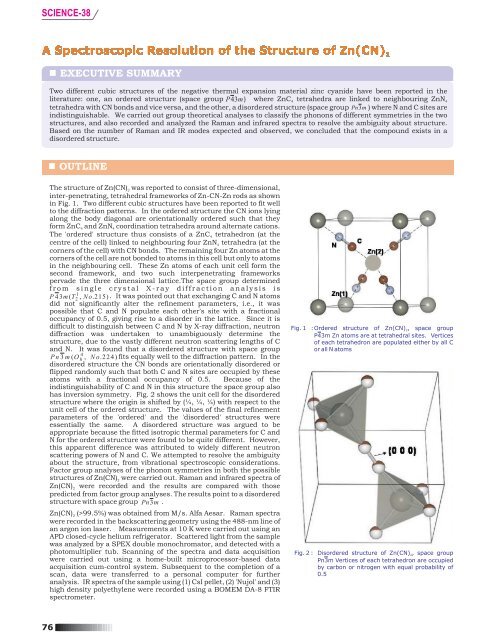
The structure <strong>of</strong> <strong>Zn</strong>(<strong>CN</strong>) 2 was reported to consist <strong>of</strong> three-dimensional,<br />
inter-penetrating, tetrahedral frameworks <strong>of</strong> <strong>Zn</strong>-<strong>CN</strong>-<strong>Zn</strong> rods as shown<br />
in Fig. 1. Two different cubic structures have been reported to fit well<br />
to <strong>the</strong> diffraction patterns. In <strong>the</strong> ordered structure <strong>the</strong> <strong>CN</strong> ions lying<br />
along <strong>the</strong> body diagonal are orientationally ordered such that <strong>the</strong>y<br />
form <strong>Zn</strong>C4and <strong>Zn</strong>N4coordination tetrahedra around alternate cations.<br />
The 'ordered' structure thus consists <strong>of</strong> a <strong>Zn</strong>C4<br />
tetrahedron (at <strong>the</strong><br />
centre <strong>of</strong> <strong>the</strong> cell) linked to neighbouring four <strong>Zn</strong>N4<br />
tetrahedra (at <strong>the</strong><br />
corners <strong>of</strong> <strong>the</strong> cell) with <strong>CN</strong> bonds. The remaining four <strong>Zn</strong> atoms at <strong>the</strong><br />
corners <strong>of</strong> <strong>the</strong> cell are not bonded to atoms in this cell but only to atoms<br />
in <strong>the</strong> neighbouring cell. These <strong>Zn</strong> atoms <strong>of</strong> each unit cell form <strong>the</strong><br />
second framework, and two such interpenetrating frameworks<br />
pervade <strong>the</strong> three dimensional lattice.The space group determined<br />
from single crystal X-ray diffraction analysis is<br />
1<br />
P 4 3m<br />
( T 2 , No.215)<br />
. It was pointed out that exchanging C and N atoms<br />
did not significantly alter <strong>the</strong> refinement parameters, i.e., it was<br />
possible that C and N populate each o<strong>the</strong>r's site with a fractional<br />
occupancy <strong>of</strong> 0.5, giving rise to a disorder in <strong>the</strong> lattice. Since it is<br />
difficult to distinguish between C and N by X-ray diffraction, neutron<br />
diffraction was undertaken to unambiguously determine <strong>the</strong><br />
structure, due to <strong>the</strong> vastly different neutron scattering lengths <strong>of</strong> C<br />
and N. It was found that a disordered structure with space group<br />
4<br />
Pn3<br />
m ( O h , No.224)<br />
fits equally well to <strong>the</strong> diffraction pattern. In <strong>the</strong><br />
disordered structure <strong>the</strong> <strong>CN</strong> bonds are orientationally disordered or<br />
flipped randomly such that both C and N sites are occupied by <strong>the</strong>se<br />
atoms with a fractional occupancy <strong>of</strong> 0.5. Because <strong>of</strong> <strong>the</strong><br />
indistinguishability <strong>of</strong> C and N in this structure <strong>the</strong> space group also<br />
has inversion symmetry. Fig. 2 shows <strong>the</strong> unit cell for <strong>the</strong> disordered<br />
structure where <strong>the</strong> origin is shifted by (¼, ¼, ¼) with respect to <strong>the</strong><br />
unit cell <strong>of</strong> <strong>the</strong> ordered structure. The values <strong>of</strong> <strong>the</strong> final refinement<br />
parameters <strong>of</strong> <strong>the</strong> 'ordered' and <strong>the</strong> 'disordered' structures were<br />
essentially <strong>the</strong> same. A disordered structure was argued to be<br />
appropriate because <strong>the</strong> fitted isotropic <strong>the</strong>rmal parameters for C and<br />
N for <strong>the</strong> ordered structure were found to be quite different. However,<br />
this apparent difference was attributed to widely different neutron<br />
scattering powers <strong>of</strong> N and C. We attempted to resolve <strong>the</strong> ambiguity<br />
about <strong>the</strong> structure, from vibrational spectroscopic considerations.<br />
Factor group analyses <strong>of</strong> <strong>the</strong> phonon symmetries in both <strong>the</strong> possible<br />
structures <strong>of</strong> <strong>Zn</strong>(<strong>CN</strong>)<br />
2<br />
were carried out. Raman and infrared spectra <strong>of</strong><br />
<strong>Zn</strong>(<strong>CN</strong>)<br />
2<br />
were recorded and <strong>the</strong> results are compared with those<br />
predicted from factor group analyses. The results point to a disordered<br />
structure with space group Pn3m .<br />
<strong>Zn</strong>(<strong>CN</strong>)<br />
2<br />
(>99.5%) was obtained from M/s. Alfa Aesar. Raman spectra<br />
were recorded in <strong>the</strong> backscattering geometry using <strong>the</strong> 488-nm line <strong>of</strong><br />
an argon ion laser. Measurements at 10 K were carried out using an<br />
APD closed-cycle helium refrigerator. Scattered light from <strong>the</strong> sample<br />
was analyzed by a SPEX double monochromator, and detected with a<br />
photomultiplier tub. Scanning <strong>of</strong> <strong>the</strong> spectra and data acquisition<br />
were carried out using a home-built microprocessor-based data<br />
acquisition cum-control system. Subsequent to <strong>the</strong> completion <strong>of</strong> a<br />
scan, data were transferred to a personal computer for fur<strong>the</strong>r<br />
analysis. IR spectra <strong>of</strong> <strong>the</strong> sample using (1) CsI pellet, (2) 'Nujol' and (3)<br />
high density polyethylene were recorded using a BOMEM DA-8 FTIR<br />
spectrometer.<br />
Fig. 1 :Ordered structure <strong>of</strong> <strong>Zn</strong>(<strong>CN</strong>)<br />
2, space group<br />
P43m <strong>Zn</strong> atoms are at tetrahedral sites. Vertices<br />
<strong>of</strong> each tetrahedron are populated ei<strong>the</strong>r by all C<br />
or all N atoms<br />
Fig. 2 : Disordered structure <strong>of</strong> <strong>Zn</strong>(<strong>CN</strong>)<br />
2, space group<br />
Pn3m Vertices <strong>of</strong> each tetrahedron are occupied<br />
by carbon or nitrogen with equal probability <strong>of</strong><br />
0.5<br />
76
SCIENCE-38<br />
RAMAN AND IR SPECTROSCOPIC TECHNIQUES<br />
Raman spectroscopy and Infrared Absorption spectroscopy are vibrational spectroscopic techniques. They are also<br />
known as 'finger printing' techniques for molecules/solids since each molecule/material has its characteristic vibrational<br />
spectrum. They arise due to <strong>the</strong> vibrational motions <strong>of</strong> chemical bonds in <strong>the</strong> material, such as C-O, C-C, C-H, O-H, C=C,<br />
etc. Since materials can have several different bonds differing in strength, one has in general several vibrational<br />
frequencies, corresponding to each <strong>of</strong> <strong>the</strong> bonds. IR is commonly employed by chemists for quick recording <strong>of</strong> spectra<br />
whereas Raman spectroscopy finds favor mainly with Physicists for <strong>the</strong> study <strong>of</strong> phase transitions in crystals, etc. Raman<br />
is a non-destructive technique but <strong>the</strong> instrumentation could be relatively expensive.<br />
Raman and IR techniques are complementary to each o<strong>the</strong>r, and for a good understanding <strong>of</strong> a material both techniques<br />
should in general be employed. For a molecule with a centre <strong>of</strong> inversion (or a solid with an inversion symmetry), Raman<br />
and IR frequencies are mutually exclusive, i.e., <strong>the</strong>y appear at different frequencies. This is <strong>the</strong> principle that has been<br />
useful in <strong>the</strong> present study to converge upon <strong>the</strong> disorder structure.<br />
FACTOR GROUP ANALYSIS AND EXPERIMENTAL MEASUREMENTS<br />
<strong>Zn</strong>(<strong>CN</strong>) 2 has two formula units per unit cell. In <strong>the</strong> ordered structure,<br />
as shown in Fig. 1, <strong>the</strong> <strong>Zn</strong>(1) atom occupies <strong>the</strong> tetrahedral 'a' sites at<br />
(0, 0, 0) and <strong>Zn</strong>(2) atom occupies <strong>the</strong> tetrahedral 'b' sites at (½, ½, ½).<br />
C and N atoms occupy distinct 4e sites with C3v<br />
site symmetry. Four<br />
C atoms around <strong>Zn</strong>(2) form a coordination tetrahedron. Similarly <strong>the</strong><br />
four N atoms around <strong>Zn</strong>(1) (three N atoms from <strong>the</strong> neighboring cells)<br />
form <strong>the</strong> o<strong>the</strong>r coordination tetrahedron. The 30 degrees <strong>of</strong> freedom<br />
arising from <strong>the</strong> 10 atoms in <strong>the</strong> cubic unit cell result in 3 acoustic<br />
phonons and 27 optical phonons. The 6 degrees <strong>of</strong> freedom<br />
corresponding to <strong>the</strong> linear molecular ion <strong>CN</strong> can be divided into 1-<br />
internal (stretching vibration), 3 rigid-translations and 2 rigid<br />
rotational degrees <strong>of</strong> freedom. Factor group analysis was carried out<br />
using Bhagavantam and Venkatarayudu method. The following<br />
irreducible representations <strong>of</strong> <strong>the</strong> acoustic and optical phonons were<br />
obtained:<br />
optical (27) = 2A<br />
1(R) + 2E(R) + 2F<br />
1<br />
+ 5F<br />
2(R, IR)<br />
The correlation method <strong>of</strong> Fately and Dollish also gave identical<br />
results. Out <strong>of</strong> <strong>the</strong>se, A<br />
1, E and F2are Raman active, and F2is also IR<br />
active. F1<br />
modes are not optically active. Thus <strong>the</strong>re are 9 optically<br />
active modes. All 9 modes are Raman active and 5 <strong>of</strong> <strong>the</strong>m are IR<br />
active as well, for this 'ordered' structure.<br />
Similar analysis <strong>of</strong> <strong>the</strong> 'disordered structure' results in 5 Raman<br />
active and 2 IR active modes. Comparison <strong>of</strong> experimental results<br />
with those expected from Factor group analysis (Table 1) establishes<br />
that <strong>the</strong> structure should be 'disordered'.<br />
Table 1.<br />
Raman and IR modes observed and expected for <strong>the</strong><br />
two structures <strong>of</strong> <strong>Zn</strong>(<strong>CN</strong>) 2.<br />
Note <strong>the</strong> mutual exclusion<br />
<strong>of</strong> Raman and IR frequencies except for <strong>the</strong> high<br />
-1<br />
frequency mode about 2220 cm . This 'anomaly' is<br />
attributed to <strong>the</strong> necessary assumption <strong>of</strong><br />
indistinguishability <strong>of</strong> C and N atoms<br />
Number <strong>of</strong> modes expected from<br />
group <strong>the</strong>ory<br />
“Ordered”<br />
“Disordered”<br />
Observed mode<br />
frequencies<br />
(cm -1 )<br />
Raman IR Raman IR Raman IR<br />
9 5 5 2<br />
-<br />
216<br />
334<br />
339<br />
-<br />
2221<br />
178<br />
-<br />
-<br />
-<br />
461<br />
2218<br />
CONVERGING ON THE STRUCTURE OF <strong>Zn</strong>(<strong>CN</strong>) 2<br />
The striking feature <strong>of</strong> Table 1 is <strong>the</strong> mutual exclusion <strong>of</strong> <strong>the</strong> observed Raman and IR frequencies, except for <strong>the</strong> high<br />
-1<br />
frequency mode about 2220 cm . This mutual exclusion points to a structure with an inversion symmetry, i.e., <strong>the</strong><br />
-1<br />
disordered structure. The IR frequency at 2218 cm is indeed expected here in spite <strong>of</strong> <strong>the</strong> arguments from factor group<br />
analysis to <strong>the</strong> contrary, since <strong>the</strong> <strong>CN</strong> has a strong dipole moment that should give rise to IR activity. Taking this into<br />
account, <strong>the</strong> structure is still concluded to be disordered, since <strong>the</strong> 'mutual exclusion' is a strong indication <strong>of</strong> a<br />
centrosymmetric structure, in this case <strong>the</strong> disordered one. It is noteworthy that several o<strong>the</strong>r cyanides such as Cu<strong>CN</strong>,<br />
Ag<strong>CN</strong> and Au<strong>CN</strong> also are known to have this 'head to tail' disorder <strong>of</strong> <strong>the</strong> <strong>CN</strong> ion.<br />
ACHIEVEMENT<br />
We have addressed a long standing controversy in <strong>the</strong> structure <strong>of</strong> <strong>Zn</strong>(<strong>CN</strong>)<br />
2, and using spectroscopic techniques, resolved<br />
<strong>the</strong> structure to be disordered. It is noteworthy that this problem could not be satisfactorily solved using X-ray diffraction<br />
or neutron diffraction techniques earlier by o<strong>the</strong>r research groups.<br />
PUBLICATIONS ARISING OUT OF THIS STUDY AND RELATED WORK<br />
T. R. Ravindran, A. K. Arora and T. N. Sairam, J. Raman Spectroscopy, 38 (2007) 283.<br />
Fur<strong>the</strong>r inquiries:<br />
Dr. T.R. Ravindran, Materials Science Division<br />
Metallurgy and Materials Group, IGCAR, e-mail: trr@igcar.gov.in<br />
77