Vol.60, Nos. 2-3 - Indira Gandhi Centre for Atomic Research
Vol.60, Nos. 2-3 - Indira Gandhi Centre for Atomic Research
Vol.60, Nos. 2-3 - Indira Gandhi Centre for Atomic Research

Create successful ePaper yourself
Turn your PDF publications into a flip-book with our unique Google optimized e-Paper software.
Trans. Indian Inst. Met.<br />
<strong>Vol.60</strong>, <strong>Nos</strong>. 2-3, April-June 2007, pp. 225-227<br />
TP 2129<br />
Metallic Nanowires Electrodeposited in Nano-Porous<br />
Polycarbonate Membrane<br />
Rena Washio 1 , Masayuki Mizumoto, Takeshi Ohgai and Akio Kagawa<br />
1<br />
Department of Materials Science, Graduate School of Science & Technology, Nagasaki University<br />
Department of Materials Science and Engineering, Nagasaki University,<br />
1-14 Bunkyo-machi, Nagasaki 852-8521, JAPAN<br />
E-Mail: renarena00110@hotmail.com<br />
(Received 30 June 2006 ; in revised <strong>for</strong>m 20 November 2006)<br />
ABSTRACT<br />
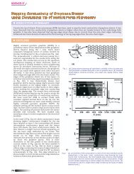
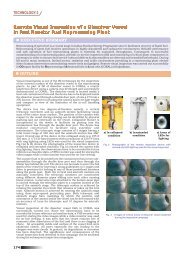
A nuclear track-etched polycarbonate membrane filter was used as a nano-porous template to obtain metallic nanowires with highaspect-ratio.<br />
To make a cathode, a gold thin film was sputtered on one side of the membrane filter with pore length of 6 mm, pore<br />
diameter of 100-150 nm and pore density of 4×10 8 pores/cm 2 . Nickel, cobalt, iron, zinc and copper were electrodeposited in the nanoporous<br />
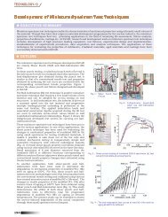
templates with numerous cylindrical nano-pores. An optimum deposition potential <strong>for</strong> growing nanowires was determined<br />
from a cathodic polarization curve of a nano-porous template. Electrodeposition of nanowires was carried out potentio-statically at<br />
an optimum cathode potential. Time-dependence of deposition current was monitored to investigate the growing process of nanowires.<br />
SEM images of nanowires revealed that the diameter and length of nanowires correspond to those of nanopores in the template. TEM<br />
images and diffraction patterns of nanowires confirmed that nickel and iron nanowires composed of a single crystal.<br />
Trans. Indian Inst. Met.<br />
<strong>Vol.60</strong>, <strong>Nos</strong>. 2-3, April-June 2007, pp. 229-233<br />
TP 2130<br />
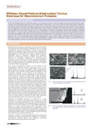
Microstructure and Mechanical Properties of Ti-Fe-(Sn)<br />
Ultrafine Eutectic Alloys<br />
J. Das, F. Ettingshausen 1 , R. Theissmann 2 , W. Löser, J. Eckert<br />
Leibniz-Institute <strong>for</strong> Solid State and Materials <strong>Research</strong> Dresden, P.O. Box 27016, D-01171, Dresden, Germany<br />
1<br />
TU Darmstadt, Institut für Werkstoffkunde, Grafenstraße 2, Darmstadt, D-64283, Germany<br />
2<br />
Forschungszentrum Karlsruhe, Institut für Nanotechnologie, Postfach 3640, Karlsruhe, Germany<br />
E-mail: j.das@ifw-dresden.de<br />
(Received 30 June 2006 ; in revised <strong>for</strong>m 20 November 2006)<br />
ABSTRACT<br />
Nano-/ultrafine-scale eutectic microstructures are prepared in (Ti 70.5<br />
Fe 29.5<br />
) 100-x<br />
Sn x<br />
alloys (x = 0, 3.85) through arc melting and cold<br />
crucible casting. The nano-/ultrafine-scale composites consist of a mixture of FeTi (B2) and -Ti (A2) solid solution phases. The<br />
average eutectic spacing of the binary Ti-Fe alloy is larger than that of the ternary Ti-Fe-Sn alloy indicating a refinement of the<br />
eutectic microstructure upon alloying. Furthermore, the room temperature plastic strain of the ternary Ti-Fe-Sn eutectic alloys is<br />
significantly larger (7.4%) than that of the binary Ti-Fe alloy. The difference in the plastic strains is correlated with the corresponding<br />
microstructure, supersaturation of the phases and elastic constants.