

Archaeorhizomycetes: a new class for a major clade of ... - IMA Fungus
Archaeorhizomycetes: a new class for a major clade of ... - IMA Fungus
Archaeorhizomycetes: a new class for a major clade of ... - IMA Fungus

Create successful ePaper yourself
Turn your PDF publications into a flip-book with our unique Google optimized e-Paper software.
<strong>Archaeorhizomycetes</strong>: a <strong>new</strong> <strong>class</strong> <strong>for</strong> a <strong>major</strong> <strong>clade</strong><br />
<strong>of</strong> soil fungi<br />
The pioneering molecular study on Alaskan<br />
soils by Schadt et al. (2003) was a huge<br />
surprise to mycologists in claiming that<br />
there were even <strong>major</strong> fungal lineages in<br />
soils that did not correspond to known<br />
fungal groups. Subsequent work by many<br />
researches has shown just how right their<br />
seemingly brash claims were. Two <strong>major</strong><br />
<strong>clade</strong>s <strong>of</strong> unnamed soil ascomycetes<br />
repeatedly emerge, that have come to be<br />
termed Soil Clone Groups 1 and 2. Group 1<br />
is the most ubiquitous, especially in boreal<br />
and tundra soils, and data from 52 studies<br />
with 162 environmental sequences have<br />
been analyzed by Rosling et al. (2011). In<br />
addition, the authors obtained cultures<br />
<strong>of</strong> one species from soil in Sweden, and<br />
studied its behaviour on Pinus roots in the<br />
laboratory. It <strong>for</strong>ms pale colonies and occurs<br />
on root surfaces, <strong>of</strong>ten mixed with other<br />
fungi, but is not mycorrhizal. No sexual<br />
spores or undeniable conidia were found,<br />
but chlamydospore-like structures were<br />
noted. The fungus seems to have seasonal<br />
tendencies which the authors suggest may<br />
be indicative <strong>of</strong> a saprobic habit in which it<br />
depends on carbon compounds released by<br />
roots in the summer.<br />
The <strong>new</strong> genus Archaeorhizomyces<br />
is introduced <strong>for</strong> A. finlayi and<br />
another unnamed species. The <strong>new</strong><br />
<strong>class</strong> <strong>Archaeorhizomycetes</strong>, order<br />
Archaeorhizomycetales, and family<br />
Archaeorhizomycetaeae are introduced to<br />
accommodate these. The <strong>class</strong> is diagnosible<br />
by rRNA sequences, and is clearly extremely<br />
ancient as it belongs to the subphylum<br />
Taphrinomycotina, which includes Neolecta,<br />
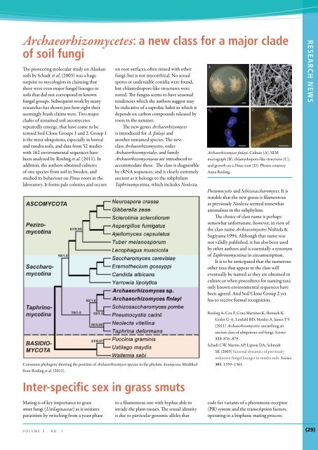
Archaeorhizomyces finlayi. Culture (A), SEM<br />
micrograph (B), chlamydospore-like structures (C),<br />
and growth on a Pinus root (D). Photos courtesy<br />
Anna Rosling.<br />
Pneumocystis and Schizosaccharomyces. It is<br />
notable that the <strong>new</strong> genus is filamentous<br />
as previously Neolecta seemed somewhat<br />
anomalous in the subphylum.<br />
The choice <strong>of</strong> <strong>class</strong> name is perhaps<br />
somewhat un<strong>for</strong>tunate, however, in view <strong>of</strong><br />
the <strong>class</strong> name Archiascomycetes Nishida &<br />
Sugiyama 1994. Although that name was<br />
not validly published, it has also been used<br />
by other authors and is essentially a synonym<br />
<strong>of</strong> Taphrinomycotina in circumscription.<br />
It is to be anticipated that the numerous<br />
other taxa that appear in the <strong>class</strong> will<br />
eventually be named as they are obtained in<br />
culture or when procedures <strong>for</strong> naming taxa<br />
only known environmental sequences have<br />
been agreed. And Soil Clone Group 2 yet<br />
has to receive <strong>for</strong>mal recognition.<br />
RESEARCH NEWS<br />
Consensus phylogeny showing the position <strong>of</strong> Archaeorhizomyces species in the phylum Ascomycota. Modified<br />
from Rosling et al. (2011).<br />
Rosling A, Cox F, Cruz-Martinez K, Ihrmark K,<br />
Grelet G-A, Lindahl BD, Menkis A, James TY<br />
(2011) <strong>Archaeorhizomycetes</strong>: unearthing an<br />
ancient <strong>class</strong> <strong>of</strong> ubiquitous soil fungi. Science<br />
333: 876–879.<br />
Schadt CW, Martin AP, Lipson DA, Schmidt<br />
SK (2003) Seasonal dynamics <strong>of</strong> previously<br />
unknown fungal lineages in tundra soils. Science<br />
301: 1359–1361.<br />
Inter-specific sex in grass smuts<br />
Mating is <strong>of</strong> key importance to grass<br />
smut fungi (Ustilaginaceae) as it initiates<br />
parasitism by switching from a yeast phase<br />
to a filamentous one with hyphae able to<br />
invade the plant tissues. The sexual identity<br />
is due to particular genomic alleles that<br />
code <strong>for</strong> variants <strong>of</strong> a pheromone-receptor<br />
(PR) system and the transcription factors,<br />
operating in a bisphasic mating process;<br />
volume 3 · no. 1<br />
(29)
RESEARCH News<br />
this involves recognition, directed hyphal<br />
growth leading to conjugation, and then<br />
plasmogamy <strong>of</strong> compatible mating partners.<br />
In order to investigate this complex and<br />
fascinating system, last December Kellner et<br />
A<br />
support values<br />
MrBayes RAxML<br />
< 90 < 50<br />
90-95 50-80<br />
> 95 > 80<br />
0.1<br />
hybrid filament<br />
pheromone response<br />
1 a1 allele<br />
2 a2 allele<br />
3 a3 allele<br />
Ustilaginaceae<br />
host age: 83-89 mya<br />
al. (2011) reported on elegant investigations<br />
designed to illuminate our understanding <strong>of</strong><br />
the evolution <strong>of</strong> the PR system. Ten species<br />
spanning 100 Myr <strong>of</strong> evolution <strong>of</strong> the system<br />
were selected <strong>for</strong> genomic and biological<br />
S. walkeri<br />
S. scitamineum<br />
S. anthracoideisporum<br />
S. mishrae<br />
S. andropogonis<br />
S. reilianum<br />
S. exsertum<br />
S. bursum<br />
U. vetiveriae<br />
U. maydis<br />
U. cynodontis<br />
U. xerochloae<br />
U. hordei<br />
U. strii<strong>for</strong>mis<br />
Me. pennsylvanicum<br />
U. fili<strong>for</strong>mis<br />
U. spermophora<br />
---------------------------------<br />
1 2 3 3 2 3<br />
---------------------------<br />
2 1 1<br />
2 2<br />
2 2<br />
------------------------ 1<br />
1 1<br />
--------------------<br />
1 1 3 3 3 1<br />
----------------------------<br />
3 1<br />
1 3 3<br />
----------------------------<br />
2<br />
1<br />
1 3<br />
study. In addition to detailed comparative<br />
in<strong>for</strong>mation on the alleles, they per<strong>for</strong>med<br />
interspecific sex tests which revealed a high<br />
potential <strong>for</strong> hybridization between species<br />
linked to pheromone signalling. While the<br />
system is optimized <strong>for</strong> within-species sex, it<br />
reveals that there are possibilities <strong>for</strong> hybrid<br />
generation which could lead to smuts with<br />
<strong>new</strong> host specificities. This possibility was<br />
confirmed by the demonstration <strong>of</strong> actual<br />
fusions between not only species <strong>of</strong> the<br />
same genus, but ones in different genera,<br />
as illustrated in the accompanying figure.<br />
The authors comment that the system now<br />
revealed may serve as a valuable model <strong>for</strong><br />
the study <strong>of</strong> the hybrid-based genesis <strong>of</strong><br />
novel genotypes.<br />
Kellner R, Vollmeister E, Feldbrügge M, Begerow D<br />
(2011) Interspecific sex in grass smuts and the<br />
genetic diversity <strong>of</strong> their pheromone-receptor<br />
system. PLoS Genetics 7: e1002436.<br />
S. consanguineum<br />
Ustilaginales<br />
host age: 113-117 mya<br />
pra1<br />
U. maydis<br />
S. reilianum<br />
S. walkeri 80<br />
Me. pennsylvanium 63<br />
U. hordei<br />
* ** *<br />
U. xerochloae<br />
U. cynodontis<br />
Us. gigantosporum<br />
*<br />
B<br />
: bt > 91<br />
0.5<br />
Malassezia pachydermatis<br />
100<br />
100<br />
100<br />
Ma. eriachnes<br />
U. williamsii<br />
Us. gigantosporum<br />
Us. standleyanum<br />
Cintractia limitata<br />
Schizonella melanogramma<br />
Malassezia globosa<br />
U. maydis<br />
S. reilianum<br />
pra2 Ma. eriachnes<br />
61*<br />
U. hordei<br />
82<br />
Us. gigantosporum<br />
71<br />
pra3<br />
S. reilianum<br />
* S. walkeri<br />
U. xerochloae<br />
Us. gigantosporum<br />
Tranzscheliella hypodytes<br />
---------------------------------<br />
1 2 3 3<br />
Melanotaenium euphorbiae<br />
C<br />
Urocystis eranthidis<br />
Figure XXX. Interspecific sex in grass smuts - modified from Kellner et al. 2011 (PLoS Genetics)<br />
(A) Multi-gene phylogeny and interspecific sexual compatibility <strong>of</strong> Ustilaginales. Concatenated Maximum<br />
Likelihood (ML) analysis <strong>of</strong> 2571 bp <strong>of</strong> ssu, ITS, lsu rDNA, ef1-α and rpb1. Circles next to branches indicate<br />
bootstrap support values and a posteriori probabilities <strong>of</strong> Bayesian and ML analyses, respectively. Branch lengths<br />
correspond to substitutions per site and abbreviated branches indicate longer branches. Connected squares<br />
illustrate<br />
It has previously<br />
hybrid filament<br />
been<br />
<strong>for</strong>mation<br />
recognized<br />
(bold lines)<br />
that selfperpetuating<br />
et al., 2005 changes (PNAS). in protein (B) Phylogeny structure <strong>of</strong> mating type-specific <strong>of</strong> the protein pheromone and can receptors. adopt Maximum an amyloid Likelihood<br />
or pheromone<br />
Sup35<br />
response<br />
that<br />
(thin<br />
is not<br />
lines).<br />
essential<br />
Numbers<br />
to<br />
in<br />
the<br />
squares<br />
function<br />
represent<br />
respective a mating types. Coloured boxes depict different phylogenetic <strong>clade</strong>s (see text). Host ages refer to<br />
Prasad<br />
analysis<br />
can be heritable<br />
<strong>of</strong> complete<br />
elements<br />
pheromone<br />
in<br />
receptor-coding<br />
yeasts separate<br />
sequences.<br />
configuration<br />
Numbers and<br />
which<br />
asterisks<br />
self-perpetuates<br />
next to branches<br />
and<br />
indicate<br />
bootstrap (bt) support values and branch lengths correspond to substitutions per site. (C) Interspecific mating <strong>of</strong><br />
from and preceding genetic change (True<br />
& Lindquist 2000); such self-perpetuating<br />
epigenetic structures are termed prions.<br />
Halfmann et al. (2012) investigated the<br />
yeast translation-termination factor prion<br />
haploid sporidia <strong>of</strong> Sporisorium reilianum (Sr) and Sporisorium scitamineum (Ss).<br />
Sr<br />
Ss<br />
1<br />
Interspecific sex in grass smuts (modified from<br />
Kellner et al. 2011). A, Multi-gene phylogeny and<br />
interspecific sexual compatibility <strong>of</strong> Ustilaginales.<br />
Concatenated Maximum Likelihood (ML) analysis<br />
<strong>of</strong> 2571 bp <strong>of</strong> ssu, ITS, lsu rDNA, ef1-a and rpb1.<br />
Circles next to branches indicate bootstrap support<br />
values and a posteriori probabilities <strong>of</strong> Bayesian and<br />
ML analyses, respectively. Branch lengths correspond<br />
to substitutions per site and abbreviated branches<br />
indicate longer branches. Connected squares<br />
illustrate hybrid filament <strong>for</strong>mation (bold lines)<br />
or pheromone response (thin lines). Numbers in<br />
squares represent respective a mating types. Coloured<br />
boxes depict different phylogenetic <strong>clade</strong>s (see text).<br />
Host ages refer to Prasad et al. (Science 310:1177–<br />
1180, 2005. B, Phylogeny <strong>of</strong> mating type-specific<br />
pheromone receptors. Maximum Likelihood analysis<br />
<strong>of</strong> complete pheromone receptor-coding sequences.<br />
Numbers and asterisks next to branches indicate<br />
bootstrap (bt) support values and branch lengths<br />
correspond to substitutions per site. C, Interspecific<br />
mating <strong>of</strong> haploid sporidia <strong>of</strong> Sporisorium reilianum<br />
(Sr) and S. scitamineum (Ss); SEM micrograph.<br />
Prions and phenotypic inheritance in wild yeasts<br />
leads to increased stops in codon readthrough;<br />
that leads to a variety <strong>of</strong> <strong>new</strong> traits.<br />
The prions had been considered an artefact<br />
<strong>of</strong> strains kept in culture, but these authors<br />
examined occurrences and screened <strong>for</strong> <strong>new</strong><br />
prions in around 700 wild Saccharomyces<br />
strains. Prions proved to occur in about<br />
one third <strong>of</strong> the wild strains examined.<br />
Modifications <strong>of</strong> the Sip35 prion were<br />
demonstrated to confer characters likely to<br />
be beneficial to the yeasts under selective<br />
pressures, that is to develop beneficial<br />
phenotypes. Indeed, 40 % <strong>of</strong> the prions in<br />
(30)<br />
<br />
ima fUNGUS
the wild yeasts were beneficial to growth<br />
under 12 sets <strong>of</strong> conditions tested. In yeasts,<br />
it has consequently now been established<br />
that prions are a naturally present<br />
supplementary source <strong>of</strong> inheritable material<br />
<strong>of</strong> adaptive value. The extent <strong>of</strong> prions in<br />
filamentous fungi as a whole has yet to be<br />
assessed, but they clearly have the potential<br />
to contribute to adaptability and fitness.<br />
Halfmann R, Jarosz DF, Jones SK, Change A,<br />
Lancaster AK, Lindquist S (2012) Prions<br />
are a common mehcnisms <strong>for</strong> phenotypic<br />
inheritance in wild yeasts. Nature 282:<br />
363–368.<br />
True HL, Lindquist SL (2000) A yeast prion<br />
provides a mechanism <strong>for</strong> genetic variation<br />
and phenotypic diversity. Nature 407:<br />
477–478.<br />
Yeast colonies, light and tramsmission electron<br />
micrograph photos <strong>of</strong> Saccharomyces cerevisiae.<br />
RESEARCH NEWS<br />
Different fungal and algal genotypes demonstrated<br />
within one lichen specimen<br />
Observations on the development <strong>of</strong> lichens<br />
in the field reveal that multiple propagules<br />
<strong>of</strong> a species developing on a surface <strong>of</strong>ten<br />
116<br />
P4T7e<br />
104<br />
P3T4c<br />
108<br />
P4T7<br />
102<br />
P3T4<br />
113<br />
P1T8<br />
103<br />
P5T3<br />
106<br />
P5T3c<br />
123<br />
P4T7d<br />
A single specimen <strong>of</strong> Parmotrema tinctorum showing the different fungal<br />
and algal genotypes determined with PCR <strong>of</strong> SSR markers. Codes prefixed<br />
by P are <strong>of</strong> the fungal partner, and those by T are <strong>of</strong> the algal partner; five<br />
fungal genotypes and one algal genotype were detected within this particular<br />
98<br />
specimen. P3T6 Adapted from Mansournia et al. (2012).<br />
coalesce to <strong>for</strong>m a single structure. This is<br />
frequently observed where the propagules<br />
are asexual soredia or isidia, which may<br />
or may not have<br />
come from the same<br />
parent, and is welldocumented.<br />
However,<br />
whether all had to be<br />
<strong>of</strong> a single genotype<br />
<strong>for</strong> this to occur was<br />
uncertain. The first<br />
study to suggest that a<br />
single lichen specimen<br />
might not just have a<br />
single fungal partner<br />
experimentally was<br />
the study <strong>of</strong> Larson<br />
& Carey (1986) who<br />
found that single<br />
115<br />
P6<br />
Tabcde<br />
120<br />
P6<br />
Tabcde<br />
124<br />
P6<br />
Tabcde<br />
specimens <strong>of</strong> two<br />
Umbilicaria species<br />
showed variations<br />
in physiological<br />
parameters and<br />
isoenzyme pr<strong>of</strong>iles.<br />
With the advent<br />
<strong>of</strong> DNA PCR<br />
technology, and<br />
especially the use <strong>of</strong><br />
microsatellite (SSR)<br />
markers, it has become<br />
possible to explore the<br />
issue <strong>of</strong> the degree <strong>of</strong><br />
individuality <strong>of</strong> single<br />
lichen specimens<br />
with respect to both<br />
the fungal and the<br />
algal populations that<br />
comprise them.<br />
Parmotrema tinctorum is a rather<br />
common tropical lichen that reproduces<br />
mainly by asexual isidia. Mansornia et<br />
al. (2012) studied populations growing<br />
on Pinus thunbergii in Japan, and used<br />
microsatellite markers to characterize the<br />
partners at different levels: within single<br />
specimens, on single trees, and within 10 x<br />
10 cm quadrats. Of particular interest were<br />
the results from single specimens in which<br />
they studied numerous small pieces <strong>of</strong> tissue.<br />
They found that a single specimen could be<br />
<strong>for</strong>med from a single fungal partner with<br />
or without changes in the algal partner,<br />
or fusion <strong>of</strong> several independent partners.<br />
In total 12 fungal genotypes and 37 algal<br />
genotypes were recognized. An example in<br />
which there were five fungal genotypes and<br />
a single algal genotype is illustrated here.<br />
Further, specimens from individual trees<br />
or which were close together tended to<br />
have similar genotypes, suggesting limited<br />
dispersal in the site.<br />
This study provides evidence to support<br />
what has long been suspected, that one<br />
cannot presume that what looks like a single<br />
individual lichen specimen represents a<br />
single fungal genotype.<br />
Larson DW, Carey CK (1986) Phenotypic variation<br />
within “individual” lichen thalli. American<br />
Journal <strong>of</strong> Botany 73: 214–223.<br />
Mansournia MR, Wu B, Matsushita N, Hogetsu T<br />
(2012) Genotypic analysis <strong>of</strong> the foliose lichen<br />
Parmotrema tinctorum using microsatellite<br />
markers: association <strong>of</strong> mycobiont and<br />
photobiont, and their reproductive modes.<br />
Lichenologist 44: 419–440.<br />
volume 3 · no. 1<br />
(31)
RESEARCH News<br />
Fungi that can trans<strong>for</strong>m lead<br />
Soil and rock-inhabiting fungi, especially<br />
lichen-<strong>for</strong>ming fungi, are well known to be<br />
able to convert different minerals to oxalates<br />
through the extracellular secretion <strong>of</strong> oxalic<br />
acid. Now, Rhee et al. 92012) have found<br />
that two fungi, Metarhizium anisopliae and<br />
Paecilomyces javanicus, are able to act directly<br />
on lead metal to <strong>for</strong>m chloropyromorphite,<br />
the most stable lead mineral known. The<br />
strains were isolated from a <strong>for</strong>mer leadmining<br />
area in Scotland, and their activity<br />
was demonstrated using incubated lead<br />
shot, and examination by two methods<br />
<strong>of</strong> X-ray analysis; it should be noted that<br />
in controls without the fungi, different<br />
compounds were <strong>for</strong>med. The lead shot was<br />
visibly corroded after one month, and minor<br />
amounts <strong>of</strong> some other lead compounds<br />
were also noted. The paper includes superb<br />
environmental scanning electron (ESEM)<br />
micrographs, and amazingly shows that the<br />
pyromorphite develops as minute spherules<br />
even inside the fungal hyphae. This finding<br />
is not only <strong>of</strong> interest in demonstrating<br />
a previously unknown biogenic step<br />
in the corrosion <strong>of</strong> lead metal and as a<br />
contribution to lead biogeochemistry, but<br />
could have applications. Soils can become<br />
lead-contaminated through, <strong>for</strong> example,<br />
the deposition <strong>of</strong> industrial wastes, battery<br />
casings, pipes, paints, inks, and shot, and<br />
lead has dangerous toxic effects on humans.<br />
The potential <strong>of</strong> using the tested strains,<br />
and other isolates <strong>of</strong> those and additional<br />
species <strong>of</strong> fungi, in the bioremediation <strong>of</strong><br />
actual lead-contaminated soils clearly merits<br />
further exploration and assessment.<br />
Rhee YJ, Hillier S, Gadd GM (2012) Lead<br />
trans<strong>for</strong>mation to pyromorphite by fungi.<br />
Current Biology 22: 1–5.<br />
Schematic representation <strong>of</strong> the processes involved in lead trans<strong>for</strong>mation by fungi. Courtesy<br />
Ge<strong>of</strong>frey M. Gadd.<br />
Nutritional value <strong>of</strong> fungi in animal diets<br />
Secondary mineral <strong>for</strong>mation on the surface <strong>of</strong> metallic lead resulting<br />
Rhee3.pdf<br />
from the activities <strong>of</strong> Metarhizium anisopliae. Photo courtesy Ge<strong>of</strong>frey<br />
Secondary M. Gadd. mineral <strong>for</strong>mation on the surface <strong>of</strong> metallic lead resulting<br />
from the activities <strong>of</strong> the fungus Metarhizium anisopliae after incubation<br />
<strong>for</strong> 1 month.<br />
Humans along with many other animals,<br />
including a wide range <strong>of</strong> terrestrial<br />
mammals, eat fungi as components <strong>of</strong><br />
their diets to various degrees. The actual<br />
nutritional value <strong>of</strong> fungi has, however, been<br />
unclear and much-debated. This is as while<br />
chemical analyses can give very positive<br />
indications, the extent to which they are<br />
digestible is unclear. In order to ascertain the<br />
extent <strong>of</strong> digestibility, Wallis et al. (2012)<br />
analyzed the fibre, amino acid composition,<br />
and both total and available nitrogen in a<br />
about 60 samples <strong>of</strong> sporocarps <strong>of</strong> diverse<br />
epigeous and hypogeous macr<strong>of</strong>ungi from<br />
Australia and the USA; they then examined<br />
the digestibility in vitro. Amongst the<br />
genera <strong>of</strong> fungi studied, were species <strong>of</strong><br />
Agaricus, Boletus, Cantharellus, Gauteria,<br />
Hysterangium, Morchella, Rhizopogon, and<br />
Tricholoma. The results showed that while<br />
in general the mushrooms and truffles tested<br />
A northern flying squirrel (Glaucomys sabrinus)<br />
holding a truffle in its paws, evidently devouring the<br />
white flesh. Photo Jim Grace.<br />
(32) ima fUNGUS
were a reasonable source <strong>of</strong> amino acids<br />
and digestible nitrogen, there were large<br />
differences between species, and the protein<br />
had a poor balance <strong>of</strong> digestible amino acids.<br />
The authors consider that this explains why<br />
mammals that are primarily mycophagous<br />
tend to eat a wide range <strong>of</strong> sporocarps,<br />
and in some cases have developed <strong>for</strong>egutfermentation<br />
to maximise the available<br />
nutritional value. In addition, they note that<br />
many mycophagous mammals supplement<br />
their diets with insects which are a source<br />
<strong>of</strong> high-quality protein. In Australia,<br />
the combination <strong>of</strong> mycophagy, <strong>for</strong>egut<br />
fermentation, and coevolution may explain<br />
the potororine marsupials which are obligate<br />
or preferential mycophagists. It is suggested<br />
that their use <strong>of</strong> hypogeous fungi enables<br />
them to survive the destructive effects <strong>of</strong><br />
devastating fires as the hypogeous fungi tend<br />
to remain in the aftermath. The authors,<br />
perhaps tactfully, largely avoid the issue <strong>of</strong><br />
the dietary value <strong>of</strong> fungal sporocarps in the<br />
human diet . . . . A single experience can<br />
hardly be taken as representative but, after<br />
repeatedly consuming meals with different<br />
mushrooms as the <strong>major</strong> component over<br />
several weeks about 15 years ago, I found I<br />
had shed quite a few pounds.<br />
Wallis IR, Claridge AW, Trappe JM (2012)<br />
Nitrogen content, amino acid composition<br />
and digestibility <strong>of</strong> fungi from a nutritional<br />
perspective in animal mycophagy. Fungal<br />
Biology 116: 590–602.<br />
RESEARCH NEWS<br />
Archaeolichenology: a novel use <strong>of</strong> lichens<br />
A novel application <strong>of</strong> lichens has just been<br />
developed by lichenologists at the Royal<br />
Botanic Garden Edinburgh. These are<br />
being used to reconstruct species’ regional<br />
distributions, and so indicate habitat types,<br />
<strong>for</strong> the historic period prior to the industrial<br />
revolution that started in the mid-18 th<br />
century. The rationale is that epiphytic lichens<br />
grow on the outer-bark surface <strong>of</strong> trees, and<br />
trees harvested and used as the frame <strong>for</strong> preindustrial<br />
buildings were not likely to have<br />
been transported far from where they were<br />
used. Consequently, where bark occurs on the<br />
timber structures <strong>of</strong> pre-industrial buildings,<br />
it might be possible to find preserved lichens<br />
which may suggest something <strong>of</strong> both past<br />
distributions and local ecologies.<br />
This proved to be the case, and Yahr et<br />
al. (2011) discovered 87 epiphytic lichen<br />
species in a survey <strong>of</strong> 78 buildings dating<br />
from the period 1300-1750 across southern<br />
England. The best-preserved material tended<br />
to be found in the ro<strong>of</strong>-spaces <strong>of</strong> lowstatus<br />
homes with continuous occupancy,<br />
where conditions are not so dissimilar to<br />
those <strong>of</strong> many herbaria today. Many <strong>of</strong> the<br />
pre-industrial records are from outside<br />
the species’ current range, and estimates<br />
suggest an 80 % loss <strong>of</strong> epiphyte diversity<br />
from areas such as south-east England (Ellis<br />
et al. 2011). This study has demonstrated<br />
an intriguing <strong>new</strong> tool <strong>for</strong> environmental<br />
reconstruction, with the potential <strong>of</strong> reevaluating<br />
environmental and conservation<br />
base-lines. This is <strong>of</strong> particular interest as<br />
current knowledge from the literature and<br />
preserved specimens is necessarily biased<br />
towards the mid-18 th century onwards, a<br />
period where industrialization was already<br />
starting to become widespread in much <strong>of</strong><br />
lowland Britain.<br />
In<strong>for</strong>mation related to changes over<br />
time can be accrued, but to ensure accuracy,<br />
it was necessary to consider the accurate<br />
dating <strong>of</strong> the timbers (based on styles <strong>of</strong><br />
carpentry), possible timber re-use within<br />
buildings, and the local transport networks.<br />
The group plans to focus on increasing the<br />
resolution <strong>of</strong> the data, using data available<br />
from wattles, and also dendrochronology.<br />
The work is being undertaken in<br />
collaboration with archaeologists at<br />
University College London.<br />
Based on material kindly supplied by Christopher<br />
J. Ellis.<br />
Ellis CJ, Yahr R, Coppins BJ (2011)<br />
Archaeobotanical evidence <strong>for</strong> a massive<br />
loss <strong>of</strong> epiphyte species richness during<br />
industrialisation in southern England.<br />
Proceedings <strong>of</strong> the Royal Society<strong>of</strong> London,<br />
Biological Science, B, 278: 3482–3489.<br />
Yahr R, Coppins BJ, Ellis CJ (2011) Preserved<br />
epiphytes as an archaeological resource in<br />
post-medieval vernacular buildings. Journal <strong>of</strong><br />
Archaeological Science 38: 1191–1198.<br />
A specimen <strong>of</strong> a Physconia species preserved on<br />
the bark <strong>of</strong> a 400 year-old timber. Photo courtesy<br />
Christopher J. Ellis.<br />
volume 3 · no. 1<br />
(33)


