

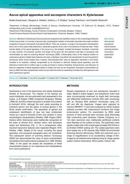
AR TICLE Ascus apical apparatus and ascospore ... - IMA Fungus
AR TICLE Ascus apical apparatus and ascospore ... - IMA Fungus
AR TICLE Ascus apical apparatus and ascospore ... - IMA Fungus

You also want an ePaper? Increase the reach of your titles
YUMPU automatically turns print PDFs into web optimized ePapers that Google loves.
doi:10.5598/imafungus.2012.03.02.04<br />
<strong>IMA</strong> <strong>Fungus</strong> · volume 3 · no 2: 125–133<br />
<strong>Ascus</strong> <strong>apical</strong> <strong>apparatus</strong> <strong>and</strong> <strong>ascospore</strong> characters in Xylariaceae<br />
Nuttika Suwannasai 1 , Margaret A. Whalley 2 , Anthony J. S. Whalley 2 , Surang Thienhirun 3 , <strong>and</strong> Prakitsin Sihanonth 2<br />
1<br />
Department of Biology (Microbiology), Faculty of Science, Srinakharinwirot University, 114 Sukhumvit 23, Bangkok, 10110, Thail<strong>and</strong>;<br />
corresponding author e-mail: snuttika@hotmail.com<br />
2<br />
Department of Microbiology, Faculty of Science Chulalongkorn University, Bangkok, Thail<strong>and</strong><br />
3<br />
Forest Products Research Division Royal Forest Department, Chatuchak, Bangkok, 10900, Thail<strong>and</strong><br />
<strong>AR</strong><strong>TICLE</strong><br />
Abstract: Members of Xylariaceae (Ascomycota) are recognized <strong>and</strong> classified mainly on the morphological features<br />
of their sexual state. In a number of genera high morphological variation of stromatal characters has made confident<br />
recognition of generic <strong>and</strong> specific boundaries difficult. There are, however, a range of microscopical characteristics<br />
which can in most cases make distinctions, especially at generic level, even in the absence of molecular data. These<br />
include details of the <strong>apical</strong> <strong>apparatus</strong> in the ascus (e.g. disc-shaped, inverted hat-shaped, rhomboid, composed<br />
of rings, amyloid, non-amyloid); position <strong>and</strong> length of the germ slit; <strong>and</strong> presence <strong>and</strong> type of <strong>ascospore</strong> wall<br />
ornamentation as seen by scanning electron microscopy (SEM). Unfortunately many of the classical studies on<br />
xylariaceous genera omitted these features <strong>and</strong> were undertaken long before the development of scanning electron<br />
microscopy. More recent studies have, however, demonstrated their value as diagnostic characters in the family.<br />
Camillea is for example, instantly recognizable by its rhomboid or diamond shaped <strong>apical</strong> <strong>apparatus</strong>, <strong>and</strong> the<br />
distinctive inverted hat or urniform type is usually prominent in Xylaria, Rosellinia, Kretzschmaria, <strong>and</strong> Nemania. At<br />
least six categories of <strong>apical</strong> <strong>apparatus</strong> based on shape <strong>and</strong> size can be recognized. Ascospore ornamentation as<br />
seen by SEM has been exceptionally useful <strong>and</strong> provided the basis for separating Camillea from Biscogniauxia <strong>and</strong><br />
other xylariaceous genera.<br />
Key words:<br />
Ascomycota<br />
<strong>ascospore</strong>s<br />
iodine reaction<br />
scanning electron<br />
microscopy<br />
systematics<br />
Xylariales<br />
Article info: Submitted: 5 July 2012; Accepted: 11 October 2012; Published: 7 November 2012.<br />
INTRODUCTION<br />
Xylariaceae is one of the best-known <strong>and</strong> widely distributed<br />
families of Ascomycota. The majority of the species are<br />
wood inhabitants, <strong>and</strong> are particularly well represented in the<br />
tropics. Ju & Rogers (1996) recognized 38 genera, Whalley<br />
(1996) 40, <strong>and</strong> the number has grown to at least 76 (Lumbsch<br />
& Huhndorf 2010), although the total varies according to<br />
individual opinion <strong>and</strong> the status of several genera in the<br />
family awaits confirmation. The separation of genera <strong>and</strong><br />
subsequent identification of taxa has been problematic<br />
mainly as a result of diversity of form <strong>and</strong> variation in many<br />
morphological characteristics (Whalley 1996, Rogers 2000).<br />
Genera within Xylariaceae were traditionally recognized on<br />
the basis of stromal form, stromal colour, <strong>and</strong> <strong>ascospore</strong><br />
shape <strong>and</strong> dimensions (Fig. 1). As a consequence other<br />
important taxonomic features were neglected (Rogers 1979,<br />
Whalley 1996). Details of the ascus, including the <strong>apical</strong><br />
<strong>apparatus</strong>, <strong>and</strong> <strong>ascospore</strong> topography were not considered.<br />
The subsequent advent of scanning electron microscopy<br />
(SEM) has demonstrated the value of spore ornamentation<br />
<strong>and</strong> details of stromatal surfaces (Læssøe et al. 1989, Whalley<br />
1996). In this paper we assess the importance of these<br />
characteristics based on our experience <strong>and</strong> extrapolations<br />
from recent publications.<br />
METHODS<br />
Squash preparations of asci <strong>and</strong> <strong>ascospore</strong>s mounted in<br />
water, Melzer’s iodine reagent, <strong>and</strong> lactophenol cotton blue<br />
were microscopically examined by bright field microscopy<br />
<strong>and</strong> differential interference contrast (DIC) light microscopy<br />
with an Olympus BH2 research microscope using x10,<br />
x40 <strong>and</strong> x60 dry objectives. Images were captured by<br />
Camera (INFINITY 1) <strong>and</strong> were analyzed by Infinity Analyze<br />
software provided with measurement functions <strong>and</strong> image<br />
enhancement options. For examination by SEM, small<br />
sections of dried stromata were mounted using Electrodag<br />
high conductivity paint (Acheson Colloids Company) on a<br />
1cm diam aluminium stub. Additionally perithecial contents<br />
were Åspread on the surface of stubs. The specimens were<br />
sputter-coated with a film of gold approximately 500 Å thick in<br />
an Emitech K550X coating unit. The coated specimens were<br />
then loaded into a FEI (Quanta 200) ESEM (Environmental<br />
Scanning Electron Microscopy, 2008) <strong>and</strong> examined over a<br />
range of magnifications at an accelerating voltage of 5kV.<br />
Images for all methods were obtained using an image capture<br />
system (Oxford Instruments, INCA system, Oxford, UK).<br />
© 2012 International Mycological Association<br />
You are free to share - to copy, distribute <strong>and</strong> transmit the work, under the following conditions:<br />
Attribution:<br />
You must attribute the work in the manner specified by the author or licensor (but not in any way that suggests that they endorse you or your use of the work).<br />
Non-commercial: You may not use this work for commercial purposes.<br />
No derivative works: You may not alter, transform, or build upon this work.<br />
For any reuse or distribution, you must make clear to others the license terms of this work, which can be found at http://creativecommons.org/licenses/by-nc-nd/3.0/legalcode. Any of the above conditions can be waived if you get<br />
permission from the copyright holder. Nothing in this license impairs or restricts the author’s moral rights.<br />
volume 3 · no. 2 125
Suwannasai et al.<br />
<strong>AR</strong><strong>TICLE</strong><br />
Fig. 1. Stromatal characteristics of some xylariaceous fungi. A. Daldinia eschscholzii (SUT 039) B. Biscogniauxia capnodes (SUT 212) C.<br />
Hypoxylon monticulosum (SUT 189). D. Rhopalostroma lekae (PK 148). Kretzschmaria clavus (PK 270). F. Annulohypoxylon bovei var.<br />
microspora (SUT 025). G. Rosellinia procera (SUT113). H. Astrocystis mirabilis (SUT 051). I. Xylaria sp.(PK 017). J. X. cubensis (PK 108). K.<br />
X. magnoliae var. microspora (PH 072). L. X. allantoidea (PK 088). Bars A–B, I–L = 1 cm; C, F–H = 5 mm; D–E = 2 mm.<br />
* Collection abbreviations: AJSW = Liverpool John Moores<br />
University, UK; SUT = Suranaree University of Technology Nakhon<br />
Ratchasima, Thail<strong>and</strong>; ST Royal Forest Department, Bangkok,<br />
Thail<strong>and</strong>; SWU Srinakharinwirot University, Bangkok, Thail<strong>and</strong> –<br />
incorporating collections from national parks <strong>and</strong> forests of Thail<strong>and</strong><br />
H (Khao Kra Yang Plantation, Phitsanulok Province), PK (Phu Kheio<br />
Wildlife Sanctuary, Chaiyaphum Province), <strong>and</strong> PH (Phu Hin Rong<br />
Kla National Park, Phitsanulok Province).<br />
RESULTS AND DISCUSSION<br />
In most of the currently recognized genera of Xylariaceae<br />
the asci contain eight spores. Exceptions include Wawelia,<br />
with 4-spored asci (Minter & Webster 1983, Lundqvist 1992)<br />
<strong>and</strong> Thuemenella with 6-spored asci (Samuels & Rossman<br />
1992). In general, the xylariaceous ascus is cylindrical <strong>and</strong><br />
possesses a stipe. In Biscogniauxia the stipe is frequently<br />
short in relation to the spore-containing part of the ascus,<br />
126 ima fUNGUS
<strong>Ascus</strong> <strong>apical</strong> <strong>apparatus</strong> <strong>and</strong> <strong>ascospore</strong> characters in Xylariaceae<br />
<strong>AR</strong><strong>TICLE</strong><br />
Fig. 2. Asci <strong>and</strong> different types of <strong>apical</strong> <strong>apparatus</strong>. A. Hypoxylon fuscum with disc-like <strong>apical</strong> <strong>apparatus</strong> stained in Melzer’s reagent (AJSW<br />
078*). B. Camillea selangorensis ascus (IMI – isotype). C. Kretzschmaria clavus ascus with <strong>apical</strong> <strong>apparatus</strong> stained in Melzer’s reagent (PK<br />
270). D. Nemania bipapillata ascus with stipe (AJSW 693). E. K. clavus showing distinctive urniform <strong>apical</strong> <strong>apparatus</strong> stained dark blue in<br />
Melzer’s reagent (PK 270). F. C. fusiformis with rhomboid <strong>apical</strong> <strong>apparatus</strong> stained in Melzer’s reagent (MAW S21, IMI) G. Hypoxylon lividicolor<br />
ascus with long stipe (ST 1047 RFD). H. Xylaria aristata ascus with <strong>apical</strong> <strong>apparatus</strong> arrowed (ST 1411 RFD). Bars A–B, F–H = 10 µm; C–D =<br />
25 µm; E = 5 µm.<br />
volume 3 · no. 2<br />
127
Suwannasai et al.<br />
<strong>AR</strong><strong>TICLE</strong><br />
whilst in Xylaria <strong>and</strong> Kretzschmaria the stipes are usually<br />
long. Hypoxylon begae, H. haematostroma <strong>and</strong> H. polyporum<br />
are notable within the genus for their very long stipes which<br />
appear to have diagnostic value (Ju & Rogers 1996). The<br />
<strong>apical</strong> tip of the ascus is usually rounded <strong>and</strong> encloses an<br />
<strong>apical</strong> <strong>apparatus</strong> which is mostly amyloid, staining blue<br />
in Melzer’s iodine reagent. There are a number of taxa in<br />
which no <strong>apical</strong> <strong>apparatus</strong> can be seen by light microscopy<br />
although the possibility of some remnant structures cannot<br />
be excluded as such taxa have not yet been studied by<br />
transmission electron microscopy. The shape <strong>and</strong> size of<br />
the <strong>apical</strong> <strong>apparatus</strong> is one of the more important taxonomic<br />
features exhibited in Xylariaceae (Fig. 2). The general<br />
appearance of the <strong>apical</strong> <strong>apparatus</strong> has been successfully<br />
applied in taxonomic studies of the family (e.g. Munk 1957,<br />
Carroll 1963, 1964, Martin 1967, 1968a, b, 1969a, b, Krug<br />
& Cain 1974a, b, Francis 1975, Rogers 1979, Læssøe et<br />
al. 1989, van der Gucht 1995, Ju & Rogers 1996, Whalley<br />
1996). Unfortunately, a number of important taxonomic<br />
studies in the family have not considered this feature. On the<br />
basis of shape <strong>and</strong> size, at least five types of amyloid <strong>apical</strong><br />
<strong>apparatus</strong> can be recognized plus a category in which there<br />
is no visible <strong>apparatus</strong>:<br />
1) Stacks of small rings, as in Hypocopra <strong>and</strong> Poronia<br />
(Krug & Cain 1974b, Jong & Rogers 1969).<br />
2) Discoid or triangular, as in most species of Hypoxylon<br />
s. str. <strong>and</strong> Daldinia (Ju & Rogers 1996, Ju et al.<br />
1997).<br />
3) Broad b<strong>and</strong> to discoid, as in Biscogniauxia (Ju et al.<br />
1998).<br />
4) Rhomboid to diamond-shaped in Camillea (Læssøe<br />
et al. 1989).<br />
5) Inverted hat or urniform, as in Xylaria, Rosellinia,<br />
Kretzschmaria <strong>and</strong> Nemania (Petrini & Muller 1986,<br />
Whalley 1996, Rogers 2000).<br />
6) No visible <strong>apical</strong> <strong>apparatus</strong> under the light microscope<br />
as in Rhopalostroma <strong>and</strong> most species of Ascotricha<br />
(Whalley & Thienhirun 1996, Hawksworth 1971)<br />
In most species the <strong>apical</strong> <strong>apparatus</strong> stains blue, usually<br />
dark blue, or occasionally reddish brown (dextrinoid) in<br />
Melzer’s iodine reagent. The significance of the iodine<br />
reaction in the <strong>apical</strong> <strong>apparatus</strong>, including Xylariaceae has<br />
been discussed by Eriksson (1966), Kohn & Korf (1975), <strong>and</strong><br />
Nannfeldt (1976). It has been shown that pre-treatment with<br />
potassium hydroxide (KOH) can induce a positive reaction in<br />
a previously iodine negative species (Nannfeldt 1976). Baral<br />
(1987) has questioned the effectiveness of Melzer’s reagent<br />
demonstrating that Lugol’s solution is superior in the detection<br />
of amyloidity in ascomycetes. Species of Xylariaceae can,<br />
however, be grouped according to the response of their<br />
<strong>apical</strong> <strong>apparatus</strong> to Melzer’s reagent as:<br />
7) Apical <strong>apparatus</strong> consistently iodine positive (blue).<br />
8) Apical <strong>apparatus</strong> varying in its reaction to iodine, i.e.<br />
some collections give a positive amyloid reaction<br />
whilst other collections of the same species do not,<br />
as in Hypoxylon cohaerens <strong>and</strong> Nemania serpens<br />
(Pouzar 1985a, b, Petrini & Rogers 1986).<br />
9) Apical <strong>apparatus</strong> consistently iodine-negative, as in<br />
Hypoxylon intermedium <strong>and</strong> H. cercidicola (Pouzar<br />
1972, Ju & Rogers 1996).<br />
The iodine positive nature of the <strong>apical</strong> <strong>apparatus</strong> is<br />
considered, however, to be a cardinal character of the<br />
Xylariaceae in spite of the presence of certain iodine negative<br />
taxa in what are undoubted taxa of the Xylariaceae (Rogers<br />
1979, 1994, 2000).<br />
The structure of the <strong>apical</strong> <strong>apparatus</strong> appears to be<br />
relatively simple when studied by transmission electron<br />
microscopy (Greenhalgh & Evans 1967, Beckett & Crawford<br />
1973, Griffiths 1973). Chadefaud (1942, 1973) proposed<br />
a much more complex structure on the basis of light<br />
microscopicy, but many of his studies were carried out on<br />
old material with degenerating asci which might also be the<br />
case here. Regardless of structure or reaction to iodine, the<br />
function of the <strong>apical</strong> <strong>apparatus</strong> is not clear. Greenhalgh &<br />
Evans (1967) <strong>and</strong> Beckett & Crawford (1973) considered<br />
the <strong>apical</strong> <strong>apparatus</strong> to act as a sphincter through which the<br />
<strong>ascospore</strong>s pass. Martin (1967a), however, was of the opinion<br />
that the <strong>ascospore</strong>s bypass the <strong>apical</strong> <strong>apparatus</strong> during<br />
discharge <strong>and</strong> that the function of the <strong>apical</strong> <strong>apparatus</strong> was<br />
therefore unclear. Rogers (1979) suggested that the <strong>apical</strong><br />
<strong>apparatus</strong> served as a strengthening device in the ascus <strong>and</strong><br />
that it becomes everted, pushed to one side, or blown off<br />
by the <strong>ascospore</strong>s once sufficient pressure has developed<br />
in the ascus. Certainly, the dimensions <strong>and</strong> shapes of many<br />
<strong>ascospore</strong>s are not suited for passage through the central<br />
channel in the <strong>apical</strong> <strong>apparatus</strong> <strong>and</strong> the suggestion of Rogers<br />
(1979) is currently the most plausible. In a study of Barron’s<br />
strain of Nemania serpens which unusually produces mature<br />
stromata in culture, Kenerley & Rogers (1976) demonstrated<br />
that the <strong>ascospore</strong>s were passively discharged under wet<br />
conditions, but forcibly discharged under dry conditions.<br />
The <strong>ascospore</strong>s of most xylariaceous fungi are described<br />
as more or less bean-shaped (phaseoliform), single-celled,<br />
smooth walled, light to dark brown, <strong>and</strong> with a conspicuous<br />
germ slit usually running the full length of the spore (Rogers<br />
1979). In reality, there is considerable variation on this basic<br />
theme (Fig. 2). In most species the ascopores are uniseriate<br />
in their arrangement in the ascus, but variation occurs in<br />
relation to their shape. The basic shape is ellipsoid, but this<br />
can become subglobose, oblong, fusiform, inequilaterally<br />
ellipsoid, navicular or broadly crescent-shaped. The ends<br />
can be narrowly or broadly rounded, attenuated, or apiculate.<br />
In Biscogniauxia species, which possess appendages, the<br />
loss of an appendage results in a truncate end (Whalley et<br />
al. 1990). In Hypoxylon s. str. <strong>and</strong> Daldinia the spores are<br />
usually inequilaterally ellipsoid, in Biscogniauxia they are<br />
more frequently subglobose, in Xylaria they are often broadly<br />
crescent-shaped, <strong>and</strong> in Rosellinia many are characterized<br />
by long attenuated ends (Petrini 1992). Most xylariaceous<br />
spores are brown, but range from light to medium or dark<br />
brown, sometimes appearing almost black. In Camillea,<br />
however, the spores are pale yellow or almost colourless,<br />
<strong>and</strong> almost all of them lack germ slits or pores, except for C.<br />
labiatrima which have a distinct slit (Rogers et al. 2002), <strong>and</strong><br />
are ornamented. Their very pale colour, lack of a germ slit<br />
<strong>and</strong> presence of spore wall ornamentation, as observed by<br />
scanning electron microscopy, drew attention to the incorrect<br />
placement of many applanate species in Hypoxylon, which<br />
were subsequently transferred to Camillea (Rogers 1977,<br />
Læssøe et al. 1989). Thus, the genus Camillea is partially<br />
128 ima fUNGUS
<strong>Ascus</strong> <strong>apical</strong> <strong>apparatus</strong> <strong>and</strong> <strong>ascospore</strong> characters in Xylariaceae<br />
<strong>AR</strong><strong>TICLE</strong><br />
Fig. 3. Variation in <strong>ascospore</strong> shape <strong>and</strong> germ slits. A. Rosellinia bunodes with elongated <strong>ascospore</strong> ends (AJSW 937). B. Entoleuca mammata<br />
with broadly rounded <strong>ascospore</strong>s (AJSW 803). C. Biscogniauxia nummularia with broadly rounded <strong>ascospore</strong>s. (AJSW 236). D. Hypoxylon<br />
comedens with straight germ slits 2/3 length of the spore (ST 1142 RFD). E. Xylaria longipes with spiral germ slit. (AJSW 576). F. H. monticulosum<br />
with spiral germ slits (SUT 189). G. Rhopalostroma kanyae with germ slit on the dorsal side of the <strong>ascospore</strong> (IMI 368200 – isotype). H.<br />
Biscogniauxia anceps showing bicelled spores <strong>and</strong> germ slit (AJSW 1009) I. H. fuscum showing dehiscent perispore following treatment with 10<br />
% KOH (AJSW 078). J. Kretzschmaria clavus with straight germ slit almost full length of the <strong>ascospore</strong> (PK 270). A–B, D–G scanning electron<br />
micrographs. C, H–J bright field light microscopy. Bars A–B, D, G–H = 10 µm; C, E = 2 µm; F = 1 µm; I–J = 15 µm.<br />
volume 3 · no. 2<br />
129
Suwannasai et al.<br />
<strong>AR</strong><strong>TICLE</strong><br />
Fig. 4. Ascospore ornamentation. A. Camillea fusiformis longitudinal reticulate. (MAW S21, IMI). B. C. tinctor poroid (SUT 260). C. C. fusiformis<br />
details of reticulate ornamentation (MAW S21, IMI).D. C. selangorensis verrucose (IMI). E. Nemania chestersii longitudinal ribbed (AJSW433).<br />
F. C. selangorensis faint ornamentation by light microscopy (IMI). G. Daldinia eschscholzii transverse (SUT 039). H. C. cyclops poroid<br />
(MAW S18) A–E, G–H scanning electron micrographs. F, bright field light microscopy. Bars A–B, F = 10 µm; C–D, G–H = 1 µm; E = 2 µm.<br />
130 ima fUNGUS
<strong>Ascus</strong> <strong>apical</strong> <strong>apparatus</strong> <strong>and</strong> <strong>ascospore</strong> characters in Xylariaceae<br />
circumscribed on the basis of <strong>ascospore</strong> wall ornamentation<br />
which may be poroid, reticulate, or ribbed (Camillea subgen.<br />
Camillea), or echinulate to verrucose (Camillea subgen.<br />
Jongiella) (Læssøe et al. 1989, Rogers et al. 1991, Whalley<br />
1995, 1996, Whalley et al. 1996, 1999).<br />
Most xylariaceous <strong>ascospore</strong>s are smooth walled, but<br />
ornamentation occurs spasmodically throughout the family<br />
(Figs 3–4). Thus, Stromatoneurospora possesses striate<br />
<strong>ascospore</strong>s (Jong & Davis 1973), <strong>and</strong> some species of<br />
Hypoxylon s. str. have <strong>ascospore</strong>s with faint transverse<br />
striations perpendicular to the long axis of the spore (Rogers<br />
& C<strong>and</strong>oussau 1982, Rogers 1985, van der Gucht & van<br />
der Veken 1992, Ju & Rogers 1996). Van der Gucht (1993)<br />
<strong>and</strong> Stadler et al. (2002) emphasized the significance of<br />
transverse striations of the <strong>ascospore</strong>s in certain species of<br />
Daldinia. A single species of Biscogniauxia, B. reticulospora,<br />
exhibits reticulately ornamented <strong>ascospore</strong>s (Ju et al. 1998),<br />
<strong>and</strong> the genera Helicogermslita <strong>and</strong> Spirodecospora were<br />
erected mainly on the presence of a spiral ornamentation on<br />
the <strong>ascospore</strong>s (Hawksworth & Lodha 1983, Lu et al. 1998).<br />
In their revision of Hypoxylon, Ju & Rogers (1996) placed<br />
considerable importance on <strong>ascospore</strong> ornamentation,<br />
noting that it can be found on the perispore, epispore,<br />
<strong>and</strong>/or beneath the epispore. Perispore ornamentation<br />
is evident in those taxa where perispores dehisce in 10<br />
% potassium hydroxide. The ornamentation falls into two<br />
major patterns, which Ju & Rogers (1996) used as one of<br />
the three major characters to delimit the two sections of<br />
Hypoxylon. Transversely orientated, coil-like ornamentation<br />
can be found in sect. Hypoxylon, whereas a thickening of<br />
the perispore situated towards one end is almost universal in<br />
sect. Annulata (Ju & Roger 1996). It was also recognized that<br />
the conspicuousness of the coil-like ornamentation in sect.<br />
Hypoxylon is an important character at species level. This<br />
feature is useful in the separation of closely related taxa such<br />
as H. anthochroum, H. duranii, H. fendleri, <strong>and</strong> H. retpela (Ju<br />
& Rogers 1996). Epispore ornamentation appears to be rare<br />
in Hypoxylon, but shallow pits can be found in H. rubellum<br />
(Rogers et al. 1987), striations in H. californicum (Ju & Rogers<br />
1996), <strong>and</strong> pleated folds in H. rectangulosporum (Rogers<br />
et al. 1992) <strong>and</strong> H. thouarsianum (Miller 1961). Transverse<br />
striations are also apparent in some Daldinia species (van<br />
der Gucht 1993, Stadler et al. 2002). Stadler et al. (2002)<br />
examined representative specimens of Daldinia species<br />
with the SEM <strong>and</strong> found that ornamentation of their outer<br />
spore layers were species-consistent. They reported them<br />
as having either smooth or transversely striated <strong>ascospore</strong>s,<br />
with the striated spores always ellipsoid-equilateral to<br />
ellipsoid-inequilateral with narrowly rounded ends. Smooth<br />
<strong>ascospore</strong>s were broadly ellipsoid to cylindrical. Daldinia<br />
concentrica was found to have very faint ornamentation, but<br />
this was only visible at ×1000 in an SEM. Ju et al. (1997)<br />
had previously found that <strong>ascospore</strong>s of some species of<br />
Daldinia undergo perispore dehiscence in 10 % potassium<br />
hydroxide <strong>and</strong> have ornamentation similar to that exhibited<br />
by members of Hypoxylon sect. Hypoxylon. In H. fragiforme a<br />
shedding or eclosion, likened to the hatching of insect pupae,<br />
of the perispore in response to specific chemical stimuli<br />
has been interpreted as part of an intricate fungus-host<br />
recognition system (Chapela et al. 1990, 1991). Whether<br />
this phenomenon occurs in other Hypoxylon species or<br />
indeed in other xylariaceous taxa has not been tested. In the<br />
coprophilous genera Poronia, Podosordaria, <strong>and</strong> Hypocopra,<br />
the <strong>ascospore</strong>s are usually surrounded by thick gelatinous<br />
sheaths which are assumed to facilitate the spores adhering<br />
to plant materials, mainly leaf lamina (Rogers 1979).<br />
Details of the asci <strong>and</strong> <strong>ascospore</strong>s, in conjunction with<br />
features of any asexual stages (Ju & Rogers 1996), have<br />
proved to be valuable in making identifications, <strong>and</strong> they also<br />
provide insights into species groups <strong>and</strong> generic separations.<br />
However, knowledge on the distribution <strong>and</strong> patterns of<br />
extrolite chemicals in Xylariaceae <strong>and</strong> application of DNA<br />
technology has been pivotal in resolving boundary issues<br />
(Whalley & Edwards 1995, Stadler & Hellwig 2005, Triebel<br />
et al. 2005).<br />
REFERENCES<br />
Baral H-O (1987) Lugol’s solution/IKI versus Melzer’s reagent:<br />
hemiamyloidity , a universal feature of the ascus wall. Mycotaxon<br />
29: 399-450.<br />
Beckett A, Crawford RM (1973) The development <strong>and</strong> fine structure<br />
of the ascus apex <strong>and</strong> its role during spore discharge in Xylaria<br />
longipes. New Phytologist 72: 357–369.<br />
Carroll G (1963) Studies on the flora of Thail<strong>and</strong> 24. Pyrenomycetes.<br />
Dansk Botansk Arkiv 23: 101–114.<br />
Carroll G (1964) Pyrenomycetes, mainly Xylariaceae, from some<br />
South Pacific Isl<strong>and</strong>s. Botanisk Tidskrift 59: 301–310.<br />
Chadefaud M (1942) Structure et anatomie comparee de 1’appareil<br />
<strong>apical</strong> des asques chez divers Discomycètes et Hypocopra, sa<br />
Pyrenomycètes. Revue Mycologie 7: 57–88.<br />
Chadefaud M (1973) Les asques et la systematique des Ascomycètes.<br />
Bulletin de la Société Mycologique de France 89: 127–170.<br />
Chapela IH, Petrini O, Hagmann L (1991) Monolignol glucosides<br />
as specific recognition messengers in fungus/plant symbioses.<br />
Physiological <strong>and</strong> Molecular Plant Pathology 39: 289–298.<br />
Chapela IH, Petrini O, Petrini LE (1990) Unusual <strong>ascospore</strong><br />
germination In Hypoxylon fragiforme first steps in the<br />
establishment of an endophytic symbiosis. Canadian Journal of<br />
Botany 68: 2571–2575.<br />
Eriksson OE (1966) On Anthostomella Sacc., Entosordaria (Sacc.)<br />
Höhn. <strong>and</strong> some related genera (Pyrenomycetes). Svensk<br />
Botanisk Tidskrift 60: 315–324.<br />
Francis SM (1975) Anthostomella Sacc. (Part I). Mycological Papers<br />
139: 1–97.<br />
Greenhalgh GN, Evans LV (1967) The structure of the ascus apex<br />
in Hypoxylon fragiforme with reference to <strong>ascospore</strong> release in<br />
this <strong>and</strong> related species. Transactions of the British Mycological<br />
Society 50: 83–188.<br />
Griffiths HB (1973) Fine structure of seven unitunicate pyrenomycete<br />
asci. Transactions of the British Mycological Society 60: 261–<br />
271.<br />
Hawksworth DL (1971) A revision of the genus Ascotricha Berk.<br />
Mycological Papers 126: 1–28.<br />
Hawksworth DL, Lodha BC (1983) Helicogermslita: a new stromatic<br />
xylariaceous genus with a spiral germ slit from India. Transactions<br />
of the British Mycological Society 81: 91–96.<br />
Jong SC, Davis EE (1973) Stromatic Neurosporas. Mycologia 65:<br />
458–464.<br />
<strong>AR</strong><strong>TICLE</strong><br />
volume 3 · no. 2<br />
131
Suwannasai et al.<br />
<strong>AR</strong><strong>TICLE</strong><br />
Jong SC, Rogers JD (1969) Poronia oedipus in culture. Mycologia<br />
60: 973–976.<br />
Ju Y-M, Rogers JD (1996) A Revision of the Genus Hypoxylon. St<br />
Paul, MN: American Phytopathological Society Press.<br />
Ju Y-M, Rogers JD, San Martin F (1997) A revision of the genus<br />
Daldinia. Mycotaxon 61: 243–293.<br />
Ju Y-M, Rogers JD, San Martin F, Granmo, A (1998) The genus<br />
Biscogniauxia. Mycotaxon 67: 1–98.<br />
Kenerley CM, Rogers JD (1976) On Hypoxylon serpens in culture.<br />
Mycologia 68: 688–691.<br />
Kohn LM, Korf RP (1975) Variation in ascomycete iodine reactions:<br />
KOH pretreatment explored. Mycotaxon 3: 165–172.<br />
Krug JC, Cain RF (1974a) A preliminary treatment of the genus<br />
Podosordaria. Canadian Journal of Botany 52: 589–605.<br />
Krug JC, Cain RF (l974b) New species of Hypocopra. Canadian<br />
Journal of Botany 52: 809–843.<br />
Læssøe T, Rogers JD, Whalley AJS (1989) Camillea, Jongiella <strong>and</strong><br />
light- spored species of Hypoxylon. Mycological Research 93:<br />
121–155.<br />
Lumbsch HT, Huhndorf SM (2010) Outline of Ascomycota – 2009.<br />
Fieldiana, Life <strong>and</strong> Earth Sciences 14: 1–64.<br />
Lu BS, Hyde KD, Ho WH (1998) Spirodecospora gen. nov.<br />
(Xylariaceae, Ascomycotina), from bamboo in Hong Kong.<br />
Fungal Diversity 1: 169–177.<br />
Lundqvist N (1992) Wawelia effusa Lundqvist spec. nov.<br />
(Xylariaceae). Persoonia 14: 417–423.<br />
Martin P (1967) Studies in the Xylariaceae: I. New <strong>and</strong> old concepts.<br />
Journal of South African Botany 33: 205–208.<br />
Martin P (1968a) Studies in the Xylariaceae III. South African <strong>and</strong><br />
foreign species of Hypoxylon section Entoleuca. South African<br />
Journal of Botany 34: 153–199.<br />
Martin P (l968b) Studies in the Xylariaceae IV. Hypoxylon, sections<br />
Papillata <strong>and</strong> Annulata. South African Journal of Botany 34:<br />
303–330.<br />
Martin P (1969a) Studies in the Xylariaceae V. Euhypoxylon. Journal<br />
of South African Botany 35: 149–206.<br />
Martin P (l969b) Studies in the Xylariaceae VI. Daldinia, Numulariola<br />
<strong>and</strong> their allies. Journal of South African Botany 35: 267-320.<br />
Miller JH (1961) A Monograph of the World Species of Hypoxylon.<br />
Athens, GA: University of Georgia Press.<br />
Minter DW, Webster J (1983) Wawelia octospora sp. nov., a<br />
xerophilous <strong>and</strong> coprophilous member of the Xylariaceae.<br />
Transactions of the British Mycological Society 80: 370–373.<br />
Munk A (1957) Danish pyrenomycetes. Dansk Botansk Arkiv 17:<br />
1–491.<br />
Nannfeldt JA (1976) Iodine reactions in ascus plugs <strong>and</strong> their<br />
taxonomic significance. Transactions of the British Mycological<br />
Society 67: 283–287.<br />
Petrini LE (1992) Rosellinia species of the temperate zones. Sydowia<br />
44: 169–281.<br />
Petrini LE, Müller E (1986) Haupt- und Nebenfruchtformen<br />
europäischer Hypoxylon-Arten (Xylariaceae, Sphaeriales) und<br />
velw<strong>and</strong>ter Pilze. Mycologia Helvetica 1: 501–627.<br />
Petrini LE, Rogers JD (1986) A summary of the Hypoxylon serpens<br />
complex. Mycotaxon 26: 409–436.<br />
Pouzar Z (1972) Hypoxylon fraxinophylum spec. nov. <strong>and</strong> H.<br />
moravicium spec. nov., two interesting species found on Fraxinus<br />
angustifolia. Česká Mykologie 26: 129–137.<br />
Pouzar Z (1985a) Reassessment of Hypoxylon serpens-complex 1.<br />
Česká Mykologie 39: 15–25.<br />
Pouzar Z (1985b) Reassessment of Hypoxylon serpens-complex II.<br />
Česká Mykologie 39: 129–134.<br />
Rogers JD (1977) Surface features of the light colored <strong>ascospore</strong>s of<br />
some applanate Hypoxylon species. Canadian Journal of Botany<br />
55: 2394–2398.<br />
Rogers JD (1979) The Xylariaceae: systematic, biological <strong>and</strong><br />
evolutionary aspects. Mycologia 71: 1–42.<br />
Rogers JD (1985) Hypoxylon duranii sp. nov. <strong>and</strong> the anamorphs of<br />
H. caries, H. papiflatum, <strong>and</strong> Rosellinia subicutata. Mycotaxon<br />
23: 429–437.<br />
Rogers JD (1994) Problem genera <strong>and</strong> family interfaces in the<br />
eupyrenomycetes In: Ascomycete Systematics: problems <strong>and</strong><br />
perspectives in the nineties (Hawksworth DL, ed.): 321–331.<br />
New York: Plenum Press.<br />
Rogers JD (2000) Thoughts <strong>and</strong> musing on tropical Xylariaceae.<br />
Mycological Research 104: 1412–1420.<br />
Rogers JD, Callan BE, Samuels GJ (1987) The Xylariaceae of the<br />
rain forests of North Sulawesi (Indonesia). Mycotaxon 29: 113–<br />
172.<br />
Rogers JD, C<strong>and</strong>oussau F (1982) Hypoxylon gillesii, a new species<br />
with ornamented <strong>ascospore</strong>s from Madagascar. Mycotaxon 15:<br />
507–514.<br />
Rogers JD, Læssøe T, Lodge DJ (1991) Camillea: new combinations<br />
<strong>and</strong> a new species. Mycologia. 83: 224–227.<br />
Rogers JD, Martin FS, Ju YM (2002) Three new taxa of Camillea<br />
from Costa Rica. Sydowia 54: 84–90.<br />
Rogers JD, Ju Y-M, Hemmes DE (1992) Hypoxylon rectangulosporum<br />
sp. nov., Xylaria psidii sp. nov., <strong>and</strong> comments on taxa of<br />
Podosordaria <strong>and</strong> Stromatoneurospora. Mycologia 84: 166–172.<br />
Samuels GJ, Rossman AY (1992) Thuemenella <strong>and</strong> Sarawakus.<br />
Mycologia 84: 26–40.<br />
Stadler M, Baumgartner M, Ide K, Popp A, Wollweber H (2002)<br />
Importance of <strong>ascospore</strong> ornamentation in the taxonomy of<br />
Daldinia. Mycological Progress 1: 31–42.<br />
Stadler M, Hellwig V (2005) Chemotaxonomy of the Xylariaceae <strong>and</strong><br />
remarkable bioactive compounds from the Xylariales <strong>and</strong> their<br />
associated asexual stages. Recent Research Developments in<br />
Phytochemistry 9: 41–93.<br />
Triebel D, Persoh D, Wollweber H, Stadler M (2005) Phylogenetic<br />
relationships among Daldinia, Entonaema, <strong>and</strong> Hypoxylon as<br />
inferred from nr DNA analyses of Xylariales. Nova Hedwigia 80:<br />
25–43.<br />
van der Gucht K, van der Veken P (1992) Contributions towards<br />
a revision of the genus Hypoxylon s. str. (Xylariaceae,<br />
Ascomycetes) from Papua New Guinea. Mycotaxon 44: 275–<br />
299.<br />
van der Gucht K (1993) Spore ornamentation makes a nice<br />
difference: Daldinia eschscholzii <strong>and</strong> D. concentrica. In: Aspects<br />
of Tropical Mycology Isaac S, Frankl<strong>and</strong> JC, Watling R, Whalley<br />
AJS, eds): 309–310. Cambridge, UK: Cambridge University<br />
Press.<br />
van der Gucht K (1995) Illustrations <strong>and</strong> descriptions of xylariaceous<br />
fungi collected in Papua New Guinea. Bulletin de la Jardin<br />
Botanique National de Bélgique 64: 219–403.<br />
Whalley AJS (1996) The xylariaceous way of life. Mycological<br />
Research 100: 897–922.<br />
Whalley AJS, Edwards RL (1995) Secondary metabolites <strong>and</strong><br />
systematic arrangements in the Xylariaceae. Canadian Journal<br />
of Botany 73 (Suppl 1): S802–S810.<br />
Whalley AJS, Læssøe T, Kile GA (1990) A new species of<br />
132 ima fUNGUS
<strong>Ascus</strong> <strong>apical</strong> <strong>apparatus</strong> <strong>and</strong> <strong>ascospore</strong> characters in Xylariaceae<br />
Biscogniauxia with appendaged <strong>ascospore</strong>s from Tasmania.<br />
Mycological Research 94: 27–239.<br />
Whalley AJS, Thienhirun S (1996) Rhopalostroma kanyae sp. nov.<br />
from Thail<strong>and</strong>. Mycological Research 100: 866–868.<br />
Whalley MA (1995) Camillea fusiformis sp. nov. from Ecuador.<br />
Sydowia 47: 82–88.<br />
Whalley MA (1996) Distinctive features of Camillea (Xylariaceae)<br />
from Cuyabeno as revealed by scanning electron microscopy.<br />
Mycologist 10: 149–151.<br />
Whalley MA, Whalley AJS, Jones EBG (1996) Camillea selangorensis<br />
sp. nov. from Malaysia. Sydowia 48: 145–15l.<br />
Whalley MA, Whalley AJS, Thienhirun S, Sihanonth P (1999) The<br />
genus Camillea in southeast Asia. Kew Bulletin 54: 715–722.<br />
<strong>AR</strong><strong>TICLE</strong><br />
volume 3 · no. 2<br />
133


