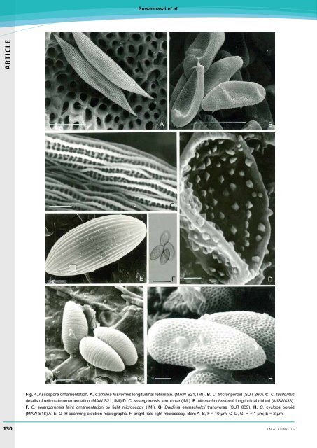
Suwannasai et al. <strong>AR</strong><strong>TICLE</strong> Fig. 4. Ascospore ornamentation. A. Camillea fusiformis longitudinal reticulate. (MAW S21, IMI). B. C. tinctor poroid (SUT 260). C. C. fusiformis details of reticulate ornamentation (MAW S21, IMI).D. C. selangorensis verrucose (IMI). E. Nemania chestersii longitudinal ribbed (AJSW433). F. C. selangorensis faint ornamentation by light microscopy (IMI). G. Daldinia eschscholzii transverse (SUT 039). H. C. cyclops poroid (MAW S18) A–E, G–H scanning electron micrographs. F, bright field light microscopy. Bars A–B, F = 10 µm; C–D, G–H = 1 µm; E = 2 µm. 130 ima fUNGUS
<strong>Ascus</strong> <strong>apical</strong> <strong>apparatus</strong> <strong>and</strong> <strong>ascospore</strong> characters in Xylariaceae circumscribed on the basis of <strong>ascospore</strong> wall ornamentation which may be poroid, reticulate, or ribbed (Camillea subgen. Camillea), or echinulate to verrucose (Camillea subgen. Jongiella) (Læssøe et al. 1989, Rogers et al. 1991, Whalley 1995, 1996, Whalley et al. 1996, 1999). Most xylariaceous <strong>ascospore</strong>s are smooth walled, but ornamentation occurs spasmodically throughout the family (Figs 3–4). Thus, Stromatoneurospora possesses striate <strong>ascospore</strong>s (Jong & Davis 1973), <strong>and</strong> some species of Hypoxylon s. str. have <strong>ascospore</strong>s with faint transverse striations perpendicular to the long axis of the spore (Rogers & C<strong>and</strong>oussau 1982, Rogers 1985, van der Gucht & van der Veken 1992, Ju & Rogers 1996). Van der Gucht (1993) <strong>and</strong> Stadler et al. (2002) emphasized the significance of transverse striations of the <strong>ascospore</strong>s in certain species of Daldinia. A single species of Biscogniauxia, B. reticulospora, exhibits reticulately ornamented <strong>ascospore</strong>s (Ju et al. 1998), <strong>and</strong> the genera Helicogermslita <strong>and</strong> Spirodecospora were erected mainly on the presence of a spiral ornamentation on the <strong>ascospore</strong>s (Hawksworth & Lodha 1983, Lu et al. 1998). In their revision of Hypoxylon, Ju & Rogers (1996) placed considerable importance on <strong>ascospore</strong> ornamentation, noting that it can be found on the perispore, epispore, <strong>and</strong>/or beneath the epispore. Perispore ornamentation is evident in those taxa where perispores dehisce in 10 % potassium hydroxide. The ornamentation falls into two major patterns, which Ju & Rogers (1996) used as one of the three major characters to delimit the two sections of Hypoxylon. Transversely orientated, coil-like ornamentation can be found in sect. Hypoxylon, whereas a thickening of the perispore situated towards one end is almost universal in sect. Annulata (Ju & Roger 1996). It was also recognized that the conspicuousness of the coil-like ornamentation in sect. Hypoxylon is an important character at species level. This feature is useful in the separation of closely related taxa such as H. anthochroum, H. duranii, H. fendleri, <strong>and</strong> H. retpela (Ju & Rogers 1996). Epispore ornamentation appears to be rare in Hypoxylon, but shallow pits can be found in H. rubellum (Rogers et al. 1987), striations in H. californicum (Ju & Rogers 1996), <strong>and</strong> pleated folds in H. rectangulosporum (Rogers et al. 1992) <strong>and</strong> H. thouarsianum (Miller 1961). Transverse striations are also apparent in some Daldinia species (van der Gucht 1993, Stadler et al. 2002). Stadler et al. (2002) examined representative specimens of Daldinia species with the SEM <strong>and</strong> found that ornamentation of their outer spore layers were species-consistent. They reported them as having either smooth or transversely striated <strong>ascospore</strong>s, with the striated spores always ellipsoid-equilateral to ellipsoid-inequilateral with narrowly rounded ends. Smooth <strong>ascospore</strong>s were broadly ellipsoid to cylindrical. Daldinia concentrica was found to have very faint ornamentation, but this was only visible at ×1000 in an SEM. Ju et al. (1997) had previously found that <strong>ascospore</strong>s of some species of Daldinia undergo perispore dehiscence in 10 % potassium hydroxide <strong>and</strong> have ornamentation similar to that exhibited by members of Hypoxylon sect. Hypoxylon. In H. fragiforme a shedding or eclosion, likened to the hatching of insect pupae, of the perispore in response to specific chemical stimuli has been interpreted as part of an intricate fungus-host recognition system (Chapela et al. 1990, 1991). Whether this phenomenon occurs in other Hypoxylon species or indeed in other xylariaceous taxa has not been tested. In the coprophilous genera Poronia, Podosordaria, <strong>and</strong> Hypocopra, the <strong>ascospore</strong>s are usually surrounded by thick gelatinous sheaths which are assumed to facilitate the spores adhering to plant materials, mainly leaf lamina (Rogers 1979). Details of the asci <strong>and</strong> <strong>ascospore</strong>s, in conjunction with features of any asexual stages (Ju & Rogers 1996), have proved to be valuable in making identifications, <strong>and</strong> they also provide insights into species groups <strong>and</strong> generic separations. However, knowledge on the distribution <strong>and</strong> patterns of extrolite chemicals in Xylariaceae <strong>and</strong> application of DNA technology has been pivotal in resolving boundary issues (Whalley & Edwards 1995, Stadler & Hellwig 2005, Triebel et al. 2005). REFERENCES Baral H-O (1987) Lugol’s solution/IKI versus Melzer’s reagent: hemiamyloidity , a universal feature of the ascus wall. Mycotaxon 29: 399-450. Beckett A, Crawford RM (1973) The development <strong>and</strong> fine structure of the ascus apex <strong>and</strong> its role during spore discharge in Xylaria longipes. New Phytologist 72: 357–369. Carroll G (1963) Studies on the flora of Thail<strong>and</strong> 24. Pyrenomycetes. Dansk Botansk Arkiv 23: 101–114. Carroll G (1964) Pyrenomycetes, mainly Xylariaceae, from some South Pacific Isl<strong>and</strong>s. Botanisk Tidskrift 59: 301–310. Chadefaud M (1942) Structure et anatomie comparee de 1’appareil <strong>apical</strong> des asques chez divers Discomycètes et Hypocopra, sa Pyrenomycètes. Revue Mycologie 7: 57–88. Chadefaud M (1973) Les asques et la systematique des Ascomycètes. Bulletin de la Société Mycologique de France 89: 127–170. Chapela IH, Petrini O, Hagmann L (1991) Monolignol glucosides as specific recognition messengers in fungus/plant symbioses. Physiological <strong>and</strong> Molecular Plant Pathology 39: 289–298. Chapela IH, Petrini O, Petrini LE (1990) Unusual <strong>ascospore</strong> germination In Hypoxylon fragiforme first steps in the establishment of an endophytic symbiosis. Canadian Journal of Botany 68: 2571–2575. Eriksson OE (1966) On Anthostomella Sacc., Entosordaria (Sacc.) Höhn. <strong>and</strong> some related genera (Pyrenomycetes). Svensk Botanisk Tidskrift 60: 315–324. Francis SM (1975) Anthostomella Sacc. (Part I). Mycological Papers 139: 1–97. Greenhalgh GN, Evans LV (1967) The structure of the ascus apex in Hypoxylon fragiforme with reference to <strong>ascospore</strong> release in this <strong>and</strong> related species. Transactions of the British Mycological Society 50: 83–188. Griffiths HB (1973) Fine structure of seven unitunicate pyrenomycete asci. Transactions of the British Mycological Society 60: 261– 271. Hawksworth DL (1971) A revision of the genus Ascotricha Berk. Mycological Papers 126: 1–28. Hawksworth DL, Lodha BC (1983) Helicogermslita: a new stromatic xylariaceous genus with a spiral germ slit from India. Transactions of the British Mycological Society 81: 91–96. Jong SC, Davis EE (1973) Stromatic Neurosporas. Mycologia 65: 458–464. <strong>AR</strong><strong>TICLE</strong> volume 3 · no. 2 131