Suwannasai et al. <strong>AR</strong><strong>TICLE</strong> whilst in Xylaria <strong>and</strong> Kretzschmaria the stipes are usually long. Hypoxylon begae, H. haematostroma <strong>and</strong> H. polyporum are notable within the genus for their very long stipes which appear to have diagnostic value (Ju & Rogers 1996). The <strong>apical</strong> tip of the ascus is usually rounded <strong>and</strong> encloses an <strong>apical</strong> <strong>apparatus</strong> which is mostly amyloid, staining blue in Melzer’s iodine reagent. There are a number of taxa in which no <strong>apical</strong> <strong>apparatus</strong> can be seen by light microscopy although the possibility of some remnant structures cannot be excluded as such taxa have not yet been studied by transmission electron microscopy. The shape <strong>and</strong> size of the <strong>apical</strong> <strong>apparatus</strong> is one of the more important taxonomic features exhibited in Xylariaceae (Fig. 2). The general appearance of the <strong>apical</strong> <strong>apparatus</strong> has been successfully applied in taxonomic studies of the family (e.g. Munk 1957, Carroll 1963, 1964, Martin 1967, 1968a, b, 1969a, b, Krug & Cain 1974a, b, Francis 1975, Rogers 1979, Læssøe et al. 1989, van der Gucht 1995, Ju & Rogers 1996, Whalley 1996). Unfortunately, a number of important taxonomic studies in the family have not considered this feature. On the basis of shape <strong>and</strong> size, at least five types of amyloid <strong>apical</strong> <strong>apparatus</strong> can be recognized plus a category in which there is no visible <strong>apparatus</strong>: 1) Stacks of small rings, as in Hypocopra <strong>and</strong> Poronia (Krug & Cain 1974b, Jong & Rogers 1969). 2) Discoid or triangular, as in most species of Hypoxylon s. str. <strong>and</strong> Daldinia (Ju & Rogers 1996, Ju et al. 1997). 3) Broad b<strong>and</strong> to discoid, as in Biscogniauxia (Ju et al. 1998). 4) Rhomboid to diamond-shaped in Camillea (Læssøe et al. 1989). 5) Inverted hat or urniform, as in Xylaria, Rosellinia, Kretzschmaria <strong>and</strong> Nemania (Petrini & Muller 1986, Whalley 1996, Rogers 2000). 6) No visible <strong>apical</strong> <strong>apparatus</strong> under the light microscope as in Rhopalostroma <strong>and</strong> most species of Ascotricha (Whalley & Thienhirun 1996, Hawksworth 1971) In most species the <strong>apical</strong> <strong>apparatus</strong> stains blue, usually dark blue, or occasionally reddish brown (dextrinoid) in Melzer’s iodine reagent. The significance of the iodine reaction in the <strong>apical</strong> <strong>apparatus</strong>, including Xylariaceae has been discussed by Eriksson (1966), Kohn & Korf (1975), <strong>and</strong> Nannfeldt (1976). It has been shown that pre-treatment with potassium hydroxide (KOH) can induce a positive reaction in a previously iodine negative species (Nannfeldt 1976). Baral (1987) has questioned the effectiveness of Melzer’s reagent demonstrating that Lugol’s solution is superior in the detection of amyloidity in ascomycetes. Species of Xylariaceae can, however, be grouped according to the response of their <strong>apical</strong> <strong>apparatus</strong> to Melzer’s reagent as: 7) Apical <strong>apparatus</strong> consistently iodine positive (blue). 8) Apical <strong>apparatus</strong> varying in its reaction to iodine, i.e. some collections give a positive amyloid reaction whilst other collections of the same species do not, as in Hypoxylon cohaerens <strong>and</strong> Nemania serpens (Pouzar 1985a, b, Petrini & Rogers 1986). 9) Apical <strong>apparatus</strong> consistently iodine-negative, as in Hypoxylon intermedium <strong>and</strong> H. cercidicola (Pouzar 1972, Ju & Rogers 1996). The iodine positive nature of the <strong>apical</strong> <strong>apparatus</strong> is considered, however, to be a cardinal character of the Xylariaceae in spite of the presence of certain iodine negative taxa in what are undoubted taxa of the Xylariaceae (Rogers 1979, 1994, 2000). The structure of the <strong>apical</strong> <strong>apparatus</strong> appears to be relatively simple when studied by transmission electron microscopy (Greenhalgh & Evans 1967, Beckett & Crawford 1973, Griffiths 1973). Chadefaud (1942, 1973) proposed a much more complex structure on the basis of light microscopicy, but many of his studies were carried out on old material with degenerating asci which might also be the case here. Regardless of structure or reaction to iodine, the function of the <strong>apical</strong> <strong>apparatus</strong> is not clear. Greenhalgh & Evans (1967) <strong>and</strong> Beckett & Crawford (1973) considered the <strong>apical</strong> <strong>apparatus</strong> to act as a sphincter through which the <strong>ascospore</strong>s pass. Martin (1967a), however, was of the opinion that the <strong>ascospore</strong>s bypass the <strong>apical</strong> <strong>apparatus</strong> during discharge <strong>and</strong> that the function of the <strong>apical</strong> <strong>apparatus</strong> was therefore unclear. Rogers (1979) suggested that the <strong>apical</strong> <strong>apparatus</strong> served as a strengthening device in the ascus <strong>and</strong> that it becomes everted, pushed to one side, or blown off by the <strong>ascospore</strong>s once sufficient pressure has developed in the ascus. Certainly, the dimensions <strong>and</strong> shapes of many <strong>ascospore</strong>s are not suited for passage through the central channel in the <strong>apical</strong> <strong>apparatus</strong> <strong>and</strong> the suggestion of Rogers (1979) is currently the most plausible. In a study of Barron’s strain of Nemania serpens which unusually produces mature stromata in culture, Kenerley & Rogers (1976) demonstrated that the <strong>ascospore</strong>s were passively discharged under wet conditions, but forcibly discharged under dry conditions. The <strong>ascospore</strong>s of most xylariaceous fungi are described as more or less bean-shaped (phaseoliform), single-celled, smooth walled, light to dark brown, <strong>and</strong> with a conspicuous germ slit usually running the full length of the spore (Rogers 1979). In reality, there is considerable variation on this basic theme (Fig. 2). In most species the ascopores are uniseriate in their arrangement in the ascus, but variation occurs in relation to their shape. The basic shape is ellipsoid, but this can become subglobose, oblong, fusiform, inequilaterally ellipsoid, navicular or broadly crescent-shaped. The ends can be narrowly or broadly rounded, attenuated, or apiculate. In Biscogniauxia species, which possess appendages, the loss of an appendage results in a truncate end (Whalley et al. 1990). In Hypoxylon s. str. <strong>and</strong> Daldinia the spores are usually inequilaterally ellipsoid, in Biscogniauxia they are more frequently subglobose, in Xylaria they are often broadly crescent-shaped, <strong>and</strong> in Rosellinia many are characterized by long attenuated ends (Petrini 1992). Most xylariaceous spores are brown, but range from light to medium or dark brown, sometimes appearing almost black. In Camillea, however, the spores are pale yellow or almost colourless, <strong>and</strong> almost all of them lack germ slits or pores, except for C. labiatrima which have a distinct slit (Rogers et al. 2002), <strong>and</strong> are ornamented. Their very pale colour, lack of a germ slit <strong>and</strong> presence of spore wall ornamentation, as observed by scanning electron microscopy, drew attention to the incorrect placement of many applanate species in Hypoxylon, which were subsequently transferred to Camillea (Rogers 1977, Læssøe et al. 1989). Thus, the genus Camillea is partially 128 ima fUNGUS
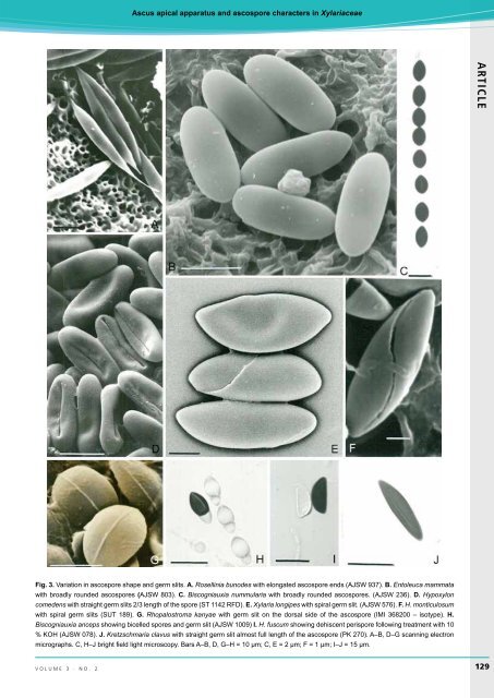
<strong>Ascus</strong> <strong>apical</strong> <strong>apparatus</strong> <strong>and</strong> <strong>ascospore</strong> characters in Xylariaceae <strong>AR</strong><strong>TICLE</strong> Fig. 3. Variation in <strong>ascospore</strong> shape <strong>and</strong> germ slits. A. Rosellinia bunodes with elongated <strong>ascospore</strong> ends (AJSW 937). B. Entoleuca mammata with broadly rounded <strong>ascospore</strong>s (AJSW 803). C. Biscogniauxia nummularia with broadly rounded <strong>ascospore</strong>s. (AJSW 236). D. Hypoxylon comedens with straight germ slits 2/3 length of the spore (ST 1142 RFD). E. Xylaria longipes with spiral germ slit. (AJSW 576). F. H. monticulosum with spiral germ slits (SUT 189). G. Rhopalostroma kanyae with germ slit on the dorsal side of the <strong>ascospore</strong> (IMI 368200 – isotype). H. Biscogniauxia anceps showing bicelled spores <strong>and</strong> germ slit (AJSW 1009) I. H. fuscum showing dehiscent perispore following treatment with 10 % KOH (AJSW 078). J. Kretzschmaria clavus with straight germ slit almost full length of the <strong>ascospore</strong> (PK 270). A–B, D–G scanning electron micrographs. C, H–J bright field light microscopy. Bars A–B, D, G–H = 10 µm; C, E = 2 µm; F = 1 µm; I–J = 15 µm. volume 3 · no. 2 129