MAKETA 5/2 po
MAKETA 5/2 po
MAKETA 5/2 po

Create successful ePaper yourself
Turn your PDF publications into a flip-book with our unique Google optimized e-Paper software.
A C T A M E D I C A M A R T I N I A N A 2 0 0 5 5/2 19<br />
After that, using a mouth opener device, thin <strong>po</strong>rtex catheter (external diameter 0.3 cm) with<br />
conducting wire was introduced into the esophagus of the animal. The catheter was tipped with<br />
cotton tam<strong>po</strong>n and its end was <strong>po</strong>sitioned in the middle part of esophagus.<br />
Capsaicin (Sigma, 1000 μM, 0.2 ml) was applied into the catheter in a manner that allowed<br />
absorption of the solutions used into the cotton tam<strong>po</strong>n. Conducting wire was then introduced<br />
into the catheter and cotton tam<strong>po</strong>n was disengaged from its tip by means of careful moving of<br />
the conducting wire. Then the catheter was pulled out from esophagus and mouth.<br />
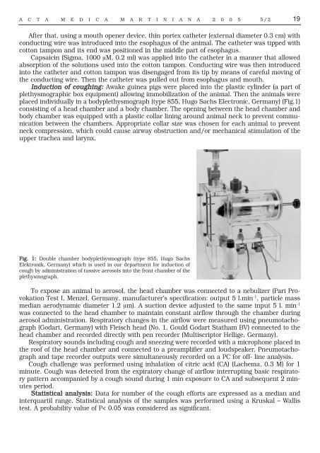
Induction of coughing: Awake guinea pigs were placed into the plastic cylinder (a part of<br />
plethysmographic box equipment) allowing immobilization of the animal. Then the animals were<br />
placed individually in a bodyplethysmograph (type 855, Hugo Sachs Electronic, Germany) (Fig.1)<br />
consisting of a head chamber and a body chamber. The opening between the head chamber and<br />
body chamber was equipped with a plastic collar lining around animal neck to prevent communication<br />
between the chambers. Appropriate collar size was chosen for each animal to prevent<br />
neck compression, which could cause airway obstruction and/or mechanical stimulation of the<br />
upper trachea and larynx.<br />
Fig. 1: Double chamber bodyplethysmograph (type 855, Hugo Sachs<br />
Elektronik, Germany) which is used in our department for induction of<br />
cough by administration of tussive aerosols into the front chamber of the<br />
plethysmograph.<br />
To ex<strong>po</strong>se an animal to aerosol, the head chamber was connected to a nebulizer (Pari Provokation<br />
Test I, Menzel, Germany, manufacturer’s specification: output 5 l.min -1 , particle mass<br />
median aerodynamic diameter 1.2 μm). A suction device adjusted to the same input 5 l. min -1<br />
was connected to the head chamber to maintain constant airflow through the chamber during<br />
aerosol administration. Respiratory changes in the airflow were measured using pneumotachograph<br />
(Godart, Germany) with Fleisch head (No. 1, Gould Godart Statham BV) connected to the<br />
head chamber and recorded directly with pen recorder (Multiscriptor Hellige, Germany).<br />
Respiratory sounds including cough and sneezing were recorded with a microphone placed in<br />
the roof of the head chamber and connected to a preamplifier and loudspeaker. Pneumotachograph<br />
and tape recorder outputs were simultaneously recorded on a PC for off- line analysis.<br />
Cough challenge was performed using inhalation of citric acid (CA) (Lachema, 0.3 M) for 1<br />
minute. Cough was detected from the expiratory change of airflow interrupting basic respiratory<br />
pattern accompanied by a cough sound during 1 min ex<strong>po</strong>sure to CA and subsequent 2 minutes<br />
period.<br />
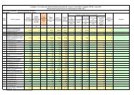
Statistical analysis: Data for number of the cough efforts are expressed as a median and<br />
interquartil range. Statistical analysis of the samples was performed using a Kruskal – Wallis<br />
test. A probability value of P< 0.05 was considered as significant.