Watching The Action - Max Planck Institute for Solid State Research
Watching The Action - Max Planck Institute for Solid State Research
Watching The Action - Max Planck Institute for Solid State Research

You also want an ePaper? Increase the reach of your titles
YUMPU automatically turns print PDFs into web optimized ePapers that Google loves.
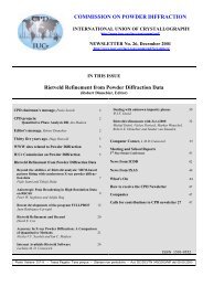
COMMISSION ON POWDER DIFFRACTION<br />
INTERNATIONAL UNION OF CRYSTALLOGRAPHY<br />
http://www.mpi-stuttgart.mpg.de/cpd/index.html<br />
NEWSLETTER No. 29, June 2003<br />
http://www.mpi-stuttgart.mpg.de/cpd/html/newsletter.html<br />
IN THIS ISSUE<br />
<strong>Watching</strong> <strong>The</strong> <strong>Action</strong><br />
(Andy Fitch, Editor)<br />
CPD Chairman’s message, Robert Dinnebier 2<br />
Editor’s message, Andy Fitch 2<br />
WWW sites related to Powder Diffraction 3<br />
IUCr Commission on Powder Diffraction 3<br />
<strong>Watching</strong> <strong>The</strong> <strong>Action</strong>:<br />
Rietveld Refinement Using Time Resolved<br />
Synchrotron X-ray Powder Diffraction Data<br />
to Study Trans<strong>for</strong>mations in Zeolites<br />
John B. Parise , Yongjae Lee, Jonathan C. Hanson,<br />
Thomas Vogt and Joseph A. Hriljac 7<br />
Beyond Rietveld Refinement – Non Crystallographic<br />
In<strong>for</strong>mation from Powder Diffraction<br />
J.S.O. Evans and S. Allen 11<br />
Structural In<strong>for</strong>mation from In-Situ Powder<br />
Diffraction Experiments<br />
Poul Norby 14<br />
Neutron Diffraction Studies of Self-Propagating<br />
High-Temperature Synthesis<br />
Erich H. Kisi and Daniel P. Riley 18<br />
Time-Resolved, In-Situ Energy Dispersive X-Ray<br />
Diffraction Studies of the Structural Trans<strong>for</strong>mations<br />
of Templated Zinc Phosphates under Hydrothermal<br />
Conditions<br />
Alexander J. Norquist and Dermot O’Hare 21<br />
In-Situ Time Resolved Powder Diffaction Studies<br />
in Heterogenous Catalysis; Coupling the Study of<br />
Long Range and Local Structural Changes<br />
Peter J. Chupas, Clare P. Grey, Jonathan C. Hanson,<br />
Jae-Yong Kim, Jose Rodriguez, Xiangyun Qiu, Simon<br />
J.L. Billinge and Peter L. Lee 24<br />
Phase Mapping and Trans<strong>for</strong>mation<br />
Dynamics in Fusion Welds<br />
Joe Wong 26<br />
SDPD Round Robin 2002 Results<br />
Armel Le Bail and Lachlan Cranswick 31<br />
<strong>The</strong> SDPD Internet Course in its Fourth<br />
Year – Overview<br />
Armel Le Bail, Yvon Laligant and Alain Jouanneaux 34<br />
A Guided Exercise of Quantitative Phase Analysis<br />
Using EXPGUI Available on the CCP14 Web Site<br />
A. F. Gualtieri 35<br />
Computer Corner, L M D Cranswick 37<br />
Quantitative Mineral Analysis by XRD---RockJock<br />
D.D. Eberl 40<br />
Quanto, a useful tool <strong>for</strong> phase quantification.<br />
First applications to pharmaceutical compounds<br />
Cinzia Giannini, Antonietta Guagliardi and<br />
Emilio Tedesco 42<br />
News from the ICDD 44<br />
What’s On 46<br />
Contact names <strong>for</strong> advertisers 47<br />
How to receive the CPD Newsletter 47<br />
Calls <strong>for</strong> contributions to CPD Newsletter 30 47<br />
ISSN 1591-9552
CPD Chairman’s Message<br />
This issue of our newsletter is a special one. From the very beginning powder diffraction was regarded a sensitive method to<br />
follow phase transitions in polycrystalline material driven by PTX. <strong>The</strong> main drawbacks of the method were always the offsets<br />
between the (fast) action and the recording and the readout. <strong>The</strong> situation has drastically changed within the last few years<br />
opening challenging new fields <strong>for</strong> the method of powder diffraction. This newsletter can be considered a state of the art reference<br />
of in situ powder diffraction. Special thanks to Andy and all the contributing authors who put a lot of ef<strong>for</strong>t into this project.<br />
This reminds me that as an incentive <strong>for</strong> prospective authors it should be noted that we have an ISSN number (see bottom<br />
of the front page) and all papers in the CPD newsletter are quotable papers.<br />
Some of you might already have noticed that I finally moved the CPD website to my home institution (see page 3). It has been<br />
slimmed down a bit to allow <strong>for</strong> better maintenance. Finally, all issues of the CPD newsletter are now available <strong>for</strong> download<br />
as PDF files. We are still in the process of correcting some of the OCR scanned pages but this task will be completed soon.<br />
I apologize <strong>for</strong> the delay in sending out the previous newsletter but we are in a learning process how to keep costs low and how<br />
to handle printing, mailing, and customs. Since the largest expenses are due to mailing costs, I ask you please to report address<br />
changes to me in good time.<br />
Robert Dinnebier<br />
CPD projects<br />
RIETVELD REFINEMENT OF ORGANIC STRUCTURES<br />
Increasing numbers of organic crystal structures are being solved and refined from powder diffraction data. <strong>The</strong> basic<br />
arrangement of the molecules in the structure can often be determined by direct methods, or by direct-space approaches.<br />
However, experience shows that problems can arise in the subsequent Rietveld refinement. For example, unless restrained by<br />
appropriate bond distances and angles molecules can distort unrealistically from a reasonable molecular structure. So how<br />
good are these Rietveld refinements? Is the problem a fundamental one of powder diffraction? eg. the ambiguities and<br />
correlations caused by peak overlap or defining the background etc. lead to inaccurate structures. Or can some of the blame be<br />
attributed to poor refinement practice? We plan to put onto the CPD web site a number of good quality powder diffraction<br />
patterns from organic compounds of known crystal structure and of different complexity. <strong>The</strong>se can be downloaded, and<br />
powder crystallographers can try out their own prowess at Rietveld refinement, by comparing their refined structures with the<br />
accepted single-crystal structures. This should be a learning exercise <strong>for</strong> us all. Any suggestions as to compounds that would<br />
appear particularly appropriate <strong>for</strong> this project are very welcome. Please contact the CPD chairman.<br />
SIZE-STRAIN ROUND ROBIN<br />
Results have been presented at international conferences (e.g. at IUCr in Geneva) and a paper has been submitted to Acta<br />
Cryst. A summary was given in CPD newsletter No. 28. A report is also on line at http://www.boulder.nist.gov/div853/balzar/.<br />
From the Editor of Newsletter 29<br />
Powder diffraction as a technique is particularly well suited to studying the behaviour of materials with changes in<br />
temperature, pressure, composition, or to follow the evolution of a system with time. For example, the method is often used to<br />
look at the structural phase transitions of a compound on heating or cooling, or to identify intermediate steps in the synthesis,<br />
dehydration or calcination of a material, maybe under conditions similar to those used in a large-scale industrial process. Such<br />
work has been carried out routinely using standard laboratory X-ray tubes and detectors. With the availability of intense<br />
synchrotron X-ray and neutron sources at central facilities, the time to measure a good quality diffraction pattern can be<br />
significantly reduced to even a few tens of milliseconds when exploiting multi-channel detectors with appropriate readout<br />
electronics. This means that fast physical or chemical processes can now be monitored in-situ. Neutrons and hard X-rays are<br />
highly penetrating, so can be used to look at bulk systems in complex sample environments. When following the structural<br />
changes that occur with temperature in a material, measurements can now be made at many temperatures, possibly at hundreds<br />
of temperatures, in a realistic period of time. Such experiments are revealing a wealth of new in<strong>for</strong>mation about the detailed<br />
evolution of crystalline systems. This newsletter contains a number of articles describing some of the experimental approaches<br />
used, and giving clear ideas of the increased understanding that results. So why not join in the action as well?<br />
Andy Fitch<br />
2
WWW sites related to powder diffraction<br />
<strong>The</strong> Commission on Powder Diffraction (CPD): http://www.mpi-stuttgart.mpg.de/cpd/index.html<br />
<strong>The</strong> International Union of Crystallography (IUCr): http://www.iucr.org/<br />
<strong>The</strong> International Centre <strong>for</strong> Diffraction Data (ICDD): http://www.icdd.com/<br />
<strong>The</strong> International X-ray Analysis Society (IXAS): http://www.ixas.org/<br />
CCP 14: http://www.ccp14.ac.uk/<br />
Submitting a proposal <strong>for</strong> neutron diffraction or synchrotron radiation X-ray diffraction is possible at many (publicly funded)<br />
large scale facilities in the world. It represents an important and frequently unique opportunity <strong>for</strong> powder diffraction experiments.<br />
A useful guide and in<strong>for</strong>mation can be accessed through the following web-site, maintained by R. Dinnebier at<br />
http://www.pulverdiffraktometrie.de/<br />
This list is far from being complete and needs input from users and readers of the CPD Newsletter. Please send comments to R.<br />
Dinnebier (r.dinnebier@fkf.mpg.de)<br />
THE IUCR COMMISSION ON POWDER DIFFRACTION - TRIENNIUM 2002-2005<br />
Chairman: Dr R. E. Dinnebier (Robert)<br />
<strong>Max</strong>-<strong>Planck</strong>-Institut für Festkörper<strong>for</strong>schung,<br />
Heisenbergstrasse 1, D-70569 Stuttgart, Germany<br />
Telephone: +49-711-689-1503 | Fax: +49-711-689-1502<br />
e-mail: r.dinnebier@fkf.mpg.de<br />
Secretary: Prof. A. N. Fitch (Andy)<br />
ESRF, BP220, F-38043 Grenoble Cedex, France<br />
Telephone : +33 476 88 25 32 | Fax: +33 476 88 25 42<br />
e-mail: fitch@esrf.fr<br />
Dr R. Delhez (Rob)<br />
Laboratory of Materials Science, Delft University of Technology,<br />
Rotterdamseweg 137 2628 AL Delft, <strong>The</strong> Netherlands<br />
Telephone: +31 15 2782261 | Fax: +31 (15) 278 6730<br />
e-mail: R.Delhez@tnw.tudelft.nl<br />
Prof. N. Masciocchi (Norberto)<br />
Dipartimento di Scienze Chimiche, Fisiche e Matematiche,<br />
Università dell'Insubria,<br />
via Valleggio 11, 22100 Como (Italy)<br />
Telephone: +39-031-326227 | Fax: +39-031-2386119<br />
e-mail: norberto.masciocchi@mail.co.uninsubria.it<br />
Dr C. R. Hubbard (Cam)<br />
Diffraction and <strong>The</strong>rmophysical Properties Group, MS<br />
6064, Bldg 4515, High Temperature Materials Laboratory,<br />
Metals & Ceramics Division, Oak Ridge National Laboratory,<br />
Oak Ridge, TN 37831-6064<br />
Telephone: 865-574-4472 | Fax: 865-574-3940<br />
e-mail: hubbardcr@ornl.gov<br />
Dr D. Balzar (Davor)<br />
Department of Physics & Astronomy<br />
University of Denver<br />
2112 E Wesley Ave, Denver, CO 80208-0202<br />
Telephone: 303-871-2137 | Fax: 303-871-4405<br />
e-mail: balzar@du.edu<br />
Prof. G. J. Kruger (Gert)<br />
Department of Chemistry & Biochemistry, Rand Afrikaans<br />
University, P O Box 524, Aucklandpark, South Africa<br />
Telephone: +27 11 489 2368 | Fax: +27 11 489 2360<br />
e-mail: gjk@na.rau.ac.za<br />
Dr. I. Madsen (Ian)<br />
CSIRO Minerals<br />
Box 312, Clayton South 3169<br />
Victoria, Australia<br />
Telephone: +61 3 9545 8785 | Fax: +61 3 9562 8919<br />
e-mail; Ian.Madsen@csiro.au<br />
Prof. W. I. F. David (Bill)<br />
Ruther<strong>for</strong>d Appleton Laboratory (CCLRC), Chilton, Oxon.<br />
OX11 OQX, United Kingdom<br />
Telephone: +44 1235 445179 | Fax: +44 1235 445383<br />
e-mail: bill.david@rl.ac.uk<br />
Prof. M. Delgado (Miguel)<br />
Laboratorio de Cristalografía, Departamento de Química,<br />
Núcleo Universitario Dr. Pedro Rincón Gutiérrez, Sector La<br />
Hechicera. Edif. "A". Nivel Patio. Universidad de Los Andes,<br />
Mérida, Venezuela<br />
Telephone: +58 274240-1372<br />
e-mail: migueld@ula.ve<br />
ICDD Representative<br />
Prof. R. L. Snyder (Bob)<br />
Department of Materials Science & Engineering, Georgia<br />
<strong>Institute</strong> of Technology, Columbus, 771 Ferst Dr. N.W., Atlanta,<br />
GA 30332-0245, USA;<br />
Telephone: +1 (404) 894-2888 | Fax: +1 (404) 894-2888<br />
e-mail: bob.snyder@mse.gatech.edu<br />
Consultants<br />
Prof. P. Scardi (Paolo)<br />
Dipartimento di Ingegneria dei Materiali e Tecnologie<br />
Industriali, Università di Trento, 38050 Mesiano (TN), Italy;<br />
Telephone: +39 0461 882417/67 | Fax: +39 (461) 881977<br />
e-mail: Paolo.Scardi@ing.unitn.it<br />
Dr F. Izumi (Fujio)<br />
National <strong>Institute</strong> <strong>for</strong> <strong>Research</strong> in Inorganic Materials<br />
1-1 Namiki, Tsukuba, Ibaraki 305-0044, Japan<br />
Telephone: +81-298-51-3354 (ext. 511); FAX: +81-298-52-<br />
7449<br />
e-mail: izumi@nirim.go.jp<br />
3
X’Pert Software with XML:<br />
Leading the way <strong>for</strong>ward<br />
in data sharing<br />
X'Pert Software<br />
<strong>for</strong> XRD data<br />
A complete family of<br />
analytical software<br />
Whether you're in research,<br />
development or production<br />
control, there are<br />
PANalytical X'Pert Software<br />
packages to support your<br />
XRD data collection and<br />
analysis.<br />
<strong>The</strong> complete range, which<br />
centers on the X’Pert Data<br />
Collector, is based on the<br />
XRDML data plat<strong>for</strong>m.<br />
A universal standard<br />
<strong>for</strong> structured data<br />
PANalytical is the first<br />
company in the XRD<br />
community to bring you an<br />
open <strong>for</strong>mat based on the<br />
universal XML standard:<br />
XRDML. It ensures that<br />
your data remain accessible,<br />
transparent and secure and<br />
it guarantees total<br />
reproducibility. In<strong>for</strong>mation<br />
on instrument type,<br />
settings, test conditions and<br />
experimental parameters is<br />
included. Moreover, XML<br />
files are human readable.<br />
To get on the way <strong>for</strong>ward<br />
in data sharing, visit:<br />
www.xrdml.com<br />
PANalytical<br />
Lelyweg 1<br />
7602 EA Almelo<br />
<strong>The</strong> Netherlands<br />
Tel.: +31 546 534 444<br />
Fax.: +31 546 534 598<br />
E-mail: info@panalytical.com<br />
www.panalytical.com
YOUR PARTNER IN X-RAY DIFFRACTION<br />
<br />
<br />
<br />
<br />
<br />
<br />
<br />
<br />
<br />
<br />
<br />
<br />
<br />
<br />
<br />
STOE & Cie GmbH P.O.Box 101302 D-64213 Darmstadt<br />
Phone: (+49) 6151 / 98870 Fax: (+49) 6151 / 988788<br />
E-mail: stoe@stoe.com Homepage: http://www.stoe.com<br />
<br />
<br />
<br />
Crystal Structure Visualization<br />
Diamond is an application <strong>for</strong> the exploration and drawing<br />
of crystal structures. With its high data capacity, its wide<br />
range of functions beginning with the generation of<br />
molecules reaching up to the construction of rather<br />
complicated inorganic structural frameworks, Diamond is<br />
the tool <strong>for</strong> molecular and solid state chemists as well as <strong>for</strong><br />
surface and material scientists.<br />
Phase Identification from Powder<br />
Match! is an easy-to-use software <strong>for</strong> phase identification<br />
from X-ray powder diffraction data. Using an elaborate<br />
algorithm, it compares the user’s diffraction pattern to the<br />
patterns stored in the ICDD PDF database in order to<br />
identify the phases which are present in the user’s sample.<br />
Single as well as multiple phases can be identified based on<br />
both peak data and raw (profile) data (if present).<br />
Match! will become available in 3 rd quarter 2003.<br />
Structure Solution from Powder<br />
Endeavour uses an innovative concept: a combined global<br />
optimization of the difference between the calculated and<br />
measured diffraction pattern and of the potential energy of<br />
the system.<br />
Since Endeavour is based upon the experienced Diamond<br />
visualization technology, the user can watch the crystal<br />
structure evolving from a completely random arrangement<br />
of atoms to the final model during the structure solution<br />
process.<br />
Free-of-charge demonstration versions are available <strong>for</strong><br />
download from our webpage.<br />
CRYSTAL IMPACT GbR<br />
Postfach 1251<br />
D-53002 Bonn<br />
Germany<br />
Tel.: +49 (228) 981 36 43<br />
Fax: +49 (228) 981 36 44<br />
E-mail: info@crystalimpact.com<br />
http://www.crystalimpact.com
• Unambiguous results using pure Kα 1<br />
radiation<br />
• Large sample variety: powders, bulk samples,<br />
capillary samples, air-sensitive samples, samples<br />
with preferred orientation, small amounts of<br />
sample, low absorbing samples, foils, fibers,<br />
liquids …<br />
• 6 predefined geometries:<br />
Bragg-Brentano reflection<br />
Bragg-Brentano capillary transmission<br />
Bragg-Brentano foil transmission<br />
High flux micro reflection<br />
High flux capillary transmission<br />
High flux foil transmission<br />
• Virtually unlimited versatility<br />
• Compact design <strong>for</strong> long-time stability<br />
• Compatible with all D8 goniometers<br />
• Compatible with all D8 attachments:<br />
sample stages, secondary optics and detectors<br />
Germany:<br />
Tel. (+49) 721/595-2888<br />
Fax (+49) 721/595-4587<br />
USA:<br />
Tel. (+1) 608/276-3000<br />
Fax (+1) 608/276-3006<br />
http://www.bruker-axs.com<br />
E-mail: info@bruker-axs.com<br />
SUPERIOR FLUX,<br />
RESOLUTION,<br />
AND VERSATILITY…<br />
… Vαrio1<br />
FOR D8 ADVANCE<br />
AND D8 DISCOVER<br />
BRUKER ADVANCED X-RAY SOLUTIONS
Rietveld Refinement Using Time Resolved<br />
Synchrotron X-ray Powder Diffraction<br />
Data to Study Trans<strong>for</strong>mations in Zeolites<br />
John B. Parise 1,2 , Yongjae Lee 3 , Jonathan C.<br />
Hanson 4 , Thomas Vogt 3 and Joseph A. Hriljac 5<br />
1 Geosciences, 2 Chemistry, <strong>State</strong> University of New<br />
York, Stony Brook NY 11794-2100, USA; Department<br />
of 3 Physics, 4 Chemistry, Brookhaven National<br />
Laboratory, Upton NY 11973, USA; 5 Department of<br />
Chemistry, University of Birmingham, Birmingham,<br />
B15 2TT, UK.<br />
John.Parise@sunysb.edu, yollee@bnl.gov<br />
Introduction<br />
Phases stable under particular conditions of pressuretemperature-composition-time<br />
(PTXt), and their<br />
physical properties under these conditions, are often<br />
best studied using time resolved diffraction. Often<br />
these trans<strong>for</strong>mations either destroy single crystals or<br />
occur in technologically important materials that<br />
crystallize as sub-micron “single” crystals. Although<br />
quench experiments provide samples more easily<br />
studied at ambient conditions, data collected in situ are<br />
required when structural states such as cation ordering<br />
are not quenchable from the PTX conditions of interest.<br />
This is particularly the case <strong>for</strong> zeolitic materials.<br />
<strong>The</strong>se open networks, exemplified by the 4- connected<br />
aluminosilicate frameworks, trans<strong>for</strong>m readily upon<br />
changing pressure, temperature, hydration-state and the<br />
type of ion-exchangeable cation occupying cavities of<br />
molecular dimension within the crystal structure.<br />
Indeed most processes, including the synthesis of the<br />
native material, hydration-dehydration and ionexchange,<br />
involve induction periods and/or are<br />
kinetically hindered. Time resolved studies reveal<br />
much about the mechanism of trans<strong>for</strong>mation.<br />
<strong>The</strong> quality of data required depends on the<br />
in<strong>for</strong>mation being sought. <strong>The</strong> optimization of<br />
synthetic conditions only requires identification of the<br />
phases, the PTX conditions over which they are stable<br />
[1,2] and many of these can be carried out in the<br />
laboratory setting, particularly since the more<br />
widespread availability of area detectors. Powder<br />
diffraction data suitable <strong>for</strong> Rietveld refinement require<br />
1) access to brighter X-ray beams at 2 nd. - and 3 rd. -<br />
generation synchrotron storage rings and 2) versatile<br />
high pressure [3,4] and hydrothermal cells [5,6].<br />
Structural models of zeolites in their various<br />
cation-exchanged <strong>for</strong>ms, in their dehydrated and<br />
hydrated states, and with and without sorbents, are<br />
important to rationalizing their mode of operation.<br />
Monitoring these structural changes in situ and as a<br />
function of time allows the mechanism of<br />
trans<strong>for</strong>mation to be followed.<br />
Experimental techniques<br />
This short article will concentrate on studies close to<br />
ambient pressure. Devices suitable <strong>for</strong> studies at high<br />
pressure and high temperatures are described elsewhere<br />
[7-9] and have been used <strong>for</strong> some important time<br />
resolved studies of martensitic and pseudo-martensitic<br />
trans<strong>for</strong>mations [8]. For studies at ambient temperature<br />
and high pressures, the DAC (diamond anvil cell) is<br />
most commonly used.<br />
A<br />
x-ray beam<br />
C<br />
B<br />
Figure 1. Photographs (top) of a Small Environmental<br />
Cell <strong>for</strong> Real Time Studies (SECReTS) [10] and (left)<br />
installed at beamline X7A, NSLS. Positions at A and B<br />
allow X-ray access, and fluids (or vacuum <strong>for</strong><br />
dehydration studies) are introduced through injection<br />
and exhaust ports C and D. <strong>The</strong> fitting F allows<br />
attachment to diffractometers and an air- or resistive<br />
heater directed at A or B allow studies to 250°C or<br />
1000°C, respectively. <strong>The</strong> scenario (right) details<br />
injection capabilities. A saturated solution of Na 2 SO 4<br />
heated to 60°C and a saturated solution of BaCl 2<br />
injected through a second capillary, Φ = 0.2 mm to<br />
precipitate BaSO 4 while scattering is simultaneously<br />
recorded [10].<br />
For powder diffraction studies of materials<br />
trans<strong>for</strong>ming under hydrothermal conditions a number<br />
of cells are described in the literature [2,5,10-17] and<br />
one example is shown in Figure 1. Safety issues limit<br />
operating conditions to below the critical point of water<br />
(22 MPa, 374°C) or below that <strong>for</strong> the solvent being<br />
used in solvothermal reaction. <strong>The</strong>y are used in time<br />
resolved studies employing achromatic or<br />
monochromatic sources, the choice predicated upon the<br />
goal of the experiment.<br />
If time resolution is a priority, energy dispersive<br />
studies using a solid-state detector at a fixed angle offer<br />
a number of advantages, including simplicity of<br />
experimental design and excellent collimation. This<br />
scheme was used in conjunction with the ability to<br />
inject into hydrothermal systems [2,10] to study the<br />
precipitation and trans<strong>for</strong>mation of FeS to FeS 2<br />
(pyrite). Several phases crystallize over a 1 − 60 minute<br />
D<br />
F<br />
E<br />
7
period and the contention that, in the absence of<br />
oxygen, the trans<strong>for</strong>mation does not take place was<br />
tested. Previous quench studies were ambiguous on this<br />
point, chiefly because sampling of the reaction<br />
potentially contaminated the sample with oxygen.<br />
Introduction of even small amounts of oxygen causes<br />
the trans<strong>for</strong>mation to pyrite to begin. In situ studies<br />
with both ED and monochromatic diffraction from<br />
reacting samples demonstrate conclusively pyrite <strong>for</strong>ms<br />
only in the presence of oxygen [2].<br />
For the study of structure at atomic resolution,<br />
bright, energetic monochromatic beams (E > 12 keV)<br />
and fast area detectors enable high quality diffraction<br />
patterns to be acquired in a short period of time.<br />
Integration of the two-dimensional diffraction pattern<br />
provides data with reasonable signal-to-noise<br />
discrimination at data rates rivaling those attainable<br />
using energy dispersive techniques. <strong>The</strong>se data are also<br />
suitable <strong>for</strong> Rietveld structural refinement.<br />
Applications to ion exchange<br />
<strong>The</strong> ion exchange properties of zeolites were amongst<br />
the first recognized and utilized [18]. <strong>The</strong>y are the basis<br />
of detergent industry and are important <strong>for</strong> the clean-up<br />
of radioactive waste as well as a number of other<br />
agricultural and industrial applications. While quench<br />
studies are useful <strong>for</strong> determining the composition of<br />
sites occupied/replaced during ion exchange, in situ<br />
time-resolved work can provide in<strong>for</strong>mation on the<br />
pathway taken. Many zeolites exchange ions at<br />
multiple sites and at different rates. Depending on the<br />
steric constraints of a particular site, exchange might be<br />
inhibited until a phase changes allows access. <strong>The</strong><br />
pathway and the changes in framework geometry<br />
accompanying these are best addressed by following<br />
the process at the atomistic level. Ion exchange into<br />
faujasite and gismondine are excellent examples of<br />
structural exchange induced by ion exchange. <strong>The</strong><br />
Faujasite (FAU) framework (Figure 2) contains a<br />
number of sites, some readily accessed through the<br />
large cage (eg II) and others accessible only after the<br />
exchanging cation negotiates the small entrance of the<br />
6-ring window of the sodalite unit (I and I’).<br />
One example of these site-specific<br />
replacements and related phase transitions is the<br />
structural investigations of potassium-exchange<br />
mechanism in sodium-<strong>for</strong>m of low-silica X (LSX)<br />
[19,20]. Compared to X and Y analogues (Figure 2),<br />
LSX exhibits enhanced ion exchange capacity due to<br />
the maximum Al content in the framework. Prior to<br />
time resolved studies ex situ synchrotron X-ray powder<br />
diffraction of Na- and K-end members of LSX as well<br />
as <strong>for</strong> the samples exchanged at the 20%, 42% and<br />
80% K + levels were undertaken [21] <strong>The</strong>se provided<br />
reasonable constraints on framework and<br />
extraframework cation positions in the subsequent<br />
refinements using the lower resolution time-resolved<br />
data. In situ synchrotron X-ray powder diffraction data<br />
(Figure 3) were collected using the SECReTS cell<br />
(Figure 1) at the beamline X7B of the NSLS. <strong>The</strong><br />
detailed pathway of the site-specific ion exchange in<br />
LSX (Figure 4) was determined from these data [19]<br />
which were collected using a combination of IP and<br />
CCD, the later providing enhanced time-resolution.<br />
Figure 2. Polyhedral representation of the Faujasite<br />
(FAU) framework Nodes represent T-sites (Al,Si)<br />
tetrahedrally coordinated to oxygen, which lies at the<br />
approximate mid-point of the straight lines. A pure<br />
silica framework would have unit cell contents<br />
Si 192 O 384 . Charge imbalance introduced when Al 3+<br />
substitutes <strong>for</strong> Si 4+ implies cations are accommodated<br />
at sites extra to the framework close to 4-rings (III’)<br />
and 6-rings (II) facing the large (super) cage, and at 6-<br />
rings within the smaller sodalite cage. <strong>The</strong>se latter<br />
sites can only be accessed from the super cage through<br />
site II and lie close to the single 6-ring (I’) or close to<br />
the center of a double 6-ring (I) column. Variations in<br />
the framework chemistry include low and high silica<br />
<strong>for</strong>ms X and Y, respectively.<br />
<strong>The</strong> mechanism revealed by these early<br />
studies has since been confirmed in the Sr-K exchange<br />
in LSX and <strong>for</strong> various ion exchange couples in the<br />
GIS system [22]. <strong>The</strong> initial stage of the exchange in<br />
K-Na-LSX involves replacement at the most easily<br />
accessible wall sites [19]. Exchange at the various sites<br />
(Figure 2) can be monitored from Rietveld refinement<br />
along with geometric changes in the framework. <strong>The</strong>se<br />
reveal (Figure 3) that replacement at the least<br />
accessible site I proceeds only after significant<br />
replacement at all other sites, and after the 6-ring<br />
access to the site has undergone considerable distortion<br />
as K-replaces Na at site I’ (Figure 2). Na → K<br />
exchange also proceeds through a two-phase region in<br />
agreement with the result of the K → Na study.<br />
Strontium exchanges sodium via a narrow miscibility<br />
gap whereas a single phase is maintained throughout<br />
the course of Sr → K exchange. Changes in occupancy<br />
at each site suggest that ion exchange in LSX is cationdependent<br />
as well as site-specific, a result that would<br />
have been difficult to obtain with static measurements<br />
alone [20].<br />
8
Cations/unit cell (in Phase B)<br />
30<br />
25<br />
20<br />
15<br />
10<br />
5<br />
K at site I<br />
K at site I´<br />
K at site II<br />
Figure 4. RHO topology (see Figure 1) with α-cages<br />
linked via double 8-rings (D8R) which accommodate<br />
charge balancing, extra-framework cations along with<br />
sites at the single 8-ring (S8R) and single 6-ring (S6R).<br />
<strong>The</strong> framework can adopt a centric (C-<strong>for</strong>m, Im-3m<br />
and acentric A-<strong>for</strong>m, I-43m) <strong>for</strong>ms shown at left and<br />
right, respectively.<br />
0<br />
0 50 100 150 200 250 300 350<br />
Time (min)<br />
Figure 3. (top) Synchrotron X-ray powder diffraction<br />
profiles as a function of time during the 6h of K-<br />
exchange into Na-LSX showing the cell collapse<br />
accompanying occupation of site I by potassium.<br />
(bottom) Variations of cation distribution during K-<br />
exchange into Na-LSX derived from Rietveld analyses<br />
using the in situ data and starting models from the ex<br />
situ study.<br />
In situ dehydration and re-hydration<br />
Zeolite RHO, (Cs, Na) 12 Al 12 Si 36 O 96 .nH 2 O; n ~ 48) is an<br />
exceptionally flexibible framework. Depending on<br />
composition (hydration state, type of exchangeable<br />
cation) T and P it adopts the centric (Im-3m) or<br />
acentric (I-43m) cubic structure above and below ~<br />
14.9 Å, respectively (Figure 4) [23]. For example in<br />
Cd-rho Cd 2+ cations relocate from a D8R-site, blocking<br />
access to the α-cages, to the S6R-site, allowing access<br />
to the pores of RHO upon heating in a “trap door”<br />
mechanism [24]. <strong>The</strong> relocation was attributed to<br />
temperature-driven phase transitions but time-resolved<br />
in situ synchrotron X-ray powder diffraction<br />
experiments, per<strong>for</strong>med using Pb-rho and Cd-rho,<br />
suggest that the phenomena result from chemical<br />
changes occurring during dehydration and re-hydration<br />
[24].<br />
In situ synchrotron X-ray powder diffraction<br />
data indicate that water plays an important role in the<br />
greater than 5 Å “trap door” motion of Cd 2+ in Cd-rho<br />
[25] and a number of other ion-excahnged <strong>for</strong>ms of this<br />
25<br />
290<br />
650<br />
450<br />
120<br />
25 4<br />
(200)<br />
6<br />
(110)<br />
zeolite. Upon heating in either air (Figures 5 and 6a)<br />
or in vacuum (Figure 6b), there is an initial decrease<br />
in cell parameter from 25°C to 75°C due to the loss of<br />
unbound water. A more gradual change in cell<br />
parameter is observed between 75°C and 450°C. A<br />
large increase in the cell parameter, which Rietveld<br />
refinement reveals is accompanied by a change from<br />
8<br />
R1~R3<br />
10<br />
C1~C3<br />
12<br />
λ λ = = 0.937 À<br />
Figure 5 Time-resolved XPD patterns typical of those<br />
collected on an IP detector (top) <strong>for</strong> Cd-rho, heated<br />
and cooled under atmosphere. See Figure 6 <strong>for</strong> cell<br />
parameter variations and derived extraframework<br />
cation positions as a function of temperature. <strong>The</strong> 1-D<br />
patterns (bottom) integrated using the Fit2D software<br />
of Hammersley [26,27], clearly show a number of<br />
phase transitions.<br />
9
the acentric to centric structure and a relocation of Cd 2+<br />
from the S8R site to the S6R site, takes place at higher<br />
temperatures. <strong>The</strong>re is a marked difference in the<br />
behavior of Cd-rho between cooling in air and vacuum<br />
(Figure 6). With the exception of a small temperature<br />
hysteresis, the reaction pathway is directly reversed<br />
upon cooling in air, while these changes do not occur<br />
upon cooling under vacuum. <strong>The</strong>se observations<br />
suggest that water may play a significant role in the<br />
relocation of cations from the S6R back to the S8R.<br />
Experiments in which rho is cooled over other gases<br />
such as N 2 and Kr did not cause the Cd to relocate from<br />
the S6R-site in the high temperature <strong>for</strong>m. It is likely<br />
that water acts as a transport agent in the original cation<br />
relocation from the S8R to the S6R site [24]<br />
Figure 6. Changes in cubic cell constant (Å) <strong>for</strong> (a)<br />
Cd-rho, heated under atmosphere, and (b) Cd-rho,<br />
heated under vacuum analyzed using in-situ<br />
synchrotron X-ray powder diffraction data. Polyhedral<br />
representations illustrate the cation distribution in the<br />
α-cage of RHO determined from Rietveld refinement.<br />
Conclusions<br />
Phase transitions driven by PTX can now be studied<br />
routinely as a function of t using synchrotron sources.<br />
In the future combinations of variables, high pressure<br />
and low temperature <strong>for</strong> example, will allow new<br />
phenomena to be explored in microporous (zeolites)<br />
and in solid-state materials in general. We have not<br />
been exhaustive but rather attempted to offer a flavor<br />
of what is possible with one class of materials. Other<br />
problems worthy of study were excluded, <strong>for</strong> example<br />
the range of earth and planetary materials occupying<br />
the pressure regimes from vacuum to TPa. For zeolites<br />
in situ studies concentrating on the atomic scale<br />
mechanisms, derived from Rietveld refinement, are<br />
helping to address some larger questions. <strong>The</strong>se include<br />
the incipient stages of crystallization, the response of<br />
the framework to environmental variables and how<br />
these affect selectivity and reactivity.<br />
Acknowledgements<br />
This work was supported by the National Science<br />
Foundation through grant NSF-DMR 0095633. This<br />
work made use of beamlines X7A and X7B at the<br />
NSLS; these facilities are supported by the U.S.<br />
Department of Energy, office of Energy Sciences,<br />
Division of Materials Sciences (X7A) and Division of<br />
Chemical Sciences (X7B) under contract DE-Ac02-<br />
98CH10886.<br />
References<br />
[1] Cahill, C. L.; Parise, J. B. (2000) On the <strong>for</strong>mation<br />
of framework indium sulfides, Dalton Trans. 9, 1475-<br />
1482.<br />
[2] Cahill, C. L.; Benning, L. G.; Barnes, H. L.; Parise,<br />
J. B. (2000) In situ, time-resolved X-ray diffraction of<br />
iron sulfides during hydrothermal pyrite growth, Chem.<br />
Geol. 167, 53-63.<br />
[3] Chen, J. H.; Kikegawa, T.; Shimomura, O.;<br />
Iwasaki, H. (1997) Application of an imaging plate to<br />
the large-volume press MAX80 at the photon factory,<br />
J. Synchrot. Radiat. 4, 21-27.<br />
[4] Shimomura, O.; Yamaoka, S.; Yagi, T.; Wakatsuki,<br />
M.; Tsuji, K.; Kawamura, H.; Hamaya, N.; Aoki, K.;<br />
Akimoto, S. In <strong>Solid</strong> <strong>State</strong> Physics Under Pressure:<br />
Recent Advances with Anvil Devices; Minomura, S.,<br />
Ed.; KTK Sci. Pub.: Tokyo, 1985, pp 351-356.<br />
[5] Parise, J. B.; Tan, K.; Norby, P.; Ko, Y.; Cahill, C.<br />
In MRS Proceedings “<strong>Solid</strong> state chemistry of<br />
inorganic materials”; Davies, P., Jackobson, A.,<br />
Toradri, C. T., Vanderah, T., Eds.: Boston Fall<br />
meeting, 1997; Vol. 453, pp 103-114.<br />
[6] Schoonen, M. A. A.; Barnes, H. L. (1991)<br />
Mechanisms of pyrite and marcasite <strong>for</strong>mation from<br />
solution:I. Nucleation of FeS 2 below 100 o C,<br />
Geochimica Cosmochimica Acta 55, 1495-1504.<br />
[7] Chen, J.; Weidner, D. J.; Vaughan, M. T.; Li, R.;<br />
Parise, J. B.; Koleda, C.; Baldwin, K. J. (1998) Time<br />
resolved diffraction measurements with an imaging<br />
plate at high pressure and temperature, Rev. High<br />
Press. Sci. Technol. 7, 272-274.<br />
[8] Chen, J. H.; Weidner, D. J.; Parise, J. B.; Vaughan,<br />
M. T.; Raterron, P. (2001) Observation of cation<br />
reordering during the olivine-spinel transition in<br />
fayalite by in situ synchrotron x-ray diffraction at high<br />
pressure and temperature, Phys. Rev. Lett. 86, 4072-<br />
4075.<br />
[9] Parise, J. B.; Chen, J. (1997) Studies of crystalline<br />
solids at high pressure and temperature using the DIA<br />
multi-anvil apparatus, Eur. J. Sol. St. Inorg. Chem 34,<br />
809-821.<br />
[10] Norby, P.; Cahill, C.; Koleda, C.; Parise, J. B.<br />
(1998) A reaction cell <strong>for</strong> in situ studies of<br />
hydrothermal titration, J. Appl. Crystallogr. 31, 481-<br />
483.<br />
10
[11] Evans, J. S. O.; Francis, R. J.; O’Hare, D.; Price, S.<br />
J.; Clark, S. M.; Gordon, J.; Neild, A.; Tang, C. C.<br />
(1994) An apparatus <strong>for</strong> the study of the Kinetics and<br />
Mechanism of Hydrothermal Reactons by in situ<br />
Energy dispersive X-ray diffraction, Rev. Sci. Inst 66,<br />
2442-2445.<br />
[12] Francis, R. J.; O’Brien, S.; Fogg, A. M.;<br />
Halasyamani, P. S.; O’Hare, D.; Loiseau, T.; Ferey, G.<br />
(1999) Time-resolved in-situ energy and angular<br />
dispersive X-ray diffraction studies of the <strong>for</strong>mation of<br />
the microporous gallophosphate ULM-5 under<br />
hydrothermal conditions, J. Am. Chem. Soc. 121,<br />
1002-1015.<br />
[13] Jensen, T. R.; Norby, P.; Hanson, J. C.; Simonsen,<br />
O.; Skou, E. M.; Stein, P. C.; Boye, H. A. (1998)<br />
Hydrothermal synthesis and crystal structure of alpha-<br />
LiZnAsO 4 , J. Mater. Chem. 8, 969-975.<br />
[14] Norby, P.; Hanson, J. C.; Fitch, A. N.; Vaughan,<br />
G.; Flaks, L.; Gualtieri, A. (2000) Formation of alphaeucryptite,<br />
LiAlSiO 4 : An in-situ synchrotron X-ray<br />
powder diffraction study of a high temperature<br />
hydrothermal synthesis, Chem. Mat. 12, 1473-1479.<br />
[15] O’Hare, D.; Evans, J. S. O.; Francis, R. J.;<br />
Halasyamani, P. S.; Norby, P.; Hanson, J. (1998) Timeresolved,<br />
in situ X-ray diffraction studies of the<br />
hydrothermal syntheses of microporous materials,<br />
Microporous Mesoporous Mat. 21, 253-262.<br />
[16] O’Hare, D.; Evans, J. S. O.; Francis, R.; Price, S.;<br />
O’Brien, S. (1998) <strong>The</strong> use of in-situ powder<br />
diffraction in the study of intercalation and<br />
hydrothermal reaction kinetics, Materials Science<br />
Forum 278-2, 367-378.<br />
[17] Schoonen, M. A. A.; Barnes, H. L. (1991)<br />
Mechanisms of pyrite and marcasite <strong>for</strong>mation from<br />
solution: III. Hydrothermal processes, Geochem.<br />
Cosmochem. Acta 55, 3491-3504.<br />
[18] Breck, D. W. (1984) Zeolite Molecular Sieves;<br />
Krieger: Malabar,FL<br />
[19] Lee, Y.; Cahill, C. L.; Hanson, J. C.; Parise, J. B.;<br />
Carr, S. W.; Myrick, M. L.; Preckwinkel, U. W.;<br />
Phillips, J. C. In Proceedings of the 12th International<br />
Zeolite Conference; Treacy, M. M. J., Marcus, B. K.,<br />
Bisher, M. E., Higgins, J. B., Eds.; Materials <strong>Research</strong><br />
Society: Baltimore, Maryland, USA, 1998; Vol. 4, p<br />
2401.<br />
[20] Parise, J. B.; Cahill, C. L.; Lee, Y. J. (2000)<br />
Dynamic powder crystallography with synchrotron X-<br />
ray sources, Can. Mineral. 38, 777-800.<br />
[21] Lee, Y. J.; Carr, S. W.; Parise, J. B. (1998) Phase<br />
transition upon K + ion exchange into Na low silica X:<br />
Combined NMR and synchrotron X-ray powder<br />
diffraction study, Chem. Mat. 10, 2561-2570.<br />
[22] Tripathi, A.; Parise, J. B.; Kim, S.-J.; Lee, Y.;<br />
Johnson, G. M.; Uh, Y.-S. (2000) Structural changes<br />
and cation site ordering in Na- and K-<strong>for</strong>m of<br />
aluminogermanates with the zeolite GIS topology,<br />
Chem. Mater. 12, 3760-3769.<br />
[23] Corbin, D. R.; Abrams, L.; Jones, G. A.; Eddy, M.<br />
M.; Harrison, W. T. A.; Stucky, G. D.; Cox, D. E.<br />
(1990) Flexibility of zeolite RHO framework. In-situ<br />
X-ray and neutron powder structural characterization of<br />
divalent cation-exchanged zeolite RHO, J. Am. Chem.<br />
Soc. 112, 4821-4830.<br />
[24] Reisner, B. A.; Lee, Y.; Hanson, J. C.; Jones, G.<br />
A.; Parise, J. B.; Corbin, D. R.; Toby, B. H.; Freitag,<br />
A.; Larese, J. Z.; Kahlenberg, V. (2000) Understanding<br />
negative thermal expansion and ’trap door’ cation<br />
relocations in zeolite rho, Chem. Commun., 2221-<br />
2222.<br />
[25] Parise, J. B.; Corbin, D. R.; Abrams, L. (1995)<br />
Structural changes upon sorption and desorption of Xe<br />
from Cd-exchanged zeolite rho: a real-time synchrotron<br />
x-ray powder diffraction study, Microporous Mater. 4,<br />
99-110.<br />
[26] Hammersley, A. P. "FIT2D: V9.129 Reference<br />
Manual V3.1," 1998.<br />
[27] Hammersley, A. P.; Svensson, S. O.; Hanfland,<br />
M.; Fitch, A. N.; Hausermann, D. (1996) Twodimensional<br />
detector software: From real detector to<br />
idealised image or two-theta scan, High Pressure Res.<br />
14, 235-248.<br />
Beyond Rietveld Refinement – Non<br />
Crystallographic In<strong>for</strong>mation from Powder<br />
Diffraction<br />
J.S.O. Evans and S. Allen<br />
Department of Chemistry, University of Durham,<br />
South Road, Durham, DH1 3LE, UK.<br />
john.evans@durham.ac.uk<br />
Over recent decades powder diffraction has evolved<br />
from a technique used to gain relatively crude<br />
analytical in<strong>for</strong>mation into one of the most powerful<br />
tools available to solid state chemists, condensed<br />
matter physicists and materials scientists. Quantitative<br />
phase analysis, microstructural in<strong>for</strong>mation, detailed<br />
structural studies from Rietveld refinement and abinitio<br />
structure solution procedures are now routinely<br />
per<strong>for</strong>med in many laboratories around the world and<br />
provide key insight into the properties of materials.<br />
With increases in X-ray and neutron fluxes at central<br />
facilities and huge improvements in detector<br />
technologies, data quality in terms of signal to noise, q-<br />
range and resolution from such experiments has<br />
improved dramatically, allowing increasingly complex<br />
problems to be tackled.<br />
In addition to tackling increasing complexity,<br />
these instrument improvements can be exploited in an<br />
alternative way: by accepting data of slightly lower<br />
quality one can obtain individual diffraction data sets<br />
extremely quickly. This allows one to per<strong>for</strong>m not just<br />
single diffraction experiments but experiments as a<br />
function of time, temperature, pressure, magnetic field,<br />
composition, or a variety of other external<br />
perturbations to a system. By following the evolution<br />
11
of diffraction data as a function of such perturbations,<br />
considerable extra in<strong>for</strong>mation on a material’s<br />
properties can often be obtained. In this short article we<br />
will highlight examples from our work in which<br />
variable temperature diffraction data have been used to<br />
give not only “crystallographic” in<strong>for</strong>mation about the<br />
evolution of structural coordinates as a function of<br />
temperature, but “non-crystallographic” additional<br />
in<strong>for</strong>mation such as the energy and magnitude of<br />
atomic vibrations in the solid state, in<strong>for</strong>mation on<br />
order-disorder phase transitions, and in<strong>for</strong>mation on<br />
activation energies <strong>for</strong> ionic migration. <strong>The</strong> examples<br />
selected are naturally biased by our own interests, but<br />
will hopefully be illustrative of the general<br />
methodology of “parametric diffraction” in which<br />
multiple data sets are collected and treated as an<br />
ensemble of evolving in<strong>for</strong>mation. <strong>The</strong>re are, of course,<br />
many other examples of diffraction=f(x) in the<br />
literature from other workers, as well as elegant<br />
examples of chemical reactions being followed in real<br />
time. We will not attempt to cover these in this article.<br />
Why per<strong>for</strong>m parametric diffraction studies?<br />
Consider the common scenario facing a researcher: one<br />
has been allocated a day at a central facility to study a<br />
powdered material as a function of temperature – what<br />
temperature points should be selected and how long<br />
should one collect at each temperature? Such a<br />
question is extremely hard to answer until the<br />
experiment has been per<strong>for</strong>med!<br />
We were faced with this dilemma when<br />
studying ZrW 2 O 8 , a material that displays isotropic<br />
negative thermal expansion (NTE), using the High<br />
Resolution Powder Diffractometer (HRPD) at the ISIS<br />
facility of the Ruther<strong>for</strong>d Appleton Laboratory.[1,2] On<br />
this instrument a “normal” data collection time <strong>for</strong><br />
Rietveld-quality data might be 3-4 hours. We decided<br />
to take an extreme approach in our experiment and<br />
collect diffraction data in 5 minute time slices. This<br />
allowed us to collect variable temperature data sets in<br />
2 K intervals at 260 experimental temperatures in a<br />
little over 24 hours of measurement time. <strong>The</strong> data<br />
from this experiment proved to contain a wealth of<br />
crystallographic and non-crystallographic in<strong>for</strong>mation.<br />
Cell Parameter<br />
9.19<br />
9.18<br />
9.17<br />
9.16<br />
9.15<br />
9.14<br />
9.13<br />
9.12<br />
0 100 200 300 400 500 600 700<br />
Temperature (K)<br />
Figure 1. Unit cell parameters of ZrW 2 O 8 (upper<br />
curve) and ZrWMoO 8 (lower curve) from 5 min.<br />
neutron powder diffraction experiments recorded in<br />
2 K steps. Error bars are smaller than the plotted<br />
points.<br />
From an initial round of Rietveld refinements it<br />
was clear that many of the parameters refined evolved<br />
smoothly with temperature (a background coefficient,<br />
<strong>for</strong> example, at T K being closely related to its value at<br />
T ± 2 K). This means that many parameters in the<br />
refinements could be modelled as smoothly-evolving<br />
functions, thus reducing the effective number of<br />
parameters refined at each temperature. Figure 1 shows<br />
perhaps the most important result of the study – the<br />
unit cell parameter as a function of temperature. <strong>The</strong><br />
material shows continual contraction from 2 K to<br />
520 K. Thanks to the extremely high precision of the<br />
cell parameter determination (a = 9.18000(3) Å at 2 K),<br />
if one assumes a simple model <strong>for</strong> the heat capacity one<br />
can use cell parameter data to extract in<strong>for</strong>mation on<br />
the Einstein or Debye temperature of the material,<br />
useful in<strong>for</strong>mation about the energy scale of the<br />
vibrational modes responsible <strong>for</strong> NTE. From the<br />
contributions required to describe a(T) fully, a crude<br />
picture of the vibrational density of states emerges.<br />
<strong>The</strong>re is, however, no need to make the a priori<br />
assumptions regarding the phonon density of states<br />
which are implicit in these simple models <strong>for</strong> the heat<br />
capacity of a material. Since it is the population of<br />
phonons of different energies, each of which has an<br />
intrinsic tendency to expand or contract the lattice<br />
(expressed via a mode Gruneisen parameter), that cause<br />
thermal expansion, it follows that the expansion data<br />
themselves must contain direct in<strong>for</strong>mation about these<br />
modes. Figure 2 follows this line of thinking and shows<br />
a maximum entropy reconstruction of the Gruneisenparameter-weighted<br />
phonon density of states derived<br />
directly from the unit cell parameter of Figure 1. From<br />
this curve one can see that the modes responsible <strong>for</strong><br />
NTE have an energy of ~5 meV (40 cm -1 ). <strong>The</strong>re are<br />
higher energy modes (~73 meV, 590 cm -1 ) which tend<br />
to expand the lattice. This unusual in<strong>for</strong>mation on the<br />
energy scale of vibrational modes is obtained directly<br />
from powder diffraction data.<br />
Weighted density-of-states<br />
0<br />
-0.5<br />
-1.0<br />
0 42.8 85.7 128.5<br />
Energy (meV)<br />
Figure 2. Gruneisen-parameter-weighted phonon<br />
density of states derived from cell parameter data of<br />
Figure 1.<br />
Since a full Rietveld refinement was per<strong>for</strong>med<br />
<strong>for</strong> each of the 260 data sets in Figure 1, the<br />
temperature evolution of structural parameters can also<br />
12
e followed. In fact, by treating the evolving data sets<br />
as an ensemble, fractional coordinates at any<br />
temperature can be determined to similar precision to<br />
per<strong>for</strong>ming traditional “long” data collections at that<br />
temperature. Detailed in<strong>for</strong>mation about bond distances<br />
and angles is there<strong>for</strong>e retained during this data<br />
collection method. Figure 3a shows one set of derived<br />
parameters - anisotropic displacement parameters of<br />
one oxygen atom as a function of temperature. <strong>The</strong>se<br />
give insight into the magnitude of the atomic vibrations<br />
responsible <strong>for</strong> contraction. Again, despite the rapid<br />
data collection times, useful in<strong>for</strong>mation about<br />
structural parameters can clearly be obtained.<br />
Figure 3b shows the fractional occupancy of a<br />
second oxygen site in the material as a function of<br />
temperature. As the material is warmed to around<br />
450 K an order-disorder transition occurs and the atom<br />
becomes dynamically disordered over two possible<br />
lattice sites. This disordering can again be followed by<br />
parametric Rietveld refinement and the order parameter<br />
can be related to simple models. By using rapid data<br />
collection times, there is a wealth of different<br />
in<strong>for</strong>mation available from a 24 hour experiment!<br />
Bij Values<br />
Occupancy<br />
4.5<br />
4.0<br />
3.5<br />
3.0<br />
2.5<br />
2.0<br />
1.5<br />
1.0<br />
0.5<br />
0.0<br />
-0.5<br />
1.1<br />
1.0<br />
0.9<br />
0.8<br />
0.7<br />
0.6<br />
0.5<br />
0.4<br />
0.3<br />
B22<br />
B33<br />
B11<br />
B12<br />
B13<br />
B23<br />
(a)<br />
0 100 200 300 400 500 600<br />
Temperature (K)<br />
0 100 200 300 400 500 600<br />
Temperature (K)<br />
Figure 3. ADP’s of one O site and fractional<br />
occupancy of another as a function of temperature<br />
from 5 minute data collections on ZrW 2 O 8 . <strong>The</strong> solid<br />
lines in (a) are a fit to a simple Einstein model and in<br />
(b) to a 3D Ising model <strong>for</strong> the order-disorder<br />
transition.<br />
Figure 1 also shows the temperature evolution<br />
of the unit cell parameter of a related material,<br />
ZrWMoO 8 , as a function of temperature.[3] A similar<br />
experiment was per<strong>for</strong>med to that described above in<br />
which a sample of ZrWMoO 8 was cooled rapidly to<br />
(b)<br />
2 K and data collected in 6 minute time slices as the<br />
sample was warmed. Again NTE is seen from very low<br />
temperatures, as had been anticipated <strong>for</strong> the material.<br />
However, in this case an unusual “hump” was observed<br />
in the cell parameter from around 200 K; by ~300 K<br />
evidence of this hump had disappeared. It’s worth<br />
noting that in a “traditional” data collection strategy,<br />
where one collected data in 30-50 K intervals, this<br />
hump could be missed entirely, or might be ignored as<br />
“one rogue point”. Using parametric methods its<br />
presence cannot be missed.<br />
Subsequent experiments showed that the origin<br />
of this hump was again the oxygen order-disorder<br />
process described above <strong>for</strong> ZrW 2 O 8 . In ZrWMoO 8 ,<br />
one oxygen is dynamically disordered over two sites at<br />
room temperature and only orders below ~270 K. In<br />
the experiment of Figure 1 we had inadvertently<br />
quenched the high temperature disordered <strong>for</strong>m on<br />
rapidly cooling the sample. During the data collections,<br />
which were per<strong>for</strong>med on warming, the material gained<br />
sufficient thermal energy <strong>for</strong> the oxygen ions to start to<br />
order. However the rapid rate of data collection meant<br />
that the material had insufficient time to order fully at<br />
any given temperature be<strong>for</strong>e it had been warmed<br />
further, leading to the “hump” in the cell parameter. In<br />
this type of experiment one is essentially collecting<br />
diffraction data as a function of both time and<br />
temperature and per<strong>for</strong>ming a kinetic study. By making<br />
various approximations about the processes involved it<br />
is actually possible to extract an estimate of the<br />
activation energy <strong>for</strong> the oxygen ion migration directly<br />
from the temperature/time dependence of the cell<br />
parameter. It is also important to note that parametric<br />
studies give conclusive evidence <strong>for</strong> oxygen migration<br />
in these materials at around 200 K – an extremely low<br />
temperature <strong>for</strong> ionic migration in the solid state.<br />
Rapid data collections, and the wealth of<br />
in<strong>for</strong>mation obtainable from them, are not restricted to<br />
experiments at central facilities. In the case of<br />
ZrWMoO 8 we have per<strong>for</strong>med variable<br />
temperature/time X-ray diffraction experiments at our<br />
home laboratory to further characterise the oxygen<br />
migration. Using a Bruker d8 diffractometer equipped<br />
with a Ge(111) incident beam monochromator, an<br />
mBraun linear psd and an Anton-Paar TTK450<br />
cryofurnace we have been able to further study the<br />
kinetics of this process in a number of ways. In one set<br />
of experiments (quench/hold experiments) we have<br />
quenched the material from 473 K (where it is fully<br />
disordered) to a series of temperatures between 200 and<br />
250 K and monitored the evolution of diffraction peaks<br />
as a function of time (Figure 4a). From the temperature<br />
dependence of the extracted rate constants one can<br />
estimate an activation energy <strong>for</strong> the migration process.<br />
Kinetic in<strong>for</strong>mation can also be extracted from<br />
temperature/time experiments similar to those<br />
producing the “hump” in Figure 1 (quench/warm<br />
experiments). In a single series of experiments we have<br />
quenched the sample to low temperature and collected<br />
diffraction data as the material is warmed at a steady<br />
rate to high temperature and then slow-cooled back to<br />
low temperature. Figure 4b shows the Rietveld-refined<br />
13
Intensity<br />
Excess Occupancy<br />
60<br />
50<br />
40<br />
30<br />
20<br />
10<br />
0<br />
0.8<br />
0.7<br />
0.6<br />
0.5<br />
0.4<br />
0.3<br />
0.2<br />
0.1<br />
(a)<br />
0 20000 40000 60000 80000 100000 120000<br />
Time (seconds)<br />
quench, warm<br />
slow<br />
cool<br />
(b)<br />
0<br />
50 100 150 200 250 300 350 400 450<br />
Temperature (K)<br />
Figure 4. (a) summed intensity of (111), (221), (301)<br />
and (401) reflections as a function of time extracted<br />
from 13 minute scans of a quenched sample of<br />
ZrWMoO 8 . (b) excess occupancy of a lattice site<br />
derived from Rietveld refinement of a quenched sample<br />
of ZrWMoO 8 heated at 15 K/hr to 400 K then cooled at<br />
19 K/hr. <strong>The</strong> solid line in (b) is a fit of excess<br />
occupancy to a simple cumulative kinetic model. Both<br />
plots are derived from laboratory X-ray diffraction<br />
data.<br />
excess occupancy <strong>for</strong> one site from these<br />
measurements. As the material is warmed the<br />
quenched-in disordered oxygen arrangement begins to<br />
order, leading to a hump in the excess occupancy with<br />
temperature; on slow cooling an equilibrium ordered<br />
state can be achieved. Again, by using a cumulative<br />
rate expression, (the total degree of ordering at any<br />
temperature, T, being dependent on the rate of ordering<br />
at that temperature and the rates at all previous<br />
temperatures the material has been exposed to), one<br />
can extract kinetic in<strong>for</strong>mation directly from these data,<br />
the solid line in Figure 4b yielding an activation energy<br />
of around 39 kJ mol -1 . Gratifyingly, the quench/hold<br />
methods of Figure 4a and the quench/warm methods of<br />
Figure 4b give rise to similar estimates of the activation<br />
energy.<br />
In summary, we hope this short article has<br />
shown some of the exciting extra in<strong>for</strong>mation that can<br />
be obtained by parametric diffraction studies. Such<br />
methods are not universally applicable, and there will<br />
always be cases in which a full understanding of a<br />
material can only be obtained by long data collections<br />
yielding the highest possible quality data. For many<br />
systems, however, this mode of data collection should<br />
at least be considered. To finish with a deliberately<br />
biased viewpoint, in the approximately 5 years we have<br />
been collecting data in this mode, we have rarely<br />
regretted adopting a parametric approach!<br />
We would like to thank a number of people<br />
whose insight/expertise/technical assistance has been<br />
invaluable in these studies including W.I.F. David,<br />
R.M. Ibberson, K.S. Knight and J. Bones at the<br />
Ruther<strong>for</strong>d Laboratory; co-workers on the publications<br />
listed below; and the EPRSC <strong>for</strong> access to central<br />
facilities and the provision of laboratory diffraction<br />
facilities via JREI GR/M35222/01.<br />
References<br />
[1] W. I. F. David; J. S. O. Evans; A. W. Sleight,<br />
Europhys. Lett., 1999, 46, 661.<br />
[2] J. S. O. Evans; W. I. F. David; A. W. Sleight, Acta<br />
Cryst. B, 1999, 55, 333.<br />
[3] J. S. O. Evans; P. A. Hanson; R. M. Ibberson; U.<br />
Kameswari; N. Duan; A. W. Sleight, Journal of the<br />
American Chemical Society, 2000, 122, 8694.<br />
Structural In<strong>for</strong>mation from In-Situ<br />
Powder Diffraction Experiments<br />
Poul Norby<br />
Department of Chemistry, University of Oslo,<br />
N-0315 Oslo, Norway<br />
p.a.norby@kjemi.uio.no<br />
In-situ experiments play an increasingly important role<br />
in studies of materials of technological interest as well<br />
as in fundamental studies of materials. <strong>The</strong> field has<br />
developed tremendously, and emphasis is shifting from<br />
method development to studies of materials and<br />
systems of scientific or technological interest. When<br />
the materials involved are crystalline, diffraction<br />
methods are often used in order to obtain in-situ<br />
structural in<strong>for</strong>mation. <strong>The</strong> access to synchrotron X-ray<br />
sources has had a major impact on the development of<br />
in-situ powder diffraction. <strong>The</strong> very high intensity of<br />
the synchrotron X-ray beam allows fast data collection.<br />
<strong>The</strong> tunability of the X-ray energy makes it possible to<br />
use hard radiation, so that container materials can be<br />
penetrated. Finally, it is possible to collect high<br />
resolution powder diffraction data, allowing even<br />
subtle structural changes to be determined in-situ.<br />
14
In-situ studies may be per<strong>for</strong>med as time<br />
resolved experiments, where a chemical reaction or<br />
physical process is followed. One examples of this type<br />
of experiments is in-situ synchrotron X-ray powder<br />
diffraction studies of hydrothermal crystallisation of<br />
zeolites and microporous materials. In-situ experiments<br />
may also be per<strong>for</strong>med under static or equilibrium<br />
conditions, where the objective is to investigate<br />
structural properties of materials, <strong>for</strong> instance catalysts,<br />
under actual working conditions.<br />
Numerous studies have demonstrated the<br />
versatility and strength of using in-situ powder<br />
diffraction in studies of materials synthesis and<br />
chemical reactions, and an increasing number of<br />
research groups are developing and using in-situ<br />
powder diffraction. Angle dispersive as well as energy<br />
dispersive synchrotron X-ray powder diffraction has<br />
been used, in addition to conventional X-ray diffraction<br />
and neutron diffraction.<br />
It is important to stress that combining different<br />
and complementary characterization methods, both insitu<br />
and ex-situ, will often give far more in<strong>for</strong>mation<br />
than using one method alone. One may use in<strong>for</strong>mation<br />
about local order from e.g. EXAFS/XANES or MAS<br />
NMR in combination with in<strong>for</strong>mation about long<br />
range order from diffraction. Another example is insitu<br />
studies of synthesis where e.g. Dynamic Light<br />
Scattering or Small Angle Scattering is used to follow<br />
the nucleation process, while the development and<br />
growth of larger, crystalline species are followed by<br />
diffraction methods.<br />
Starting at the Chemistry beamline X7B at the<br />
National Synchrotron Light Source, NSLS, at<br />
Brookhaven National Laboratory, BNL and continuing<br />
at the European Synchrotron Radiation Facility, ESRF,<br />
in Grenoble, we have developed equipment and<br />
techniques <strong>for</strong> time resolved in-situ powder diffraction<br />
using synchrotron X-ray radiation. A capillary-based<br />
micro reaction cell was designed, allowing control over<br />
the environment of the sample. <strong>The</strong> cell is very<br />
versatile, and has been used to study <strong>for</strong> instance<br />
hydrothermal synthesis, ion exchange, intercalation,<br />
adsorption, solid state reactions and solid-gas reactions.<br />
In order to be able to collect time resolved powder<br />
diffraction data suitable <strong>for</strong> Rietveld analysis with a<br />
time resolution of a few seconds, a Translating Imaging<br />
Plate system was developed [e.g. 1-3]. By translating<br />
an imaging plate behind a screen with a vertical slit<br />
(Figure 1), a continuous series of powder diffraction<br />
patterns is collected. <strong>The</strong> time resolution is dependent<br />
on the size of the slit and the translation speed. Later, a<br />
modified version of the Translating Imaging Plate<br />
System was built at the Italian beam line, GILDA, at<br />
ESRF [4], and a Rotating Slit device <strong>for</strong> a MAR-345<br />
imaging plate system, allowing a time resolution below<br />
100ms, was developed <strong>for</strong> the Swiss-Norwegian beam<br />
line, SNBL, at ESRF [5].<br />
<strong>The</strong> following examples demonstrate how insitu<br />
studies may reveal surprising and important<br />
in<strong>for</strong>mation about structural rearrangement under nonambient<br />
conditions. It illustrates how Rietveld<br />
refinement of time resolved synchrotron X-ray powder<br />
diffraction data may be used to obtain real time<br />
structural in<strong>for</strong>mation and how this in<strong>for</strong>mation may be<br />
used together with in-situ and ex-situ MAS NMR<br />
spectroscopy to understand cation migration and cation<br />
distribution in zeolites.<br />
Figure 1. Principle sketch of the Translating Imaging<br />
Plate system. An imaging plate is translated behind a<br />
steel screen with a vertical opening. As only a small<br />
strip of the imaging plate is exposed to the diffracted<br />
beam, a time resolved series of powder diffraction<br />
patterns is recorded.<br />
Zeolites and other microporous materials are<br />
used extensively as e.g. adsorbents and catalysts, as<br />
they have crystallographically ordered channels and<br />
cages of molecular dimensions. From crystal structure<br />
determinations, the size and shape of channels and<br />
cages, as well as the positions of the extra-framework<br />
cations, may be determined. It is well known from<br />
studies of hydrated and dehydrated zeolites that the<br />
presence or absence of water molecules may<br />
dramatically alter the position and coordination of<br />
extra-framework cations. <strong>The</strong> coordination<br />
requirements of the cations and the strong interaction<br />
between water molecules and cations mean that when<br />
water is removed, the cations may move in order to<br />
obtain more energetically favourable coordination. In a<br />
number of studies the dehydration of zeolites has been<br />
followed using in-situ powder diffraction and Rietveld<br />
refinement. This allows real-time observation of the<br />
dehydration process, where disappearance of individual<br />
water molecules is observed together with cation<br />
migration and framework relaxation [e.g. 6,7].<br />
It is surprising, however, that adsorption of<br />
organic molecules may also lead to significant cation<br />
migration and redistribution. We have per<strong>for</strong>med a<br />
number of studies, where synchrotron X-ray powder<br />
diffraction was used in combination with 23 Na MAS-<br />
NMR spectroscopy to follow the adsorption of<br />
15
hydrofluorocarbons (e.g. HFC-134, CHF 2 CHF 2 ) in<br />
zeolites with faujasite type structure. A threedimensional<br />
representation of the powder diffraction<br />
patterns during adsorption of HFC-134 in zeolite Y is<br />
shown in Figure 2. <strong>The</strong> most visible feature is the<br />
sudden decrease in intensity of the first reflection (111)<br />
when exposing the dehydrated zeolite to HFC-134.<br />
However, significant changes in the higher angle<br />
reflections are observed as well. Structure refinements<br />
of zeolite Y with adsorbed HFC-134 revealed that the<br />
interaction between the HFC molecules and the cations<br />
were sufficiently strong to cause migration of the<br />
cations [8]. This demonstrates clearly that it is crucial<br />
not to rely on structural studies per<strong>for</strong>med on e.g.<br />
dehydrated zeolites at ambient conditions when<br />
predicting adsorption sites. It is necessary to take into<br />
account that adsorption may cause redistribution of the<br />
cation sites as well as relaxation of the framework.<br />
<strong>for</strong> this was clearly seen when the crystal structure<br />
zeolite Cs(Na)-Y was followed in-situ during heating<br />
under vacuum to 500ºC.<br />
Figure 3. Observed and difference powder diffraction<br />
pattern after Rietveld refinement of a series of in-situ<br />
powder diffraction patterns collected during<br />
dehydration of zeolite Cs(Na)-Y in vacuum.<br />
Figure 2. Three-dimensional representation of time<br />
resolved powder diffraction patterns collected during<br />
adsorption of HFC-134 on zeolite Na-Y. <strong>The</strong> very<br />
dramatic and fast decrease in intensity of the first<br />
reflection corresponds to the time when the sample is<br />
exposed to HFC-134.<br />
In a related study, cation migration and cation<br />
distribution in zeolite Cs(Na)-Y were investigated as a<br />
function of temperature and hydration state using 133 Cs<br />
and 23 Na MAS-NMR and in-situ synchrotron X-ray<br />
powder diffraction [9]. A sample of partially Cs-ionexchanged<br />
zeolite Y (aqueous solution, 0.1 M, 80ºC)<br />
was heated under vacuum in a capillary-based micro<br />
reaction cell. Powder diffraction data were collected<br />
during heating and subsequent cooling under vacuum<br />
using the Translating Imaging Plate system at X7B,<br />
NSLS. Figure 3 shows the results of a Rietveld<br />
refinement of a series of powder diffraction patterns<br />
collected during dehydration. <strong>The</strong> observed patterns are<br />
shown together with the difference between observed<br />
and calculated patterns in a 3-dimensional<br />
representation.<br />
An interesting observation when ion exchanging<br />
zeolite Na-Y with cesium cations in aqueous solution,<br />
is that repeated ion-exchange/calcination cycles<br />
increase the cesium content of the zeolite. <strong>The</strong> reason<br />
Figure 4. <strong>The</strong> framework structure of zeolite Y<br />
(faujasite, FAU) shown as connection of the<br />
tetrahedral framework atoms. To the right, some cation<br />
positions are shown: SII in the supercage, just outside<br />
the six-ring opening to the sodalite cage. SII’ on the<br />
opposite side of the six-ring inside the sodalite cage. SI<br />
in the hexagonal prism and SI’ in the sodalite cage,<br />
facing the double six-ring.<br />
Figure 4 shows the framework structure of<br />
zeolite Y (Faujasite structure) and some of the extra<br />
framework cation positions. Only the tetrahedral atoms<br />
are shown in the Figure. <strong>The</strong> structure may be seen as<br />
sodalite cages interconnected by hexagonal prisms<br />
<strong>for</strong>ming larger super-cages. Figure 5 shows refined<br />
populations of the various cation positions <strong>for</strong> cesium<br />
and sodium during heating [9]. Initially, after the first<br />
ion exchange, all cesium cations are situated in the<br />
large super-cage while the sodium cations are situated<br />
in the small sodalite cage. This may be understood<br />
when looking at the size of the rings giving access to<br />
the cages (<strong>The</strong> number of atoms in the rings in zeolite<br />
type materials is conventionally given counting only<br />
16
the tetrahedral atoms, i.e. a six-ring consists of six<br />
tetrahedral atoms and six oxygen atoms). <strong>The</strong> smaller<br />
sodalite cages are interconnected with double six-rings<br />
and have six-rings openings to the large super-cage.<br />
<strong>The</strong> super-cages are also interconnected in a threedimensional<br />
arrangement, but the openings between the<br />
cages are 12-rings. <strong>The</strong> cesium cation is too large to<br />
pass through a six-ring at ambient conditions. Aqueous<br />
ion exchange will there<strong>for</strong>e result in only the sodium<br />
cations in the large super-cages being exchanged,<br />
which explains why only partial ion exchange is<br />
accomplished.<br />
24<br />
n(Cs)<br />
35<br />
30<br />
25<br />
20<br />
15<br />
Cs(total)<br />
Cs(super cage)<br />
Cs(SII)<br />
22<br />
20<br />
Na(total)<br />
10<br />
Cs(SIII’)<br />
n(Na)<br />
n(Cs)<br />
18<br />
16<br />
14<br />
12<br />
10<br />
8<br />
6<br />
4<br />
2<br />
Na(SI)<br />
Na(SI’)<br />
Na(SII)<br />
0<br />
100 150 200 250 300 350 400 450 500<br />
Temperature (°C)<br />
35<br />
30<br />
25<br />
20<br />
15<br />
10<br />
5<br />
Cs(SII)<br />
Cs(SIII’)<br />
Cs(SI’) Cs(SII’’)<br />
0<br />
100 150 200 250 300 350 400 450 500<br />
Temperature (°C)<br />
Cs(total)<br />
Cs (super cage)<br />
Cs(sodalite cage)<br />
Cs(SII’)<br />
Figure 5. Refined population of sodium (top) and<br />
cesium (bottom) in the various cation sites during<br />
dehydration to 500ºC under vacuum.<br />
5<br />
100 200 300 400 500<br />
Temperature (°C)<br />
Figure 6. Refined populations of the cesium cations in<br />
sites SII and SIII’ (in the supercage) during cooling<br />
from 500ºC to room temperature under vacuum.<br />
However, when heating the sample a migration<br />
of cations is observed. <strong>The</strong> redistribution of cesium<br />
cations below 250ºC occurs exclusively between sites<br />
in the super-cage. However, above 250ºC migration of<br />
cesium cations between the super-cage and the sodalite<br />
cage is observed. At the same time sodium cations<br />
migrate into the super-cage. This explains why the<br />
calcination steps are necessary when increasing the<br />
cesium exchange by repeated ion exchange cycles.<br />
After dehydration under vacuum to 500ºC, the<br />
sample was cooled (still under vacuum) while<br />
collecting powder diffraction data. Figure 6 shows the<br />
cesium cation populations <strong>for</strong> the anhydrous zeolite<br />
during cooling. A significant redistribution of cesium<br />
cations in the super-cage is observed between site SII<br />
(in front of the 6-ring entrance to the sodalite cage) and<br />
SIII (in front of a 4-ring in the super-cage). At high<br />
temperatures the number of cesium cations per unit cell<br />
in the two sites is approximately equal. As the<br />
temperature is lowered, site SII becomes increasingly<br />
populated, and this is the predominant site at low<br />
temperatures.<br />
Redistribution of the cesium cations in the<br />
sodalite cage is also observed during cooling. <strong>The</strong><br />
redistribution of cesium cations in the sodalite cage and<br />
in the super-cage is, of course, related. For instance,<br />
when the SII sites is occupied by a cesium cation, site<br />
SII’ (in the sodalite cage on the other side of the sixring)<br />
is less likely to be occupied because of the<br />
proximity of the two cation sites. Steric and<br />
electrostatic considerations may be used, together with<br />
knowledge about the temperature dependent occupancy<br />
of the various sites, to build up a model of possible<br />
local arrangements of the cations [9]. In addition to<br />
obtaining in<strong>for</strong>mation about the population of the<br />
various cation sites, structural details, such as unit cell<br />
17
variations and changes in individual bond distances and<br />
angles, may be followed in-situ [9].<br />
An important observation in this study is that<br />
the distribution and preferred site occupancy of cations<br />
in zeolites is very dependent on temperature and e.g.<br />
adsorbed molecules. One implication of this in relation<br />
to catalysis and adsorption is that using structural<br />
in<strong>for</strong>mation obtained at ambient conditions to predict<br />
adsorption sites or catalytic reaction mechanisms may<br />
well lead to incorrect conclusions. It is necessary to<br />
per<strong>for</strong>m in-situ experiments, and to determine the<br />
structure at operating conditions in order to understand<br />
the interactions fully. In addition, the use of<br />
complementary characterization tools (in this case X-<br />
ray diffraction and MAS-NMR) is in many cases<br />
critical in order to obtain reliable structural in<strong>for</strong>mation<br />
from in-situ experiments.<br />
References<br />
[1] P. Norby "Synchrotron powder diffraction using<br />
Imaging Plates; Crystal structure determination and<br />
Rietveld refinement" J. Appl. Cryst. 30 (1997) 21-30.<br />
[2] P. Norby “Hydrothermal Conversion of Zeolites:<br />
An In-situ Synchrotron X-ray Powder Diffraction<br />
Study” J. Amer. Chem. Soc. 119 (1997) 5215-5221.<br />
[3] P. Norby and J. C. Hanson, "In-situ Synchrotron<br />
Powder Diffraction Studies of Hydrothermal Synthesis<br />
of Zeolites and Zeolite-like Materials: Formation of<br />
CoAPO-5." Catalysis Today 39 (1998) 301-309.<br />
[4] C. Meneghini, G. Artioli, P. Norby, A. Balerna, A.<br />
Gualtieri and S. Mobilio “A Translating Imaging Plate<br />
System <strong>for</strong> In-situ Experiments at the GILDA<br />
Beamline.” J. Synchr. Rad. 8 (2001) 1162-1166.<br />
[5] P. Norby, H. Fjellvåg and H. Emerich “Fast time<br />
resolved in-situ powder diffraction studies of hightemperature<br />
oxidation/reduction reactions” ESRF<br />
Highlights 2001 (2002) 88-89.<br />
[6] K. Ståhl and J.C. Hanson ”Multiple cation sites in<br />
dehydrated brewsterite. An in-situ X-ray synchrotron<br />
powder diffraction study” Microporous Mesoporous<br />
Mater. 32 (1999) 147-158.<br />
[7] G. Artioli, K. Ståhl, G. Cruciani, A. Gualtieri and<br />
J.C. Hanson “In situ dehydration of yugawaralite”<br />
Amer. Mineral. 86 (2001) 185-192.<br />
[8] C.P. Grey, F.I. Poshni, A.F. Gualtieri, P. Norby,<br />
J.C. Hanson and D.R. Corbin. "Combined MAS NMR<br />
and X-ray Powder Diffraction Structural<br />
Characterization of Hydrofluorocarbon-134 Adsorbed<br />
on Zeolite NaY: Observation of Cation Migration and<br />
Strong Sorbate-Cation Interactions" J. Amer. Chem.<br />
Soc. 119 (1997) 1981-1989.<br />
[9] P. Norby, F.I. Poshni, C.P. Grey, A.F. Gualtieri and<br />
J.C. Hanson "Cation migration in zeolites; a combined<br />
in-situ synchrotron X-ray powder diffraction and MAS<br />
NMR investigation of dehydration of zeolite Cs(Na)-<br />
Y." J. Phys. Chem. B. 102 (1998) 839-856.<br />
Neutron Diffraction Studies of Self-<br />
Propagating High-Temperature Synthesis<br />
Erich H. Kisi and Daniel P. Riley<br />
School of Engineering<br />
<strong>The</strong> University of Newcastle,<br />
Callaghan NSW 23087, Australia<br />
meehk@cc.newcastle.edu.au<br />
Introduction<br />
Self-propagating High-temperature Synthesis (SHS),<br />
uses the heat of reaction as the primary energy source<br />
in the production of a material. Once ignited by an<br />
ignition source (laser, electron beam, furnace or<br />
electric arc) the reaction becomes self-sustaining and<br />
converts reactants to products very rapidly (0.1-100 s).<br />
<strong>The</strong> extreme rapidity and violently exothermic nature<br />
of the reactions makes it difficult to determine the<br />
precise kinetic sequence using standard laboratory<br />
techniques (eg. it is extremely difficult to halt the<br />
process to examine intermediate products using XRD<br />
or microscopy, without influencing the microstructure).<br />
In-situ diffraction experiments offer the ideal solution<br />
giving access to phase identification, phase<br />
quantification, crystal structure data and occasionally<br />
microstructural data in a real-time sequence.<br />
Rapid powder diffraction data collection has<br />
until recently been the preserve of synchrotron X-ray<br />
instruments. However, some relatively recent<br />
developments in instrumentation have allowed SHS to<br />
be studied using neutrons [1]. Neutron diffraction has<br />
some useful characteristics <strong>for</strong> the study of SHS<br />
reactions:<br />
i) the low absorption coefficient <strong>for</strong> most materials<br />
allows large samples to be used (SHS is difficult<br />
to sustain in small samples),<br />
ii) large samples also overcome problems<br />
associated with volatilisation which causes the<br />
surface of the sample to be different from the<br />
bulk,<br />
iii) large ancillaries such as furnaces are readily<br />
iv)<br />
used,<br />
neutron diffractometers generally cover a large<br />
range of d-spacing.<br />
In addition, neutron scattering lengths <strong>for</strong> the elements<br />
bear different relationships to each other than do the X-<br />
ray <strong>for</strong>m factors, making the techniques<br />
complementary in solving multi-site occupancy<br />
problems.<br />
18
<strong>The</strong> particular material chosen as an example<br />
here is Ti 3 SiC 2 and related ternary carbides [2]. <strong>The</strong><br />
material has an unusual combination of properties that<br />
is neither ceramic nor metallic. It has the refractory and<br />
high temperature properties normally associated with<br />
ceramics, combined with the electrical and thermal<br />
conductivity of a metal [2]. <strong>The</strong> material exhibits<br />
sufficient ductility to enable it to be readily machined<br />
with ordinary machine tools and has a range of other<br />
attractive properties [2].<br />
<strong>The</strong> heat of <strong>for</strong>mation <strong>for</strong> Ti 3 SiC 2 is quite<br />
large making it an excellent candidate <strong>for</strong> production<br />
by SHS. Here we summarise the results of in-situ<br />
neutron powder diffraction studies of Ti 3 SiC 2 at 380 ms<br />
and 900 ms time resolution.<br />
<strong>The</strong> Experiments<br />
<strong>The</strong> experiments were conducted on the instrument<br />
D20 at the Institut Laue-Langevin, Grenoble, France<br />
[1]. D20 is equipped with a 1500 element microstrip<br />
detector spanning 0-160 o 2θ and can record an entire<br />
diffraction pattern (diffracted intensity permitting) at<br />
arbitrarily short times down to a lower limit of 30 µs.<br />
Cold-pressed pellets of the reactants were heated in the<br />
D20 furnace at ~100 o C/min through the critical range<br />
800-1000 o C to initiate the reaction whilst diffraction<br />
patterns were simultaneously recorded. In one series of<br />
experiments, 0.5 s patterns were followed by 0.4 s <strong>for</strong><br />
data download and in later work 0.3 s patterns were<br />
able to be downloaded in 0.08 s. <strong>The</strong> systems studied<br />
have included Ti 3 SiC 2 <strong>for</strong>mation from 3Ti-SiC-C, and<br />
3Ti-Si-2C mixtures and Ti 3 AlC 2 <strong>for</strong>mation from 12Ti-<br />
Al4C3-C5 mixtures. <strong>The</strong> results shown here are <strong>for</strong> the<br />
system 3Ti-SiC-C. However the behaviour of the other<br />
systems is very similar.<br />
Results<br />
An overview of part of the diffraction patterns <strong>for</strong> the<br />
SHS reaction 3Ti+SiC+C → Ti3SiC2 is shown in<br />
Figure 1 and a three dimensional rendering of a similar<br />
region is shown in Figure 2 [3]. <strong>The</strong> data are rich in<br />
structural, microstructural and kinetic detail that can be<br />
used in a number of ways.<br />
1. Reaction Mechanisms<br />
From close inspection of the data it was clear that the<br />
SHS reactions proceed in five stages. Pre-heating of<br />
the reactants assists in the ignition by reducing the<br />
overall heat requirement. <strong>The</strong> diffraction patterns show<br />
only the expected thermal expansion below 880 o C. <strong>The</strong><br />
α→β phase trans<strong>for</strong>mation in Ti <strong>for</strong>ms the next stage.<br />
This phase transition is readily seen at the left-hand end<br />
of Figure 2 and was found to be a necessary precursor<br />
to the SHS reaction in all cases studied. <strong>The</strong> preignition<br />
reaction stage consists of local reaction of Ti<br />
with C. As this reaction is exothermic, it provides local<br />
temperature increases that are the real ignition sources.<br />
<strong>The</strong> fourth stage is the <strong>for</strong>mation of a single solid<br />
intermediate phase in
n<br />
−Ct<br />
f = 1 − e<br />
(1)<br />
is also shown. To extract meaningful values <strong>for</strong> the<br />
Avrami exponent, n and the constant C, the sample<br />
temperature as a function of time also needs to be<br />
known (see section 3 below). Similar data are available<br />
<strong>for</strong> all of the phases within the samples.<br />
T (K)<br />
2600<br />
2200<br />
1800<br />
(a)<br />
1400<br />
f(Ti 3<br />
SiC 2<br />
)<br />
0.8<br />
0.6<br />
0.4<br />
T (K)<br />
R<br />
1000<br />
170<br />
70<br />
(b)<br />
Figure 3. Fraction of the sample converted to Ti 3 SiC 2<br />
during the post-combustion period (t has been zeroed<br />
at combustion).<br />
3. Diffraction<strong>The</strong>rmometry and DTA<br />
Additional data on the state of the sample is available<br />
from the lattice parameters obtained during Rietveld<br />
analyses. Using known thermal expansion coefficients,<br />
the mean sample temperature can be estimated. Typical<br />
data are shown in Figure 4 which shows the SHS<br />
combustion temperature (2596 K) followed by post-<br />
SHS cooling of the sample. A pronounced lattice<br />
parameter anomaly that occurs during the precipitation<br />
of Ti 3 SiC 2 is thought to be due to the release of latent<br />
heat and has been used to propose a diffraction based<br />
thermal analysis technique [5]. By solving the<br />
differential equation:<br />
dT<br />
0.2<br />
-0.1<br />
0 10 20 30 40<br />
t (s)<br />
− M∆H<br />
= nC<br />
R<br />
R n−1<br />
−Ct<br />
(2)<br />
dt mC<br />
p<br />
an estimate <strong>for</strong> the enthalpy of <strong>for</strong>mation can be<br />
obtained. Here M is the sample mass, m the mass of<br />
Ti 3 SiC 2 <strong>for</strong>med, C p is the heat capacity of the phase<br />
assembly present, ∆ H is the <strong>for</strong>mation enthalpy, T<br />
R<br />
R is<br />
the difference between the sample temperature and the<br />
interpolated baseline cooling curve (shown in Figure<br />
4), and n and C are the Avrami constants above. It is<br />
anticipated that other kinetic studies may benefit from<br />
this type of analysis.<br />
t<br />
e<br />
n<br />
-30<br />
0 20 40 60 80 100<br />
t (s)<br />
Figure 4. (a) Post-combustion sample temperature<br />
(estimated from lattice parameters) superimposed on<br />
an interpolated cooling curve. (b) <strong>The</strong> difference<br />
between the sample temperature and the baseline<br />
cooling curve, a diffraction DTA curve.<br />
Conclusions<br />
Rapid in-situ diffraction studies can reveal all of the<br />
complexities of a combustion synthesis reaction in a<br />
single snap-shot. Further analysis is used to extract<br />
phase quantities, trans<strong>for</strong>mation kinetics and the<br />
sample temperature. Crystal structure data as a function<br />
of time and temperature is also available. In some<br />
cases, the transition enthalpy may be estimated from<br />
diffraction DTA.<br />
References<br />
[1] http://whisky.ill.fr/YellowBook/D20/<br />
[2] T. El-Raghy and M.W. Barsoum, “Processing and<br />
Mechanical Properties of Ti 3 SiC 2 : I, Reaction Path and<br />
Microstructure Evolution”, J. Am. Ceram. Soc., 82,<br />
2849-2854, 1999.<br />
[3] D.P. Riley, E.H. Kisi, T.C. Hansen and A.W.<br />
Hewat “Ultra-high speed neutron diffraction study of<br />
self-propagating high-temperature synthesis of Ti 3 SiC 2 :<br />
I Reaction Mechanism. J. Am. Ceram. 85, 2417-2424,<br />
2002.<br />
[4] R.J. Hill and C.J. Howard, “Quantitative Phase<br />
Analysis from Neutron Powder Diffraction Data Using<br />
the Rietveld Method” J. Appl. Crystallogr. 20, 467-<br />
474, 1987.<br />
[5] E.H. Kisi and D.P. Riley “Diffraction <strong>The</strong>rmometry<br />
and Differential <strong>The</strong>rmal Analysis”, J. Appl. Crystallogr.<br />
35, 664-668, 2002.<br />
20
Time-Resolved, In-Situ Energy<br />
Dispersive X-Ray Diffraction Studies of the<br />
Structural Trans<strong>for</strong>mations of Templated<br />
Zinc Phosphates under Hydrothermal<br />
Conditions<br />
Alexander J. Norquist and Dermot O’Hare<br />
Inorganic Chemistry Laboratory, University of<br />
Ox<strong>for</strong>d, Ox<strong>for</strong>d OX1 3QR UK.<br />
dermot.ohare@chem.ox.ac.uk<br />
Introduction<br />
A great deal of ef<strong>for</strong>t is focussed upon understanding<br />
crystallisation mechanisms and kinetics of microporous<br />
solid <strong>for</strong>mation. <strong>The</strong>se materials have attracted this<br />
attention owing to their desirable optical, catalytic or<br />
sorption properties [1]. It is hoped that an<br />
understanding of the mechanisms leading to the<br />
<strong>for</strong>mation of these zeolite-like structures will lead to<br />
the rational synthesis of new functional materials [2].<br />
Such open-framework materials are usually synthesised<br />
under hydrothermal condition, often in the presence of<br />
aggressive mineralizers, such as HF aq or H 3 PO 4 ,<br />
requiring the use of thick-walled reaction vessels. In<br />
recent years we have been developing reaction cells<br />
that allow us non-invasively to probe these<br />
experimentally challenging reaction conditions by in<br />
situ diffraction methods [3].<br />
Synchrotron generated X-rays are produced with<br />
high intensity over a wide energy range (typically 5 to<br />
120 keV). This allows researchers to per<strong>for</strong>m powder<br />
diffraction experiments in the energy dispersive mode,<br />
which utilises the full spectrum of the incident X-ray<br />
flux simultaneously. An energy dispersive X-ray<br />
diffraction (EDXRD) experiment involves the<br />
measurement of the intensity spectrum of scattered X-<br />
rays by a fixed angle solid-state photon counting and<br />
energy dispersive detector, with which each Bragg<br />
reflection is characterised by an energy that is<br />
dependent upon the angle of the detector [4]. Minimal<br />
collimation of the incident beam is required owing to<br />
the lack of monochromation. <strong>The</strong> flux incident on the<br />
sample is high (~10 10 photon s -1 ) and sufficiently<br />
intense to penetrate large volume samples in thickwalled<br />
reaction vessels. <strong>The</strong> use of fixed angle<br />
detectors negates the need <strong>for</strong> large windows required<br />
<strong>for</strong> the passage of X-rays through the apparatus, and<br />
devices with bulky heaters and pressure containment<br />
vessels can be constructed [5]. <strong>The</strong> EDXRD method is<br />
widely used <strong>for</strong> the study of pressure-induced phase<br />
transitions of inorganic solids using confined cells [6].<br />
However, ‘laboratory-sized’ reaction vessels can be<br />
employed, which are identical in design to apparatus<br />
used in preparative chemistry. This limits the number<br />
of additional reaction variables introduced by<br />
variations in vessel design. We have constructed a<br />
large-volume hydrothermal reaction cell from which<br />
EDXRD data may be collected at temperatures up to<br />
250 ºC and 20 bar autogeneous pressure [7]. This<br />
reaction vessel is virtually identical in design and<br />
construction to the 23 mL Parr hydrothermal<br />
autoclaves widely used in research laboratories. A<br />
schematic of the experimental apparatus is shown in<br />
Figure 1. This cell was designed to operate on the<br />
energy dispersive beam line 16.4 at the SRS, Daresbury<br />
Laboratory, UK. <strong>The</strong> beam line is equipped with three<br />
energy discriminating detector which allow us to<br />
measure a wide range of d-spacing simultaneously, <strong>for</strong><br />
our experiments it is typically 1 – 14 Å, although this<br />
‘window’ can be adjusted by moving the fixed angle of<br />
the detectors. A small area of the autoclave walls has<br />
been thinned to approximately 0.4 mm which gives us<br />
acceptable attenuation of the incident and diffracted X-<br />
ray beam. We still have sufficient X-ray beam intensity<br />
to collect individual EDXRD scans on the order to 30<br />
seconds to 1 minute. Each reaction is contained in a<br />
Teflon sleeve, from which a characteristic Bragg<br />
reflection at 4.9 Å is observed. <strong>The</strong> cell has been used<br />
by us <strong>for</strong> a variety of studies, including the elucidation<br />
of crystallisation mechanisms and the determination of<br />
reaction kinetics under real laboratory conditions,<br />
without introducing additional reaction variables<br />
through quenching studies [8].<br />
Energy (KeV)<br />
Top<br />
Middle<br />
Bottom<br />
Collimators<br />
Aluminium<br />
block<br />
Diffracted<br />
beam<br />
Pressure Transducer<br />
Temperature probe<br />
Heating cassettes<br />
Stirred reaction solution<br />
Incident ‘white’<br />
X-ray beam<br />
Magnetic stir bar<br />
Magnetic stirrer<br />
Figure 1. Schematic showing the experimental<br />
apparatus used <strong>for</strong> recording in situ EDXRD data on<br />
hydrothermal reactions.<br />
In situ study of a hydrothermal reaction<br />
<strong>The</strong> incorporation of transition metal centres into<br />
microporous solids has enabled researchers to<br />
introduce chemically reactive sites into these porous<br />
structures. <strong>The</strong>y exhibit a range of interesting and<br />
useful reactions including free-radical chemistry,<br />
photochemical reactions or redox chemistry. A host of<br />
transition metal centres have been synthesised,<br />
including Ti [9], V [10] and Zn [11] to name only<br />
three. <strong>The</strong> extended structures containing such metal<br />
centres can often be described in terms of oligomeric<br />
groups of coordination polyhedra, denoted secondarybuilding<br />
units or SBUs [12]. Férey demonstrated that a<br />
few types of SBU (M 2 P 2 tetramers, M 3 P 3 hexamers and<br />
M 4 P 4 octamers) could be used to describe the complex<br />
and varied structures in organically templated gallium<br />
phosphates, and Taulelle et al. have identified<br />
oligomeric ‘building blocks’ using NMR spectroscopy<br />
21
[13]. C. N. R. Rao et al. recently began exploring the<br />
use of amine phosphates as precursors <strong>for</strong> the<br />
<strong>for</strong>mation of organically templated zinc phosphates<br />
[14]. This investigation led to the isolation of two M 2 P 2<br />
tetrameric SBUs that can trans<strong>for</strong>m to structures of<br />
higher dimensionality [15].<br />
<strong>The</strong>se studies were conducted ex situ. <strong>The</strong><br />
tetrameric starting material ([C 6 N 2 H 18 ][Zn(HPO 4 )<br />
(H 2 PO 4 ) 2 ] or ZPM-I ) was placed in an autoclave with<br />
another amine template and deionised water. <strong>The</strong><br />
reactants were heated <strong>for</strong> several hours, typically<br />
between 24 and 48 h. <strong>The</strong> solid products were<br />
recovered, isolated by filtration and characterised. No<br />
kinetic or mechanistic data can be obtained from such<br />
investigations owing to the robust containers required<br />
to contain the autogenous pressures generated in<br />
hydrothermal reactions.<br />
ZnPO-PIP-I<br />
0 1 1 0 1 0 ZPM-I<br />
1 0 1<br />
1 1 1<br />
Energy (KeV)<br />
45<br />
50<br />
55<br />
60<br />
30<br />
25<br />
20<br />
15<br />
10<br />
5<br />
Time (min)<br />
Figure 2. A stack plot of the EDXRD data measured<br />
during the hydrothermal trans<strong>for</strong>mation of a molecular<br />
zinc phosphate monomer (ZPM-I) to an extended zinc<br />
phosphate network structure (ZnPO-PIP-I) at 60 ºC.<br />
<strong>The</strong> first reaction we studied involved the<br />
trans<strong>for</strong>mation of SBU-1 to [C 4 N 2 H 12 ][Zn(HPO 4 ) 2<br />
(H 2 O)] (ZnPO-PIP-I). ZPM-I was reacted with<br />
piperazine in deionised water. An autoclave containing<br />
the reaction gel of composition ZPM-I : piperazine :<br />
250 H 2 O was placed in a heating block at 60 ºC. A<br />
stack plot of the time-resolved EDXRD data <strong>for</strong> this<br />
trans<strong>for</strong>mation is shown in Figure 2. Characteristic<br />
Bragg reflections of ZPM-I, the 010, 101 and 111, are<br />
observed in the first pattern. <strong>The</strong> intensity of these<br />
reflections decreased dramatically over the next 5<br />
minutes; at which point no reflections from the starting<br />
materials are observed. At this point one new Bragg<br />
reflection is observed, which corresponds to the 011<br />
reflection of trans<strong>for</strong>mation product, ZnPO-PIP-I. <strong>The</strong><br />
intensity of this Bragg reflection increases over the<br />
next 10-15 minutes, at which point the intensity<br />
remains constant. <strong>The</strong> Bragg reflections are integrated<br />
<strong>for</strong> each scan. <strong>The</strong> area of each reflection from a given<br />
phase is proportional to the amount of that phase<br />
present in the reaction at that time. <strong>The</strong> integrated peak<br />
intensities <strong>for</strong> the trans<strong>for</strong>mation of ZPM-I to ZnPO-<br />
PIP-I are shown in Figure 3. <strong>The</strong> decay of the starting<br />
material and growth of the final product do not mirror<br />
one another. In fact, the ZPM-I starting material is<br />
almost completely consumed be<strong>for</strong>e any ZnPO-PIP-I,<br />
the final product, is observed, suggesting a dissolution<br />
reprecipitation process, not a solid state trans<strong>for</strong>mation.<br />
Peak Area<br />
15000<br />
10000<br />
5000<br />
0<br />
ZnPO-PIP-I<br />
ZPM-I<br />
0 5 10 15 20 25 30 35 40<br />
Time (min)<br />
Figure 3. Integrated peak areas of the ZPM-I (111)<br />
and ZnPO-PIP-I (011) Bragg reflections. <strong>The</strong> unfilled<br />
squares represent ZPM-I and the filled red squares<br />
represent ZnPO-PIP-I.<br />
A number of expressions have been proposed <strong>for</strong><br />
the determination of mechanistic and kinetic<br />
parameters in solid-state reactions. <strong>The</strong> most common<br />
is the Avrami-Erofe’ev expression [16]. <strong>The</strong><br />
mechanism of nuclei growth and rate of nucleation [17]<br />
can be obtained using a Sharp Hancock plot [18]. <strong>The</strong><br />
analysis of the trans<strong>for</strong>mation of ZPM-I to ZnPO-PIP-I<br />
indicates that the reaction is phase boundary controlled,<br />
meaning that the rate limiting step is reaction at each<br />
crystallite, not diffusion of reactants or nucleation<br />
itself.<br />
<strong>The</strong> second trans<strong>for</strong>mation studied involved heating<br />
ZPM-I in water, without adding an additional<br />
templating species. A reaction gel of composition<br />
ZPM-I : 100 water was placed in an autoclave, which<br />
in turn was placed in a heating block preset to 100 ºC.<br />
ZnPO-TMED-I<br />
45<br />
Energy (KeV)<br />
50<br />
0 0 2<br />
55<br />
60<br />
30<br />
25<br />
20<br />
0 1 0<br />
15<br />
10<br />
1 0 1<br />
Time (min)<br />
5<br />
ZPM<br />
1 1 1<br />
Figure 4. A stack plot of the EDXRD data measured<br />
during the trans<strong>for</strong>mation of ZPM-I to ZnPO-TMED-I<br />
at 100 ºC.<br />
22
A stack plot of the EDXRD data is shown in<br />
Figure 4. Just as in the previous reaction, Bragg<br />
reflections from ZPM-I are observed in the first scan.<br />
<strong>The</strong> intensity of the 010 101 and 111 reflections<br />
declines quickly, their intensities fall to zero after<br />
approximately 5 or 6 minutes. A single Bragg<br />
reflection from [C 6 N 2 H 18 ][Zn 3 (H 2 O) 4 (HPO 4 ) 4 ] (ZnPO-<br />
TMED-I) [15a] is observed after only 2 or 3 minutes.<br />
<strong>The</strong> intensity of this reflection increases <strong>for</strong><br />
approximately 10 minutes, at which time it remains<br />
constant <strong>for</strong> the duration of the experiment. A plot<br />
showing the integrated peak areas of both the ZPM-I<br />
starting material and ZnPO-TMED-I product as a<br />
function of time is shown in Figure 5. This data can be<br />
used to suggest that the trans<strong>for</strong>mation of ZPM-I to<br />
ZnPO-TMED-I is a dissolution reprecipitation<br />
mechanism, owing to the differences between the<br />
decline and growth curves.<br />
Peak Area<br />
20000<br />
18000<br />
16000<br />
14000<br />
12000<br />
10000<br />
8000<br />
6000<br />
4000<br />
2000<br />
0<br />
ZnPO-TMED-I<br />
ZPM-I<br />
0 5 10 15 20 25 30<br />
Time/min<br />
Figure 5. Integrated peak areas of ZPM-I (111) and<br />
ZnPO-TMED-I (002) Bragg reflections. <strong>The</strong> red circles<br />
represent ZPM-I and the green squares represent<br />
ZnPO-TMED-I.<br />
<strong>The</strong> reaction mechanism was analysed using<br />
Sharp-Hancock plots in conjunction with the Avarmi-<br />
Erofe’ev equation. <strong>The</strong> trans<strong>for</strong>mation of ZPM-I to<br />
ZnPO-TMED-I is a diffusion-controlled process, where<br />
the rate limiting step is the transport of reactants<br />
through solution.<br />
<strong>The</strong> use of EDXRD in conjunction with the in<br />
situ hydrothermal cell described above has enabled us<br />
to monitor crystallisations and trans<strong>for</strong>mations in an<br />
important family of compounds, organically templated<br />
zinc phosphates. This technique has allowed us to gain<br />
crucial understanding into the reaction kinetics and<br />
dynamics. <strong>The</strong> trans<strong>for</strong>mation of ZPM-I to both ZnPO-<br />
PIP-I and ZnPO-TMED-I under hydrothermal<br />
conditions occurs via a dissolution reprecipitation<br />
mechanism. <strong>The</strong> trans<strong>for</strong>mation to ZnPO-PIP-I is phase<br />
boundary controlled while the trans<strong>for</strong>mation to ZnPO-<br />
TMED-I is diffusion controlled. <strong>The</strong> accessibility of<br />
this in<strong>for</strong>mation is crucial if any rational design of open<br />
framework metal phosphate structures is to occur.<br />
Conclusions<br />
<strong>The</strong> measurement of powder diffraction data during the<br />
synthesis of many inorganic materials requires the<br />
design and construction of specialised apparatus, but in<br />
the last five years much progress has been made by us<br />
and others. Analysis of the diffraction data and detailed<br />
quantitative kinetic measurements are first steps<br />
required in proposing reaction mechanisms, but clearly<br />
much more work is required in this new field be<strong>for</strong>e the<br />
rational design of new materials is possible.<br />
References<br />
[1] Cheetham, A. K.; Science 1994, 264, 794; Bierlein, J. D.;<br />
Arweiler, C. B.; Appl. Phys. Lett. 1987, 49, 917; Schollhorm,<br />
R.; Chem. Mater. 1996, 8, 1747.<br />
[2] Davis, M. E.; Stud. Surf. Sci. Catal. 1995, 97, 35; Francis,<br />
R. J.; O’Hare, D.; J. Chem. Soc. Dalton Trans. 1998, 3133.<br />
[3] Walton, R. I.; O’Hare, D.; Chem. Comunm. 2000, 2283.<br />
[4] Giessen, B. C.; Gordon, G. E. Science 1968, 159, 973.<br />
[5] Clark, S. M.; Nield, A.; Rathbone, T.; Flaherty, J., Tang,<br />
C. C.; Evans, J. S. O.; Francis, R. J.; O’Hare, D. Nucl.<br />
Instrum. Methods B 1995, 97, 98.<br />
[6] Parise, J. B.; Weidner, D. J.; Chen, J.; Liebermann, R. C.<br />
Ann. Rev. Mater. Sci. 1998, 28, 349.<br />
[7] Evans, J. S. O.; Francis, R. J.; O’Hare, D.; Price, S. J.;<br />
Clarke, S. M.; Flaherty, J.; Gordon, J.; Nield, A.; Tang, C. C.<br />
Rev. Sci. Instrum. 1995, 66, 2442.<br />
[8] Francis, R. J.; O’Brien, S.; Fogg, A. M.; Halasyamani, P.<br />
S.; O’Hare, D.; Loiseau, T.; Férey, G.; J. Am. Chem. Soc.<br />
1999, 121, 1002; Walton, R. I.; Loiseau, T.; O’Hare, D.;<br />
Férey, G.; Chem. Mater. 1999, 11, 3201.<br />
[9] Ekambaram, S.; Serre, C.; Férey, G.; Sevov, S. C.; Chem.<br />
Mater. 2000, 12, 444.<br />
[10] Khan, M. I.; Meyer, L. M.; Haushalter, R. C.;<br />
Schweitzer, A. L.; Zubieta, J.; Dye, J. L.; Chem. Mater. 1996,<br />
8, 43; Do, J.; Bontchev, R. P.; Jacobson, A. J.; J. <strong>Solid</strong> <strong>State</strong><br />
Chem.2000, 154, 514.<br />
[11] Rao, C. N. R.; Natarajan, S.; Neeraj, S.; J. Am. Chem.<br />
Soc. 2000, 122, 2810.<br />
[12] Férey, G; J. Fluorine Chem. 1995, 72, 187; Oliver, S.;<br />
Kuperman, A.; Ozin, G. A.; Angew. Chem. Int. Ed. 1998, 37,<br />
46; Férey, G; Chem. Mater. 2001, 13, 3084.<br />
[13] Taulelle, R.; Haouas, M.; Gerardin, C.; Estournes, C.;<br />
Louiseau, T.; Férey, G.; Colloids Surf. A 1999, 158, 299.<br />
[14] Neeraj, S.; Natarajan, S.; Rao, C. N. R.; Angew. Chem.<br />
Int. Ed. 1999, 38, 3480; Rao, C. N. R.; Natarajan, S.; Neeraj,<br />
S.; J. <strong>Solid</strong> <strong>State</strong> Chem. 2000, 152, 302; Natarajan, S.;<br />
Neeraj, S.; Rao, C. N. R.; <strong>Solid</strong> <strong>State</strong> Sci. 2000, 2, 87.<br />
[15] Neeraj, S.; Natarajan, S.; Rao, C. N. R.; J. <strong>Solid</strong> <strong>State</strong><br />
Chem. 2000, 150, 417; Ayi, A. A.; Choudhury, A.; Natarajan,<br />
S.; Neeraj. S.; Rao, C. N. R.; J. Mater. Chem. 2001, 11, 1181.<br />
[16] Avrami, M. J. Phys. Chem. 1940, 8, 212; Avrami, M. J.<br />
Phys. Chem. 1941, 9, 177; Erofe’ev, B. V.; Dokl, C. R.;<br />
Acad. Sci. URSS 1946, 52, 511.<br />
[17] Hulbert, S. F.; J. Br. Ceram. Soc. 1969, 6, 11.<br />
[18] Hancock, J. D.; Sharp, J. H.; J. Am. Ceram. Soc. 1972,<br />
55, 74.<br />
23
In-Situ Time Resolved Powder Diffaction<br />
Studies in Heterogenous Catalysis;<br />
Coupling the Study of Long Range and<br />
Local Structural Changes<br />
Gas Flow<br />
Pyrex/Quartz<br />
Cappilary<br />
Catalyst<br />
Sample<br />
<strong>The</strong>rmocouple<br />
Peter J. Chupas 1 , Clare P. Grey 1 , Jonathan C.<br />
Hanson 2 , Jae-Yong Kim 2 , Jose Rodriguez 2 , Xiangyun<br />
Qiu 3 , Simon J.L. Billinge 3 and Peter L. Lee 4<br />
1 Chemistry Department, <strong>State</strong> University of New York<br />
at Stony Brook; 2 Chemistry Department, Brookhaven<br />
National Laboratory; 3 Department of Physics and<br />
Astronomy, Michigan <strong>State</strong> University; 4 Advanced<br />
Photon Source, Argonne National Laboratory, USA.<br />
pchupas@ic.sunysb.edu<br />
Introduction<br />
<strong>The</strong> application of in-situ time resolved powder<br />
diffraction to study structural changes that occur during<br />
chemical reactions has become a standard method at<br />
synchrotron sources. With respect to heterogeneous<br />
catalysis, the understanding of the structural changes,<br />
both on the local and long range length scales, that are<br />
the consequence of reaction conditions, is paramount to<br />
understanding fully the functioning of many catalysts.<br />
Time resolved diffraction experiments which combine<br />
high-energy X-rays (>80 keV) with area detectors open<br />
the possibility of coupling both reciprocal and real<br />
space methods to probe both long range and local<br />
structural changes simultaneously [1]. This is<br />
particularly advantageous in the study of chemically<br />
induced structural changes, commonly encountered in<br />
heterogeneous catalysis. Here we illustrate the<br />
application of the approach to monitor the reduction of<br />
CuO, a material that is ubiquitous in compositions of<br />
methanol synthesis and water-gas shift catalysts [2].<br />
Activation of the catalysts requires reduction of the<br />
CuO, producing small metallic Cu particles which are<br />
the active centers <strong>for</strong> catalysis. We have there<strong>for</strong>e been<br />
studying the reduction of CuO under controlled<br />
conditions using H 2 to examine the <strong>for</strong>mation of<br />
centers active <strong>for</strong> catalysis (3).<br />
Experimental<br />
All diffraction experiments were per<strong>for</strong>med at the 1-ID<br />
beamline at the Advanced Photon Source (APS)<br />
located at Argonne National Laboratory, Argonne, IL<br />
(USA). High energy X-rays were selected using a<br />
double bent Laue monochrometer capable of providing<br />
a flux of 10 12 photons/second and operating with X-<br />
rays in the energy range of 50-200 keV.<br />
In-situ diffraction experiments were per<strong>for</strong>med<br />
with a specially designed “flow cell” (Figure 1), which<br />
allows the flow of reactant gas over the sample during<br />
acquisition of powder diffraction data. <strong>The</strong> temperature<br />
was controlled in steps as follows; 2 hour ramp from<br />
room temperature to 350ºC. During the experiment<br />
4%H 2 in He was passed over the sample at a rate of 8<br />
cm 3 per minute.<br />
Figure 1. Diagram of flow cell <strong>for</strong> in-situ experiments.<br />
A MAR345 image plate camera was mounted<br />
orthogonal to the beam path, with the beam centered on<br />
the IP. When using the 99 keV X-rays, a LaB 6 standard<br />
was used to calibrate the sample to detector distance<br />
and tilt of the IP relative to the beam path, using the<br />
software Fit2D [4]. All raw two-dimensional image<br />
data were integrated using the software Fit2D and files<br />
output as intensity versus 2θ. Ten second exposures<br />
yielded data that provided very good statistics <strong>for</strong> the<br />
refinement techniques described below.<br />
Results<br />
<strong>The</strong> in-situ reduction of CuO was followed using high<br />
energy X-rays coupled with a two dimensional image<br />
plate (IP) detector. <strong>The</strong> results are shown in Figure 2.<br />
Time<br />
To Goniometer Head<br />
Intensity (a.u)<br />
To Mass Spec.<br />
2 4 6 8 10<br />
2θ<br />
Figure 2. Time resolved in-situ powder diffraction of<br />
the reduction of CuO.<br />
<strong>The</strong> reduction of CuO to Cu metal takes place at<br />
approximately 280ºC. <strong>The</strong> reduction to Cu is quite fast<br />
and occurs on the order of minutes. During this period<br />
both CuO and Cu are evident, and we are currently<br />
investigating the structural changes that occur.<br />
24
High energy X-rays provide several<br />
experimental advantages in the structural analysis of<br />
time resolved data. Utilization of high-energy X-rays<br />
allow <strong>for</strong> larger values of momentum transfer to be<br />
probed (> 18 Å -1 ), opening up the possibility <strong>for</strong> the<br />
application of real space methods (pair distribution<br />
function, PDF, analysis) to probe changes to local<br />
structure during reaction conditions. <strong>The</strong> G(r)s<br />
obtained from the diffraction patterns (IP data) at the<br />
beginning of the in-situ experiment and at the end of<br />
the in-situ experiment are shown in Figure 3. For<br />
example, the first Cu-O peak drops dramatically and a<br />
new peak at 2.6 Å (Cu-Cu) is observed. Traditional<br />
Bragg refinement of the same diffraction data used <strong>for</strong><br />
PDF analysis provides <strong>for</strong> an improved parameter to<br />
data ratio. Exceptional Bragg profile refinements of<br />
beginning and final phases can be obtained, but<br />
disorder in mixed phase results in poorer fits and<br />
significant features in the difference electron density<br />
maps, which we are attempting to interpret. Following<br />
the reduction using real space methods may open up<br />
the further understanding of the reduction process, as<br />
the changes to the Cu-O coordination during the final<br />
stages of reduction may not be evident in the Bragg<br />
profile refinements.<br />
Conclusions<br />
High energy X-rays open up the possibility <strong>for</strong><br />
qualitatively new time-resolved experiments which<br />
couple both reciprocal and real space approaches to<br />
structural analysis. <strong>The</strong> application of X-rays with<br />
energies between 80-120 keV allows <strong>for</strong> collection of<br />
powder diffraction data to high momentum transfer<br />
(>18 Å -1 ) using commercial image plate cameras at<br />
sampling rates which will allow monitoring relevant<br />
structure changes under process conditions. This allows<br />
<strong>for</strong> both Rietveld and PDF analysis to be carried out on<br />
the same data sets probing both long range and local<br />
structural changes simultaneously.<br />
Acknowledgements<br />
Financial support from the Basic Energy Sciences<br />
program of the Department of Energy (D.O.E.) is<br />
gratefully acknowledged (DE-FG0296ER14681, DE-<br />
AC02-98CH10086 and DE-FG02-97ER45651). Use of<br />
the Advanced Photon Source was supported by the<br />
U.S. D.O.E., Office of Basic Energy Sciences, under<br />
contract No. W-31-109-Eng-38.<br />
G(r) (Å -2 )<br />
G(r) (Å -2 )<br />
8<br />
4<br />
0<br />
-4<br />
8<br />
4<br />
0<br />
-4<br />
2 4 6 8<br />
r(Å)<br />
(a)<br />
(b)<br />
Figure 3. G(r) <strong>for</strong> (a) CuO at the beginning of the time<br />
resolved experiment and G(r) <strong>for</strong> (b) Cu <strong>for</strong>med at the<br />
end of the time resolved experiment. <strong>The</strong> guideline<br />
shows the Cu-Cu first shell distance.<br />
References<br />
[1] P.J. Chupas, X. Qiu, P.L. Lee, J.C. Hanson, C.P.<br />
Grey, S.J.L. Billinge, in preparation.<br />
[2] J.M. Thomas and W.J. Thomas, Principles and<br />
Practice of Heterogeneous Catalysis, VCH, 1997.<br />
[3] J.A. Rodriguez, J.-Y. Kim, J.C. Hanson, M. Perez<br />
and A.I. Frenkel, Catalysis Letters, (2003) 85, 247-254.<br />
(and references therein).<br />
[4] A.P. Hammersley, ESRF Internal Report,<br />
ESRF98HA01T, "FIT2D V9.129 Reference Manual<br />
V3.1", 1998. A.P. Hammersley, S.O. Svenson, M.<br />
Hanfland, A.N. Fitch and D. Hausermann, High<br />
Presssure <strong>Research</strong>. 1996, 14, 235-248.<br />
[5] I.-K. Jeong, J. Thompson, Th. Proffen, A. Perex<br />
and S.J.L. Billinge, "PDFgetX, a program <strong>for</strong> obtaining<br />
the atomic Pair Distribution Function from X-ray<br />
powder diffraction data."<br />
25
Phase Mapping and Trans<strong>for</strong>mation<br />
Dynamics in Fusion Welds<br />
Joe Wong<br />
Lawrence Livermore National Laboratory,<br />
University of Cali<strong>for</strong>nia, P.O. Box 808, Livermore,<br />
CA 94551 USA.<br />
wong10@LLNL.gov<br />
Introduction<br />
Site-specific and time-resolved diffraction techniques<br />
utilizing intense synchrotron radiation have recently<br />
been employed to map the phases and their solid-state<br />
trans<strong>for</strong>mation in the heat-affected zone in a number of<br />
fusion welds in-situ during the welding process. <strong>The</strong>se<br />
in-situ probes yield kinetics and microstructural<br />
evolution data not previously obtained with<br />
conventional ex-situ, post-mortem methods. Thus far,<br />
the synchrotron data provide not only some of the<br />
crucial experimental in<strong>for</strong>mation <strong>for</strong> understanding the<br />
phase dynamics and microstructural development in<br />
fusion welds, but also realistic inputs <strong>for</strong> simulations as<br />
well as validating various kinetic models of phase<br />
trans<strong>for</strong>mation in welding metallurgy. <strong>The</strong><br />
experimental progress to date in these synchrotron<br />
diffraction studies will be reviewed in this paper.<br />
Synchrotron Radiation<br />
Synchrotron radiation is emitted as the major loss<br />
mechanism from charged particles such as electrons<br />
and positrons, in circular motion at relativistic energies.<br />
<strong>The</strong> properties of synchrotron light emitted from<br />
electrons with velocities near that of light are<br />
drastically different from the classical dipole radiation<br />
[1] and demonstrate its importance as a novel and<br />
powerful light source [2]. Synchrotron radiation was<br />
experimentally discovered by Elder et al. [3] using the<br />
General Electric 70 MeV betatron and studied<br />
theoretically by Schwinger [1]. <strong>The</strong> properties of<br />
synchrotron radiation may be summarized [4] as<br />
follows: i) broad spectral distribution tunable from IR<br />
to the x-ray region; ii) higher intensity, > 10 6 times that<br />
of an x-ray tube; iii) plane polarized, with the electric<br />
vector in the orbital plane of the circulating particles;<br />
iv) high natural collimation, which is important <strong>for</strong><br />
small specimens and v) sharply pulsed time structure.<br />
A detailed historical account of the discovery has been<br />
given by Pollock [5].<br />
<strong>The</strong> spectral distribution of synchrotron<br />
radiation from the Stan<strong>for</strong>d Positron-Electron<br />
Accumulation Ring (SPEAR) [4] is shown in Figure 1<br />
with the electron beam energy E e from 1.5 to 4.5 GeV<br />
as the parameter. As can be seen, the radiation is an<br />
intense continuous distribution extending from the<br />
infrared into the hard X-ray regime of the<br />
electromagnetic spectrum. Compared with the<br />
Bremsstrahlung output of a 12 kW standard X-ray tube,<br />
synchrotron radiation is higher in intensity by a factor<br />
Figure 1. Spectral distribution of synchrotron<br />
radiation from SPEAR having a radius of curvature of<br />
12.7 m. After Doniach et al. [4].<br />
of 10 6 or more. This reduces the measurement time <strong>for</strong><br />
a typical diffraction measurement from hours to<br />
seconds or less.<br />
Fusion Welds<br />
During fusion welding, rapid thermal cycling induces<br />
solid state phase trans<strong>for</strong>mations both on heating and<br />
on cooling, and causes melting and solidification in<br />
those parts of the weld where the liquidus temperature<br />
has been exceeded. From a practical standpoint, solid<br />
state phase trans<strong>for</strong>mations play an important role in<br />
welding related problems such as sub-solidus<br />
cracking, cold cracking and distortion caused by<br />
residual stresses [6-8]. Solution and understanding of<br />
these problems will greatly be facilitated by the<br />
development of experimental methods capable of<br />
determining phase trans<strong>for</strong>mation behavior in the<br />
steep thermal gradients and at the high cooling rates<br />
that occur during welding. Until a few years ago [9]<br />
no direct method existed <strong>for</strong> investigating solid state<br />
phase trans<strong>for</strong>mations that are taking place in fusion<br />
welds.<br />
In this paper, we briefly describe a couple of<br />
novel synchrotron-based diffraction techniques that<br />
have been developed and applied successfully to<br />
investigate the phase distribution, trans<strong>for</strong>mation<br />
kinetics and chemical dynamics in the fusion welds of<br />
a number of metallurgical systems. <strong>The</strong>se include a<br />
commercially pure titanium, and AISI 1005 carbonmanganese<br />
steel.<br />
Synchrotron Diffraction Methods<br />
Space-resolved X-ray diffraction (SRXRD).<br />
<strong>The</strong> high intensity provided by synchrotron radiation<br />
emitted from a multi-pole wiggler insertion device<br />
was used to advantage to produce a sub-millimeter<br />
probe to record diffraction patterns as a function of<br />
position in the weld during welding to follow the<br />
phases and map their location in the heat affected zone<br />
(HAZ) or the fusion zone (FZ). <strong>The</strong> measurements<br />
were first per<strong>for</strong>med on the 31-pole wiggler beam line,<br />
BL 10-2 [10] at Stan<strong>for</strong>d Synchrotron Radiation<br />
Laboratory (SSRL) with SPEAR operating at an<br />
electron energy of 3.0 GeV and an injection current of<br />
26
~100 mA. <strong>The</strong> synchrotron white beam was first<br />
focused by a toroidal mirror to the source size of<br />
~1mm vertical x 2mm horizontal, and<br />
monochromatized downstream with a double Si(111)<br />
crystal. <strong>The</strong> focused monochromatic beam was then<br />
passed through a tungsten pinhole to render a submillimeter<br />
beam on the sample at an incident angle of<br />
~25°. A photon energy of 12.0 keV (λ = 0.1033 nm)<br />
was chosen to maximize the number of Bragg peaks in<br />
a selected 2θ window to facilitate phase<br />
identification, and to be far enough in energy above<br />
the Ti and Fe-edges to minimize the background<br />
contribution due to Ti or Fe K-fluorescence from the<br />
sample (K-edge: Ti = 4.966 keV, Fe = 7.112 keV)<br />
[11].<br />
SRXRD patterns were measured during welding<br />
by positioning the beam at a pre-determined location<br />
with respect to the welding electrode. A 50 mm, 2048-<br />
element position sensitive silicon photodiode array<br />
detector was used to record the diffraction patterns.<br />
<strong>The</strong> detector together with the associated ST121 data<br />
acquisition system were manufactured by Princeton<br />
Instruments (now Roper Scientific) [12], and was used<br />
to store and display the X-ray diffraction data in real<br />
time. By incrementally jogging the weld to new<br />
locations in 200 µm intervals, a series of spaceresolved<br />
X-ray diffraction patterns was collected along<br />
a lineal scan direction perpendicular to and away from<br />
the centerline of the weld. A schematic of the SRXRD<br />
setup is shown in Figure 2. Details of the apparatus has<br />
been given elsewhere [13 ].<br />
Weld Pool<br />
200 µm<br />
Pinhole<br />
GTA Welder<br />
XRD Pattern<br />
5 cm X-ray<br />
Photodiode<br />
Array<br />
indicative of the liquid state. <strong>The</strong> δ(110) reflection<br />
appeared at jog #9. This was followed by 11 frames of<br />
pure γ(fcc), beyond which, γ and α coexisted <strong>for</strong><br />
another 9 frames. At frame 29 and beyond, only<br />
α persisted as the X-rays now probed the base metal.<br />
Intensity, (counts)<br />
19500<br />
13000<br />
6500<br />
δ(110)<br />
γ(111)<br />
700 1400<br />
α(110)<br />
γ(200)<br />
Bragg angle, (pixel #)<br />
Figure 3. An experimental SRXRD scan recording the<br />
sequence of phase trans<strong>for</strong>mation from the liquid →<br />
δÀEFFà → γÀfcc) → αÀEFFà DVà Dà IXQFWLRQà RI<br />
position in a carbon-manganese fusion weld at 1.9 kW.<br />
Time-resolved x-ray diffraction (TRXRD).<br />
<strong>The</strong> TRXRD procedure consists of first positioning the<br />
X-ray beam at a pre-determined location in the FZ or<br />
HAZ using the electrode position as center of the liquid<br />
pool. Experimentally, a time resolution of 100 ms in<br />
conjunction with a 260 µm pinhole was adequate to<br />
capture the α→γ phase trans<strong>for</strong>mation upon heating<br />
and the γ→α trans<strong>for</strong>mation upon cooling in the HAZ<br />
of plain carbon steel welds. For phase trans<strong>for</strong>mations<br />
in the FZ, a 50 ms time resolution using a 730 µm<br />
pinhole was used to incorporate more grains into the<br />
diffraction beam [14]. <strong>The</strong> TRXRD technique was<br />
originally developed <strong>for</strong> chemical dynamics study of<br />
fast, high temperature solid combustion reactions<br />
[15,16].<br />
10<br />
α(200)<br />
20<br />
30<br />
40<br />
Jog Number<br />
50<br />
Phase Mapping<br />
Synchrotron Beam<br />
10 cm dia. Metal Bar<br />
Figure 2. Schematics of the SRXRD setup <strong>for</strong> in-situ<br />
phase mapping and real time observation of phase<br />
trans<strong>for</strong>mation in fusion welds [13].<br />
Figure 3 is a plot of an experimental SRXRD<br />
run of a carbon-manganese fusion weld at 1.9 kW. <strong>The</strong><br />
starting position was 2.0 mm from the center of the<br />
weld. <strong>The</strong> data were collected using a 10 keV photon<br />
beam, a pinhole of 180 µm, jog size 200 µm/step and<br />
an integration time of 20 s/scan. It can be seen that in<br />
the first 8 jogs, no Bragg peaks were recorded<br />
Commercially Pure Titanium.<br />
In commercially pure grade 2 titanium, the allotropic<br />
trans<strong>for</strong>mation from a hcp α-phase to a bcc β-phase<br />
occurs at ~915 o C. A transition from a β-Ti bcc pattern<br />
at high temperature inside the HAZ, to a α+β-mixed<br />
zone, and eventually to the α-Ti hcp pattern have been<br />
recorded in real time. In addition to phase<br />
trans<strong>for</strong>mations, the material undergoes annealing and<br />
re-crystallization in the cooler region and outside of the<br />
HAZ. SRXRD data obtained from different starting<br />
positions with respect to the weld center may be<br />
classified into five principal diffraction patterns.<br />
Examples <strong>for</strong> each of the principal patterns are<br />
27
displayed in Figure 4 together with one <strong>for</strong> the α+βcoexistence<br />
region.<br />
Normalized Intensity<br />
Figure 4. Principal diffraction patterns observed in<br />
various locations in the HAZ of commercially pure Ti<br />
fusion weld. Intensity is normalized to unity <strong>for</strong> the<br />
highest peak in each pattern [18].<br />
In Figure 5 the completed phase map presenting the<br />
locations of four principal diffraction patterns plus β L<br />
with respect to the HAZ of the weld is shown. Twenty<br />
one individual scans, each having 40 XSRXRD<br />
patterns, are displayed together with two calculated<br />
weld isotherms. For scans presented by two bars sideby-side,<br />
the left bar represents the presence of α-Ti and<br />
the right bar <strong>for</strong> β-Ti. Each bar again is subdivided into<br />
regions with different shadings to indicate the different<br />
types of diffraction patterns that were present: (α AR ) is<br />
the annealed and recrystallized α-Ti; (α RG ) denotes a<br />
recrystallized α-Ti phase which exhibits large<br />
diffraction domains; (α BT ) is the back trans<strong>for</strong>med α-Ti<br />
that <strong>for</strong>ms from the region of the HAZ that once<br />
contained β-Ti; (β) is the β-Ti phase; (β L ) is the β-Ti<br />
that coexists with α-Ti in low amounts predominantly<br />
together with α BT . <strong>The</strong> α pattern is not shown in this<br />
phase map since the SRXRD data were not recorded<br />
far enough in to the cold region of the HAZ. <strong>The</strong><br />
principal diffraction patterns and associated<br />
microstructure have been described in detail elsewhere<br />
[17,18].<br />
y [mm]<br />
1.0<br />
0.0<br />
14.0<br />
12.0<br />
10.0<br />
8.0<br />
6.0<br />
4.0<br />
2.0<br />
0.0<br />
-8<br />
(002)<br />
-6<br />
(101)<br />
-4<br />
αAR<br />
(100)<br />
β L<br />
-2<br />
(110)<br />
(102)<br />
αBT<br />
βL<br />
αAR<br />
β<br />
αRG<br />
(101)<br />
35 40 45 50 55<br />
Diffraction angle 2θ, (deg.)<br />
Liquid<br />
0 2 4 6 8 10 12 14 16 18 20<br />
x [mm]<br />
Figure 5. Complete phase map of a 1.9 kW fusion weld of<br />
commercially pure titanium and calculated<br />
trans<strong>for</strong>mation isotherms [18].<br />
β<br />
(200)<br />
1670 ºC<br />
α RG pattern<br />
α AR pattern<br />
α pattern<br />
β pattern<br />
α + β coexistence<br />
915 ºC<br />
α BT pattern<br />
αRG<br />
αBT<br />
AISI 1005 Carbon-Manganese Steel.<br />
In Figure 6 the completed phase map and the<br />
distribution of the α-Fe, γ-Fe, δ-Fe, and liquid phases<br />
in the HAZ are shown. <strong>The</strong> coexistence of γ-Fe with α-<br />
Fe, or γ-Fe with δ-Fe, was identified at numerous<br />
SRXRD locations as evidenced by simultaneous<br />
recording of the fcc and bcc diffraction patterns. <strong>The</strong>se<br />
regions indicate either a phase is in the process of<br />
trans<strong>for</strong>ming or that two phases are coexisting in a twophase<br />
region of the HAZ. Superimposed on Figure 6<br />
are three major weld isothermal boundaries calculated<br />
using an analytical heat flow model described<br />
elsewhere [19]. <strong>The</strong> calculated isotherm at 1529°C<br />
represents the liquid weld pool boundary, which<br />
extends 4.4 mm from the weld centerline, and was<br />
made equal to the actual weld pool width by adjusting<br />
the heat source distribution parameter [19]. <strong>The</strong> γ/(γ+α)<br />
boundary is represented by the 882°C isotherm, and the<br />
Fe 3 C/(α+γ) eutectoid is represented by the 720°C<br />
isotherm. <strong>The</strong>se calculated boundaries represent the<br />
locations where phase trans<strong>for</strong>mations would occur<br />
under equilibrium conditions. However, since the<br />
kinetics of the phase trans<strong>for</strong>mations require a finite<br />
time to take place, the location where the phase<br />
trans<strong>for</strong>mation is finally completed is displaced behind<br />
the calculated isotherms. Thus, the calculated isotherms<br />
represent the point where the phase trans<strong>for</strong>mations<br />
can begin to occur; the locations where the<br />
trans<strong>for</strong>mations are complete can be determined by<br />
SRXRD measurements. <strong>The</strong> difference between the<br />
calculated isotherms (start locations) and the SRXRD<br />
completion locations is related to the kinetics of a<br />
given phase trans<strong>for</strong>mation.<br />
Y, (mm)<br />
12<br />
11<br />
10<br />
9<br />
8<br />
7<br />
6<br />
5<br />
4<br />
3<br />
2<br />
1<br />
0<br />
-6<br />
-5<br />
-4<br />
-3<br />
-2<br />
-1<br />
δ<br />
α(bcc)<br />
γ<br />
Liquid<br />
← Welding Direction 1529 o C 882 o C 720 o C<br />
0 1 2 3 4 5 6 7 8 9 10 11 12 13 14 15 16 17<br />
X, (mm)<br />
Figure 6. SRXRD map showing the locations of the α-<br />
Fe, γ-Fe, δ-Fe and liquid phases present in the AISI<br />
1005 steel fusion weld at 1.9 kW. <strong>The</strong> calculated weld<br />
isotherms are superimposed on to the critical phase<br />
trans<strong>for</strong>mation temperatures. Notations - α: thin line;<br />
γ: dotted line; δ: filled; liquid: thick line [19].<br />
Phase Dynamics<br />
AISI 1005 carbon-manganese steel may be considered<br />
as a pseudo-binary Fe-C system containing 0.05 wt%<br />
of carbon. With increasing temperature, the system<br />
undergoes the following trans<strong>for</strong>mations: α(bcc) →<br />
γ(fcc) → δ(bcc) → liquid. <strong>The</strong> phase distribution and<br />
microstructural evolution in the vicinity of this fusion<br />
weld has been studied using SRXRD [19]. <strong>The</strong> nature<br />
28
of the solidification product from the liquid pool, and<br />
the chemical dynamics associated with the α→γ<br />
trans<strong>for</strong>mation in a positive thermal gradient (heating)<br />
and the γ→α trans<strong>for</strong>mation in a negative thermal<br />
gradient (cooling) have also been recently elucidated in<br />
detail with a time resolution down to 50 ms [14]. With<br />
an experimental weld pool width of 8.5 mm, and by<br />
positioning the X-ray beam at a position 5 mm and<br />
another at 3 mm from the electrode, one can now probe<br />
the dynamics of phase trans<strong>for</strong>mation in the HAZ and<br />
FZ of the steel weld respectively. <strong>The</strong> results are<br />
summarized as follows.<br />
Phase Trans<strong>for</strong>mation in the HAZ.<br />
Figure 7 shows a series of TRXRD patterns recorded in<br />
the HAZ at a location 5 mm away from the center of<br />
the liquid pool. <strong>The</strong> data were recorded with a time<br />
resolution of 100 ms. For clarity of presentation, only<br />
every 15 th of the 600 TRXRD patterns is shown in the<br />
pseudo 3D plot given in Figure 7a. As the arc was<br />
turned on and off, diffraction patterns were<br />
continuously recorded to follow the annealing and<br />
phase trans<strong>for</strong>mations in real time in both the heating<br />
and cooling cycles. Frame-by-frame qualitative<br />
analysis of the TRXRD data yielded a real time<br />
sequence of events in the HAZ, schematically given in<br />
Figure 7b. In this plot, t = 0 corresponds to the start of<br />
TRXRD measurement at room temperature. At t =<br />
1.0 s, the arc was turned on and heating began.<br />
Annealing of the bcc α-phase took place in the next<br />
7.1 s. At t = 8.1 s, the fcc γ-phase first appeared,<br />
marking the start of the α→γ trans<strong>for</strong>mation. This<br />
trans<strong>for</strong>mation was completed within 1.1 s, and at t =<br />
9.2 s, only γ-Fe existed and persisted <strong>for</strong> the whole<br />
length of time the arc was on.<br />
At t = 25.0 s, the arc was turned off. <strong>The</strong> γ-<br />
phase cooled and at t = 25.7 s, the first backtrans<strong>for</strong>med<br />
α-Fe was observed, signifying the start of<br />
the reverse γ→α trans<strong>for</strong>mation. At t = 26.1 s, all γ-Fe<br />
disappeared, denoting completion of the γ→α.<br />
trans<strong>for</strong>mation. From then, only α-Fe existed and<br />
cooled with time. It is interesting to note that the α→γ<br />
trans<strong>for</strong>mation upon heating takes about twice as long<br />
as the γ→α trans<strong>for</strong>mation upon cooling.<br />
Phase Trans<strong>for</strong>mation in the FZ.<br />
In Figure 8a, the TRXRD patterns recorded in the FZ at<br />
a position 3 mm away from the center of the liquid<br />
pool are shown. <strong>The</strong> data were recorded using a time<br />
resolution of 50 ms [14]. Again, <strong>for</strong> graphic clarity,<br />
only every 15 th of the 600 TRXRD patterns is shown.<br />
Frame-by-frame qualitative analysis of the TRXRD<br />
data yielded the following real time sequence of events<br />
in the FZ schematically show in Figure 8b.<br />
Again, t =0 corresponds to the start of TRXRD<br />
measurement at room temperature. At t = 1.20 s, the<br />
arc was turned on and heating began. Annealing of the<br />
bcc α-phase took place in the next 1.30 s, which was<br />
much shorter than that (7.1 s) in the HAZ. At t = 2.50 s,<br />
the fcc γ-phase first appeared, marking the start of the<br />
α→γ trans<strong>for</strong>mation in the FZ on heating. Similar to<br />
Counts<br />
1800<br />
1350<br />
900<br />
450<br />
60.0<br />
Temperature →<br />
α (110)<br />
50.0<br />
Time, (s)<br />
0.0<br />
40.0<br />
882 o C →<br />
720 o C →<br />
30.0<br />
α → γ<br />
1.1 s<br />
| |<br />
α (200)<br />
α anneals<br />
← arc on<br />
20.0<br />
10.0<br />
γ only<br />
α (211)<br />
γ (111)<br />
γ (200)<br />
that in the HAZ, this trans<strong>for</strong>mation was completed<br />
within 1.10 s, and at t = 3.60 s, only γ-Fe existed and<br />
persisted <strong>for</strong> an interval of 10.50 s. At t = 14.10 s, all γ<br />
disappeared and no diffraction peaks were observed<br />
signifying the system melted.<br />
At 17.35 s, the arc was turned off. <strong>The</strong> γ-phase<br />
reappeared and cooled. At = 18.10 s, the first back<br />
trans<strong>for</strong>med α-Fe was observed, signifying the start of<br />
the γ→α trans<strong>for</strong>mation. At t = 18.65 s, all γ-Fe<br />
disappeared, denoting completion of the<br />
γ→α trans<strong>for</strong>mation. From then, only α-Fe existed and<br />
cooled with time. It is interesting to note that, also in<br />
the FZ, the α→γ trans<strong>for</strong>mation upon heating takes<br />
about twice as long as the γ→α trans<strong>for</strong>mation upon<br />
cooling.<br />
Profile analysis [14] of the major Bragg reflections<br />
recorded in these TRXRD patterns reveals similarities<br />
and differences in the microstructural evolution with<br />
time in the HAZ and in the FZ. <strong>The</strong> latter undergoes<br />
melting and solidification in addition to solid state<br />
trans<strong>for</strong>mations. With increasing temperature, the cell<br />
constant of the α-phase prior to and during the α→γ<br />
trans<strong>for</strong>mation and that of the γ-phase just after the<br />
same trans<strong>for</strong>mation exhibit a decrease. This unusual<br />
lattice contraction with temperature rise may be<br />
attributed to dynamical processes involving diffusion<br />
of impurities or alloying elements. In the FZ, the γ-Fe<br />
that <strong>for</strong>ms has a preferential (200)-texture upon<br />
solidification of the liquid, whereas upon cooling in the<br />
28<br />
← arc off<br />
32<br />
36<br />
Arc off<br />
↓<br />
40<br />
(a)<br />
||<br />
α cools<br />
γ → α<br />
0.5 s<br />
44<br />
48<br />
2θ, (deg)<br />
(b)<br />
Arc on<br />
↓<br />
10.0 20.0 30.0 40.0 50.0 60.<br />
Time, (s)<br />
Figure 7. (a) TRXRD patterns recorded in the HAZ of<br />
a 1.9 kW carbon-manganese steel spot weld at a<br />
position 5 mm from the weld center with 100 ms time<br />
resolution. For clarity, only every 15 th of the 600<br />
frames is plotted. (b) Temperature-time schematics<br />
depicting the recorded TRXRD events shown in (a).<br />
After [14].<br />
52<br />
29
HAZ, the γ-Fe retains largely a (111)-texture induced<br />
in the α→γ trans<strong>for</strong>mation on heating. Upon cooling in<br />
the HAZ, the width of the γ(111) reflection increases<br />
initially, indicative of micro-strain developing in the<br />
fcc lattice, but decreases as expected with reduction of<br />
thermal disorder upon further cooling all the way to the<br />
completion of the γ→α trans<strong>for</strong>mation. In the FZ,<br />
however, the micro-strain in the γ-phase increases<br />
steadily upon solidification, and more rapidly in the<br />
entire duration of the γ→α trans<strong>for</strong>mation on further<br />
cooling. <strong>The</strong> final microstructure of the FZ is likely to<br />
consist of a single α-phase dispersed in two<br />
morphological entities, whereas in the HAZ, the α-<br />
phase persists in one morphological entity in the final<br />
microstructure [14].<br />
Counts<br />
3000<br />
2000<br />
1000<br />
0.0<br />
Temperature →<br />
(a)<br />
γ (111)<br />
γ (200)<br />
α (110)<br />
γ (220)<br />
α (110) α (200) α (211)<br />
28 32 36 40 44 48 52<br />
2θ, (deg)<br />
(b)<br />
α → γ<br />
1.15 s<br />
← 720 o C<br />
← arc on<br />
← 882 o C<br />
α anneals<br />
1529 o C →<br />
γ only<br />
Liquid<br />
← arc off<br />
γ → α<br />
0.60 s<br />
α (200)<br />
Figure 8. (a) TRXRD patterns recorded in the FZ of a<br />
1.9kW carbon-manganese steel weld at a position 3<br />
mm from the weld center with a 50 ms time resolution.<br />
For clarity, only every 15 th of the 600 frames is plotted.<br />
(b) Temperature-time schematic depicting the recorded<br />
TRXRD events shown in (a).<br />
Concluding Remarks<br />
1. <strong>The</strong> synchrotron SRXRD and TRXRD methods<br />
have produced <strong>for</strong> the first time both real space<br />
and real time data on the phases, their location and<br />
trans<strong>for</strong>mation time in the HAZ and FZ of fusion<br />
welds.<br />
2. Profile analysis of the Bragg reflections associated<br />
with various occurring phases yielded detailed<br />
in<strong>for</strong>mation on their microstructure in terms of<br />
annealing, re-crystallization, grain growth,<br />
texturing as well as micro-strain with the grains.<br />
3. <strong>The</strong>se in-situ crystallographic and microstructural<br />
data must be taken into account <strong>for</strong> realistic and<br />
5.0<br />
← Arc on<br />
10.0<br />
α cools<br />
15.0<br />
α (211)<br />
20.0<br />
← Arc off<br />
0 4.0 8.0 12.0 16.0 20.0 24.0 28.0<br />
Time, (s)<br />
25.0<br />
Time, (s)<br />
30.0<br />
meaningful modeling of trans<strong>for</strong>mation kinetics in<br />
fusion welds [20] and serve as test beds <strong>for</strong><br />
evaluating models <strong>for</strong> phase trans<strong>for</strong>mation in<br />
these technologically important systems.<br />
Acknowledgements<br />
This work was per<strong>for</strong>med under the auspices of the US<br />
Department of Energy by the University of Cali<strong>for</strong>nia,<br />
Lawrence Livermore National Laboratory under<br />
Contract No. W-7405-Eng-48. <strong>The</strong> author would like<br />
to thank J.W. Elmer, E.M. Larson, M. Froeba, T.<br />
Ressler, P.A. Waide and A. Teruya <strong>for</strong> their<br />
collaboration and technical support in the course of<br />
developing these synchrotron tools <strong>for</strong> welding<br />
metallurgy.<br />
References<br />
[1] J. Schwinger, Phys. Rev., 75, 1912 (1949).<br />
[2] H. Winick, in H. Winick and S. Doniach (eds),<br />
Synchrotron Radiation <strong>Research</strong>, Plenum Press, New York,<br />
(1980), p. 11.<br />
[3] F. Elder, R. Langmuir and H.C. Pollock, Phys. Rev., 74,<br />
52 (1948).<br />
[4] S. Doniach, I. Lindau, W.R. Spicer and H. Winick, J. Vac.<br />
Sci. Technol., 12, 1123 (1975).<br />
[5] H. C. Pollock, Am. J. Phys., 51, 278 (1983).<br />
[6] International Conference on Trends in Welding Science<br />
and Technology, ed. S.A David and J.M. Vitek, ASM Intern.,<br />
1993<br />
[7] B.K. Damkroger, G.R. Edwards and B.B. Rath, Welding<br />
Journal, 68, 290, (1989).<br />
[8] M.C. Mcquire, M.L. Santella and B.K Damkroger, in <strong>The</strong><br />
Science of Metal Joining, ed. by M. H., Cieslak, J. Perepezko,<br />
S. Kang and M.E. Glicksman, TMS (1992), p.41.<br />
[9] Joe Wong, J. W. Elmer, P. Waide and E.M. Larson, Adv.<br />
X-Ray Analysis, 37, 479 (1994).<br />
[10] V. Karpenko, J.H. Kinney, S. Kulkarni, K. Neufeld, C.<br />
Poppe, K.G. Tirsell, Joe Wong, J. Cerino, T. Troxel, J. Yang,<br />
E. Hoyer, M. Green, D. Humpries, S. Marks and D. Plate,<br />
Rev. Sci. Instrum., 60, 1451 (1980).<br />
[11] J. A. Bearden and A. F. Burr, Rev. Mod. Physics, 39,<br />
125 (1967).<br />
[12] ST121 Detector and Controller Manual, Princeton<br />
Instruments Rev. B. (1996).<br />
[13] Joe Wong, M. Fröba, J.W. Elmer, P.A. Waide, E.<br />
Larson, J. Mater. Sc., 32, 1493 (1997).<br />
[14] Joe Wong, T. Ressler and J.W. Elmer, J. Synchrotron<br />
Rad. 10, 154 (2003).<br />
[15] Joe Wong, E. M. Larson, J.Holt, P.Waide, B. Rupp and<br />
R. Frahm, Science, 249, 1906 (1990).<br />
[16] E.M. Larson, Joe Wong, J. Holt, P. Waide, B. Rupp and<br />
L. Terminello, J. Mater. Res. 8, 1533 (1993).<br />
[17] J. W. Elmer, Joe Wong and Thorsten Ressler, Met. Mat.<br />
Trans. 29A, 2761 (1998).<br />
[18] T. Ressler, Joe Wong and J.W. Elmer, J. Phys. Chem. B,<br />
102, 10724 (1998).<br />
[19] J. W. Elmer, Joe Wong and T. Ressler, Metal. Mater.<br />
Trans. 32A, 1175 (2001).<br />
[20] Z. Yang, J.W. Elmer, Joe Wong, and T.DebRoy,<br />
Welding Journal, 79, 97s (2000).<br />
30
SDPD Round Robin 2002 Results<br />
Armel Le Bail, (1) and Lachlan Cranswick (2)<br />
(1) Université du Maine, CNRS UMR 6010, Avenue<br />
O. Messiaen, 72085 Le Mans, France<br />
alb@cristal.org<br />
(2) CCP14 - School of Crystallography, Birkbeck<br />
College, Malet Street, Bloomsbury, WC1E 7HX,<br />
London, UK<br />
l.m.d.cranswick@dl.ac.uk<br />
Introduction<br />
<strong>The</strong> total number of structure determinations by<br />
powder diffractometry (SDPD) is now more than 600.<br />
It has almost doubled during the last 4 years, since the<br />
first 1998 SDPD round robin (RR) [1-2]. Four years<br />
ago, the routine solution of 15-30 non-hydrogen-atom<br />
structures from powder data was not demonstrated at<br />
the SDPDRR-1. Although more than 70 potential<br />
participants downloaded the data, only two solutions<br />
were provided (by using the DASH program and the<br />
CSD package - software descriptions and Internet<br />
hyperlinks can be found at the CCP14 [4]) <strong>for</strong> the<br />
pharmaceutical sample (≈30 non-H atoms), and none<br />
<strong>for</strong> the inorganic one (15 non-H atoms). Four years<br />
later, we observe that the number of structures solved<br />
by SDPD remains below 100 per annum, in spite of an<br />
explosion of the number of different software packages<br />
available <strong>for</strong> powder structure solution, commercial as<br />
well as in the public domain or even open source,<br />
suggesting that a user could be lost in the maze and<br />
might not know which button to press. By the end of<br />
the XIXth IUCr congress (Geneva, August 2002), it<br />
was thus considered timely to verify if SDPD on<br />
demand had really become routine, meaning that<br />
software in hands other than those of their developers<br />
would produce solutions with some regularity and ease.<br />
Samples and schedule<br />
<strong>The</strong> first SDPD round robin was made knowing the cell<br />
and space group parameters. Thus, the indexing<br />
bottleneck was avoided. A number of attendees at the<br />
IUCr 2002 Geneva congress were interested in an<br />
indexing round robin, <strong>The</strong> SDPDRR-2 was thus<br />
organized in two steps [3] :<br />
1- Indexing - September 9 to October 13, 2002 - 8<br />
powder patterns<br />
2- Structure solution and refinement - September 9 to<br />
November 17, 2002 - 3 powder patterns<br />
<strong>The</strong> results of the indexing step were disclosed on<br />
October 14, 2002, because the experts in indexing<br />
could be different from the experts in structure<br />
solution. Owing to the presupposed larger difficulty at<br />
step 2, only the first three of the eight powder patterns<br />
to be indexed at step 1 were considered <strong>for</strong> the<br />
structure solution step 2. <strong>The</strong> description of the<br />
samples was the following :<br />
Sample 1 - probable <strong>for</strong>mula Al 2 F 10 [C 6 N 4 H 20 ] -<br />
probable impurity : pyrochlore structure, cubic, Fd3m,<br />
a ≈ 9.8 , with possible <strong>for</strong>mula Al(F,OH) 3 .xH 2 O -<br />
Cartesian coordinates provided <strong>for</strong> the C 6 N 4 H 20<br />
molecule, as found in C 6 N 4 H 20 .Cl 4 - crystal chemistry :<br />
the usual behaviour of Al in F environment is to <strong>for</strong>m<br />
AlF 6 octahedra - reflection laboratory X-ray data from<br />
a Bruker D8 Advance diffractometer, CuKalpha,<br />
recorded from a micro sample (40 mg).<br />
Sample 2 - probable <strong>for</strong>mula Sr 5 V 3 (F/O/OH/H 2 O) 22 -<br />
capillary synchrotron data, λ = 0.79764 Å.<br />
Sample 3 - probable <strong>for</strong>mula C 61 Br 2 - Cartesian<br />
coordinates provided <strong>for</strong> the C 60 molecule - capillary<br />
synchrotron data, λ = 0.79764 Å.<br />
Sample 4 - probable <strong>for</strong>mula C 20 H 12 O 6 - capillary<br />
synchrotron data, λ = 0.79776 Å.<br />
Sample 5 - probable <strong>for</strong>mula C 16 H 26 O 6 - capillary<br />
synchrotron data, λ = 0.79776 Å.<br />
Sample 6 - probable <strong>for</strong>mula C 21 H 15 Bi 2 O 9 - capillary<br />
synchrotron data, λ = 0.79764 Å.<br />
Sample 7 - probable <strong>for</strong>mula C 7 H 5 BiO 4 - capillary<br />
synchrotron data, λ = 0.79764 Å.<br />
Sample 8 - probable <strong>for</strong>mula Rb 7 Cr 6 F 25 - reflection<br />
laboratory X-ray data from a Bruker D8 Advance<br />
diffractometer, CuKalpha.<br />
<strong>The</strong> hunt <strong>for</strong> accurate peak positions is softwareand<br />
user-dependent. <strong>The</strong> round robin participants were<br />
encouraged to use the software of their choice, though<br />
a list of peak positions was provided <strong>for</strong> each sample,<br />
obtained by using PowderX in default peak-finding<br />
mode. At step 1, we expected only one cell proposal<br />
from every participant, <strong>for</strong> each sample (or at least <strong>for</strong><br />
samples 1-3 from people making SDPDs routinely),<br />
together with the list of indexed peaks, and figures of<br />
merit, the name of the software (<strong>for</strong> indexing and peak<br />
position hunting), a statement telling if the cell was<br />
ascertained by applying CHEKCELL or/and a whole<br />
pattern fitting decomposition program (if yes, the<br />
software name and the method name - Pawley or Le<br />
Bail fit, etc), and finally a space group proposal (telling<br />
if estimated manually or with the help of software,<br />
giving the software name, if any), plus explanations<br />
judged necessary (use of databases like CSD, ICSD,<br />
ICDD, etc). However, giving a list of possible<br />
unchecked cells ordered by decreasing value of figures<br />
of merit was accepted too. Robin Shirley passed on<br />
CDT files (converted from the PowderX peak list)<br />
ready <strong>for</strong> using the CRYSFIRE suite (which links to<br />
over eight indexing programs). One can note that a new<br />
Monte Carlo indexing software (McMaille) was born<br />
during the round robin and that improvements of Index,<br />
by Joerg Bergmann, were proposed. For step 2, all<br />
details of the structure solution and refinement were<br />
expected to be provided by the participants on a <strong>for</strong>m<br />
available at the SDPDRR-2 Web site [3]. Slightly more<br />
than 100 participants downloaded the data.<br />
Results and discussion<br />
A citation is appropriate, from a chapter of the recent<br />
book “Structure Determination from Powder<br />
31
Diffraction Data” [5] : "Sadly, despite the excellent<br />
quality of synchrotron data and the ingenuity of the<br />
scientists who collect it, not all structures can be<br />
solved. Every laboratory has a supply of powder data<br />
sets which stubbornly refuse to yield to structure<br />
solution. It is the existence of these sets that will act as<br />
a continual challenge to both instrumental and<br />
algorithmic developments." We completely subscribe<br />
to that statement. It suggests that the more than 600<br />
structures determined ab initio from powder diffraction<br />
during the last 20 years are the tip of the iceberg.<br />
SDPD is known to be quite difficult, but scientists are<br />
secret about their failures, so that SDPD could even be<br />
much more difficult than usually admitted. It was thus<br />
decided to include in this second SDPD round robin<br />
some of those (splendid) "data sets which stubbornly<br />
refuse to yield to structure solution." <strong>The</strong>se are samples<br />
4-8 <strong>for</strong> which we had no structure solution, so that the<br />
cell proposals themselves cannot be confirmed. Of<br />
course, some samples having known (unpublished)<br />
solutions were also incorporated (samples 1 to 3).<br />
Step 1 : <strong>The</strong> indexing bottleneck - Six participants<br />
sent results in due time <strong>for</strong> step 1, giving generally cell<br />
parameters <strong>for</strong> samples 1 to 3. Only one participant<br />
suggested cell parameters <strong>for</strong> samples 5, 7 and 8 of the<br />
supplementary series (samples 4 to 8). <strong>The</strong>se<br />
contributions appeared so scarce (6% return rate) that it<br />
was announced that post-deadline proposals would be<br />
accepted.<br />
Table 1. Participant (Pn) cell proposals to samples<br />
1-8 (Sn)<br />
S1 S2 S3 S4 S5 S6 S7 S8<br />
P1 X X X<br />
P2 X X X<br />
P3 X X X<br />
P4 X<br />
P5 X X X X X X<br />
P6 X X X<br />
For samples 1-2, all the returned cell proposals<br />
were correct (sample 1, monoclinic : a = 10.323 Å, b =<br />
7.395 Å, c = 8.535 Å, β = 91.29°; sample 2,<br />
monoclinic : a = 11.239 Å, b = 8.194 Å, c = 19.943 Å,<br />
β = 106.727°). Sample 3 is cubic (a = 18.88 Å), it was<br />
proposed by three of the five participants having sent a<br />
cell proposal, while the two remaining ones proposed a<br />
tetragonal subcell (possibly due to default volume<br />
restrictions in the software used - but an application of<br />
the LePage algorithm via Chekcell should have<br />
revealed the error). Software or program suites used<br />
were CRYSFIRE (P1, P3), DICVOL (P2), ITO (P4),<br />
Index (P5) and X-Cell (P6).<br />
For samples 4 to 8, only one response was<br />
obtained in due time <strong>for</strong> three of the five data - using<br />
the Index software of Joerg Bergmann [6].<br />
More post-deadline results were sent by<br />
participants 5 and 6 (the latter using X-Cell [7]).<br />
Without any crystal structure, we can neither confirm<br />
nor invalidate these cell proposals. <strong>The</strong>se samples 4-8<br />
are part of the great mass of iceberg under the surface.<br />
Table 2. Post-deadline cell proposals to samples 1-8.<br />
S1 S2 S3 S4 S5 S6 S7 S8<br />
P5 X X X X X X X<br />
P6 X X X X X X X X<br />
It appears that powder indexing is not easy and not<br />
routine. It is known since three decades that bad data<br />
are the main cause of indexing failures. "Bad" data<br />
means both inaccurate and impure. Ef<strong>for</strong>ts are made in<br />
some indexing software in order to be more efficient<br />
with at least a two-phases mixture, since inaccuracy<br />
can be fought by more ef<strong>for</strong>ts at the recording step <strong>for</strong><br />
well crystallized samples. However, the main problem<br />
is that an indexing software user may not have the<br />
in<strong>for</strong>mation that the data are either inaccurate or<br />
impure or both.<br />
Nowadays, indexing is not simply the application<br />
of an indexing software which may produce several<br />
different cells with neighbouring figures of merit. <strong>The</strong><br />
whole suite of operations after recording a powder<br />
pattern and after failing to identify any known<br />
compound (search/match process [8]) is (in brackets<br />
are given the techniques/software selected by the<br />
participants) : peak search [PowderX - as provided with<br />
the round robin data; WinPlotr; CMPR], indexing<br />
sensu stricto [see list above], cell reduction [LePage],<br />
visual checking and more [Chekcell], whole pattern fit<br />
checking [Fullprof; Rietica; Materials Studio;<br />
Pawley/Le Bail methods], space group proposal<br />
[Chekcell, Materials Studio, manual]. <strong>The</strong> cell<br />
proposals have to be checked against the whole pattern,<br />
not only the 20-30 first peak positions, and this<br />
produces cell parameters refinement as well as more<br />
data <strong>for</strong> estimating systematic extinctions from which<br />
one or several possible space groups are deduced. <strong>The</strong><br />
next step (structure solution) can be quite timeconsuming,<br />
so that a powder diffractionist will not<br />
make any further ef<strong>for</strong>t if the probability to have found<br />
the true cell and space group is not high (this is a<br />
question of self-convincing, as indexing is still<br />
considered as an art).<br />
Step 2 - Structure solution and refinement - Two<br />
participants sent successful results <strong>for</strong> samples 1 and 2<br />
(none <strong>for</strong> sample 3). This is no more than <strong>for</strong> the<br />
SDPDRR-1 in 1998. <strong>The</strong> software used are FOX (P2)<br />
[9] and TOPAS (P7) [10].<br />
Table 3 - Participant (Pn) structure proposals to<br />
samples 1-3.<br />
S1 S2 S3<br />
P2 X X<br />
P7 X X<br />
32
Participant 2 solved the sample 1 structure by<br />
using FOX according to the following strategy :<br />
location of structural units in direct space by simulated<br />
annealing in the parallel-tempering mode. Three<br />
independent entities (two AlF 6 octahedra and the<br />
C 6 N 4 H 20 molecule) were allowed to rotate and move in<br />
the P2/c space group, corresponding to 18 degrees of<br />
freedom. <strong>The</strong> final Rietveld refinement was made by<br />
using the FULLPROF software. Much more details are<br />
given by the participants at the SDPDRR-2 Web site<br />
[3].<br />
Participant 7 solved the sample 1 structure by<br />
using TOPAS according to the following strategy :<br />
molecule location in direct space, simulated annealing,<br />
structure determination using step intensity data,<br />
starting with 3 "rigid" bodies (including 2 <strong>for</strong> AlF 6<br />
octahedra). For the C 6 N 4 H 20 molecule, 3 rotational and<br />
3 translational degrees of freedom and 4 torsion angles<br />
were used.<br />
Participant 2 solved the sample 2 structure with<br />
FOX in a similar way as <strong>for</strong> sample 1 : either 3<br />
tetrahedra VO 4 and 5 cubes SrO 8 or 3 octahedra VO 6<br />
and 5 cubes SrO 8 were rotated and translated (48<br />
degrees of freedom in both cases). Both models have<br />
yielded the same correct positions <strong>for</strong> the cations. <strong>The</strong><br />
positions of anions were determined by third modeling<br />
using free atoms.<br />
Participant 7 solve the sample 2 structure with<br />
TOPAS by putting into the cell 30 independent atoms<br />
(no constraint/restraint) at random positions, locating<br />
them by simulated annealing.<br />
Computer programs used by the organizers <strong>for</strong> the<br />
step 1 (indexing) were TREOR, DICVOL, ITO,<br />
CRYSFIRE, McMaille, and <strong>for</strong> step 2 (structure<br />
solution) SHELXS (sample 2) and ESPOIR (samples 1<br />
and 3). Sample 1 was solved in the Pc space group by<br />
using 3 objects in the ESPOIR program (one AlF 6<br />
octahedron, one AlF 4 square plane and the C 6 N 4<br />
molecule - fig. 1). SHELXS solved the sample 2<br />
structure by direct methods, quite easily. ESPOIR<br />
could also solve the sample 2 structure in "scratch"<br />
mode (all atoms random at the beginning, going to<br />
their right place by Monte Carlo - fig. 2). Sample 3<br />
could not be "solved" unless a global scattering factor<br />
<strong>for</strong> a sphere was used <strong>for</strong> modelling a disordered C 60 .<br />
<strong>The</strong> Br atoms appeared located partially at 2 positions<br />
in the I-43m space group (fig. 3).<br />
Figure 1. Projection of the sample 1 structure along<br />
the b axis (H atoms not represented)<br />
Figure 2. Projection of the sample 2 structure along<br />
the b axis.<br />
Figure 3. <strong>The</strong> C 60 CBr 2 mean structure model <strong>for</strong><br />
sample 3. Big spheres represent the C 60 molecules.<br />
Small spheres are the Br atoms.<br />
Figure 4. Molecular <strong>for</strong>mula <strong>for</strong> sample 7.<br />
<strong>The</strong> SDPDRR-2 is still open <strong>for</strong> post-deadline<br />
structure solution results <strong>for</strong> samples 4-8. More details<br />
can be found on these samples which could help. For<br />
instance, sample 7 is a commercial bismuth subsalicylate<br />
[CAS: 14882-18-9] whose <strong>for</strong>mula<br />
C 7 H 5 BiO 4 can also be found written as 2-<br />
(HO)C 6 H 4 CO 2 BiO (pepto-bismol, a drug <strong>for</strong> traveler's<br />
diarrhea - can give you a black tongue). It was shown<br />
(together with sample 3) at the IUCR XVIII<br />
conference, (Glasgow, 1999), in a poster comparing the<br />
merits and advantages of synchrotron versus<br />
conventional X-ray [11]. Its molecular <strong>for</strong>mula appears<br />
quite simple (fig. 4).<br />
Conclusion<br />
<strong>The</strong> two months allocated <strong>for</strong> solving at least one of 3<br />
structures by SDPD methodology would have been<br />
33
quite enough if SDPD was routine (a rainy evening<br />
would have been sufficient). Only two final answers<br />
<strong>for</strong> samples 1 and 2 suggest that either SDPD experts<br />
are lazy or SDPD is still not routine. This will be the<br />
main SDPDRR-2 conclusion. Anyway, three computer<br />
programs certainly deserve special mention because<br />
they could provide exceptional results in due time :<br />
Index <strong>for</strong> indexing (participant 5) and FOX (participant<br />
2) as well as TOPAS (participant 7) <strong>for</strong> structure<br />
solution. <strong>The</strong> SDPDRR-2 seems to establish the<br />
triumph of direct space methods (FOX, TOPAS). But<br />
statistics cannot seriously be deduced from two<br />
contributions (owing to the number of data downloads,<br />
the return is less than 2%). Considering that still fewer<br />
than 100 SDPDs are published per year, it was clearly<br />
utopian to expect that 3% of them could be solved in 2<br />
months by people extremely busy at solving their own<br />
difficult problems. <strong>The</strong> final examination of the SDPD<br />
Internet Course [12] involves a complete SDPD, the<br />
subscribers having two weeks <strong>for</strong> the structure<br />
determination of a moderatly complex case. Some are<br />
able to finish the job in less time (4-5 days). Since<br />
subscribers at the end of the course become experts,<br />
this may define the mean time needed <strong>for</strong> a simple<br />
SDPD. That prohibiting time of several days may lead<br />
to a choice : either participants solve their own<br />
problems or they solve the round robin problems.<br />
Ordinary users could have preferred to solve their own<br />
problems. But software developers have other<br />
motivations which are to promote their product, in<br />
principle, so that wasting days to solve the round robin<br />
problems may appear worthwhile to them. Is the<br />
absence of software like EXPO, DASH,<br />
POWDERSOLVE, PSSP, MRIA, FOCUS,<br />
SIMULATED ANNEALING, ENDEAVOUR, EAGER,<br />
OCTOPUS, SAFE (etc) indicative of failure to solve or<br />
of lack of willing to solve ? We do not have answers.<br />
For those wanting to continue to play with the<br />
SDPDRR-2 data sets, they will remain available [3], in<br />
the hope that they will not definitely stay in a category<br />
"which stubbornly refuse to yield to structure solution".<br />
Anyway, a doubling of the total number of SDPDs is<br />
expected again during the next four years, this will<br />
have to correspond to a new revolution that would<br />
allow 200 structures to be determined per year. A<br />
rendezvous is given <strong>for</strong> the SDPDRR-3 in 2006,<br />
expecting more successful participants than just two or<br />
three out of over a hundred participants.<br />
References<br />
[1] http://www.cristal.org/SDPDRR/<br />
[2] http://www.iucr.org/iucrtop/comm/cpd/Newsletters/no25jul2001/cpd25.pdf<br />
[3] http://www.cristal.org/sdpdrr2/<br />
[4] CCP14: http://www.ccp14.ac.uk/<br />
[5] P.W. Stephens, D.E. Cox & A.N. Fitch, in Structure<br />
Determination from Powder Diffraction Data, eds.<br />
W.I.F David, K. Shankland, L.B. McCusker & Ch.<br />
Baerlocher, Ox<strong>for</strong>d Science Publications, Chapter 4<br />
(2002) 49-87.<br />
[6] http://www.bgmn.de/related.html<br />
[7] M.A. Neumann, J Appl. Cryst. (2003). 36, 356.<br />
[DOI 10.1107/S0021889802023348]<br />
[8] http://www.cristal.org/smrr/<br />
[9] http://objcryst.source<strong>for</strong>ge.net/<br />
[10] http://www.brukeraxs.de/index.html?page=/products/gd/topas.php<br />
[11] http://sdpd.univlemans.fr/glasgow/poster/glasgowposter.html<br />
[12] http://sdpd.univ-lemans.fr/course/<br />
<strong>The</strong> SDPD Internet Course in its Fourth<br />
Year - Overview<br />
Armel Le Bail, Yvon Laligant and Alain Jouanneaux<br />
Université du Maine, Avenue O. Messiaen, 72085<br />
Le Mans, France<br />
alb@cristal.org<br />
<strong>The</strong> Structure Determination by Powder Diffractometry<br />
(SDPD) Internet Course [1-3] is a distance<br />
teaching/learning service which started in September<br />
1999 at Le Mans University (France). A total number<br />
of 30 PhD-level participants have subscribed (12 in<br />
2000, 6 in 2001 as well as in 2002 and 2003). <strong>The</strong>y<br />
come from 18 countries: Algeria (1), Belgium (1),<br />
Canada (1), China (1), Cuba (1), UK (2), France (1),<br />
Germany (1), Greece (1), India (2), Israel (1), Italy (1),<br />
Japan (1), Korea (3), Portugal (1), Spain (2), Sweden<br />
(2) and USA (7), and five more have declared a recent<br />
interest, which could increase the participant number to<br />
35, if confirmed. <strong>The</strong> English content of the course (in<br />
10 sessions) mainly covers: phase identification,<br />
indexing, intensity extraction, structure solution using<br />
conventional (Patterson and direct methods) and directspace<br />
techniques, structure completion and refinement<br />
by the Rietveld method. Participants are guided<br />
(instructions and documents sent by email) through<br />
lectures and solved exercises and are provided with a<br />
list of software to download. At the end of every<br />
session, they have to solve exercises by themselves.<br />
<strong>The</strong> course is asynchronous (every participant<br />
going at his own speed), and registration is open all the<br />
year. <strong>The</strong> minimum duration <strong>for</strong> the course is 3 months<br />
(10 sessions of one week and a final examination of<br />
two weeks consisting of a complete SDPD), but there<br />
is no maximum limit. As can be seen in the figure<br />
giving the last session attained by participants, listed<br />
34
to pay the fees only if they decide to continue after the<br />
completion of the first two sessions. Another change is<br />
the new reduced fees <strong>for</strong> groups of subscribers coming<br />
from a single university or company.<br />
A part of the course is also taught in the face to<br />
face mode to a few local first year PhD students, (6<br />
students in 3 years), but the allocated time (15 hours)<br />
does not allow us to cover more than half of the<br />
Internet course content. Completing that course<br />
requires very high motivation and willingness, since<br />
SDPD is far from being yet a routine task, as shown by<br />
the recent round robin [4].<br />
according to their registration order, only 3 among the<br />
12 subscribers in 2000, 2 among 6 in 2001 and one<br />
among 6 in 2002 have completed the course. This does<br />
not mean that all the others have given up; only a few<br />
really have and <strong>for</strong> some of them very soon after<br />
session 1 or 2. This is slightly better than usual results<br />
<strong>for</strong> a distance-learning course, where 80% quit.<br />
However, a change was recently made in the<br />
registration arrangements: participants are now invited<br />
[1] SDPD Internet Course, http://sdpd.univlemans.fr/course/<br />
[2] A. Le Bail, Y. Laligant and A. Jouanneaux, Mat.<br />
Sci. Forum, 378-381 (2001) 47-52.<br />
[3] SDPD Internet Course, ECM-20 Krakow<br />
proceedings, http://sdpd.univlemans.fr/ecm20/sdpd.html<br />
[4] SDPD Round Robin 2002,<br />
http://www.cristal.org/sdpdrr2/<br />
A Guided Exercise of Quantitative Phase<br />
Analysis UsingEXPGUI Available on the<br />
CCP14 Web Site<br />
A. F. Gualtieri<br />
Dipartimento di Scienze dellla Terra, Università di<br />
Modena e R.E, Via S.Eufemia 19, 41100 Modena, Italy<br />
alex@unimo.it<br />
Introduction<br />
In the last decade, quantitative phase analysis (QPA)<br />
using the Rietveld method [1-4] has been applied to<br />
systems of gradually increasing complexity (in the<br />
number of phases, their structural complexity and even<br />
their degree of structure order). In QPA using the<br />
Rietveld method, the weight fraction w i of each i th<br />
crystalline component in the mixture is calculated from<br />
the corresponding refined scale parameter S i ,<br />
according to the equation<br />
wi<br />
=<br />
SiMiVi<br />
SjMjVj<br />
∑<br />
j<br />
with M i and V i the unit cell mass and volume,<br />
respectively. This algorithm is based on the<br />
normalisation condition Σ i w i = 1. <strong>The</strong> scattering<br />
contribution of any amorphous component possibly<br />
existing in the sample is a part of the background and<br />
the above normalisation equation does not include it.<br />
<strong>The</strong> specific problem is usually solved using a suitably<br />
chosen internal standard, which is added in a known<br />
amount to the investigated mixture and treated as a<br />
mixture component itself. At the end of the Rietveld<br />
process, the refined phase fractions are converted into<br />
weight fractions and rescaled to the values of the<br />
original mixture by the ratio between the refined (X s,c )<br />
and the known (X s ) amount of added standard.<br />
Whenever an amorphous phase exists in the system, the<br />
values of the weight fractions are overestimated to<br />
satisfy the normalisation condition. <strong>The</strong> percentage of<br />
the amorphous phase X a in the original mixture can be<br />
calculated directly from the weight of the internal<br />
standard according to the equation<br />
X<br />
a<br />
=<br />
100<br />
(100<br />
⎛<br />
⎜1<br />
−<br />
− X )<br />
s<br />
⎝ s , c ⎠<br />
One of the major challenges of QPA is to<br />
achieve accurate weights in complex systems<br />
containing crystalline and amorphous phases, as shown<br />
by the number of papers recently published, using an<br />
internal standard [e.g. 5-11] or using empirical<br />
functions to fit the contribution of the amorphous phase<br />
[12,13]. <strong>The</strong> interest in this area is predicted to increase<br />
if one considers that the appealing nano-science world<br />
describes crystal-size- dependent properties of systems<br />
that cover a broad range between the crystalline and the<br />
amorphous state. <strong>The</strong>se analyses can be per<strong>for</strong>med<br />
using the existing free software GSAS and EXPGUI.<br />
This short summary introduces an extended version of<br />
a guided training exercise designed <strong>for</strong> the beginner<br />
who wants to make the first step into the world of<br />
quantitative phase analysis. <strong>The</strong> full guided training<br />
exercise of a ternary mixture containing an amorphous<br />
phase using EXPGUI is available online at CCP14,<br />
http://www.ccp14.ac.uk/solution/gsas/files/expgui_qua<br />
nt_gualtieri.pdf .<br />
X<br />
X<br />
s<br />
⎞<br />
⎟<br />
35
<strong>The</strong> GSAS and EXPGUI Packages<br />
GSAS (General Structure Analysis System) is a<br />
comprehensive system <strong>for</strong> the refinement of structural<br />
models from X-ray and/or neutron diffraction data.<br />
GSAS has been written by Allen C. Larson and Robert<br />
B. Von Dreele [14]. Executable versions of GSAS are<br />
distributed freely, but the source code is not<br />
distributed. <strong>The</strong> CCP14 GSAS mirror site is<br />
ftp://ftp.ccp14.ac.uk/ccp14/ftp-mirror/gsas/public/gsas/<br />
where the manual can also be found.<br />
EXPGUI by Brian Toby, [15] is a graphical<br />
user interface (GUI) editor <strong>for</strong> GSAS experiment<br />
(.EXP) files and shell which allows all the other GSAS<br />
programs to be executed within a GUI. EXPGUI is not<br />
a replacement <strong>for</strong> the GSAS program EXPEDT.<br />
EXPGUI is written in the Tcl/Tk scripting language, so<br />
it is largely plat<strong>for</strong>m independent. Documentation is<br />
found at<br />
http://www.ncnr.nist.gov/xtal/software/expgui/expgui.h<br />
tml<br />
If you are new to GSAS & EXPGUI, it is<br />
recommended to start with the user-friendly tutorial at<br />
http://www.ncnr.nist.gov/xtal/software/expgui/tutorial3<br />
. <strong>The</strong>re are also three tutorials in the GSAS manual as<br />
well as several on the CCP14 site. Two of the tutorials<br />
from the GSAS manual are repeated as examples <strong>for</strong><br />
EXPGUI,<br />
http://www.ncnr.nist.gov/xtal/software/expgui/tutorial1<br />
and<br />
http://www.ncnr.nist.gov/xtal/software/expgui/tutorial2<br />
<strong>The</strong>se tutorials are intended to be read in<br />
parallel with the GSAS manual. A web page covers<br />
problems related to GSAS & EXPGUI: NCNR GSAS<br />
FAQ page. Send your contributions (preferably in<br />
HTML) <strong>for</strong> <strong>for</strong> inclusion on this page to<br />
crystal@NIST.gov. Software bugs should be reported<br />
<strong>for</strong> GSAS to Bob Von Dreele (vondreele@anl.gov) and<br />
<strong>for</strong> EXPGUI to Brian Toby (brian.toby@NIST.gov).<br />
A Real Case as a Training Exercise<br />
<strong>The</strong> full guided training exercise avalable as a PDF file<br />
on the CCP14 web site is designed <strong>for</strong> the beginner of<br />
GSAS and EXPGUI. <strong>The</strong> ternary mixture contains<br />
corundum, hydroxyapatite and an amorphous phase.<br />
<strong>The</strong> refinement is described in detail step by step. <strong>The</strong><br />
amorphous glass fraction must be determined indirectly<br />
with the aid of an internal standard [9]. <strong>The</strong> mixture is<br />
composed of 45 wt% glass, 10 wt% corundum NIST<br />
676 (the internal standard) and 45 wt% hydroxyapatite<br />
NIST 2910. <strong>The</strong> glass has composition SiO 2 = 20.72%;<br />
BaO = 8.09%; PbO = 71.13%; Al 2 O 3 = 0.02%; CaO =<br />
0.02%; K 2 O = 0.02%. <strong>The</strong> powder was obtained by<br />
carefully weighing the crystalline and the amorphous<br />
phases using an analytical balance and homogenized by<br />
hand grinding in an agate mortar. One of the problems<br />
in the quantification of the amorphous phase using the<br />
internal standard method is that the standard may not<br />
be fully crystalline. Corundum NIST 676 was annealed<br />
to increase the crystallinity essentially to 100%.<br />
Similarly, hydroxyapatite NIST 2910 was annealed <strong>for</strong><br />
1 day at 250°C to eliminate the certified amorphous<br />
fraction of 7.5(4.1)%.<br />
Special attention is devoted to the refinement strategy<br />
which is of paramount importance to achieve a sound<br />
result. Practical advices and suggestions are also given<br />
together with literature citations. It is strongly<br />
encouraged to time some time to read the papers by<br />
McCusker et al. [16], Madsen et al. [17] and of course<br />
the GSAS manual be<strong>for</strong>e the refinement.<br />
References<br />
[1] Rietveld, H.M. (1969). J. Appl. Cryst., 2, 65-71.<br />
[2] Bish, D.L. & Howard, S.A. (1988). J. Appl. Cryst.,<br />
21, 86-91.<br />
[3] Bish, D.L. & Post, J.B. (1993). Am. Min., 78, 932-<br />
940.<br />
[4] Hill, R.J. (1991). Powder Diff., 6(2), 74-77.<br />
[5] Artioli, G., Kern, A., Marchi, M. & Dapiaggi, M.<br />
(2002). Collected Abstracts, Proc. EPDIC-8, <strong>The</strong> 8th<br />
European Powder Diffraction Conference, Uppsala<br />
(SW) 23-26 May 2002, 202.<br />
[6] De La Torre, A.G., Bruque, S. & Aranda, M.A.G.<br />
(2000). J. Appl. Cryst., 34, 196-202.<br />
[7] De la Torre, A.G., Pajares, I., Martinez-Ramírez,<br />
S., Bruque, S. & Aranda, M.A.G. (2002). Collected<br />
Abstracts, Proc. EPDIC-8, <strong>The</strong> 8th European Powder<br />
Diffraction Conference, Uppsala (SW) 23-26 May<br />
2002, 201.<br />
[8] Feret, F.R. & Roy, D. (2002). Spectrochimica<br />
Acta., 57, 551-559.<br />
[9] Gualtieri, A.F. (2000). J. Appl. Cryst., 33, 267-278.<br />
[10] Guirado, F., Galí, S. & Chinchón, S. (2000).<br />
Cement and Concrete Res., 30, 1023-1029.<br />
[11] Orlhac, X., Fillet, C., Deniard, P., Dulac, A.M. &<br />
Brec, R. (2000). J. Appl. Cryst., 34, 114-118.<br />
[12] Riello, P., Canton, P. & Fagherazzi, G. (1998a). J.<br />
Appl. Cryst., 31, 78-82.<br />
[13] Riello, P., Fagherazzi, G. & Canton, P. (1998b).<br />
Acta Cryst., A54, 219-224.<br />
[14] Larson, A.C,. & Von Dreele, R.B. (1994). Los<br />
Alamos National Laboratory Report LAUR 86-748.<br />
[15] Toby, B.H. (2001). “EXPGUI, a graphical user<br />
interface <strong>for</strong> GSAS”, J. Appl. Cryst. 34, 210-213.<br />
[16] McCusker, L.B., Von Dreele, R.B., Cox, D.E., D<br />
Louër, & Scardi P. (1999). J. Appl. Cryst., 32, 36-50.<br />
[17] Madsen, I.C., Scarlett, N.V.Y. & Cranswick,<br />
L.M.D., Thaung Lwin (2001). J. Appl. Cryst., 34, 409-<br />
426.<br />
36
Computer Corner<br />
Updates on Freely Available Crystallographic and Powder<br />
Diffraction Software<br />
(Suggestions, corrections, comments appreciated;<br />
especially if you know of new program features, program<br />
updates and announcements that should be mentioned<br />
here).<br />
per<strong>for</strong>m a wide variety of data analysis, including<br />
interlinking with other packages including powder indexing<br />
and structure refinement. A demonstration version of the<br />
Windows version of AXES is available and pricing varies<br />
depending on the type of institution: academic /<br />
government or commercial.<br />
Lachlan M. D. Cranswick<br />
Neutron Program <strong>for</strong> Materials <strong>Research</strong> (NPMR),<br />
National <strong>Research</strong> Council (NRC),<br />
Building 459, Station 18, Chalk River Laboratories,<br />
Chalk River, Ontario, Canada, K0J 1J0<br />
Tel: (613) 584-8811; Fax: (613) 584-4040<br />
E-mail: l.m.d.cranswick@dl.ac.uk<br />
WWW: http://neutron.nrc-cnrc.gc.ca/<br />
Updated DrawXTL <strong>for</strong> Windows, Mac and UNIX by<br />
Larry Finger and Martin Kroeker<br />
<strong>The</strong> latest feature updated and bugfix version of DrawXTL<br />
3.2 is available on the web at via<br />
http://www.lwfinger.net/drawxtl and CCP14 mirrors. <strong>The</strong><br />
program can read in crystal structure <strong>for</strong>mats such as<br />
including CIF, SHELXTL, GSAS and CSD; then output<br />
Povray and VRML files of selected views with ball and<br />
stick and/or polyhedral rendering. While controlled using<br />
an ASCII file, there are also basic control interfaces <strong>for</strong><br />
DrawXTL that run on Windows, Mac and UNIX.<br />
DrawXTL can be very effective <strong>for</strong> exact rendering of<br />
crystal structure diagrams, including those with<br />
complicated polyhedral connectivity.<br />
Fig 2: Screen image of Pattern Fitting within AXES.<br />
Latest WinGX single crystal suite now interlinks with<br />
the Gnu Xtal System.<br />
<strong>The</strong> lastest WinGX single crystal suite by Louis Farrugia at<br />
the University of Glasgow now interlinks with the Xtal<br />
single crystal suite, adding a variety of tools that are not<br />
only useful to single crystal types, but also powder<br />
diffraction people. <strong>The</strong> WinGX homepage is viewable via<br />
http://www.chem.gla.ac.uk/~louis/software/ and CCP14<br />
mirrors while the Xtal suite homepage is now at<br />
http://xtal.source<strong>for</strong>ge.net/<br />
A tutorial on installing and spawning the Gnu Xtal System<br />
within WinGX is on the CCP14 website at:<br />
http://www.ccp14.ac.uk/tutorial/wingx/xtal_suite_within_w<br />
ingx.html<br />
Fig 1: An example of a publication quality structure<br />
drawing of heulandite (hydrated Calcium Sodium<br />
Aluminum Silicate) generated by DrawXTL and rendered in<br />
Povray. To appear in H.-R. Wenk and A. Bulakh (2003).<br />
Minerals, their Constitution and Origin. Cambridge<br />
University Press (in press)<br />
AXES <strong>for</strong> Windows by Hugo Mandar<br />
An MS-Windows version of AXES by Hugo Mandar is<br />
now available via the web (as well as the older DOS<br />
version) at http://www.physic.ut.ee/~hugo/axes/ and CCP14<br />
mirrors.<br />
Functionality includes the ability to import powder<br />
diffraction data in a wide variety of file <strong>for</strong>mats; then<br />
Fig 3: At the click of a button within WinGX: running the<br />
Creduc cell reduction software (LePage and Flack)<br />
provided within the Gnu Xtal suite.<br />
Ad-aware “anti ad-ware” software <strong>for</strong> Windows<br />
Most users of PCs would know the necessity of installing<br />
an anti-virus program. But another annoyance and threat<br />
from those browsing the web is that of spy-ware and ad-<br />
37
ware. <strong>The</strong>se programs can be inadvertantly installed due to<br />
dubious webpages, or as part of other freeware programs<br />
(File sharing programs using the Gnutella protocol can be<br />
notorious <strong>for</strong> this). Effects of this include the screening of<br />
web adverts at random intervals, sending personal data to<br />
third party organisations and slowing down of the<br />
computer.<br />
been experienced with new update RPMs <strong>for</strong> Redhat 9.<br />
Also, removing un-needed Internet deamons such as<br />
“sendmail, portmap, etc” can be easily achieved by using<br />
the “setup” program within Redhat. Using “nmap –sS<br />
127.0.0.1” can in<strong>for</strong>m you if any errant internet deamons<br />
are still running.<br />
A good program that can be used <strong>for</strong> eliminating spyware<br />
and freely run <strong>for</strong> personal and ad-ware is that of is that of<br />
Ad-aware available at http://www.lavasoftusa.com/. In<br />
conjunction with an up to date anti-virus program, this can<br />
help minimize the effects of malicious programs.<br />
Fig 4: Screen image of Ad-aware in action. In this case,<br />
allowing the view of “Quarantined Objects”. On first<br />
running Ad-aware on “problem” PCs, 100s of problem<br />
files and Windows registry settings can often be found.<br />
Redhat Linux 9.0 released<br />
<strong>The</strong> free Linux operating system (it is completely free if<br />
your time has zero value) is getting very slick and is<br />
becoming a match <strong>for</strong> Windows on desktop systems (it has<br />
been a superior general server system over MS-Windows<br />
<strong>for</strong> quite some time). An example of this is the recently<br />
released Redhat Linux 9, which includes the Open Office<br />
suite as discussed below. Installation seems to be getting<br />
easier, and the setup of printers is getting as intuitive as<br />
MS-Windows (in UNIX of old, the set up of printers could<br />
be a major and time consuming pain).<br />
People interested in making it more difficult <strong>for</strong> Microsoft<br />
to pillage their research and computing may wish to check<br />
out Redhat and other Linux distributions such as SuSE,<br />
Mandrake, etc. Many of these operating systems seem to<br />
have a higher per<strong>for</strong>mance and standard of reliability than<br />
MS-Windows (my personal preference is to use the KDE<br />
“X” interface and not Gnome). Using programs such as<br />
FIPS to make a spare partition at the end of a large harddisk<br />
with Windows installed on it; Linux can then be<br />
installed using the “Force LBA32” boot loading option.<br />
Redhat Linux is available via http://www.redhat.com. At<br />
the time of writing, at least one mirror of the Redhat 9 ISO<br />
CD images was at<br />
ftp://mirror.phy.bnl.gov/pub/redhat/pub/redhat/linux/9/en/is<br />
o/i386/ . Other Redhat mirrors have been slow to update<br />
with the latest distribution. One caveate is that it is<br />
important to keep a Linux installation up to date and<br />
patched to restrict the ability of hackers to successfully<br />
attack the computer. Please note that some problems have<br />
Fig 5: Screen image of Redhat 9 with the KDE X-Windows<br />
interface running Bob von Dreele and Alan Larson’s GSAS<br />
Rietveld via Brian Toby’s EXPGUI interface (installed from<br />
Brian Toby’s Linux RPM on his web/ftp site)<br />
Open Office (free open source office suite <strong>for</strong> Windows,<br />
Mac and UNIX)<br />
For an office PC, one of the big computer costs after the<br />
hardware, is that of purchasing an office suite with word<br />
processor, spreadsheet program, drawing and presentation<br />
software. A suite worth watching and evaluating at various<br />
intervals is the Open Office suite at<br />
http://www.openoffice.org/ ; which is available <strong>for</strong><br />
Windows, Linux, Mac OS X and Solaris.<br />
While version 1.0 is the official release, the 1.1 Beta that is<br />
available <strong>for</strong> download seems to almost match MS-Office<br />
in functionality. An advantage of GPL software such as<br />
this is that it includes the source code. If bugs are found or<br />
new features are required, there is always the option of<br />
making your own code changes (or finding someone to<br />
make the code changes <strong>for</strong> you)<br />
Fig 6: Editing the Computer Corner article in Open Office<br />
1.1Beta .(importing a file originally created in MS-Word).<br />
38
Rietveld Software Updates (as of late April 2003):<br />
Hugo Rietveld website:<br />
http://home.wxs.nl/~rietv025/<br />
BGMN (21 st March 2003)<br />
http://www.bgmn.de/<br />
DBWS (22 nd February 2000)<br />
http://www.physics.gatech.edu/downloads/young/downl<br />
oad_dbws.html<br />
Debvin (25 th May 2001)<br />
ftp://ftp.cc.uniud.it/DEBVIN/<br />
GSAS (2 nd January 2003)<br />
ftp://ftp.lanl.gov/public/gsas/<br />
EXPGUI (10 th April 2003)<br />
http://www.ncnr.nist.gov/programs/crystallography/<br />
Jana (4 th February 2003)<br />
http://www-xray.fzu.cz/jana/jana.html<br />
LHPM-Rietica (27 th November 2001)<br />
ftp://ftp.ansto.gov.au/pub/physics/neutron/rietveld/Rieti<br />
ca_LHPM95/<br />
MAUD <strong>for</strong> Java (GPL’d) (15 th March 2003)<br />
http://www.ing.unitn.it/~luttero/maud/<br />
Prodd (12 th March 2002)<br />
http://www.ccp14.ac.uk/ccp/web-mirrors/prodd/~jpw22/<br />
Profil (24 th May 2001)<br />
ftp://img.cryst.bbk.ac.uk/pdpl/<br />
Rietan 2000 (GPL’d) (29 th January 2003)<br />
http://homepage.mac.com/fujioizumi/rietan/angle_dispe<br />
rsive/angle_dispersive.html<br />
Winplotr/Fullprof (23 rd July 2002) (if Fullprof FTP site is<br />
unavailable, use the CCP14 Mirrors)<br />
http://www-llb.cea.fr/winplotr/winplotr.htm<br />
ftp://bali.saclay.cea.fr/pub/divers/fullprof.2k/<br />
Winmprof (21 st June 2001)<br />
http://lpec.univ-lemans.fr/WinMProf/<br />
XND (10 th April 2003)<br />
http://www-cristallo.polycnrs-gre.fr/xnd/xnd.html<br />
ftp://ftp.polycnrs-gre.fr/pub/xnd/ (if XND FTP site is<br />
unavailable, use the CCP14 Mirrors)<br />
All the above Rietveld programs are also available via the<br />
CCP14 based mirrors in UK, USA, Australia and Canada<br />
(http://www.ccp14.ac.uk/mirror/).<br />
Summary lists of some software available via the<br />
EPSRC funded CCP14 website:<br />
“What do you want to do?“ (lists of software by single<br />
crystal and powder methods)<br />
http://www.ccp14.ac.uk/mirror/want_to_do.html<br />
Anharmonic <strong>The</strong>rmal Refinement Software<br />
http://www.ccp14.ac.uk/solution/anharmonic/<br />
Data Conversion <strong>for</strong> Powder Diffraction<br />
http://www.ccp14.ac.uk/solution/powderdataconv/<br />
Image Plate Software<br />
http://www.ccp14.ac.uk/solution/image-plate/<br />
Incommensurate Structure Software<br />
http://www.ccp14.ac.uk/solution/incomm.htm<br />
Indexing Software <strong>for</strong> Powders<br />
http://www.ccp14.ac.uk/solution/indexing/<br />
LeBail Method <strong>for</strong> Intensity Extraction<br />
http://www.ccp14.ac.uk/solution/lebail/<br />
Pawley Method <strong>for</strong> Intensity Extraction<br />
http://www.ccp14.ac.uk/solution/pawley/<br />
PDF, High Q Powder diffraction Analysis Software<br />
http://www.ccp14.ac.uk/solution/high_q_pdf/<br />
Peak Find/Profiling Software <strong>for</strong> Powder Diffraction<br />
http://www.ccp14.ac.uk/solution/peakprofiling/<br />
Pole Figure and Texture Analysis Software<br />
http://www.ccp14.ac.uk/solution/pole_figure/<br />
Powder Diffraction Data Visualisation<br />
http://www.ccp14.ac.uk/solution/powder_data_visual/<br />
Rietveld Software<br />
http://www.ccp14.ac.uk/solution/rietveld_software/<br />
Search-Match Phase Identification Software<br />
http://www.ccp14.ac.uk/solution/search-match.htm<br />
Single Crystal Structure Solution Software relevant to<br />
Chemical Crystallography<br />
http://www.ccp14.ac.uk/solution/xtalsolution/<br />
Single Crystal Structure Refinement Software relevant to<br />
Chemical Crystallography<br />
http://www.ccp14.ac.uk/solution/xtalrefine/<br />
Single Crystal Suites linking to multiple programs relevant<br />
to Chemical Crystallography<br />
http://www.ccp14.ac.uk/solution/xtalsuites/<br />
Spacegroup and Symmetry operator determination software<br />
and source code<br />
http://www.ccp14.ac.uk/recomm/sym_operators_to_sp<br />
acegroups.html<br />
http://www.ccp14.ac.uk/recomm/spacegroups_to_sym<br />
_operators.html<br />
Spacegroup and Structure Trans<strong>for</strong>mation Software<br />
http://www.ccp14.ac.uk/solution/trans<strong>for</strong>m/<br />
Structure Conversion and Trans<strong>for</strong>mation<br />
http://www.ccp14.ac.uk/solution/structconv/<br />
Structure Drawing and Visualisation<br />
http://www.ccp14.ac.uk/solution/structuredrawing/<br />
Unit Cell Refinement of Powder Diffraction Data<br />
http://www.ccp14.ac.uk/solution/unitcellrefine/<br />
COD (Crystallography Open Database)<br />
Armel le Bail<br />
Laboratoire des Fluorures, URA CNRS 449<br />
Universite du Maine, Faculte des Sciences<br />
72017 Le Mans Cedex, France; E-mail: alb@cristal.org<br />
In the IUCr Newsletter 10-4 (2002), Bill Duax (Letter from<br />
the President) wrote : "Emerging nations can benefit from<br />
the use of the powerful techniques of X-ray crystallography<br />
in order to analyze, understand and use the unique natural<br />
resources within their countries whether mineralogical,<br />
chemical, or biological in nature."<br />
This generous wish would more easily be achieved by a<br />
free access to a global crystallography open database<br />
(inorganic/metallic/organometallic/organic) which is the<br />
purpose of a new web server located at<br />
http://www.crystallography.net/ and offering atomic<br />
coordinates in the IUCr CIF files <strong>for</strong>mat, together with a<br />
search engine.<br />
39
Fig. 1: <strong>The</strong> http://www.crystallography.net/ Web page.<br />
An international advisory board supervises that good<br />
practices are respected (M. Berndt, D. Chateigner, X. Chen,<br />
M. Ciriotti, L.M.D. Cranswick, R.T. Downs, A. Le Bail, L.<br />
Lutterotti, H. Rajan, B.H. Toby, A.F.T. Yokochi). Rather<br />
than to systematically copy the atomic coordinates from the<br />
literature, individual authors and laboratories are requested<br />
to send their own CIFs prior to (or after) publication, which<br />
is possible if you have not sold your experimental results<br />
exclusively (facts and raw scientific data are in principle<br />
not copyrightable). Thanks to a few major donators (the<br />
American Mineralogist Crystal Structure Database <strong>for</strong> 3725<br />
entries, the Institut de Physique de la Matière Condensée -<br />
Grenoble- <strong>for</strong> ~1200 entries, the CRISMAT -Caen- <strong>for</strong><br />
~850 entries and the Laboratoire des Fluorures -Le Mans<strong>for</strong><br />
~450 entries), as well as to many individual<br />
contributors, the COD can already propose more than 8.000<br />
CIFs. This is however only 2 % of an expected total<br />
number close to 400.000 entries.<br />
<strong>The</strong> search in COD is realized by two powerful options :<br />
1 - by combining in the way you choose : text (2 words or<br />
parts of words), elements (1 to 8, with <strong>for</strong>mula numbers or<br />
not), volume (min and max), and strict number of elements.<br />
Fig. 2: Part of the COD simple but powerful search <strong>for</strong>m.<br />
2 - By cell parameters ranges a min -a max , b min -b max , etc.<br />
Given that the COD was built mainly <strong>for</strong> verifying if,<br />
knowing your sample cell, you are not redetermining an<br />
already known structure, these above search possibilities<br />
should fulfil basic needs (especially option 2 and using<br />
volume ranges in option 1, needing the cell<br />
knowledge). However, a real efficiency urgently requires<br />
completeness, so please upload your CIFs !<br />
<strong>The</strong> whole COD system is based on open source software<br />
(Apache/MySQL/PHP) and can be downloaded by<br />
interested parties (contact cod@cristal.org). It is simple to<br />
install and would be quite useful to laboratories wanting to<br />
build their crystal database from their own crystal structure<br />
determinations, <strong>for</strong> either an intranet or an internet external<br />
access. Collecting all such laboratory fragments in the COD<br />
would serve invaluably the crystallographers community in<br />
the world and particularly in emerging nations. <strong>The</strong><br />
CRYSTMET, ICSD and CSD commercial databases offer<br />
much more complete and powerful services than the COD<br />
which is limited to the minimum allowing a<br />
crystallographer to survive at low cost (though a not so<br />
cheap internet access is necessary).<br />
Quantitative Mineral Analysis by XRD---RockJock<br />
D.D. Eberl<br />
U. S. Geological Survey, 3215 Marine St.<br />
Boulder, CO 80303-1066, USA<br />
E-mail: ddeberl@usgs.gov<br />
Fig 1: From the RockJock instruction manual. Photo by K.<br />
Mystkowski.<br />
RockJock, a free program <strong>for</strong> calculating mineral weight<br />
percents from powder X-ray diffraction data, now is<br />
available <strong>for</strong> downloading from the following USGS ftp<br />
site: ftp://brrcrftp.cr.usgs.gov/pub/ddeberl/ . <strong>The</strong> program<br />
is almost entirely automatic, requiring only that the X-ray<br />
intensities <strong>for</strong> a sample be entered into the program, that<br />
minerals likely to be present be chosen from a list, and that<br />
a button be clicked. RockJock is written in Microsoft Excel<br />
2000 macro language, and requires a fast computer (>1<br />
GHz), either a PC (Windows 98 of higher) or a Macintosh<br />
(System X). RockJock joins two other programs that also<br />
are available at the ftp site. MudMaster calculates<br />
crystallite size distributions and strain from the shapes of<br />
X-ray diffraction peaks using a modified version of the<br />
Bertaut-Warren-Averbach technique, and Galoper simulates<br />
the shapes of crystal size distributions from crystal growth<br />
mechanisms. Other associated programs also are available<br />
at the site.<br />
RockJock (Eberl, 2003) offers a relatively simple and<br />
mostly automatic method <strong>for</strong> determining quantitatively the<br />
mineralogical composition of samples, including clay<br />
minerals, to within a few weight percent from powder X-<br />
ray diffraction data. <strong>The</strong> calculations are based on three<br />
previously published methods: (1) the matrix flushing<br />
technique of Chung (1974), in which integrated intensities<br />
of the unknown minerals are compared to that of an internal<br />
standard (in the case of RockJock, ZnO, zincite), thereby<br />
obviating the need <strong>for</strong> measuring the mass absorption<br />
coefficient <strong>for</strong> a sample; (2) the whole-pattern fitting<br />
routine of Smith and others (1987) <strong>for</strong> measuring integrated<br />
intensities by fitting the sum of pure mineral patterns to that<br />
of the measured XRD pattern, except that in RockJock key<br />
parts of the pattern which contain the 060 reflections <strong>for</strong><br />
clay minerals also are fitted separately; and (3) the<br />
quantitative method of Srodon and others (2001) <strong>for</strong> sample<br />
preparation, and <strong>for</strong> the method of measuring clay mineral<br />
content from 060 reflections rather than from the more<br />
commonly used basal reflections.<br />
<strong>The</strong> program operates as follows. After the measured<br />
pattern has been entered, the minerals present chosen from<br />
40
a list, and the Start button clicked, the measured pattern is<br />
justified in two-theta with respect to the peak positions of<br />
the internal standard. <strong>The</strong>n stored, standard patterns <strong>for</strong> the<br />
chosen minerals are called from a library. <strong>The</strong>se patterns<br />
each are multiplied by a separate factor, and the resulting<br />
patterns are summed to match the measured pattern. <strong>The</strong><br />
factors are adjusted by the Solver tool in Excel until the<br />
degree of fit parameter between the calculated and<br />
measured pattern is minimized. One also can include peak<br />
shifts in the Solver solution to account <strong>for</strong> changes in<br />
sample peak positions due to isomorphous substitutions, or<br />
these shifts can be made manually. Once the multiplication<br />
factors have been determined, then the integrated intensity<br />
<strong>for</strong> each standard pattern is known, and the weight percent<br />
of each mineral is calculated from previously determined<br />
mineral intensity factors (MIFs, which are the same as<br />
reference intensity rations, or RIRs, of earlier literature)<br />
that are stored in the program.<br />
<strong>The</strong> Solver solution is made twice, once <strong>for</strong> the Full<br />
Pattern region (20 to 65 degrees two-theta using Cu K-<br />
alpha radiation) to measure non-clays, and again <strong>for</strong> the<br />
Clay region (58 to 65 degrees two-theta) to measure the<br />
clay content. Aluminous smectite + illite + mica are<br />
reported together, because their reflections are similar in<br />
the 060 (Clay) region. <strong>The</strong> more commonly used basal<br />
clay reflections are avoided during this analysis because<br />
their intensities often strongly depend on degree of crystal<br />
orientation, on mixed layering and on clay mineral<br />
chemistry.<br />
One drawback to the RockJock approach is that the<br />
calculation is slow, taking up to half an hour or more <strong>for</strong><br />
each sample. Calculation time is a function of the number<br />
of minerals present. However, the program can operate in<br />
an automatic mode, analyzing many XRD patterns in a<br />
single run, and there<strong>for</strong>e can be left to calculate overnight.<br />
A poem by Wallace Stevens in the instruction manual also<br />
helps pass the time.<br />
<strong>The</strong> user eventually will want to develop their own set of<br />
standards <strong>for</strong> the program; but adequate results may be<br />
obtained using the standards currently present in the<br />
program, as is shown in the table below. Table 1 gives<br />
RockJock results <strong>for</strong> artificial mixtures prepared by D.<br />
McCarty <strong>for</strong> the Reynolds Cup competition, an<br />
international quantitative analysis contest first held in 2002<br />
and now to be held every other year. <strong>The</strong> columns labelled<br />
zero contain the actual values used to make the three<br />
samples. <strong>The</strong> columns labelled 1 contain results <strong>for</strong> a<br />
Siemens D500 instrument, the same instrument on which<br />
the standards were run. <strong>The</strong> columns labeled 2 are <strong>for</strong> a<br />
Nicolet instrument, which has a different geometry from the<br />
Siemens, using the Siemens generated standards. Bias is<br />
defined as the sum of the absolute difference in weight<br />
percent between the actual values and the measured values.<br />
Eberl, D.D., 2003, User’s guide to RockJock—a program<br />
<strong>for</strong> determining quantitative mineralogy from powder X-ray<br />
diffraction data. U.S. Geological Survey Open File Report<br />
03-78, 37 p.<br />
Chung, F.H., 1974, Quantitative interpretation of X-ray<br />
diffraction patterns of mixtures. I. Matrix flushing method<br />
<strong>for</strong> quantitative multicomponent analysis: Journal of<br />
Applied Crystallography, v. 7, p. 519-525.<br />
Smith, D.K., Johnson, G.G., Jr., Scheible, W., Wims,<br />
A.M., Johnson, J.L., and Ullmann, G., 1987, Quantitative<br />
X-ray powder diffraction method using the full diffraction<br />
pattern: Powder Diffraction, v. 2, p. 73-77.<br />
Snyder, R.L. and Bish, D.L., 1989, Quantitative analysis,<br />
in, Bish, D.L. and Post, J.E., eds., Modern powder<br />
diffraction: Mineralogical Society of America Reviews in<br />
Mineralogy, v. 20, p. 101-144.<br />
Srodon, J., Drits, V.A., McCarty, D.K., Hsieh, J.C.C.,<br />
and Eberl, D.D., 2001, Quantitative X-ray diffraction<br />
analysis of clay-bearing rocks from random preparations:<br />
Clays and Clay Minerals, v. 49, p. 514-528.<br />
Sample: RC-AR1 RC-AR2 RC-AR3<br />
Diffractometer: 0 1 2 0 1 2 0 1 2<br />
Microcline 8.0 8.5 7.7 10.0 12.3 11.7 2.0 1.8 1.9<br />
Albite 8.0 9.5 10.1 10.0 10.7 11.9 2.0 2.8 3.9<br />
Calcite 5.0 4.9 5.1 3.0 2.5 2.6 17.0 15.4 16.4<br />
Dolomite 3.0 3.4 3.3 2.0 2.3 1.8 5.0 4.9 5.6<br />
Halite 3.0 3.3 2.4 2.0 1.1 0.5 5.0 2.0 2.0<br />
Pyrite 2.0 1.5 1.7 3.0 2.5 2.7 2.0 1.4 1.9<br />
Siderite 3.0 2.6 2.0 2.0 1.8 1.7 5.0 4.1 3.6<br />
Barite 2.0 1.8 2.4 2.0 2.0 2.2 2.0 2.8 2.3<br />
Quartz 20.0 20.0 19.7 40.0 40.3 38.1 15.0 14.8 12.8<br />
Gypsum 0.0 0.0 0.0 0.0 0.0 0.0 5.0 2.8 5.2<br />
Kaolinite 9.0 8.1 9.7 7.0 6.2 8.3 10.0 7.3 9.4<br />
Fe-smectite 7.0 6.7 7.2 6.0 6.5 5.6 5.0 5.7 4.4<br />
Illite 25.0 23.6 25.2 9.0 6.5 10.5 28.0 25.7 31.2<br />
Chlorite 5.0 5.3 3.7 4.0 4.2 4.0 0.0 0.7 0.0<br />
Total 100.0 99.2 100.2 100.0 98.9 101.6 100.0 91.1 100.6<br />
Bias 0.0 6.8 7.8 0.0 9.6 15.0 0.0 13.3 11.8<br />
Table 1: RockJock results <strong>for</strong> artificial mixtures prepared by D. McCarty <strong>for</strong> the Reynolds Cup competition.<br />
41
QUANTO, A USEFUL TOOL FOR PHASE<br />
QUANTIFICATION. FIRST APPLICATIONS TO<br />
PHARMACEUTICAL COMPOUNDS<br />
Cinzia Giannini & Antonietta Guagliardi<br />
Istituto di Cristallografia, CNR<br />
Via Orabona 4, Campus Univ., 70125 Bari, ITALY<br />
E-mail: antonella.guagliardi@ic.cnr.it<br />
WWW: http://www.ic.cnr.it/<br />
Emilio Tedesco<br />
Novartis Pharma AG, Chemical & Analytical Dep.<br />
Lichtstrasse 35, 4056 Basel, SWITZERLAND<br />
In memory of our friend Emilio<br />
INTRODUCTION<br />
QUANTO [1] is a Rietveld [2] program specialized <strong>for</strong><br />
quantitative phase analysis (QPA) of polycrystalline<br />
mixtures [3,4]. It has been designed <strong>for</strong> automatic runs<br />
and special care has been dedicated <strong>for</strong> making<br />
interactive runs as user-friendly as possible. Good<br />
per<strong>for</strong>mances of the automatic QUANTO approach were<br />
obtained <strong>for</strong> a large variety of mixtures, which allowed us<br />
to test a wide range of difficulties in terms of number of<br />
phases, low concentration detection, peak overlapping<br />
degree and quality of experimental pattern. Up to now the<br />
program has never been experimented on organic<br />
compounds. <strong>The</strong> collaboration with Novartis Pharma<br />
gave us this opportunity and the first applications of<br />
QUANTO to samples of pharmaceutical interest are here<br />
presented, together with a brief and general description of<br />
the program. Besides the standard Rietveld approach, a<br />
special method [5] is implemented in the program to<br />
adopt the Rietveld QPA even in case of unknown or<br />
imperfectly known crystal structure, provided that the<br />
experimental pattern of the pure phase is available. <strong>The</strong><br />
method is able to combine intensities calculated from the<br />
structure of phases with a suitable model and “observed”<br />
intensities extracted from the pure pattern and read from<br />
an external file, <strong>for</strong> phases lacking of a suitable crystal<br />
structure. It was tested on zeolite mixtures and here<br />
applied to the investigated samples to check its<br />
effectiveness <strong>for</strong> this kind of compounds.<br />
PROGRAM DESCRIPTION<br />
<strong>The</strong> program is available <strong>for</strong> Microsoft Windows, Linux<br />
and Unix plat<strong>for</strong>ms; it is free of charge <strong>for</strong> Academic<br />
Institutions at the website http://www.ic.cnr.it./<br />
Some technical details and general remarks of the<br />
distributed version are summarized:<br />
• user-friendly Graphic Interface<br />
• user management of the Quanto Data<br />
Bank(collecting structure models in .pha files)<br />
• reading .CIF files and writing .pha files<br />
• maximum number of crystalline phases: 10<br />
• maximum number of counts: 10000<br />
• atomic scattering factors available (up to Cf)<br />
• analytical peak shape functions: Pearson VII,<br />
Pseudo-Voigt, Gaussian, Lorentzian<br />
• background: Young and Chebishev polynomials,<br />
linear interpolation<br />
• preferred orientation correction: March-Dollase<br />
model (max 3 planes)<br />
• microabsoprtion correction: Brindley model<br />
• automatic definition of peak range (to avoid<br />
truncation peak tails effects)<br />
• amorphous content estimation: internal standard<br />
method<br />
• refinable parameters: Global (zero-shift, background<br />
coefficients); Phase Dependent (profile shape, width<br />
and asymmetry; atomic fractional coordinates,<br />
isotropic thermal factor and occupancy)<br />
• Help on line available<br />
(http://www.ic.cnr.it/Varie/quantohelp/)<br />
• Le Bail algorithm available<br />
A graphic representation of the input/output QUANTO<br />
in<strong>for</strong>mation is given in Fig. 1, together with the flowchart<br />
of the automatic procedure<br />
GLIIUDFWRPHWHU<br />
([S&RQGLWLRQV<br />
λNα Nα ÃJHRPHWU\<br />
1ÃSKDVHV<br />
UDZ GDWD<br />
,QSXWÃILOHÃTXD<br />
5HIHUHQFH ² UDQJH<br />
SURFHGXUH<br />
,QLWLDO<br />
VFDOHÃ]HURFRUUHFWLRQ<br />
SHDNÃVKDSHÃDQGÃZLGWK<br />
%DFNJURXQGÃGHILQLWLRQ<br />
3RO\QRPLDOÃGHJUHHÃDQGÃ<br />
VWDUWLQJÃFRHIILFLHQWV<br />
5HILQHPHQW<br />
:È<br />
4XDQWRÃ'DWDÃ%DQN<br />
6WUXFWXUHÃ<br />
PRGHOV SKD<br />
Fig. 1: Schematic illustration of the minimal in<strong>for</strong>mation<br />
required in input by the program (experimental<br />
conditions, raw data and crystal structure of phases) and<br />
the 3 main steps of the automatic procedure : 1) the<br />
reference range, to select and fit, <strong>for</strong> each phase, an<br />
isolated peak or a limited range of the whole pattern, to<br />
get initial values of peak shape and width; 2) automatic<br />
background definition; 3) Whole pattern refinement.<br />
APPLICATIONS<br />
<strong>The</strong> investigated samples are binary mixtures of large and<br />
complex structures; their crystal model references and<br />
chemical details will not be given, because of the<br />
confidential nature of pharmaceutical compounds; they<br />
will be conventionally named phase A and phase B; some<br />
crystallographic in<strong>for</strong>mation are given in the Table<br />
below.<br />
42
Unit Cell<br />
Content<br />
Cl O N C<br />
Unit Cell<br />
Volume<br />
(A°) 3<br />
Space<br />
Group<br />
H<br />
Phase A 4 48 4 172 280 4748.8 P 2 1 2 1 2 1<br />
Phase B 2 22 2 86 136 2226.0 P 2 1<br />
All datasets were collected with a Stoe diffractometer in<br />
WUDQVPLVVLRQÃ JHRPHWU\Ã Ã Ã Ã Å, 2 Ã UDQJHÃ ƒ<br />
step size of 0.02°.<br />
All the investigated samples showed preferred<br />
orientations, with weak effects along (110) direction <strong>for</strong><br />
phase A and stronger along (010) and (100) <strong>for</strong> phase B,<br />
respectively.<br />
<strong>The</strong> automatic refinement procedure, as briefly described<br />
in Fig. 1, was carried out but taking care of the following<br />
conditions: a) cell parameters of phase A were kept fixed<br />
when it is in low concentration (Mix-1 and Mix-2) and<br />
the values refined by other datasets were adopted; b)<br />
preferred orientation corrections were refined (according<br />
to the March-Dollase model).<br />
<strong>The</strong> estimated weight fractions of the test mixtures are<br />
shown in Table I, together with the profile agreement<br />
index R wp , the Goodness of fit (GoF) and the absolute<br />
error percentage (Err) with respect to the known mixture<br />
composition. <strong>The</strong> plot of the final fit <strong>for</strong> Mix-3 is shown<br />
in Fig. 2. For most mixtures the absolute error is less than<br />
1%; the largest errors refer to the mixtures with the<br />
highest concentration of phase A (Mix-7 and<br />
%w Known<br />
Mixture phases<br />
A/B<br />
Mix-1 3.39<br />
96.61<br />
Mix-2 5.33<br />
94.67<br />
Mix-3 10.09<br />
89.91<br />
Mix-4 19.68<br />
80.32<br />
Mix-5 48.09<br />
51.91<br />
Mix-6 76.63<br />
23.37<br />
Mix-7 87.28<br />
12.72<br />
Mix-8 92.66<br />
7.34<br />
%w Q R wp<br />
Gof<br />
3.04(52) 5.26<br />
96.96(1) 3.47<br />
6.11(26) 5.03<br />
93.89(2) 3.19<br />
10.05(24) 5.91<br />
89.95(4) 4.27<br />
19.25(19) 4.84<br />
80.75(5) 3.24<br />
46.92(16) 4.50<br />
53.08(15) 2.81<br />
76.36(8) 5.35<br />
23.64(28) 3.58<br />
85.19(4) 6.50<br />
14.81(22) 4.73<br />
89.60(4) 6.31<br />
10.40(26) 4.63<br />
Table I: QUANTO standard run results.<br />
Err%<br />
0.35<br />
0.78<br />
0.04<br />
0.43<br />
1.17<br />
0.27<br />
2.09<br />
3.06<br />
Fig. 2: Final plot corresponding to the QUANTO standard run<br />
of Mix-3.<br />
Mix-8). Indeed, the structural model of phase A revealed<br />
to be not properly suitable <strong>for</strong> reproducing the<br />
corresponding powder pattern, as highlighted by the pure<br />
phase profile fitting (not shown here) and confirmed by<br />
the increased R wp of Mix-7 and Mix-8. We applied the<br />
method [5], referred in the introduction section, to test its<br />
effectiveness <strong>for</strong> this kind of compounds. We used the Le<br />
Bail algorithm implemented in QUANTO <strong>for</strong> extracting the<br />
reflection intensities from the phase A pattern; in<br />
successive runs we used the program feature of reading<br />
the external intensities file of the previous run and<br />
exploiting the available structural model to place the<br />
“observed” values on the absolute scale. <strong>The</strong> structural<br />
model of phase B was maintained to calculate the<br />
corresponding Bragg peak intensities. <strong>The</strong> procedure was<br />
applied automatically without including any preferred<br />
orientation correction <strong>for</strong> phase A. It is worthwhile noting<br />
that, the Wilson-plot technique <strong>for</strong> estimating absolute<br />
scale was also tested and proved to be not effective, in<br />
WKLVÃFDVHÃEHFDXVHÃRIÃWKHÃUHGXFHGÃ ÃUDQJHÃ7KHÃUHVXOWVÃIRU<br />
Mix-7 and Mix-8 are reported in Table II, confirming the<br />
applicability of the method to organic compounds.<br />
Mixture<br />
Mix-7<br />
Mix-8<br />
%w Known<br />
phases<br />
A/B<br />
87.28<br />
12.72<br />
92.66<br />
7.34<br />
%w Q R wp<br />
Gof<br />
89.03(4) 5.99<br />
10.97(16) 3.37<br />
91.38(3)<br />
8.62(20)<br />
6.17<br />
4.51<br />
Err%<br />
1.75<br />
1.28<br />
Table II: QUANTO results when “observed” reflection<br />
intensities are used <strong>for</strong> phase A instead of those calculated from<br />
the structural model.<br />
References<br />
[1] Altomare, A., Burla, M.C., Giacovazzo, C., Guagliardi, A.,<br />
Moliterni, A.G.G., Polidori, P. & Rizzi, R. (2001). J.Appl.Cryst.<br />
34, 392-397<br />
[2] Rietveld, H.M. (1969). J.Appl.Cryst. 2, 65-71<br />
[3] Hill, R.J. & Howard, C.J. (1987). J.Appl.Cryst. 20, 467-474<br />
[4] Bish, D.L., & Howard, S.A. (1988). J.Appl.Cryst. 21, 86-91<br />
[5] Giannini, C., Guagliardi A. & Millini, R. (2002)<br />
J.Appl.Cryst. 21, 86-91<br />
43
News from the<br />
International Centre <strong>for</strong> Diffraction Data<br />
(ICDD)<br />
12 Campus Boulevard www.icdd.com<br />
Newtown Square, PA 19073-3273, U.S.A.<br />
www.dxcicdd.com<br />
Phone: +610.325.9814<br />
www.ixas.org<br />
Fax: +610.325.9823<br />
E-mail: info@icdd.com<br />
Ludo Frevel Crystallography Scholarship<br />
Recipients Named<br />
<strong>The</strong> ICDD Ludo Frevel Crystallography Scholarship<br />
Committee selected six recipients <strong>for</strong> the 2003 Scholarship<br />
awards. <strong>The</strong>se recipients were selected, on a<br />
competitive basis, from 34 commendable applications<br />
received by the ICDD Scholarship Committee. <strong>The</strong><br />
recipients, along with their chosen field of research, are:<br />
■ Kacey Claborn from <strong>The</strong> University of Washington,<br />
Seattle, Washington, USA with research<br />
involving, “Measurement of Optical Rotation in<br />
the Achiral Crystals of Pentaerythritol”<br />
■ Sean Dalrymple from <strong>The</strong> University of Calgary,<br />
Calgary, Alberta, Canada with exploration into<br />
“Flexible Hydrogen Bonded Networks via Second<br />
Sphere Coordination”<br />
■ Desiree Fong from McGill University, Montreal,<br />
Quebec, Canada with major interest in “Substrate<br />
Binding Properties and Reaction Mechanism<br />
of an Aminoglycoside-Modifying Kinase”<br />
■ Erwann Jeanneau from <strong>The</strong> University of Rennes<br />
I, Rennes cedex, France with studies focusing on<br />
“Design of New Mixed Oxalates With Open-<br />
Framework Structures Based on MO 8 Building Units”<br />
■ Chong Lim from <strong>The</strong> University of Illinois at<br />
Urbana-Champaign, Urbana, Illinois, USA<br />
with research concerning “Crystallographic and<br />
Structural Studies of Cobalt Silicide Formation<br />
on Si(001)”<br />
■ Andrew Locock from <strong>The</strong> University of Notre<br />
Dame, South Bend, Indiana, USA, who’s investigating<br />
“Crystal Structure and Synchrotron<br />
Radiation Study of Uranyl Oxysalts of Phosphate<br />
and Arsenate-Implications <strong>for</strong> Remediation”<br />
Each of these students received a check in the amount<br />
of $2,250 to assist in the continuation of studies in<br />
their selected fields of crystallographic research.<br />
Scholarship awards are made possible by donations<br />
from both individuals and corporations. Since the program’s<br />
inception in 1991, over $107,000 in scholarships<br />
have been awarded to 51 graduate students. As a reflection<br />
of the international scope of this award, students<br />
representing ten different countries have benefited from<br />
these scholarships.<br />
X-ray Clinics<br />
In pursuing its dynamic commitment to education, the<br />
ICDD recently held its annual training clinics in XRF<br />
and XRD at its headquarters in Newtown Square,<br />
Pennsylvania.<br />
<strong>The</strong>se week-long<br />
clinics, held in<br />
late April and<br />
early June, offer<br />
both novices and<br />
professionals in the<br />
field of X-ray<br />
analysis an opportunity<br />
to learn Kaduk.<br />
Clinic Instructors. Left to right: John Faber and Jim<br />
techniques from the experts as well as the opportunity<br />
to discuss field experiences with peers.<br />
<strong>The</strong> instructors <strong>for</strong> the XRF Clinic included: John<br />
Anzelmo of Anzelmo & Associates; Larry Arias of<br />
Bruker AXS, Inc.; Gene Bertin, Emeritus, RCA Labs;<br />
Richard Bostwick of SPEX CertiPrep, Inc.; Larry<br />
Creasy of Titanium Metals Corp.; Tim Fawcett of<br />
ICDD; Al Martin of Rigaku/MSC, Inc.; and Mary<br />
Ann Zaitz of IBM Microelectronics.<br />
<strong>The</strong> instructors <strong>for</strong> the XRD Clinic included: Tom<br />
Blanton, Eastman Kodak Company; C. M. Foris,<br />
DuPont Central <strong>Research</strong> & Development; Richard<br />
Hamilton, Emeritus, Air Products and Chemicals,<br />
Inc.; James A. Kaduk, BP Amoco, Mark Rodriguez,<br />
Sandia National Laboratory; Susan Quick and Earle<br />
Ryba, both from <strong>The</strong> Pennsylvania <strong>State</strong> University;<br />
Bernie Squires, Rigaku/MSC, Inc.; and Harlan Clark,<br />
John Faber, Tim Fawcett, Suri Kabekkodu, Frank<br />
McClune, Fangling Needham, Chuck Weth, all from<br />
the ICDD.<br />
<strong>The</strong> ICDD wishes to thank Bruker AXS, Inc.,<br />
McCrone Microscopes and Accessories, Osmic, Inc.,<br />
Ox<strong>for</strong>d Instruments, PANalytical, Inc. and
Rigaku/MSC, Inc. <strong>for</strong> their assistance in providing X-ray<br />
instruments and equipment <strong>for</strong> use during the courses.<br />
Through these generous donations, the clinic curriculums<br />
were rein<strong>for</strong>ced by providing hands-on experiences<br />
to the attendees.<br />
ICDD Annual Spring Meetings<br />
ICDD held its annual spring meetings during the<br />
week of 17-21 March 2003. For the first time in the<br />
ICDD's history, a new meeting <strong>for</strong>mat was introduced<br />
to maximize member interactions. A Plenary session, a<br />
poster session, and a tour of historical Philadelphia<br />
were added to the typical schedule of committee, subcommittee,<br />
and task group meetings.<br />
A major announcement, delivered by Cam Hubbard,<br />
ICDD’s Chairman, launched the meetings. In pursuing<br />
ICDD’s long-term goal of supporting total pattern<br />
analysis, Dr. Hubbard announced a new collaborative<br />
agreement that was recently negotiated with Material<br />
Phases Data Systems (MPDS), distributors of the<br />
Linus Pauling File (LPF). This agreement will result in<br />
the incorporation of inorganic structural data (Sentry)<br />
from the LPF into the PDF-4 relational database,<br />
including atomic coordinates, crystallographic,<br />
and bibliographic data. Including this select LPF data<br />
will add new materials to the PDF database as well as<br />
provide complimentary atomic, crystallographic and<br />
bibliographic data to existing material sets. With integrated<br />
software, PDF-4 database users will be able to<br />
identify unknown materials and then quantitate the<br />
components by either the Reference Intensity Ratio<br />
(RIR) method or Rietveld method. <strong>The</strong> first collaboration<br />
product, scheduled to be released in 2005, will<br />
include the first 100,000 atomic coordinate data sets,<br />
visualization software, and enhanced digital pattern<br />
calculation in the PDF-4 database.<br />
Several awards, recognizing the contributions of the<br />
Editors and Consulting Editors to the various PDF<br />
products, were announced at the Annual Meeting of<br />
Members and presented to those in attendance. In<br />
addition, ICDD's Editor-in-Chief, Frank McClune,<br />
was named a Distinguished Fellow in recognition of<br />
his 34 years of diligent service.<br />
Cam Hubbard presents an award to Frank McClune, recognizing his<br />
contributions and those of the entire ICDD staff, to the PDF.<br />
New Student Online Subscription Rates<br />
Published by the ICDD, Powder Diffraction is a quarterly<br />
journal devoted to the use of the powder<br />
method <strong>for</strong> materials characterization. With feature<br />
articles covering a wide range of applications, from<br />
mineral analysis to epitactic growth of thin films to<br />
advances in application software, this journal offers a<br />
wide range of practical applications. <strong>The</strong> ICDD is<br />
introducing a new annual student rate of $35.00 <strong>for</strong><br />
online subscriptions.<br />
PDF-4 Product Highlights<br />
Did you know…<br />
Combined, today's PDF-4/Full File and PDF-<br />
4/Organics features:<br />
■ Nearly 280,000 material data sets! (with patterns)<br />
■ 34 million d-spacings<br />
■ 513,000 literature citations from 1,602 journals<br />
<strong>The</strong> PDF-4 product line offers total pattern analysis<br />
with unprecedented power to do full data mining<br />
with Boolean searches of all data fields. Learn more<br />
about the PDF-4 family of products by visiting the<br />
ICDD web site <strong>for</strong> an online overview.<br />
A meet & greet with Ben Franklin was a highlight of ICDD’s tour of historical<br />
Philadelphia. Left to right: Tim Fawcett, Ben Franklin, Cam Hubbard.<br />
ICDD, the ICDD logo & PDF are registered in the U.S. Patent and Trademark Office.<br />
Further In<strong>for</strong>mation<br />
To learn more about the ICDD, its products and services, please visit our web sites: www.icdd.com and<br />
www.dxcicdd.com.
WHAT’S ON<br />
<strong>The</strong> following web-site contains an up-to-date and semi-exhaustive list of conference and workshops in<br />
crystallography and related areas. http://www.iucr.org/cww-top/mtg.date.html<br />
26 – 31 July 2003<br />
American Crystallographic Association Annual Meeting,<br />
ACA 2003<br />
Northern Kentucky Convention Center, Cincinnati, Ohio,<br />
USA<br />
http://www.hwi.buffalo.edu/ACA/<br />
E-mail: jeanette.krause@uc.edu<br />
4 – 6 August 2003<br />
Polarised Neutrons and Synchrotron X-rays <strong>for</strong><br />
Magnetism - PNSXM<br />
Venice International University, San Servolo, Venice, Italy<br />
http://venice.infm.it/<br />
E-mail: massimo.altarelli@elettra.trieste.it<br />
4 – 8 August 2003<br />
52nd Annual Denver X-ray Conference<br />
Denver Marriott Tech Center Hotel, Denver, Colorado, USA<br />
http://www.dxcicdd.com/<br />
E-mail: dxc@icdd.com<br />
10 – 13 August 2003<br />
AsCA’03/Crystal-23 : Asian Crystallographic<br />
Association and the Society <strong>for</strong> Crystallographers in<br />
Australia and New Zealand (SCANZ)<br />
Cable Beach Club resort, Broome, Western Australia,<br />
Australia<br />
http://www.crystal.uwa.edu.au/CrystalsDownUnder/<br />
E-mail: srh@crystal.uwa.edu.au<br />
24 – 30 August 2003<br />
21st European Crystallographic Meeting<br />
International Conference Center, Durban, South Africa<br />
http://www.ecm21-africa.co.za/index.html<br />
E-mail: jboeyens@postino.up.ac.za<br />
8 – 13 September 2003<br />
Aperiodic-2003<br />
Universidade Federal de Minas Gerais (UFMG), Belo<br />
Horizonte, Brazil.<br />
http://agora.grude.ufmg.br/aperiodic2003<br />
8 – 18 September 2003<br />
8th Ox<strong>for</strong>d School on Neutron Scattering<br />
Univ. of Ox<strong>for</strong>d, Mansfield College, Ox<strong>for</strong>d, UK.<br />
http://www.isis.rl.ac.uk/conferences/osns2003/<br />
E-mail: C.C.Wilson@rl.ac.uk<br />
1 – 4 September 2003<br />
XIX Conference in Applied Crystallography<br />
Campanile Hotel, Krakow, Poland<br />
http://www.us.edu.pl/uniwersytet/konferencje/2003/cac/inde<br />
x.php<br />
E-mail: dana @us.edu.pl<br />
4 – 7 September 2003<br />
Summer School on Polycrystalline Structure<br />
Determination by Direct Methods<br />
Campanile Hotel, Krakow, Poland<br />
http://www.us.edu.pl/uniwersytet/konferencje/2003/cac<br />
E-mail: dana @us.edu.pl<br />
7 – 9 September 2003<br />
MECA-SENS, Stress Evaluation By Neutron and<br />
Synchrotron Radiation<br />
Craw<strong>for</strong>d House, Manchester, UK<br />
http://www.mecasens.org/<br />
E-mail: admin@mecasens.org<br />
14 – 19 September 2003<br />
Structure Solution from Powder Diffraction Data -<br />
SSPD’03<br />
Congress Center Academia, Stara Lesna, Slovak Republic<br />
http://www.sspd-03.sav.sk/<br />
E-mail: Lubomir.Smrcok@savba.sk<br />
Clinics at ICDD, Newton Square, Pennsylvania, USA.<br />
26 – 30 April 2004<br />
Practical X-ray Fluorescence Spectrometry<br />
Covering basics of X-ray spectra, instrumentation design,<br />
methods of qualitative and quantitative analysis, specimen<br />
preparation, review of mathematical matrix correction<br />
procedures, applications <strong>for</strong> both wavelength and energy<br />
dispersive spectrometry and new developments in XRF.<br />
7 – 11 June 2004<br />
Fundamentals of X-ray Powder Diffraction<br />
Covering instrumentation, specimen preparation, data<br />
acquisition, and qualitative phase analysis.<br />
14 – 18 June 2004<br />
Advanced Methods in X-ray Powder Diffraction<br />
Emphasizing computer-based methods of data collection<br />
and interpretation, both <strong>for</strong> qualitative and quantitative<br />
phase analysis.<br />
http://www.icdd.com/education/clinics/<br />
E-mail: clinics@icdd.com<br />
9 – 20 June 2004<br />
Diversity amidst Similarity: a Multidisciplinary<br />
Approach to Polymorphs, Solvates and Phase<br />
Relationships: the 35th crystallographic course at the<br />
Ettore Majorana Centre<br />
http://www.crystalerice.org/Erice2004/Diversity.htm<br />
E-mail: Yoel@bgumail.bgu.ac.il<br />
2 – 6 August 2004<br />
<strong>The</strong> 53rd Denver X-ray Conference<br />
Sheraton Steamboat Resort, Steamboat Springs, Colorado,<br />
USA<br />
http://www.dxcicdd.com/04/<br />
E-mail: flaherty@icdd.com<br />
2 – 5 September 2004<br />
46
EPDIC-9<br />
Prague, Czech Republic<br />
http://www.xray.cz/epdic/<br />
E-mail: kuzel@karlov.mff.cuni.cz<br />
23 – 31, August 2005<br />
IUCr XX Congress and General Assembly<br />
of the International Union of Crystallography<br />
Congress Citidal, Florence, Italy<br />
http://www.iucr2005.it<br />
1 – 4 September 2006<br />
EPDIC-10<br />
Geneva, Switzerland<br />
E-mail: Radovan.Cerny@cryst.unige.ch<br />
13 – 15 September 2006<br />
European Congress on Residual Stresses,ECRS7<br />
TU Berlin, Germany<br />
http://www.ecrs7.de/<br />
E-mail: Walter.Reimers@TU-Berlin.de<br />
How to receive the IUCr CPD Newsletter<br />
If you wish to be added to the mailing list <strong>for</strong> the Newsletter of the IUCr Commission on Powder<br />
Diffraction or have changed address, please contact the Chairman:<br />
Robert Dinnebier (r.dinnebier@fkf.mpg.de).<br />
<strong>The</strong> Newsletter can also be downloaded in electronic <strong>for</strong>mat, as a .pdf file, from the CPD web-site.<br />
Companies<br />
If you would like to advertise in this twice-yearly newsletter, please contact Robert Dinnebier<br />
e-mail: r.dinnebier@fkf.mpg.de<br />
Telephone: +49-711-689-1503<br />
Fax: +49-711-689-1502<br />
Call <strong>for</strong> contributions to the next CPD Newsletter (No 30)<br />
<strong>The</strong> next issue of the CPD Newsletter will be edited by Dr. C.R. Hubbard, to appear in December 2003.<br />
Cam will greatly appreciate contributions from readers on matters of interest to the powder diffraction<br />
community, e.g. meeting reports, future meetings, developments in instruments, techniques, and news<br />
of general interest. Please contact him <strong>for</strong> sending articles and suggestions. Software developments can<br />
be directly addressed to Lachlan Cranswick or to the Editor of Newsletter No 30 (addresses are given<br />
below).<br />
Dr C. R. Hubbard<br />
Diffraction and <strong>The</strong>rmophysical Properties Group, MS 6064, Bldg 4515, High Temperature Materials<br />
Laboratory, Metals & Ceramics Division, Oak Ridge National Laboratory, Oak Ridge, TN 37831-6064<br />
Telephone: 865-574-4472 | Fax: 865-574-3940<br />
e-mail: hubbardcr@ornl.gov<br />
Dr Lachlan M. D. Cranswick<br />
Neutron Program <strong>for</strong> Materials <strong>Research</strong> (NPMR), National <strong>Research</strong> Council (NRC),<br />
Building 459, Station 18, Chalk River Laboratories, Chalk River, Ontario, Canada, K0J 1J0<br />
Telephone: (613) 584-8811 Fax: (613) 584-4040<br />
e-mail: l.m.d.cranswick@dl.ac.uk<br />
WWW: http://neutron.nrc-cnrc.gc.ca/<br />
47
No<br />
Internet access? No<br />
JUNE 2003 EDITION<br />
OUT NOW!<br />
problem<br />
If you’re working in the<br />
developing world and<br />
have limited or no access<br />
to the Internet, then the<br />
free Xtal Nexus CD-ROM is<br />
here to help.<br />
Packed with a host of crystallographic software<br />
packages and resources, these virtual Internet<br />
CD-ROMs have been produced with the permission of<br />
software and website custodians. <strong>The</strong> organizer of the Nexus<br />
project, the IUCr Committee on Electronic Publishing, Dissemination and Storage of In<strong>for</strong>mation, invites<br />
academics and students using single-crystal, powder diffraction and other crystallographic analysis methods<br />
in developing countries that are isolated from the Internet to register their interest. <strong>The</strong> CD-ROMs are free of<br />
charge and available while stocks last.<br />
<strong>The</strong> CD-ROM is sponsored by the IUCr, the EPSRC-funded Collaborative Computational Project No. 14<br />
(CCP14) <strong>for</strong> Single Crystal and Powder Diffraction, and the Neutron Program <strong>for</strong> Materials <strong>Research</strong> of the<br />
National <strong>Research</strong> Council of Canada.<br />
Contents<br />
Multiple software packages and suites relevant to single-crystal, powder diffraction,<br />
crystallographic structure validation and crystallographic teaching, including<br />
• single-crystal structure solution and refinement suites <strong>for</strong> PC<br />
• crystallographic structure visualization programs<br />
• powder diffraction pattern visualization, peak profiling and indexing software<br />
• powder diffraction structure solution<br />
• classic and modern crystallographic program source code<br />
• Rietveld refinement packages<br />
• structure validation and checking software<br />
• powder pattern simulation and journal-quality structure plotting and photorealistic<br />
rendering programs<br />
• educational websites dealing with crystallographic teaching<br />
• webpage-based tutorials on how to use many of the programs within the CD-ROM<br />
• part of the IUCr website including IUCr Commissions.<br />
Send your request <strong>for</strong> a free<br />
Xtal Nexus CD-ROM (preferably via<br />
e-mail or by conventional mail) to<br />
Lachlan M. D. Cranswick<br />
NPMR, NRC<br />
Building 459, Station 18<br />
Chalk River Laboratories, Chalk River<br />
Ontario, Canada, K0J 1J0<br />
E-mail: lachlan.cranswick@nrc.gc.ca<br />
Tel: +1 (613) 584-8811 ext. 3719<br />
Fax: +1 (613) 584-4040<br />
WWW: http://neutron.nrc.ca/<br />
<strong>The</strong> CD-ROMs, and regular updates,<br />
will be sent by airmail.