Dipole - dipole interaction between cold Rydberg atoms
Dipole - dipole interaction between cold Rydberg atoms
Dipole - dipole interaction between cold Rydberg atoms

Create successful ePaper yourself
Turn your PDF publications into a flip-book with our unique Google optimized e-Paper software.
<strong>Dipole</strong> - <strong>dipole</strong> <strong>interaction</strong><br />
<strong>between</strong> <strong>cold</strong> <strong>Rydberg</strong> <strong>atoms</strong><br />
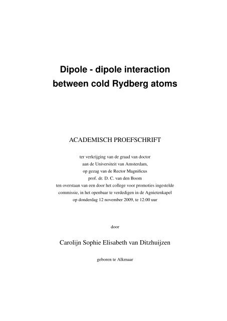
ACADEMISCH PROEFSCHRIFT<br />
ter verkrijging van de graad van doctor<br />
aan de Universiteit van Amsterdam,<br />
op gezag van de Rector Magnificus<br />
prof. dr. D. C. van den Boom<br />
ten overstaan van een door het college voor promoties ingestelde<br />
commissie, in het openbaar te verdedigen in de Agnietenkapel<br />
op donderdag 12 november 2009, te 12:00 uur<br />
door<br />
Carolijn Sophie Elisabeth van Ditzhuijzen<br />
geboren te Alkmaar
Promotiecommissie:<br />
Promotor:<br />
Copromotor:<br />
Overige leden:<br />
prof. dr. H. B. van Linden van den Heuvell<br />
prof. dr. L. D. Noordam<br />
prof. dr. M. Weidemüller<br />
prof. dr. L. Kouwenhoven<br />
dr. E. J. D. Vredenbregt<br />
prof. dr. M. S. Golden<br />
dr. T. W. Hijmans<br />
dr. R. J. C. Spreeuw<br />
Faculteit der Natuurwetenschappen, Wiskunde en Informatica<br />
ISBN: 978-90-9024636-9<br />
A full colour version of this thesis can be downloaded from<br />
http://www.science.uva.nl/research/aplp<br />
On the cover is depicted a Stark map: the energy levels of <strong>Rydberg</strong> states with<br />
principal quantum numbers <strong>between</strong> 32 and 35 in a static electric field. The<br />
rainbow color scheme was inspired by work from the artist Jen Stark.<br />
The research reported in this thesis was carried out at the group ”Quantum Gases<br />
& Quantum Information”, Van der Waals-Zeeman Instituut, Universiteit van<br />
Amsterdam, Valckenierstraat 65, 1018 XE Amsterdam, The Netherlands, where<br />
a limited number of copies of this thesis is available. The work was part of<br />
the research program of the ”Stichting voor Fundamenteel Onderzoek der Materie”<br />
(FOM), which is financially supported by the ”Nederlandse Organisatie<br />
voor Wetenschappelijk Onderzoek” (NWO).
Inhoudsopgave<br />
1 Introduction 1<br />
1.1 Quantum computing . . . . . . . . . . . . . . . . . . . . . . . . . . . . 1<br />
1.2 <strong>Rydberg</strong> <strong>atoms</strong> . . . . . . . . . . . . . . . . . . . . . . . . . . . . . . . 2<br />
1.3 Experimental context . . . . . . . . . . . . . . . . . . . . . . . . . . . . 4<br />
1.4 This thesis . . . . . . . . . . . . . . . . . . . . . . . . . . . . . . . . . . 5<br />
2 Theoretical background 7<br />
2.1 Introduction . . . . . . . . . . . . . . . . . . . . . . . . . . . . . . . . . 8<br />
2.2 <strong>Rydberg</strong> wavefunctions . . . . . . . . . . . . . . . . . . . . . . . . . . . 9<br />
2.2.1 Hydrogen wavefunctions . . . . . . . . . . . . . . . . . . . . . . 10<br />
2.2.2 Analytical wavefunctions for alkali <strong>atoms</strong> . . . . . . . . . . . . . 11<br />
2.2.3 Numerical calculation of the radial wavefunctions . . . . . . . . . 12<br />
2.2.4 Expectation values . . . . . . . . . . . . . . . . . . . . . . . . . 14<br />
2.3 <strong>Rydberg</strong> <strong>atoms</strong> in a static electric field . . . . . . . . . . . . . . . . . . . 16<br />
2.3.1 The Stark effect . . . . . . . . . . . . . . . . . . . . . . . . . . . 16<br />
2.3.2 Perturbation theory . . . . . . . . . . . . . . . . . . . . . . . . . 17<br />
2.3.3 Calculation of a Stark map . . . . . . . . . . . . . . . . . . . . . 18<br />
2.3.4 Measurement of <strong>dipole</strong>-transitions to the manifold states . . . . . 20<br />
2.4 <strong>Dipole</strong>-<strong>dipole</strong> <strong>interaction</strong>s <strong>between</strong> <strong>Rydberg</strong> <strong>atoms</strong> . . . . . . . . . . . . 22<br />
2.4.1 A suitable <strong>dipole</strong>-<strong>dipole</strong> transition . . . . . . . . . . . . . . . . . 23<br />
2.5 Simulations . . . . . . . . . . . . . . . . . . . . . . . . . . . . . . . . . 27<br />
2.5.1 <strong>Dipole</strong>s distributed over long cylinders . . . . . . . . . . . . . . 27<br />
2.5.2 Frustrated diffusion . . . . . . . . . . . . . . . . . . . . . . . . . 31<br />
2.6 Summary . . . . . . . . . . . . . . . . . . . . . . . . . . . . . . . . . . 33<br />
3 Experimental Setup 35<br />
3.1 Introduction . . . . . . . . . . . . . . . . . . . . . . . . . . . . . . . . . 36<br />
3.2 The Magneto-Optical Trap . . . . . . . . . . . . . . . . . . . . . . . . . 40<br />
3.2.1 Diode laser system for the MOT . . . . . . . . . . . . . . . . . . 42<br />
3.2.2 Measurement of the MOT density . . . . . . . . . . . . . . . . . 45<br />
3.3 <strong>Rydberg</strong> excitation . . . . . . . . . . . . . . . . . . . . . . . . . . . . . 46<br />
3.3.1 Two-photon excitation . . . . . . . . . . . . . . . . . . . . . . . 46<br />
3.3.2 Nd:YAG pumped pulsed dye lasers . . . . . . . . . . . . . . . . 47<br />
3.3.3 Wavelength scan . . . . . . . . . . . . . . . . . . . . . . . . . . 49<br />
3.3.4 Size of the <strong>Rydberg</strong> volumes . . . . . . . . . . . . . . . . . . . . 51<br />
3.4 The role of electric and magnetic fields . . . . . . . . . . . . . . . . . . . 53<br />
3.4.1 Electric field . . . . . . . . . . . . . . . . . . . . . . . . . . . . 53<br />
3.4.2 Magnetic field . . . . . . . . . . . . . . . . . . . . . . . . . . . 55<br />
iii
iv<br />
INHOUDSOPGAVE<br />
3.4.3 Black Body Radiation and Natural Lifetime . . . . . . . . . . . . 56<br />
3.5 <strong>Rydberg</strong> detection . . . . . . . . . . . . . . . . . . . . . . . . . . . . . . 56<br />
3.6 Summary . . . . . . . . . . . . . . . . . . . . . . . . . . . . . . . . . . 59<br />
4 Simultaneous position and state measurement of <strong>Rydberg</strong> <strong>atoms</strong> 61<br />
4.1 Introduction . . . . . . . . . . . . . . . . . . . . . . . . . . . . . . . . . 62<br />
4.2 State and position determination . . . . . . . . . . . . . . . . . . . . . . 62<br />
4.3 Experimental Setup . . . . . . . . . . . . . . . . . . . . . . . . . . . . . 63<br />
4.4 Results . . . . . . . . . . . . . . . . . . . . . . . . . . . . . . . . . . . . 65<br />
4.5 Conclusions . . . . . . . . . . . . . . . . . . . . . . . . . . . . . . . . . 68<br />
5 Spatially resolved observation of <strong>dipole</strong>-<strong>dipole</strong> <strong>interaction</strong> 69<br />
5.1 Introduction . . . . . . . . . . . . . . . . . . . . . . . . . . . . . . . . . 70<br />
5.2 Experimental setup . . . . . . . . . . . . . . . . . . . . . . . . . . . . . 70<br />
5.3 Simulations . . . . . . . . . . . . . . . . . . . . . . . . . . . . . . . . . 72<br />
5.4 Results . . . . . . . . . . . . . . . . . . . . . . . . . . . . . . . . . . . . 72<br />
5.5 Conclusions . . . . . . . . . . . . . . . . . . . . . . . . . . . . . . . . . 75<br />
6 Radio-frequency driven <strong>dipole</strong>-<strong>dipole</strong> <strong>interaction</strong>s in spatially separated volumes<br />
77<br />
6.1 Introduction . . . . . . . . . . . . . . . . . . . . . . . . . . . . . . . . . 78<br />
6.2 Radiofrequency Assisted <strong>Dipole</strong>-<strong>Dipole</strong> Interactions . . . . . . . . . . . 79<br />
6.3 Improved Experimental Setup . . . . . . . . . . . . . . . . . . . . . . . 82<br />
6.4 Diabatic Switching of the <strong>Dipole</strong>-<strong>Dipole</strong> Interaction . . . . . . . . . . . . 84<br />
6.5 Observation of RF-assisted <strong>dipole</strong>-<strong>dipole</strong> transitions . . . . . . . . . . . . 85<br />
6.6 Conclusion . . . . . . . . . . . . . . . . . . . . . . . . . . . . . . . . . 88<br />
7 Observation of Stückelberg oscillations in <strong>dipole</strong>-<strong>dipole</strong> <strong>interaction</strong>s 89<br />
7.1 Introduction . . . . . . . . . . . . . . . . . . . . . . . . . . . . . . . . . 90<br />
7.2 A coupled two-level system in an oscillating field . . . . . . . . . . . . . 90<br />
7.2.1 Numerical solutions in the time domain . . . . . . . . . . . . . . 92<br />
7.2.2 Floquet approach . . . . . . . . . . . . . . . . . . . . . . . . . . 93<br />
7.2.3 Classical limit of the sideband population distribution . . . . . . . 97<br />
7.2.4 Coupling <strong>between</strong> dressed and undressed state . . . . . . . . . . 99<br />
7.2.5 Stückelberg oscillations . . . . . . . . . . . . . . . . . . . . . . 102<br />
7.3 Experiment . . . . . . . . . . . . . . . . . . . . . . . . . . . . . . . . . 108<br />
7.3.1 Spectroscopy . . . . . . . . . . . . . . . . . . . . . . . . . . . . 108<br />
7.3.2 Experimental Setup . . . . . . . . . . . . . . . . . . . . . . . . . 110<br />
7.3.3 Simulations for multiple atom pairs . . . . . . . . . . . . . . . . 110<br />
7.3.4 Results . . . . . . . . . . . . . . . . . . . . . . . . . . . . . . . 111<br />
7.4 Conclusions . . . . . . . . . . . . . . . . . . . . . . . . . . . . . . . . . 115<br />
Summary 117<br />
References 119<br />
Samenvatting 127<br />
Dankwoord 131<br />
List of Publications 133
1 Introduction<br />
1.1 Quantum computing<br />
The combination of quantum mechanics and technology has many promises of which the<br />
quantum computer might be the most spectacular one. Despite this claim, the quantum<br />
computer is not existing yet. The reason is that there are competing requirements from<br />
quantum mechanics and from technology. The bit of a quantum computer, the qubit, can<br />
have the value |0〉 and |1〉 at the same time, whereas the classical computer bit is either 0<br />
or 1. This is called superposition. Secondly, the qubits are entangled, which means their<br />
values are connected. The advantage of the quantum computer lies in the combination of<br />
entanglement and superposition: complicated calculations are performed with all qubits<br />
at the same time, while they also have all possible values at the same time. This makes<br />
the quantum computer much faster than a classical computer.<br />
The qubits in a quantum computer should be realized with quantum mechanical objects<br />
and they should be able to have an unperturbed coherent evolution. In other words,<br />
they should be light, <strong>cold</strong> and isolated. Hardware implementation requires, on the other<br />
hand, a system that is sufficiently large and sufficiently strongly coupled to a measuring<br />
device. This conflict is very generic and there are various proposals for a solution from<br />
a surprisingly wide range of physics. Quantum information can be encoded in, for instance,<br />
the various spins of electrons in a molecule (the NMR approach) [96], the spins<br />
of an electron in a solid state [53] or the internal state of a trapped ion [15]. But there<br />
are many more proposals [44], including ones that seem very exotic at first sight, such<br />
as a quantum bit on basis of the topology of an N-particle configuration in a 2D system<br />
[75, 8]. This thesis investigates the idea to use the states of gas-phase <strong>Rydberg</strong> <strong>atoms</strong> as<br />
qubits, which are <strong>atoms</strong> in highly excited states.<br />
A quantum computer requires operations involving multiple qubits, notably the XORoperation,<br />
which requires <strong>interaction</strong> <strong>between</strong> qubits. A system of interacting <strong>Rydberg</strong><br />
<strong>atoms</strong> can perform this task and has some unique advantages:<br />
• The <strong>interaction</strong> <strong>between</strong> particles is unusually strong for quantum systems.<br />
• The <strong>interaction</strong> <strong>between</strong> particles can be switched on and off, e.g. by exciting an<br />
atom to a <strong>Rydberg</strong> state and de-exciting back to the ground state, or by tuning an<br />
electric field.<br />
• A system can have typical dimensions of micrometers instead of nanometers, which<br />
makes it technologically easily accessible.<br />
1
2 Introduction<br />
• The building blocks (<strong>Rydberg</strong> <strong>atoms</strong>) are neutral, and have therefore a relatively<br />
weak <strong>interaction</strong> with the environment.<br />
The possible relevance of <strong>Rydberg</strong> <strong>atoms</strong> for quantum information was first pointed out<br />
by the Zoller group in Innsbruck [46, 65]; they sketched a potential solution for the XOR<br />
gate, which makes use of the so-called <strong>dipole</strong>-blockade; alternative proposals followed<br />
[81, 84]. This thesis focuses on the <strong>dipole</strong>-<strong>dipole</strong> <strong>interaction</strong> <strong>between</strong> <strong>Rydberg</strong> <strong>atoms</strong>,<br />
which is the cause of the <strong>dipole</strong>-blockade. In short, the goal of the research presented<br />
in this thesis is to investigate the <strong>interaction</strong> <strong>between</strong> <strong>Rydberg</strong> <strong>atoms</strong> and to explore the<br />
possibilities and limitations of the system of interacting <strong>Rydberg</strong> <strong>atoms</strong> in its future contribution<br />
to quantum information processing.<br />
V dd<br />
01<br />
10<br />
20 Μm<br />
Figure 1.1: A schematic representation of two <strong>Rydberg</strong> <strong>atoms</strong> that are used as qubits (not to scale).<br />
<strong>Dipole</strong>-<strong>dipole</strong> <strong>interaction</strong> (V dd ) causes the left atom to make a transition from |0〉 to |1〉 while the right<br />
atom makes a transition from |1〉 to |0〉.<br />
The <strong>dipole</strong>-<strong>dipole</strong> <strong>interaction</strong> occurs in many places and systems in nature, but it is<br />
most easily studied and manipulated in <strong>Rydberg</strong> <strong>atoms</strong>, because of the accessible distance<br />
scale. A well-known form of the <strong>dipole</strong>-<strong>dipole</strong> <strong>interaction</strong> is the Van der Waals<strong>interaction</strong>.<br />
This <strong>interaction</strong> is very weak due to the non-resonant character, and only<br />
significant at smaller distances; it drops off with the sixth or seventh power of distance.<br />
In the familiar case of droplet formation in liquids, the <strong>interaction</strong> occurs <strong>between</strong> particles<br />
at distances comparable to their own size. Here we focus on a resonant and thus<br />
stronger type of <strong>dipole</strong>-<strong>dipole</strong> <strong>interaction</strong>. In addition, the <strong>Rydberg</strong> <strong>atoms</strong> are a thousand<br />
times larger than regular <strong>atoms</strong> and therefore have an <strong>interaction</strong> that is a million times<br />
stronger. Due to the unusually strong <strong>interaction</strong> we can work with distances that are four<br />
orders of magnitude larger compared to the normal distance in a liquid or a solid. This<br />
makes the <strong>dipole</strong>-<strong>dipole</strong> <strong>interaction</strong> twelve orders of magnitude smaller - the <strong>interaction</strong><br />
decreases with the third power of the atom-atom distance. Still, the <strong>interaction</strong> energy<br />
is Planck’s constant times hundreds of kHz; <strong>interaction</strong>s that are detectable within 10’s<br />
of microseconds. Two <strong>dipole</strong>-interacting <strong>Rydberg</strong> <strong>atoms</strong> are schematically depicted in<br />
figure 1.1.<br />
1.2 <strong>Rydberg</strong> <strong>atoms</strong><br />
The name ”<strong>Rydberg</strong> <strong>atoms</strong>” honors the Swedish physicist Johannes <strong>Rydberg</strong>. He introduced<br />
the <strong>Rydberg</strong> formula, which gives the wavenumber of the spectral lines in hydro-
1.2 <strong>Rydberg</strong> <strong>atoms</strong> 3<br />
gen, but also other <strong>atoms</strong>, especially alkali <strong>atoms</strong>. For hydrogen, the <strong>Rydberg</strong> formula<br />
is<br />
(<br />
1 1<br />
λ = Ry − 1 )<br />
, (1.1)<br />
n 2 1<br />
n 2 2<br />
with 1/λ the wavenumber, n 1 and n 2 integer values and Ry the <strong>Rydberg</strong> constant. For<br />
high values of n equation 1.1 is most accurate, which is why <strong>atoms</strong> with high n are called<br />
<strong>Rydberg</strong> <strong>atoms</strong>.<br />
<strong>Rydberg</strong> <strong>atoms</strong> have highly exaggerated properties compared to ground-state <strong>atoms</strong>.<br />
They are large, very sensitive to electromagnetic fields and, as we study in this thesis, they<br />
interact with each other at large distances. Some of their properties can easily be deduced<br />
from the Bohr model. The Bohr model is not as accurate as quantum mechanics, but it<br />
is much easier and it works as a first approximation for the hydrogen atom. Quantization<br />
of angular momentum (mvr = n) is combined with classical mechanics – the Coulomb<br />
force equals the centripetal force on the electron:<br />
This leads to an expression of the radius of the electron orbit<br />
1<br />
4πɛ 0<br />
e 2<br />
r 2 = mv2<br />
r . (1.2)<br />
r = 4πɛ 0n 2 2<br />
e 2 m = a 0n 2 , (1.3)<br />
where a 0 is the Bohr radius. The energy of such an electron is negative, because the<br />
electron is bound to the positively charged nucleus<br />
E = −<br />
e4 m<br />
(4πɛ 0 ) 2 2 1<br />
2n 2 = −Ry n 2 . (1.4)<br />
Bohr had hereby expressed the <strong>Rydberg</strong> constant in terms of already known constants.<br />
We see that with increasing n, the radius increases as n 2 and the binding energy decreases<br />
as 1/n 2 . Also note that the energy levels of subsequent n states come increasingly closer<br />
together for higher n; the energy difference <strong>between</strong> two adjacent n states scales like 1/n 3 .<br />
The larger distance of the electron to the nucleus makes the atom more sensitive to<br />
external electric fields; an external electric field can easily overcome the Coulomb field<br />
of the nucleus far away. It can easily be shown from the Bohr atom that the electron can<br />
escape the atom for an external field of<br />
F ion = 1 Ry 2<br />
4πɛ 0 4n = 1 a.u., (1.5)<br />
4 16n4 where the right hand side is the more usual form in atomic units (see section 2.1).<br />
The round trip time of the electron around the nucleus scales as n 3 , simply from T =<br />
2πr/v for uniform circular motion. The radiative lifetime of the <strong>Rydberg</strong> state also scales<br />
like n 3 . This is explained by the fact that the electron spends most of its time far away<br />
from the nucleus, where it is almost a free electron. Free electrons can not radiate, so the<br />
electron can only radiatively decay when it’s close to the core, which occurs once every<br />
round trip.
4 Introduction<br />
The <strong>dipole</strong> moment µ is given by the separation of charge, so this scales equally as the<br />
electron-core distance: µ∼n 2 . The <strong>dipole</strong>-<strong>dipole</strong> <strong>interaction</strong> energy <strong>between</strong> two similar<br />
<strong>dipole</strong>s is deduced from the energy of a <strong>dipole</strong> in the electric field of the other <strong>dipole</strong>:<br />
V dd =−µ·F d . The field of a <strong>dipole</strong> at distance R is the sum of the field of the nucleus 1/R 2<br />
and the field of the electron −1/(R + n 2 ) 2 , which approximately adds up to F d ∼ n 2 /R 3 .<br />
The <strong>dipole</strong>-<strong>dipole</strong> <strong>interaction</strong> energy therefore becomes V dd ∼n 4 /R 3 .<br />
All these scaling laws for n show that for highly excited states (high n) many properties<br />
are exaggerated. This enables interesting experiments, which are not possible with ground<br />
state <strong>atoms</strong>. The long lifetime gives the opportunity to study the <strong>atoms</strong> for a long time.<br />
The sensitivity to external fields makes it relatively simple to study their behavior in very<br />
modest fields. The large <strong>dipole</strong> moments make it possible to study <strong>interaction</strong>s <strong>between</strong><br />
<strong>atoms</strong> at very large distances. Some values for n=50 are given in table 1.1.<br />
Table 1.1: <strong>Rydberg</strong> atom properties.<br />
Property n-dependence value for n=50<br />
radius n 2 0.2 µm<br />
binding energy n −2 5.4 meV<br />
natural lifetime n 3 ∼0.2 ms<br />
ionization field n −4 51 V/cm<br />
maximum permanent <strong>dipole</strong> moment n 2 9300 debye<br />
<strong>dipole</strong>-<strong>dipole</strong> <strong>interaction</strong> frequency n 4 /R 3 2π 1.6 MHz (R=20 µm)<br />
1.3 Experimental context<br />
In the 20 th century many experiments with <strong>Rydberg</strong> <strong>atoms</strong> have been performed already,<br />
but the development of the magneto-optical trap has set off a new wave of <strong>Rydberg</strong> atom<br />
experiments in the 21 st century. In the magneto-optical trap ground-state <strong>atoms</strong> are cooled<br />
down to ∼100 µK by means of laser cooling. This provides more control over the position<br />
of the <strong>atoms</strong>, since they move slower. Having obtained such a <strong>cold</strong> cloud of <strong>atoms</strong>, the<br />
<strong>atoms</strong> can be excited to <strong>Rydberg</strong> states with a laser [7, 69]. Another approach is deceleration<br />
and trapping of <strong>Rydberg</strong> <strong>atoms</strong> by static electric fields [97, 42]. The main focus is<br />
on <strong>interaction</strong>s <strong>between</strong> <strong>Rydberg</strong> <strong>atoms</strong>, in most cases electric <strong>dipole</strong>-<strong>dipole</strong> <strong>interaction</strong>s.<br />
<strong>Dipole</strong>-<strong>dipole</strong> <strong>interaction</strong>s can occur <strong>between</strong> static <strong>dipole</strong>s, but in <strong>Rydberg</strong> atom experiments<br />
often transition <strong>dipole</strong>s are used, where both <strong>atoms</strong> undergo a transition <strong>between</strong><br />
two angular momentum states. Due to energy conservation, the energy that one<br />
atom gains with the transition equals the energy that the other atom loses. This is called<br />
a Förster resonance. In <strong>Rydberg</strong> <strong>atoms</strong>, there is a lot of choice for initial energy levels<br />
and, furthermore, the energy levels can easily be tuned with an electric field, so a resonant<br />
two-atom transition is easily found. Resonant <strong>dipole</strong>-<strong>dipole</strong> <strong>interaction</strong>s have been studied<br />
with room-temperature <strong>Rydberg</strong> <strong>atoms</strong> in the past, where the transition was brought into<br />
resonance with a static electric field [85, 90] or an oscillating field, i.e. electro-magnetic<br />
radiation [34, 16, 73].
1.4 This thesis 5<br />
Several interesting effects are observed in a sample of ultra<strong>cold</strong> <strong>Rydberg</strong> <strong>atoms</strong>. While<br />
in the older ”hot” experiments the <strong>interaction</strong>s were two-atom collisions, in the new<br />
”frozen gases” many-body <strong>interaction</strong>s play a role [7, 69]. Secondly, mechanical effects<br />
could be observed now, due to the dipolar forces [31, 61, 104], as well as the angular<br />
dependence of the <strong>dipole</strong>-<strong>dipole</strong> <strong>interaction</strong> [21]. The <strong>dipole</strong> forces lead to spontaneous<br />
plasma formation for high densities of <strong>cold</strong> <strong>atoms</strong> [61], and also the other way round:<br />
starting from an ultra<strong>cold</strong> plasma, <strong>Rydberg</strong> <strong>atoms</strong> can be spontaneously formed [70].<br />
Such an ultra<strong>cold</strong> plasma [82] is a new and exotic type of matter and could give insights<br />
in plasma’s that occur naturally in astrophysical systems. Another possibility is that two<br />
<strong>cold</strong> <strong>Rydberg</strong> <strong>atoms</strong> can form an enormous molecule [36, 18, 26, 30, 11].<br />
The already mentioned <strong>dipole</strong>-blockade proposal [46, 65] has received a lot of interest.<br />
<strong>Dipole</strong>-blockade is the inhibition of optical transitions from ground state <strong>atoms</strong> to <strong>Rydberg</strong><br />
states due to the presence of a nearby <strong>Rydberg</strong> atom. This <strong>dipole</strong> blockade effect is<br />
observed by several groups [92, 86, 62, 98, 49, 103]. It is suggested that using oscillating<br />
fields instead of static fields to tune the <strong>interaction</strong> into resonance has some benefits for<br />
quantum information processing [4, 17]. However, all these experiments are performed<br />
with randomly organized ”frozen” gases, without direct control over the interatomic distances.<br />
In this thesis we aim for <strong>interaction</strong>s over a fixed and controlled distance. During<br />
the final stages of writing this thesis two groups achieved a large amount of control of resonant<br />
<strong>dipole</strong>-<strong>dipole</strong> <strong>interaction</strong> [94, 33], where coherent evolution was observed <strong>between</strong><br />
two <strong>Rydberg</strong> <strong>atoms</strong> fixed in space.<br />
Quantum information with <strong>dipole</strong>-interacting particles is also investigated in other<br />
fields of physics. For example, molecules can be used [41] or self-assembled quantum<br />
dots, instead of real <strong>atoms</strong> [20]. In nanophotonics, coherent <strong>dipole</strong>-<strong>dipole</strong> coupling<br />
<strong>between</strong> carefully placed polarizable plasmonic nanoparticles is pursued as a tool<br />
to create ultrasmall optical circuits [76, 19, 40, 52]. Resonant <strong>dipole</strong>-<strong>dipole</strong> <strong>interaction</strong>s<br />
even occur in biological systems: in bacterial light harvesting complexes, responsible<br />
for photosynthesis, resonant <strong>dipole</strong>-<strong>dipole</strong> <strong>interaction</strong>s mediate the ultrafast energy flow<br />
[95, 39, 28, 59]. <strong>Dipole</strong>-<strong>dipole</strong> <strong>interaction</strong>s <strong>between</strong> fluorophores, as first described by<br />
Förster [32], are now a workhorse tool in biological imaging to measure nanoscale distances<br />
[47]. We believe that our research might help in understanding these complex<br />
biological systems.<br />
In most of these systems the <strong>dipole</strong> moments are so small that the <strong>interaction</strong> can<br />
only be observed if the separation of the <strong>dipole</strong>s is at a nanometer scale. Moreover, the<br />
<strong>interaction</strong>s typically occur on a femtosecond timescale. These properties make these<br />
experiments technologically very challenging. With the ability to control the distance<br />
<strong>between</strong> <strong>Rydberg</strong> <strong>atoms</strong>, which have an <strong>interaction</strong> over a distance of tens of micrometers<br />
and an <strong>interaction</strong> time of several microseconds, the study of coherent <strong>dipole</strong>-<strong>dipole</strong><br />
<strong>interaction</strong> is more feasible.<br />
1.4 This thesis<br />
In the experiments described in this thesis we use <strong>Rydberg</strong> states <strong>between</strong> n = 40 and<br />
50 which have <strong>dipole</strong> moments of the order of 1000a 0 e and radiative lifetimes around
6 Introduction<br />
100 µs. The <strong>dipole</strong>-<strong>dipole</strong> <strong>interaction</strong> time is then typically 15 µs for a distance of 40 µm.<br />
The gas-phase <strong>atoms</strong> must have such a low speed that they move only a small fraction<br />
of their distance (∼40 µm) during the <strong>interaction</strong> time (∼20 µs). To achieve such low<br />
speeds (≪2 m/s) we use laser-cooling of the ground-state <strong>atoms</strong> prior to the experiments.<br />
Once we have prepared a <strong>cold</strong> cloud of ground state <strong>atoms</strong> - in a magneto-optical trap -<br />
we excite <strong>atoms</strong> to two different <strong>Rydberg</strong> states with the use of two separate lasers. Each<br />
laser is focused in the cloud to create a narrow region of <strong>Rydberg</strong> <strong>atoms</strong> (∼ 15 µm in<br />
diameter). <strong>Rydberg</strong> <strong>atoms</strong> from one volume are allowed to interact resonantly with <strong>atoms</strong><br />
in the other volume, tens of micrometers away, for a controlled amount of time, typically<br />
several microseconds, after which they are detected.<br />
In the following chapter, chapter 2, we will provide the theoretical background of the<br />
experiments described in chapters 4 - 7. We describe calculations of experimental parameters,<br />
such as the energy levels of the <strong>Rydberg</strong> states in an electric field, needed to find<br />
an appropriate <strong>dipole</strong>-<strong>dipole</strong> resonance, and relevant transition <strong>dipole</strong> moments. The analytic<br />
version of this is a new generalization of the Gordon expression [12]. Furthermore<br />
we describe simulations, which provide basic understanding of the experiments.<br />
In chapter 3 we describe the experimental setup we have build for our experiments,<br />
and we investigate some of its properties, such as the density in the <strong>cold</strong> atom cloud, the<br />
size of the <strong>Rydberg</strong> volumes and the homogeneity of the electric field.<br />
In chapter 4 we present a novel technique that we developed, with which we can<br />
simultaneously detect the quantum state of the <strong>Rydberg</strong> <strong>atoms</strong> as well as their position.<br />
The technique combines state-selective field ionization to deduce the principal quantum<br />
number and time-of-flight of the ionization products (electrons) to deduce the original<br />
position of the <strong>atoms</strong> with an accuracy of about 10 µm.<br />
Chapter 5 is in our view the most important chapter of this thesis. It describes the<br />
first realization of <strong>dipole</strong>-<strong>dipole</strong> <strong>interaction</strong> <strong>between</strong> <strong>Rydberg</strong> <strong>atoms</strong> in separate volumes.<br />
The resonant <strong>interaction</strong> 41d+49s ↔ 42p+49p is observed for volume separations up to<br />
50 µm. We measured the amount of <strong>interaction</strong> products as a function of static electric<br />
field, volume separation and time. The data fit the extensive many-body simulations.<br />
In the last two chapters we investigated the possibility to manipulate the <strong>dipole</strong>-<strong>dipole</strong><br />
<strong>interaction</strong> 41d+49s ↔ 42p+49p with radio-frequency fields. Instead of using a static<br />
electric field, the <strong>interaction</strong> can be tuned into resonance with an RF-field. In this case<br />
multiple resonances are observed due to multi RF-photon transitions. In chapter 6 we<br />
show that the <strong>dipole</strong>-<strong>dipole</strong> <strong>interaction</strong> can be switched on and off by rapidly switching<br />
the field. Secondly, we show that RF-fields can be used to perform sub-MHz spectroscopy<br />
on the involved <strong>Rydberg</strong> states and we observe strong AC-Stark shifts and multi-photon<br />
transitions with up to 5 photons.<br />
In chapter 7 we use an RF-field to demonstrate that the interacting <strong>atoms</strong> are coherent<br />
for at least 0.6 µs, by making use of so-called Stückelberg oscillations. We present two<br />
different methods to calculate the <strong>interaction</strong> strength as a function of static field and RFfield.<br />
This calculation is essentially different for states with a linear Stark shift as for<br />
states with a quadratic Stark shift, which is the case for the angular momentum states<br />
involved here. The variations of <strong>interaction</strong> strength are in fact Stückelberg oscillations<br />
and the measured <strong>interaction</strong> strength fits the calculations very well. These calculations<br />
culminate in a rather unusual result: the generalized Bessel function.
2 Theoretical background<br />
Calculations of <strong>Rydberg</strong> wavefunctions and Stark shifts enable us<br />
to choose a suitable resonant <strong>dipole</strong>-<strong>dipole</strong> transition for our experiments:<br />
41d + 49s ↔ 42p + 49p is resonant at a field of only<br />
∼0.4 V/cm and the relevant transition <strong>dipole</strong> moments are of the<br />
order of ∼1000 a.u. Both the calculation of the wavefunctions<br />
as well as the Stark shift are covered extensively in this chapter.<br />
Simulations of <strong>dipole</strong>s distributed over two parallel elongated volumes<br />
show that the short-time energy transfer probability scales<br />
like P ∝ t 2 /d 5 , with d the distance <strong>between</strong> the volumes. In a<br />
simulation of excitation diffusion in a randomly organized system<br />
we observe localization resulting from the <strong>dipole</strong> blockade effect.<br />
7
8 Theoretical background<br />
2.1 Introduction<br />
This chapter describes the theoretical background and the calculations we have performed.<br />
They both provide us with physical insight as well as the necessary experimental parameters.<br />
The main goal was to find a suitable set of states for <strong>dipole</strong>-<strong>dipole</strong> <strong>interaction</strong> among<br />
<strong>Rydberg</strong> states. We aimed for a strong <strong>interaction</strong>, allowing us to measure at large atomic<br />
distances. To find resonant <strong>interaction</strong>s, one needs to know the exact energies of the <strong>atoms</strong><br />
as a function of electric field, for which the wavefunctions of the <strong>atoms</strong> should be known.<br />
The calculation of the <strong>Rydberg</strong> atom wavefunctions are described in section 2.2. The<br />
potential for the electron is not exactly known, but by making use of the experimentally<br />
known binding energies an approximate wavefunction can be obtained. We present two<br />
methods, one numerical and one analytical solution of the Schrödinger equation. The following<br />
section, section 2.3, deals with the Stark effect: the shift of atomic energy levels<br />
in an electric field. The Stark shift can be calculated by means of perturbation theory; in<br />
contrast to the situation with lower excited states, this perturbation couples many states<br />
and the calculation involves quite large matrices.<br />
In section 2.4 <strong>dipole</strong>-<strong>dipole</strong> <strong>interaction</strong>s are explained. Here we also pick a suitable<br />
resonant transition, that we will use in our experiments. The transition of our preference is<br />
41d+49s ↔ 42p+49p. For this transition we calculated the resonance fields (0.3807 V/cm<br />
and 0.4112 V/cm) and the transition <strong>dipole</strong> moments of both involved atomic transitions<br />
41d ↔ 42p and 49s ↔ 49p. Both <strong>dipole</strong> moments are about 1000 a 0 e, leading to a<br />
<strong>dipole</strong>-<strong>dipole</strong> <strong>interaction</strong> strength of V dd = 2π 35 kHz at a particle distance of 40 µm.<br />
Table 2.1: CODATA (”Committee on Data for Science and Technology”) values of atomic units (a.u.)<br />
from [68], with the reduced Planck constant, µ B the Bohr magneton and ɛ 0 the electric constant.<br />
Name Origin Value<br />
length Bohr radius a 0 0.529 177 208 59(36)·10 −10 m<br />
time /E h 2.418 884 326 505(16)·10 −17 s<br />
mass electron mass m e 9.109 382 15(45)·10 −31 kg<br />
energy<br />
Hartree energy E h = e2<br />
4πɛ 0 a 0<br />
4.359 743 94(22)·10 −18 J<br />
charge elementary charge e 1.602 176 487(40)·10 −19 C<br />
electric <strong>dipole</strong> moment a 0 e 8.478 352 81(21)·10 −30 C m<br />
electric field E h /a 0 e 5.142 206 32(13)·10 9 V/cm<br />
magnetic <strong>dipole</strong> moment 2µ B =e/m e 1.854 801 830(46)·10 −27 J/G<br />
magnetic field /a 2 0 e 2.350 517 382(59)·109 G<br />
The last section 2.5 presents some simulations with <strong>dipole</strong>-<strong>dipole</strong> interacting <strong>atoms</strong>.<br />
In the experiments described in this thesis, we don’t have two single <strong>Rydberg</strong> <strong>atoms</strong>, but<br />
two very thin and long cylinder-like volumes with tens of <strong>Rydberg</strong> <strong>atoms</strong>. Straightforward<br />
two-particle simulations predict the experimental data from chapter 5 quite well. Furthermore<br />
we analytically derive a scaling law for the transition probability for short times as a<br />
function of distance d:P ∼ t 2 /d 5 . Another simulation we performed describes excitation<br />
diffusion through <strong>dipole</strong>-<strong>dipole</strong> <strong>interaction</strong>. This simulation compares the diffusion in an<br />
ordered lattice of particles to a randomly organized particle configuration, where the latter
2.2 <strong>Rydberg</strong> wavefunctions 9<br />
Table 2.2: Energy conversions used in this thesis. The full precision and uncertainty can be found in<br />
[68].<br />
MHz<br />
cm −1<br />
1 MHz 1 MHz 3.335 641·10 −5 cm −1<br />
1 cm −1 2.997 925·10 4 MHz 1 cm −1<br />
1 eV 2.417 989 5·10 8 MHz 8.065 545·10 3 cm −1<br />
1 a.u. 6.579 683 920 7·10 9 MHz 2.194 746 313 7·10 5 cm −1<br />
1 J 1.509 190 5·10 27 MHz 5.034 117·10 22 cm −1<br />
Table 2.2: continued<br />
eV a.u. J<br />
1 MHz 4.135 667·10 −9 eV 1.519 829 846 0·10 −10 a.u. 6.626 069·10 −28 J<br />
1 cm −1 1.239 841 9·10 −4 eV 4.556 335 252 8·10 −6 a.u. 1.986 446·10 −23 J<br />
1 eV 1 eV 3.674 932 5·10 −2 a.u. 1.602 176 5·10 −19 J<br />
1 a.u. 27.211 384 eV 1 a.u. 4.359 744·10 −18 J<br />
1 J 6.241 510·10 18 eV 2.293 713·10 17 a.u. 1 J<br />
case shows an interesting result: a localization effect which is due to a <strong>dipole</strong>-blockade<br />
mechanism.<br />
In this thesis atomic units are used unless stated otherwise. Atomic units form a<br />
convenient system of units in atomic physics; all quantities are measured relative to the<br />
properties of atomic hydrogen. In practice it means that the values for the electron charge,<br />
the electron mass, the reduced Planck constant are all set to unity as well as 1/4πɛ 0 and<br />
2µ B . This leads to a significant simplification of all relevant equations.<br />
In table 2.1 the conversion factors from atomic units to SI units are given for a selection<br />
of quantities relevant in this thesis. Values are taken from the ‘2006 CODATA<br />
recommended values’ as published in [68].<br />
The energy unit Joule is usually impractical for atomic physics. Apart from atomic<br />
units we often use MHz or cm −1 , also eV are common. In table 2.2 some energy conversions<br />
are given.<br />
2.2 <strong>Rydberg</strong> wavefunctions<br />
Alkali <strong>atoms</strong> are effectively one-electron <strong>atoms</strong>, just as hydrogen. Especially when the<br />
electron of the atom is in a highly excited state, or <strong>Rydberg</strong> state, there are many similarities.<br />
For rubidium in its ground state we have a nucleus of charge +37, and the first 36<br />
electrons around it form a very stable configuration around the nucleus, as in the noble<br />
gas krypton, where all electron shells up to 4p are filled. The last electron is on his own in<br />
a new shell, the 5s state. When the atom is (laser) excited to a <strong>Rydberg</strong> state, this single<br />
electron is moved to a shell with a much higher principal quantum number n. Already<br />
in the 5s ground state, but especially in the higher excited states, we can consider the<br />
inner part of the atom as a point particle with charge +1, equivalent with the hydrogen<br />
nucleus. Therefore we will first discuss the hydrogen wavefunctions, which can be cal-
10 Theoretical background<br />
culated exactly (subsection 2.2.1). Then we will make some small adjustments for the<br />
wavefunctions that enter the core region of the atom: the low angular momentum states.<br />
These wavefunctions are not exact and we will discuss two different methods to calculate<br />
these wavefunctions, an analytical and a numerical method (resp. subsections 2.2.2 and<br />
2.2.3). The last part of this section describes how the expectation values of powers of<br />
the electron position are obtained (subsection 2.2.4). The general expression for these<br />
expectation values enables us to calculate the <strong>interaction</strong> with a static electric or magnetic<br />
field as well as with electromagnetic radiation. Especially the <strong>interaction</strong> with an electric<br />
field is important, because this leads to an energy shift of the states resulting in multiple<br />
resonances of the <strong>dipole</strong>-<strong>dipole</strong> <strong>interaction</strong>. The expectation values will also be necessary<br />
to calculate the <strong>interaction</strong> strength <strong>between</strong> two <strong>Rydberg</strong> <strong>atoms</strong>.<br />
2.2.1 Hydrogen wavefunctions<br />
The potential for the electron is the Coulomb potential V = −1/r due to the attraction<br />
to the nucleus. To compute the wavefunctions one needs to solve the time-independent<br />
Schrödinger equation<br />
− 1 2 ∇2 ψ(r) − 1 ψ(r) = Eψ(r). (2.1)<br />
r<br />
Given the spherically symmetric potential, we can use spherical coordinates and separate<br />
the Schrödinger equation in a radial and an angular part:<br />
ψ(r) = R(r)Y lm (θ, φ). (2.2)<br />
The spherical harmonics Y lm (θ, φ) are independent of the exact potential as long as it is<br />
spherically symmetric. The solution of the angular part of the Schrödinger equation is<br />
given by [12]<br />
Y lm (θ, φ) =<br />
√<br />
(2l + 1) (l − m)!<br />
4π (l + m)! Pm l (cos θ) eimφ , (2.3)<br />
with P m l<br />
the associated Legendre function [2]. From boundary conditions for θ and φ<br />
follows l ≥ 0 and |m| ≤ l. l is the orbital angular momentum quantum number and m is<br />
the magnetic quantum number.<br />
For the radial part of the Schrödinger equation it is practical to rewrite u(r) = rR(r)<br />
and we obtain (<br />
− 1 d 2 l(l + 1)<br />
+ − 1 )<br />
u(r) = Eu(r). (2.4)<br />
2 dr2 2r 2 r<br />
The solutions [12] ∗ are in the form of an exponent of r, a power of r and a generalized<br />
Laguerre polynomial L b a [2]<br />
R nl (r) =<br />
√ (2<br />
n<br />
) 3 ( ) l<br />
(n − l − 1)! 2r<br />
2n(n + l)! e−r/n L 2l+1<br />
n−l−1<br />
n<br />
( ) 2r<br />
n<br />
(2.5)<br />
∗ Note that Bethe and Salpeter [12] use a different definition of the generalized Laguerre polynomial. We<br />
use here the Abramowitz and Stegun [2] definition, resulting in a slightly different expression for R nl .
2.2 <strong>Rydberg</strong> wavefunctions 11<br />
and the energy is<br />
E n = − 1<br />
2n 2 , (2.6)<br />
with n the principal quantum number. The orbital angular momentum quantum number<br />
l can not get larger than n − 1, or 0 ≤ l ≤ n − 1. The n −2 dependence of the binding<br />
energy (Eq. 2.6) was first noted in the 1880’s by Johann Balmer and Johannes <strong>Rydberg</strong><br />
and further developed by Niels Bohr in 1913 [105]. The associated wavenumber of a<br />
photon that just ionizes the atom is given by the <strong>Rydberg</strong> constant Ry. It is equivalent to<br />
half an atomic unit of energy (in units of cm −1 ), given in table 2.2. So the binding energy<br />
can also be written as E n =−Ry/n 2 .<br />
2.2.2 Analytical wavefunctions for alkali <strong>atoms</strong><br />
The adjustments for alkali <strong>atoms</strong> heavier than hydrogen lie in the low angular momentum<br />
states. Of all states, these low-l states have the largest probability near the core of the<br />
atom, and here, the nucleus is not fully screened by the core electrons. Also electronelectron<br />
<strong>interaction</strong> is largest close to the core. The potential near the core is not exactly<br />
known, but gives rise to a change in energy, which is known experimentally. Already<br />
noted by <strong>Rydberg</strong>, the binding energy Eq. 2.6 is modified by replacing n by n ∗ =n − δ nl j<br />
1<br />
E nl j = −<br />
2(n − δ nl j ) ; (2.7)<br />
2<br />
δ nl j is called the quantum defect. It depends mainly on l, but the spin orbit coupling<br />
gives rise to a small dependence on j; the number changes only slightly with n. The<br />
quantum defects we use in our calculations are based on measurements in 2003 and 2006<br />
[60, 38, 3]. They are given by<br />
δ nl j = δ 0 +<br />
δ 2<br />
(n − δ 0 ) 2 , (2.8)<br />
where δ 0 and δ 2 depend on l and j. The experimentally obtained values for δ 0 and δ 2 for<br />
85 Rb are given in table 2.3.<br />
Table 2.3: The 85 Rb quantum defects δ nl j =δ 0 + δ 2 /(n − δ 0 ) 2 from [60, 38, 3].<br />
State δ 0 δ 2<br />
ns 1/2 3.1311804(10) 0.1784(6)<br />
np 1/2 2.6548849(10) 0.2900(6)<br />
np 3/2 2.6416737(10) 0.2950(7)<br />
nd 3/2 1.34809171(40) -0.60286(26)<br />
nd 5/2 1.34646572(30) -0.59600(18)<br />
nf 5/2 0.0165192(9) -0.085(9)<br />
nf 7/2 0.0165437(7) -0.086(7)<br />
ng 7/2,9/2 0.00405(6)<br />
The radial wavefunction is also modified by replacing n by n ∗ in equation 2.5. However,<br />
the Laguerre polynomials need an integer bottom index. Therefore, we also replace
12 Theoretical background<br />
l by l ∗ =l − δ nl j + I(l) [10, 54] † . This modifies the radial potential in such a way that the<br />
binding energy is an exact solution of the Schrödinger equation again. In this approach<br />
the modification of the potential is proportional to the centrifugal potential, keeping the<br />
changes localized near the core. I(l) is an integer fixing the number of radial nodes, for<br />
which we have the restrictions δ nl j −l− 1 2 < I(l) ≤ n min−l−1 with n min the principal quantum<br />
number of the ground state. For rubidium we use for I(l) the nearest integer below or<br />
equal to δ nl j , which gives the best agreement <strong>between</strong> the calculated and experimentally<br />
known value for the lifetime of the 5s – 5p transition. We have<br />
√ ( ) 3 ( ) 2<br />
R n ∗ l ∗(r) = (n ∗ − l ∗ l ∗ ( )<br />
− 1)! 2r<br />
n ∗ 2n ∗ (n ∗ + l ∗ )! e−r/n∗ L 2l∗ +1 2r<br />
n ∗ n ∗ −l ∗ −1<br />
. (2.9)<br />
n ∗<br />
The angular part of the wavefunction is equal to the one for hydrogen (Eq. 2.3), i.e. not<br />
l ∗ , but just l should be used.<br />
We have depicted the radial wavefunctions of n=9 in figure 2.1. Note that the number<br />
of radial zero-crossings is given by n ∗ −l ∗ −1. Furthermore it is visible that for higher l the<br />
wavefunction starts further away from the core, due to the centrifugal part of the potential<br />
(the second term in equation 2.4).<br />
2.2.3 Numerical calculation of the radial wavefunctions<br />
A numerical method to calculate the radial wavefunctions of heavy alkali <strong>atoms</strong> is described<br />
by Zimmerman et al. [109]. Here the Numerov method is used to numerically<br />
solve the radial Schrödinger equation (Eq. 2.4). Numerov’s method is a numerical method<br />
to solve second-order differential equations, where the first-order term does not appear,<br />
like we have here. Zimmerman’s method uses a logarithmic scaling for r, which is most<br />
efficient, because the wavefunction oscillates more and more when going inward. The<br />
very inner part of the potential and of the wavefunction are unknown, but the wavefunction<br />
can be calculated using the semi-empirically known energy (Eq. 2.7) and simply the<br />
hydrogen potential – the Coulomb potential and the centrifugal barrier, respectively the<br />
third and the second term in equation 2.4. The calculation is started from the outside<br />
and uses the fact that the outer part of the wavefunction (beyond the classical outer turning<br />
point r out = 2n 2 ) decreases exponentially. The starting point for the calculation is<br />
at r s = 2n(n + 15) and from there the wavefunction is calculated step-by-step by going<br />
inwards with a logarithmic step size of h = 0.001 until the classical inner turning point<br />
r in =n ( ∗ n ∗ − √ [<br />
(n ∗ ) 2 −(l+1/2) 2]) is reached.<br />
Specifically, with x=ln r and X= √ rR the radial Schrödinger equation (Eq. 2.4) becomes<br />
d 2 X<br />
= g(x)X, (2.10)<br />
dx2 where<br />
g(x) = 2e 2x (<br />
− 1 e x + 1<br />
2(n ∗ ) 2 )<br />
+ (l + 1 2 )2 (2.11)<br />
† In [54] the quantum defect depends on l only, such that a complete orthonormal set of wavefunctions<br />
is obtained, however, we prefer to use the n- and j-dependent version for a better match of the energy.
2.2 <strong>Rydberg</strong> wavefunctions 13<br />
rRn ⋆ ⋆<br />
r a 0 <br />
0 50 100 150 200<br />
0.15<br />
⩵0<br />
0.1<br />
0.05<br />
0<br />
0.05<br />
0.1<br />
0.15<br />
0 50 100 150 200<br />
⩵1<br />
0 50 100 150 200<br />
⩵2<br />
0.15<br />
0.1<br />
0.05<br />
0<br />
0.05<br />
0.1<br />
0.15<br />
0.15<br />
0.1<br />
0.05<br />
0<br />
0.05<br />
0.1<br />
0.15<br />
⩵3<br />
⩵4<br />
⩵5<br />
0.15<br />
0.1<br />
0.05<br />
0<br />
0.05<br />
0.1<br />
0.15<br />
rRn ⋆ ⋆<br />
0.15<br />
0.1<br />
0.05<br />
0<br />
0.05<br />
0.1<br />
0.15<br />
⩵6<br />
⩵7<br />
⩵8<br />
0.15<br />
0.1<br />
0.05<br />
0<br />
0.05<br />
0.1<br />
0.15<br />
0 50 100 150 200<br />
r a 0 <br />
0 50 100 150 200<br />
0 50 100 150 200<br />
Figure 2.1: The radial wavefunctions of n = 9 for rubidium-85. We have plotted rR n ∗ l ∗<br />
(Eq. 2.9) versus<br />
the radius in units of the Bohr radius a 0 , such that we get the total wavefunction for the whole spherical<br />
surface with radius r. The insets show a small part of the analytical wavefunction together with the<br />
numerically calculated wavefunction in red, where for small l a small phase difference is visible.<br />
and the Numerov algorithm is<br />
X i+1 = (10T i + 2)X i + (T i−1 − 1)X i−1<br />
1 − T i+1<br />
, (2.12)<br />
where we used T i = (h 2 /12)g i . The index i is the number of the step x i = ln r s − ih. The<br />
normalization constant N is obtained by<br />
∑<br />
N 2 h Xi 2 ri 2 = 1. (2.13)<br />
i<br />
The resulting wavefunctions Ξ= NX resemble the analytical wavefunctions quite well,<br />
especially for higher l states. In figure 2.1 a small phase difference is visible <strong>between</strong> the
14 Theoretical background<br />
numerically calculated and the analytical wavefunction of the p and d state, due to the<br />
different assumptions of both methods near the atom core. In the analytical version the<br />
phase of the radial wavefunction is chosen such that the function nicely goes to zero at<br />
r = 0, whereas the numerical calculation starts on the outside and stops abruptly near the<br />
core. It is not clear which method reflects the reality better, but the numerical method is<br />
used most often. In most calculations that follow in this thesis, we have used the numerical<br />
method to calculate the wavefunctions and expectation values.<br />
2.2.4 Expectation values<br />
The wavefunctions are used for the calculation of expectation values. The expectation<br />
value of some parameter A is defined as 〈ψ|A|ψ〉. Often, ψ is written as a composition of<br />
several eigenfunctions ψ j and the atom can make a transition from one eigenstate ψ i , the<br />
initial state, to another state, the final state ψ f . Here we discuss the expectation values of<br />
powers of the electron position.<br />
∫<br />
〈ψ f |r d |ψ i 〉 = ψ ∗ f (r)rd ψ i (r) dr. (2.14)<br />
We separate this integral in an angular and a radial part. For the numerically calculated<br />
radial wavefunctions the expectation value becomes a simple summation ‡<br />
∑<br />
X nl,n ′ l ′ = 〈n′ l ′ |r d |nl〉 = h Ξ j Ξ ′ jr 2+d<br />
j , (2.15)<br />
where the accents refer to the final state. For the analytical wavefunction we get for the<br />
radial part<br />
∫<br />
R i, f = 〈n f l f |r d |n i l i 〉 = R n ∗<br />
f l ∗rd+2 R<br />
f n ∗<br />
i li ∗ dr. (2.16)<br />
Inspired by [54, 74] we have worked out this integral as<br />
√<br />
1 (n<br />
∗<br />
i<br />
−li ∗−1)!(n∗ f −l∗ f −1)! ⎞<br />
i<br />
2<br />
⎛⎜⎝ ⎟⎠l ∗+l∗ f +3+d ( ) ( )<br />
2 1+d (n ∗ i +l∗ i )!(n∗ f +l∗ f )! n ∗ (n ∗<br />
i +n∗ i ) l∗ f +1+d (n ∗ f )l∗ i<br />
n +1+d ∗<br />
i<br />
+li<br />
∗ n<br />
∗<br />
n ∗ f<br />
i −l∗ i −1 f +l∗ f<br />
n ∗ f −l∗ f −1<br />
⎛<br />
× (li+l ∗ ∗ f +2+d)! F 2⎜⎝li+l ∗ ∗ f +3+d;−n∗ i+l i+1,−n ∗ ∗ f +l∗ f +1; 2l∗ i+2, 2l ∗ f +2; 2n ∗ ⎞<br />
f 2n ∗ i<br />
n ∗ , ⎟⎠<br />
i +n∗ f<br />
n ∗ , (2.17)<br />
i +n∗ f<br />
where F 2 is the Appell hypergeometric function of the second kind [29]<br />
j<br />
F 2 (α; β, β ′ ; γ, γ ′ ; x, y) =<br />
∞∑<br />
∞∑<br />
p=0 q=0<br />
(α) p+q (β) p (β ′ ) q<br />
p!q!(γ) p (γ ′ ) q<br />
x p y q . (2.18)<br />
The symbol (a) p =Γ(a+ p)/Γ(a) is the Pochhammer symbol. For the upper limit of the<br />
summations in the Appell function in 2.17 we use p = n ∗ i −l∗ i −1 and q = n∗ f −l∗ f<br />
−1 (the<br />
factors (β) p and (β ′ ) q are zero for larger p and q). This result is in fact a generalization of<br />
‡ Note that ∑ j R j R ′ j r2+d j<br />
∆r j can be rewritten as h ∑ j Ξ j Ξ ′ j r2+d j
2.2 <strong>Rydberg</strong> wavefunctions 15<br />
the Gordon formula [12] and this Appell hypergeometric function is a generalization of<br />
the hypergeometric function.<br />
The angular part of equation 2.14 is given by [27]<br />
√ ∫ 4π π ∫ 2π<br />
A i, f = sin(θ)dθ dφ Y l<br />
3<br />
f m f<br />
(θ, φ) Y 1q (θ, φ) Y li m i<br />
(θ, φ). (2.19)<br />
0<br />
0<br />
Note that Y 1q (θ, φ) originates from the conversion to Cartesian coordinates<br />
√<br />
4π<br />
3 Y 10(θ, φ) = cos θ = ẑ, (2.20)<br />
√<br />
4π<br />
3 Y 1±1(θ, φ) = ∓ e±iφ sin θ<br />
√<br />
2<br />
= ∓<br />
ˆx ± iŷ<br />
√<br />
2<br />
. (2.21)<br />
In other words q is related to the polarization of the external electromagnetic wave; q=±1<br />
for circularly polarized radiation and q=0 for linearly polarized radiation. This light can<br />
be either light that pumps the transition, or light that is spontaneously emitted from the<br />
atom that makes the transition from the initial to the final state. The integral Eq. 2.19 can<br />
be written in terms of a Wigner 3j-symbol<br />
(<br />
)<br />
A i, f = (−1) l f −m f<br />
l<br />
√max(l i , l f ) f 1 l i<br />
; (2.22)<br />
−m f q m i<br />
this Wigner 3j-symbol is zero unless ∆l =l f −l i =0, ±1 and ∆m=m f −m i =q.<br />
Due to spin-orbit <strong>interaction</strong> we get a splitting of the l states into states with the new<br />
quantum number j, which denotes the total electronic angular momentum. Possible values<br />
for j run from l−s to l+s in steps of 1. The angular part of the wavefunction becomes<br />
∑<br />
|l jm j 〉 = C i Y lml (θ, φ), (2.23)<br />
i<br />
with C i the Clebsch-Gordan coefficients. The angular integral, now including the spinorbit<br />
<strong>interaction</strong>, can be worked out as follows<br />
√<br />
A i, f = (−1) l f −m f +1/2<br />
max(l i , l f ) √ { } ( )<br />
l<br />
(2 j i +1)(2 j f +1) f j f s ji 1 j f<br />
, (2.24)<br />
j i l i 1 m i q −m f<br />
where m i and m f refer now to the value of m j of the initial, respectively final state. Equivalently<br />
we have ∆ j=0, ±1 and ∆m=q from the Wigner 3j-symbol. The array <strong>between</strong> the<br />
curly braces is the Wigner or Racah 6j-symbol. This 6j-symbol is nonzero for ∆l =0, ±1.<br />
As a test case of both methods we calculate the spontaneous emission rate of the 5p<br />
state. The spontaneous emission rate is given in a.u. by [64]<br />
Γ i, f = 4 3 α3 (∆E i, f ) 3 |〈ψ f |r|ψ i 〉| 2 , (2.25)<br />
where α is the fine structure constant. The final state is in this case the ground state 5s.<br />
For ∆E i, f we use 1/λ with the known wavelength of the transition; λ = 780 nm for the
16 Theoretical background<br />
5P 3/2 state and λ = 795 nm for 5P 1/2 . The angular integral A 5p,5s (Eq. 2.22) is 1/ √ 3 for<br />
all three possibilities of the magnetic quantum number m l of the 5p state § . For the radial<br />
part we can compare the analytical version R 5p,5s (Eq. 2.17) with the numerical one X 5p,5s<br />
(Eq. 2.15). As a result we get for the 5P 1/2 state a lifetime of τ 1/2 = 1/Γ 5p1/2 ,5s 1/2<br />
= 30.9 ns<br />
if we use the analytical calculation and τ 1/2 = 26.1 ns if we use the Numerov method.<br />
For the 5P 3/2 state we get τ 3/2 = 29.7 ns analytically and τ 3/2 = 24.8 ns numerically. All<br />
numbers are a few nanoseconds off from the experimentally known values τ 1/2 = 27.70(4)<br />
τ 3/2 = 26.24(4) [99]. The difference is due to the different phase of the radial wavefunctions,<br />
visible in figure 2.1, as already mentioned at the end of subsection 2.2.3. For<br />
high lying <strong>Rydberg</strong> states, the difference becomes much smaller; we will see in table 2.5<br />
that the difference in the transition <strong>dipole</strong> moments is only about 0.6% for n <strong>between</strong> 40<br />
and 50. This is because the high-n states are much less influenced by the – unknown –<br />
properties of the core region.<br />
In the next section, section 2.3, we use the expectation value of z to calculate the<br />
energy levels in the presence of an externally applied electric field in the z-direction. In<br />
section 2.4 we calculate the transition <strong>dipole</strong> moments a 0 e〈ψ f |r|ψ i 〉, which are the subjects<br />
in the resonant <strong>dipole</strong>-<strong>dipole</strong> <strong>interaction</strong>.<br />
2.3 <strong>Rydberg</strong> <strong>atoms</strong> in a static electric field<br />
In <strong>Rydberg</strong> experiments the coupling with an electrical field is usually important, because<br />
it is very strong. Due to this coupling the energy levels shift, which is called the Stark<br />
effect. This shift enables for resonances in <strong>dipole</strong>-<strong>dipole</strong> <strong>interaction</strong>s. First we will give a<br />
general description of the Stark effect (subsection 2.3.1). The energy shift in rubidium can<br />
be calculated with perturbation theory (subsections 2.3.2 and 2.3.3). In the last subsection<br />
we describe an experiment we performed, which maps out a part of a Stark map, making<br />
use of <strong>dipole</strong>-<strong>dipole</strong> interacting <strong>atoms</strong> (subsection 2.3.4).<br />
2.3.1 The Stark effect<br />
The coupling with the external field occurs for <strong>dipole</strong>s. A <strong>dipole</strong> µ in an external electric<br />
field F has an <strong>interaction</strong> potential of<br />
V dip = −µ · F = −µ z F, (2.26)<br />
where we have chosen the field to be in the z-direction. The <strong>dipole</strong> moment of an atom<br />
µ is in fact the expectation value of r times the charge of the electron −e, or, if r is<br />
in atomic units, µ = −a 0 e〈r〉 and µ z = −a 0 e〈z〉, with a 0 the Bohr radius. For pure<br />
angular momentum states the expectation value of r is always zero. However, states with<br />
a well defined angular momentum are not stable in an electric field. The atomic states<br />
are coupled to each other by means of transition <strong>dipole</strong> moments. The transition <strong>dipole</strong><br />
§ Note that if A 5p1/2 ,5s 1/2<br />
(Eq. 2.24) is used, the total spontaneous emission rate is the sum over both Γ i, f ’s<br />
for the two possible m j states of the 5s state. This gives the same result.
2.3 <strong>Rydberg</strong> <strong>atoms</strong> in a static electric field 17<br />
moments are given by µ 1,2 = −a 0 e〈ψ 1 |r|ψ 2 〉. This second-order coupling (the secondorder<br />
Stark effect) gives rise to a quadratic behavior of the energy levels.<br />
∆E = − 1 2 αF2 , (2.27)<br />
with α the polarizability. This polarizability can be calculated with perturbation theory<br />
described in the next subsection.<br />
For states that are degenerate at zero field, the Stark effect will cause a splitting of<br />
the states. The energy shift of these states is linear, in other words, these states have<br />
a permanent <strong>dipole</strong> moment. In hydrogen this is the case for all angular momentum<br />
states with the same principal quantum number n and in alkali’s for all higher angular<br />
momentum states with equal n. For the case of hydrogen it is convenient to replace<br />
the angular momentum quantum number l by the parabolic quantum number k, which<br />
originates from the conversion to a parabolic coordinate system. The state |nkm〉 can be<br />
expressed as a linear superposition of |nlm〉 states [72, 43]<br />
∑<br />
|nkm〉 = C kl |nlm〉, (2.28)<br />
l<br />
with the Clebsch-Gordan coefficient being<br />
C kl = (−1) √ ( 1 m 2<br />
2l + 1<br />
(n − 1) 1<br />
)<br />
(n − 1) l<br />
2<br />
1<br />
(m + k) 1<br />
(m − k) m . (2.29)<br />
2 2<br />
The number k runs from n−|m|−1 to −n+|m|+1 in steps of 2. The Stark shift of such a<br />
state in a field is given by [12]<br />
∆E = 3 nkF. (2.30)<br />
2<br />
So the quantum number k is directly related to the permanent <strong>dipole</strong> moment µ = − 3nk<br />
2<br />
(in a.u.). In the heavier alkali <strong>atoms</strong> we will see the same linear effect in the high angular<br />
momentum states which are degenerate at zero field. However, there is no simple expression<br />
in terms of a the quantum number k. So for the case of heavier alkali’s it is most<br />
practical to use perturbation theory to calculate the energies in an electrical field.<br />
2.3.2 Perturbation theory<br />
Perturbation theory provides us with an approximate method to calculate a complicated<br />
quantum system in terms of a simpler one. The solution of the unperturbed Schrödinger<br />
equation is already known, namely we have the unperturbed energies E 1 , E 2 . . . , E N and<br />
the unperturbed wavefunctions ψ 1 , ψ 2 . . . , ψ N from section 2.2. We use these energies and<br />
wavefunctions and then we simply add the perturbation H ′ to the unperturbed Hamiltonian<br />
H 0 (from Eq. 2.1)<br />
⎛<br />
⎛<br />
E 1 0 . . . 0 H ′ 11<br />
H ′ 12<br />
. . . H ′ ⎞<br />
1N<br />
H = H 0 + H ′ =<br />
⎜⎝<br />
0 E 2 . . . 0 H<br />
.<br />
. . ..<br />
⎞⎟⎠<br />
′ 21<br />
H ′ 22<br />
. . . H ′ 2N<br />
+<br />
.<br />
. . ..<br />
⎜⎝<br />
0 0 E N H ′ N1<br />
H ′ N2<br />
H<br />
NN<br />
′<br />
. (2.31)<br />
⎟⎠
18 Theoretical background<br />
The matrix elements of the perturbation are H<br />
i ′ j = 〈ψ i|H ′ |ψ j 〉. The elements on the diagonal<br />
are the first-order perturbations, the off-diagonal matrix elements are the second-order<br />
perturbations. With this Hamiltonian we can calculate the energies of the perturbed states<br />
E ′ and the new wavefunctions Ψ ′ in terms of the old ones Ψ. We have the Schrödinger<br />
equation HΨ ′ = E ′ Ψ ′ and we can solve it by diagonalizing the Hamiltonian. E ′ is the set<br />
of eigenvalues and Ψ ′ is the set of eigenfunctions, obtained from the matrix product of C,<br />
the set of eigenvectors, and Ψ, the set of the original wavefunctions. Specifically, we have<br />
⎛<br />
E 1 + H ′ 11<br />
H ′ 12<br />
. . . H ′ ⎞ ⎛<br />
1N<br />
ψ ′ ⎞ ⎛<br />
H ′ 21<br />
E 2 + H ′ 22<br />
. . . H ′ 1<br />
E ′ ⎞ ⎛<br />
2N<br />
ψ ′ 1<br />
0 . . . 0 ψ ′ ⎞<br />
2<br />
0 E ′ 1<br />
2<br />
. . . 0 ψ ′ 2<br />
.<br />
. . ..<br />
= .<br />
. . . ..<br />
(2.32)<br />
.<br />
⎜⎝<br />
⎟⎠ ⎜⎝ ⎟⎠ ⎜⎝<br />
⎟⎠ ⎜⎝ ⎟⎠<br />
H ′ N1<br />
H ′ N2<br />
E N + H<br />
NN<br />
′ ψ ′ N<br />
0 0 E<br />
N<br />
′<br />
ψ ′ N<br />
and<br />
⎛<br />
Ψ ′ =<br />
⎜⎝<br />
ψ ′ 1<br />
ψ ′ 2<br />
.<br />
ψ ′ N<br />
⎞ ⎛<br />
= CΨ =<br />
⎟⎠ ⎜⎝<br />
c 11 ψ 1 + c 12 ψ 2 + . . . + c 1N ψ N<br />
c 21 ψ 1 + c 22 ψ 2 + . . . + c 2N ψ N<br />
. (2.33)<br />
. .<br />
.<br />
⎞⎟⎠<br />
c N1 ψ 1 + c N2 ψ 2 + . . . + c NN ψ N<br />
2.3.3 Calculation of a Stark map<br />
For the calculation of the energy of rubidium <strong>Rydberg</strong> levels in an external electric field<br />
the perturbation is H ′ = z F. The zero-field energies E 1 , E 2 . . . , E N are given by equation<br />
2.7. All terms H<br />
ii ′ on the diagonal in the second matrix of equation 2.31 are zero, since<br />
we use the angular momentum wavefunctions to start with – from either subsection 2.2.2<br />
or 2.2.3. The off-diagonal matrix elements are<br />
H ′ i j = F〈ψ i |z|ψ j 〉. (2.34)<br />
The angular part of this integral is given by Eq. 2.24 with q = 0 (see Eq. 2.20). So we<br />
have only non-zero values for ∆m = 0. Therefore, we only have to use states with the<br />
same value for m in our calculation (in this case we work with m j ). Furthermore we have<br />
the selection rule ∆l = ±1. At equation 2.24 we mentioned that ∆l = 0, ±1, but parity<br />
considerations require odd ∆l for electric <strong>dipole</strong> transitions.<br />
We need to work with quite a lot of states. For one principal quantum number n<br />
and m j = 1/2 we have 2n−1 states – there are n different l states and each one is split<br />
in two j-sublevels, except for the s state, which is not split. Secondly, coupling occurs<br />
<strong>between</strong> states several n-manifolds apart; we usually work with two or three manifolds<br />
above and below the state we’re interested in. For larger ∆n the coupling strengths become<br />
negligible. For example, if we’re interested in a state in the n = 40 manifold, we have a<br />
Hamiltonian of approximately 550 × 550 elements. Fortunately, most elements are zero,<br />
due to the selection rule ∆l =±1, which saves calculation time.<br />
In figure 2.2 we depicted the result of such a calculation, a so-called Stark map. Depicted<br />
are the binding energies versus electric field around n = 21. Clearly visible are the<br />
fan-like structures of high angular momentum states for each n-manifold, due to the linear<br />
Stark effect. Note that these states are no longer pure angular momentum states, but they
2.3 <strong>Rydberg</strong> <strong>atoms</strong> in a static electric field 19<br />
-220<br />
25p<br />
Energy (cm -1 )<br />
-230<br />
-240<br />
-250<br />
-260<br />
-270<br />
-280<br />
22<br />
25s<br />
23d<br />
24p<br />
21<br />
24s<br />
22d<br />
23p<br />
20<br />
23s<br />
21d<br />
0 500 1000 1500 2000<br />
Electric field (V/cm)<br />
Figure 2.2: A rubidium Stark map around n = 21 for m j = 1/2. We used the Numerov method [109]<br />
to calculate the wavefunctions. The nl states are labeled in red. The blue line on the right had side<br />
depicts the classical ionization limit V ion =−2 √ F.<br />
are strongly mixed, like the k-states in hydrogen. The three lowest angular momentum<br />
states, s, p and d, are clearly visible, due to their large quantum defect. These states experience<br />
a purely quadratic and therefore weak Stark shift until they come close to other<br />
states. The point where two fan-like structures – or manifolds – of subsequent n start to<br />
cross each other is given by the Inglis-Teller limit. It can easily be shown using the shift<br />
of the hydrogen k-states that this occurs at a field of approximately<br />
F IT = 1<br />
3n 5 . (2.35)<br />
When the external field is as strong as the Coulomb field from the core, the atom will be<br />
able to ionize. This occurs for a potential of V ion =−2 √ F, depicted as a blue line in figure<br />
2.2. With a binding energy of −1/2n 2 we obtain an ionization field of<br />
F ion = 1<br />
16n 4 . (2.36)<br />
This is called the classical ionization limit. In quantum mechanics, states with positive<br />
k-values can live beyond this limit, when the k-states are not coupled (as in hydrogen)<br />
[35]. However, for rubidium, these k-states are coupled and they all have short lifetimes.<br />
If the Stark map is examined closely, no two lines will be found that cross each other.<br />
Due to the coupling <strong>between</strong> all states the crossings are avoided crossings. This is however<br />
only true for a static field. If the field would be swept quickly over such a crossing<br />
the two levels now do cross each other and the coupling is ”ignored”. This is called the
20 Theoretical background<br />
diabatic case, opposed to the adiabatic case, where the eigenenergies are followed and<br />
the crossing is an avoided crossing, which occurs for a slow sweep of the field. This has<br />
implications for the method of State-selective Field Ionization (SFI), which uses the n<br />
dependence of the ionization field F ion (Eq. 2.36) to detect the state of the <strong>atoms</strong> (see<br />
chapter 3, section 3.5). In this method the field is swept and the different n-states are<br />
detected subsequently when they ionize. Due to the (a)diabaticity, the exact ionization<br />
field depends on the sweep speed, which is examined in [9]. More about diabaticity and<br />
adiabaticity is explained in chapter 7, subsection 7.2.5.<br />
2.3.4 Measurement of <strong>dipole</strong>-transitions to the manifold states<br />
As an illustration of the Stark map we have performed a measurement, which shows the<br />
structure of the Stark-manifold, as first performed by [21]. In our setup we cannot resolve<br />
the energy of the manifold states with our laser, however, we can resolve the s- and d-<br />
states, especially at zero field, since they are the only allowed laser transitions (section<br />
3.3.3). We make use of the resonant two atom transitions in rubidium like<br />
nd + nd ↔ (n−2)m + (n+2)p,<br />
nd + nd ↔ (n−1)m + (n+1)p,<br />
(2.37a)<br />
(2.37b)<br />
where the m refers to the manifold states. Both transitions are already close to resonant at<br />
zero field and if we scan the electric field, we see quite a number of manifold states. With<br />
these transitions we can now partly resolve the manifold states, after having created the<br />
d-states with our laser. The transitions are caused by <strong>dipole</strong>-<strong>dipole</strong> <strong>interaction</strong>, which is<br />
explained in the next section (section 2.4).<br />
In the experiment we excite about 50 laser-cooled <strong>atoms</strong> up to the 32d-state, using one<br />
dye-laser beam (see chapter 3). We studied the process<br />
32d + 32d ↔ 34p + 30m (2.38)<br />
as a function of an externally applied static electric field. We let the <strong>atoms</strong> interact for<br />
3 µs in this field and afterward we detected the <strong>atoms</strong>, resolving the 34p state from the<br />
32d state. In figure 2.3 we have depicted the fraction of 34p <strong>atoms</strong> (a fraction of the total<br />
number of <strong>atoms</strong>) versus the applied electric field.<br />
In figure 2.3 we observe multiple peaks, which correspond well to the resonances<br />
shown in the upper panel of the figure. This gives us the trust that our Stark map calculation<br />
is accurate. The upper panel is constructed from a Stark map for m j = 1 and shows<br />
2<br />
the binding energy of the manifold states as well as the difference of the binding energies<br />
of the other involved states E 32d + E 32d − E 34p , such that a resonance occurs of two lines<br />
cross. In reality more values of m j can occur, which we did not take into account in the<br />
upper panel for clarity reasons.<br />
In the calculation in the lower panel, we did take into account all relevant m j values;<br />
with the limitation of ∆m j =0, ±1 for each atom we obtain about 5000 resonances <strong>between</strong><br />
0 and 4 V/cm. The theoretical spectrum shown in the figure is a sum of Lorentzian peaks<br />
as given in equations 2.53 and 2.54. For the <strong>interaction</strong> strength, which determines the<br />
height and/or width of the peak, we used V = |µ A ||µ B |/R 3 . This is a simplified form of the
2.3 <strong>Rydberg</strong> <strong>atoms</strong> in a static electric field 21<br />
2<br />
Energy (GHz)<br />
1<br />
0<br />
−1<br />
−2<br />
0.15<br />
34p fraction<br />
0.1<br />
0.05<br />
0<br />
0 0.5 1 1.5 2 2.5 3 3.5<br />
Electric field (V/cm)<br />
Figure 2.3: Upper panel: Energy levels of the n = 30 manifold in blue and the difference energy<br />
E 32d + E 32d − E 34p in red for all possible combinations of j. These are only the m j =1/2 levels; in reality<br />
more m j values are involved. The resonances are depicted as circles. Lower panel: Measurement of<br />
the 34p fraction versus the static electric field (blue), with a theoretical calculation in red.<br />
<strong>dipole</strong>-<strong>dipole</strong> <strong>interaction</strong>, without angular dependence (section 2.4). µ A is the transition<br />
<strong>dipole</strong> moment <strong>between</strong> 32d and 34p with the relevant j and m j and µ A is the transition<br />
<strong>dipole</strong> moment <strong>between</strong> 32d and the relevant n = 30 manifold state. To obtain a peak<br />
width that fits the measurement we used a distance of R = 1.2 µm. Secondly we adjusted<br />
the height of the total spectrum.<br />
Despite all the simplifications in our model, the theoretical spectrum matches the measurement<br />
remarkably well in the lower panel of figure. The distance of 1.2 µm seems<br />
rather small; a more realistic number for the average inter-particle distance would probably<br />
lie <strong>between</strong> 5 and 10 µm. Due to the 1/R 3 dependence of the <strong>interaction</strong>, the smaller<br />
distances will make a much stronger contribution. However, probably other broadening<br />
effects in the experiment play the largest role, like stray fields etc. (see section 3.4);<br />
the average particle distance is therefore strongly underestimated. The height of the total<br />
spectrum was adjusted by factor of 0.002, which is sensible since it is not correct to<br />
simply add up all 5000 peaks.<br />
The experiment presented in figure 2.3 is a repetition of a measurement from [21], but<br />
the calculation we performed is much simpler. Secondly, we do now understand the peak<br />
at 0.4 V/cm, which is the resonance 32d+32d → 34p + 30g; at the time of [21] (2004) the<br />
g-quantum defect was not accurately known yet, it was measured in 2006 [3].
e - e -<br />
22 Theoretical background<br />
2.4 <strong>Dipole</strong>-<strong>dipole</strong> <strong>interaction</strong>s <strong>between</strong> <strong>Rydberg</strong> <strong>atoms</strong><br />
<strong>Dipole</strong>-<strong>dipole</strong> <strong>interaction</strong>s are one of the most important <strong>interaction</strong>s <strong>between</strong> <strong>atoms</strong> or<br />
molecules. The Van der Waals forces are <strong>dipole</strong>-<strong>dipole</strong> <strong>interaction</strong>s of induced <strong>dipole</strong>s;<br />
two <strong>atoms</strong> or molecules induce a <strong>dipole</strong> moment to each other and these <strong>dipole</strong>s will attract.<br />
Here we will discuss resonant <strong>dipole</strong>-<strong>dipole</strong> <strong>interaction</strong>s. Two dipolar <strong>atoms</strong> (or<br />
molecules) can, next to attracting or repelling each other, exchange state – and transfer<br />
energy – through <strong>dipole</strong>-<strong>dipole</strong> <strong>interaction</strong>. This resonant energy transfer is sometimes<br />
called Förster Resonance Energy Transfer (FRET) [47], which occurs for example <strong>between</strong><br />
molecules in biological systems [95, 39, 28, 59]. <strong>Dipole</strong>-<strong>dipole</strong> <strong>interaction</strong> can<br />
also occur <strong>between</strong> magnetic <strong>dipole</strong>s [55], but here we limit ourselves to electric <strong>dipole</strong>s.<br />
For two atomic <strong>dipole</strong>s, schematically depicted in figure 2.4, we can construct the<br />
Hamiltonian from all six Coulomb <strong>interaction</strong>s <strong>between</strong> both electrons and both cores.<br />
We assume that the distance <strong>between</strong> the cores R is much larger than both distances from<br />
the core to its electron, r 1 and r 2 , and the cores are fixed in space<br />
H = H A + H B + V dd ,<br />
V dd = 1 R − 1<br />
|R + r B | − 1<br />
|R − r A | + 1<br />
|R + r B − r A |<br />
≈ r A · r B − 3(r A · ˆR)(r B · ˆR)<br />
R 3 , (2.39)<br />
where the Coulomb <strong>interaction</strong>s <strong>between</strong> the core and its electron are contained in H A and<br />
H B and the <strong>interaction</strong> <strong>between</strong> the <strong>atoms</strong> is contained in V dd , the <strong>dipole</strong>-<strong>dipole</strong> <strong>interaction</strong><br />
potential.<br />
A<br />
+<br />
Ar<br />
R<br />
B<br />
r B<br />
+<br />
Figure 2.4: Two atomic <strong>dipole</strong>s A and B schematically depicted as a red core, charge +1, and a blue<br />
electron.<br />
We use perturbation theory (subsection 2.3.2) to calculate the properties of the two<br />
atom system. The full quantum method would be to use the <strong>dipole</strong>-<strong>dipole</strong> <strong>interaction</strong> V dd<br />
as an operator, operating on the combined wavefunction of the two <strong>atoms</strong> and the matrix<br />
elements of the perturbation would be 〈ψ ′ A , ψ′ B |V dd|ψ A , ψ B 〉. This method is described in<br />
[81] and can take into account multi-body <strong>interaction</strong>s. Here we describe a simpler, semiclassical<br />
method, providing a basic understanding of the dynamics. This simplification is<br />
well justified for the low densities we have. We use the operator r to calculate the <strong>dipole</strong>
2.4 <strong>Dipole</strong>-<strong>dipole</strong> <strong>interaction</strong>s <strong>between</strong> <strong>Rydberg</strong> <strong>atoms</strong> 23<br />
matrix elements µ per atom<br />
Then we calculate the <strong>dipole</strong>-<strong>dipole</strong> <strong>interaction</strong> with<br />
µ A = 〈ψ ′ A |r|ψ A〉, (2.40a)<br />
µ B = 〈ψ ′ B |r|ψ B〉. (2.40b)<br />
V dd = µ A · µ B − 3(µ A · ˆR)(µ B · ˆR)<br />
R 3 . (2.41)<br />
So V dd is now simply a number, instead of an operator.<br />
The µ’s in equation 2.41 could be either permanent <strong>dipole</strong>s or transition <strong>dipole</strong>s; from<br />
first- respectively second-order perturbation theory (subsection 2.3.2). In other words, a<br />
<strong>dipole</strong> is a permanent <strong>dipole</strong> if ψ ′ is equal to ψ in equation 2.40, for different initial and<br />
final wavefunctions we have transition <strong>dipole</strong>s. Large permanent <strong>dipole</strong>s are for example<br />
the k-states in hydrogen – or the equivalent manifold states in heavier alkali’s.<br />
In this thesis we will mainly deal with transition <strong>dipole</strong>s, which can be transitions<br />
<strong>between</strong> any two states with ∆l =±1. These transition <strong>dipole</strong>s can be interpreted as oscillating<br />
<strong>dipole</strong>s. The atom is in a coherent superposition of e.g. the s- and p-state and the<br />
sum of these wavefunctions will have a <strong>dipole</strong> moment, while the separate pure angular<br />
momentum states do not. Due to the energy- and therefore phase-difference <strong>between</strong> these<br />
wavefunctions, the <strong>dipole</strong> moment will change direction with a frequency corresponding<br />
to this energy difference. Two of these oscillating <strong>dipole</strong>s can just as well <strong>dipole</strong>-interact.<br />
For both the permanent <strong>dipole</strong> as well as the transition <strong>dipole</strong>, µ scales like n 2 , which<br />
results in a scaling law for the <strong>dipole</strong>-<strong>dipole</strong> <strong>interaction</strong>: V dd ∼ n 4 /R 3 .<br />
2.4.1 A suitable <strong>dipole</strong>-<strong>dipole</strong> transition<br />
In this section we describe how we have chosen a suitable <strong>dipole</strong>-<strong>dipole</strong> transition to<br />
use in our experiments, which was critical for the success of this work. We wanted our<br />
<strong>dipole</strong>s to be strong enough to be able to work at large distances. At the same time, there<br />
are some practical limitations, which have to do with laser excitation and detection, but<br />
also we want to avoid other interfering processes. At the end of the section we will give<br />
some properties of the chosen states.<br />
Our original idea to study <strong>dipole</strong>-<strong>dipole</strong> <strong>interaction</strong>s in a position resolved manner<br />
was to make use of two states with a maximum permanent <strong>dipole</strong>, which would exchange<br />
state. Like the following<br />
60 (k =−59) + 61 (k =−60) ↔ 61 (k =−60) + 60 (k =−59), (2.42)<br />
as is described in [81]. We could can call the n = 60 state the |0〉 qubit and the n = 61<br />
state the |0〉-qubit. In rubidium there is no such thing as a k = −60 or k = −59 state, but<br />
we can use the lowest manifold state. We can produce this state by exciting with the laser<br />
to the d-state and subsequently ramping the electric field to just beneath the Inglis-Teller<br />
limit (see figure 2.2 and equation 2.35). To measure a process like this a position resolved<br />
detection system is needed. We have developed such a detection method (chapter 4), but
24 Theoretical background<br />
1.11<br />
Energy (cm -1 )<br />
1.1<br />
1.09<br />
1.08<br />
1.07<br />
1.06<br />
d 5/2<br />
→ p 1/2<br />
d → p 3/2 1/2<br />
s → p 1/2 1/2<br />
d 5/2<br />
→ p 3/2<br />
s 1/2<br />
→ p 3/2<br />
1.05<br />
d → p 3/2 3/2<br />
1.04<br />
0 0.5 1 1.5 2<br />
F (V/cm)<br />
Figure 2.5: All energy level crossings for the 41d + 49s → 49p + 42p transition. In red we depicted<br />
the difference in binding energy for one atom E 41d − E 42p , in blue the difference for the other atom<br />
E 49p − E 49s . Indicated are the exact j values of the transitions. The circles indicate the resonances.<br />
it is unfortunately not as easy to operate as we hoped. Secondly, we could not confirm<br />
that we were really producing the lowest manifold state.<br />
We have chosen a different approach, which involves four different states, such that it<br />
does not need the position resolved detection technique.<br />
n 1 l 1 + n 2 l 2 ↔ n 3 l 3 + n 4 l 4 . (2.43)<br />
Now we can probe the <strong>interaction</strong> by just using state-selective field ionization (SFI), which<br />
can distinguish the four states. A process like n 1 d + n 2 d → n 3 p + n 4 m, comparable to the<br />
measurement described in subsection 2.3.4, is not practical, because there are so many<br />
resonances. Furthermore, the processes from equation 2.37 <strong>between</strong> two nd <strong>atoms</strong> of<br />
the same kind occur, which severely disturbs the signal. For this reason, we search for<br />
<strong>interaction</strong>s <strong>between</strong> s-, p- and d-states only.<br />
One possible candidate is the transition 25s + 33s ↔ 24p + 34p, as investigated in<br />
[7], with an <strong>interaction</strong> strength of V dd ∼1.5 kHz at 20 µm. However, we prefer a stronger<br />
<strong>interaction</strong>, enabling us to work at larger distances, which will be achieved if n is increased<br />
(V dd ∼ n 4 /R 3 ). Up to n=50 we can clearly distinguish subsequent n-states using SFI. We<br />
considered transitions <strong>between</strong> n = 40 and 50 that had the smallest energy difference at<br />
zero field (the energy difference <strong>between</strong> the left- and right hand side of equation 2.43).<br />
Secondly we have to check if there existed an actual resonance for the transition and if<br />
the transition <strong>dipole</strong> moments were large enough.<br />
In rubidium, the process 41d + 49s ↔ 42p + 49p seemed most suitable. Note that this<br />
type of transition can still be written in the form of two qubits, just as for the transition<br />
of equation 2.42, despite the fact that there are now four states involved. Atom A starts<br />
in the 41d state, or the |1〉 state and changes to the lower lying 42p state, the |0〉 state,<br />
while atom B starts in 49s, |0〉, and is excited to 49p, |1〉. The fact that |0〉 and |1〉 have a<br />
different meaning for each atom is not important, as long as the energy difference is equal.<br />
In figure 2.5 we have depicted the difference energies for the transitions of both <strong>atoms</strong>.
2.4 <strong>Dipole</strong>-<strong>dipole</strong> <strong>interaction</strong>s <strong>between</strong> <strong>Rydberg</strong> <strong>atoms</strong> 25<br />
F 1<br />
F 2<br />
Figure 2.6: The two-atom energy levels of the |41d, 49s〉 and |42p, 49p〉 system. The red line depicts<br />
the |42p 1 1<br />
2 2 , 49p 3 3<br />
2 2 〉 state, where we use the quantum numbers nl j|m j|. Green is |42p 1 1<br />
2 2 , 49p 3 1<br />
2 2<br />
〉. The<br />
blue solid line represents the |41d 3 1<br />
2 2 , 49s 1 1<br />
2 2 〉 state and the blue dashed line is the |41d 3 3<br />
2 2 , 49s 1 1<br />
2 2 〉 state.<br />
The last one is not excited by the laser in our experiment and therefore does not play a role. The inset<br />
shows the avoided crossings due to the <strong>dipole</strong>-<strong>dipole</strong> coupling <strong>between</strong> the <strong>atoms</strong>, zoomed in around<br />
the two relevant resonances F 1 and F 2 .<br />
Around 0.4 V/cm there is a group of four resonances. The specific transition including<br />
the quantum number j for these resonances is<br />
41d 3/2 + 49s 1/2 ↔ 42p 1/2 + 49p 3/2 . (2.44)<br />
In figure 2.6 we zoomed in around these resonances and in this case we depicted<br />
the sum of the binding energies of the two <strong>atoms</strong>, either in the |41d 3, 49s 1 〉 state or the<br />
2 2<br />
|42p 1, 49p 3〉 state. Both states split up in two, for the different |m 2 2 j| values. However,<br />
the initial state that is excited by the laser is only the |41d 3 1<br />
, 49s 1 1〉 state (the quantum<br />
2 2 2 2<br />
numbers here are nl j|m j |). This is because the laser is linearly polarized and we have<br />
∆m j = 0 starting for both <strong>atoms</strong> from the |5s 1 1〉 state. Therefore we have in the experiment<br />
only two resonances that play a role; they occur at fields of F 1 = 0.3807 V/cm and<br />
2 2<br />
F 2 = 0.4112 V/cm.<br />
All involved states have a purely quadratic Stark shift. We fitted the energy levels<br />
from the Stark map with a parabola and obtained all polarizabilities α (see Eq. 2.27). The<br />
fact that energy levels shift in a field means that they are not pure l states anymore, but<br />
they are mixed states. The amplitudes of the states that are mixed-in can be read off in<br />
the eigenstates when diagonalizing the Hamiltonian for the Stark map. At 0.4 V/cm the<br />
mixing is still very small, all involved states still have an population of 0.998 or higher<br />
in the original l state. This mixing in of other states also leads to a permanent <strong>dipole</strong><br />
moment, which is equal to the slope of the line in the Stark map. We have summarized
26 Theoretical background<br />
state α (MHz/(V/cm) 2 ) µ perm (MHz/(V/cm)) µ perm (a.u.)<br />
41d 3/2,1/2 1.1 0.43 0.34<br />
49s 1/2,1/2 44 17 14<br />
42p 1/2,1/2 83 33 26<br />
49p 3/2,1/2 309 124 97<br />
49p 3/2,3/2 260 104 81<br />
Table 2.4: The polarizability α of each involved state and the permanent <strong>dipole</strong> moment µ perm at<br />
0.4 V/cm (both in MHz/(V/cm) as well as atomic units) for all relevant states of the process given in<br />
Eq. 2.44. These values µ perm are in fact the local derivative of the binding energy, or the slope in figure<br />
2.6, i.e. µ perm = 2αF. The indices in the first column refer to j and |m j |.<br />
both the polarizability as well as the permanent <strong>dipole</strong> moment at 0.4 V/cm in table 2.4.<br />
The resulting permanent <strong>dipole</strong> moments are not very large, at least, compared to the<br />
transition <strong>dipole</strong> moments, which we will give below. We assume we can ignore effects<br />
due to the permanent <strong>dipole</strong> moment.<br />
transition µ tr (a.u.) polarization resonance<br />
41d 3/2,1/2 ↔ 42p 1/2,1/2 1000(3) linear F 1 and F 2<br />
41d 3/2,1/2 ↔ 42p 1/2,−1/2 707(2) circular F 1 and F 2<br />
49s 1/2,1/2 ↔ 49p 3/2,1/2 1135(3) linear F 1<br />
49s 1/2,1/2 ↔ 49p 3/2,−1/2 803(2) circular F 1<br />
49s 1/2,1/2 ↔ 49p 3/2,3/2 1390(4) circular F 2<br />
Table 2.5: The size and the polarization of the various transition <strong>dipole</strong> moments µ tr in the process<br />
2.44 and the resonance where the transition occurs. Note that here the sign of m j does matter, so the<br />
indices refer now to j and m j . We have given only initial m j values of 1/2; the initial m j values of -1/2<br />
give equivalent values for the transition <strong>dipole</strong> moments. The errors are an estimate of the accuracy,<br />
based on the difference <strong>between</strong> the analytical and the numerical calculation.<br />
The transition <strong>dipole</strong> moments are calculated with the numerical and the analytical<br />
method described in subsection 2.2.4. The results are given in table 2.5. Both <strong>dipole</strong>s<br />
are of the order of 1000 a.u., equal to ∼2500 D (debye). These numbers are enormous<br />
when compared to ground-state molecules or <strong>atoms</strong>; also, they are an order of magnitude<br />
larger than for the 25s+33s <strong>interaction</strong>, where we have <strong>dipole</strong> moments of only ∼60 and<br />
∼200 a.u. Secondly, note that the error of the calculation, i.e. half the difference <strong>between</strong><br />
the numerical and the analytical method, is only 0.3%. If we calculate the <strong>dipole</strong>-<strong>dipole</strong><br />
<strong>interaction</strong> strength (Eq. 2.41) for both transitions linearly polarized and both <strong>dipole</strong>s<br />
parallel to the distance vector R we get V dd = 2π 35 kHz for a distance of 40 µm. This<br />
means that the energy is transferred from one atom to the other and back in 14.5 µs, in<br />
other words the Rabi oscillation period of each atom is 14.5 µs. The radiation that drives<br />
the Rabi oscillation is a photon which is emitted by the 41d atom and absorbed by the<br />
49s atom. The photon energy can be read off from figure 2.5: 1.1 cm −1 , corresponding<br />
to a wavelength of λ = 0.91 cm and a frequency of 32.8 GHz. This wavelength is much<br />
larger than the typical separation <strong>between</strong> the <strong>atoms</strong>, tens of microns, so it is a near-field<br />
<strong>interaction</strong>.
2.5 Simulations 27<br />
The energy transfer period of 14.5 µs is shorter than the natural lifetime of the states<br />
as well as the black-body transition lifetime (see subsection 3.4.3). This leads to the<br />
conclusion that the <strong>interaction</strong> is strong enough for this particular transition, even at the<br />
enormous distance of 40 µm; which is a distance we should be able to control and measure.<br />
2.5 Simulations<br />
In order to understand what would happen in the experimental setup and to tune the experimental<br />
parameters for observable results, we have performed several simulations. We<br />
will start with a simple 2-body calculation in subsection 2.5.1. We will compare two situations,<br />
namely one, where both particles are fixed in space, and the other, where again both<br />
particles are fixed in space, but one on a random location within a cigar-shaped ellipsoid<br />
at some distance d from the other particle. The latter, averaged over lots of realizations,<br />
should represent the real experiment we are doing. In some limiting cases, the calculation<br />
can be done analytically.<br />
In subsection 2.5.2 we describe a many-body simulation with one atom in state A<br />
among many <strong>atoms</strong> in state B, where the <strong>atoms</strong> are either arranged in a random way, like<br />
a ”frozen” gas or in an ordered way, like a crystal. The calculations predict a localization<br />
effect for the randomly organized system. Unfortunately we have no experimental<br />
verification of the effect.<br />
2.5.1 <strong>Dipole</strong>s distributed over long cylinders<br />
In general a coupled two-level system can be described by the Hamiltonian<br />
H =<br />
( ) −δ/2 V<br />
, (2.45)<br />
V δ/2<br />
where V is the <strong>interaction</strong> <strong>between</strong> the unperturbed states and δ is the unperturbed energy<br />
difference <strong>between</strong> the two states. The detuning δ could, for example, vary as a function<br />
of field. If we diagonalize H we obtain for the eigenenergies an avoided crossing at δ=0<br />
with a width 2V. The amplitude of both states evolve in time as follows<br />
( ) (<br />
c1 (t)<br />
= Ψ<br />
c 2 (t)<br />
( ) )<br />
c1 (0)<br />
e −iEt/ Ψ, (2.46)<br />
c 2 (0)<br />
where Ψ is the matrix containing the eigenvectors of H and E is the vector containing the<br />
eigenvalues of H. Starting with 100% probability in the first state the probability of the<br />
second state becomes<br />
(<br />
P 2 (t) = |c 2 (t)| 2 4V 2 1 √<br />
)<br />
=<br />
4V 2 + δ 2 sin2 4V2 + δ<br />
2<br />
2 t<br />
(2.47)<br />
and P 1 (t) = 1 − P 2 (t). This oscillation of the probability is called a Rabi oscillation.
28 Theoretical background<br />
In our experiments the two states are the two two-atom states |41d, 49s〉 and |42p, 49p〉<br />
or |ds〉 and |pp ′ 〉 for short. V is the <strong>dipole</strong>-<strong>dipole</strong> <strong>interaction</strong>. For two single <strong>atoms</strong> the<br />
<strong>dipole</strong>-<strong>dipole</strong> <strong>interaction</strong> is (see also Eq. 2.41)<br />
V = γ µ Aµ B<br />
R 3 , (2.48)<br />
where µ A and µ B are the transition <strong>dipole</strong> strengths |〈42p|r|41d〉| and |〈49p|r|49s〉| and γ is<br />
a factor <strong>between</strong> -2 and 1, determined by the angles <strong>between</strong> the <strong>dipole</strong>s and the distance<br />
vector R <strong>between</strong> the <strong>atoms</strong>. At resonance (δ=0) the |pp ′ 〉 probability becomes<br />
(<br />
P pp ′ = sin γ µ )<br />
Aµ 2 B<br />
t ≈ γ 2 µ 2 t 2<br />
R 3<br />
A µ2 B<br />
R , (2.49)<br />
6<br />
where the approximation is valid for small Vt. So, for small Vt, P pp ′ scales with t 2 /R 6 .<br />
In the experiment multiple <strong>atoms</strong> are distributed over long cylinders. The way we<br />
calculate the |pp ′ 〉 probability of the system is by having one atom in a cylinder, with its<br />
center on (x, y, z) = (0, d, 0) and one atom on position (0, 0, 0). Interactions within one<br />
cylinder could play a role, but here we do not take them into account. We use for the<br />
<strong>dipole</strong>-<strong>dipole</strong> <strong>interaction</strong><br />
V = γ(x, y, z)<br />
µ A µ B<br />
(<br />
x2 + (y + d) 2 + z 2) 3/2 , (2.50)<br />
where γ(x, y, z) contains the angular dependence. The cylinder is oriented along the z-axis<br />
with line density λ and radius ρ. We can integrate the short-time probability P pp ′ = V 2 t 2<br />
to get the total probability P cyl for the system. In reality the cylinders are about 500 µm<br />
long and have a radius of about 8 µm. The distance d <strong>between</strong> the cylinders is typically<br />
varied from 0 to 80 µm. Therefore, using infinitely long cylinders is well justified, but the<br />
radius of the cylinders should be taken into account. In cylinder coordinates we get<br />
P cyl = λµ2 A µ2 B<br />
πρ 2<br />
= λµ2 A µ2 B<br />
d 5<br />
∫ ∞ ∫ π ∫ ρ<br />
−∞<br />
−π<br />
(<br />
(η − 1) 2 K<br />
γ ′ (η) 2<br />
0<br />
γ(x, y, z) 2<br />
(<br />
r2 sin 2 θ + (d + r cos θ) 2 + z 2) 3 t2 r dr dθ dz<br />
)<br />
4η<br />
(η+1) 2<br />
− ( 7η 2 + 1 ) (<br />
E<br />
)<br />
4η<br />
(η+1) 2<br />
12 (η − 1) 3 ( η + η 2) 2<br />
t 2 , (2.51)<br />
where we have replaced ρ/d with η and γ ′ (η) is now the angular dependence factor. K(m)<br />
and E(m) are the complete elliptic integrals of the first and second kind. We do not<br />
know what γ ′ (η) is, but for η → 0 we can do the integration that does include the angular<br />
dependence. For <strong>dipole</strong>s oriented in the y-direction we have<br />
P lin = λµ 2 A µ2 B<br />
∫ ∞<br />
−∞<br />
(<br />
1 − 3d 2 /(d 2 +z 2 ) ) 2<br />
(<br />
d2 +z 2) 3<br />
t 2 dz = 123π<br />
128<br />
λµ 2 A µ2 B<br />
d 5 t 2 , (2.52)<br />
which means that |γ ′ (0)| ≈ 1.6 for <strong>dipole</strong>s in the y-direction. So for <strong>interaction</strong> <strong>between</strong><br />
<strong>atoms</strong> on lines, the probability scales like t 2 /d 5 for small Vt. So this probability drops
2.5 Simulations 29<br />
not as strongly with the distance as for the case of two point particles, where we have<br />
P ∼ t 2 /R 6 .<br />
For a calculation of the |pp ′ 〉 probability for large Vt, the behavior becomes oscillatory<br />
and analytical integrations are no longer possible; we need to perform numerical<br />
simulations of the system. For this we used a three-state system, equivalent with the<br />
49s+41d→49p+42p system, with two avoided crossings 4 MHz apart (see Fig. 2.6). In<br />
figures 2.7, 2.8 and 2.9 we compared the cylinder configuration to a simple two particle<br />
system.<br />
0.3<br />
a<br />
1.<br />
0.8<br />
b<br />
0.2<br />
0.6<br />
P<br />
P<br />
0.1<br />
0.4<br />
0.2<br />
0.<br />
4 2 0 2 4<br />
detuning MHz<br />
0.<br />
4 2 0 2 4<br />
detuning MHz<br />
Figure 2.7: a) The excited state probability as a function of detuning for one point particle and one<br />
particle distributed over a cylinder at 20 µm distance. The red line is a sum of two Lorentzians with<br />
width 2V, but for the calculation of V we used a distance of 25 µm. b) The same for two particles at<br />
20 µm distance. The red Lorentzian here is plotted for 20 µm<br />
We randomly placed one particle in an elongated ellipsoid, Gaussian in 3D with a<br />
half length of 200 µm and a radius of 8 µm (both numbers are 1/ √ e half widths or σ) at<br />
some distance of a probe particle. For the <strong>interaction</strong> strength we used equation 2.50 with<br />
γ(x, y, z)µ A µ B = 4000 MHz·µm 3 . We evaluated the |pp ′ 〉 probability by diagonalizing the<br />
Hamiltonian for 10000 random cases.<br />
For the first simulation we had the probe particle at 20 µm distance from the elongated<br />
ellipsoid and we determined the average |pp ′ 〉 probability after 10 µs for various detunings<br />
(e.g. various static field values). We multiplied the total probability by 3 because<br />
this gives a more realistic picture; this is justified because in the real experiment there<br />
are a few tens of <strong>atoms</strong> in each volume instead of one (however the probability does not<br />
scale linearly with the density). The result is shown in figure 2.7a. We also performed the<br />
same calculation for two particles at a fixed distance of 20 µm, shown in figure 2.7b. Here<br />
we observe an oscillating function underneath a Lorentzian envelope with a half width of<br />
2V =2 × 4000/20 3 MHz, as is expected from equation 2.47. In figure 2.7a the oscillations<br />
are almost averaged out, because of all the different occurring distances and therefore<br />
different <strong>interaction</strong> strengths. Surprisingly, the oscillations have not disappeared completely.<br />
Another difference is that the Lorentzian envelope is narrower, we plotted here<br />
one with a width of 2 × 4000/25 3 MHz. Apparently, due to the low filling of the cylinder,<br />
the effective distance is 25 µm. This also gives rise to the lower probability of ∼0.3 at<br />
resonance.
30 Theoretical background<br />
0.3<br />
a<br />
P<br />
.1<br />
1.<br />
0.8<br />
b<br />
P<br />
0.2<br />
0.1<br />
.01<br />
30 40 50 60 80<br />
d Μm<br />
P<br />
0.6<br />
0.4<br />
0.2<br />
0.<br />
80 40 0 40 80<br />
d Μm<br />
0.<br />
80 40 0 40 80<br />
d Μm<br />
Figure 2.8: a) The excited state probability as a function of distance for one point particle and one<br />
particle distributed over a thin, long cylinder. In the inset of a) a log-log-plot of the data together with<br />
a a plot of equations 2.51 in green and 2.52 in red, showing that the simulation data indeed match the<br />
d −5 scaling for large d. b) The excited state probability as a function of distance for two particles.<br />
The Lorentzian line shape can be deduced from equation 2.47. For varying V, the<br />
oscillations average out and a Lorentzian remains. For large Vt – we call the transition<br />
saturated – sin 2 averages to 1 and we have<br />
2<br />
P 2 ≈ 1 2<br />
4V 2<br />
4V 2 + δ 2 . (2.53)<br />
This probability as a function of detuning resembles a peak with a height of 1 and a half<br />
2<br />
width of 2V. In the case of small Vt the equation becomes P 2 ≈ V 2 t 2 sinc 2 1 δt; this is<br />
2<br />
roughly similar to the Lorentzian shape<br />
P 2 ≈<br />
4<br />
4 + δ 2 t 2 V2 t 2 . (2.54)<br />
This peak has a half width of 2/t and a height of V 2 t 2 .<br />
For figure 2.8a and 2.8b we kept the detuning at one of the resonances and varied the<br />
distance <strong>between</strong> the probe particle and the cylinder resp. the other particle. The |pp ′ 〉<br />
probability is plotted after 10 µs. In figure 2.8a we multiplied the probability by 3. Again<br />
we see oscillations in figure 2.8b. In the center it is actually oscillating very rapidly, which<br />
is why we have left the center part out. In figure 2.8a the oscillations seem to have damped<br />
out completely. In the inset in figure 2.8a we made a comparison to the simplifications<br />
2.52 and 2.51, which both fit quite well for larger distances.<br />
In figure 2.9 we depicted the evolution of the |pp ′ 〉 probability at resonance for several<br />
distances. Again for a particle and a cylinder (a), where we multiplied the probability with<br />
3 and for just two particles (b). Also here the oscillations are not visible for the case of<br />
the cylinder. For large distances or short times the quadratic behavior of the probability is<br />
visible. If we wouldn’t have multiplied with three, the simulated probabilities all go to 0.5<br />
for infinite times as expected. In the inset of figure 2.9a we depicted a double logarithmic<br />
plot of the transfer rate 1/τ, defined as P(τ) = 0.05, again together with equations 2.52<br />
and 2.51, showing that the d −5/2 scaling of this parameter indeed fits.
2.5 Simulations 31<br />
P<br />
0.4<br />
0.3<br />
0.1<br />
0.3<br />
20 30 50 80 0.03<br />
distance Μm<br />
0.2<br />
0.1<br />
a<br />
1<br />
1Τ MHz<br />
P<br />
1.0<br />
0.8<br />
0.6<br />
0.4<br />
0.2<br />
b<br />
0.0<br />
0 5 10 15 20 25<br />
<strong>interaction</strong> time Μs<br />
0.0<br />
0 5 10 15 20 25<br />
<strong>interaction</strong> time Μs<br />
Figure 2.9: a) The excited state probability as a function of time for one point particle and one ellipsoid<br />
of particles at a distance of 20 (red), 30 (green), 40 (blue), 50 (orange), 60 (purple), 70 (black) and<br />
80 µm (brown). In b) the same for two particles at the same range of distances. In the inset of a) a<br />
log-log-plot the rate 1/τ, defined as P(τ) = 0.05, i.e. the dotted line in the main figure, together with a a<br />
plot of equations 2.51 (green) and 2.52 (red).<br />
Despite the fact that we used a strongly simplified calculation we could still predict the<br />
behavior of our system quite well. Qualitatively, but also quantitatively the calculations<br />
are not far off from the experimental data presented in chapter 5. Moreover, these simple<br />
calculations, especially the ones in analytical form gives us an intuitive understanding of<br />
the system. It appears that, for particles distributed over two cylinders oscillations will<br />
not appear in the experimental data, because each pair will have a different <strong>interaction</strong><br />
strength, and hence a different oscillation frequency. Therefore it is not directly possible<br />
to measure if the <strong>interaction</strong>s are coherent or not. In chapter 5 results of more extensive<br />
simulations are depicted, performed by J. V. Hernández and F. Robicheaux. Here the<br />
angular dependence, as well as many body <strong>interaction</strong>s are taken into account.<br />
2.5.2 Frustrated diffusion<br />
In this section we study the difference <strong>between</strong> a random organization of <strong>dipole</strong>s compared<br />
to a lattice-like organization in the propagation of an excitation by <strong>dipole</strong>-<strong>dipole</strong><br />
<strong>interaction</strong>. We placed one atom in the excited state (e.g. the 41d state) in the center of<br />
a 100 µm cube and 124 lower state <strong>atoms</strong> (e.g. in the 49s state) around it. In one case<br />
the 124 lower state <strong>atoms</strong> are distributed randomly, while in the other case they are distributed<br />
in a lattice with unit cell of 20 µm. The excited state can diffuse now over the<br />
cube through <strong>dipole</strong>-<strong>dipole</strong> <strong>interaction</strong>.<br />
The system consists of 125 different basis states, namely the excited atom being one<br />
of the 125 <strong>atoms</strong>. All states have the same unperturbed energy and two of such states are<br />
coupled through the <strong>dipole</strong>-<strong>dipole</strong> <strong>interaction</strong> <strong>between</strong> the two <strong>atoms</strong> that exchange the<br />
excitation. We diagonalize the Hamiltonian and calculate the evolution of the system in<br />
time, as like equation 2.46 (in this case we have amplitudes c 1 , c 2 , . . . , c 125 ). In figure 2.10<br />
we depicted the excited state probability versus position z (the direction of the <strong>dipole</strong>s),<br />
averaged over times <strong>between</strong> t = 40–110 µs, to eliminate the oscillations. For the case of
32 Theoretical background<br />
0.050<br />
P exc<br />
0.020<br />
0.010<br />
0.005<br />
40 20 0 20 40<br />
distance Μm<br />
Figure 2.10: The diffusion of an excited state in a 3D-volume with <strong>atoms</strong> in a lower state. We compare<br />
two configurations, which are illustrated on the right hand side, a random organization of <strong>atoms</strong> in blue<br />
and an ordered or lattice-like organization in red. The green filled circle represents the atom that is in<br />
the excited state at t = 0. In the main figure, the blue line depicts the excited state probability in the<br />
z-direction for the randomly organized situation, the red line is for the case of the ordered situation.<br />
Note that the vertical scale is logarithmic.<br />
the random gas the probability is averaged over 1000 realizations.<br />
1.0<br />
a<br />
1.0<br />
b<br />
0.8<br />
0.8<br />
P exc<br />
0.6<br />
0.4<br />
P exc<br />
0.6<br />
0.4<br />
0.2<br />
0.2<br />
0.0<br />
0 20 40 60 80<br />
t Μs<br />
0.0<br />
0 20 40 60 80<br />
t Μs<br />
Figure 2.11: The excited state probability as a function of time for the particles arranged on a 20×40 µm<br />
rectangle as depicted in the inset. In figure a) one particle is missing and the excited state probability<br />
is only weakly transferred to the green particle on the right hand side. In figure b) the excitation is<br />
transferred to the right hand side, due to the more regular structure of the arrangement of the particles.<br />
In the ordered lattice case the excited state diffuses virtually homogeneously over the<br />
whole cube, with a bit more probability in the center. In the randomly distributed case<br />
however, the excited state is strongly localized in the center. This is due to the fact that<br />
the energy transfer is <strong>dipole</strong>-blockaded: <strong>atoms</strong> at small distances are shifted in energy and<br />
can not resonantly transfer energy to further lying <strong>atoms</strong>.<br />
This effect is more clearly illustrated in figures 2.11a and 2.11b. Here a similar simulation<br />
is performed with only a few <strong>atoms</strong> in a 20×40 µm rectangle. The excited state<br />
starts on the atom in the lower left corner (depicted in red). In the three-atom case the
2.6 Summary 33<br />
energy is hardly transferred to the atom on the right hand side. This is because the two<br />
<strong>atoms</strong> on the left hand side have shifted energy levels due to the strong <strong>dipole</strong>-<strong>dipole</strong> <strong>interaction</strong>.<br />
This makes the <strong>interaction</strong> with the right-hand side particle no longer resonant<br />
and blockade occurs. In the four atom case, figure b, the excited state does reach the right<br />
hand side of the rectangle, because on the right hand side the energy levels are just as<br />
much shifted as on the left side, making the transfer resonant again. In a randomly distributed<br />
gas asymmetric situations like the three atom configuration in figure 2.11a occur<br />
very often, resulting in a localization of the excitation in the area where it started.<br />
This blockade effect can actually be used when just one atom per volume is desired.<br />
If the volumes are small enough and the laser power is tuned such that it excites 0, 1<br />
or 2 <strong>atoms</strong> per volume, e.g. like a Poisson distribution, then only in the 1 atom per volume<br />
case the <strong>dipole</strong>-<strong>dipole</strong> <strong>interaction</strong> signal <strong>between</strong> the volumes is significant. When<br />
there is more than one atom per volume, the <strong>interaction</strong> within a volume induces a level<br />
shift, making the <strong>interaction</strong> <strong>between</strong> volumes non-resonant. The term ”<strong>dipole</strong> blockade”<br />
is mainly used for the effect that with a narrow bandwidth laser the number of excited<br />
<strong>Rydberg</strong> <strong>atoms</strong> in a certain volume is limited. In this case the ground state <strong>atoms</strong> that<br />
surround one <strong>Rydberg</strong> atom can no longer be excited, because this <strong>Rydberg</strong> atom shifts<br />
the <strong>Rydberg</strong> levels of the other <strong>atoms</strong> due to the <strong>dipole</strong>-<strong>dipole</strong> <strong>interaction</strong>. This makes the<br />
laser excitation no longer resonant.<br />
The fact that no diffusion occurs in a randomly ordered system is rather interesting<br />
and unexpected and is a similar effect as Anderson localization [5]. In [70] the claim is<br />
made that diffusion does occur, but it is not mentioned how far it extends. Secondly, the<br />
transition the authors use is essentially different – a three-state system like ns + ns →<br />
np + (n−1)p. In our experiments we have not been able to proof this localization effect.<br />
2.6 Summary<br />
We have presented two methods to calculate the <strong>Rydberg</strong> wavefunctions, a numerical and<br />
an analytical method. There are only small differences <strong>between</strong> the results of both methods,<br />
assuring us that both type of calculations are accurate enough for our purposes. The<br />
wavefunctions form the basis for the calculation of e.g. the Stark shift or the (transition)<br />
<strong>dipole</strong> moments. The structure of the Stark map is demonstrated in a measurement of<br />
<strong>dipole</strong>-<strong>dipole</strong> <strong>interaction</strong> resonances. We used the Stark map calculation and the calculation<br />
of the transition <strong>dipole</strong> moments to find a suitable resonant <strong>dipole</strong>-<strong>dipole</strong> transition<br />
for the experiments in this thesis. We have chosen the transition 41d + 49s ↔ 42p + 49p<br />
which is resonant at a field of only ∼0.4 V/cm. The relevant transition <strong>dipole</strong> moments are<br />
of the order of ∼1000 a.u., resulting in a Rabi oscillation period of 14.5 µs for a distance<br />
of 40 µm. Simulations of <strong>dipole</strong> interacting <strong>atoms</strong> distributed over elongated volumes<br />
provide us insight in their behavior in a realistic experimental setup. It appears that for<br />
<strong>dipole</strong>s distributed over two thin parallel lines separated by the distance d, the short-time<br />
transition probability scales like P ∝ t 2 /d 5 . A simulation of excitation diffusion through<br />
<strong>dipole</strong>-<strong>dipole</strong> <strong>interaction</strong> shows a localization effect for randomly organized systems, due<br />
to the <strong>dipole</strong> blockade effect.
34 Theoretical background
3 Experimental Setup<br />
The experimental setup used in the experiments described in this<br />
thesis consists of a rubidium magneto-optical trap (MOT), two<br />
pulsed Nd:YAG pumped dye lasers, a vacuum system in which<br />
the experiments take place and a detection system. The MOT provides<br />
<strong>cold</strong> ground-state <strong>atoms</strong> (83(7) µK); the cooling is necessary<br />
to keep the <strong>atoms</strong> in place during the experiments. These <strong>atoms</strong><br />
are excited to <strong>Rydberg</strong> states by two focused dye laser beams,<br />
with spectral linewidths of 5.4(4) and 0.51(3) GHz, creating two<br />
separate elongated volumes of <strong>Rydberg</strong> <strong>atoms</strong>, with diameters of<br />
11.6(4) µm and 16.3(5) µm. After a controlled <strong>interaction</strong> time,<br />
the <strong>Rydberg</strong> <strong>atoms</strong> are detected by means of state-selective field<br />
ionization that can distinguish the principal quantum number of<br />
the state. The <strong>dipole</strong>-<strong>dipole</strong> <strong>interaction</strong> experiments demand an<br />
accuracy and stability for the electric fields (
36 Experimental Setup<br />
3.1 Introduction<br />
The aim of the experimental setup is to have control over all parameters of the <strong>dipole</strong><strong>dipole</strong><br />
<strong>interaction</strong>: the position of the <strong>atoms</strong>, the <strong>interaction</strong> time, the size of the <strong>dipole</strong><br />
moments and the resonance condition. In this chapter we will describe how we have<br />
achieved control over these parameters. An illustration of the experiment and the important<br />
parameters is depicted in figure 3.1. The vacuum chamber with all its components is<br />
shown in figure 3.2.<br />
a º J 1 r N1ê3 ` 25 mm<br />
fl r p 10 7 cm -3<br />
F<br />
τ ~5 µs<br />
1/τ<br />
w<br />
1t<br />
DE ` º 200 kHz<br />
fl DF ` 2 mV/cm<br />
fl DB ` 0.15 G<br />
w ` d<br />
v `<br />
dt<br />
º 5 m/s<br />
fl T ` 100 mK<br />
d~25 µm<br />
Figure 3.1: In the experiment, two different types of <strong>Rydberg</strong> <strong>atoms</strong>, drawn here as red and orange<br />
filled circles, are created in separate volumes from a cloud of <strong>cold</strong> ground state <strong>atoms</strong> (blue circles).<br />
The <strong>dipole</strong>-<strong>dipole</strong> <strong>interaction</strong> <strong>between</strong> the different <strong>Rydberg</strong> <strong>atoms</strong> typically takes place on a timescale<br />
of τ ∼ 5 µs for a distance of d = 25 µm. This requires that the average speed v of the <strong>atoms</strong> is much<br />
slower than several meters per second, and thus a temperature below 100 mK. Secondly, the width w<br />
of the volumes must be smaller than the <strong>interaction</strong> distance d. An electric field F brings the <strong>dipole</strong><strong>dipole</strong><br />
<strong>interaction</strong> into resonance, which must be accurate up to the level of millivolts per centimeter for<br />
a typical resonance, because this corresponds to 1/τ = 200 kHz; the magnetic field B is for the same<br />
reason required to be below 0.15 G. The density ρ of the atom cloud must be at least such that the<br />
average inter-particle distance a is much smaller than the <strong>interaction</strong> distance d, to ensure a sufficient<br />
number of interacting <strong>atoms</strong>.<br />
The position of the <strong>atoms</strong> is controlled in two dimensions by the creation of a small focus<br />
in the laser beam that excites the <strong>atoms</strong> to <strong>Rydberg</strong> states. This ensures that the <strong>atoms</strong><br />
are created in a cylinder-like volume of a few micrometers diameter. This is small enough,<br />
since <strong>Rydberg</strong> <strong>atoms</strong> can interact over tens of micrometers distance. The typical <strong>interaction</strong><br />
time in our experiment is several microseconds. During this time, room temperature<br />
<strong>atoms</strong> would have flown off already. Therefore, we also need to control the speed of the<br />
<strong>atoms</strong>. This is done by using laser-cooled <strong>atoms</strong>, here realized with a magneto-optical<br />
trap (MOT). These <strong>atoms</strong>, the majority in the 5s ground state, form a ∼1 mm cloud with a<br />
temperature of ∼100 µK. The working principle of the MOT, as well as the experimental
3.1 Introduction 37<br />
a)<br />
MOT coils<br />
MOT<br />
beams<br />
MCP<br />
electron<br />
flight tube<br />
<strong>Rydberg</strong><br />
excitation<br />
beam<br />
Turbo<br />
pump<br />
Moving coils<br />
B-field<br />
Compensation<br />
coils<br />
b)<br />
P2<br />
MOT cloud<br />
P1<br />
c)<br />
+V<br />
dispenser<br />
block<br />
Rb<br />
dispenser<br />
-V<br />
Figure 3.2: a) A cut-open view of the stainless steel vacuum chamber. The main reaction center is<br />
inside the right-hand 6-way cross, where all the orange and red laser beams come together and where<br />
the circular field plates are visible. On the outside we have two bigger MOT coils and four smaller<br />
MOT-moving coils. Inside, toward the left, the flight tube, which can be set at a voltage to slow down<br />
the electrons from the ionized <strong>Rydberg</strong> <strong>atoms</strong> on their way to the MCP. A turbo pump underneath the<br />
larger 6-way cross takes care of the ultra-high vacuum (∼3·10 −8 mbar). Large compensation coils are<br />
placed around the setup to realize zero magnetic field. b) The setup zoomed in at the reaction center.<br />
The two plates P 1 and P 2 have a hole to allow one pair of MOT beams to go through, as well as<br />
ionization products. The plates are separately supplied by a voltage, together with the pillars that hold<br />
up the plates. c) The inside of the dispenser block. Charged particles are bent off by the two plates<br />
at opposite voltage. The cover is grounded and the neutral <strong>atoms</strong> go through the slit on top, which is<br />
aligned with the MOT center.
38 Experimental Setup<br />
implementation and a measurement of the number of <strong>atoms</strong> is described in section 3.2.<br />
The excitation to <strong>Rydberg</strong> states – states with high principal quantum number n – is<br />
achieved with pulsed dye lasers. These lasers operate at ∼594 nm, which gets the groundstate<br />
<strong>atoms</strong> up to <strong>Rydberg</strong> states with two photons, schematically depicted in figure 3.3.<br />
The pulse length of only 8 ns provides an sharp starting time for the <strong>interaction</strong>. The<br />
wavelength can be tuned to excite only one nl state (l is the angular momentum quantum<br />
number). Two separate lasers are focused in the MOT cloud to make two different states.<br />
The lasers operate at a repetition rate of 10 Hz, which is consequently the repetition rate<br />
of the experimental data acquisition. More about the <strong>Rydberg</strong> excitation is described in<br />
section 3.3.<br />
We have control over the size of the <strong>dipole</strong> moments, and consequently the <strong>interaction</strong><br />
strength, by choosing specific <strong>Rydberg</strong> states. In the case of an <strong>interaction</strong> like n 1 k 1 +<br />
n 2 k 2 ↔ n 2 k 2 + n 1 k 1 , with k the parabolic quantum number, the <strong>interaction</strong> is always<br />
resonant – the <strong>atoms</strong> have just exchanged their states – and every n can be chosen. The<br />
<strong>dipole</strong> moment of an atom in such a state nk is 3 nk, where k can have values <strong>between</strong><br />
2<br />
n<br />
s p d<br />
F Ν MHz<br />
9<br />
8<br />
7<br />
8<br />
7<br />
6<br />
7<br />
6<br />
5<br />
5 2 P 32<br />
4<br />
3<br />
2<br />
1<br />
102<br />
20<br />
84<br />
117<br />
6<br />
594 nm<br />
5<br />
4<br />
5 2 P 12<br />
780.0 nm<br />
794.7 nm<br />
3<br />
2<br />
150<br />
213<br />
780 nm<br />
795 nm<br />
3<br />
1269<br />
5 2 S 12<br />
5<br />
2<br />
1776<br />
Figure 3.3: The energy level scheme of rubidium<br />
for s, p and d states. The laser transitions at<br />
780 nm and 795 nm are used for the MOT. The<br />
594 nm laser couples the 5s state with either ns<br />
or nd-states through a two-photon transition via a<br />
virtual p-state.<br />
Figure 3.4: A more detailed picture of the MOT<br />
transitions in Rb-85. The nuclear spin I=5/2 gives<br />
rise to a hyperfine splitting depicted by F, with the<br />
hyperfine shift ∆ν in MHz.
3.1 Introduction 39<br />
−n + 1 and n − 1. These <strong>dipole</strong> moments are independent of the electric field (at least for<br />
a large range of electric fields), i.e. they are permanent <strong>dipole</strong>s. Another possibility is to<br />
study <strong>interaction</strong>s like n 1 l 1 + n 2 l 2 ↔ n 3 l 3 + n 4 l 4 , with l the angular momentum quantum<br />
number. These states have a <strong>dipole</strong> moment that increases linearly with electric field,<br />
which means their energy changes quadratically with the field. These <strong>dipole</strong> moments are<br />
not very large, but the different field dependence of these states can provide for resonances<br />
in these type of <strong>interaction</strong>s. In these <strong>interaction</strong>s the transition <strong>dipole</strong> moments – <strong>between</strong><br />
n 1 l 1 and n 3 l 3 and <strong>between</strong> n 2 l 2 and n 4 l 4 – are very strong. These <strong>dipole</strong> moments are of<br />
the order n 2 , like the permanent <strong>dipole</strong>s in the previously mentioned case. Since the<br />
resonances are usually rather narrow, the electric field has to be very accurate. Obtaining<br />
this accuracy is described in section 3.4. The motivation to select a specific combination<br />
of <strong>Rydberg</strong> states is described in chapter 2.<br />
Figure 3.5: A picture of the Coulomb potential −1/r, as experienced by the electron in a <strong>Rydberg</strong> atom,<br />
included are iso-potential lines and the classical trajectory of an electron. Figure a) is without external<br />
field, b) is with constant external electric field, where the electron can escape the atom.<br />
A few µs after the lasers have excited the <strong>atoms</strong> to the states n 1 and n 2 – the initial<br />
states in the <strong>dipole</strong>-<strong>dipole</strong> <strong>interaction</strong> process – we want to determine the fraction in the<br />
final states n 3 and n 4 . This is achieved by ionizing the <strong>Rydberg</strong> <strong>atoms</strong> with an electric<br />
field [35]. Since the outer electron is very loosely bound, only a modest electric field<br />
is needed (e.g. ∼100 V/cm for n = 40). A sketch of a two-dimensional potential for the<br />
electron in an atom is shown in figure 3.5a). This potential combined with an external<br />
homogeneous electric field is shown in figure 3.5b). Depending on the state of the atom<br />
(or energy of the electron), the electron escapes the atom at a different field. The electron<br />
flies against the direction of the field, toward a micro-channel plate (MCP), which detects<br />
the single electrons. When a field is applied that increases with time, one can deduce<br />
the original state of the atom from the arrival time of the electron. More details on the<br />
detection method are described in section 3.5. With an extended field ionization scheme<br />
together with time-of-flight data, the original position of the <strong>atoms</strong> can also be deduced.<br />
This is described in chapter 4.
40 Experimental Setup<br />
3.2 The Magneto-Optical Trap<br />
If an atom absorbs a photon, it does not only absorb its energy, but also its momentum. In<br />
most cases this is negligible, since the momentum of a room-temperature rubidium atom<br />
(∼300 m/s) is a factor of ∼50 000 more than the momentum of a visible photon. However,<br />
with methods developed in the 1980’s under the name ”laser cooling”, rewarded with<br />
the Nobel prize in 1997, the photon momentum can be used to slow down <strong>atoms</strong> to about<br />
0.3 m/s and trap them [77, 67]. With this low velocity we can regard the <strong>atoms</strong> as ”frozen”,<br />
in other words we have control over the position of the <strong>atoms</strong> in the sense that they don’t<br />
move on the relevant time and distance scales (see Fig. 3.1).<br />
795 nm<br />
I<br />
780 nm<br />
B<br />
y<br />
I<br />
x<br />
z<br />
Figure 3.6: A schematic representation of the MOT setup, including the direction of the current I<br />
through the coils, the resulting direction of the magnetic field B and the polarizations of the laser<br />
beams.<br />
In the experimental setup, the <strong>atoms</strong> are obtained from a SAES Getters rubidium<br />
dispenser (Fig. 3.2). This is a ∼1 cm long, ∼1 mm thick tube, containing a mixture of<br />
rubidium chromate (Rb 2 CrO 4 ) and a reducing agent (Zr 84% - Al 16%) [83]. They are<br />
mainly manufactured for the photo-tube industry, but are widely used in the laser-cooling<br />
community. When running a current of 4.5 - 7.5 A through the dispenser, it heats up to<br />
a few hundred degrees Celsius and atomic rubidium comes out of the slit at the front.<br />
However, we observed that charged particles come out as well, which is a problem for the<br />
field-sensitive <strong>Rydberg</strong> <strong>atoms</strong>. To solve this problem we have placed two plates in front<br />
of the dispenser, at voltages of +6 V and -6 V followed by a grounded plate with a ∼1 mm<br />
slit. The charged particles are bent off in the field, while the neutral <strong>atoms</strong> go straight<br />
through the slit of the plate in front. The whole dispenser assembly can be seen in figure<br />
3.2c.<br />
The light used to cool the <strong>atoms</strong> stems from a 780 nm laser diode. The laser is 12 MHz<br />
red detuned from the 5 2 S 1/2 (F=3) → 5 2 P 3/2 (F=4) resonance in 85 Rb. This transition is de-
3.2 The Magneto-Optical Trap 41<br />
picted in figure 3.4. The red detuning ensures that an atom moving toward the laser beam<br />
is resonant with the light, due to the Doppler effect. After multiple processes of stimulated<br />
absorption and spontaneous emission of a photon, the net effect is a large absorbed<br />
momentum in the direction of the laser beam. This is because all the absorptions are in<br />
the same direction and add up, while all the emissions are in all directions, isotropically<br />
distributed, so they average out. There is a very small probability that an atom decays into<br />
the F=2 ground state, and then it can not longer be excited by the 780 nm laser. However,<br />
the small probability becomes a high probability after tens of thousands of cycles, so the<br />
atom will be lost for the cooling process. To solve this problem we add a 795 nm diode<br />
laser beam in the MOT center; the so-called repumper laser. This laser couples the 5 2 S 1/2<br />
(F=2) state with the 5 2 P 1/2 (F=3) state, from here it has some probability to decay back<br />
into the 5 2 S 1/2 (F=3) state, from where it will continue to be cooled further. More details<br />
the diode laser system can be found in section 3.2.1.<br />
To collect all the <strong>atoms</strong> in the same place (∼1 mm) a magnetic field is added, which is<br />
zero in the center and increasing in all directions (figure 3.6). Such a field is obtained with<br />
two coils in the anti-Helmholtz configuration, i.e. two coils with opposing currents. The<br />
coils consist of 125(5) windings with a radius of 5.5(0.3) cm at a distance ranging from<br />
5.6 to 11.2 cm from the center. With a current of 9 A this should give a field gradient of<br />
12 G/cm on the z-axis and 6 G/cm on the x and y axes. We will qualitatively describe how<br />
this field, combined with six laser beams of diameter ∼1 cm pointing toward the center,<br />
creates a trap for the Rubidium <strong>atoms</strong>.<br />
The positive m states of the 5P 3/2 (F=4) state shift down in energy in a negative B-field<br />
– due to the Zeeman effect – and σ + polarized light couples to these states, because of the<br />
selection rule ∆m=+1. At the same field, negative m states are shifted upwards and σ −<br />
light couples to them. So an atom at +z in figure 3.6 (negative field) will be more resonant<br />
to the σ + polarized laser beam, because of the red detuning of the laser. The beam at the<br />
right hand side in the picture will therefore push it toward the center.<br />
The Zeeman shift depends on the magnetic field as ∆E =m F g F µ B B, with m F the magnetic<br />
quantum number, g F the Landé g-factor, µ B the Bohr magneton and B the magnetic<br />
field. Most of the cooling occurs <strong>between</strong> 5 2 S 1/2 (F=3,m F =±3) and 5 2 P 3/2 (F=4,m F =±4),<br />
which have a shift of respectively ±µ B B and ±2µ B B. At the edge of a beam, 0.5 cm away<br />
from the center on the z-axis, the shift of the resonance frequency becomes ∓8.4 MHz.<br />
This number is comparable to the laser detuning as well as to the Doppler shift for an<br />
atom velocity v=10 m/s; the Doppler shift is given by v/λ, resulting in 12.8 MHz.<br />
The MOT cloud is located at the point where the field is zero. This point can be moved<br />
in the z-direction, by changing the current in one of the MOT coils. To move the zeropoint<br />
in the x-y plane two sets of smaller Helmholtz coils (10 windings) around the x and<br />
y-axis arms of the 6 way cross can be used (figure 3.2a). These coils just add a constant<br />
field in the x or y direction, which effectively moves the zero-point.<br />
The number of <strong>atoms</strong> in the MOT cloud is typically 5.5(2.2)·10 10 cm −3 , as we determined<br />
by fluorescence imaging described in section 3.2.2. The temperature of the MOT<br />
cloud is determined only in the final stage of writing this thesis [25]. Here, the temperature<br />
was obtained from real-time imaging of the expansion of the MOT cloud, after<br />
switching off the laser beams, with a result of 83(7) µK. In all experiments described in<br />
this thesis the temperature was assumed to be below 300 µK, which is typical for a ru-
42 Experimental Setup<br />
bidium MOT [100]. These temperatures of 300 µK resp. 83 µK correspond to an average<br />
speed of 0.27 µm/µs resp. 0.14 µm/µs. Both speeds are low enough to regard the particles<br />
as ”frozen”, when studying <strong>dipole</strong>-<strong>dipole</strong> <strong>interaction</strong>s at distances of 20 - 50 µm in<br />
10 - 20 µs.<br />
3.2.1 Diode laser system for the MOT<br />
LD<br />
gra<br />
780 nm<br />
PD<br />
APP<br />
isolator<br />
Rb<br />
λ/2<br />
PBS<br />
λ/2<br />
PBS<br />
AOM<br />
80 MHz<br />
+25<br />
dia<br />
PBS<br />
λ/2<br />
dia<br />
λ/2<br />
PBS<br />
795 nm<br />
λ/4<br />
λ/4<br />
λ/4<br />
dia<br />
f=200<br />
f=25<br />
λ/4<br />
MOT<br />
λ/4<br />
f=150<br />
f=25<br />
λ/4<br />
Figure 3.7: The diode laser setup. The light from the laser diode (LD) travels via the grating (gra) onto<br />
a mirror, which is attached to the grating, in order to keep the alignment of the exiting beam. The beam<br />
proceeds through a achromatic prism pair (APP), to change the beam profile from elliptic to circular.<br />
The following isolator prevents reflected laser light to enter the laser diode, which would give unwanted<br />
feedback. The combination of a λ/2 waveplate plus polarizing beam splitter cube (PBS) is used several<br />
times for splitting the beam and control the power in both ends. Here a weak beam is split off for the<br />
frequency locking setup, described in the main text. The Rubidium cell (Rb) transmission is monitored<br />
by a photodiode (PD). The complete setup in the blue rectangle is identical for both the 780 nm laser<br />
as the 795 nm laser. The 795 nm beam is then simply directed on the MOT. The 780 nm beam passes<br />
the 80 MHz acousto-optical modulator (AOM), where the +1 st order is selected with a diaphragm (dia).<br />
The z-beam is split off first, and then both arms pass a telescope, which enlarges the beam size. One<br />
beam is split in two again for the x and y directions. After passing a λ/4 waveplate the beams enter the<br />
vacuum chamber. On the other side of the chamber the beams are reflected back onto the MOT cloud,<br />
passing another λ/4 twice.
3.2 The Magneto-Optical Trap 43<br />
A scheme of the cw-laser setup is depicted in figure 3.7. For both the cooling laser at<br />
780 nm and the repumper laser at 795 nm we use grating stabilized diode lasers DL 100<br />
from Toptica in Littrow configuration [106, 66, 80]. The light from the laser diode with a<br />
linewidth of ∼100 MHz is collimated by a lens and then strikes a reflection grating, where<br />
the first order is focused back into the diode. This grating, together with the rear facet of<br />
the diode forms now the laser cavity, with a longer length than the original cavity – the<br />
diode itself. This results in a narrower linewidth of about 1 MHz. The angle of the grating<br />
determines the exact laser frequency and is automatically adjusted with a piezo actuator.<br />
Course adjustment over a few nm is performed with a screw on the grating.<br />
0.14<br />
Voltage (V)<br />
0.12<br />
0.1<br />
0.08<br />
1<br />
a)<br />
1 a<br />
2 b c 3<br />
Rb−87<br />
D2 line<br />
2d3e f 4<br />
Rb−85<br />
D2 line<br />
Voltage (V)<br />
0.5<br />
0<br />
−0.5<br />
b)<br />
−1000 −500 0 500 1000<br />
f (MHz)<br />
Figure 3.8: In panel a) we depicted the signal of the photodiode in the laser-lock setup, while the laser<br />
frequency is scanned. Two Doppler-broadened absorption dips can be observed from the D2 line of<br />
the two most occurring rubidium isotopes. The small peaks in the absorption signal – the Lamb peaks<br />
– occur due to the counter propagating pump beam in the rubidium cell. In these much narrower peaks<br />
the hyperfine levels and crossovers are resolved. In panel b) the signal from the PDD is depicted,<br />
which is in fact the derivative of the photodiode signal. The dotted lines depict the hyperfine levels of<br />
the upper state as well as the crossovers (see also Fig. 3.4). The hyperfine resonances are denoted<br />
with the quantum number F and the crossovers are denoted with letters. These crossovers are located<br />
exactly <strong>between</strong> the two involved resonances. Note that the lowest hyperfine state is not populated,<br />
since this transition is not allowed. We lock the laser on the Rb-85 2-4 crossover, depicted with the<br />
solid line denoted by ”e”.<br />
A small amount of the laser light is deflected into a Doppler-free FM spectroscopy<br />
setup [13, 14]. Here part of the light, the probe beam, goes through a heated Rb vapor<br />
cell and then strikes a fast Thorlabs photodiode (1 ns rise time). The other fraction of the<br />
light, the pump beam, goes through the Rb cell in the opposite direction, overlapping with<br />
the probe beam. This creates narrow peaks on top of the Doppler-broadened absorption<br />
spectrum from the probe beam, known as ”Lamb dips” (see Fig. 3.8a). These Dopplerfree<br />
peaks are so narrow that the hyperfine splitting can be resolved. The peaks arise from
44 Experimental Setup<br />
<strong>atoms</strong> with zero longitudinal velocity that absorb laser light from both beams at the same<br />
time, therefore less absorption is visible from the probe beam. Apart from the resonances<br />
itself, also crossover peaks are visible, located exactly <strong>between</strong> two resonances. This is<br />
when <strong>atoms</strong> with some velocity are resonant with one transition of the pump beam and<br />
with another transition of the probe beam.<br />
A so-called Pound-Drever-Hall detector (PDD) superimposes a 20 MHz signal on the<br />
current of the photodiode, creating a modulation of the laser frequency, which is converted<br />
into amplitude modulation of the photodiode signal in the vicinity of a resonance. This<br />
signal is in fact the derivative of the absorption spectrum, with a zero crossing at the<br />
position of the resonance (Fig. 3.8b). With a home-built lock-in amplifier system, this<br />
signal from the PDD is fed back into the piezo actuator, and sets the laser is back onto the<br />
resonance.<br />
The cooling laser is locked on the F=2, F=4 crossover peak, located at -93 MHz from<br />
the F=4 resonance (see figures 3.4 and 3.8). The main beam goes through an Isomet<br />
80 MHz Acousto-Optical Modulator (AOM). This is a crystal through which an acoustic<br />
wave travels transversely to the light. The acoustic wave creates Bragg diffraction, where<br />
several beams exit the crystal under a different angle, as well as at a different frequency.<br />
These frequencies differ 80 MHz times the order number. We use the +1 st order, so that<br />
the actual detuning of the laser becomes −93 + 80 = −13 MHz. This is about twice the<br />
natural linewidth. We lock the repumper laser on the F=3 resonance in the same manner,<br />
and direct the beam onto the MOT center without any further manipulation. Both lasers<br />
remain in lock for several hours, and therefore the MOT remains stable for the same<br />
amount of time. A few hours of stability is required for running extensive automated<br />
measurement series like the data presented in chapters 5,6 and 7, where each data point is<br />
averaged over a few hundred 10 Hz dye-laser shots.<br />
After passing through the AOM, the laser beam is split into three parts, one for each<br />
orthogonal axis. To increase the maximum trapping velocity, the beams are expanded in<br />
a telescope. The telescope consists of two positive lenses, increasing the beam size with<br />
the ratio of the focal distances of the two lenses. These are a factor 8 resp. 6 obtaining<br />
diameters of 1.3(1) and 1.0(1) cm (see Fig. 3.7). The z-beam is expanded less, because<br />
this beam is limited by the 14 mm holes in the field plates P1 and P2 (Fig. 3.2). The<br />
telescopes are set up such that the beams are slightly converging, to compensate for the<br />
fact that at each surface of the the vacuum chamber windows 4% of the power is lost.<br />
This way the retro-reflected beams strike the MOT center with the same intensity as the<br />
incoming beams. The intensity of the x and y-beams are about 5 mW/cm 2 and for the<br />
z-beam it is about 7 mW/cm 2 . These intensities are a few times larger than the saturation<br />
intensity of the 5 2 S 1/2 –5 2 P 3/2 transition, which is I s =1.64 mW/cm 2 .<br />
At all three incoming beams, as well as at the three end mirrors for the retro-reflection,<br />
we placed quarter waveplates to change the polarization from linear to circular. At the incoming<br />
beams the plates have to be set at the correct angles to make either right handed or<br />
left handed circular polarization, see Fig. 3.6. At the retro-reflection mirrors, the rotation<br />
angle of the waveplates can be set arbitrary. This is because the light goes through the<br />
waveplate twice in opposite direction, which ensures the polarization remains the same<br />
with respect to the direction of the light.
3.2 The Magneto-Optical Trap 45<br />
3.2.2 Measurement of the MOT density<br />
We would like to have a inter-particle distance that is at least the same order of magnitude<br />
to the separation of the <strong>Rydberg</strong> volumes in the experiment, such that the distance<br />
<strong>between</strong> two <strong>atoms</strong> in the separate volumes is approximately equal to the distance <strong>between</strong><br />
the volumes. If we take 10 µm for the inter-particle distance, we obtain a density<br />
of approximately 2×10 8 cm −3 .<br />
The number of <strong>atoms</strong> in the MOT and the size of the cloud is obtained by measuring<br />
the fluorescence coming from the cloud of <strong>atoms</strong>. The cloud is imaged on the Prosilica<br />
EC750 CMOS camera, with an f=50 mm lens at a distance of about 25 cm from the MOT.<br />
A typical picture of the MOT cloud is depicted in figure 3.9.<br />
200 Μm<br />
Figure 3.9: A picture of the fluorescing MOT with 1.8(4)·10 7 <strong>atoms</strong>.<br />
The number of <strong>atoms</strong> is determined through the relation [71]<br />
N =<br />
F<br />
η ω ρ Γ , (3.1)<br />
where F is the fluorescence signal, η the detection efficiency, ω the photon energy, ρ is<br />
the fraction of <strong>atoms</strong> in the 5p state and Γ = 5.98 MHz the natural linewidth of the 5p<br />
state.<br />
We calibrated the camera by illuminating the CMOS chip directly with a small and<br />
weak, 780 nm laser beam. The chip has 480×752 pixels and each pixel has an output value<br />
<strong>between</strong> 0 and 255 that represents the amount of light. We compared the integrated pixel<br />
signal divided by the camera’s shutter time with the power of the laser beam, measured<br />
with the Newport 818 power meter, for several laser intensities. We obtained 51(5) aJ per<br />
unit of integrated pixel signal. This energy corresponds approximately to 200 780–nm–<br />
photons.<br />
The detection efficiency η is dominated by the geometrical efficiency. This is the<br />
fraction of all spontaneously emitted light that hits the camera. In front of the camera lens<br />
is a 12.5(1) mm pinhole, which is at 22.0(0.5) cm from the MOT. Since the spontaneous<br />
emission is isotropic, the efficiency is the fraction of the pinhole area over the area of the
46 Experimental Setup<br />
total imaginary sphere with radius 22 cm. Including the loss of 4% at each surface of the<br />
vacuum chamber window we obtain an efficiency of η=1.86(0.12)·10 −4 .<br />
The excited state fraction is not straightforward to obtain. We use [93]<br />
ρ = 1 C 2 I/I s<br />
2 1 + C 2 I/I s + (2δ/Γ) . (3.2)<br />
2<br />
Here I s =1.64 mW/cm 2 is the saturation intensity for the Rb 5S 1/2 – 5P 3/2 transition,<br />
I = 30(3) mW/cm 2 is the total intensity of all six laser beams together and δ = 13(1) MHz<br />
is the detuning of the laser. C is the average Clebsch-Gordan coefficient, taking into account<br />
all transitions <strong>between</strong> the various Zeeman sublevels, laser polarizations, etcetera.<br />
This number is rather challenging to calculate, however, it is likely that C 2 has a value<br />
<strong>between</strong> 0.4 and 1. We obtain an excited state fraction of ρ=0.20(5).<br />
The number of <strong>atoms</strong> we obtain from the total signal in picture 3.9 is 1.8(4)·10 7 . The<br />
size of the cloud is obtained by fitting a 2D Gaussian profile. The pixel size corresponds to<br />
19.4(2) µm in reality, calibrated from the 14 mm holes in the plates P 1 and P 2 , visible in the<br />
complete 752×480 picture. We obtained 0.54(6) mm for the 1/ √ e diameter, which results<br />
in a center density of 5.5(2.2)·10 10 cm −3 . This density is sufficient for the experiment. It<br />
corresponds to an inter-particle distance of approximately 1.5 µm.<br />
3.3 <strong>Rydberg</strong> excitation<br />
For the excitation to the <strong>Rydberg</strong> states we use pulsed dye lasers. The benefits of these<br />
lasers are that they are relatively easy to operate, the pulse energy (2–3 mJ) is more than<br />
enough for our purposes and the pulse length of 8 ns is much shorter than the typical<br />
<strong>interaction</strong> time that we study. Furthermore, these type of lasers have been widely used<br />
for decades already, and we happened to own two of these lasers already. We use a<br />
wavelength of 594 nm, such that two of these photons can excite from the 5s ground<br />
state to a chosen <strong>Rydberg</strong> s- or d-state (see Fig. 3.3). This wavelength is also easy to<br />
work with, since the needed laser dye, Sulforhodamine-B, is one of the most efficient,<br />
common and long-lived. Another option for rubidium used in other laboratories is a<br />
continuous or pulsed laser at 480 nm, which would excite to <strong>Rydberg</strong> states from the 5p<br />
state, the excited state present in the MOT cloud. One of the most important benefits<br />
of a continuous 480 nm laser is the much narrower linewidth. In the following sections<br />
we will describe some properties of the two-photon process, followed by a section about<br />
the working principles of the dye lasers and the optics setup used. After that, two typical<br />
wavelength scans are described, which are used to calibrate the laser, followed by a section<br />
about the determination of the size of the <strong>Rydberg</strong> volumes.<br />
3.3.1 Two-photon excitation<br />
There are some obvious differences <strong>between</strong> a two-photon process and a one-photon process.<br />
The Rabi frequency of a two-photon process follows from the dressed-state approach<br />
[23] and is given in a simplified form by<br />
Ω = Ω 1Ω 2<br />
2δ , (3.3)
3.3 <strong>Rydberg</strong> excitation 47<br />
with Ω 1 the Rabi frequency of one photon from the ground state to the intermediate state<br />
and Ω 2 the Rabi frequency of the other photon from the intermediate state to the <strong>Rydberg</strong><br />
state and δ is the detuning from the intermediate state (we assume that the detuning to<br />
the final state is negligible). In reality there are a lot of intermediate states, and the Rabi<br />
frequency is the sum over all such terms. In the case of the two-photon excitation of the<br />
rubidium nd- or ns-series the 5p state is assumed to play the most important role. We need<br />
quite a large intensity (typically ∼100 MW/cm 2 ), because the 5p–nd/ns transition <strong>dipole</strong><br />
moment is rather small, but mostly because the detuning from the 5p state is very large.<br />
This high intensity can easily be obtained from a pulsed laser.<br />
The excitation probability scales with the intensity squared, opposed to a single photon<br />
process, where it scales linearly with the intensity. This follows from the fact that the<br />
excitation probability is proportional to Ω 2 for low power and/or short excitation times<br />
and Ω 1 and Ω 2 are proportional to the electric field amplitude. An interesting property<br />
for a two-photon process in a focused beam is that the density of <strong>Rydberg</strong> <strong>atoms</strong> along<br />
the length of the beam decreases outside the focus, whereas in a single photon process<br />
the line density of <strong>Rydberg</strong> <strong>atoms</strong> is constant over the length of the beam. For this reason<br />
we can regard the <strong>Rydberg</strong> volume simply as a cylinder or cigar-shaped ellipsoid and we<br />
can align the focus on the MOT cloud by simply looking for the region with the largest<br />
amount of <strong>Rydberg</strong> <strong>atoms</strong>. Another difference is that the width of the <strong>Rydberg</strong> volume<br />
is √ 2 smaller than the laser waist for a two-photon process. A drawback from using a<br />
two photon process is that also a three photon process is not unlikely to occur, which will<br />
ionize the <strong>atoms</strong>. The resulting electrons and ions could disturb the experiment. We use<br />
low intensities, such that less than 1% of our field-ionization signal is from a three-photon<br />
ionization process.<br />
3.3.2 Nd:YAG pumped pulsed dye lasers<br />
We have two sets of dye lasers, since we want to have two different <strong>Rydberg</strong> states in separate<br />
volumes (Fig. 3.1). Both dye lasers are pumped with Q-switched Nd:YAG lasers. In<br />
these lasers neodymium-doped yttrium aluminum garnet (Nd:YAG) is the active medium.<br />
The Nd ions in the crystal are excited by a flash lamp during approximately 200 µs. The<br />
excited ions immediately drop down to a long-lived state, the upper level of the lasing<br />
transition. The most probable transition from this state is at 1064 nm to the lower level of<br />
the lasing transition, from which it decays to the ground state. At the point of maximum<br />
population inversion an electro-optic Q-switch is triggered. This Q-switch consists of a<br />
Pockels cell and a polarizer positioned at one of the cavity mirrors. When a high-voltage<br />
is applied to the Pockels cell, the polarization of the light is changed, and therefore the<br />
output through the polarizer. When the Q-switch is triggered, the cavity is operational,<br />
and the 1064 nm light is fed back into the active medium resulting in lasing. The output<br />
pulse is about 8 ns long and the peak optical power is tens of megawatts [89, 87].<br />
The laser light is frequency doubled in a nonlinear crystal KD ∗ P (potassium dideuterium<br />
phosphate). The light and the crystal should be ”phase matched”, which means<br />
that the index of refraction for the fundamental and the second harmonic should be the<br />
same. The resulting 532 nm (green) light is used as a pump for the dye laser.<br />
The Quanta-Ray PDL-3 Pulsed Dye Laser (in short: PDL) [88] and the Lambda
48 Experimental Setup<br />
Physik ScanMate Dye Laser (SCM) [56] have the same principle of operation, which<br />
we shortly discuss below. In the dye lasers, the green pump beam is split into two parts<br />
and both beams are focused with a cylindrical lens onto a dye cuvette. The first beam, the<br />
so-called oscillator beam, will start the lasing; the second beam will amplify it. The laser<br />
dye, Sulforhodamine-B dissolved in methanol, absorbs the pump light and emits light in<br />
the 580–620 nm range. This orange light is amplified in the laser cavity, passing through<br />
the gain medium – the laser dye – several times. The laser cavity consists of a grating at<br />
one end and an output mirror at the other end. The grating is hit under grazing incidence<br />
after being expanded (Littrow-scheme), where a higher order reflection is fed back into<br />
the gain medium. The angle of the grating determines the exact wavelength of the laser<br />
light, which can be set at 1 pm precision by a stepper motor. A problem that arises in<br />
high gain lasers like these is amplified spontaneous emission (ASE). This ASE gives a<br />
broadband background contribution to the laser pulse. Both the SCM and PDL laser have<br />
different methods to minimize the contribution of ASE, which we will not discuss here.<br />
The output beam of the oscillator cavity is now again guided through a dye cuvette, and<br />
amplified by the second beam of green laser pump light. In both lasers an output pulse<br />
energy of 2–3 mJ is obtained. The SCM laser has an option of an intra-cavity etalon,<br />
which reduces the linewidth of the laser light. An etalon (or a Fabry-Pérot interferometer)<br />
consists of two parallel reflecting surfaces and the transmission depends on the wavelength<br />
and the angle of incidence. At a certain angle, only a narrow distribution around<br />
the wanted wavelength is transmitted. The angle of the etalon is automatically synchronized<br />
with the grating angle by means of a stepper motor. The resulting linewidth of the<br />
lasers is determined in section 3.3.3.<br />
We want to excite the <strong>atoms</strong> to <strong>Rydberg</strong> states in a small region in space (∼10 µm),<br />
in order to study the spatial dependence of the <strong>interaction</strong>. The smallest focus size is<br />
obtained with the largest beam size and the strongest lens. For gaussian beams the waist,<br />
defined as the 1/ √ e diameter of the intensity profile, is given by<br />
w 0 = 2 λ π<br />
F<br />
d , (3.4)<br />
with λ the wavelength of the light, F the focal length of the lens and d the diameter of<br />
the beam at the lens. In practice the waist is larger, because the beam is usually not<br />
perfectly Gaussian and, to a much lesser extent, because of lens errors. The lens we use<br />
is an achromatic doublet of F = 200 mm, which limits the effects of spherical aberration.<br />
Because we use the lens outside the vacuum chamber for practical reasons, we are forced<br />
to use a long focal length F. For the diameter of the beam we use 8 mm. A much larger<br />
beam is impractical, moreover, lens errors become more important, since the diameter<br />
of the lens is only 30 mm. The beam profile of both lasers is not nicely Gaussian. It is<br />
strongly elongated and has some brighter and darker areas on the outside. Fortunately, the<br />
fluence of the laser pulses is much larger than necessary, so after enlarging the beam size<br />
by a factor 8 in a telescope, we select only a small fraction of the center of the beam with<br />
an iris diaphragm, set to 8 mm (see figure 3.10).<br />
Both beams are joined on a 50/50 beam splitter cube, where one of the exiting beams<br />
is directed onto achromatic lens and focused on the MOT cloud, the other exiting beam<br />
goes through a similar lens and is focused onto a Philips CMOS webcam (from which
3.3 <strong>Rydberg</strong> excitation 49<br />
cam<br />
f=25<br />
f=200<br />
dia<br />
filter<br />
f=200<br />
PDL<br />
λ/2<br />
PBS<br />
dump<br />
f=200<br />
dia<br />
50/50<br />
f=200<br />
MOT<br />
SCM<br />
dump<br />
f=25<br />
SM<br />
λ/2<br />
PBS<br />
Figure 3.10: A schematic drawing of dye-lasers setup. The ScanMate laser beam (SCM) is first<br />
expanded by a factor 8 in a telescope, where after a center section is cut out by an iris diaphragm (dia)<br />
set to 8 mm. The second telescope lens is moved laterally by a stepper motor (SM). The power in the<br />
beam is controlled by a polarizing beam splitter cube (PBS) together with a half wave plate. The PDL<br />
laser beam is split off in a PBS first to control the power and then enlarged by a similar telescope. Both<br />
beams are joined in a 50/50 beam splitter cube (50/50), where both output beams are focused with<br />
similar lenses on the MOT cloud and through a gray filter on a webcam (cam). The triangles in the<br />
figure represent prisms, which are used as mirrors.<br />
the original camera lens is removed), to image both waists. Two prisms in the PDL beam<br />
path, functioning as mirrors, can be precisely adjusted to overlap both beams just after the<br />
50/50 cube as well as at the webcam. The second lens of the telescope in the ScanMate<br />
laser beam is attached to a stepper motor, which can move the lens laterally. Since this lens<br />
has the same focal length as the final focusing lens, a displacement of this lens translates<br />
to the exact same displacement of the focus in the MOT as well as on the webcam. The<br />
exact width of the beams as well as the separation <strong>between</strong> the beams is calibrated by a<br />
two-photon mixing process using one photon from each beam described in section 3.3.4.<br />
3.3.3 Wavelength scan<br />
To calibrate the wavelength and the linewidth of the laser we use the known spectra of<br />
the <strong>atoms</strong> as a reference. The angle of the grating in the laser can be automatically tuned<br />
with a stepper-motor, thereby scanning the wavelength. These scans are performed on<br />
a daily basis, because the laser frequency depends on the exact atmospheric pressure as<br />
well as the humidity, furthermore the laser itself might be drifting. During these scans<br />
the laser focus is positioned approximately 1 cm behind the MOT cloud, such that a large<br />
atom number is illuminated at low fluence. In this way we can assume an unsaturated<br />
excitation and a negligible AC-Stark shift or -broadening, while keeping a large signal.<br />
Within a few microseconds the <strong>atoms</strong> are ionized by an electrical field pulse and detected<br />
by a micro-channel plate. The total signal of the particles is counted and this number is<br />
depicted as a function of wavelength in figure 3.11.
50 Experimental Setup<br />
Number of <strong>atoms</strong><br />
18<br />
16<br />
14<br />
12<br />
10<br />
8<br />
6<br />
4<br />
a)<br />
51d 50d<br />
49d<br />
53s<br />
52s<br />
51s<br />
594.24 594.26 594.28 594.3 594.32<br />
Wavelength (nm)<br />
Number of <strong>atoms</strong><br />
40<br />
35<br />
30<br />
25<br />
20<br />
15<br />
b)<br />
58d 57d 56d<br />
60s 59s 58s 57s<br />
10<br />
5<br />
594.06 594.08 594.1 594.12 594.14<br />
Wavelength (nm)<br />
Figure 3.11: Result of two wavelength scans. a) performed with the PDL laser b) performed with the<br />
ScanMate laser with intra-cavity etalon.<br />
In figure 3.11 we observe a regular peak structure, connected to the binding energies<br />
of the concerning <strong>Rydberg</strong> states. Starting from the 5s ground state, only s- and d-states<br />
can be excited with two photons, because each photon has an angular momentum of 1.<br />
We observe that the excitation probability of the d-state is about five times larger than of<br />
the s-state. The binding energy of the states given by<br />
E n = − Ry , (3.5)<br />
n∗2 with Ry = 109736.605 cm −1 the <strong>Rydberg</strong> constant for rubidium [35], and n ∗ the effective<br />
principal quantum number n ∗ = n − δ l , with n the principal quantum number and δ l the<br />
quantum defect depending the angular momentum quantum number l of the state (see also<br />
chapter 2). The resonance wavelength is given by λ=2×10 7 /(E n − E g ) with E g the energy<br />
of the ground state, equal to the ionization potential of rubidium E g = -33690.798(2) cm −1<br />
[35]. The factor 10 7 arises from the conversion from cm to nm; the factor 2 from the 2-<br />
photon process. The wavelength has to be corrected for the index of refraction in air, since<br />
the laser operates in the air, while the <strong>atoms</strong> are in vacuum. We use λ air = λ/ν. For this<br />
index we used ν = 1.000276, where the last digit is uncertain, due to the dependence on<br />
atmospheric pressure and temperature [1].<br />
The wavelength scan is fitted with a series of Gaussian peaks centered around the<br />
expected values of λ air with equal widths, heights and vertical offset. We assume the<br />
calibration error of the wavelength is constant; on this scale a refractive index error would<br />
give a constant error. The resonance peaks come closer together for higher n, which<br />
enables us to label the resonances with the right quantum numbers. If the scan consists<br />
of at least three succeeding nd peaks the labeling is already reliable. From the width of<br />
the peaks we can deduce the linewidth of the laser. The fitted widths of the peaks (2σ<br />
or the 1/ √ e full width) in the PDL scan is 6.4(5) pm, corresponding to a linewidth of<br />
0.181(14) cm −1 or 5.4(4) GHz. The linewidth specified in the manual [88] is 0.07 cm −1 ;<br />
much less than what we measure. This could be due to alignment errors of the laser.<br />
The SCM scan is performed with the intra-cavity etalon in place and fitted with double<br />
Gaussian peaks, a narrow peak on top of broad peak, which we think reflects the frequency
3.3 <strong>Rydberg</strong> excitation 51<br />
profile of the laser. Here we get 9.2(1.6) pm for the broad peaks and 0.60(4) pm for the<br />
narrow peak, corresponding to 0.017(1) cm −1 or 0.51(3) GHz. This linewidth is actually<br />
smaller than the value specified in the manual [56], which is 0.024 cm −1 for the laser with<br />
intra-cavity etalon. Both linewidths are suitable for the 41d+49s experiments, which are<br />
described in chapters 5, 6 and 7, where we use the PDL for the 41d excitation and the<br />
SCM for the 49s excitation.<br />
3.3.4 Size of the <strong>Rydberg</strong> volumes<br />
The focused laser beam excites the MOT <strong>atoms</strong> to <strong>Rydberg</strong> states, creating an elongated<br />
volume of <strong>Rydberg</strong> <strong>atoms</strong>. The length is determined by the size of the MOT cloud, because<br />
outside the MOT cloud the rubidium density is orders of magnitude lower; this<br />
size is ∼500 µm. For the diameter of the <strong>Rydberg</strong> volumes 1/ √ 2w n is expected for unsaturated<br />
excitation, (with w n the laser beam waist), because of the two-photon process.<br />
However, if the excitation is saturated, the diameter is slightly larger. The exact diameter<br />
is important for the spatially resolved measurements (chapter 5).<br />
# of 41d <strong>atoms</strong><br />
10 1<br />
10 2 E (μJ)<br />
a)<br />
b)<br />
E sat<br />
=2.6 μJ<br />
# of 49s <strong>atoms</strong><br />
10 0<br />
10 1 E (μJ)<br />
E sat<br />
=3.1 μJ<br />
10 0<br />
10 0 10 1<br />
10 0 10 1<br />
Figure 3.12: The number of <strong>atoms</strong> versus laser pulse energy E. Fig. a) depicts the number of 41d<br />
<strong>atoms</strong> excited with the PDL laser, Fig. b) depicts the number of 49s <strong>atoms</strong> excited with the SCM laser.<br />
The red lines are fits through the data, with the fit parameter E sat indicated.<br />
To determine the saturation of the 41d and 49s excitation we measured the number<br />
of <strong>Rydberg</strong> <strong>atoms</strong> versus laser pulse energy (see Fig. 3.12). The density profile of the<br />
excited <strong>atoms</strong> is modeled as<br />
ρ(r) = 1 2<br />
( )<br />
1 − e<br />
−F (r) 2 /Fsat<br />
2 , (3.6)<br />
with F (r) the fluence profile of the laser and F sat the saturation fluence. For a Gaussian<br />
laser beam ρ(r) becomes<br />
ρ(r) = 1 2)<br />
(se<br />
(1 − e − −2r2 /w 2) , (3.7)<br />
2<br />
with w the waist of the laser beam, and s the saturation parameter s= E/E sat . E is the pulse<br />
energy of the laser and E sat the saturation energy E sat = 1 2 πw2 F sat . The 1/ √ e diameter of
52 Experimental Setup<br />
the density profile is<br />
√<br />
( 1<br />
d = w 2 log (s) − log<br />
2 − log ( √ e − 1 + e<br />
−s 2)) . (3.8)<br />
The measurements (Fig. 3.12) are fitted to the integral of the density profile ρ(r). For<br />
41d we get E sat = 2.6(0.1) µJ and for 49s E sat = 3.1(0.2) µJ (see Fig. 3.12). At first<br />
sight one would expect a lower value for 41d, but the linewidth of the 41d laser is about<br />
twice as large, leading to a similar value (the SCM laser was used without the intra-cavity<br />
etalon). In the experiments described in chapters 5, 6 and 7 we use E tot = 5.0(1) µJ for 41d<br />
and 3.0(1) µJ for 49s. Assuming Gaussian beams the diameter of the 49s beam becomes<br />
0.85 × w 49 and the 41d beam 1.19 × w 41 (see Eq. 3.8).<br />
The laser waists are determined by a so-called two-photon overlap measurement. For<br />
this we detune the 49s laser by 20 GHz to the blue and we make sure that the laser pulses<br />
overlap in time, such that absorbing one photon of each laser leads to excitation to the<br />
44d state. By moving the 49s beam over the 41d beam one obtains the convolution of the<br />
two laser beams in the 44d signal. A measurement is depicted in Fig. 5.3 and fitted to a<br />
Gaussian profile with a 1/ √ e full width of 22.8(6) µm.<br />
35<br />
Width in μm<br />
30<br />
25<br />
20<br />
0 5 10 15<br />
Laser pulse energy (μJ)<br />
Figure 3.13: The width of the photon overlap versus laser pulse energy √ E 1 E 2 .<br />
The two-photon overlap measurement is in fact a convolution of the laser beams, or<br />
∫ 1<br />
2 (1 − e−F 1(x,y)F 2 (x−x 0 ,y)/F 2 sat ) dxdy, (3.9)<br />
which is known analytically if the fluence profiles F 1 and F 2 of both lasers are Gaussian.<br />
In Fig. 3.13 the width of the photon overlap peak is plotted versus the laser pulse energy<br />
and fitted with the width of the convolution Eq. 3.9. The agreement with the measured<br />
data in Fig. 3.13 show the validity of the model. The fit-parameters are E sat =2.55(10) µJ<br />
and the unsaturated 1/ √ e diameter: 19.4(4) µm. Assuming identical laser beams the<br />
waists are 1/ √ 2 times this number or 13.7(4) µm. Including the saturation effect, this<br />
gives <strong>Rydberg</strong> volume diameters of 11.6(4) µm for 49s and 16.3(5) µm for 41d. Relevant<br />
for the experiments are not the widths of the individual <strong>Rydberg</strong> volumes, but the width<br />
of the convolution. The upper limit of this convolution is 23 µm, without assumptions on
3.4 The role of electric and magnetic fields 53<br />
the separate beams. Due to the finite temperature of the <strong>atoms</strong> (≤ 300 µK) the diameter<br />
of the cylinders increases at most 2 µm in 25 µs.<br />
These widths of 11.6(4) µm and 16.3(5) µm are small enough to measure the <strong>dipole</strong><strong>dipole</strong><br />
<strong>interaction</strong> <strong>between</strong> separate volumes. In chapter 5 we measured a significant<br />
signal at a distance <strong>between</strong> the volumes of 40 µm, which is significantly larger than the<br />
upper limit of the convolution of the <strong>Rydberg</strong> volumes of 23 µm. In the future we would<br />
like to decrease the volume size. If the volumes would be 5 µm in every dimension it<br />
should be possible to observe coherence, as described in [81]. This could be achieved by<br />
using two crossed laser beams, where one photon from each beam results in excitation to<br />
the <strong>Rydberg</strong> state, drastically shortening the length of the volumes. Furthermore the focus<br />
size should be decreased by using a lens with a smaller focal length (inside the vacuum<br />
chamber) and/or improving the beam profile.<br />
3.4 The role of electric and magnetic fields<br />
A static electric field brings the <strong>dipole</strong>-<strong>dipole</strong> <strong>interaction</strong> into resonance, at least in the<br />
case of an <strong>interaction</strong> <strong>between</strong> angular momentum states like n 1 l 1 + n 2 l 2 ↔ n 3 l 3 + n 4 l 4 .<br />
In the case of an <strong>interaction</strong> <strong>between</strong> permanent <strong>dipole</strong>s, i.e. <strong>between</strong> <strong>atoms</strong> in parabolic<br />
states, a small field might be needed to align the <strong>dipole</strong>s or be able to excite to these<br />
states with a laser. The parabolic states have a <strong>dipole</strong> moment of 3 nk, which means their<br />
2<br />
energy changes by 3 nk when the field is changed by 1 atomic unit. If, for example, the<br />
2<br />
<strong>interaction</strong> is studied <strong>between</strong> two subsequent n states in the highest k state (k = n − 1),<br />
the difference in their <strong>dipole</strong> moments is 3n. For n = 50 this means that their energy<br />
difference has shifted by ∼190 kHz for a field change of only 1 mV/cm. For the 41d + 49s<br />
<strong>interaction</strong> this number is ∼130 kHz. These numbers are of the same order of magnitude<br />
of the <strong>dipole</strong>-<strong>dipole</strong> <strong>interaction</strong> strength itself, and thus with an inhomogeneous field, the<br />
resonances are significantly broadened.<br />
Here we describe how we calculated and optimized the dimensions of the electric field<br />
plates and how we reduced the temporal noise of the field. In section 3.4.2 we describe<br />
how the magnetic field influences <strong>dipole</strong>-<strong>dipole</strong> <strong>interaction</strong>s and in section 3.4.3 we describe<br />
how electromagnetic radiation of the background (black-body radiation) reduces<br />
the lifetime of the <strong>Rydberg</strong> <strong>atoms</strong>, together with their natural lifetime.<br />
3.4.1 Electric field<br />
We aim for a design of the electric field plates, such that the field is nicely homogeneous<br />
and the <strong>atoms</strong> that are spread out over the MOT cloud experience the same electric field,<br />
with a maximum difference of 2 mV/cm, because this would give a broadening of about<br />
200 kHz, approximately the same value as the <strong>interaction</strong> strength (Fig. 3.1). The MOT<br />
sets a few limits on the design of the field plates. We want the MOT beams to be at least<br />
1 cm in diameter, so this is the minimum distance <strong>between</strong> the plates. Next we need to<br />
make a hole in the plates for the beam in the z-direction. We choose to have this beam<br />
under a slight angle so that it just misses the MCP and the electrons can go straight to the<br />
detector. So this hole will have to be slightly larger than 1 cm.
54 Experimental Setup<br />
20<br />
a<br />
10<br />
z mm<br />
0<br />
10<br />
20<br />
40 20 0 20 40<br />
20<br />
10<br />
b<br />
x mm<br />
z mm<br />
0<br />
10<br />
20<br />
40 20 0 20 40<br />
x mm<br />
Figure 3.14: a) 2D calculation of the electric potential <strong>between</strong> two circular plates with holes. The color<br />
depicts the voltage from red (0 V) to purple (1 V). b) Same as a) for two infinite plates in Cartesian<br />
coordinates with a slit and a dispenser block on 0 V on the side. The black isopotential lines are from<br />
a ten order polynomial fit.<br />
The electric field F <strong>between</strong> two field plates is calculated numerically in two dimensions.<br />
We apply Gauss’ law in vacuum ∇ · F = 0, and with F = −∇V we get Laplace’s<br />
equation<br />
∂ 2 V<br />
∂x + ∂2 V<br />
= 0, (3.10)<br />
2 ∂z2 with V the electric potential or voltage [45]. The calculation is done with the finite element<br />
method using the program FreeFEM. This method solves partial differential equations<br />
very efficiently by using a triangular mesh.<br />
The optimization of the field homogeneity was done in an iterative process. We optimized<br />
on the smallest curvature in both directions, giving the best homogeneity around<br />
the center region. In figure 3.14a the result of the calculation is plotted for an optimal<br />
configuration: two plates with outside diameter 5.5 cm, a hole of 1.4 cm diameter and<br />
a distance of 2.5 cm. This configuration is also the one we built in our setup (see Fig.
3.4 The role of electric and magnetic fields 55<br />
3.2b). The calculation is done in cylindrical coordinates with a 0 V border at 8 cm from<br />
the center. This border distance has also been decreased to 4 and 6 cm, because in reality<br />
there might be grounded objects closer to the plates. The numerical result is fitted with<br />
a polynomial of ten orders in two dimensions. The black isopotential lines in the figure<br />
are drawn from this fit. To get an estimate of the influence of the dispenser block, we also<br />
performed a calculation in Cartesian coordinates with a strip added on the side at 0 V. The<br />
result is shown in figure 3.14b.<br />
With 1 V on one of the plates, the field in the center is 0.371(5) V/cm, pointing in the<br />
z-direction, in other words the effective plate distance is 2.69(3) cm. The error arises from<br />
the uncertainty of the real distance <strong>between</strong> the plates in the vacuum chamber, which is<br />
0.3 mm; the field does not change much with the different distances of the 0 V border in<br />
the calculation. The curvature in the z-direction is 10 mV/cm 2 , but with the smaller border<br />
distance it becomes 20 mV/cm 2 . In the calculation with dispenser a field of 14.5 mV/cm<br />
arises in the x-direction (note that this increases the total field by only 0.1%); the curvature<br />
is not increased. Furthermore, we can verify that within the MOT size of 0.5 mm diameter,<br />
the electric field is constant within 2 mV/cm, giving a broadening of less than 200 kHz.<br />
Both plates P1 and P2 (Fig. 3.2b) are connected to separate power supplies. The<br />
power supplies used for chapters 5, 6 and 7 have a reduced noise level, below 3 mV,<br />
which translates to ∼1 mV/cm or 130 kHz broadening. The Agilent 33250A arbitrary<br />
pulseform generator has a noise level that is low enough already. This power supply is<br />
connected to plate P2 and provides the constant or oscillatory fields to tune the <strong>dipole</strong><strong>dipole</strong><br />
<strong>interaction</strong> into resonance. Plate P1 is connected to a home-built fast high-voltage<br />
pulse generator (SFI-box) to ionize the <strong>atoms</strong> for detection (see section 3.5). This power<br />
supply used to pick up a lot of noise from the environment. This problem was solved by<br />
integrating a line filter in the apparatus and shielding the apparatus, by building a metal<br />
box around it, serving as a Faraday cage.<br />
3.4.2 Magnetic field<br />
The Zeeman shift of the <strong>Rydberg</strong> states is of the same order of magnitude of the Zeeman<br />
shift of the ground state or 5p state, because it does not depend on the principal quantum<br />
number n. The Zeeman shift is given as ∆E = m j g j µ B B (see section 3.2). Since we use<br />
low-m states the shift is of the order of µ B B. The magnetic field in the MOT cloud with<br />
0.5 mm size varies about half a Gauss and therefore the Zeeman shift gives a broadening<br />
to the E-field <strong>dipole</strong>-<strong>dipole</strong> <strong>interaction</strong> resonances of the order of 1 MHz, which is<br />
significant. For the measurements described in chapters 6 and 7 the MOT magnetic field<br />
is switched off just before the laser excitation to the <strong>Rydberg</strong> states. The MOT coils are<br />
switched off with a fast home-built IGBT switch. This ensures a low B-field environment<br />
during the <strong>interaction</strong> process of the <strong>Rydberg</strong> <strong>atoms</strong>. The exact procedure is further described<br />
in section 6.3, as well as the difference in broadening with and without the MOT<br />
magnetic field.<br />
To compensate for the earth magnetic field of about 0.5 G we placed two large sets of<br />
compensation coils around the vacuum chamber as depicted in figure 3.2. The currents<br />
through the coils have been optimized for the lowest field in both directions, measured<br />
with a Stefan Mayer Instruments Fluxmaster. The remaining fields are below 0.14 G
56 Experimental Setup<br />
throughout the chamber, resulting in a broadening of ∼200 kHz. The low field is also<br />
important for the ionized electrons to fly straight to the detector.<br />
3.4.3 Black Body Radiation and Natural Lifetime<br />
A remaining field to disturb the sensitive <strong>Rydberg</strong> <strong>atoms</strong> is the black body radiation field.<br />
At room temperature this radiation is mainly in the GHz–THz range. These frequencies<br />
correspond to the energy spacings <strong>between</strong> the <strong>Rydberg</strong> levels, and therefore the radiation<br />
can induce transitions to energetically nearby states. An approximation of the black-body<br />
rate (the rate at which the concerned <strong>Rydberg</strong> state is depopulated) is given by [35]<br />
1<br />
= 4α3 k B T<br />
, (3.11)<br />
τ bb 3n 2<br />
with α the fine structure constant. The equation is accurate for large principal quantum<br />
number n (n > 15). Interestingly this rate depends on the principal quantum number only.<br />
For n <strong>between</strong> 40 and 50 we obtain rates <strong>between</strong> 8 and 12 kHz at 300 K.<br />
Most relevant is the black body transition rate <strong>between</strong> 49s and 49p, because in the<br />
experiments described in chapters 5, 6 and 7 this transition is used as a measure for the<br />
<strong>dipole</strong>-<strong>dipole</strong> <strong>interaction</strong> rate. The stimulated emission and absorption rate for black body<br />
radiation from the initial state |nl〉 to the final state |n ′ l ′ 〉 is given by [35]<br />
1<br />
τ n ′ l ′ ,nl<br />
= 4 3 α3 k B T l max<br />
2l + 1 ω2 n ′ l ′ ,nl |〈n′ l ′ |r|nl〉| 2 , (3.12)<br />
where l max is the larger of l and l ′ . Note that for 〈n ′ l ′ |r|nl〉 only the radial part of the<br />
wavefunctions is needed. We have |〈49p|r|49s〉| ≈ 2420(30) and ω 49p,49s =2π 32.4(4) GHz,<br />
resulting in a rate of ∼ 2.9(2) kHz.<br />
The <strong>atoms</strong> can also spontaneously decay to the ground state or another lower lying<br />
state. The spontaneous decay rate depends on n ∗ and l. It is given by [35]<br />
τ = τ 0 n ∗α , (3.13)<br />
where τ 0 is for rubidium 1.43, 2.76, 2.09 or 0.76 ns and α is 2.94, 3.02, 2.85 or 2.95<br />
respectively for the s, p, d or f states. For the relevant states in the described experiments<br />
we obtain τ 49s = 9.1 kHz, τ 49p = 3.4 kHz, τ 41d = 13.3 kHz and τ 42p = 5.5 kHz. So for these<br />
states, the black body rate and the natural lifetime are of the same order of magnitude, but<br />
most importantly both are much smaller than the typical <strong>interaction</strong> strength of 200 kHz.<br />
3.5 <strong>Rydberg</strong> detection<br />
The <strong>Rydberg</strong> <strong>atoms</strong> are detected with the method of State-selective Field Ionization (SFI)<br />
[35], which enables us to distinguish different <strong>Rydberg</strong> states. In hydrogen the outer<br />
electron is bound to the nucleus by a Coulomb potential, −1/r in atomic units. If an<br />
electric field F is applied in the z-direction the potential seen by the electron is<br />
V = − 1 + Fz. (3.14)<br />
r
3.5 <strong>Rydberg</strong> detection 57<br />
The potential has a saddle point on the z-axis at z = −1/ √ F with a value of V = −2 √ F.<br />
This means that if an electron has an energy equal or larger than −2 √ F it can escape from<br />
the atom and the atom is ionized. If we simply use the binding energy at zero field, we<br />
have −1/(2n 2 )=−2 √ F and we obtain<br />
F = 1<br />
16n 4 . (3.15)<br />
However, when the field is increased the Stark effect will play a role, leading to a different<br />
ionization field. The minimum and maximum Stark shift is approximately given by ∆E =<br />
± 3 2 n2 F and the ionization field becomes F =1/(9n 4 ) or F =(11 − 4 √ 7)/(9n 4 ).<br />
In non-hydrogenic <strong>atoms</strong> all states are coupled leading to avoided crossings <strong>between</strong><br />
the states. Depending on the speed at which the field is increased the atom ionizes at<br />
a different field value. In an experiment performed with sodium <strong>atoms</strong> in the d-state<br />
[48] two peaks are observed in the field-ionization signal. Part of the <strong>atoms</strong> ionize at<br />
F = 1/(9n ∗4 ), because the d-state strongly couples to a state with a minimum Stark shift.<br />
The other part ionizes around the classically expected value of F =1/(16n ∗4 ). The first part<br />
follows a (mainly) diabatic path, because this state with the minimum Stark shift ignores<br />
all the couplings and when the field is increased, the atom stays in the same quantum<br />
state. The last part follows an adiabatic path, because the <strong>atoms</strong> remains at approximately<br />
the same energy and it only wiggles a bit through the Stark map (see Fig. 2.2). ∗ In case of<br />
rubidium, however, a two-peak structure in an SFI measurement is not visible, probably<br />
because the couplings <strong>between</strong> the states is stronger than in sodium. The states ionize at<br />
a value somewhere <strong>between</strong> 1/(16n ∗4 ) and 1/(9n ∗4 ).<br />
In our experiments we create a field ramp by applying a voltage ramp on the field plate<br />
P1 (see Fig. 3.2), provided by a home-built fast high-voltage pulse generator (SFI-box).<br />
The maximum voltage can be set up to 2 kV (resulting in a field of ∼750 V/cm) and the<br />
rise time is usually set around 10 µs. This field ramp is not exactly linear in time, but has a<br />
slight curvature. In some cases we used an Agilent 33250A arbitrary pulseform generator,<br />
amplified by a home-built fast high-voltage amplifier (1 MHz bandwidth, 400 V output),<br />
which gives a cleaner, linear ramp, but has a lower maximum voltage. The field pulse is<br />
triggered just after the <strong>Rydberg</strong> <strong>atoms</strong> are created or after a desired <strong>interaction</strong> time. The<br />
released electrons fly toward a micro-channel plate detector and are detected (see Fig.<br />
3.2).<br />
The Hamamatsu micro-channel plate detector (MCP) consists of two slabs with a<br />
regular array of small channels, that function as electron multipliers under a strong electric<br />
field (we apply 2.3 kV). If an electron hits one channel, it starts a cascade of electrons that<br />
propagates through the channel, which amplifies the original signal by a factor about 10 3 .<br />
After the second channel, ∼10 6 electrons hit an anode and this current peak is recorded by<br />
an HP Infiniium oscilloscope. Some reflections appear in the electronics, such that each<br />
∗ In the Stark map Fig. 2.2 none of the lines actually cross each other. The adiabatic path is when<br />
one would exactly follow one of the lines depicted. These lines only wiggle a bit, but go approximately<br />
horizontal and stay at the same energy as at zero field (especially at high fields and higher n). The diabatic<br />
path would be visible if looked at the figure from a distance and one recognizes the fan structure. A diabatic<br />
path would follow these straight lines. Which path the atom follows depends on the speed at which the field<br />
is increased, according to Landau [57] and Zener [107].
58 Experimental Setup<br />
Voltage [mV]<br />
0<br />
-1<br />
-2<br />
-3<br />
-4<br />
-5<br />
-6<br />
-7<br />
2.2 2.4 2.6 2.8 3 3.2<br />
Time [μs]<br />
Figure 3.15: An example of a non-averaged scope trace. Indicated with red circles are the electron<br />
hits as recognized by the described procedure.<br />
electron that hits the detector results in a damped oscillation. We set the oscilloscope to<br />
the 30 MHz bandwidth limited setting (a bandwidth filter) and the signal appears as a<br />
single peak. Most MCP’s have an electron detection efficiency of about 50%.<br />
To get a reliable number of detected electrons we save all single scope traces and count<br />
the single peaks in the signal afterward. This method is favorable over simply averaging<br />
the signal if the signal to noise ratio is very low. An example of a scope trace is given<br />
in figure 3.15. The analysis consists of finding local minima, sticking out below -1 mV.<br />
The minima are then selected on basis of the slope (one data point has to be 10% lower<br />
than the preceding point) and the height (10% below the nearby maxima). In the figure<br />
the fourth and the last hit might be incorrectly tagged as single electron hits. The analysis<br />
procedure could be further improved by filtering low frequencies out and defining better<br />
restrictions for the slopes and heights of the peaks. The arrival times of the electrons<br />
are saved and counted for the relevant time windows, that belong to the specific <strong>Rydberg</strong><br />
states, calibrated in an SFI measurement.<br />
In figure 3.16 measurements are depicted of the field ionization of d-states for various<br />
values of n. For this measurement we used the home-built fast high-voltage pulse<br />
generator (SFI-box) connected to the field plate P1, triggered just after the laser excitation.<br />
The ramp is recorded via the other field plate P2, which is weakly capacitively<br />
connected to P1. The transfer function is first calibrated at a lower voltage, and we assume<br />
that the frequency dependence is negligible for these ramps. The average ramp<br />
speed is 36 (V/cm)/µs. The measurements are averaged over 200 laser shots and the oscilloscope<br />
time trace is transformed to a signal versus field picture. In this transformation,<br />
the flight time of the electrons over 28 cm to the detector is also taken into account. With<br />
V the applied voltage on the plate P1, the potential energy of the electrons that start in<br />
the middle <strong>between</strong> the plates is 1 eV, with e the electron charge. We can ignore the short<br />
2<br />
flight <strong>between</strong> the plates and immediately transform the potential energy into kinetic energy<br />
1 2 m(L/t)2 with m the electron mass, L the flight path (28 cm) and the flight time t<br />
becomes 0.668 µs √ V. The ionization time, the time for the electron to find the saddle
3.6 Summary 59<br />
1.0<br />
50d<br />
49d<br />
48d<br />
47d<br />
46d<br />
45d<br />
0.8<br />
Signal a.u.<br />
0.6<br />
0.4<br />
0.2<br />
0.0<br />
70 80 90 100 110<br />
F Vcm<br />
Figure 3.16: Signal of the <strong>Rydberg</strong> <strong>atoms</strong> in arbitrary units versus ionization field. In fact the depicted<br />
data are time traces transformed to field, using the field ramp.<br />
point in the potential, is negligible. As a result, the average field values of ionization<br />
(depicted by the black dots) vary here from 1/(12.4n ∗4 ) for 50d to 1/(12.8n ∗4 ) for 45d.<br />
But most importantly, it shows that we can accurately distinguish the <strong>Rydberg</strong> states of<br />
subsequent n in the n = 50 range. In chapter 4 a novel method of SFI is described, in<br />
order to determine the position of the <strong>atoms</strong> simultaneously with the state. In [9] the state<br />
selective field ionization is studied for different linear ramp speeds.<br />
3.6 Summary<br />
We have built a setup with which we have control – up to a certain level – over <strong>dipole</strong><strong>dipole</strong><br />
<strong>interaction</strong>s <strong>between</strong> <strong>Rydberg</strong> <strong>atoms</strong>. With tightly focused laser beams we create<br />
narrow cigar-shaped volumes of <strong>Rydberg</strong> <strong>atoms</strong>, 11.6(4) µm resp. 16.3(5) µm in diameter.<br />
The <strong>atoms</strong> remain in position during the µs experiment, because they are ultra-<strong>cold</strong><br />
(83(7) µK, v = 0.14 µm/µs), due to the fact that we use magneto-optically trapped <strong>atoms</strong>.<br />
Consequently, we have obtained control over the position of the <strong>atoms</strong> in two dimensions<br />
for a duration of tens of microseconds. The exact state of the <strong>Rydberg</strong> <strong>atoms</strong> is controlled<br />
by the frequency of the laser light, which is accurate to 5.4(4) GHz resp. 0.51(3) GHz,<br />
clearly resolving the nd and ns states around n=50. For the <strong>atoms</strong> in both volumes to undergo<br />
resonant <strong>dipole</strong>-<strong>dipole</strong> <strong>interaction</strong> with each other, the resonance condition should<br />
be accurately fulfilled. Since the <strong>dipole</strong>-<strong>dipole</strong> <strong>interaction</strong> itself has a strength of the order<br />
of 100’s of kHz, the broadening due to background fields should be of the same order of<br />
magnitude or less. This is obtained by applying an accurately homogeneous electric field<br />
(curvatures below 20 mV/cm 2 ) with a low noise level (∼1 mV/cm) and by having a low<br />
magnetic field (below 0.14 G). The respective broadenings are 260, 130 and 200 kHz,<br />
all three comparable to the typical <strong>interaction</strong> strength. The <strong>Rydberg</strong> <strong>atoms</strong> are probed<br />
by ionizing them in a field ramp and detecting the released electrons. Around n = 50<br />
we are capable to resolve the principle quantum number of the <strong>Rydberg</strong> states, due to<br />
the different field values at which they ionize. This enables us to probe the <strong>dipole</strong>-<strong>dipole</strong>
60 Experimental Setup<br />
<strong>interaction</strong>.
4<br />
Simultaneous<br />
position and state<br />
measurement of <strong>Rydberg</strong> <strong>atoms</strong><br />
We present a technique for state-selective position detection of<br />
<strong>cold</strong> <strong>Rydberg</strong> <strong>atoms</strong>. Ground state Rb <strong>atoms</strong> in a magneto-optical<br />
trap are excited to a <strong>Rydberg</strong> state and are subsequently ionized<br />
with a tailored electric field pulse. This pulse selectively ionizes<br />
only <strong>atoms</strong> in for instance the 54d state and not in the 53d state.<br />
The released electrons are detected after a slow flight towards a<br />
micro channel plate. From the time of flight of the electrons the<br />
position of the <strong>atoms</strong> is deduced. The state selectivity is about<br />
20:1 when comparing 54d with 53d and the one-dimensional position<br />
resolution ranges from 6 to 40 µm over a range of 300 µm.<br />
This state selectivity and position resolution are sufficient to allow<br />
for the observation of coherent quantum excitation transport.<br />
This chapter is based on the paper: C. S. E. van Ditzhuijzen, A. F. Koenderink, L. D. Noordam, and H. B.<br />
van Linden van den Heuvell, Simultaneous position and state measurement of <strong>Rydberg</strong> <strong>atoms</strong>, Eur. Phys.<br />
J. D 40, 13-17 (2006)<br />
61
62 Simultaneous position and state measurement of <strong>Rydberg</strong> <strong>atoms</strong><br />
4.1 Introduction<br />
In this chapter we present experimental results that are a first step towards exploration of<br />
the coherent evolution of systems of <strong>Rydberg</strong> <strong>atoms</strong>. We show that the position-dependent<br />
readout of |0〉 and |1〉 states is indeed possible. The possibility to create and measure <strong>Rydberg</strong><br />
<strong>atoms</strong> in a position and state resolved way is demonstrated here by exciting a narrow<br />
region in a <strong>cold</strong> cloud of rubidium <strong>atoms</strong> to the 54d state. The one-dimensional position<br />
sensitive detection is done with a time-of-flight technique. Time of flight is our choice in<br />
preference to a position-sensitive multi-channel plate detector because of simplicity and<br />
compatibility with existing equipment.<br />
The time-of-flight approach makes it difficult to combine these measurements with<br />
other time-resolved techniques. State resolution is therefore based on the threshold character<br />
of field ionization, rather than timing as in conventional ramped state selective field<br />
ionization (SFI) [35]. The combination of techniques leads to conflicting optimizations.<br />
In the remainder of this paper these problems will be quantified and solved.<br />
4.2 State and position determination<br />
We first focus on the state determination by SFI [35]. The electric field E in which a<br />
<strong>Rydberg</strong> atom ionizes is roughly given by the classical ionization threshold, which is at<br />
an energy of U = −2 √ E, in atomic units, and corresponds to a field of E = 1/16n 4 . Due<br />
to Stark shifts, however, the energy of the state changes with the electric field. Therefore<br />
the ionization field is different. In non-hydrogenic <strong>atoms</strong> many Stark states couple with<br />
each other, forming a level scheme with many avoided crossings. The best state selectivity<br />
is achieved when the crossings are traversed either all adiabatically or all diabatically. In<br />
the latter case the field has to be ramped up faster than technically convenient, so we<br />
choose to ramp the field very slowly. In the adiabatic case the energy stays approximately<br />
the same and the ionization field will just be around 1/16n 4 .<br />
For the time-of-flight method we use the same electric field that ionizes the atom. This<br />
electric field pushes the electron through one of the ionizing electrodes into a field-free<br />
flight tube. The time it takes for the electron to fly through the tube is a measure for<br />
its kinetic energy at the beginning of the tube, and therefore a measure for the potential<br />
energy at its starting position. At the position of the atom cloud the electric potential<br />
decreases linearly with position (see Fig. 4.1). Consequently the electric potential, and<br />
thus the arrival time of the electron on the detector is a measure for the original position<br />
of the <strong>Rydberg</strong> atom. To magnify the arrival time differences beyond the instrumental<br />
time resolution we use a long flight tube and take care that the electrons are slow during<br />
flight. This is achieved by setting a potential on the flight tube that is almost the same as<br />
the potential at the starting position. The arrival time of the electron as a function of the<br />
initial position x is approximately<br />
√<br />
t(x) = L<br />
m e<br />
2e(Ex + V 0 ) , (4.1)<br />
with L the length of the flight tube e the charge and m e the mass of the electron, E the<br />
electric field and V 0 the potential at x=0 minus the potential on the flight tube.
4.3 Experimental Setup 63<br />
For the time-of-flight technique the time of ionization has to be well defined. This is<br />
in contradiction with the slow field ramp needed for the state selective field ionization.<br />
Therefore we use a more advanced scheme for the combination of the two techniques: a<br />
slow field ramp brings the field just below the ionization limit of the |1〉 state, followed by<br />
a fast pulse to go above the limit of the |1〉 state. In addition, this pulse has to stay below<br />
the ionization limit of the |0〉 state, in order to detect the <strong>atoms</strong> in state |1〉 exclusively.<br />
Figure 4.1: Schematic representation of the components that provide the electric field together with<br />
equipotential lines, which are calculated numerically. The applied voltages are: -115 V on the first plate<br />
(P1), +10 V on the second plate (P2), -48 V on the flight tube and +90 V on the mesh in front of the<br />
MCP. Below is a graph of the electric potential on axis. Also depicted are the position of the MOT (black<br />
dot) and the dye laser beam.<br />
4.3 Experimental Setup<br />
To make sure the <strong>atoms</strong> don’t move on the relevant timescale of the experiment we use a<br />
magneto-optical trap (MOT) of 85 Rb <strong>atoms</strong> as our <strong>cold</strong> atom source. A typical rubidium<br />
MOT has a temperature below 300 µK [100], which corresponds to an average velocity<br />
of 0.3 µm/µs. The <strong>atoms</strong> are loaded from a dispenser into the MOT, created at the intersection<br />
of three orthogonal pairs of counterpropagating σ + - σ − laser beams at the center<br />
of a magnetic quadrupole field. The background pressure is 3 10 −8 mbar. Charged particles<br />
from the dispenser are removed by deflection in an electric field of 50 V/cm, which<br />
is shielded from its environment. The laser frequency is tuned about 13 MHz below the<br />
5S 1/2 (F=3) → 5P 3/2 (F=4) resonance. A repumping laser beam tuned to the 5S 1/2 (F=2)<br />
→ 5P 1/2 (F=3) resonance is added at the MOT center. The cooling laser and the repumping<br />
laser are both Toptica DL100 diode lasers at resp. 780 nm and 795 nm and are locked<br />
to the rubidium resonance with Doppler-free FM spectroscopy. During all measurements
64 Simultaneous position and state measurement of <strong>Rydberg</strong> <strong>atoms</strong><br />
the cooling laser is blocked, so that all <strong>atoms</strong> are pumped to the 5S 1/2 (F=3) ground state<br />
by the repumping laser.<br />
To create <strong>atoms</strong> in a <strong>Rydberg</strong> state we excite <strong>cold</strong> <strong>atoms</strong> with an 8 ns, ∼2 µJ laser pulse<br />
at 594 nm by a two-photon process from the 5s state to 53d or 54d (resp. |0〉 and |1〉). This<br />
light is provided by a Lambda Physik dye laser pumped with a Spectra Physics frequencydoubled,<br />
Q-switched Nd:YAG laser with a repetition rate of 10 Hz. The linewidth of the<br />
dye laser pulse is 0.15 cm −1 , which is well below the energy spacing <strong>between</strong> 53d an 54d<br />
(1.55 cm −1 ). A small contamination of the nearest s-state, which lies 0.32 cm −1 higher,<br />
can not be excluded but is of no further relevance. The dye laser beam is focused in<br />
the MOT cloud by a lens placed on a micrometer translation stage. The beam waist is<br />
determined to be approximately 23(1) µm, measured with the knife-edge technique. The<br />
polarization of the light is parallel to the electric field that will be applied after the laser<br />
pulse.<br />
The <strong>Rydberg</strong> <strong>atoms</strong> are field ionized by applying voltage pulses on two field plates P1<br />
and P2 spaced by 2.5 cm (see Fig. 4.1). The circular stainless steel plates have a diameter<br />
of 5.5 cm and have a 1.4 cm hole in the middle for the transmission of one of the pairs of<br />
counterpropagating MOT laser beams. The released electrons go through the positively<br />
charged field plate P2 into a 40 cm long stainless steel tube, which is at negative potential<br />
and serves to slow down the electrons. After a 45 cm flight the electrons are detected on<br />
a Hamamatsu Micro Channel Plate (MCP). A copper mesh is placed in front of the MCP.<br />
The mesh is 95% open and set at +90 Volt.<br />
Additional coils outside the vacuum chamber compensate for background magnetic<br />
field. This is important for the electrons to fly straight towards the detector. The axis of<br />
the magnetic quadrupole field for the MOT is in the same direction as the electric field<br />
<strong>between</strong> the field plates.<br />
For the simultaneous position and state detection we use the pulse scheme depicted in<br />
the lower half of Fig. 4.2. Shortly after the dye laser pulse we apply a slow voltage ramp<br />
on the field plate P1 which goes from 0 V to -115 V in 1.2 µs. It then remains -115 V for<br />
1.3 µs, while a 10 V pulse is applied on the other plate P2 with a duration of 30 ns and a<br />
rise time of 10 ns. After that the voltage on the plate P1 is increased to -230 V in 1.2 µs, to<br />
ionize all <strong>atoms</strong> that remained unionized. With the fast pulse we aim to ionize the upper<br />
state <strong>atoms</strong> exclusively at a well defined time. The slow ramp is provided by an Agilent<br />
33240A arbitrary waveform generator amplified by a home-built amplifier system. The<br />
10 V pulse is provided by an HP 8114A high power pulse generator.<br />
For the calibration of the conversion from time – of – flight to position we shift the<br />
<strong>Rydberg</strong> – production volume along the flight path by moving the lens that focuses the<br />
dye laser beam in the MOT cloud and record the signal of the MCP for every lens position.<br />
The voltage on the flight tube is carefully tuned, such that the electrons created at the edge<br />
of the MOT nearest to the plate P2 are still clearly detected on the MCP. This usually<br />
means that they have a flight time of ≈1 µs, or equivalently, an energy of ≈0.5 eV. At<br />
lower velocities the signal density drops.<br />
Before describing our experimental results, we consider the effect of the timing of our<br />
experiment on the achievable position resolution. First the thermal spreading of <strong>atoms</strong><br />
during the slow ramp of the electric field is approximately 0.9 µm, assuming a MOT<br />
temperature of 300 µK. A second concern is that <strong>atoms</strong> may move during the ramp due
4.4 Results 65<br />
Figure 4.2: In the lower panel the applied voltage pulses on the plates P1 and P2 are shown. The<br />
laser pulse is shown as L. In the upper panel the MCP signal for the |0〉 and |1〉 state (resp. 53d and<br />
54d) are depicted. The first two peaks a and b are both resulting from the fast pulse on P2 at t=0.<br />
The peaks c originate from the <strong>atoms</strong> that are left over. The voltage on the flight tube is -48 V and the<br />
position of the dye laser beam is 280 µm on the scale of Fig. 4.3.<br />
to electrostatic forces on their <strong>dipole</strong> moments [97]. These forces can arise from the field<br />
gradient associated with small inhomogeneities in the applied electric field. In our setup<br />
the gradient is approximately 20 V/cm 2 at a field of 50 V/cm, which gives a displacement<br />
of less than 0.06 µm over the duration of the slow field ramp. Both numbers are negligible<br />
compared to the required position resolution of 20 µm.<br />
4.4 Results<br />
In the upper panel of Fig. 4.2 the MCP signal during the voltage pulses is depicted for two<br />
different dye laser wavelengths. The upper trace is the signal for λ=594.166 nm, which<br />
excites to the 54d state or the |1〉 state and the trace below this is the signal for λ=594.193<br />
nm, leading to excitation of the 53d state or the |0〉 state.<br />
In the upper trace of Fig. 4.2 three different peaks can be distinguished, labelled a,<br />
b and c. Apparently <strong>atoms</strong>, originally in the 54d state, can be in different states at the<br />
time of ionization. This is because the <strong>atoms</strong> have traversed the avoided crossings in the<br />
Stark map differently during the slow field ramp, ending up in a distribution over blue<br />
and red states in the Stark manifold. The fast pulse sequentially ionizes some of these<br />
states within a very short time window (during the 10 ns rise time of the pulse). As the<br />
potential at the time of electron release is different, electrons acquire different velocities<br />
depending on whether they originate from red or blue states, which results in peak a (the
66 Simultaneous position and state measurement of <strong>Rydberg</strong> <strong>atoms</strong><br />
Figure 4.3: Measured time-of-flight spectra for different laser focus positions x L . The peaks correspond,<br />
apart from a minus, to the peak b1 as defined in Fig. 4.2. The time t=0 is when the 30 ns pulse<br />
is applied to plate P2.<br />
fast electrons released first) and peak b (the slowest electrons). Peak c results from <strong>atoms</strong><br />
in a red state that remain unionized by the fast pulse and are ionized by the subsequent<br />
ramp. This slow ramp is used for estimating the ionization efficiency during the earlier<br />
pulses.<br />
For the time-of-flight measurements we will focus on peak b, because this peak is<br />
most sensitive to the position of the <strong>Rydberg</strong>-production volume. Peak b1 consists of<br />
30% of the total signal of state |1〉. When the <strong>atoms</strong> are prepared in state |0〉, the same<br />
measurement should ideally give no signal (b0). From our measurements we find an upper<br />
bound that is 1% or 2% of the total signal. The ratio of the genuine and spurious |1〉 signal<br />
is therefore about 20. In future experiments we will attempt to improve this ratio further.<br />
The spurious signal could result from <strong>atoms</strong> that are actually excited to the |1〉 state, due<br />
to the finite laser linewidth, or from <strong>atoms</strong> in the |0〉 state that ionize in a smaller field.<br />
Fig. 4.3 shows time-of-flight spectra for the state |1〉. The peaks correspond to the<br />
peak b1 as defined in Fig. 4.2 for several positions of the dye laser focus. It can be seen<br />
that for faster electrons the peaks are narrower, but also start to overlap more. The peaks<br />
in the time-of-flight spectrum appeared to be narrowest when the duration of the fast pulse<br />
applied on plate P2 was 30 ns.<br />
We transformed the time-of-flight spectra to the position domain using Eq. 4.1 with<br />
L=40 cm. As the potential is significantly more complicated (see Fig. 4.1) than assumed<br />
for the simple model of Eq. 4.1, we use an effective field E and V 0 as fit parameters. Also<br />
we add a short offset time of 13 ns, based on numerical calculations, for the parts of the<br />
flight that are outside the flight tube. Our fit is based on more traces than depicted in<br />
Fig. 4.3, since the measurements were taken for lens positions 20 µm apart. The peaks<br />
in the position domain can be excellently fitted with gaussian profiles, as shown in Fig.<br />
4.4. Optimal agreement of the center positions of the fitted gaussians with the known 20
4.4 Results 67<br />
Figure 4.4: The peaks of Fig. 4.3 are transformed to the position domain using Eq. 4.1. Solid lines<br />
show gaussian fits. In the inset the 1/e-widths of these gaussians ∆x TOF are plotted against the center<br />
position x TOF . The solid line is a fit to the data with Eq. 4.2. The dashed line w shows the fitted width<br />
of the <strong>Rydberg</strong>-production volume (21 µm).<br />
micron <strong>between</strong> lens positions is obtained for E=50.2 V/cm and V 0 =-2.2 V.<br />
It can be seen that in the position domain the peaks get narrower for larger x (i.e.<br />
”downhill” on the potential in Fig. 4.1), while the corresponding peaks in the time domain<br />
(i.e. later times) get wider. For large x the broadening due to the finite width of<br />
the <strong>Rydberg</strong>-production volume w (due to a finite laser beam waist) is dominant. For<br />
small x the time broadening effect τ is dominant. This time width τ and not the <strong>Rydberg</strong>production<br />
volume w is the limit for the spatial resolution of our time-of-flight system.<br />
Therefore we decouple τ from the widths ∆x TOF of the gaussians by fitting the two broadening<br />
effects to the following expression<br />
√<br />
( ) 2 dx(t)<br />
∆x TOF = w 2 + τ<br />
dt<br />
2 , (4.2)<br />
with x(t) the inverse function of Eq. 4.1. In the inset of Fig. 4.4 ∆x TOF is plotted against<br />
the center position x TOF together with the fitted dependence. The fitted parameters are<br />
τ=20(1) ns and w=21(1) µm, which is comparable to 1/ √ 2 times the measured laser<br />
waist (the 1/ √ 2 factor originates from the two-photon character excitation).<br />
In Fig. 4.5 we plot the center positions of the gaussians x TOF against the read-off<br />
position of the lens that focuses the dye laser beam into the MOT cloud (x L ), together<br />
with the line x TOF = x L . The data points show an excellent agreement with the straight<br />
line, which shows that the simple model Eq. 4.1 is well suited to convert flight times<br />
into position. The small horizontal error bar is an estimate of the read-off error (2 µm).<br />
The vertical error bars provide the spatial resolution of our system and are obtained by<br />
transforming the time width τ to position. The spatial resolution ranges from 6 to 40 µm
68 Simultaneous position and state measurement of <strong>Rydberg</strong> <strong>atoms</strong><br />
Figure 4.5: The center positions of the fitted gaussians of Fig. 4.4 x TOF plotted against the read-off<br />
position x L . The solid line is x TOF = x L . The small horizontal error bar is an estimate of the read-off<br />
error (2 µm). The vertical error bars are obtained by transforming the fitted time width τ to position and<br />
give the spatial resolution of our system.<br />
over a distance of 300 µm (1/e diameter), and it is better than 20 µm over a range of 150<br />
µm.<br />
4.5 Conclusions<br />
We have demonstrated a technique for state-selective position detection of <strong>cold</strong> <strong>Rydberg</strong><br />
<strong>atoms</strong>. The state selectivity is about 20:1 when comparing 54d with 53d and might be<br />
improved by using a narrower spectral linewidth of the <strong>Rydberg</strong>-exciting laser or by optimizing<br />
the electric field pulse sequence. The position resolution ranges from 6 to 40 µm<br />
over a distance of 300 µm. These n-state and position resolution are sufficient to allow for<br />
the observation of long range hopping transport by coherent <strong>dipole</strong>-<strong>dipole</strong> <strong>interaction</strong>s in<br />
<strong>cold</strong> <strong>Rydberg</strong> systems [81].
5<br />
Spatially<br />
resolved observation<br />
of <strong>dipole</strong>-<strong>dipole</strong> <strong>interaction</strong><br />
We have observed resonant energy transfer <strong>between</strong> <strong>cold</strong> <strong>Rydberg</strong><br />
<strong>atoms</strong> in spatially separated cylinders. Resonant <strong>dipole</strong>-<strong>dipole</strong><br />
coupling excites the 49s <strong>atoms</strong> in one cylinder to the 49p state<br />
while the 41d <strong>atoms</strong> in the second cylinder are transferred down<br />
to the 42p state. We have measured the production of the 49p state<br />
as a function of separation of the cylinders (0 - 80 µm) and the <strong>interaction</strong><br />
time (0 - 25 µs). In addition we measured the width of<br />
the electric field resonances. A full many-body quantum calculation<br />
reproduces the main features of the experiments.<br />
This chapter is based on the paper: C. S. E. van Ditzhuijzen, A. F. Koenderink, J. V. Hernández, F.<br />
Robicheaux, L. D. Noordam, and H. B. van Linden van den Heuvell, Spatially resolved observation of<br />
<strong>dipole</strong>-<strong>dipole</strong> <strong>interaction</strong> <strong>between</strong> <strong>Rydberg</strong> <strong>atoms</strong>, Phys. Rev. Lett. 100, 243201 (2008)<br />
69
70 Spatially resolved observation of <strong>dipole</strong>-<strong>dipole</strong> <strong>interaction</strong><br />
5.1 Introduction<br />
In the experiment presented in this chapter, we control the spatial separation <strong>between</strong><br />
<strong>Rydberg</strong> <strong>atoms</strong> in two dimensions and study the dynamics of resonant excitation transfer<br />
by <strong>dipole</strong>-<strong>dipole</strong> <strong>interaction</strong>s. Distinct <strong>Rydberg</strong> states (49s and 41d) are created from a<br />
magneto-optical trap (MOT) of ground-state Rb <strong>atoms</strong> by pulsed two-photon excitation<br />
using two independent focused laser beams, separated by tens of microns (see Fig. 5.1).<br />
<strong>Dipole</strong>-<strong>dipole</strong> coupling causes a transition of a <strong>Rydberg</strong> atom (49s) in one volume to a<br />
higher state (49p) and a simultaneous transition of an atom (41d) in the second volume<br />
to a lower state (42p). This transition occurs over separations <strong>between</strong> the volumes of<br />
<strong>Rydberg</strong> <strong>atoms</strong> of up to 50 µm, over 10 µs time scales.<br />
z<br />
x<br />
40 μm<br />
15 μm<br />
MOT<br />
49p<br />
λ∼1 cm<br />
49s<br />
41d<br />
42p<br />
Figure 5.1: Two pulsed laser beams excite <strong>cold</strong> <strong>atoms</strong> to <strong>Rydberg</strong> states. The 41d <strong>atoms</strong> in one<br />
volume interact with the 49s <strong>atoms</strong> in the other volume. The resonant <strong>dipole</strong>-<strong>dipole</strong> <strong>interaction</strong> giving<br />
rise to a change of states 41d + 49s → 42p + 49p is in the near field limit (R∼40 µm ≪ λ=0.9 cm). We<br />
resolve the <strong>interaction</strong> in time and space by varying the beam separation.<br />
<strong>Dipole</strong>-<strong>dipole</strong> <strong>interaction</strong>s in <strong>Rydberg</strong> <strong>atoms</strong> can be induced by tuning two transitions<br />
into resonance using a static electric field. A transition in rubidium which has particularly<br />
large transition <strong>dipole</strong> moments and a resonance at a low electric field (∼0.4 V/cm) is<br />
41d 3/2 + 49s 1/2 ↔ 42p 1/2 + 49p 3/2 . (5.1)<br />
The resonant transition frequency is 33 GHz. The strength of the <strong>dipole</strong>-<strong>dipole</strong> <strong>interaction</strong><br />
is given by<br />
V = µ 1 · µ 2 − 3(µ 1 · ˆR)(µ 2 · ˆR)<br />
R 3 , (5.2)<br />
where R is the distance vector <strong>between</strong> the interacting particles, µ 1 (µ 2 ) is the <strong>dipole</strong><br />
moment of the 41d 3/2 →42p 1/2 (49s 1/2 →49p 3/2 ) transition. Both <strong>dipole</strong> moments are of<br />
the order of 1000a 0 e. The transition probability P∝sin 2 (Vt) shows quantum beats with<br />
a period of ∼14 µs for R=40 µm (if µ 1 , µ 2 ‖ R). For short times (Vt≪1), we can make<br />
the approximation P ∝ t 2 /R 6 . For <strong>dipole</strong>s on parallel lines at distance d, the short-time<br />
probability averages to P∝t 2 /d 5 .<br />
5.2 Experimental setup<br />
A standard 85 Rb MOT is used, which has a temperature of 300 µK or less. This temperature<br />
corresponds to an average speed of 0.3 µm/µs, so the <strong>atoms</strong> hardly move on the
5.2 Experimental setup 71<br />
timescale of the experiment. The <strong>cold</strong> ground state <strong>atoms</strong> (5s) are excited to a <strong>Rydberg</strong><br />
state by an 8 ns laser pulse of 594 nm in a two-photon process. There are two separate<br />
lasers, one for each of the two <strong>Rydberg</strong> states, focused next to each other in the MOT<br />
cloud. The one that excites to the 49s state (from now on called the 49s beam or 49s laser<br />
as opposed to the 41d beam or laser) has a linewidth of 0.09(1) cm −1 and the 41d laser<br />
has a linewidth of 0.21(2) cm −1 . The foci are imaged on a CCD camera, with a pixel size<br />
of 5.6 µm. The 49s beam can be laterally moved by a motorized lens.<br />
An electric field surrounding the MOT cloud is created by applying a differential voltage<br />
on two 5.5 cm diameter circular plates, 2.5 cm apart, perforated by a 14 mm hole<br />
to allow MOT beams and ionization products to pass. This electric field, parallel to the<br />
laser beam separation and polarization, brings the <strong>dipole</strong>-<strong>dipole</strong> transfer (Eq. 5.1) into<br />
resonance. After a variable <strong>interaction</strong> time an electric field pulse (from 0 to 150 V/cm in<br />
5 µs) ionizes the <strong>atoms</strong> and the released electrons are detected by a micro-channel plate<br />
(MCP). This gives different electron arrival times for the 49p and 49s <strong>atoms</strong>; the signal of<br />
the 41d and the 42p states are not distinguished. We use the 49p signal as a measure of<br />
the <strong>dipole</strong>-<strong>dipole</strong> <strong>interaction</strong>. Every electron is timed separately (the detection efficiency<br />
for MCP’s is typically 40-70%) and all data are averaged over 200 laser shots.<br />
The initial number of measured 49s 1/2 <strong>atoms</strong> per shot is 12.5(2.5) and the total number<br />
of <strong>atoms</strong> in the 41d volume is 70(10). Of these <strong>atoms</strong> 15(3) are in the 43p state and<br />
we presume the same number in the 42p state. We get 24(5) <strong>atoms</strong> in the 41d 5/2 state<br />
and 16(3) <strong>atoms</strong> in the relevant 41d 3/2 state, based on the ratio that is given by our laser<br />
polarization. All mentioned atom numbers do not take the finite detection efficiency of<br />
the MCP into account. During the experiment, the number of <strong>Rydberg</strong> <strong>atoms</strong> slightly<br />
decreases due to spontaneous decay (rates around 8 kHz). Secondly, black body radiation<br />
induces transitions to nearby lying <strong>Rydberg</strong> states with rates around 10 kHz [35]. Reference<br />
measurements are done with an isolated 41d beam, resulting in 0.5 49s atom and 0.5<br />
49p atom, which are subtracted from the data.<br />
The <strong>Rydberg</strong> volumes are cigar-like ellipsoids. Their length is determined by the size<br />
of the MOT cloud (∼0.5 mm). For unsaturated excitation we expect that the diameter<br />
of the <strong>Rydberg</strong> volume is 1/ √ 2 times the laser beam waist, because of the two-photon<br />
process. However, laser fluence dependent measurements show that both excitations are<br />
slightly saturated in our experiment, resulting in slightly larger diameters. The laser waists<br />
are determined with a two-photon overlap measurement. For this we detune the 49s laser<br />
by 20 GHz to the blue and we make sure that the laser pulses overlap in time, so that<br />
absorbing one photon of each laser leads to excitation to the 44d state. By moving the<br />
49s beam over the 41d beam one obtains the convolution of the two laser beams in the<br />
44d signal. A measurement is depicted in Fig. 5.3 and fitted to a Gaussian profile with<br />
a 1/ √ e full width of 22.8(0.6) µm. From another measurement, not saturated, we obtain<br />
a laser waist of 13.7(0.4) µm for each laser waist, assuming identical beams. Taking the<br />
measured saturation with increasing power into account, the <strong>Rydberg</strong> volume diameters<br />
in our experiment are 11.6(0.4) µm for 49s and 16.3(0.5) µm for 41d. Without assuming<br />
identical beams, the convolution of the two saturated <strong>Rydberg</strong> volumes (relevant for the<br />
experiment) has a full width of less than 23(1) µm. Due to the finite temperature, the<br />
diameter of the cylinders increases at most 2 µm in 25 µs. See also section 3.3.4.
72 Spatially resolved observation of <strong>dipole</strong>-<strong>dipole</strong> <strong>interaction</strong><br />
5.3 Simulations<br />
For the interpretation of the data, we simulated the populations in each state by performing<br />
a fully quantum calculation with a limited number of <strong>atoms</strong> fixed in space. The<br />
matrix elements were computed using standard angular momentum algebra and numerical<br />
integration for the radial matrix elements. We used the energy levels of Ref. [60]<br />
to determine the radial functions needed for the matrix elements. No adjustments were<br />
made to get better agreement with the measurements. We randomly placed 25 <strong>atoms</strong> in<br />
each cigar-shaped ellipsoid (Gaussian in 3D) and performed a series of calculations starting<br />
with one randomly picked 49s atom. We added neighboring <strong>atoms</strong> in the calculation<br />
until the time dependent probability for the 49s → 49p transition converged. We obtained<br />
good results with two 49s <strong>atoms</strong> and the two nearest 41d <strong>atoms</strong>, which are depicted in the<br />
figures; full convergence was obtained with one additional 41d atom. This demonstrates<br />
that many-body effects are important. Adding more electronic energy levels per atom or<br />
adding <strong>atoms</strong> initially in the 42p or 43p state slightly slowed down the transition times<br />
(∼20%).<br />
5.4 Results<br />
0.3<br />
Detuning (MHz)<br />
−6 −4 −2 0 2 4 6<br />
0.25<br />
49p fraction<br />
0.2<br />
0.15<br />
0.1<br />
0.05<br />
0<br />
F 1<br />
F 2<br />
0.34 0.36 0.38 0.4 0.42 0.44<br />
E−field (V/cm)<br />
Figure 5.2: The 49p fraction after 10 µs as a function of electric field for different beam separations;<br />
21, □ 31, 41 and 51 µm. Solid lines are Lorentzian fits. Red dashed (20 µm), green dashdotted<br />
(30 µm), blue dotted (40 µm) and purple dashed (50 µm) lines are simulations. Resonances are<br />
denoted as F 1 (0.38 V/cm) and F 2 (0.41 V/cm). On the top axis a relative energy scale is given.
5.4 Results 73<br />
As a first experiment we monitored the 49p fraction N 49p /(N 49s + N 49p ), resulting from<br />
the resonant <strong>dipole</strong>-<strong>dipole</strong> <strong>interaction</strong>, after 10 µs as a function of the applied static electric<br />
field. The measurements are performed with beam separations <strong>between</strong> 21 and 51 µm and<br />
are depicted in Fig. 5.2. Two resonances can be seen, due to a small difference in the Stark<br />
shift of the 49p 3/2 |m j | states. At the field F 1 (F 2 ), |m j | = 1( 3) is resonant. The |m 2 2 j| splitting<br />
of the 41d 3/2 state is not visible, because only |m j | = 1 is excited by the laser. The field<br />
2<br />
values have a systematic error of 2%, due to the uncertainty in the effective plate distance.<br />
The calculated resonances fit within this error, confirming that the creation of 49p in<br />
the 49s volume requires the tuning into resonance with the transition in the adjacent 42p<br />
volume. For the top axis a conversion of 127 MHz/(V/cm) is used, based on the calculated<br />
difference of the total polarizabilities of the initial and final states.<br />
It is clearly visible in Fig. 5.2 that the resonance peaks become higher and broader as<br />
the distance <strong>between</strong> the foci is reduced (FWHM are resp. 18(2), 20(1), 29(1) and 29(1)<br />
mV/cm). This is consistent with the notion that the <strong>dipole</strong>-<strong>dipole</strong> <strong>interaction</strong> (Eq. (5.2))<br />
gets stronger with shorter distance. These features are also reproduced in the simulations.<br />
However, the widths of the calculated resonance peaks are much narrower than observed<br />
in the experiment. This is mainly due to the magnetic field of the MOT (∼1.4 MHz). It will<br />
further be discussed in connection with the time dependent measurement, as well as the<br />
origin of the background of 0.1. The increasing background at shorter distance, visible<br />
in simulation and measurement, is due to an increase of overlap <strong>between</strong> the <strong>Rydberg</strong><br />
volumes, as well as presumably many-body effects.<br />
49p fraction<br />
Position →<br />
−80 −60 −40 −20 0 20 40 60 80<br />
0.4<br />
.1<br />
0.35<br />
.01<br />
0.6<br />
.001<br />
0.3<br />
30 50 80<br />
0.25<br />
0.4<br />
0.2<br />
0.2<br />
0.15<br />
0.1<br />
0<br />
−80 −60 −40 −20 0 20 40 60 80<br />
Position (μm)<br />
Two−photon overlap (arbitrary units)<br />
Figure 5.3: The 49p fraction after 10 µs as a function of the position of the 49s beam at the resonance<br />
F 1 () and F 2 (□). Blue solid (F 1 ) and green dashed (F 2 ) lines are simulations. The measured beam<br />
overlap () is fitted to a Gaussian (red dotted line). Above the graph CCD pictures of the laser foci are<br />
depicted corresponding to the horizontal axis tick marks. The log-log plot inset depicts the wings of the<br />
data together with a fit to d −5 .<br />
To investigate the distance dependance of the <strong>interaction</strong>, we tuned the field to each of
74 Spatially resolved observation of <strong>dipole</strong>-<strong>dipole</strong> <strong>interaction</strong><br />
the resonances and measured the transferred fraction after 10 µs as a function of the separation<br />
(Fig. 5.3). The most important result in this figure is that a range of distances exists,<br />
where the overlap of the lasers vanishes, while the <strong>interaction</strong> is still clearly present. This<br />
demonstrates <strong>dipole</strong>-<strong>dipole</strong> energy transfer <strong>between</strong> <strong>Rydberg</strong> <strong>atoms</strong> in separate volumes.<br />
Fig. 5.3 also shows simulations of the experiment. The effective range of <strong>interaction</strong><br />
is well reconstructed, but slightly overestimated in the calculation. The inset shows that<br />
the wings of the data follow a d −5 behavior, as expected. A striking discrepancy with<br />
the simulation is that the experimental signal decreases where the beams overlap. This<br />
is because non-resonant processes take place in the high density 41d beam, reducing the<br />
number of 49s <strong>atoms</strong> from 12.5(2.5) to 10(2); we particularly observe 4(1) <strong>atoms</strong> in the<br />
47p state, but possibly Penning ionization occurs as well [101]. Besides, there are 5(1)<br />
44d <strong>atoms</strong> due to the two-laser excitation. ∗ Within the accuracy of the experiment there<br />
is no significant difference <strong>between</strong> the resonance F 1 () and the resonance F 2 (□). The<br />
asymmetry of the measured curve might be due to a deviation from a Gaussian of the laser<br />
beam profiles or a decrease in the density for positive positions (∼10%).<br />
measured 49p fraction<br />
0.45<br />
0.4<br />
0.35<br />
0.3<br />
0.25<br />
0.2<br />
0.15<br />
0.1<br />
0.05<br />
0<br />
1<br />
0.5<br />
0.2<br />
0.1<br />
20 30 50<br />
0.5<br />
0.4<br />
0.3<br />
0.2<br />
0 5 10 15 20 25 0 0.1<br />
<strong>interaction</strong> time (μs)<br />
simulated 49p fraction<br />
Figure 5.4: The growth of the 49p fraction for different beam separations, measured data are 0, <br />
20, 30, □ 40, 50 µm and △ with only 49s <strong>atoms</strong> created (comparable to infinite beam separation).<br />
The <strong>interaction</strong> time is controlled by varying the delay <strong>between</strong> the laser excitation and the field ionizing<br />
ramp. The lines are simulations: solid red 0 µm, dashed yellow 20 µm, dotted green 30 µm, dash-dot<br />
dark green 40 µm and solid blue 50 µm. The left axis refers to the measured data, the right axis refers<br />
to the simulations. The inset show a log-log plot of the transfer rate 1/τ in MHz versus effective distance<br />
together with a fit to d −5/2 .<br />
Next we will discuss the main result in this chapter, the temporal evolution of the<br />
<strong>interaction</strong>. In Fig. 5.4 the evolution of the 49p fraction is measured for various beam<br />
separations with the field tuned to the resonance F 1 . The figure clearly shows that the rate<br />
of transfer slows down for larger distance, but even at a 50 µm separation an apprecia-<br />
∗ Another explanation of the decreased signal is given in an article that appeared recently [22].
5.5 Conclusions 75<br />
ble transfer is observed. The rates slow down because the <strong>interaction</strong> strength decreases<br />
strongly with distance (Eq. (5.2)). Quantum beat oscillations, which are expected on basis<br />
of the coherent coupling <strong>between</strong> <strong>atoms</strong>, are not observed. A spread in distances of the<br />
interacting <strong>atoms</strong>, and hence a spread in <strong>interaction</strong> strength, causes dephasing of these<br />
oscillations. The transition rate 1/τ in the inset is based on the time τ where the measured<br />
49p fraction has reached 0.17. It is plotted versus distance, slightly corrected for the finite<br />
cylinder width. The data points nicely fit the scaling law d −5/2 . The minor deviation might<br />
be related to the different asymptotes of the curves.<br />
Fig. 5.4 also shows results of the simulations. The curves reproduce the experimental<br />
data in a qualitative way, but the calculated growth rate is slightly higher and the final<br />
production is somewhat different. An important factor to note is that temporal fluctuations<br />
in the electric field, that are fast with respect to the <strong>dipole</strong>-<strong>dipole</strong> <strong>interaction</strong> rate, will slow<br />
down the transfer rate. These fluctuations diabatically detune an interacting atom pair in<br />
and out of resonance, thereby reducing the fraction of time during which the <strong>interaction</strong><br />
actually takes place. Indeed, in early runs of the experiment, where the high-frequency<br />
noise level of the field generated by the field plates was 10 rather than 2 mV/cm, the<br />
observed transfer times were a factor 2.8(5) longer. Moving ions in the MOT cloud might<br />
also contribute to electrical field noise. Furthermore, the initially present 42p and 43p<br />
<strong>atoms</strong> also slow down the transfer rate, as indicated by the simulations.<br />
The final 49p fraction is not 50%, which one would expect on basis of binary <strong>interaction</strong>.<br />
This reduced transfer is due to time-independent broadening effects, mainly the<br />
magnetic field (see also Fig. 5.2). However, since the transferred fraction is also below<br />
50% in the simulations, it is probable that also many-body effects play a role. The main<br />
process can be accompanied by <strong>interaction</strong>s <strong>between</strong> the initial 49s and the reaction product<br />
49p, as well as <strong>between</strong> 41d and 42p. These processes are always resonant and occur<br />
within a single cylinder. Spurious processes within the 49s beam can be observed when<br />
the 41d laser is turned off, depicted as △ in Fig. 5.4. This is mainly due to black body<br />
radiation; the transition from 49s to 49p has a rate of 6.7 kHz [35], but possibly also<br />
non-resonant two-body processes play a role.<br />
5.5 Conclusions<br />
Up till now distance dependence of <strong>dipole</strong>-<strong>dipole</strong> <strong>interaction</strong>s in <strong>Rydberg</strong> <strong>atoms</strong> has only<br />
been measured indirectly by varying the density. Here, we have taken the next step by<br />
having <strong>interaction</strong> <strong>between</strong> <strong>atoms</strong> in different volumes at a well defined separation and<br />
orientation. This is remarkable since an <strong>interaction</strong> is measured <strong>between</strong> two mesoscopic<br />
gaseous systems at a macroscopic distance. We map the <strong>interaction</strong> by varying the electric<br />
field, mutual separation and <strong>interaction</strong> time and our data fit the here presented simulations<br />
as well as a straightforward scaling law. The distance restriction is not strong enough to<br />
show the coherence of the underlying processes. Furthermore, due to the fact that only<br />
<strong>interaction</strong>s at large distances play a role, this experiment is more sensitive to manybody<br />
<strong>interaction</strong>s and external fields. When this approach is combined with positionsensitive<br />
measurements (chapter 4) it allows for many variations with more complexity<br />
and/or reduced dimensionality and hence coherent evolution.
76 Spatially resolved observation of <strong>dipole</strong>-<strong>dipole</strong> <strong>interaction</strong>
6<br />
Radio-frequency<br />
driven<br />
<strong>dipole</strong>-<strong>dipole</strong> <strong>interaction</strong>s in<br />
spatially separated volumes<br />
Radio-frequency (RF) fields in the MHz range are used to induce<br />
resonant energy transfer <strong>between</strong> <strong>cold</strong> <strong>Rydberg</strong> <strong>atoms</strong> in spatially<br />
separated volumes. After laser preparation of the <strong>Rydberg</strong> <strong>atoms</strong>,<br />
<strong>dipole</strong>-<strong>dipole</strong> coupling excites the 49s <strong>atoms</strong> in one cylinder to<br />
the 49p state while the 41d <strong>atoms</strong> in the second cylinder are transferred<br />
down to the 42p state. The energy exchanged <strong>between</strong> the<br />
<strong>atoms</strong> in this process is 33 GHz. An external RF-field brings this<br />
energy transfer into resonance. The strength of the <strong>interaction</strong> has<br />
been investigated as a function of amplitude (0–1 V/cm) and frequency<br />
(1–30 MHz) of the RF-field and as a function of a static<br />
field offset. Multi-photon transitions up to fifth order as well as<br />
selection rules prohibiting the process at certain fields have been<br />
observed. The width of the resonances has been reduced compared<br />
to earlier results by switching off external magnetic fields<br />
of the magneto-optical trap, making sub-MHz spectroscopy possible.<br />
All features are well reproduced by theoretical calculations<br />
taking the strong AC-Stark shift due to the RF-field into account.<br />
This chapter is based on the paper: Atreju Tauschinsky, C. S. E. van Ditzhuijzen, L. D. Noordam, H. B.<br />
van Linden van den Heuvell, Radio-frequency driven <strong>dipole</strong>-<strong>dipole</strong> <strong>interaction</strong>s in spatially separated<br />
volumes, Phys. Rev. A 78, 063409 (2008)<br />
77
78 Radio-frequency driven <strong>dipole</strong>-<strong>dipole</strong> <strong>interaction</strong>s in spatially separated volumes<br />
6.1 Introduction<br />
In chapter 5 we investigated the <strong>dipole</strong> – coupling <strong>between</strong> <strong>Rydberg</strong> <strong>atoms</strong> in two separated<br />
volumes as a function of static electric field, minimum particle distance and <strong>interaction</strong><br />
time. In the present chapter we report how a radio-frequency field can provide<br />
additional control over the <strong>interaction</strong> strength. Radio – frequency and microwave fields<br />
have previously been used to investigate the properties of both isolated and interacting<br />
<strong>Rydberg</strong> <strong>atoms</strong> e.g. in [17, 34, 108, 16, 73]. The techniques presented in this article will<br />
allow precision spectroscopy on 2 – atom states and might be useful in quantum computation<br />
schemes to minimize electric field inhomogeneities which influence the dephasing<br />
of qubits implemented as <strong>Rydberg</strong> <strong>atoms</strong> [17].<br />
In the experiment presented in this chapter we create two spatially separated volumes<br />
of <strong>Rydberg</strong> <strong>atoms</strong> by exciting <strong>cold</strong> ground-state <strong>atoms</strong> from a magneto-optical trap. This<br />
is achieved by focusing two dye lasers tuned to different <strong>Rydberg</strong> states into the trap<br />
(see Fig. 6.1). This procedure creates a few tens of <strong>Rydberg</strong> <strong>atoms</strong> in each laser focus.<br />
After letting the <strong>atoms</strong> interact for a fixed period of time, they are field-ionized, providing<br />
information on the states present. From this one can determine the number of <strong>atoms</strong> in the<br />
initial and final states, and hence the fraction of <strong>atoms</strong> having interacted. The <strong>interaction</strong><br />
is influenced by external fields during the <strong>interaction</strong> time.<br />
Figure 6.1: Schematic of the experiment. Fields are represented by the labelled arrows. The two<br />
upright areas symbolize the laser beams creating the <strong>Rydberg</strong> <strong>atoms</strong>. Only two <strong>atoms</strong> are shown for<br />
simplicity while in reality there are some tens of <strong>atoms</strong> in total. The <strong>Rydberg</strong> volumes are typically<br />
25 µm apart at a width of 15 µm. The change of states of the <strong>Rydberg</strong> <strong>atoms</strong> is also depicted.<br />
Fig. 6.1 shows a schematic picture of the experiment. The photon associated with<br />
the <strong>dipole</strong>-<strong>dipole</strong> <strong>interaction</strong> is symbolized by the arrow <strong>between</strong> the two <strong>atoms</strong>. The<br />
other arrows represent external RF- and static electric fields. During the <strong>interaction</strong> time<br />
the <strong>interaction</strong> is tuned into resonance by these externally applied fields. An additional<br />
external field due to black body radiation is always present in the experiment.
6.2 Radiofrequency Assisted <strong>Dipole</strong>-<strong>Dipole</strong> Interactions 79<br />
Fig. 6.2 shows an illustration depicting the order of magnitude of the typical energy<br />
density of all time-dependent fields. Note that the energy density has been multiplied<br />
by ω giving units of J/m 3 as this leads to constant intervals dω in the logarithmic plot.<br />
Also note that the scale of the y-axis covers more than 20 orders of magnitude. This<br />
shows that while there is a contribution of the black body-radiation in the background, the<br />
contribution of the <strong>dipole</strong>-<strong>dipole</strong> and RF-fields is much larger in the relevant frequency<br />
ranges.<br />
Figure 6.2: Energy density spectrum of the three different radiation fields, to which the <strong>atoms</strong> are<br />
exposed. The blue line (long dashes) represents the always and everywhere present black body radiation,<br />
depicted for 300 K. Resonances of the vacuum chamber and the metallic field plates are not<br />
taken into account for this illustration. The red line (solid) represents the photon that goes from one<br />
atom to the other, due to the <strong>dipole</strong>-<strong>dipole</strong> <strong>interaction</strong> at 25 µm. The line shape is lorentzian based<br />
on experimental parameters. The green line (dashed) represents the oscillating field that is externally<br />
applied.<br />
The process in our experiment can be interpreted as amplitude modulation of the<br />
<strong>dipole</strong> – field by the external RF-field. Surprisingly, the amplitude of the carrier wave<br />
(<strong>dipole</strong>-<strong>dipole</strong> radiation) is much smaller than the amplitude of the modulating RF-field.<br />
6.2 Radiofrequency Assisted <strong>Dipole</strong>-<strong>Dipole</strong> Interactions<br />
The initial states chosen in this experiment are 41d and 49s, created in separate volumes.<br />
They exchange a photon in a resonant <strong>dipole</strong>-<strong>dipole</strong> <strong>interaction</strong> forming the states 42p<br />
and 49p in one of the processes<br />
41d 3<br />
2 , 1 2 + 49s 1 2 , 1 2 → 42p 1 2 , 1 2 + 49p 3 2 , 1 2 , (6.1a)<br />
41d 3<br />
2 , 1 2 + 49s 1 2 , 1 2 → 42p 1 2 , 1 2 + 49p 3 2 , 3 2 , (6.1b)
80 Radio-frequency driven <strong>dipole</strong>-<strong>dipole</strong> <strong>interaction</strong>s in spatially separated volumes<br />
where the notation denotes nl j,|m j |. Both the 41d 3<br />
2<br />
and the 49p 3<br />
2<br />
states split in an electric<br />
field into two substates |m j | = 1/2 and |m j | = 3/2. Note that the initial 41d 3<br />
2 , 3 -state is not<br />
2<br />
excited by the laser, because of its polarization, so it does not play a role in our experiment<br />
and was ignored in Eq. 6.1. The approach of using four different states makes it possible to<br />
control the distance <strong>between</strong> the <strong>atoms</strong>, by having separate volumes for the two different<br />
initial states. A position-resolved detection technique is then not necessary; state selective<br />
detection of the final 49p-state only will probe the <strong>dipole</strong>-<strong>dipole</strong> <strong>interaction</strong>.<br />
The binding energies and polarizabilities of the states from Eq. 6.1 are obtained from<br />
a calculated Stark map, using the Numerov method [109] and making use of the Rubidium-85<br />
quantum defects given in [60, 38, 3]. In Fig. 6.3 the sum of the binding energies<br />
of the two <strong>atoms</strong> is plotted versus the electric field. Both the initial state (41d + 49s) as<br />
well as the final state (42p + 49p) undergo a purely quadratic Stark shift, which is much<br />
stronger for the final pp-state than for the initial sd-state. This is caused by the strong<br />
Stark shift of the 49p-state.<br />
Figure 6.3: Schematic representation of two-atom energy versus electric field. An external RF-field of<br />
10 MHz is symbolized by the arrows. At a purely static field of 0.38 V/cm and 0.41 V/cm the <strong>dipole</strong><strong>dipole</strong><br />
transition is resonant. At 0.3 V/cm it is brought into resonance by one RF-photon of 10 MHz,<br />
while at 0.15 V/cm two such photons bring the system into resonance. The inset shows the avoided<br />
crossings due to <strong>dipole</strong>-<strong>dipole</strong> <strong>interaction</strong> at a separation of 25 µm.<br />
For the <strong>dipole</strong>-<strong>dipole</strong> transfer to become resonant the energy difference <strong>between</strong> the<br />
initial and final two-atom state has to be zero. This energy difference can be written as<br />
W = W 0 − 1 2 αF2 , (6.2)<br />
where W 0 is the zero-field energy difference, W 0 = 25.15(13) MHz, F the total electric<br />
field and α the difference polarizability, i.e. the difference in polarizability <strong>between</strong> the<br />
initial and the final two-atom state. The nonlinear term in the field is due to the purely<br />
quadratic Stark shift of the energy levels. The energy difference at zero field (W 0 ) is
6.2 Radiofrequency Assisted <strong>Dipole</strong>-<strong>Dipole</strong> Interactions 81<br />
already very small, which is one of the reasons we have chosen this particular process.<br />
The difference polarizabilities are α a = 347.04(4) MHz/(V/cm) 2 for process (6.1a) and<br />
α b = 297.40(4) MHz/(V/cm) 2 for process (6.1b); this makes the process resonant at the<br />
static fields F a = 0.3807(15) V/cm and F b = 0.4113(16) V/cm respectively.<br />
For non-zero electric fields the quantum number l does not describe a proper eigenstate<br />
of the system. We label those states as having the angular momentum quantum<br />
number l that connect adiabatically to the zero-field states with that quantum number.<br />
However at an electric field of 0.4 V/cm the mixing in of other l-states is still negligible.<br />
The strength of the <strong>dipole</strong>-<strong>dipole</strong> <strong>interaction</strong>, and therefore half the size of the avoided<br />
crossing is given in atomic units by<br />
V = µ 1 · µ 2 − 3(µ 1 · ˆR)(µ 2 · ˆR)<br />
R 3 , (6.3)<br />
where R is the distance vector <strong>between</strong> the interacting particles, µ 1 is the <strong>dipole</strong> moment<br />
of the 41d → 42p transition and µ 2 the <strong>dipole</strong> moment of the 49s → 49p transition. The<br />
<strong>dipole</strong> moments are rather large, of the order of 1000 a 0 e, which is another advantage of<br />
this particular process eqn. 6.1. The exact value and polarization of each <strong>dipole</strong> µ depends<br />
on the ∆m j of the transition. The <strong>dipole</strong> is either linearly polarized (µ = µ z ) or circularly<br />
polarized (µ = µ x ± iµ y ). For the field strength of the oscillating <strong>dipole</strong>s we typically<br />
obtain 4 µV/cm at a separation of 25 µm. We can consider the <strong>dipole</strong>-<strong>dipole</strong> <strong>interaction</strong><br />
process as the exchange of a photon from one atom to the other. This photon has an<br />
energy of 32.8 GHz. The inset in Fig. 6.3 shows the avoided crossing due to the <strong>dipole</strong><strong>dipole</strong><br />
<strong>interaction</strong> for this separation. The frequency of the quantum beat oscillation of<br />
the system (or the Rabi oscillation of the single <strong>atoms</strong>) is ω QB = 2V ≈ 2π × 200 kHz.<br />
In the presence of an external RF-field the sd-state can couple to the pp-state even<br />
far away from the static field resonance, as symbolized by the arrows in Fig. 6.3. This is<br />
the case if the energy of the RF-photon equals the energy difference <strong>between</strong> the sd- and<br />
pp-state. The energy difference of those states for an RF-field is<br />
W = W 0 − 1 2 α (F S + F RF sin ωt) 2 , (6.4)<br />
where F S is the static field offset and F RF the amplitude of the radiofrequency field. As<br />
long as ω QB ≪ ω we can average Eq. 6.4 over time [108]; this yields<br />
W = W 0 − 1 (F<br />
2 α S 2 + 1 )<br />
2 F2 RF , (6.5)<br />
and we define an effective field<br />
F eff =<br />
√<br />
F 2 S + 1 2 F2 RF . (6.6)<br />
This effective field is defined such that the Stark shift for a static field equal to F eff is the<br />
same as the time-averaged Stark shift of the time-dependent field. The RF-field brings the<br />
<strong>dipole</strong>-<strong>dipole</strong> <strong>interaction</strong> into resonance, if an integer number of RF-photons matches the<br />
energy difference W <strong>between</strong> the states, so<br />
Nω = W 0 − 1 2 αF2 eff , (6.7)
82 Radio-frequency driven <strong>dipole</strong>-<strong>dipole</strong> <strong>interaction</strong>s in spatially separated volumes<br />
where N is the number of photons involved in the transition and ω is the frequency of the<br />
external RF-photon. Note that in the case of a purely linear Stark shift the behavior is<br />
essentially different. In that case the time-average of the RF-field becomes zero and only<br />
the static field F S is relevant. The term 1 4 αF2 RF<br />
in Eq. 6.5 is also called the AC-Stark shift.<br />
6.3 Improved Experimental Setup<br />
The setup used is similar as previously described in chapters 3, 4 and 5. A Magneto-<br />
Optical Trap (MOT) is used to create a <strong>cold</strong> cloud of 85 Rb <strong>atoms</strong>. Rubidium <strong>atoms</strong><br />
from a dispenser 3 cm away are trapped and cooled by three orthogonal pairs of counterpropagating<br />
laser beams in a quadrupole magnetic field. The <strong>cold</strong> atom cloud contains<br />
typically several 10 7 <strong>atoms</strong> at a temperature well below 300 µK. Charged particles emitted<br />
by the dispenser are deflected by statically charged plates in front of the dispenser. The<br />
background pressure is below 3 × 10 −8 mbar. These conditions ensure that the <strong>atoms</strong> in<br />
the MOT do not move on the relevant timescales of the experiment.<br />
<strong>Rydberg</strong> <strong>atoms</strong> are created by 8 ns laser pulses at 594 nm in a two-photon process<br />
from the <strong>cold</strong> ground-state Rb <strong>atoms</strong>. There are two separate sets of Nd:YAG-pumped<br />
dye lasers, one which excites <strong>atoms</strong> to the 49s state and one which excites to the 41d<br />
state. The laser creating the 49s <strong>atoms</strong> has a linewidth of 0.017(11) cm −1 and a pulse<br />
energy of 3.0(1) µJ; the laser creating 41d <strong>atoms</strong> has a linewidth of 0.21(2) cm −1 and a<br />
pulse energy of 5.0(1) µJ. The repetition rate of both lasers is 10 Hz. Each can be focused<br />
into a different region of the MOT to create two spatially separated, cigar-shaped volumes<br />
of <strong>Rydberg</strong> <strong>atoms</strong> within the magneto-optical trap. The focus of the laser creating the<br />
49s <strong>atoms</strong> can be moved laterally within the trap by use of a stepper motor. The beam<br />
separation can accurately be measured in a two-photon mixing process using one photon<br />
from each beam. For this measurement we detune the laser creating the 49s <strong>atoms</strong> by<br />
20 GHz to the blue and make sure that the laser pulses overlap in time. This leads to the<br />
excitation of the 44d state as described in chapter 5. As reported there the diameter of the<br />
<strong>Rydberg</strong> volumes is 11.6(0.4) µm for 49s- and 16.3(0.5) µm for 41d-<strong>atoms</strong>.<br />
Figure 6.4: Schematic of the timing of one experimental cycle. First the magnetic field is switched off,<br />
a laser pulse excites <strong>Rydberg</strong> states which then are field ionized after 20 µs <strong>interaction</strong> time. Finally<br />
the magnetic field is switched on again.<br />
The MOT is situated <strong>between</strong> two circular stainless steel plates with a diameter of
6.3 Improved Experimental Setup 83<br />
5.5 cm which are spaced 2.50(5) cm apart. Both plates are perforated by a 14 mm hole in<br />
the center to allow ionization products to pass through, as well as to enable optical access<br />
to the MOT. These plates serve to generate the static- and radio-frequency fields used to<br />
manipulate the <strong>dipole</strong>-<strong>dipole</strong> <strong>interaction</strong> as well as for the field ionization.<br />
The manipulating fields are generated by an Agilent 33250A Arbitrary Waveform<br />
Generator attached to one of the plates. The amplitude of these external fields is in the<br />
range of 0–1 V/cm and the frequency of the external RF-field is in the range of 1–30 MHz.<br />
The polarization of the RF-field is always parallel to the separation <strong>between</strong> the <strong>Rydberg</strong><br />
volumes as they are separated along the symmetry axis of the plates.<br />
The other plate is connected to a home-built fast high-voltage pulse generator to provide<br />
a negative field ramp that ionizes the <strong>Rydberg</strong> <strong>atoms</strong> after the <strong>interaction</strong> time. The<br />
ramp rises from 0 to 150 V/cm in 5 µs. Electrons are detected on a Hamamatsu Multichannel<br />
Plate (MCP). Since each state ionizes at a different electric field the signal arises<br />
at a different time for different states [35]. This allows to distinguish <strong>between</strong> 49s, 49p<br />
and 41d <strong>atoms</strong>. Electrons from 41d <strong>atoms</strong> arrive at the same time as electrons from 42p,<br />
so this method does not allow a distinction <strong>between</strong> these two states. We use the final<br />
state fraction N 49p /(N 49p +N 49s ) as a measure of the <strong>dipole</strong>-<strong>dipole</strong> <strong>interaction</strong>.<br />
It is important that both power supplies connected to the plates have a low noise level<br />
during the <strong>interaction</strong> time. This was realized by a metal shielding box around the home<br />
built pulse generator and an implemented line filter. The Agilent 33250A had a low<br />
noise level already. The total level of electric field noise is below 2 mV/cm. We did not<br />
observe any influence of the RF-field on the SFI process as the amplitude of the RF-field<br />
is negligible compared with the magnitude of the SFI pulse, and as the frequency of the<br />
RF-field is too low to induce transitions to neighboring <strong>Rydberg</strong> states.<br />
We have greatly improved our setup by adding the ability to switch the coils generating<br />
the magnetic quadrupole field for the MOT off during the <strong>dipole</strong>-<strong>dipole</strong> <strong>interaction</strong> time.<br />
Previously the magnetic field varied by ≈ 0.5 G over the length of the <strong>Rydberg</strong> volumes.<br />
This resulted in a broadening of the resonances by ≈ 1.5 MHz, due to Zeeman shifts of<br />
the involved states. For all results reported here the coils were switched off for 6 ms in<br />
each 100 ms cycle using a fast home-built IGBT switch. The current in the coils decays<br />
with a decay time of 25 µs. Measurements are performed 2.2 ms after the coils were<br />
switched. The magnetic field has been measured to be below 0.14 G at this time. The<br />
remaining field is assumed to be due to background stray fields. The timing sequence of<br />
the experiment is shown in Fig. 6.4.<br />
Fig. 6.5 shows the 49p-fraction N 49p /(N 49p +N 49s ) as a function of static electric field<br />
for various separations of the dye laser beams creating the <strong>Rydberg</strong> <strong>atoms</strong>. The widths<br />
of the resonances have been improved significantly compared to the results presented<br />
in chapter 5 and are given in table 6.1. Resonance widths are extracted from the data by<br />
fitting two independent Lorentz profiles to the measurements. The improvement is clearly<br />
due to the switching of the magnetic field. Our results are now in good agreement with<br />
the results of simulations (chapter 5) which are also given in table 6.1. The remaining<br />
difference might be due to remaining variations in the electric field, or due to ions in the<br />
MOT-cloud. Note that a width of 5 mV/cm corresponds to only 661 kHz for resonance<br />
F a and 612 kHz for resonance F b . Both the simulations as well as the measurements<br />
yield widths bigger than the theoretical predictions of 200 kHz. This discrepancy can be
84 Radio-frequency driven <strong>dipole</strong>-<strong>dipole</strong> <strong>interaction</strong>s in spatially separated volumes<br />
Figure 6.5: Static field scans at 20 (blue circles) 30 (red squares) and 40 (green triangles) µm separation<br />
<strong>between</strong> the 41d and the 49s volumes. Solid lines are fits with two independent Lorentz profiles.<br />
Dashed lines are theoretical computations (chapter 5). The inset shows the energy level diagram for<br />
two <strong>atoms</strong> at 25 µm. The energy scale is relative to the energy of the 41d + 49s state at zero field.<br />
FWHM(F a ) mV/cm FWHM(F b ) mV/cm<br />
experiment theory experiment theory<br />
20 µm 12.3(1.3) 11.0(1.3) 17.3(1.9) 11.5(1.5)<br />
30 µm 7.3(0.8) 8.1(0.9) 8.5(1.3) 7.8(1.0)<br />
40 µm 6.4(0.9) 4.2(0.3) 5.3(1.2) 4.4(0.5)<br />
Table 6.1: Width (FWHM) of the resonances F a and F b for varying beam separations. Experimental<br />
and theoretical values are given. Theoretical data is taken from chapter 5<br />
attributed to many-body effects as well as the finite width of the <strong>Rydberg</strong> volumes. Both<br />
effects are taken into account in the simulations. In all further measurements discussed in<br />
this article a beam separation of 25 µm is used.<br />
6.4 Diabatic Switching of the <strong>Dipole</strong>-<strong>Dipole</strong> Interaction<br />
In a first experiment we investigated the switching of the <strong>dipole</strong>-<strong>dipole</strong> <strong>interaction</strong> by<br />
repeatedly changing the electric field diabatically from the resonance F a to a value F off =<br />
0 V/cm. The results are shown in Fig. 6.6. This figure depicts the 49p-fraction as a<br />
function of <strong>interaction</strong> time. The main curve in Fig. 6.6 (red solid lines, triangles) shows<br />
the 49p-fraction for an electric field that is switched several times from 0 V/cm to F a and
6.5 Observation of RF-assisted <strong>dipole</strong>-<strong>dipole</strong> transitions 85<br />
Figure 6.6: 49p-fraction as a function of <strong>interaction</strong> time for different fields F = F a (green circles),<br />
F = 0 (blue squares) and F alternating <strong>between</strong> <strong>between</strong> F a and 0 in the manner depicted on top of<br />
the figure (red triangles). Lines depict a smooth spline fitted to the data.<br />
back again. The switching is done at a very fast rate of 76 V/cm/µs. This is much faster<br />
than the frequency of the quantum beat oscillations times the width of the resonance<br />
(2 × 10 −4 V/cm/µs) ensuring that the system follows the field diabatically. Clearly the<br />
49p-fraction rises during the times where F = F a and stagnates when F = F off . This<br />
shows how the <strong>dipole</strong>-<strong>dipole</strong> <strong>interaction</strong> can be switched on and off by rapidly changing<br />
the electric field. Reference measurements are performed for F = F a at all times (green<br />
dashed curves, circles) and for F = 0 at all times (blue dashed-dotted curves, squares). In<br />
the first case the 49p-fraction rises in the manner already measured in chapter 5. When<br />
the field is off-resonant at all times there is a small background contribution due to black<br />
body radiation (see figure 6.2).<br />
6.5 Observation of RF-assisted <strong>dipole</strong>-<strong>dipole</strong> transitions<br />
Fig. 6.7 shows the 49p-fraction as a function of the frequency of an external sinusoidal<br />
RF-field with constant amplitude and offset, here F S = 260 mV/cm and F RF = 80 mV/cm.<br />
In this case amplitude and offset are chosen such that the field never reaches the resonance<br />
fields F a or F b . The figure shows a series of double peaks; each sub-peak corresponds to<br />
one of the two resonances from the different |m j | substates, and each set corresponds to a<br />
different number of photons (N) needed to make the transition resonant, as noted in the<br />
figure. Up to 5-photon transitions are clearly visible in the figure. The frequency of each<br />
N-photon transition is 1/N-th of the one-photon transition frequency. The position of the<br />
peaks agrees very well with theoretical calculations based on eqn. 6.7 indicated by vertical<br />
lines (dashed: F a , dotted: F b ) calculated for an effective field of F eff = 266.1 mV/cm;<br />
Fig. 6.8 again shows the 49p-fraction (normalized to the highest measured signal) as<br />
a function of frequency of the RF-field. The lower panel shows a frequency scan for
86 Radio-frequency driven <strong>dipole</strong>-<strong>dipole</strong> <strong>interaction</strong>s in spatially separated volumes<br />
Figure 6.7: Observed frequency dependence of RF-assisted <strong>dipole</strong> transitions at F S = 0.26 V/cm.<br />
Multi-photon peaks with n=1–5 involved photons are clearly distinguishable, the number of involved<br />
photons is denoted in the figure. The double peak structure corresponds to the two resonances F a and<br />
F b . The solid line is a smooth spline fitted to the data.<br />
F S = 0 V/cm and F RF = 331 mV/cm, hence no static field is present in this measurement.<br />
The upper panel uses a small static field of F S = 60 mV/cm and F RF = 320 mV/cm so<br />
both measurements have the same effective field F eff = 234 mV/cm. All peaks with odd<br />
photon number are clearly suppressed in the lower panel, but easily distinguishable in the<br />
upper panel. This can be understood on the basis of selection rules: The <strong>dipole</strong>-<strong>dipole</strong><br />
<strong>interaction</strong> Eq. 6.1 requires that one unit of angular momentum is transferred from one<br />
atom to the other, 49s → 49p requires ∆l = +1 and 41d → 42p requires ∆l = −1. To<br />
achieve this, an odd number of photons must be involved in the process. Since there is<br />
already one photon transmitting the <strong>dipole</strong>-<strong>dipole</strong> <strong>interaction</strong> the number of external RFphotons<br />
must be even if the static field is zero. Already at static fields of only 60 mV/cm<br />
this selection rule becomes invalid as a static-field photon can account for the missing<br />
angular momentum without transferring any energy.<br />
In Fig. 6.8 the importance of the AC-Stark shift can clearly be observed. E.g. for resonance<br />
a, the energy difference <strong>between</strong> the sd- and the pp-state is 24.53 MHz at 60 mV/cm.<br />
Without AC-Stark shift the 1,2 and 3-photon resonances are expected at 24.53, 12.26 and<br />
8.17 MHz. Including the AC-Stark shift<br />
∆ω = 1 4 αF2 RF , (6.8)<br />
we get significantly different numbers, i.e. 15.64, 7.82 and 5.31 MHz. These resonant<br />
positions clearly correspond to the observed peaks. The theoretical predictions including<br />
this AC-Stark shift are depicted in the figure as dash-dotted lines (resonance a) and dotted<br />
lines (resonance b).<br />
In Fig. 6.9 the 1-photon resonance is used to perform spectroscopy on the 2-atom<br />
states, measuring the energy difference <strong>between</strong> initial and final states for several values
6.5 Observation of RF-assisted <strong>dipole</strong>-<strong>dipole</strong> transitions 87<br />
Figure 6.8: Measurements at small static field F S (upper panel) and at F S = 0 (lower panel) showing<br />
selection rules prohibiting odd photon number transitions (see text). The solid lines are a smooth spline<br />
fitted to the data.<br />
of static field offset. The figure shows the 49p-fraction as a function of both RF- and<br />
static field. The position of the peaks corresponds to the energy levels in Fig. 6.3 as depicted<br />
by the dashed lines. These dashed lines are the same as the difference <strong>between</strong><br />
the energy levels depicted in Fig. 6.3, but now with the x- and y axis interchanged. In<br />
the upper panel the amplitude of the radio-frequency field is F RF = 80 mV/cm. In the<br />
lower panel F RF = 160 mV/cm was used since the signal dropped too much for the lower<br />
amplitude at these small static field values. This leads to an appreciable contribution of<br />
the AC-Stark shift in the lower panel. The dashed lines show the theoretical resonance<br />
position taking AC-Stark shift into account on basis of Eq. 6.7. The +1 and −1 photon<br />
resonance position as a function of static field allows us to determine the total difference<br />
polarizability and difference energy <strong>between</strong> initial and final state. For the states investigated<br />
in this chapter this yields W 0 = 25.07(10) MHz, α a = 347.2(2.1) MHz/(V/cm) 2 and<br />
α b = 300.5(1.9) MHz/(V/cm) 2 . Note that for these results the field strength was calibrated<br />
using static field scans such as shown in Fig. 6.5. For accurate spectroscopy independent<br />
measurements of the electric field strength would be needed.
88 Radio-frequency driven <strong>dipole</strong>-<strong>dipole</strong> <strong>interaction</strong>s in spatially separated volumes<br />
Figure 6.9: +1 and −1 photon resonances at various effective field values. In the lower panel (small<br />
static fields) the RF-amplitude had to be increased leading to a larger AC-Stark shift. The fitted resonance<br />
positions are marked by black dots. Some 2-photon resonances are also visible and marked in<br />
the figure.<br />
6.6 Conclusion<br />
The results presented in this chapter have been improved significantly compared to chapter<br />
5 by switching off the magnetic field coils during measurements. The widths of the<br />
resonance peaks are now in good agreement with the simulations presented in chapter<br />
5. This improvement makes sub-MHz spectroscopy possible. We have shown that the<br />
<strong>dipole</strong>-<strong>dipole</strong> <strong>interaction</strong> can be manipulated by rapidly switching the electric field. Furthermore<br />
we have shown multi-photon resonances in RF-assisted <strong>dipole</strong>-<strong>dipole</strong> <strong>interaction</strong><br />
with up to 5 involved photons where the interacting <strong>dipole</strong>s are localized in separate<br />
volumes. For large amplitudes of the RF-field significant AC-Stark shifts can be observed<br />
due to the quadratic field dependence of the involved states. The positions of the resonance<br />
peaks agree well with the theory presented here. The methods explored in this<br />
chapter allow to perform spectroscopy on Stark states of two <strong>Rydberg</strong> <strong>atoms</strong> and we have<br />
determined the energy difference and difference in polarizability with good precision.<br />
In this chapter we have shown several versatile methods of manipulating the <strong>interaction</strong>s<br />
<strong>between</strong> <strong>Rydberg</strong> <strong>atoms</strong> in separated volumes by external fields. These methods<br />
should prove to be valuable for the manipulation of <strong>dipole</strong>-<strong>dipole</strong> <strong>interaction</strong> <strong>between</strong><br />
neutral <strong>atoms</strong>.
7<br />
Observation<br />
of Stückelberg<br />
oscillations in <strong>dipole</strong>-<strong>dipole</strong><br />
<strong>interaction</strong>s<br />
We have observed Stückelberg oscillations in the <strong>dipole</strong>-<strong>dipole</strong><br />
<strong>interaction</strong> <strong>between</strong> <strong>Rydberg</strong> <strong>atoms</strong> with an externally applied<br />
radio-frequency field. The oscillating RF field brings the <strong>interaction</strong><br />
<strong>between</strong> <strong>cold</strong> <strong>Rydberg</strong> <strong>atoms</strong> in two separated volumes in<br />
resonance. We observe multi-photon transitions when varying the<br />
amplitude of the RF-field and the static electric field offset. The<br />
angular momentum states we use show a quadratic Stark shift,<br />
which leads to an essentially different behavior than linearly shifting<br />
states. Both cases are studied theoretically using the Floquet<br />
approach and are compared. The amplitude of the sidebands, related<br />
to the <strong>interaction</strong> strength, is given by the Bessel function in<br />
the linearly shifting case and by the generalized Bessel function<br />
in the quadratically shifting case. The oscillatory behavior of both<br />
functions correspond to Stückelberg oscillations, an interference<br />
effect described by the semi-classical Landau-Zener-Stückelberg<br />
model. The measurements prove coherent <strong>dipole</strong>-<strong>dipole</strong> <strong>interaction</strong><br />
during at least 0.6 µs.<br />
This chapter is based on the paper: C. S. E. van Ditzhuijzen, Atreju Tauschinsky, H. B. van Linden van den<br />
Heuvell, Observation of Stückelberg oscillations in <strong>dipole</strong>-<strong>dipole</strong> <strong>interaction</strong>s, submitted for publication<br />
(2009), preprint arXiv:0907.4275<br />
89
90 Observation of Stückelberg oscillations in <strong>dipole</strong>-<strong>dipole</strong> <strong>interaction</strong>s<br />
7.1 Introduction<br />
In a previous experiment we prepared two different types of <strong>Rydberg</strong> <strong>atoms</strong> in separate<br />
volumes and we observed energy transfer from one sample L to the other sample R (chapter<br />
5). In other words bit L changed from 0 to 1, while bit R changed from 1 to 0. One<br />
of the motivations to study <strong>dipole</strong>-<strong>dipole</strong> transfer from spatially separated volumes is to<br />
investigate the feasibility of <strong>Rydberg</strong> <strong>atoms</strong> for quantum information applications. To<br />
that end the phase of the involved states and not just the population should be a relevant<br />
parameter. In this paper we investigate Stückelberg oscillations in <strong>dipole</strong>-<strong>dipole</strong> <strong>interaction</strong>s.<br />
Such oscillations are a sign of the phase memory of the bits, hence of the quantum<br />
character of the information that is carried by the bits. The oscillations can be observed<br />
for quantum states that are subjected to a time-varying field and that are degenerate at<br />
some points during this time-dependence of the field.<br />
First we will give a theoretical description of polar and polarizable systems in a RF<br />
field, so we compare the situation for states with a linear versus a quadratic Stark shift.<br />
We use the Floquet approach, which is applicable for periodically varying fields. In this<br />
framework a two-level system plus oscillating field is replaced by an infinite number of<br />
sidebands on the two states. For a quadratic Stark shift the result is essentially different<br />
and more complicated than for a linear Stark shift. Next we compare the Floquet description<br />
with the classical limit. In addition we interpret the oscillations of the population of<br />
the sidebands as Stückelberg oscillations.<br />
In the subsequent section we describe the details of the experiment and present the<br />
results of the measurements. Related experiments have been performed with transitions<br />
within single <strong>Rydberg</strong> <strong>atoms</strong>, in most cases for states with a linear Stark shift [16, 58,<br />
102], but also quadratic shifts have been studied [108]. Collisions of <strong>Rydberg</strong> <strong>atoms</strong> in<br />
an oscillating field have been studied in an atomic beam setup [79, 50], as well as in a<br />
<strong>cold</strong> cloud [17, 6]. In our experiment we control the <strong>interaction</strong> strength, by maintaining<br />
a fixed distance <strong>between</strong> the <strong>Rydberg</strong>-atom volumes. Because of the small unperturbed<br />
energy mismatch <strong>between</strong> initial and final states, we use a rather low frequency for the<br />
oscillating field; it lies in the radio frequency range rather than in the microwave range,<br />
giving the opportunity to check the coherence during a longer period of time.<br />
7.2 A coupled two-level system in an oscillating field<br />
Inspired by the forthcoming experiment, we recall the two-level system [35], coupled by<br />
an <strong>interaction</strong> V in the presence of a field F(t)<br />
F(t) = F S + F RF cos ωt, (7.1)<br />
with an oscillating and a static part. We write the full Hamiltonian as<br />
H = H 0 + H FS + H FRF + V, (7.2)<br />
where H 0 is the Hamiltonian of the two-level system without <strong>interaction</strong>, H FS the <strong>interaction</strong><br />
with the static field and H FRF the <strong>interaction</strong> with the oscillating field. We want<br />
to know the population in both states, so we need to know the full wavefunction ψ(r, t),
7.2 A coupled two-level system in an oscillating field 91<br />
by solving the Schrödinger equation. We solve the problem by starting from the known<br />
static solution from H 0 + H FS , as described below, and then we add the other two terms<br />
as perturbation. In the next sub-section 7.2.1 ”Numerical solutions in the time domain”<br />
we work out the problem by first adding the <strong>interaction</strong> term V to the Hamiltonian, and<br />
then the oscillating field H FRF . This provides us with two coupled equations which can<br />
be solved numerically in the time domain. In the subsequent sub-sections we work in<br />
the reverse order. In section 7.2.2 ”Floquet approach” we add the oscillating field H FRF<br />
first and work out the problem in the frequency domain, which basically provides us with<br />
an infinite number of steady states in the form of sidebands. The classical limit of the<br />
sideband population, section 7.2.3, gives some physical insight in its behavior. Then, in<br />
section 7.2.4 ”Coupling <strong>between</strong> dressed and undressed state” we add the <strong>interaction</strong> term<br />
V to the Hamiltonian. The oscillations in the coupling as a function of RF amplitude can<br />
be understood in terms of Stückelberg oscillations, explained in section 7.2.5.<br />
We will start treating the perturbation of the Hamiltonian H 0 in a static field. With just<br />
the first two terms of the Hamiltonian H = H 0 + H FS we get a solution<br />
ψ(r, t) = a ψ 1 (r, t) + b ψ 2 (r, t), (7.3)<br />
where ψ 1 and ψ 2 are the eigenstates of the system and a and b are their normalized timeindependent<br />
amplitudes,<br />
ψ 1 (r, t) = ψ 1 (r)e −iW 1t ,<br />
ψ 2 (r, t) = ψ 2 (r)e −iW 2t ,<br />
(7.4a)<br />
(7.4b)<br />
where W 1 and W 2 are the eigenenergies of the two stationary states.<br />
a<br />
2<br />
W 0<br />
b<br />
2<br />
W 0<br />
Energy<br />
0<br />
1<br />
t<br />
t<br />
Energy<br />
0<br />
t<br />
1<br />
t<br />
F S F RF<br />
0 F s F S F RF<br />
Electric Field<br />
F S F RF<br />
0 F S F eff F S F RF<br />
Electric Field<br />
Figure 7.1: Energy level diagrams of state |1〉 and |2〉 as a function of electric field. State |1〉 has no<br />
Stark shift, state |2〉 has a linear Stark shift in Fig. a) and a quadratic Stark shift in Fig. b). Due to the<br />
coupling <strong>between</strong> the states, the crossing becomes an avoided crossing, depicted in the inset. When<br />
an oscillating field is applied, depicted in purple, this translates as an oscillating energy for state |2〉,<br />
depicted on the right hand side in red. The orange dotted lines depict the average energy during the<br />
oscillation and the corresponding field; in a) this field is simply F S , the average field, but in b) it is<br />
different and we define it as the effective field F eff , given in Eq. 7.25.<br />
We will discuss two cases, one for states with a linear Stark shift and one for states<br />
with a quadratic Stark shift. The first case is depicted in Fig. 7.1a. State |1〉 always has
92 Observation of Stückelberg oscillations in <strong>dipole</strong>-<strong>dipole</strong> <strong>interaction</strong>s<br />
energy 0 and state |2〉 has energy W 0 without field and a linear energy shift as a function<br />
of electric field F (all equations in this chapter are in atomic units):<br />
W 1 = 0,<br />
W 2 = W 0 − kF.<br />
(7.5a)<br />
(7.5b)<br />
Here k is the permanent <strong>dipole</strong> moment of state |2〉. This system is in fact equivalent<br />
to two states with difference energy W 0 and a difference in <strong>dipole</strong> moment of k, which<br />
makes this two-level system applicable to many realistic systems. The two levels cross at<br />
the field<br />
F lin = W 0<br />
k . (7.6)<br />
The second case is sketched in Fig. 7.1b. Again, state |1〉 has energy 0 and state |2〉 has<br />
energy W 0 at zero field but it has an energy shift that decreases quadratically as a function<br />
of electric field F:<br />
W 1 = 0,<br />
W 2 = W 0 − 1 2 αF2 .<br />
(7.7a)<br />
(7.7b)<br />
Here α is the polarizability of state |2〉. Similar to the linear case this system is equivalent<br />
to two states with difference energy W 0 and a difference in polarizability of α. The two<br />
levels cross at<br />
√<br />
2W0<br />
F quad =<br />
α . (7.8)<br />
7.2.1 Numerical solutions in the time domain<br />
The most direct method to calculate the evolution of the wavefunction in an oscillating<br />
field is to numerically solve the two coupled differential equations for both states. For<br />
this we first add the coupling V and then the time dependent field perturbation H FRF .<br />
When we add the <strong>interaction</strong> V, the degeneracy at the crossing is lifted and an avoided<br />
crossing arises, which has a width Ω 0 . This Ω 0 can also be interpreted as a quantum beat<br />
oscillation frequency: the population of state ψ 1 and ψ 2 oscillate against each other with<br />
this frequency after an appropriate initial condition. The avoided crossing is depicted in<br />
the inset of Fig. 7.1. The width is given by<br />
Ω 0 = 2〈ψ 2 (r)|V|ψ 1 (r)〉. (7.9)<br />
When we add a time-dependent field it is convenient to separate the time and spatial<br />
dependence of the wavefunction (Eq. 7.3)<br />
ψ(r, t) = T 1 (t) ψ 1 (r) + T 2 (t) ψ 2 (r). (7.10)<br />
ψ 1 (r) and ψ 2 (r) are the two original states without <strong>interaction</strong>. This separation of timeand<br />
spatial dependence is only allowed if both spatial states remain approximately the
7.2 A coupled two-level system in an oscillating field 93<br />
same despite the applied fields. We will see in section 7.3.1 that this is well validated.<br />
Using this wavefunction in the time-dependent Schrödinger equation we obtain<br />
i Ṫ 1 (t) = W 1 (t) + Ω 0<br />
2 T 2(t), (7.11a)<br />
i Ṫ 2 (t) = W 2 (t) + Ω 0<br />
2 T 1(t). (7.11b)<br />
Now the energies W 1 and W 2 depend on time. For the linear case we have<br />
and for the quadratic case<br />
W 2 (t) = W 0 − k (F S + F RF cos ωt) , (7.12)<br />
W 2 (t) = W 0 − 1 2 α ( F 2 S + 2F S F RF cos ωt + F 2 RF cos 2 ωt ) (7.13)<br />
and in both cases W 1 (t)=0.<br />
We have numerically solved the two coupled equations in Eq. 7.11. However, the<br />
results are less informative than the approach described in the next section, the Floquet<br />
approach. First of all, because numerical calculations are in general less intuitive than<br />
analytic solutions. Furthermore, although equivalent, an answer in terms of a coherent<br />
superposition of stationary states is more informative than the time-dependence of<br />
a wavefunction. However, this numerical approach can be used for all time-dependent<br />
fields, whereas the Floquet approach only works for periodic functions.<br />
7.2.2 Floquet approach<br />
In the Floquet approach we add the perturbations in the opposite order as in the numerical<br />
calculation; so we first perturb the states ψ 1 and ψ 2 with the oscillating field and<br />
subsequently add the <strong>interaction</strong> V <strong>between</strong> the two states [35, 108]. As in the previous<br />
section we separate the time and spatial dependence, but this time we keep two separate<br />
wavefunctions<br />
ψ 1 (r, t) = T 1 (t) ψ 1 (r),<br />
ψ 2 (r, t) = T 2 (t) ψ 2 (r).<br />
(7.14a)<br />
(7.14b)<br />
Using these in the time-dependent Schrödinger equation we obtain<br />
i Ṫ 1 (t) ψ 1 (r) = W 1 (t) T 1 (t) ψ 1 (r),<br />
i Ṫ 2 (t) ψ 2 (r) = W 2 (t) T 2 (t) ψ 2 (r).<br />
(7.15a)<br />
(7.15b)<br />
We can factor out the spatial dependent part and integrate Eqs. 7.15<br />
T 1 (t) = 1,<br />
T 2 (t) = e −i ∫ W 2 (t) dt ,<br />
(7.16a)<br />
(7.16b)
94 Observation of Stückelberg oscillations in <strong>dipole</strong>-<strong>dipole</strong> <strong>interaction</strong>s<br />
Energy MHz<br />
40<br />
20<br />
0<br />
20<br />
40<br />
Static field Vcm<br />
0 0.2 0.4 0.6 0.8 1<br />
F RF ⩵0.1 Vcm<br />
Static field Vcm<br />
0 0.2 0.4 0.6 0.8 1<br />
F RF ⩵0.2 Vcm<br />
Static field Vcm<br />
0 0.2 0.4 0.6 0.8 1<br />
F RF ⩵0.3 Vcm<br />
40<br />
20<br />
0<br />
20<br />
40<br />
Energy MHz<br />
40<br />
F RF ⩵0.4 Vcm<br />
F RF ⩵0.5 Vcm<br />
F RF ⩵0.6 Vcm<br />
40<br />
Energy MHz<br />
20<br />
0<br />
20<br />
40<br />
20<br />
0<br />
20<br />
40<br />
Energy MHz<br />
0 0.2 0.4 0.6 0.8 1<br />
Static field Vcm<br />
0 0.2 0.4 0.6 0.8 1<br />
Static field Vcm<br />
0 0.2 0.4 0.6 0.8 1<br />
Static field Vcm<br />
Figure 7.2: The sidebands of state |2〉 for the linear case in an oscillating field of 8 MHz for various<br />
RF amplitudes. The darkness of a line represents the population of the regarding sideband, where the<br />
original state, or the n = 0 sideband, is colored blue. The red lines depict the borders of the classically<br />
allowed region. We used k = 2π × 60 MHz/(V/cm) and W 0 = 2π × 25 MHz.<br />
where we used that W 1 (t)=0 for both examples.<br />
For the linearly Stark shifted case we use equation 7.12 and we get<br />
T 2 (t) = e −i(W 0−kF S )t e i kF RF<br />
ω sin ωt . (7.17)<br />
The last exponent can be written as an expansion of Bessel functions [2] and we get for<br />
the wavefunction<br />
∞∑ ( )<br />
ψ 2 (r, t) = ψ 2 (r) e −i(W 0−kF S )t kFRF<br />
J n e inωt . (7.18)<br />
ω<br />
This expression shows that the wavefunction consists of the original spatial wavefunction,<br />
but a modified time dependence in the form of an infinite number of sidebands. This is<br />
the steady-state solution of the problem, consisting ( of a number of states with different<br />
amplitudes, given by the Bessel function J<br />
kFRF<br />
)<br />
n ω , all with a different energy<br />
n=−∞<br />
W 2,n = W 0 − kF S − nω. (7.19)<br />
In Fig. 7.2 the sidebands of ψ 2 are plotted for various RF amplitudes. The original<br />
energy of ψ 2 , or in other words the n = 0 sideband, is depicted in blue. The color strength<br />
depicts the population of the sideband J n (kF RF /ω) 2 . It is clearly visible that more sidebands<br />
are populated when a larger RF amplitude is applied. The red lines indicate the
7.2 A coupled two-level system in an oscillating field 95<br />
Energy MHz<br />
40<br />
20<br />
0<br />
20<br />
40<br />
Static field Vcm<br />
0 0.2 0.4 0.6 0.8 1<br />
F RF ⩵0.1 Vcm<br />
Static field Vcm<br />
0 0.2 0.4 0.6 0.8 1<br />
F RF ⩵0.2 Vcm<br />
Static field Vcm<br />
0 0.2 0.4 0.6 0.8 1<br />
F RF ⩵0.3 Vcm<br />
40<br />
20<br />
0<br />
20<br />
40<br />
Energy MHz<br />
40<br />
F RF ⩵0.4 Vcm<br />
F RF ⩵0.5 Vcm<br />
F RF ⩵0.6 Vcm<br />
40<br />
Energy MHz<br />
20<br />
0<br />
20<br />
40<br />
20<br />
0<br />
20<br />
40<br />
Energy MHz<br />
0 0.2 0.4 0.6 0.8 1<br />
Static field Vcm<br />
0 0.2 0.4 0.6 0.8 1<br />
Static field Vcm<br />
0 0.2 0.4 0.6 0.8 1<br />
Static field Vcm<br />
Figure 7.3: The sidebands of state |2〉 for the quadratic case case in an oscillating field of 8 MHz<br />
for different RF amplitudes. The population of the sideband is depicted as the darkness of the line.<br />
The n = 0 sideband is depicted in blue. The red lines depict the position of the asymptotes of the<br />
distribution of occurring energies. We used the values corresponding to the experimental values of the<br />
left resonance, as mentioned at Eq. 7.54.<br />
classical limits, the maximum and minimum field values that occur in the oscillation. This<br />
is equivalent with the order and absolute argument of the Bessel function being equal.<br />
The case of the quadratic Stark shift is quite different and more complicated. Combining<br />
Eq. 7.16b and 7.13 we get<br />
T 2 (t) = e −i(W 0− 2 1 α(F2 S + 1 2 F2 RF))t e i αF RF F S<br />
ω<br />
Here we have two exponents to expand in terms of Bessel functions<br />
∞∑<br />
∞∑<br />
e ix sin ωt e iy sin 2ωt = J m ′(x)e im′ ωt<br />
J m (y)e i2mωt<br />
=<br />
m ′ =−∞<br />
∞∑<br />
n=−∞<br />
e inωt ⎛⎜⎝<br />
∞∑<br />
m=−∞<br />
sin ωt e i αF2 RF<br />
8ω sin 2ωt . (7.20)<br />
m=−∞<br />
⎞<br />
J n−2m (x)J m (y) ⎟⎠ , (7.21)<br />
where n = m ′ + 2m is used on the second line. The amplitude of the sideband, so the inner<br />
summation in the above expression, is known as the generalized Bessel function J n (x, y).<br />
It was first used by Reiss [78] and investigated by e.g. Dattoli et al. [24]. It is defined here<br />
as<br />
∞∑<br />
J n (x, y) = J n−2m (x)J m (y). (7.22)<br />
m=−∞
96 Observation of Stückelberg oscillations in <strong>dipole</strong>-<strong>dipole</strong> <strong>interaction</strong>s<br />
In practice the summation can be taken up to |m| = y+3y 1/3 +3 or |m| = 1 2 (n+ x+3x1/3 +3),<br />
because beyond these points the Bessel function is negligible ∗ . Instead of evaluating<br />
the sum, it is also possible to calculate the generalized Bessel function on basis of the<br />
recurrence relation [63] in analogy with the well-known approach to calculate the regular<br />
Bessel function on basis of its recurrence relation.<br />
The wavefunction becomes<br />
ψ 2 (r, t) = ψ 2 (r) e −i(W 0− 1 2 α(F2 S + 1 2 F2 RF)) t<br />
∞∑<br />
n=−∞<br />
e inωt J n<br />
(<br />
αFRF F S<br />
ω<br />
)<br />
, αF2 RF<br />
. (7.23)<br />
8ω<br />
A similar description of sidebands on quadratically shifted states is given in ref. [108].<br />
If we compare this wavefunction with the linear case it is the generalized Bessel function<br />
instead of the regular Bessel function that determines the sideband amplitude. The<br />
result depends on both the static field F S as well as the RF amplitude F RF , whereas for<br />
the linear case it depends on the RF amplitude only. The energy of the sidebands is also<br />
essentially different<br />
W 2,n = W 0 − 1 (F<br />
2 α S 2 + 1 )<br />
2 F2 RF − nω. (7.24)<br />
Where in the linear case the energy of a particular sideband shifts with the static field F S<br />
only, in the quadratic case there is an additional term that depends on the RF amplitude<br />
1<br />
4 αF2 RF<br />
. This term corresponds to the AC-Stark shift of the state. It is convenient to define<br />
an effective field F eff , such that the energy shift of such a static field corresponds to the<br />
energy shift of a static plus an RF field:<br />
F 2 eff = F2 S + 1 2 F2 RF. (7.25)<br />
The different behavior is visible in Fig. 7.3, where the sidebands are plotted versus static<br />
field for several RF amplitudes. Again, the color strength depicts the population of the<br />
sideband: J n<br />
(<br />
αFRF F S /ω, αF 2 RF /(8ω)) 2<br />
. As in the linear case more sidebands are populated<br />
with a larger RF field, but a clear difference is that the population of a sideband is<br />
now not only a function of the RF amplitude, but also of the DC field, visible as the changing<br />
color strength within one sideband. Secondly, the different positions of the sidebands<br />
in each subfigure illustrates that the sideband energy also depends on F RF (note that the<br />
original state, or the n=0 sideband, in blue, is shifted downwards for larger F RF ).<br />
The next step would be to add the coupling <strong>between</strong> state |1〉 and state |2〉 (section<br />
7.2.4), but first we will look closer into the population of the sidebands for different frequencies.<br />
This provides us with insight in the range of populated sidebands and other<br />
details on the behavior of the (generalized) Bessel function.
7.2 A coupled two-level system in an oscillating field 97<br />
Population<br />
Population<br />
1.5<br />
1.<br />
0.5<br />
30MHz<br />
0.<br />
20 0 20 40<br />
Energy MHz<br />
0.1<br />
0.05<br />
1MHz<br />
0.<br />
20 0 20 40<br />
Energy MHz<br />
Population<br />
Population<br />
0.6<br />
0.4<br />
0.2<br />
10MHz<br />
0.<br />
20 0 20 40<br />
Energy MHz<br />
0.04 0.3MHz<br />
0.03<br />
0.02<br />
0.01<br />
0.<br />
20 0 20 40<br />
Energy MHz<br />
Population<br />
Population<br />
0.2<br />
0.1<br />
3MHz<br />
0.<br />
20 0 20 40<br />
Energy MHz<br />
0.01<br />
0.1MHz<br />
0<br />
20 0 20 40<br />
Energy MHz<br />
Figure 7.4: Depicted in black is the sideband population of state |2〉 as a function of the sideband<br />
energy (Eqs. 7.19 and 7.24) for different frequencies. ω is 2π times the mentioned frequency. We<br />
used again k = 2π × 60 MHz/(V/cm) and W 0 = 2π × 25 MHz and field values of F S = 0.2 V/cm and<br />
F RF = 0.3 V/cm. In red is depicted the distribution of occurring energies, or in other words the classical<br />
limit, calculated with the method of Eq. 7.26.<br />
7.2.3 Classical limit of the sideband population distribution<br />
To illustrate the sideband population and to obtain some physical insight in its behavior,<br />
we depicted the sideband population for a fixed field amplitude and offset as a function<br />
of the sideband energy (Eqs. 7.19 and 7.24) and various frequencies in figures 7.4 and<br />
7.5. In other words, we plot the squared (generalized) Bessel function J n (x) 2 respectively<br />
J n (x, y) 2 for fixed x and y (per subfigure) as a function of n. The pictures can also be seen<br />
as a vertical cut through a subfigure of Fig. 7.2 or 7.3 with the appropriate parameters<br />
F S , F RF and ω. For large frequencies, i.e. small x and y, the function peaks at n = 0<br />
and drops off on both sides, so there are not many populated sidebands. For smaller<br />
frequencies (or large x,y), more and more sidebands arise. Around n = 0 the population<br />
oscillates and near |n| = |x| the Bessel function reaches a maximum, after which it drops<br />
exponentially to zero. Interestingly, the sideband populations extend further in energy<br />
beyond |n| > |x| for the higher frequencies as for the lower frequencies. For the number<br />
of populated sidebands beyond |n| = |x| the opposite is true. These extensions can be<br />
understood as a quantum mechanical effect, whereas the energy levels are in a classically<br />
forbidden region. The generalized Bessel function also has these features. In addition, it<br />
has a more asymmetric structure, which we will discuss further below.<br />
For low frequencies the (generalized) Bessel function approaches the classical limit,<br />
meaning it approaches the density distribution of the occurring energies. To calculate<br />
such a density distribution it is useful to know how a distribution, f (x), changes under a<br />
∗ 1/3 n−x<br />
The Bessel function J n (x) is roughly proportional to Ai(2 ) for n ≥ x. The Airy function Ai(z)<br />
n 1/3<br />
drops of as exp ( − 2 3 z3/2) for large z. Combining this, we obtain that the larger-order Bessel function is<br />
smaller than some error ɛ, with ɛ = exp(−Q) if n > x + Q 2/3 x 1/3 + 1 2<br />
Q. This drop-off limits the required<br />
summation range.
98 Observation of Stückelberg oscillations in <strong>dipole</strong>-<strong>dipole</strong> <strong>interaction</strong>s<br />
Population<br />
Population<br />
1.5<br />
1.<br />
0.5<br />
30MHz<br />
0.<br />
60 40 20 0 20 40<br />
Energy MHz<br />
0.1<br />
0.05<br />
1MHz<br />
0.<br />
60 40 20 0 20 40<br />
Energy MHz<br />
Population<br />
Population<br />
0.6<br />
0.4<br />
0.2<br />
10MHz<br />
0.<br />
60 40 20 0 20 40<br />
Energy MHz<br />
0.04 0.3MHz<br />
0.03<br />
0.02<br />
0.01<br />
0.<br />
60 40 20 0 20 40<br />
Energy MHz<br />
Population<br />
Population<br />
0.2<br />
0.1<br />
3MHz<br />
0.<br />
60 40 20 0 20 40<br />
Energy MHz<br />
0.01<br />
0.1MHz<br />
0<br />
60 40 20 0 20 40<br />
Energy MHz<br />
Figure 7.5: A similar figure as Fig. 7.4. We used the experimental values for the left resonance (Eq.<br />
7.54) and field values of F S =0.2 V/cm and F RF =0.45 V/cm. The distribution of occurring energies in<br />
red is calculated with Eq. 7.27.<br />
change of variable, from x to y. If y = g(x) the resulting density function becomes<br />
P(y) =<br />
1<br />
∣<br />
g ′ (g −1 (y)) ∣ f (g−1 (y)), (7.26)<br />
with g ′ the derivative and g −1 the inverse function. For non-monotonic functions g(x) we<br />
have<br />
n∑<br />
∣ P(y) =<br />
1 ∣∣∣∣∣ ∣g ′ (g −1<br />
k<br />
(y)) f (g −1<br />
k (y)), (7.27)<br />
k<br />
where n is the number of solutions in x for g(x) = y and g −1 (y) are these solutions.<br />
Population<br />
0.010<br />
50kHz<br />
0.008<br />
0.006<br />
0.004<br />
0.002<br />
0.000<br />
13 14 15 16 17 18 19 20<br />
Energy MHz<br />
k<br />
Figure 7.6: The same type of plot as in Fig. 7.5, but now at an even lower frequency and zoomed in<br />
around the middle asymptote. The blue curve depicts the moving average of the sideband population<br />
depicted in black.<br />
We could choose g to be the energy as a function of time, given by Eq. 7.12 and 7.13.<br />
It is, however, instructive and easier to first calculate the distribution of the occurring<br />
fields. So we use g(t) = F(t) = F S + F RF cos ωt. We start with f as a uniform normalized<br />
distribution. It is sufficient to have instances during half a cycle of the cosine, so the
7.2 A coupled two-level system in an oscillating field 99<br />
distribution becomes<br />
f (t) =<br />
{ ω<br />
π<br />
if 0 ≤ ωt ≤ π<br />
0 elsewhere<br />
(7.28)<br />
Applying these functions f and g to eqn. 7.26 we obtain for the probability density<br />
distribution of the field<br />
⎧<br />
⎪⎨<br />
P(F) =<br />
√ 1<br />
if F S − F RF ≤ F ≤ F S + F R F<br />
π FS ⎪⎩<br />
2 −(F−F RF) 2<br />
(7.29)<br />
0 elsewhere.<br />
This function diverges at F = F S ±F RF . These asymptotes correspond to the turning points<br />
of the field. Here the function F(t) is flat; the derivative is zero.<br />
The function F(t) is sketched in the energy level diagrams, Fig. 7.1, as a purple line.<br />
How the field oscillation translates to an oscillation in energy (Eq. 7.12 and 7.13) is depicted<br />
on the right hand side as the red line. In the linear case, the function simply remains<br />
a cosine, but in the quadratic case the function is distorted. The latter one has a third turning<br />
point when the field goes through zero, i.e. when F RF > F S . The probability density<br />
distribution of the energy is for the linear case in essence the same as the distribution of<br />
the field, but for the quadratic case it is different. For F RF < F S it looks similar to the linear<br />
case, but it is somewhat asymmetric, for F RF > F S the density distribution consists of two<br />
parts that are added up (Eq. 7.27 is needed). Both are again an asymmetric version of the<br />
linear case; one runs from W 0 − 1 2 α(F S+F RF ) 2 to W 0 , and the other from W 0 − 1 2 α(F S−F RF ) 2<br />
to W 0 , so this distribution has three asymptotes. Both functions are plotted together with<br />
the strength of the sidebands in Figs. 7.4 and 7.5. Here they are multiplied by the frequency<br />
to match the average height of the sidebands. The asymptotes of the distribution<br />
functions are also depicted in Figs. 7.2 and 7.3 as red lines.<br />
We observe in Figs. 7.4 and 7.5 that for the lower frequencies the sideband population<br />
indeed converges nicely to the distribution functions. The height of the peaks oscillate<br />
around the red line. In most cases values occur <strong>between</strong> 0 and twice the red line. In the<br />
case of the generalized Bessel function, on the right hand part of the plot some much<br />
higher peaks occur. However, the moving average still approaches the red line. This is<br />
depicted in Fig. 7.6, where we zoomed in around the asymptote W 0 − 1 2 α(F S −F RF ) 2 . The<br />
moving average of the sideband population is depicted in blue.<br />
7.2.4 Coupling <strong>between</strong> dressed and undressed state<br />
Now that we know the wavefunctions in an oscillating field, we can add the coupling V<br />
<strong>between</strong> ψ 1 and ψ 2 . Every sideband of ψ 2 crosses with ψ 1 . After introducing the coupling<br />
V, every crossing becomes an avoided crossing. When we use the wavefunctions given<br />
by Eq. 7.14 we get<br />
〈ψ 1 (r, t)| V |ψ 2 (r, t)〉 = 〈ψ 1 (r)| V |ψ 2 (r)〉 T ∗ 1 T 2. (7.30)<br />
The n th sideband of the linear case (see Eq. 7.18) becomes<br />
〈ψ 1 (r, t)| V |ψ 2 (r, t)〉 n = 〈ψ 1 (r)| V |ψ 2 (r)〉 e −i(W 0−kF S −nω)t J n<br />
(<br />
kFRF<br />
ω<br />
)<br />
. (7.31)
100 Observation of Stückelberg oscillations in <strong>dipole</strong>-<strong>dipole</strong> <strong>interaction</strong>s<br />
1.2<br />
1.0<br />
RF amplitude Vcm<br />
0.8<br />
0.6<br />
0.4<br />
0.2<br />
0.0<br />
0.0 0.2 0.4 0.6 0.8<br />
Static field Vcm<br />
Figure 7.7: Calculated <strong>interaction</strong> strength as a function of static and oscillating field for the linear case.<br />
The vertical lines depict the nω resonances, where n varies from −6 (the one most right) to 6 (the one<br />
most left) and ω is fixed to 2π×4 MHz. The darker the color the stronger the <strong>interaction</strong>. Destructive<br />
interference <strong>between</strong> the two states is depicted by the curved lines in red and green, according to<br />
Stückelberg theory described in subsection 7.2.5. The blue lines give the boundaries of the classically<br />
allowed region; in other words these lines depict field combinations where the crossing (Eq. 7.6) occurs<br />
at an extremum of the RF oscillation.<br />
The resonance condition is seen in the exponent<br />
nω = W 0 − kF S . (7.32)<br />
The coupling strength at this resonance (the half width of the n th avoided crossing) is<br />
given by the time independent part of Eq. 7.31<br />
( )<br />
Ω n<br />
2 = 〈ψ kFRF<br />
1(r)| V |ψ 2 (r)〉 J n<br />
ω<br />
= Ω ( )<br />
0<br />
2 J kFRF<br />
n . (7.33)<br />
ω<br />
Here we used Eq. 7.9, which gives the coupling strength Ω 0 without oscillating field. So<br />
the total coupling 1Ω 2 0 is distributed over all avoided crossings, according to the Bessel<br />
function. In a similar manner, we get the resonance condition for the quadratic case<br />
nω = W 0 − 1 (F<br />
2 α S 2 + 1 )<br />
2 F2 RF<br />
(7.34)
7.2 A coupled two-level system in an oscillating field 101<br />
1.2<br />
1.0<br />
RF amplitude Vcm<br />
0.8<br />
0.6<br />
0.4<br />
0.2<br />
0.0<br />
0.0 0.2 0.4 0.6 0.8<br />
Static field Vcm<br />
Figure 7.8: Calculated <strong>interaction</strong> strength as a function of static and oscillating field for the quadratic<br />
case. The quarter circles in varying gray depict the nω resonances. Here n runs from −25 (in the<br />
upper right corner) to 3 (in the lower left corner); the frequency of the RF field is here ω = 2π×8 MHz.<br />
The darker the color the stronger the <strong>interaction</strong>. Destructive Stückelberg interference <strong>between</strong> the<br />
two states is depicted in red, green and orange. The blue lines give all field combinations where the<br />
crossing is reached during an extremum of the RF oscillation.<br />
and the coupling strength is<br />
Ω n<br />
2 = Ω ( )<br />
0<br />
2 J αFRF F S<br />
n , αF2 RF<br />
. (7.35)<br />
ω 8ω<br />
In Fig. 7.7 we plot the coupling resonances as a function of RF amplitude, F RF , and<br />
static field, F S , for a fixed frequency ω for the linear case. A grayscale depicts the squared<br />
coupling strength or J n (kF RF /ω) 2 . The resonances are just vertical lines, since the resonance<br />
depends on the static field only. The blue lines indicate the boundaries of the<br />
classically allowed region, which correspond to the regions where the field F(t) (Eq. 7.1)<br />
reaches the static field resonance F lin (Eq. 7.6). These classical boundaries correspond to<br />
the boundaries depicted as red lines in Fig. 7.2 and to the asymptotes depicted in red in<br />
Fig. 7.4. Above these lines the coupling strength oscillates as a function of F RF , which<br />
we can interpret as Stückelberg oscillations, described in the next section.<br />
Fig. 7.8 shows the corresponding plot for the quadratic case. Here the resonance lines<br />
are curved, in fact, each line follows a line of constant effective field, defined in Eq. 7.25.<br />
The boundaries of the classically allowed region are again indicated with the two lower
102 Observation of Stückelberg oscillations in <strong>dipole</strong>-<strong>dipole</strong> <strong>interaction</strong>s<br />
blue lines. They correspond to the lines F S +F RF = F quad and F S −F RF = F quad (with F quad<br />
defined in Eq. 7.8). There is a third blue line, higher up in the figure, which indicates<br />
the boundary F S −F RF = −F quad , above this line both the crossing at −F quad as well as the<br />
crossing at +F quad are involved. Note that the third boundary or asymptote from Figs. 7.3<br />
and 7.5, the one with value W 0 , is not relevant, because it is never involved in the crossing<br />
with state |1〉. Within the classically allowed region, i.e. above the lower two blue lines,<br />
we see the Stückelberg oscillations.<br />
At this point there is enough information to start the measurements; it is known where<br />
to expect resonances and how strong they are. However, we will first explain why the<br />
oscillations in the <strong>interaction</strong> strength can be interpreted as Stückelberg oscillations.<br />
7.2.5 Stückelberg oscillations<br />
Stückelberg oscillations [91] are due to an interference effect <strong>between</strong> two quantum states,<br />
which occur when an avoided crossing is traversed at least twice. Imagine we start with<br />
all population in the state |1〉 and subsequently we sweep the field through the resonance<br />
and back (Fig. 7.1). When sweeping through, part of the wavefunction will follow the<br />
state |1〉 diabatically and ”ignores” the avoided crossing, while the other part will follow<br />
the eigen-energy adiabatically and ends up in the state |2〉. When coming back again to<br />
the crossing, both parts of the wavefunction have built up a different phase, due to their<br />
energy difference and due to a difference in passing the crossing. This phase difference<br />
leads to interference, which can be observed when varying for example the duration or<br />
the amplitude of the field sweep. In this particular case we have a sinusoidal oscillation<br />
of the field with a fixed frequency and we vary both F S and F RF .<br />
To calculate the interference patterns we use the Landau-Zener-Stückelberg (LZS)<br />
model [57, 107, 91], as described in e.g. [51] and apply it to the situations sketched in<br />
Fig. 7.1. It is assumed that the avoided crossing (Eq. 7.9) is small compared to the range<br />
of occurring energies during an RF oscillation (Eq. 7.12 or 7.13). In that case we can<br />
separate the phase evolution due to energy differences and phase jumps due to avoided<br />
crossings. The Hamiltonian of the two-level system in a diabatic description is<br />
( )<br />
Wa (t) V<br />
H =<br />
V ∗ , (7.36)<br />
W b (t)<br />
where W a and W b are the unperturbed eigen-energies of respectively state |a〉 and |b〉. In<br />
order to better exploit the symmetry of the problem, we choose a definition of energies<br />
that is slightly different than before: W b = −W a = 1W 2 2, with W 2 defined in Eq. 7.12 or<br />
7.13. The energies W a and W b during an RF oscillation are illustrated in Figs. 7.9 and<br />
7.11 for a specific choice of F S and F RF .<br />
In the LZS model we describe the evolution of the wavefunction with transfer matrices.<br />
The transition at a level crossing is described by the transfer matrix [51]<br />
( √ √ )<br />
1−ɛ ɛ e<br />
−iφ<br />
M =<br />
− √ √<br />
ɛ e iφ . (7.37)<br />
1−ɛ<br />
The matrix M is applicable for crossings where state |a〉 crosses |b〉 from the lower-energy<br />
side. In the opposite case the transpose of M, M T should be used. The phase angle φ is
7.2 A coupled two-level system in an oscillating field 103<br />
the so-called Stokes phase<br />
φ = π + arg Γ(1 − iδ) + δ(ln(δ) − 1) (7.38)<br />
4<br />
with values <strong>between</strong> 0 and π/4. And ɛ is the Landau-Zener adiabatic transfer probability<br />
ɛ = 1 − e −2πδ . (7.39)<br />
Both quantities depend on the adiabaticity parameter δ which in its turn depends on the<br />
coupling strength V and the rate of change of the difference of the diabatic energies<br />
δ =<br />
V 2<br />
|d(W b − W a )|/dt . (7.40)<br />
In the diabatic limit δ is small and subsequently ɛ as well, while φ approaches the value<br />
π/4. In the adiabatic limit ɛ approaches 1 and φ approaches 0.<br />
After the crossing, the fraction in state |b〉 develops a different phase compared to<br />
the fraction in state |a〉, before coming back to the crossing. Both phase evolutions are<br />
described by the matrix<br />
( ) ( )<br />
e<br />
−iΘ ai j<br />
0 e<br />
iΘ i j<br />
0<br />
G i j =<br />
0 e −iΘ =<br />
bi j<br />
0 e −iΘ , (7.41)<br />
i j<br />
with Θ bi j = ∫ t 2<br />
√<br />
Wb (t) 2 + V 2 dt ≈ ∫ t 2<br />
W<br />
t 1 t b (t)dt and equivalent for Θ ai j . The approximation<br />
1<br />
is valid for small V. Because W b = −W a also Θ ai j = −Θ bi j . We use the shorter notation<br />
Θ i j , which equals Θ bi j . Note that Θ i j is a function of F S and F RF . The matrices M and<br />
G which describe the phase development at and <strong>between</strong> the crossings are indicated in<br />
figures 7.9 and 7.11. The colored surfaces indicate the phase of state |b〉 as it develops<br />
<strong>between</strong> the crossings.<br />
For the case of states with a linear Stark shift the situation is depicted in figure 7.9<br />
and, assuming that we start with all probability in |a〉, the amplitude of both states after N<br />
oscillations is given by<br />
( ) ( )<br />
1 1<br />
(G 23 MG 12 M T ) N = S N , (7.42)<br />
0 0<br />
where S describes the time evolution of the wavefunction over one RF oscillation. The<br />
matrix S is unimodular (det S = 1). Therefore we can write S N in terms of Chebychev<br />
polynomials U n (ξ) [37]<br />
S N = S U N−1 (ξ) − I U N−2 (ξ), (7.43)<br />
where<br />
ξ = Tr(S). (7.44)<br />
The solution of the full wavefunction in the linearly Stark-shifting case after N oscillations<br />
can be expressed in terms of above defined quantities. In view of the size of the<br />
expression, we just give the population of state |b〉<br />
P b = 4(1−ɛ)ɛ sin 2 (Θ 12 +φ) U 2 N−1 (ξ) (7.45)
104 Observation of Stückelberg oscillations in <strong>dipole</strong>-<strong>dipole</strong> <strong>interaction</strong>s<br />
0 t 1 t 2 t 3<br />
a<br />
12<br />
23<br />
Energy<br />
b<br />
M T<br />
G 12 M G 23<br />
0 t 1 t 2 t 3<br />
time<br />
Figure 7.9: The energies of states |a〉 and |b〉 as a function of time during the RF-oscillation for the<br />
case of linear shifting Stark states. Indicated are the times where the two states cross, t 1 , t 2 and t 3 , and<br />
the matrices that describe the evolution of the wavefunction at the indicated time intervals: M T , G 12 ,<br />
M and G 23 . The green and red colored areas indicate the phase evolution of the state |b〉, Θ i j , which<br />
is contained in the G i j matrix.<br />
with the trace<br />
ξ = cos(Θ 12 +φ) cos(Θ 23 −φ) − (1−2ɛ) sin(Θ 12 +φ) sin(Θ 23 −φ). (7.46)<br />
In the near diabatic limit (ɛ ≪1) the population P b reduces to<br />
P b = 4ɛ sin 2 (Θ 12 +φ) sin2 (N(Θ 12 +Θ 23 ))<br />
. (7.47)<br />
sin 2 (Θ 12 +Θ 23 )<br />
In figure 7.10 we depicted the population of state |b〉 as a function of the static field F S<br />
and the RF amplitude F RF for N = 3. Note the similarity with figure 7.7. For larger N the<br />
vertically elongated areas of large population become narrower.<br />
The quadratic case allows for a similar approach, but there is a twist. In cases where<br />
the states cross each other at only one field, either +F quad or −F quad (Eq. 7.8), the same<br />
equations are used as for the linear case. However, for large RF amplitudes it is possible<br />
that the two states cross at two different fields: F = ±F quad . The speed at which each<br />
crossing is crossed is usually different, so we have different values for the adiabaticity<br />
parameter δ which will be labelled + and −. The same holds for parameters that depend<br />
on δ: φ, ɛ and M. The situation is illustrated in Fig. 7.11. After N oscillations the<br />
wavefunction has evolved to<br />
(G 45 M +<br />
G 34 M T − G 23M −<br />
G 12 M T + )N ( 1<br />
0<br />
)<br />
. (7.48)
7.2 A coupled two-level system in an oscillating field 105<br />
Figure 7.10: The population of state |b〉 as a function of the static field F S and the RF amplitude F RF<br />
(Eq. 7.47) for N =3, where red is a large population and blue is a population of 0.<br />
The parameter of the Chebychev polynomial now becomes<br />
ξ =(1−ɛ −<br />
) cos(2Θ 12 +Θ 23 +Θ 45 ) + ɛ −<br />
cos(2φ −<br />
+2Θ 12 −Θ 23 +Θ 45 )<br />
[<br />
+ 2 sin(Θ 45 −φ +<br />
) ɛ +<br />
sin(2Θ 12 +Θ 23 +φ +<br />
) − 2 sin(Θ 23 −φ −<br />
)<br />
×<br />
(ɛ +<br />
ɛ −<br />
cos(2Θ 12 +φ −<br />
+φ +<br />
) + √ (1−ɛ −<br />
)ɛ −<br />
(1−ɛ +<br />
)ɛ +<br />
)]<br />
, (7.49)<br />
where we have used Θ 12 =Θ 34 . After N oscillations the |b〉 population is<br />
P b =4<br />
[(1−2ɛ +<br />
) √ (1−ɛ −<br />
)ɛ −<br />
sin(Θ 23 −φ −<br />
) − √ (<br />
(1−ɛ +<br />
)ɛ +<br />
sin(2Θ 12 +Θ 23 +φ +<br />
)<br />
2<br />
− 2ɛ −<br />
cos(2Θ 12 +φ +<br />
+φ −<br />
) sin(Θ 23 −φ −<br />
))]<br />
U 2 N−1 (ξ). (7.50)<br />
For near-diabatic transitions ɛ −<br />
, ɛ +<br />
≪1 the transferred population reduces to<br />
P b = 4 sin2 (N(2Θ 12 +Θ 23 +Θ 45 ))<br />
[ √ɛ+<br />
sin(2Θ<br />
sin 2 12 +Θ 23 +φ +<br />
) − √ ] 2<br />
ɛ −<br />
sin(Θ 23 −φ −<br />
) . (7.51)<br />
(2Θ 12 +Θ 23 +Θ 45 )
106 Observation of Stückelberg oscillations in <strong>dipole</strong>-<strong>dipole</strong> <strong>interaction</strong>s<br />
0 t 1 t 2 t 3 t 4 t 5<br />
a<br />
Energy<br />
12<br />
23<br />
34<br />
45<br />
b<br />
M <br />
T<br />
T<br />
G 12 M G 23 M <br />
G 34 M G 45<br />
0 t 1 t 2 t 3 t 4 t 5<br />
time<br />
Figure 7.11: A similar figure as figure 7.9, now for the case of quadratically shifting Stark states and<br />
large RF amplitudes, such that both the +F quad and the −F quad crossing occur. At t 1 and t 4 the field is at<br />
the +F quad crossing and at t 2 and t 3 the field is at the −F quad crossing. Furthermore, it is clearly visible<br />
that Θ 12 and Θ 34 are equal, they cover the section <strong>between</strong> +F quad and −F quad .<br />
This |b〉 population is plotted as a function of the static field F S and the RF amplitude F RF<br />
for N = 3 in figure 7.12. This figure shows a similar structure as figure 7.8.<br />
In the expressions for the probability of state |b〉 (Eqs. 7.47 and 7.51) we recognize<br />
two factors. Firstly, the familiar sin 2 (Nx)/ sin 2 (x) factor which gives rise to the resonance<br />
condition x = nπ; a very ubiquitous phenomenon. Working out this resonance condition<br />
we get exactly the same result as before: equations 7.32 and 7.34. Secondly, we identify<br />
what we can call the Stückelberg interference factor:<br />
I 1 = 4ɛ sin 2 (Θ 12 +φ) (7.52)<br />
for the linear case or the quadratic case where only one crossing occurs and<br />
I 2 = 4 [ √ ɛ +<br />
sin(2Θ 12 +Θ 23 +φ +<br />
) − √ ɛ −<br />
sin(Θ 23 −φ −<br />
) ] 2<br />
(7.53)<br />
for two-crossings case. Note that these interference factors are independent of N and they<br />
occur already for one single oscillation.<br />
In figure 7.7 we have depicted the regions of destructive interference. The green lines<br />
are the destructive interference regions for the situation described above. The red lines<br />
give the destructive interference regions for the case of a different starting point of the RF<br />
oscillation: <strong>between</strong> t 1 and t 2 (Fig. 7.9). Note that in this case the phase difference that<br />
results in the interference (Eq. 7.52) involves Θ 23 (the red area in Fig. 7.9) instead of Θ 12<br />
(the green area). This is because only the phase difference that has developed <strong>between</strong><br />
the first two crossings is relevant; the last part gives just an overall phase factor to the<br />
wavefunction.<br />
In figure 7.8 the double crossings case occurs in the upper left corner, above the upper<br />
blue line. Below this blue line (but above the lower two blue lines) the one-crossing case<br />
is valid. In this lower part we have equivalently plotted the destructive interference in red
7.2 A coupled two-level system in an oscillating field 107<br />
Figure 7.12: The population of state |b〉 as a function of the static field F S and the RF amplitude F RF<br />
(Eq. 7.51) for N = 3, where red is a large population and blue is a population of 0.<br />
and green. For the two-crossings case the interference factor I 2 (Eq. 7.52) involves the<br />
first three colored area’s from figure 7.11, Θ 12 , Θ 23 and Θ 34 (which is equal to Θ 12 ). So<br />
here the phase difference that has developed <strong>between</strong> t 1 and t 4 is relevant. The orange<br />
regions in figure 7.8 correspond to the regions where this factor I 2 = 0. Starting <strong>between</strong><br />
t 2 and t 3 , the interference factor I 2 would contain Θ 45 instead of Θ 23 and the relevant phase<br />
difference has developed <strong>between</strong> t 3 and t 6 (t 6 is the moment of the crossing after t 5 ); this<br />
is indicated as the red regions in figure 7.8. Other starting points are possible, but not<br />
indicated in the figure since no new physics is involved.<br />
The semi-classical Landau-Zener-Stückelberg (LZS) model we described is an alternative<br />
for the Floquet model, described in sections 7.2.2 and 7.2.4. Both models give<br />
approximately the same results, as we compare figure 7.10 with figure 7.7 and figure 7.12<br />
with figure 7.8. Secondly, the regions of destructive interference correspond nicely with<br />
the minima of the (generalized) Bessel function in figures 7.7 and 7.8. This shows that<br />
both theories work well in the description of our measurements. The Floquet approach<br />
is valid for an infinite number of RF periods. In the experiment we work with an 8 MHz<br />
oscillation during 20 µs, i.e. 160 periods, so we believe that the Floquet approach is very<br />
well validated. The LZS description is not fully quantum mechanical - the exact time<br />
evolution near the crossings is not taken into account - and it does not give an answer
108 Observation of Stückelberg oscillations in <strong>dipole</strong>-<strong>dipole</strong> <strong>interaction</strong>s<br />
outside the classically allowed regions, i.e. below the lower blue lines in figures 7.7 and<br />
7.8. However, the LZS description might give more physical insight in the problem, in the<br />
interpretation of the occurrence of maxima and minima in the <strong>interaction</strong> strength, while<br />
the Floquet approach is more a mathematical exercise.<br />
7.3 Experiment<br />
7.3.1 Spectroscopy<br />
In the following part we will describe the performed experiments and compare the results<br />
with the above presented theory. For the two interacting states we have used two two-atom<br />
states, which are coupled through <strong>dipole</strong>-<strong>dipole</strong> <strong>interaction</strong>. We prepare <strong>atoms</strong> in the 41d<br />
state in a confined region in space, and some tens of microns away, we prepare <strong>atoms</strong> in<br />
the 49s state; in both cases by laser excitation. Around 0.4 V/cm the binding energy of<br />
one 41d 3/2 atom plus the binding energy of one 49s 1/2 atom equals the sum of binding<br />
energies of one 42p 1/2 atom and one 49p 3/2 atom. The two-atom energy level diagram<br />
is depicted in Fig. 7.13. Both the 41d 3/2 and the 49p 3/2 have Stark splitting of the two<br />
|m j | states, giving four two-atom states. The 42p 1/2,1/2 + 49p 3/2,1/2 state (notation: nl j,|m j |)<br />
is plotted in green and the 42p 1/2,1/2 + 49p 3/2,3/2 state is plotted in red. The 41d 3/2,1/2 +<br />
49s 1/2,1/2 state is shown in blue. The 41d 3/2,3/2 state is not excited by the laser, so the<br />
41d 3/2,3/2 + 49s 1/2,1/2 state, depicted with a blue dotted line, does not play a role in our<br />
experiment. The energies and polarizabilities are calculated with the Numerov method<br />
[109] and we get W 0 = 25.15(13) MHz. The transition<br />
41d 3/2,1/2 + 49s 1/2,1/2 ↔ 42p 1/2,1/2 + 49p 3/2,1/2 (7.54)<br />
has a difference polarizability of α 1 = 347.04(4) MHz/(V/cm) 2 and is therefore resonant<br />
at a field of F 1 = 0.3807(15) V/cm. The transition<br />
41d 3/2,1/2 + 49s 1/2,1/2 ↔ 42p 1/2,1/2 + 49p 3/2,3/2 (7.55)<br />
has a difference polarizability of α 2 = 297.40(4) MHz/(V/cm) 2 and is resonant at a field<br />
of F 2 = 0.4113(16) V/cm.<br />
The states couple through the <strong>dipole</strong>-<strong>dipole</strong> <strong>interaction</strong>. The single-atom states don’t<br />
have a <strong>dipole</strong> moment on their own (at zero field), but one atom oscillates <strong>between</strong> the<br />
49s and the 49p state and the other atom <strong>between</strong> the 41d and the 42p state. These superpositions<br />
of two states do have a <strong>dipole</strong> moment, a so called transition <strong>dipole</strong> moment<br />
µ: µ sp = 〈49s|r|49p〉 and µ dp = 〈41d|r|42p〉. Transition <strong>dipole</strong> moments oscillate with a<br />
frequency that is given by the energy difference of the contributing states. Both <strong>dipole</strong><br />
moments are around 1000a 0 e but can have different polarizations; µ = µ z for ∆m j = 0 or<br />
µ = µ x ±iµ y for ∆m j = ±1. Note that, in contrast to the situation in a constant field, here<br />
the sign of m j is relevant. The strength of the <strong>dipole</strong>-<strong>dipole</strong> <strong>interaction</strong> is given by the<br />
well-known expression<br />
V dd = µ sp · µ dp − 3(µ sp · ˆR)(µ sd · ˆR)<br />
R 3 (7.56)
7.3 Experiment 109<br />
F 1<br />
F 2<br />
Figure 7.13: The two-atom energy levels of the 41d+49s and 42p+49p system. In the inset we have<br />
zoomed in on the two relevant crossings, which become avoided crossings due to the <strong>dipole</strong>-<strong>dipole</strong><br />
<strong>interaction</strong> at 25 µm.<br />
and is approximately equal to 2π 100 kHz for a distance of 25 µm. This results in typical<br />
values of the adiabaticity parameter δ <strong>between</strong> 10 −5 and 10 −3 (depending on the values of<br />
F S and F RF ). This means that the crossings are traversed in a highly diabatic manner. The<br />
adiabatic transfer probability ɛ is <strong>between</strong> 10 −4 and 10 −2 , so the approximations eqs. 7.47<br />
and 7.51 are well validated. The Stokes phase φ lies <strong>between</strong> 0.248π and 0.25π.<br />
All 5 involved <strong>Rydberg</strong> states have a purely quadratic downwards Stark shift. This<br />
energy shift is due to the coupling with several l states in a field. This means for the<br />
eigenstates in a field l is not a good quantum number and the orbital angular momentum<br />
is not a conserved quantity. It is then not allowed to split the spatial and temporal<br />
dependence of the wavefunction (as in Eq. 7.14), because the field changes with time,<br />
and therefore ψ(r) changes with time. This is not a problem with linearly Stark shifted<br />
states, because here these states have k as a good quantum number, the parabolic quantum<br />
number which determines the <strong>dipole</strong> moment of the state. So in the linear case ψ(r)<br />
does not change with the field (and therefore not with time). Fortunately, we need to use<br />
only very small fields. In the field of 0.4 V/cm we calculated the eigenstates of a whole<br />
range of <strong>Rydberg</strong> states and it appears that all involved states are more than 99.8% pure,<br />
e.g. the population of the 49p part of the wavefunction we call 49p is 99.8%. With this<br />
number we were confident that the separation of the space and time dependent part of the<br />
wavefunction is well validated (see Eq. 7.10).<br />
In the experiment we explored the coupling strength as a function of the applied static<br />
field and the intensity of the radio frequency wave for this quadratically shifted system.<br />
In fact we made a few cuts though Fig. 7.8; a few horizontal scans, one scan along the<br />
vertical axis and finally several scans, where we followed a curved resonance line and<br />
could clearly observe the oscillations. The results (section 7.3.4) are discussed after we<br />
have described the experimental setup in section 7.3.2 and the simulations we have done
110 Observation of Stückelberg oscillations in <strong>dipole</strong>-<strong>dipole</strong> <strong>interaction</strong>s<br />
on a system of multiple atom pairs in 7.3.3.<br />
7.3.2 Experimental Setup<br />
To control the <strong>dipole</strong> <strong>dipole</strong> <strong>interaction</strong> strength V dd (Eq. 7.56) we confined the <strong>atoms</strong> in<br />
space by using focused laser beams for the creation of the <strong>Rydberg</strong> <strong>atoms</strong> as described in<br />
chapters 3 and 5 . Two sets of pulsed dye-lasers excite ground-state <strong>atoms</strong> in a magnetooptical<br />
trap (MOT) to the <strong>Rydberg</strong> states. By using <strong>cold</strong> <strong>atoms</strong>, we ensure that the <strong>atoms</strong><br />
hardly move on the timescale of the experiment. The cylinder-like volumes that contain<br />
the <strong>Rydberg</strong> <strong>atoms</strong> are approximately 15 µm in diameter, determined by the laser focus.<br />
The length is about 0.5 mm, determined by the diameter of the MOT cloud. Each volume<br />
contains about 25 interacting <strong>atoms</strong>. The distance <strong>between</strong> the two laser beams is chosen<br />
to be 25 µm. Several ms before the laser excitation the magnetic field for the MOT is<br />
switched off, because this field broadens the resonances due to position-dependent and m j<br />
dependent variation of the Zeeman effect.<br />
The <strong>atoms</strong> are located <strong>between</strong> two field plates, on which the 8-MHz oscillating field is<br />
applied as well as an ionizing field ramp to detect the <strong>atoms</strong> and their states (state selective<br />
field ionization or SFI [35]). The oscillating field is applied by an Agilent 33250A Arbitrary<br />
Waveform Generator, controlled by an NI Labview program. The distance vector<br />
<strong>between</strong> the cylinders is in the same direction as the electric field. We use the fraction of<br />
49p <strong>atoms</strong> (N 49p / [ N 49s + N 49p<br />
]<br />
) as a measure for the <strong>interaction</strong> strength. The exact field<br />
is calibrated with a static field scan over the known resonances F 1 and F 2 . The <strong>atoms</strong> are<br />
detected after 20 µs of <strong>interaction</strong> time. All data are averaged over 200 realizations. More<br />
details on the experimental setup can be found in chapters 3, 5 and 6.<br />
7.3.3 Simulations for multiple atom pairs<br />
In section 7.2.4 we derived expressions for the coupling strength as a function of the<br />
oscillating field. However, what we measure in the experiment is the 49p fraction, which<br />
is a measure for the 49p+42p state (pp-state) population. The population of this pp-state<br />
on resonance is given by<br />
P pp = sin 2 Ω t, (7.57)<br />
2<br />
since we start with 100% in the sd-state. The <strong>interaction</strong> strength Ω is given by Eq. 7.35,<br />
which contains the <strong>interaction</strong> V (Eq. 7.9). This <strong>interaction</strong> is in this case the <strong>dipole</strong><strong>dipole</strong><br />
<strong>interaction</strong> strength V dd (eqn. 7.56), which depends strongly on the atom-atom<br />
distance, which is restricted but not fixed. To match the theory better on the actual realistic<br />
situation, we performed simulations with multiple atom pairs. We have two elongated<br />
volumes, with the <strong>atoms</strong> sparsely distributed, giving varying distances for the atom pairs.<br />
V is also angular dependent, this complicates the problem a lot, also because µ sp and µ dp<br />
have several possible polarizations. We will assume the angular dependence is averaged<br />
out and we use for V dd<br />
µ sd µ pp<br />
V dd = (<br />
x2 + (y + d) 2 + z 2) , (7.58)<br />
3/2<br />
with d the distance <strong>between</strong> the two cylinders.
7.3 Experiment 111<br />
We randomly placed one particle in an elongated ellipsoid, Gaussian in 3D with full<br />
length 400 µm and radius 8 µm (both numbers are 1/ √ e widths) at 25 µm distance of a<br />
probe particle. For the <strong>interaction</strong> strength we used Eq. 7.35, with Ω 0 /2 simply equal to<br />
V dd (Eq. 7.58). For µ sd µ pp we used 800 2 a 2 0 e2 . We varied the mixing angle, while keeping<br />
the effective field (Eq. 7.25) constant on resonance for different n and ω=8 MHz. The<br />
mixing angle is defined as<br />
( )<br />
FRF<br />
θ F = arctan √ . (7.59)<br />
2FS<br />
In Fig. 7.8 θ F runs from 0 to π/2, where 0 corresponds to the horizontal axis and π/2<br />
corresponds to the vertical axis. We evaluated the |pp〉 probability after 20 µs for 10000<br />
random cases. The result is shown in Fig. 7.16 as the red lines, where only the height is<br />
fitted to the experimental data.<br />
7.3.4 Results<br />
As a first experiment we measured the 49p fraction, which is a measure for the <strong>dipole</strong><strong>dipole</strong><br />
<strong>interaction</strong> strength, as a function of static field, for various values of the RF field,<br />
given in Fig. 7.14. The measured data are depicted in black and are shown on top of a<br />
diagram of the resonances, like in Fig. 7.8. The red lines belong to the transition in Eq.<br />
7.54 and the blue lines belong to Eq. 7.55. The green lines depict the exact RF amplitudes<br />
of the performed scans, which are F RF = 0.049, 0.17, 0.4 and 0.585 V/cm. The green dots<br />
indicate where the resonances are expected, i.e. where the green lines cross the red and<br />
blue lines.<br />
We observe that the positions of the peaks correspond nicely to the expected positions,<br />
the green dots. The exact field values F RF and F S have been slightly adjusted by a multiplication<br />
factor (maximum 2% different from 1) to fit the expected positions, because<br />
the exact effective plate distance is not accurately known. The measurements show that<br />
indeed more sidebands occur for a stronger RF field, or in other words, more multi-photon<br />
transitions can be observed. With an amplitude of only 0.049 V/cm just two sidebands<br />
are populated, the one above and the one below the original states. We can also say that<br />
we have observed the n=1 transition, where the two atom system absorbs one RF photon,<br />
and we have the n=0 transition, the original resonance, and there is the n=−1 transition,<br />
where the two atom system emits one RF photon through stimulated emission. When<br />
we increase the RF amplitude to 0.17 V/cm we observe sidebands from n = 2 down to<br />
n = −5. Surprisingly, at this field the original state is completely depleted: the n = 0<br />
peaks are missing. At F RF = 0.4 V/cm the peaks around n = −4 are missing, while at<br />
F RF = 0.585 V/cm the peaks around n=−9 have disappeared. All these missing peaks are<br />
nicely predicted in Fig. 7.8 by the green, red and orange areas or the white parts in the<br />
resonance curves. Here the generalized Bessel function and therefore the strength of the<br />
transition goes to zero, or in other words destructive Stückelberg interference occurs.<br />
As a second experiment, we measured the 49p fraction as a function of RF amplitude<br />
F RF for F S =0. In other words, we made a vertical cut through Fig. 7.8, along the<br />
vertical axis. The results are depicted in Fig. 7.15. The observed peaks fit nicely with<br />
the expected resonances, depicted in red (transition in Eq. 7.54) and blue (transition in<br />
Eq. 7.55) again after a small adjustment of the field. This figure shows in fact the pure
112 Observation of Stückelberg oscillations in <strong>dipole</strong>-<strong>dipole</strong> <strong>interaction</strong>s<br />
0.7<br />
0.6<br />
RF amplitude [V/cm]<br />
0.5<br />
0.4<br />
0.3<br />
0.2<br />
49p fraction [a. u.]<br />
0.1<br />
0<br />
0 0.1 0.2 0.3 0.4 0.5 0.6 0.7<br />
Static Field [V/cm]<br />
Figure 7.14: The 49p fraction in arbitrary units, depicted in black, is measured as a function of static<br />
field for different RF amplitudes. The red and blue resonance lines correspond to respectively equation<br />
7.54 and 7.55 and run from the +3 photon transition (lower left corner) to the -14 or -11 photon transition<br />
(upper right corner). The 0-photon transition is depicted with thicker lines. The resonance peaks are<br />
expected where the green lines cross with red and blue, indicated with green dots.<br />
AC-Stark shift of the states, which is 1 4 αF2 RF<br />
(see Eq. 7.24). Interestingly, we observe<br />
only even-photon transitions. In the absence of a static field only an even number of photons<br />
can be transferred, due to the selection rules of photon transitions, as was the case in<br />
chapter 6 and is also visible in Fig. 7.8. Both <strong>atoms</strong> make a ∆l = ±1 transition, and one<br />
photon – with angular momentum 1 – goes from one atom to the other. Adding an even<br />
number of photons the transition is possible, because then the total angular momentum<br />
of these RF photons can add up to zero. With an odd number of RF photons, however,<br />
some angular momentum remains, and the photons can not be absorbed by the two <strong>atoms</strong>.<br />
Already if a small static field is added, odd-photon transitions are allowed, as can be seen<br />
in Fig. 7.8 because the static field can contain some angular momentum (but no energy).<br />
In other words, with a static field other l states are mixed in the atomic wavefunctions,<br />
as described in section 7.3.1, and the photons can couple these parts of the wavefunction.<br />
Note that only a tiny fraction of other l states is already enough to break up the selection<br />
rules. In other words, for a small field the radial part of the wavefunction is hardly<br />
changed, but the time dependent part is changed significantly.<br />
A third experiment illustrates the Stückelberg oscillations most clearly. Here we keep<br />
the effective field (Eq. 7.25) constant and fixed to a resonance and we vary the mixing<br />
angle (Eq. 7.59). This requires adjustments of both the field amplitude and the static field.<br />
In Fig. 7.16 the result is depicted for mixing angles <strong>between</strong> 0 and π/2 for the n = −3 to<br />
n = +2 photon transitions of Eq. 7.54. In fact the measurements run over the gray lines
7.3 Experiment 113<br />
0.3<br />
2<br />
{}}{<br />
0<br />
{}}{<br />
−2<br />
{}}{<br />
−4<br />
{}}{<br />
−6<br />
{}}{<br />
49p fraction<br />
0.25<br />
0.2<br />
0.15<br />
0.1<br />
0 0.2 0.4 0.6 0.8<br />
RF amplitude [V/cm]<br />
Figure 7.15: The 49p fraction as a function of RF amplitude at zero static field. The red and blue<br />
vertical lines depict the expected positions of the even photon transitions for both transitions given in<br />
equations 7.54 and 7.55.<br />
in Fig. 7.8 starting on the bottom. The clear presence of the Stückelberg interference<br />
patterns in the data, especially at the positions of the destructive interference, proves that<br />
our system is coherent for at least one period of the RF oscillation, i.e. 125 ns. Since<br />
the crossing is traversed mostly diabatically, many oscillations are needed to build up the<br />
pp population. It can not be proven from these data if this build up occurs coherently or<br />
incoherently. However, if we look at figures 7.14 and 7.15, we can estimate the ratio of<br />
peak width to peak distance to be approximately 5. This means that N in equation 7.51<br />
is at least 5, so we can say that the <strong>interaction</strong> is coherent for at least 5 RF periods, i.e.<br />
0.6 µs. In reality the coherence time could be longer, because the resonance peaks are<br />
probably broadened through other mechanisms.<br />
The red lines in Fig. 7.16 show the results of the simulations, described in section<br />
7.3.3. Here only the height and the vertical offset are adjusted to fit the data. The simulations<br />
fit the data nicely, proving that this straightforward calculation reflects the real<br />
experimental situation quite well. The blue lines Fig. 7.16 depict the squared generalized<br />
Bessel function directly (Eqs. 7.22 and 7.23). This reflects a situation of weak or short <strong>interaction</strong>,<br />
since the population of the pp-state (Eq. 7.57) then becomes P pp ≈V 2 J 2 nt 2 . When<br />
the <strong>interaction</strong> is stronger, the pp probability oscillates <strong>between</strong> 0 and 1: the process is<br />
saturated. However, because multiple distances occur and therefore different <strong>interaction</strong><br />
strengths, these oscillations are washed out. The result is that the bumps of the blue lines<br />
are broadened, as visible in the red lines of the simulation.<br />
In the figure we see again that for small angles (DC-dominated cases) the population<br />
is fully in the n = 0 state. With increasing RF amplitude the population in this state
114 Observation of Stückelberg oscillations in <strong>dipole</strong>-<strong>dipole</strong> <strong>interaction</strong>s<br />
0.3<br />
0.25<br />
0.2<br />
0.3<br />
0.25<br />
n=2<br />
n=1<br />
49p fraction<br />
0.35 n=0<br />
0.3<br />
0.25<br />
0.35 n=−1<br />
0.3<br />
0.25<br />
0.35<br />
0.3<br />
0.25<br />
0.2<br />
0.3 n=−3<br />
0.25<br />
0.2<br />
n=−2<br />
0 pi/8 pi/4 3pi/8 pi/2<br />
arctan[F RF /( √ 2F S )]<br />
Figure 7.16: The 49p fraction measured for different mixing angles (Eq. 7.59). From left to right we<br />
have increasing F RF and decreasing F S . The effective field is kept constant at the n-photon resonance<br />
for each plot. The red lines show the result of the simulation discussed in section 7.3.3 and the blue<br />
lines depict the squared generalized Bessel function (Eqs. 7.22 and 7.23) directly. The green vertical<br />
lines depict the boundaries of the classically allowed region and correspond to the lower blue lines in<br />
figure 7.8.<br />
decreases and more and more higher order states are populated. For higher |n| the start of<br />
the increasing population occurs at a larger RF amplitude (or larger mixing angle). This<br />
is also visible in the classical boundaries, depicted as green lines, equivalent to the blue<br />
lines in Fig. 7.8. Beyond these boundaries we observe the Stückelberg oscillations. On the<br />
right hand side of the plot we see again that the odd photon transitions go to a minimum<br />
and the even photon transitions go to a maximum, because on this side the static field is<br />
zero and the selection rules apply as explained in connection with Fig. 7.15.
7.4 Conclusions 115<br />
7.4 Conclusions<br />
We have studied both experimentally and theoretically <strong>interaction</strong>s <strong>between</strong> <strong>atoms</strong> under<br />
the influence of an oscillating electromagnetic field. States with a quadratic Stark shift<br />
(polarizable states) show an essentially different behavior as states with a linear Stark<br />
shift (states with a permanent <strong>dipole</strong> moment). A comparison <strong>between</strong> the two cases has<br />
been made using the Floquet approach. Where in the linear case the resonance frequency<br />
depends solely on the static field and the coupling strength depends solely on the amplitude<br />
of the field, in the case of the quadratic shift, both the resonance frequency and the<br />
coupling strength depend on both the amplitude and the static offset of the field, and no<br />
separation of variables can be made. For the coupling strength in the quadratic case we<br />
invoked the generalized Bessel function. Its role is similar to the regular Bessel function<br />
for the linear case. The behavior of both functions is studied as a function of frequency<br />
and compared to the classical limit; the energy values that occur the most in the oscillating<br />
field correspond to the resonances with the strongest coupling strength, apart from<br />
some oscillatory behavior. The oscillatory behavior of the Bessel function as well as the<br />
generalized Bessel function can be explained in terms of Stückelberg oscillations, which<br />
is an interference effect <strong>between</strong> the developed phases of the two interacting states.<br />
In the experiment the resonance positions and the <strong>interaction</strong> strength fully fit the<br />
described theory. Destructive Stückelberg interference is clearly observed, which proofs<br />
that our system is coherent for at least one period of the RF oscillation, 125 ns. From<br />
the ratio of the mutual distance the peaks and the width of the peaks, the lower bound<br />
is increased by a factor of 5 to 0.6 µs. Simulations with multiple atom pairs at different<br />
distances, corresponding to our experimental situation of two cylinders of <strong>atoms</strong>, fit the<br />
observed interference patterns well.
116 Observation of Stückelberg oscillations in <strong>dipole</strong>-<strong>dipole</strong> <strong>interaction</strong>s
Summary<br />
In this thesis we study <strong>dipole</strong>-<strong>dipole</strong> <strong>interaction</strong>s <strong>between</strong> <strong>Rydberg</strong> <strong>atoms</strong>, in connection<br />
with feasibility studies of a quantum computer [46, 65]. We have built an experimental<br />
setup that allows us to control some important parameters of the <strong>dipole</strong>-<strong>dipole</strong> <strong>interaction</strong><br />
<strong>between</strong> <strong>Rydberg</strong> <strong>atoms</strong>. The use of <strong>Rydberg</strong> <strong>atoms</strong> enables us to use relatively lowtech<br />
solutions, since these type of <strong>atoms</strong> have strongly enlarged properties. We excite<br />
laser-cooled ground-state <strong>atoms</strong> at 83(7) µK to <strong>Rydberg</strong> states with two tightly focused<br />
lasers, such that two separate elongated volumes of <strong>cold</strong> <strong>Rydberg</strong> <strong>atoms</strong> are created with<br />
a diameter of 15 µm. The <strong>Rydberg</strong> <strong>atoms</strong> in the two different volumes, typically 30 <strong>atoms</strong><br />
per volume with principal quantum numbers <strong>between</strong> 40 and 50, interact with each other<br />
over distances of tens of micrometers. The <strong>interaction</strong> results in a change of the quantum<br />
state of the <strong>atoms</strong>, which occurs in several microseconds, and this state change is what<br />
we measure. The experiments described in this thesis are not yet real qubit operations,<br />
however, they provide us with more insight in the <strong>Rydberg</strong> <strong>interaction</strong>s and they show<br />
which technological challenges will have to be overcome.<br />
To be able to measure state-changing <strong>dipole</strong>-<strong>dipole</strong> <strong>interaction</strong>s <strong>between</strong> <strong>atoms</strong> in<br />
(multiple) separated volumes like proposed in [81], a detection system is needed that reveals<br />
the state of the <strong>atoms</strong> as well as their position. We succeeded in making and testing<br />
such a detection system, which combined the techniques of state-selective field ionization<br />
[35] and time-of-flight. The system enables us to measure the atom’s position with an accuracy<br />
that ranges from 6 to 40 µm over a length of 300 µm and to simultaneously detect<br />
the state of the atom with a selectivity of 20:1 <strong>between</strong> n=53 and 54.<br />
However, in the experiments that followed, we did not need this twofold detection system,<br />
since we used a transition with four states involved and only two separate volumes;<br />
so this <strong>interaction</strong> can be probed solely with state-selective field ionization. Specifically,<br />
we used the transition<br />
41d + 49s ↔ 42p + 49p, (8.1)<br />
which we found after extensive calculations on the energy levels of a large number of<br />
<strong>Rydberg</strong> states in an electric field. The transition is resonant at a very low electric field,<br />
0.4 V/cm, and the transition <strong>dipole</strong> moments <strong>between</strong> 41d and 42p and <strong>between</strong> 49s and<br />
49p are both very large, approximately 1000 a.u. (2500 Debye).<br />
With two cylinder-like volumes of <strong>Rydberg</strong> <strong>atoms</strong>, one volume with <strong>atoms</strong> in the 41d<br />
state and the other with <strong>atoms</strong> in the 49s state, we measure the amount of 49p <strong>atoms</strong><br />
present after a certain <strong>interaction</strong> time, as a signature of the <strong>dipole</strong>-<strong>dipole</strong> <strong>interaction</strong>. In a<br />
first experiment, we varied the static electric field, the spatial separation <strong>between</strong> the volumes<br />
and the <strong>interaction</strong> time. We observed, for the first time, <strong>dipole</strong>-<strong>dipole</strong> <strong>interaction</strong>s<br />
<strong>between</strong> <strong>atoms</strong> in separated volumes, and at distances that are enormous, up to 50 µm.<br />
All data are well understood and supported by well-fitting many-body simulations. For<br />
117
118 Summary<br />
short times or large distances the data fitted the scaling law of the transition probability<br />
P ∝ t 2 /d 5 for infinitely thin and long cylinders at a distance d, as opposed to the case of<br />
two point particles, where P∝t 2 /d 6 .<br />
In the following experiments we added a time-dependent electric field to the system,<br />
to explore more properties of the <strong>dipole</strong>-interacting <strong>atoms</strong> and to investigate the tunability<br />
and switchability of the <strong>dipole</strong>-<strong>dipole</strong> <strong>interaction</strong>. The transition eq. 8.1 is not only resonant<br />
for a static electric field of 0.4 V/cm, but also for several different combinations of<br />
specific frequencies, amplitudes and offsets of a radio-frequency field. In such cases, one<br />
or multiple RF-photons are absorbed or stimulatively emitted by the two-atom system,<br />
when undergoing the <strong>dipole</strong> transition.<br />
In the RF experiments we observe transitions with up to about 10 photons involved.<br />
Secondly, having increased the parameter space, we could perform more extensive spectroscopy<br />
on the two-atom energy levels, with the resonances being 10 mV/cm (1.3 MHz).<br />
The involved states all have a purely quadratic Stark shift in the studied field regime,<br />
which results in a significant AC-Stark shift of the resonances. The exact positions of<br />
the resonances as well as the oscillatory behavior in the coupling strength are well reproduced<br />
with Floquet calculations. The coupling strength is given by the generalized<br />
Bessel function, which reflects our intuition in the classical limit: the energy values that<br />
occur the most in the oscillating field correspond to the resonances with the strongest coupling<br />
strength, apart from an oscillatory behavior. The oscillatory behavior is explained in<br />
terms of Stückelberg oscillations, which are due to interference <strong>between</strong> the two interacting<br />
two-atom states. In this view we can proof coherence for at least several RF periods,<br />
which is 0.6 µs.
Bibliography<br />
[1] http://emtoolbox.nist.gov/.<br />
[2] M. Abramowitz and I. A. Stegun, Handbook of Mathematical Functions, National<br />
Bureau of Standards, 1972.<br />
[3] K. Afrousheh, P. Bohlouli-Zanjani, J. A. Petrus, and J. D. D. Martin, Determination<br />
of the 85 Rb ng-series quantum defect by electric-field-induced resonant energy<br />
transfer <strong>between</strong> <strong>cold</strong> <strong>Rydberg</strong> <strong>atoms</strong>, Phys. Rev. A 74 (2006), 062712.<br />
[4] K. Afrousheh, P. Bohlouli-Zanjani, D. Vagale, A. Mugford, M. Fedorov, and<br />
J. D. D. Martin, Spectroscopic Observation of Resonant Electric <strong>Dipole</strong>-<strong>Dipole</strong><br />
Interactions <strong>between</strong> Cold <strong>Rydberg</strong> Atoms, Phys. Rev. Lett. 93 (2004), 233001.<br />
[5] P. W. Anderson, Absence of Diffusion in Certain Random Lattices, Phys. Rev. 109<br />
(1958), 1492.<br />
[6] W. R. Anderson, M. P. Robinson, J. D. D. Martin, and T. F. Gallagher, Dephasing<br />
of resonant energy transfer in a <strong>cold</strong> <strong>Rydberg</strong> gas, Phys. Rev. A 65 (2002), 063404.<br />
[7] W. R. Anderson, J. R. Veale, and T. F. Gallagher, Resonant <strong>Dipole</strong>-<strong>Dipole</strong> Energy<br />
Transfer in a Nearly Frozen <strong>Rydberg</strong> Gas, Phys. Rev. Lett. 80 (1998), 249.<br />
[8] E. Ardonne and K. Schoutens, Wavefunctions for topological quantum registers,<br />
Ann. Phys. 322 (2007), 201.<br />
[9] Atreju Tauschinsky, <strong>Rydberg</strong> Atoms in External Electric Fields, Master’s thesis,<br />
Universiteit van Amsterdam, 2008.<br />
[10] D. R. Bates and Agnete Damgaard, The Calculation of the Absolute Strengths of<br />
Spectral Lines, Philos. Trans. R. Soc. London 242 (1949), 101.<br />
[11] V. Bendkowsky, B. Butscher, J. Nipper, J. P. Shaffer, R. Löw, and T Pfau,<br />
Novel binding mechanism for ultra-long range molecules, 2008, preprint<br />
arXiv:0809.2961v1.<br />
[12] H. A. Bethe and E. E. Salpeter, Quantum Mechanics of one- and two-electron<br />
<strong>atoms</strong>, 1 ed., Plenum, 1977.<br />
[13] G. C. Bjorklund, Frequency-modulation spectroscopy: a new method for measuring<br />
weak absorptions and dispersions, Opt. Lett. 5 (1980), 15.<br />
119
120 BIBLIOGRAPHY<br />
[14] G. C. Bjorklund, M. D. Levenson, W. Lenth, and C. Ortiz, Frequency modulation<br />
(FM) spectroscopy; theory of lineshapes and signal-to-noise analysis, Appl. Phys.<br />
B 32 (1983), 145.<br />
[15] R. Blatt and D. J. Wineland, Entangled states of trapped atomic ions, Nature 453<br />
(2008), 1008.<br />
[16] L. A. Bloomfield, R. C. Stoneman, and T. F. Gallagher, Microwave Multiphoton<br />
Transitions <strong>between</strong> <strong>Rydberg</strong> States of Potassium, Phys. Rev. Lett. 57 (1986), 2512.<br />
[17] P. Bohlouli-Zanjani, J. A. Petrus, and J. D. D. Martin, Enhancement of <strong>Rydberg</strong><br />
Atom Interactions Using ac Stark Shifts, Phys. Rev. Lett. 98 (2007), 203005.<br />
[18] Christophe Boisseau, Ionel Simbotin, and Robin Côté, Macrodimers: Ultralong<br />
Range <strong>Rydberg</strong> Molecules, Phys. Rev. Lett. 88 (2002), 133004.<br />
[19] Mark L. Brongersma, John W. Hartman, and Harry A. Atwater, Electromagnetic<br />
energy transfer and switching in nanoparticle chain arrays below the diffraction<br />
limit, Phys. Rev. B 62 (2000), R16356.<br />
[20] Guido Burkard and Atac Imamoglu, Ultra-long-distance <strong>interaction</strong> <strong>between</strong> spin<br />
qubits, Phys. Rev. B 74 (2006), 041307(R).<br />
[21] Thomas J. Carroll, Katherine Claringbould, Anne Goodsell, M. J. Lim, and<br />
Michael W. Noel, Angular Dependence of the <strong>Dipole</strong>-<strong>Dipole</strong> Interaction in a<br />
Nearly One-Dimensional Sample of <strong>Rydberg</strong> Atoms, Phys. Rev. Lett. 93 (2004),<br />
153001.<br />
[22] Thomas J. Carroll, Christopher Daniel, Leah Hoover, Timothy Sidie, and<br />
Michael W. Noel, Simulations of the <strong>Dipole</strong>-<strong>Dipole</strong> Interaction <strong>between</strong> Two Spatially<br />
Separated Groups of <strong>Rydberg</strong> Atoms, 2009, preprint arXiv:0907.5405v2.<br />
[23] C. Cohen-Tannoudji and S. Reynaud, Dressed-atom description of resonance fluorescence<br />
and absorption spectra of a multi-level atom in an intense laser beam, J.<br />
Phys. B 10 (1977), 345.<br />
[24] G. Dattoli, L. Giannessi, L. Mezi, and A. Torre, Theory of generalized Bessel functions,<br />
Nuovo Cim. B 105 (1990), 327.<br />
[25] M. de Bruin, Characterization of a 85 Rb magneto-optical trap, Bachelor’s Thesis,<br />
Universiteit van Amsterdam, 2009.<br />
[26] A. L. de Oliveira, M. W. Mancini, V. S. Bagnato, and L. G. Marcassa, <strong>Rydberg</strong> Cold<br />
Collisions Dominated by Ultralong Range Potential, Phys. Rev. Lett. 90 (2003),<br />
143002.<br />
[27] A. R. Edmonds, Angular Momentum in Quantum Mechanics, 1 ed., Princeton University<br />
Press, 1996.
BIBLIOGRAPHY 121<br />
[28] Gregory S. Engel, Tessa R. Calhoun, Elizabeth L. Read, Tae-Kyu Ahn, Tomas<br />
Mancal, Yuan-Chung Cheng, Robert E. Blankenship, and Graham R. Fleming, Evidence<br />
for wavelike energy transfer through quantum coherence in photosynthetic<br />
systems, Nature 446 (2007), 782.<br />
[29] A. Erdelyi, W. Magnus, F. Oberhettinger, and F. G. Tricomi, Tables of Integral<br />
Transforms, Vols. I and II, McGraw-Hill, New York, Toronto, and London, 1954.<br />
[30] S. M. Farooqi, D. Tong, S. Krishnan, J. Stanojevic, Y. P. Zhang, J. R. Ensher, A. S.<br />
Estrin, C. Boisseau, R. Côté, E. E. Eyler, and P. L. Gould, Long-Range Molecular<br />
Resonances in a Cold <strong>Rydberg</strong> Gas, Phys. Rev. Lett. 91 (2003), 183002.<br />
[31] A. Fioretti, D. Comparat, C. Drag, T. F. Gallagher, and P. Pillet, Long-Range Forces<br />
<strong>between</strong> Cold Atoms, Phys. Rev. Lett. 82 (1999), 1839.<br />
[32] T. Förster, Zwischenmolekulare energiewanderung und fluoreszenz, Ann. Phys. 2<br />
(1948), 55.<br />
[33] Alpha Gaëtan, Yevhen Miroshnychenko, TatjanaWilk, Amodsen Chotia, Matthieu<br />
Viteau, Daniel Comparat, Pierre Pillet, Antoine Browaeys, and Philippe Grangier,<br />
Observation of collective excitation of two individual <strong>atoms</strong> in the <strong>Rydberg</strong> blockade<br />
regime, Nat. Phys. 5 (2009), 115.<br />
[34] T. F. Gallagher, K. A. Safinya, F. Gounand, J. F. Delpech, W. Sandner, and<br />
R. Kachru, Resonant <strong>Rydberg</strong>-atom / <strong>Rydberg</strong>-atom collisions, Phys. Rev. A 25<br />
(1982), 1905.<br />
[35] Thomas F. Gallagher, <strong>Rydberg</strong> Atoms, Cambridge University Press, 1994.<br />
[36] Chris H. Greene, A. S. Dickinson, and H. R. Sadeghpour, Creation of Polar and<br />
Nonpolar Ultra-Long-Range <strong>Rydberg</strong> Molecules, Phys. Rev. Lett. 85 (2000), 2458.<br />
[37] D. J. Griffiths and C. A. Steinke, Waves in locally periodic media, Am. J. Phys. 69<br />
(2001), 137.<br />
[38] Jianing Han, Yasir Jamil, D. V. L. Norum, Paul J. Tanner, and T. F. Gallagher, Rb nf<br />
quantum defects from millimeter-wave spectroscopy of <strong>cold</strong> 85 Rb <strong>Rydberg</strong> <strong>atoms</strong>,<br />
Phys. Rev. A 74 (2006), 054502.<br />
[39] J. L. Herek, W. L. Wohlleben, R. J. Cogdell, D. Zeidler, and M. Motzus, Quantum<br />
control of the energy flow in light harvesting, Nature 417 (2002), 533.<br />
[40] J. V. Hernández, L. D. Noordam, and F. Robicheaux, Asymmetric response in a line<br />
of optically driven metallic nanospheres, J. Phys. Chem. B 109 (2005), 15808.<br />
[41] C. Hettich, C. Schmitt, J. Zitzmann, S. Kühn, I. Gerhardt, and V. Sandoghdar,<br />
Nanometer resolution and coherent optical <strong>dipole</strong> coupling of two individual<br />
molecules, Science 298 (2002), 385.
122 BIBLIOGRAPHY<br />
[42] S. D. Hogan and F. Merkt, Demonstration of Three-Dimensional Electrostatic<br />
Trapping of State-Selected <strong>Rydberg</strong> Atoms, Phys. Rev. Lett. 100 (2008), 043001.<br />
[43] J. W. B. Hughers, Stark states and O(4) symmetry of hydrogenic <strong>atoms</strong>, Proc. Phys.<br />
Soc. 91 (1967), 810.<br />
[44] R. J. Hughes (ed.), A quantum information science and technology roadmap, Part<br />
1: Quantum Computation, ARDA, 2004.<br />
[45] J. D. Jackson, Classical Electrodynamics, 3 ed., Wiley, 1998.<br />
[46] D. Jaksch, J. I. Cirac, P. Zoller, S. L. Rolston, R. Côté, and M. D. Lukin, Fast<br />
Quantum Gates for Neutral Atoms, Phys. Rev. Lett. 85 (2000), 2208.<br />
[47] E. A. Jares-Erijman and T. M. Jovin, Fret imaging, Nat. Biotechnol. 21 (2003),<br />
1387.<br />
[48] T. H. Jeys, G. W. Foltz, K. A. Smith, E. J. Beiting, F. G. Kellert, F. B. Dunning, and<br />
R. F. Stebbings, Diabatic Field Ionization of Highly Excited Sodium Atoms, Phys.<br />
Rev. Lett. 44 (1980), 390.<br />
[49] T. A. Johnson, E. Urban, T. Henage, L. Isenhower, D. D. Yavuz, T. G. Walker, and<br />
M. Saffman, Rabi Oscillations <strong>between</strong> Ground and <strong>Rydberg</strong> States with <strong>Dipole</strong>-<br />
<strong>Dipole</strong> Atomic Interactions, Phys. Rev. Lett. 100 (2008), 113003.<br />
[50] R. Kachru, N. H. Tran, and T. F. Gallagher, Microwave-Assisted Resonant Collisional<br />
Energy Transfer in Na <strong>Rydberg</strong> States, Phys. Rev. Lett. 49 (1982), 191.<br />
[51] Yosuke Kayanuma, Phase coherence and nonadiabatic transition at a level crossing<br />
in a periodically driven two-level system, Phys. Rev. B 47 (1993), 9940.<br />
[52] A. Femius Koenderink and Albert Polman, Complex response and polariton-like<br />
dispersion splitting in periodic metal nanoparticle chains, Phys. Rev. B 74 (2006),<br />
033402.<br />
[53] F. H. L. Koppens, C. Buizert, K. J. Tielrooij, I. T. Vink, K. C. Nowack, T. Meunier,<br />
L. P. Kouwenhoven, and L. M. K. Vandersypen, Driven coherent oscillations of a<br />
single electron spin in a quantum dot, Nature 442 (2006), 766.<br />
[54] V. Alan Kostelecký and Michael Martin Nieto, Analytical wave functions for<br />
atomic quantum-defect theory, Phys. Rev. A 32 (1985), 3243.<br />
[55] Thierry Lahaye, Tobias Koch, Bernd Frohlich, Marco Fattori, Jonas Metz, Axel<br />
Griesmaier, Stefano Giovanazzi, and Tilman Pfau, Strong dipolar effects in a quantum<br />
ferrofluid, Nature 448 (2007), 672.<br />
[56] Lambda Physik, Dye Laser ScanMate Instruction Manual, 1996.<br />
[57] L. Landau, Theory of energy transfer. II, Phys. Z. Sowjetunion 2 (1932), 46.
BIBLIOGRAPHY 123<br />
[58] G. M. Lankhuijzen and L. D. Noordam, Half-Cycle Microwave Excitation of Rubidium<br />
<strong>Rydberg</strong> Atoms, Phys. Rev. Lett. 74 (1995), 355.<br />
[59] Hohjai Lee, Yuan-Chung Cheng, and Graham R. Fleming, Coherence Dynamics in<br />
Photosynthesis: Protein Protection of Excitonic Coherence, Science 316 (2007),<br />
1462.<br />
[60] Wenhui Li, I. Mourachko, M. W. Noel, and T. F. Gallagher, Millimeter-wave spectroscopy<br />
of <strong>cold</strong> Rb <strong>Rydberg</strong> <strong>atoms</strong> in a magneto-optical trap: Quantum defects of<br />
the ns, np and nd series, Phys. Rev. A 67 (2003), 052502.<br />
[61] Wenhui Li, Paul J. Tanner, and T. F. Gallagher, <strong>Dipole</strong>-<strong>Dipole</strong> Excitation and Ionization<br />
in an Ultra<strong>cold</strong> Gas of <strong>Rydberg</strong> Atoms, Phys. Rev. Lett. 94 (2005), 173001.<br />
[62] T. Cubel Liebisch, A. Reinhard, P. R. Berman, and G. Raithel, Atom Counting<br />
Statistics in Ensembles of Interacting <strong>Rydberg</strong> Atoms, Phys. Rev. Lett. 95 (2005),<br />
253002.<br />
[63] Erik Lotstedt and Ulrich D. Jentschura, Recursive algorithm for arrays of generalized<br />
Bessel functions: Numerical access to Dirac-Volkov solutions, Phys. Rev. E<br />
79 (2009), 026707.<br />
[64] Rodney Loudon, The quantum theory of light , 3 ed., Oxford University Press,<br />
Oxford, 2000.<br />
[65] M. D. Lukin, M. Fleischhauer, R. Côté, L. M. Duan, D. Jaksch, J. I. Cirac, and<br />
P. Zoller, <strong>Dipole</strong> Blockade and Quantum Information Processing in Mesoscopic<br />
Atomic Ensembles, Phys. Rev. Lett. 87 (2001), 037901.<br />
[66] K. B. MacAdam, A. Steinbach, and C. Wieman, A narrow-band tunable diode<br />
laser system with grating feedback, and a saturated absorption spectrometer for<br />
Cs and Rb, Am. J. Phys. 60 (1992), 1098.<br />
[67] H. J. Metcalf and P. van der Straten, Laser Cooling and Trapping, 1 ed., Springer-<br />
Verlag, 1999.<br />
[68] Peter J. Mohr, Barry N. Taylor, and David B. Newell, CODATA recommended<br />
values of the fundamental physical constants: 2006, Rev. Mod. Phys. 80 (2008),<br />
633.<br />
[69] I. Mourachko, D. Comparat, F. de Tomasi, A. Fioretti, P. Nosbaum, V. M. Akulin,<br />
and P. Pillet, Many-Body Effects in a Frozen <strong>Rydberg</strong> Gas, Phys. Rev. Lett. 80<br />
(1998), 253.<br />
[70] Marcel Mudrich, Nassim Zahzam, Thibault Vogt, Daniel Comparat, and Pierre<br />
Pillet, Back and Forth Transfer and Coherent Coupling in a Cold <strong>Rydberg</strong> <strong>Dipole</strong><br />
Gas, Phys. Rev. Lett. 95 (2005), 233002.<br />
[71] P. A. Molenaar, Photo Associative Reactions of Laser-Cooled Sodium, Ph.D. thesis,<br />
Utrecht University, 1996.
124 BIBLIOGRAPHY<br />
[72] D. Park, Relation <strong>between</strong> the parabolic and spherical eigenfunctions of hydrogen,<br />
Z. Phys 159 (1960), 155.<br />
[73] P. Pillet, R. Kachru, N. H. Tran, W. W. Smith, and T. F. Gallagher, Radiative<br />
<strong>Rydberg</strong>-atom–<strong>Rydberg</strong>-atom collisions in the strong-field regime, Phys. Rev. A<br />
36 (1987), 1132.<br />
[74] Lee Poh-aun, Seng-hung Ong, and H. M. Srivastava, Some Integrals of the products<br />
of laguerre polynomials, Intern. J. Computer Math. 78 (2001), 303.<br />
[75] John Preskill, Lecture Notes for Physics 219: Quantum Computation, Chapter 9:<br />
Topological quantum computation, 2004.<br />
[76] M. Quinten, A. Leitner, J. R. Krenn, and F. R. Aussenegg, Electromagnetic energy<br />
transport via linear chains of silver nanoparticles, Opt. Lett. 23 (1998), 1331.<br />
[77] E. L. Raab, M. Prentiss, Alex Cable, Steven Chu, and D. E. Pritchard, Trapping of<br />
Neutral Sodium Atoms with Radiation Pressure, Phys. Rev. Lett. 59 (1987), 2631.<br />
[78] Howard R. Reiss, Absorption of Light by Light, J. Math. Phys. 3 (1962), 59.<br />
[79] M. J. Renn and T. F. Gallagher, Ramsey Interference Fringes in radiatively Assited<br />
Collisions of K <strong>Rydberg</strong> <strong>atoms</strong>, Phys. Rev. Lett. 67 (1991), 2287.<br />
[80] L. Ricci, M. Weidemüller, T. Esslinger, A. Hemmerich, C. Zimmermann,<br />
V. Vuletic, W. Konig, and T. Hänsch, A compact grating-stabilized diode laser<br />
system for atomic physics, Opt. Commun. 117 (1995), 541.<br />
[81] F. Robicheaux, J. V. Hernández, T. Topcu, and L. D. Noordam, Simulation of coherent<br />
<strong>interaction</strong>s <strong>between</strong> <strong>Rydberg</strong> <strong>atoms</strong>, Phys. Rev. A 70 (2004), 042703.<br />
[82] M. P. Robinson, B. Laburthe Tolra, Michael W. Noel, T. F. Gallagher, and P. Pillet,<br />
Spontaneous Evolution of <strong>Rydberg</strong> Atoms into an Ultra<strong>cold</strong> Plasma, Phys. Rev.<br />
Lett. 85 (2000), 4466.<br />
[83] Saes Getters, Alkali Metal Dispensers Brochure, 2006.<br />
[84] M. Saffman and T. G. Walker, Entangling single- and N-atom qubits for fast quantum<br />
state detection and transmission, Phys. Rev. A 72 (2005), 042302.<br />
[85] K. A. Safinya, J. F. Delpech, N. Gounand, W. Sandner, and T. F. Gallagher, Resonant<br />
<strong>Rydberg</strong>-Atom-<strong>Rydberg</strong>-Atom Collisions, Phys. Rev. Lett. 47 (1981), 405.<br />
[86] Kilian Singer, Markus Reetz-Lamour, Thomas Amthor, Luis Gustavo Marcassa,<br />
and Matthias Weidemüller, Suppression of Excitation and Spectral Broadening Induced<br />
by Interactions in a Cold Gas of <strong>Rydberg</strong> Atoms, Phys. Rev. Lett. 93 (2004),<br />
163001.<br />
[87] Spectra Physics, Quanta-Ray GCR-3 and GCR-4 Pulsed Nd:YAG Laser Instruction<br />
Manual, 1989.
BIBLIOGRAPHY 125<br />
[88] , Quanta-Ray PDL-3 Pulsed Dye Laser Instruction Manual, 1989.<br />
[89] , Pulsed Nd:YAG Lasers User’s Manual, GCR-100 series, GCR-200 series,<br />
1995.<br />
[90] R. C. Stoneman, M. D. Adams, and T. F. Gallagher, Resonant-collision spectroscopy<br />
of <strong>Rydberg</strong> <strong>atoms</strong>, Phys. Rev. Lett. 58 (1987), no. 13, 1324.<br />
[91] E. C. G. Stückelberg, Theorie der unelastischen Stösse zwischen Atomen, Helv.<br />
Phys. Acta 5 (1932), 369.<br />
[92] D. Tong, S. M. Farooqi, J. Stanojevic, S. Krishnan, Y. P. Zhang, R. Côté, E. E.<br />
Eyler, and P. L. Gould, Local Blockade of <strong>Rydberg</strong> Excitation in an Ultra<strong>cold</strong> Gas,<br />
Phys. Rev. Lett. 93 (2004), 063001.<br />
[93] C. G. Townsend, N. H. Edwards, C. J. Cooper, K. P. Zetie, C. J. Foot, A. M. Steane,<br />
P. Szriftgiser, H. Perrin, and J. Dalibard, Phase-space density in the magnetooptical<br />
trap, Phys. Rev. A 52 (1995), 1423.<br />
[94] E. Urban, T. A. Johnson, T. Henage, L. Isenhower, D. D. Yavuz, T. G.Walker, and<br />
M. Saffman, Observation of <strong>Rydberg</strong> blockade <strong>between</strong> two <strong>atoms</strong>, Nat. Phys. 5<br />
(2009), 110.<br />
[95] A. van Oijen, M. Ketelaars, J. Köhler, T. J. Aartsma, and J. Schmidt, Unraveling<br />
the electronic structure of individual photosynthetic pigment-protein complexes,<br />
Science 285 (1999), 400.<br />
[96] Lieven M. K. Vandersypen, Matthias Steffen, Gregory Breyta, Costantino S.<br />
Yannoni, Mark H. Sherwood, and Isaac L. Chuang, Experimental realization of<br />
Shor’s quantum factoring algorithm using nuclear magnetic resonance, Nature 414<br />
(2001), 883.<br />
[97] E. Vliegen, H. J. Wörner, T. P. Softley, and F. Merkt, Nonhydrogenic Effects in the<br />
Deceleration of <strong>Rydberg</strong> Atoms in Inhomogeneous Electric Fields, Phys. Rev. Lett.<br />
92 (2004), 033005.<br />
[98] Thibault Vogt, Matthieu Viteau, Jianming Zhao, Amodsen Chotia, Daniel Comparat,<br />
and Pierre Pillet, <strong>Dipole</strong> Blockade at Förster Resonances in High Resolution<br />
Laser Excitation of <strong>Rydberg</strong> States of Cesium Atoms, Phys. Rev. Lett. 97 (2006),<br />
083003.<br />
[99] U. Volz and H. Schmoranzer, Precision lifetime measurements on alkali <strong>atoms</strong> and<br />
on helium by beam–gas–laser spectroscopy, Phys. Scr. T65 (1996), 48.<br />
[100] C. D. Wallace, T. P. Dinneen, K. Y. N. Tan, A. Kumarakrishnan, P. L. Gould, and<br />
J. Javanainen, Measurements of temperature and spring constant in a magnetooptical<br />
trap, J. Opt. Soc. Am. B 11 (1994), 703.<br />
[101] A. Walz-Flannigan, J. R. Guest, J.-H Choi, and G. Raithel, Cold-<strong>Rydberg</strong>-gas dynamics,<br />
Phys. Rev. A 69 (2004), 063405.
126 BIBLIOGRAPHY<br />
[102] R. B. Watkins, W. M. Griffith, M. A. Gatzke, and T. F. Gallagher, Multiphoton<br />
Resonance with One to Many Cycles, Phys. Rev. Lett. 77 (1996), 2424.<br />
[103] Hendrik Weimer, Robert Löw, Tilman Pfau, and Hans Peter Büchler, Quantum<br />
Critical Behavior in Strongly Interacting <strong>Rydberg</strong> Gases, Phys. Rev. Lett. 101<br />
(2008), 250601.<br />
[104] S. Westermann, T. Amthor, A. L. de Oliveira, J. Deiglmayr, M. Reetz-Lamour, and<br />
M. Weidemüller, Dynamics of resonant energy transfer in a <strong>cold</strong> <strong>Rydberg</strong> gas, Eur.<br />
Phys. J. D 40 (2006), 37.<br />
[105] H. E. White, Introduction to Atomic Spectra, McGraw-Hill, New York, 1934.<br />
[106] C. E. Wieman and L. Hollberg, Using diode lasers for atomic physics, Rev. Sci.<br />
Instrum. 62 (1991), 1.<br />
[107] Clarence Zener, Non-Adiabatic Crossing of Energy Levels, Proc. R. Soc. London,<br />
Ser. A 137 (1932), 696.<br />
[108] Y. Zhang, M. Ciocca, L.-W. He, C. E. Burkhardt, and J. J. Leventhal, Measurement<br />
of atomic polarizabilities using Floquet spectroscopy, Phys. Rev. A 50 (1994),<br />
1101.<br />
[109] Myron L. Zimmerman, Michael G. Littman, Michael M. Kash, and Daniel Kleppner,<br />
Stark structure of the <strong>Rydberg</strong> states of alkali-metal <strong>atoms</strong>, Phys. Rev. A 20<br />
(1979), 2251.
Samenvatting<br />
In dit proefschrift wordt dipool-dipool wisselwerking tussen <strong>Rydberg</strong>atomen onderzocht.<br />
We willen onder meer weten of deze wisselwerkende <strong>Rydberg</strong>atomen gebruikt kunnen<br />
worden voor een quantumcomputer. Een quantumcomputer is een computer die rekent<br />
met quantumbits, verkort qubits. Zo’n qubit kan tegelijkertijd de waarde 0 en 1 hebben,<br />
dat heet superpositie, terwijl de klassieke (de ’gewone’) computerbit óf 0 óf 1 is. Daarnaast<br />
zijn de qubits in een quantumcomputer met elkaar ’verstrengeld’, dat wil zeggen dat<br />
de waarden van de qubits met elkaar samenhangen. In deze combinatie van eigenschappen<br />
schuilt de kracht van de quantumcomputer; hij kan met alle qubits tegelijk rekenen,<br />
terwijl deze ook nog eens alle mogelijke waarden tegelijk hebben.<br />
Nanofysica op micrometerschaal<br />
Qubits moeten met quantummechanische objecten gerealiseerd worden; dat zijn meestal<br />
heel kleine deeltjes zoals elektronen of atomen. Wij gebruiken een speciaal soort atomen,<br />
zogeheten <strong>Rydberg</strong>atomen - naar de Zweedse fysicus Johannes <strong>Rydberg</strong> (1854-1919). Dit<br />
zijn heel grote atomen. De grootte van een atoom wordt bepaald door de elektronenwolk<br />
die om de kern heen zit. Een elektronenwolk is normaliter ongeveer een Ångström of 0.1<br />
nanometer groot (1 nanometer is 10 −9 meter of 1 miljardste meter). De <strong>Rydberg</strong>atomen<br />
die wij gebruiken daarentegen hebben een elektronenwolk van wel 0.2 micrometer (1 micrometer<br />
is een miljoenste meter, of een duizendste millimeter). Dit is enorm, zo’n 1000<br />
keer groter dan normaal. Door deze grootte zijn ze technologisch een stuk makkelijker<br />
bereikbaar, terwijl het nog steeds quantummechanische deeltjes zijn. We kunnen hiermee<br />
dus typische nano-natuurkunde doen op een micrometerschaal. Misschien is hiermee de<br />
ontwikkeling van een quantumcomputer op eenvoudiger wijze haalbaar.<br />
Dipool-dipoolinteractie<br />
Om een quantumcomputer te maken moeten we twee toestanden kiezen voor de <strong>Rydberg</strong>atomen,<br />
die we dan 0 en 1 noemen, en we moeten de <strong>Rydberg</strong>atomen met elkaar verstrengelen.<br />
De verstrengeling gebeurt door middel van elektrische dipool-dipoolinteractie.<br />
Deze interactie (wisselwerking) komt veel voor bij atomen en moleculen. De bekende<br />
Van der Waals-interactie is een vorm daarvan; het is de oorzaaak dat druppels water bijeen<br />
blijven en gekko’s tegen een muur op kunnen lopen. Een elektrische dipool is een<br />
object, waarbij de positieve en negatieve lading uit elkaar zit. Er bestaan ook magnetische<br />
<strong>dipole</strong>n; elke magneet is er een. Net als bij twee magneten, kunnen elektrische <strong>dipole</strong>n<br />
elkaar aantrekken of afstoten. De dipool-dipoolinteractie kan, naast deze afstotende en<br />
127
128 Samenvatting<br />
aantrekkende krachten, er ook voor zorgen dat de atomen van toestand veranderen. Dat is<br />
de eigenschap waar wij in dit onderzoek gebruik van maken.<br />
Atoomtoestanden en licht<br />
De elektronen in een atoom kunnen zich in verschillende banen, ook wel ’schillen’, bevinden.<br />
Deze banen zijn genummerd, beginnend bij 1 en oneindig oplopend. Hoe groter het<br />
nummer, hoe groter de baan en hoe zwakker het elektron gebonden is. De toestand van<br />
een atoom is de verdeling van alle elektronen over de verschillende banen. In ons geval<br />
varieert alleen de baan van het buitenste elektron. Wij gebruiken het element rubidium,<br />
dat heeft normaliter het buitenste elektron in de 5e baan; deze toestand heeft de laagste<br />
energie. Met licht van de juiste kleur kun je het atoom in een hogere toestand brengen,<br />
waarbij één foton wordt geabsorbeerd. Een foton is een lichtdeeltje, en zijn energie hangt<br />
samen met de kleur van het licht. Bij de juiste kleur komt de energie van het foton precies<br />
overeen met het verschil in energie tussen de hogere en de lagere toestand. Wij gebruiken<br />
een laser om de atomen in een hogere toestand te brengen, omdat een laser heel veel licht<br />
uitzendt met een hele smalle bandbreedte, oftewel heel precies die ene kleur die je wilt.<br />
In ons experiment gebruiken we twee zorgvuldig uitgekozen toestanden, 41d en 49s.<br />
Het cijfer is het baannummer, de letter zegt iets over de draaiing van het elektron. Bij<br />
een zwak elektrisch veld kunnen de twee atomen van toestand veranderen door middel<br />
van de dipool-dipoolinteractie. Het 41d atoom gaat in energie omlaag naar de toestand<br />
42p (vreemd genoeg een schil hoger, maar toch een lagere energie) en het 49s atoom gaat<br />
omhoog naar 49p. Dit proces is een quantummechanisch proces en er is eigenlijk niet één<br />
moment waarop de toestandsverandering plaatsvindt; het ene atoom zit in de 41d en 42p<br />
toestand tegelijk, terwijl het andere atoom in de 49s en 49p toestand tegelijk zit. Of beter,<br />
door de verstrengeling kunnen we beter spreken over het systeem van de twee atomen<br />
samen, dit systeem zit tegelijkertijd in de 41d+49s toestand én in de 42p+49p toestand.<br />
Experiment<br />
We creëren eerst een koud wolkje atomen. Het is belangrijk dat de atomen koud zijn, want<br />
dan blijven ze een tijdje op hun plaats, want hoe lager de temperatuur, hoe langzamer<br />
atomen en moleculen bewegen. Door middel van laserbundels en een magnetisch veld<br />
hebben we de rubidiumatomen op ongeveer één tienduizendste graad boven het absolute<br />
nulpunt van temperatuur gebracht: behoorlijk koud dus. Vervolgens schijnen we twee<br />
andere laserbundels in het koude wolkje (zie figuur 1). De ene laser brengt atomen in<br />
de 41d toestand en de andere in de 49s toestand. Hierdoor ontstaan twee langgerekte<br />
gebieden met elk ongeveer 30 <strong>Rydberg</strong>atomen. We detecteren de atomen na ze een tijdje<br />
te hebben laten wisselwerken. De detectie gebeurt door een elektrisch veld aan te brengen<br />
dat toeneemt in de tijd. Hierdoor komen één voor één de elektronen vrij, eerst van de<br />
atomen in de hoogste toestanden, want die zijn het zwakst gebonden, en daarna die van<br />
lagere toestanden. Die elektronen komen op een detector en uit hun aankomsttijd kunnen<br />
we herleiden in welke toestand de atomen zaten. We hebben ook een techniek ontwikkeld<br />
om tegelijkertijd de plaats van de atomen te herleiden, echter, dit is enigszins complex en<br />
we hebben het pas nodig bij meerdere gebiedjes met atomen.
Samenvatting 129<br />
40 μm<br />
15 μm<br />
MOT<br />
49p<br />
49s<br />
41d<br />
42p<br />
Figuur 1: Een schets van het experiment. Twee verschillende laserbundels maken <strong>Rydberg</strong>atomen uit<br />
een koud wolkje atomen. De atomen uit de twee gebiedjes kunnen vervolgens een toestandsverandering<br />
ondergaan van 49s naar 49p en van 41d naar 42p. µm is kort voor micrometer.<br />
In de experimenten meten we het aantal atomen in de 49p toestand terwijl we ofwel de<br />
afstand tussen de laserbundels, ofwel de interactietijd, of het elektrisch veld variëren. We<br />
kunnen dus verschillende aspecten van de dipool-dipoolinteractie bestuderen, zodat we<br />
het proces beter kunnen begrijpen. Het elektrisch veld is belangrijk, omdat de atomen een<br />
iets andere energie krijgen in het veld. Deze verandering in energie is grafisch afgebeeld<br />
op het omslag van dit proefschrift. Bij een bepaald elektrisch veld, het resonantieveld, is<br />
het energieverschil tussen 41d en 42p precies gelijk aan het energieverschil tussen 49s en<br />
49p. Omdat energie altijd behouden moet blijven, kan alleen bij deze waarde van het veld<br />
de interactie plaatsvinden.<br />
Als het elektrisch veld goed is afgesteld variëren we de afstand tussen de laserbundels<br />
(zie figuur 2). Bij zeer grote afstanden, bijvoorbeeld 50 micrometer – ongeveer een<br />
haardikte, vindt er nog steeds interactie plaats tussen de atomen in de gescheiden gebieden.<br />
Deze meting van atomaire interactie op zulke afstanden is heel bijzonder en nog<br />
nooit eerder vertoond. Wanneer we de hoeveelheid 49p atomen meten als functie van de<br />
49p fractie<br />
Positie →<br />
−80 −60 −40 −20 0 20 40 60 80<br />
0.4<br />
0.35<br />
0.6<br />
0.3<br />
0.4<br />
0.25<br />
0.2<br />
0.2<br />
0.15<br />
0.1<br />
0<br />
−80 −60 −40 −20 0 20 40 60 80<br />
Positie in micrometer<br />
Overlap<br />
gemeten 49p fractie<br />
0.35<br />
0.3<br />
0.25<br />
0.2<br />
0.15<br />
0.1<br />
0.05<br />
0<br />
0 5 10 15 20 25<br />
interactie tijd in microseconden<br />
Figuur 2: De 49p populatie na 10 microseconden<br />
in blauw (1 microseconde is 1 miljoenste<br />
seconde). In rood de overlap tussen de twee<br />
atoomgebiedjes. Boven de figuur de bijbehorende<br />
foto’s van de doorsneden van de laserbundels.<br />
Figuur 3: De gemeten 49p populatie als functie<br />
van de tijd voor verschillende afstanden; 0, <br />
20, 30, □ 40 en 50 micrometer.
130 Samenvatting<br />
tijd zien we een gestage groei en daarna een afvlakking (figuur 3). De snelheid van de<br />
groei heeft te maken met de sterkte van de interactie: hoe kleiner de afstand, hoe sterker<br />
de interactie en hoe sneller de groei van de 49p populatie. Echter, in het geval van twee<br />
atomen op een vaste afstand, zou je niet een gestage groei verwachten, maar een oscillatie:<br />
de populatie gaat op en neer tussen 0 en 1 op regelmatige wijze. Dit heeft te maken met<br />
de quantummechanische processen van superpositie en verstrengeling. In ons experiment<br />
is dit niet zichtbaar, omdat we niet twee atomen, maar twee langgerekte gebieden hebben<br />
met meerdere atomen. Hierdoor komen verschillende afstanden en daarmee verschillende<br />
interactiesterktes voor en zodoende kunnen we het quantumkarakter niet goed aantonen.<br />
In de nabije toekomst is wel het doel om slechts twee atomen te maken in veel kleinere<br />
gebiedjes, zodat we deze oscillatie kunnen zien.<br />
0.35<br />
0.3<br />
0.25<br />
0 RF-fotonen<br />
49p fractie<br />
0.35 1 RF-foton<br />
0.3<br />
0.25<br />
0.35<br />
0.3<br />
0.25<br />
0.2<br />
0.3<br />
0.25<br />
0.2<br />
2 RF-fotonen<br />
3 RF-fotonen<br />
amplitude E-veld<br />
Figuur 4: Interferentiepatronen tussen de 41d+49s toestand en de 42p+49p toestand voor een verschillend<br />
aantal uitgezonden RF fotonen.<br />
In vervolgexperimenten hebben we de afstand en de interactietijd vast gehouden en<br />
een oscillerend elektrisch veld in het radio-frequentiegebied (RF) aangebracht. Hierbij<br />
kunnen de twee atomen samen één of meerdere RF-fotonen absorberen of uitzenden. Met<br />
deze metingen konden we interferentie aantonen tussen de twee toestanden 41d+49s en<br />
42p+49p. Interferentie is een effect dat plaatsvindt tussen golven. Als twee gelijke golven<br />
samenkomen tellen twee toppen op tot een twee keer zo grote top en twee dalen tot een<br />
twee keer zo diep dal (constructieve interferentie). Als de ene golf daarentegen een stukje<br />
achter loopt op de ander, dat wil zeggen, ze hebben een verschillende fase, kan een top<br />
van de één precies het dal van de ander opheffen en is het resultaat nul (destructieve interferentie).<br />
De intensiteit van het resultaat is dus afhankelijk van het faseverschil van de<br />
golven. Quantummechanische objecten gedragen zich ook als golven. In het oscillerende<br />
elektrische veld kunnen de twee toestanden 41d+49s en 42p+49p een verschillende fase<br />
ontwikkelen en op het punt dat het elektrisch veld gelijk is aan het resonantieveld (waar de<br />
dipool-dipoolinteractie plaats kan vinden) interfereren de twee toestanden. Afhankelijk<br />
van de eigenschappen van het oscillerende veld is deze interferentie constructief of destructief,<br />
en dit betekent dat je meer of juist minder in de 42p+49p toestand meet (figuur<br />
4). Met deze meting hebben we bewezen dat ons systeem zich daadwerkelijk quantummechanisch<br />
gedraagt gedurende ten minste een halve microseconde.
Dankwoord<br />
Bij dit proefschrift zijn vele mensen betrokken, mensen die het mogelijk hebben gemaakt,<br />
mensen die geholpen hebben, mensen die een aandeel hebben geleverd en mensen die<br />
voor afleiding, gezelligheid en steun hebben gezorgd. Ik wil hen allen bedanken.<br />
Allereerst wil ik mijn begeleider en promotor Ben van Linden van den Heuvell bedanken.<br />
Ben, je hebt mij heel veel geholpen en we hebben veel werk samen gedaan. Je<br />
gaf mij het gevoel dat we als gelijken samenwerkten en dat heb ik als zeer prettig ervaren.<br />
Ik durf je al mijn domme vragen te stellen en ik heb veel van je geleerd; je intuïtieve<br />
kijk op de natuurkunde is erg inspirerend. Bart Noordam was als co-promotor een goede<br />
aanvulling op Ben. Wanneer ik af en toe in de details verzoop, zag jij altijd weer de grote<br />
lijnen in het experiment en stuurde aan op efficiëntie. Veel ideeën voor experimenten<br />
kwamen van jou. Ook op het persoonlijke vlak vond ik het prettig samenwerken.<br />
Op AMOLF heb ik in de eerste maanden de basisprincipes van <strong>Rydberg</strong> atomen,<br />
gepulste kleurstoflasers en MCP’s geleerd van Anouk Wetzels en Andreas Gürtler, met<br />
technische ondersteuning van Rob Kemper en Ad de Snaijer. Ook later heb ik nog vaak<br />
voor hulp kunnen aankloppen. Voor de laserkoeling heb ik veel hulp gehad van Ronald<br />
Cornelussen, Iuliana Barb, Vlad Ivanov, Aaldert van Amerongen, en tenslotte René Gerritsma.<br />
René, je bent een goede vriend waar ik uren mee kan praten over natuurkunde,<br />
andere wetenschap én persoonlijke zaken.<br />
Gedurende mijn tweede jaar werd ik bijgestaan door Femius Koenderink als postdoc.<br />
Met zijn tweeën lasers uitlijnen is vaak een stuk fijner. Ondanks dat jouw expertise meer<br />
in een ander vakgebied ligt, heb je veel bijgedragen in het experiment en met simulaties.<br />
Een enthousiaste en stimulerende, harde werker. Some time later Atreju Tauschinsky<br />
came to accompany me in the dark lab in the basement. You could pick up the atomic<br />
physics quickly, as well as the automation and other technical stuff of the experiment. We<br />
have achieved nice results together. Richard Newell brings all the fun in the lab. Apart<br />
from that, I like that you are trying to really understand the heart of quantum physics.<br />
I wish you all the best with the cute little giant <strong>atoms</strong>. Mathijs de Bruin, dank voor de<br />
temperatuurmetingen.<br />
Dank aan alle andere collega’s, die mij met grote en kleine zaken hebben geholpen.<br />
Antje Ludewig, een fijne roommate met Afrikaans accent. With Thomas Fernholz and<br />
Shannon Whitlock (and Ange) I had a lot of fun evenings enjoying beers. Sebastian<br />
Kraft, you are a pleasant guy to drink coffee or play Bartók duets with. Jan Joris van Es,<br />
het was een leuke dag met de dino’s bij Calgary. Mikhail Baranov helped me with several<br />
calculations. Also thanks to Philipp Wicke, Paul Cleary, Piotr Deuar, Tobias Tiecke en<br />
Steve Gensemer.<br />
Voor ingewikkelde natuurkundige problemen kon ik ook terecht bij Jook Walraven,<br />
Klaasjan van Druten, Tom Hijmans en Robert Spreeuw. Tom, ik heb veel om je cynische<br />
131
132 Dankwoord<br />
verhalen gelachen. Robert, je hebt ons met veel interesse vaak geholpen met quantummechanische<br />
problemen. The collaboration with Francis Robicheaux and Jesús Hernández<br />
was very fruitful; your simulations and discussions gave us a lot of insight in the experiment.<br />
Tom Gallagher gave us some nice new ideas on a short visit.<br />
Bij zo’n experiment is veel technische ondersteuning nodig. Bert Zwart, de vacuümman,<br />
heeft veel geholpen. Mattijs Bakker en Hans Ellermeijer hebben het binnenwerk<br />
in de vacuümkamer getekend en ontworpen. Hans Agema heeft alle ruisbronnen in het<br />
lab weten op te sporen en ook Pieter Sannes, Johan te Winkel, Herman Prins, en Alof<br />
Wassink hebben veel elektronica gemaakt. Ben Harrison en Gerrit Hardeman hebben mij<br />
geholpen met software voor de automatisering. Op de mechanische werkplaats kon je<br />
altijd lachen. Cees van den Biggelaar, Harry Beukers, Diederick Kwakkestijn, Daan de<br />
Zwarte, Joost Overtoom, Tjerk van Goudoever, Henk Luijten en Farah Kuckulus hebben<br />
altijd snel de benodigde spullen voor mij in elkaar kunnen draaien.<br />
Dank ook aan Mark Golden, Mariet Bos, Luuk Lusink, Rita Vinig en Ineke Baas voor<br />
allerlei organisatorische zaken en voor het financieel mogelijk maken van dit onderzoek.<br />
With Stan Konings, Asmae Khaldoun, Hua Guo (Tracy) and Paul Cleary we have set up<br />
the ’social club’ of the WZI. I had fun organising things with you.<br />
Buiten de academische wereld wil ik de floraboys & -girls bedanken voor alle nodige<br />
afleiding en steun. Dank aan Victor, Marije, Merlijn, David, Joost, Jeannine, Tim, Laura,<br />
Wybren, Tijmen, Lara, Michiel, Alexa, Amber, Misha, Kirsten, Sicco, Wendy, Frank<br />
Bloem, Frank Pitstra, Rigtje, Olivier en Berend. RSI klachten kon ik bestrijden met<br />
roeien, en ook dank voor de gezelligheid, Evelien, Annemarie, Jorindel, Jessica en Insula.<br />
Veel steun heb ik gekregen van mijn ouders, ze tonen veel trots en doen hun best te<br />
begrijpen waar dit boekje over gaat. Ook van Olga, die opschept bij vrienden dat haar<br />
kleine zusje raketgeleerde is. Jurriaan heeft mij veel steun en liefde gegeven. Dank voor<br />
je begrip en geduld en de nodige schoppen onder mijn kont.
List of publications<br />
C. S. E. van Ditzhuijzen, A. F. Koenderink, L. D. Noordam and H. B. van Linden van den<br />
Heuvell<br />
Simultaneous position and state measurement of <strong>Rydberg</strong> <strong>atoms</strong><br />
The European Physical Journal D, vol. 40, page 13 (2006)<br />
C. S. E. van Ditzhuijzen, A. F. Koenderink, J. V. Hernández, F. Robicheaux, L. D. Noordam,<br />
and H. B. van Linden van den Heuvell<br />
Spatially Resolved Observation of <strong>Dipole</strong>-<strong>Dipole</strong> Interaction <strong>between</strong> <strong>Rydberg</strong> Atoms<br />
Physical Review Letters, vol. 100, page 243201 (2008)<br />
Atreju Tauschinsky, C. S. E. van Ditzhuijzen, L. D. Noordam, and H. B. van Linden van<br />
den Heuvell<br />
Radio-frequency-driven <strong>dipole</strong>-<strong>dipole</strong> <strong>interaction</strong>s in spatially separated volumes<br />
Physical Review A, vol. 78, page 063409 (2008)<br />
C. S. E. van Ditzhuijzen, Atreju Tauschinsky, and H. B. van Linden van den Heuvell<br />
Observation of Stückelberg oscillations in <strong>dipole</strong>-<strong>dipole</strong> <strong>interaction</strong>s<br />
submitted in 2009, preprint arXiv:0907.4275<br />
133