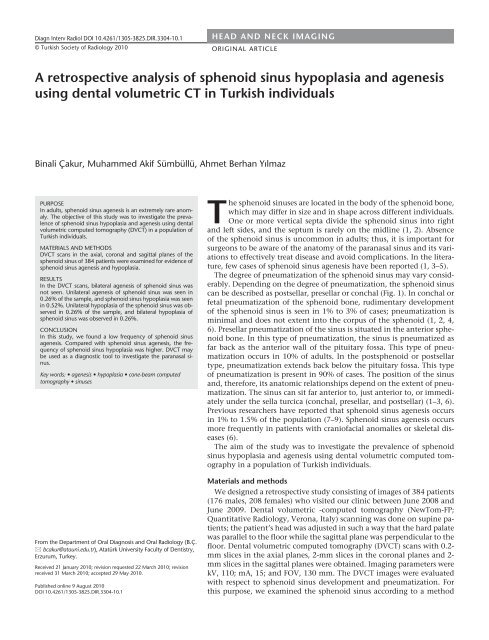
A retrospective analysis of sphenoid sinus hypoplasia and agenesis ...
A retrospective analysis of sphenoid sinus hypoplasia and agenesis ...
A retrospective analysis of sphenoid sinus hypoplasia and agenesis ...

You also want an ePaper? Increase the reach of your titles
YUMPU automatically turns print PDFs into web optimized ePapers that Google loves.
Diagn Interv Radiol DOI 10.4261/1305-3825.DIR.3304-10.1<br />
© Turkish Society <strong>of</strong> Radiology 2010<br />
HEAD AND NECK IMAGING<br />
ORIGINAL ARTICLE<br />
A <strong>retrospective</strong> <strong>analysis</strong> <strong>of</strong> <strong>sphenoid</strong> <strong>sinus</strong> <strong>hypoplasia</strong> <strong>and</strong> <strong>agenesis</strong><br />
using dental volumetric CT in Turkish individuals<br />
Binali Çakur, Muhammed Akif Sümbüllü, Ahmet Berhan Yılmaz<br />
PURPOSE<br />
In adults, <strong>sphenoid</strong> <strong>sinus</strong> <strong>agenesis</strong> is an extremely rare anomaly.<br />
The objective <strong>of</strong> this study was to investigate the prevalence<br />
<strong>of</strong> <strong>sphenoid</strong> <strong>sinus</strong> <strong>hypoplasia</strong> <strong>and</strong> <strong>agenesis</strong> using dental<br />
volumetric computed tomography (DVCT) in a population <strong>of</strong><br />
Turkish individuals.<br />
MATERIALS AND METHODS<br />
DVCT scans in the axial, coronal <strong>and</strong> sagittal planes <strong>of</strong> the<br />
<strong>sphenoid</strong> <strong>sinus</strong> <strong>of</strong> 384 patients were examined for evidence <strong>of</strong><br />
<strong>sphenoid</strong> <strong>sinus</strong> <strong>agenesis</strong> <strong>and</strong> <strong>hypoplasia</strong>.<br />
RESULTS<br />
In the DVCT scans, bilateral <strong>agenesis</strong> <strong>of</strong> <strong>sphenoid</strong> <strong>sinus</strong> was<br />
not seen. Unilateral <strong>agenesis</strong> <strong>of</strong> <strong>sphenoid</strong> <strong>sinus</strong> was seen in<br />
0.26% <strong>of</strong> the sample, <strong>and</strong> <strong>sphenoid</strong> <strong>sinus</strong> <strong>hypoplasia</strong> was seen<br />
in 0.52%. Unilateral <strong>hypoplasia</strong> <strong>of</strong> the <strong>sphenoid</strong> <strong>sinus</strong> was observed<br />
in 0.26% <strong>of</strong> the sample, <strong>and</strong> bilateral <strong>hypoplasia</strong> <strong>of</strong><br />
<strong>sphenoid</strong> <strong>sinus</strong> was observed in 0.26%.<br />
CONCLUSION<br />
In this study, we found a low frequency <strong>of</strong> <strong>sphenoid</strong> <strong>sinus</strong><br />
<strong>agenesis</strong>. Compared with <strong>sphenoid</strong> <strong>sinus</strong> <strong>agenesis</strong>, the frequency<br />
<strong>of</strong> <strong>sphenoid</strong> <strong>sinus</strong> <strong>hypoplasia</strong> was higher. DVCT may<br />
be used as a diagnostic tool to investigate the paranasal <strong>sinus</strong>.<br />
Key words: • <strong>agenesis</strong> • <strong>hypoplasia</strong> • cone-beam computed<br />
tomography • <strong>sinus</strong>es<br />
From the Department <strong>of</strong> Oral Diagnosis <strong>and</strong> Oral Radiology (B.Ç.<br />
bcakur@atauni.edu.tr), Atatürk University Faculty <strong>of</strong> Dentistry,<br />
Erzurum, Turkey.<br />
Received 21 January 2010; revision requested 22 March 2010; revision<br />
received 31 March 2010; accepted 29 May 2010.<br />
Published online 9 August 2010<br />
DOI 10.4261/1305-3825.DIR.3304-10.1<br />
The <strong>sphenoid</strong> <strong>sinus</strong>es are located in the body <strong>of</strong> the <strong>sphenoid</strong> bone,<br />
which may differ in size <strong>and</strong> in shape across different individuals.<br />
One or more vertical septa divide the <strong>sphenoid</strong> <strong>sinus</strong> into right<br />
<strong>and</strong> left sides, <strong>and</strong> the septum is rarely on the midline (1, 2). Absence<br />
<strong>of</strong> the <strong>sphenoid</strong> <strong>sinus</strong> is uncommon in adults; thus, it is important for<br />
surgeons to be aware <strong>of</strong> the anatomy <strong>of</strong> the paranasal <strong>sinus</strong> <strong>and</strong> its variations<br />
to effectively treat disease <strong>and</strong> avoid complications. In the literature,<br />
few cases <strong>of</strong> <strong>sphenoid</strong> <strong>sinus</strong> <strong>agenesis</strong> have been reported (1, 3–5).<br />
The degree <strong>of</strong> pneumatization <strong>of</strong> the <strong>sphenoid</strong> <strong>sinus</strong> may vary considerably.<br />
Depending on the degree <strong>of</strong> pneumatization, the <strong>sphenoid</strong> <strong>sinus</strong><br />
can be described as postsellar, presellar or conchal (Fig. 1). In conchal or<br />
fetal pneumatization <strong>of</strong> the <strong>sphenoid</strong> bone, rudimentary development<br />
<strong>of</strong> the <strong>sphenoid</strong> <strong>sinus</strong> is seen in 1% to 3% <strong>of</strong> cases; pneumatization is<br />
minimal <strong>and</strong> does not extent into the corpus <strong>of</strong> the <strong>sphenoid</strong> (1, 2, 4,<br />
6). Presellar pneumatization <strong>of</strong> the <strong>sinus</strong> is situated in the anterior <strong>sphenoid</strong><br />
bone. In this type <strong>of</strong> pneumatization, the <strong>sinus</strong> is pneumatized as<br />
far back as the anterior wall <strong>of</strong> the pituitary fossa. This type <strong>of</strong> pneumatization<br />
occurs in 10% <strong>of</strong> adults. In the post<strong>sphenoid</strong> or postsellar<br />
type, pneumatization extends back below the pituitary fossa. This type<br />
<strong>of</strong> pneumatization is present in 90% <strong>of</strong> cases. The position <strong>of</strong> the <strong>sinus</strong><br />
<strong>and</strong>, therefore, its anatomic relationships depend on the extent <strong>of</strong> pneumatization.<br />
The <strong>sinus</strong> can sit far anterior to, just anterior to, or immediately<br />
under the sella turcica (conchal, presellar, <strong>and</strong> postsellar) (1–3, 6).<br />
Previous researchers have reported that <strong>sphenoid</strong> <strong>sinus</strong> <strong>agenesis</strong> occurs<br />
in 1% to 1.5% <strong>of</strong> the population (7–9). Sphenoid <strong>sinus</strong> <strong>agenesis</strong> occurs<br />
more frequently in patients with crani<strong>of</strong>acial anomalies or skeletal diseases<br />
(6).<br />
The aim <strong>of</strong> the study was to investigate the prevalence <strong>of</strong> <strong>sphenoid</strong><br />
<strong>sinus</strong> <strong>hypoplasia</strong> <strong>and</strong> <strong>agenesis</strong> using dental volumetric computed tomography<br />
in a population <strong>of</strong> Turkish individuals.<br />
Materials <strong>and</strong> methods<br />
We designed a <strong>retrospective</strong> study consisting <strong>of</strong> images <strong>of</strong> 384 patients<br />
(176 males, 208 females) who visited our clinic between June 2008 <strong>and</strong><br />
June 2009. Dental volumetric -computed tomography (NewTom-FP;<br />
Quantitative Radiology, Verona, Italy) scanning was done on supine patients;<br />
the patient’s head was adjusted in such a way that the hard palate<br />
was parallel to the floor while the sagittal plane was perpendicular to the<br />
floor. Dental volumetric computed tomography (DVCT) scans with 0.2-<br />
mm slices in the axial planes, 2-mm slices in the coronal planes <strong>and</strong> 2-<br />
mm slices in the sagittal planes were obtained. Imaging parameters were<br />
kV, 110; mA, 15; <strong>and</strong> FOV, 130 mm. The DVCT images were evaluated<br />
with respect to <strong>sphenoid</strong> <strong>sinus</strong> development <strong>and</strong> pneumatization. For<br />
this purpose, we examined the <strong>sphenoid</strong> <strong>sinus</strong> according to a method
a<br />
b<br />
c<br />
Figure 1. a–c. Conchal or fetal pneumatization (a), presellar or juvenile pneumatization (b), postsellar or post<strong>sphenoid</strong> pneumatization (c).<br />
Figure 2. Sphenoid <strong>sinus</strong> <strong>hypoplasia</strong> was<br />
defined as an oval-shaped <strong>sinus</strong> with<br />
pneumatization limited to the pre<strong>sphenoid</strong>,<br />
i.e., anterior to the vertical plane <strong>of</strong> the<br />
tuberculum sella (vertical line) on the<br />
scanograms.<br />
the third <strong>and</strong> fourth fetal months. The<br />
<strong>sphenoid</strong> <strong>sinus</strong> appears in the fourth<br />
month <strong>of</strong> gestation (3). The <strong>sphenoid</strong><br />
<strong>sinus</strong>es arise from within the nasal capsule<br />
<strong>of</strong> the embryonic nose (4). At birth,<br />
the <strong>sphenoid</strong> <strong>sinus</strong>es are recognized as<br />
a cavity with a maximum diameter <strong>of</strong><br />
2 mm <strong>and</strong> remains so until age 3. At<br />
approximately 3 years <strong>of</strong> age, aeration<br />
begins in the <strong>sphenoid</strong> bone. By age 7,<br />
pneumatization reaches the sella turcica.<br />
By age 12, <strong>sphenoid</strong> pneumatization<br />
reaches complete development, <strong>and</strong> the<br />
<strong>sinus</strong>es attain their full size with a volume<br />
<strong>of</strong> 7.5 mL (23x20x17 mm) (3, 4).<br />
The lack <strong>of</strong> <strong>sinus</strong> pneumatization by the<br />
age <strong>of</strong> 10 years suggests the possibility <strong>of</strong><br />
<strong>sphenoid</strong> pathology (5). Sphenoid <strong>sinus</strong><br />
<strong>agenesis</strong> is an extremely rare phenomenon.<br />
Previous researchers have reported<br />
that <strong>sphenoid</strong> <strong>sinus</strong> <strong>agenesis</strong> occurs<br />
in 1% to 1.5% <strong>of</strong> cases (7–9). However,<br />
most <strong>of</strong> these studies date back to the<br />
first half <strong>of</strong> the 20th century <strong>and</strong> lack<br />
the support <strong>of</strong> CT. In these anatomical<br />
studies, conchal pneumatization was<br />
interpreted as <strong>agenesis</strong>; therefore, the<br />
estimated frequency would be different<br />
than that obtained in studies using<br />
CT (4, 6). Because <strong>of</strong> its deep location<br />
<strong>and</strong> intimate structural relationship<br />
with intracranial structures, pneumaproposed<br />
by Eggesbø et al. (10). In this<br />
method, <strong>sphenoid</strong> <strong>sinus</strong> <strong>hypoplasia</strong><br />
was defined as an oval-shaped <strong>sinus</strong><br />
with pneumatization limited to the<br />
pre-<strong>sphenoid</strong>, i.e., anterior to the vertical<br />
plane <strong>of</strong> the tuberculum sellae (vertical<br />
line) on the images, <strong>and</strong> the absence<br />
<strong>of</strong> septa (Fig. 2). Sphenoid <strong>sinus</strong><br />
<strong>agenesis</strong> was defined as a complete lack<br />
<strong>of</strong> pneumatization in the body <strong>of</strong> the<br />
<strong>sphenoid</strong> bone (Fig. 3). We designated<br />
the age <strong>of</strong> 15 years old as the minimum<br />
age to enroll in the study because <strong>sphenoid</strong><br />
<strong>sinus</strong>es do not complete development<br />
until puberty.<br />
Results<br />
In the axial, coronal <strong>and</strong> sagittal<br />
crani<strong>of</strong>acial DVCT scans, we found bilateral<br />
<strong>sphenoid</strong> <strong>sinus</strong> <strong>hypoplasia</strong> in one<br />
case (a 27-year-old male, Fig. 4). In one<br />
patient, both unilateral <strong>sphenoid</strong> <strong>sinus</strong><br />
<strong>hypoplasia</strong> <strong>and</strong> unilateral <strong>sphenoid</strong> <strong>sinus</strong><br />
<strong>agenesis</strong> were found (a 28-year-old<br />
ii • Diagnostic <strong>and</strong> Interventional Radiology<br />
Figure 3. Agenesis <strong>of</strong> the <strong>sphenoid</strong> <strong>sinus</strong>.<br />
female, Fig. 5). In these two cases <strong>of</strong><br />
<strong>sphenoid</strong> <strong>sinus</strong> <strong>hypoplasia</strong>, there were<br />
no crani<strong>of</strong>acial abnormalities. In our<br />
study, bilateral <strong>sphenoid</strong> <strong>sinus</strong> <strong>agenesis</strong><br />
was not seen. The results from the<br />
present study are summarized in Table.<br />
Discussion<br />
The development <strong>of</strong> paranasal <strong>sinus</strong>es<br />
begins as an evagination <strong>of</strong> the<br />
mucosa from the nasal cavities during<br />
Table. Frequency <strong>of</strong> <strong>sphenoid</strong> <strong>sinus</strong> aplasia/<strong>agenesis</strong><br />
Sphenoid <strong>sinus</strong> aplasia<br />
Unilateral<br />
Sphenoid <strong>sinus</strong> <strong>agenesis</strong><br />
Unilateral<br />
Sex n Bilateral Right Left Bilateral Right Left<br />
Male 176 1 (0.57%) - - - - -<br />
Female 208 - 1 (0.48%) - - - 1(0.48%)<br />
Total<br />
384 1 (0.26%) 1 (0.26%) - 1 (0.26%)<br />
384 2 (0.52%) 1 (0.26%)<br />
Çakur et al.
a<br />
b<br />
c<br />
Figure 4. a–c. Bilateral <strong>hypoplasia</strong> <strong>of</strong> the <strong>sphenoid</strong><br />
<strong>sinus</strong> in a 27-year-old male; limited bilateral<br />
aeration (a <strong>and</strong> c, white arrows), with axial (a),<br />
sagittal (b), <strong>and</strong> coronal (c) views.<br />
tization <strong>of</strong> the <strong>sphenoid</strong> <strong>sinus</strong>es can<br />
make radiographic diagnosis difficult,<br />
particularly with conventional radiographic<br />
techniques or panoramic radiography.<br />
In recent anatomical studies<br />
<strong>of</strong> the paranasal <strong>sinus</strong>es based on CT<br />
findings, <strong>sphenoid</strong> <strong>sinus</strong> <strong>agenesis</strong> was<br />
not reported (11–15). Terra et al. (16)<br />
performed computerized tomography<br />
with axial scans obtained at 5.0-mm<br />
thickness <strong>and</strong> coronal scans at 3.0-mm<br />
thickness to derive a better diagnosis.<br />
In DVCT scans, the thicknesses <strong>of</strong> the<br />
obtained axial <strong>and</strong> coronal scans are<br />
thinner than that <strong>of</strong> conventional CT.<br />
In this study, we obtained DVCT scans<br />
with 0.2-mm slices in the axial scans<br />
<strong>and</strong> 2-mm slices in the coronal scans.<br />
Therefore, the incidence <strong>of</strong> <strong>sphenoid</strong><br />
<strong>sinus</strong> <strong>agenesis</strong> in a population is more<br />
accurate in studies using CT <strong>and</strong> DVCT<br />
(3). In the literature, there are few cases<br />
<strong>of</strong> <strong>sphenoid</strong> <strong>sinus</strong> <strong>agenesis</strong> identified by<br />
CT (4–6, 17). Antoniades et al. (17) first<br />
described <strong>sphenoid</strong> <strong>sinus</strong> <strong>agenesis</strong> in a<br />
patient with H<strong>and</strong>-Schuller-Christian<br />
disease. Keskin et al. (6) also presented<br />
a case in which <strong>sphenoid</strong> <strong>sinus</strong> <strong>agenesis</strong><br />
was diagnosed during the evaluation <strong>of</strong><br />
endoscopic trans<strong>sphenoid</strong>al hypophysectomy.<br />
In another report presented<br />
by Degirmenci et al. (4), the patients<br />
diagnosed with <strong>sphenoid</strong> <strong>sinus</strong> <strong>agenesis</strong><br />
had no crani<strong>of</strong>acial anomalies or<br />
systemic skeletal diseases. No <strong>sphenoid</strong><br />
<strong>sinus</strong> <strong>agenesis</strong> was reported in patients<br />
with cystic fibrosis or controls from all<br />
age groups in a study by Eggesbø et al.<br />
(10). Anık et al. (5) reported the MRI<br />
<strong>and</strong> CT findings <strong>of</strong> a case <strong>of</strong> <strong>sphenoid</strong><br />
<strong>sinus</strong> <strong>agenesis</strong> without any anomaly or<br />
syndrome. S<strong>of</strong>t-tissue structures <strong>of</strong> the<br />
paranasal <strong>sinus</strong>es <strong>and</strong> their adjacent<br />
structures are more visible on MR imaging<br />
as compared to CT (5). Aydinlioğlu<br />
et al. (3) reported that the bilateral absence<br />
<strong>of</strong> the <strong>sphenoid</strong> <strong>sinus</strong> was not observed<br />
in their study, but they found two<br />
cases in which there was a unilateral absence<br />
<strong>of</strong> a <strong>sphenoid</strong> <strong>sinus</strong> on the right.<br />
Recently, Orhan et al. (18) investigated<br />
<strong>agenesis</strong> <strong>of</strong> the paranasal <strong>sinus</strong>es in 20<br />
male adult cadavers. They reported that<br />
in a 50-year-old man, bilateral absence<br />
<strong>of</strong> the <strong>sphenoid</strong> <strong>sinus</strong>es was observed<br />
on the macroscopic examination.<br />
Sphenoid <strong>sinus</strong> <strong>agenesis</strong> might be<br />
more common in patients with crani<strong>of</strong>acial<br />
anomalies due to less well-developed<br />
paranasal <strong>sinus</strong>es (19, 20). Furthermore,<br />
deficient resorption <strong>of</strong> the corpus<br />
<strong>sphenoid</strong>ale is postulated as the pathogenesis<br />
<strong>of</strong> the <strong>sphenoid</strong> <strong>sinus</strong>es without<br />
any crani<strong>of</strong>acial anomalies (4, 17).<br />
In conclusion, this study showed a<br />
low frequency <strong>of</strong> <strong>sphenoid</strong> <strong>sinus</strong> <strong>hypoplasia</strong>,<br />
especially <strong>agenesis</strong>. Thus,<br />
surgeons should consider the possibility<br />
<strong>of</strong> <strong>agenesis</strong> or <strong>hypoplasia</strong> <strong>of</strong><br />
the <strong>sphenoid</strong> <strong>sinus</strong> before planning<br />
surgical procedures to avoid serious<br />
complications. DVCT may be used<br />
as a diagnostic tool to investigate the<br />
<strong>sphenoid</strong> <strong>sinus</strong>.<br />
Sphenoid <strong>sinus</strong> <strong>hypoplasia</strong> <strong>and</strong> <strong>agenesis</strong> • iii
a<br />
b<br />
c<br />
Figure 5. a–c. On the right, unilateral <strong>hypoplasia</strong> <strong>of</strong><br />
the <strong>sphenoid</strong> <strong>sinus</strong> (white arrows) in a 28-year-old<br />
female, with axial (a), sagittal (b) <strong>and</strong> coronal (c)<br />
views; on the left, <strong>agenesis</strong> <strong>of</strong> the <strong>sphenoid</strong> <strong>sinus</strong> in<br />
a 28-year-old female (thick white arrow).<br />
References<br />
1. Yune HY, Holden RW, Smith JA. Normal<br />
variations <strong>and</strong> lesions <strong>of</strong> the <strong>sphenoid</strong> <strong>sinus</strong>.<br />
Am J Roentgenol Radium Ther Nucl<br />
Med 1975; 124:129–138.<br />
2. Yonetsu K, Watanabe M, Nakamura T.<br />
Age-related expansion <strong>and</strong> reduction in<br />
aeration <strong>of</strong> the <strong>sphenoid</strong> <strong>sinus</strong>: volume<br />
assessment by helical CT scanning. Am J<br />
Neuroradiol 2000; 21:179–182.<br />
3. Aydinlioğlu A, Erdem S. Maxillary <strong>and</strong><br />
<strong>sphenoid</strong> <strong>sinus</strong> aplasia in Turkish individuals:<br />
A <strong>retrospective</strong> review using computed<br />
tomography. Clin Anat 2004; 17:618–622.<br />
4. Degirmenci B, Haktanır A, Acar M,<br />
Albayrak R, Yucel A. Agenesis <strong>of</strong> <strong>sphenoid</strong><br />
<strong>sinus</strong>: three cases. Surg Radiol Anat 2005;<br />
27:351–353.<br />
5. Anik I, Anik Y, Koc K, Ceylan S. Agenesis<br />
<strong>of</strong> <strong>sphenoid</strong> <strong>sinus</strong>es. Clin Anat 2005;<br />
18:217–219.<br />
iv • Diagnostic <strong>and</strong> Interventional Radiology<br />
6. Keskin G, Ustundag E, Ciftci E. Agenesis<br />
<strong>of</strong> <strong>sphenoid</strong> <strong>sinus</strong>es. Surg Radiol Anat<br />
2002; 24:324–326.<br />
7. Grünwald L. Deskriptive und topographische<br />
Anatomie der Nase und ihrer<br />
Nebenhöhlen. In: Denker A, Kahler<br />
O, eds. H<strong>and</strong>buch der Hals-Nasen-<br />
Ohrenheilkunde, vol 1. Berlin: Springer/<br />
Bergmann, 1925; 74–75.<br />
8. Peele JC. Unusual anatomical variations <strong>of</strong><br />
the <strong>sphenoid</strong> <strong>sinus</strong>es. Laryngoscope 1957;<br />
67:208–237.<br />
9. Lang J. Klinische Anatomie der Nase,<br />
Nasenhöhle und Nebenhöhlen. Stuttgart:<br />
Thieme, 1988; 87–88.<br />
10. Eggesbø HB, Søvik S, Dølvik S, Eiklid K.<br />
Kolmannskog F. CT Characterization <strong>of</strong><br />
developmental variations <strong>of</strong> the paranasal<br />
<strong>sinus</strong>es in cystic fibrosis. Acta Radiol 2001;<br />
42:482–493.<br />
11. Pérez-Piñas I, Sabate J, Carmona A,<br />
Catalina-Herrera CJ, Jiménez-Castellanos J.<br />
Anatomical variations in the human paranasal<br />
<strong>sinus</strong> region studied by CT. J Anat<br />
2000; 197:221–227.<br />
12. Arslan H, Aydinlioğlu A, Bozkurt M,<br />
Egeli E. Anatomic variations <strong>of</strong> the paranasal<br />
<strong>sinus</strong>es: CT examination for endoscopic<br />
<strong>sinus</strong> surgery. Auris Nasus Larynx<br />
1999; 26:39–38.<br />
13. Sivasli E, Sirikci A, Bayazit YA,<br />
Gümüsburun E, Erbagci H, Bayram M,<br />
Kanlikama M. Anatomic variations <strong>of</strong> the<br />
paranasal <strong>sinus</strong> area in pediatric patients<br />
with chronic <strong>sinus</strong>itis. Surg Radiol Anat<br />
2003; 24:400–405.<br />
14. Sirikci A, Bayazit YA, Bayram M, Mumbuç<br />
S, Güngör K, Kanlikama M. Variations <strong>of</strong><br />
<strong>sphenoid</strong> <strong>and</strong> related structures. Eur Radiol<br />
2000; 10:844–848.<br />
15. Kantarcı M, Karasén RM, Alper F, Onbas<br />
O, Okur A, Karaman A. Remarkable anatomic<br />
variations in paranasal <strong>sinus</strong> region<br />
<strong>and</strong> their clinical importance. Eur J Radiol<br />
2004; 50:296–302.<br />
16. Terra ER, Guedes FR, Manzi FR, Boscolo<br />
FN. Pneumatization <strong>of</strong> the <strong>sphenoid</strong> <strong>sinus</strong>;<br />
case report. Dentomaxill<strong>of</strong>ac Radiol 2006;<br />
35:47–49.<br />
17. Antoniades K, Vahtsevanos K, Psimopoulou<br />
M, Karakasis D. Agenesis <strong>of</strong> <strong>sphenoid</strong> <strong>sinus</strong>.<br />
Case report. J Otorhinolaryngol Relat Spec<br />
1996; 58:347–349.<br />
18. Orhan M, Govsa F, Saylam C. A quite rare<br />
condition: absence <strong>of</strong> <strong>sphenoid</strong>al <strong>sinus</strong>es.<br />
Surg Radiol Anat 2010; 32:551–553.<br />
19. Rivero PVP, Yánez TK, García MM, Ruíz<br />
GT, Romero GP, Huelva AB. Melanoma <strong>of</strong><br />
the <strong>sphenoid</strong> <strong>sinus</strong>. Report <strong>of</strong> a case <strong>and</strong><br />
literature review. Acta Otorrinolaringol<br />
Esp 2004; 55:45–48.<br />
20. Tan HKK, Ong Y, Teo MSK, Fook-Chong<br />
SMC. The development <strong>of</strong> <strong>sphenoid</strong><br />
<strong>sinus</strong> in Asian children. Int J Pediatr<br />
Otorhinolaryngol 2003; 67:1295–1302.<br />
Çakur et al.