Whole issue in PDF - Europhysics News
Whole issue in PDF - Europhysics News
Whole issue in PDF - Europhysics News

You also want an ePaper? Increase the reach of your titles
YUMPU automatically turns print PDFs into web optimized ePapers that Google loves.
FEATURES<br />
Absorption and phase imag<strong>in</strong>g with<br />
synchrotron radiation<br />
Peter Cloetens 1 , Elodie Boller 1 , Woifgang Ludwig 1 , Jose BarucheZJ and Michel Schlenker<br />
1 European Synchrotron Radiation Facility, RP. 220, F-38043 Grenoble, France<br />
2 Laboratoire Louis Neel du CNRS, RP. 166, F-38042 Grenoble, France<br />
All users agree on two essential virtues of synchrotron radiation:<br />
its high <strong>in</strong>tensity <strong>in</strong> the X-ray range, and its cont<strong>in</strong>uous<br />
spectrum. The new, third generation, sources have another, at first<br />
sight less spectacular feature: the small divergence ofthe beam as<br />
seen from the sample. This characteristic is due to the very small<br />
cross-sectional area ofthe electron beam that acts as the source of<br />
radiation, and to the large source - sample distance. All three of<br />
these qualities lead to novel possibilities, among others <strong>in</strong> X-ray<br />
imag<strong>in</strong>g. We will discuss some of the approaches developed <strong>in</strong><br />
hard X-ray synchrotron radiation imag<strong>in</strong>g, and some ofits applications<br />
<strong>in</strong> quite diverse fields.<br />
Synchrotron radiation refers to the electromagnetic radiation<br />
emitted by ultrarelativistic electrons (energies of several GeV),<br />
circulat<strong>in</strong>g <strong>in</strong> storage r<strong>in</strong>gs, at those parts ofthe r<strong>in</strong>gs where they<br />
are accelerated by a magnetic field. This can be uniform over a<br />
part ofthe trajectory<strong>in</strong> the bend<strong>in</strong>g magnets, or spatially oscillat<strong>in</strong>g<br />
<strong>in</strong> the "<strong>in</strong>sertion devices". The spectrum of the light thus<br />
produced extends from the <strong>in</strong>fra red <strong>in</strong>to the X-ray range, the latter<br />
part be<strong>in</strong>g to most users the more valuable one. Emission is<br />
strongly concentrated <strong>in</strong> the forward direction with respect to the<br />
velocity ofthe emitt<strong>in</strong>g electrons, the characteristic angular open<strong>in</strong>g<br />
be<strong>in</strong>g mC-lE, with E the energy of the electrons, m their rest<br />
mass. Three mach<strong>in</strong>es <strong>in</strong> the world belong to the category ofthird<br />
generation, high energy sources : the European Synchrotron<br />
Radiation Facility (ESRF) <strong>in</strong> Grenoble, France, at 6 GeV; the<br />
Advanced Photon Source <strong>in</strong> Argonne, IL, USA at 7 Gev; and<br />
SPRING-8 <strong>in</strong> Japan, at 8 GeY. They are characterised bythe th<strong>in</strong>ness<br />
of the electron beam that produces the radiation (source<br />
dimensions < 0.1 mm), and by the provision, <strong>in</strong> between the<br />
bend<strong>in</strong>g magnets, ofmanystraight sections. These allow the position<strong>in</strong>g<br />
of<strong>in</strong>sertion devices, viz. wigglers orundulators,which can<br />
provide each experiment with the best suited beam.<br />
Imag<strong>in</strong>g is normally associated, <strong>in</strong> our m<strong>in</strong>ds, with lenses.<br />
Unlike visible light or electrons, efficient lenses are not (yet?) available<br />
for hard X-rays, essentially because they <strong>in</strong>teract weakly with<br />
matter, result<strong>in</strong>g <strong>in</strong> a refractive <strong>in</strong>dexvery close (to with<strong>in</strong> 10. 5 or<br />
10- 6 ) to unity. Nevertheless X-ray imag<strong>in</strong>g plays an immense role.<br />
Radiographs of hands of attendants made at the lectures on X<br />
rays <strong>in</strong> 1896 are the historical banner ofX-rays, while each of us<br />
benefited from medical radiography and enjoys the leak-tightness<br />
which <strong>in</strong>dustrial radiography controls <strong>in</strong> pipel<strong>in</strong>es. X-ray tomography<br />
is used on a rout<strong>in</strong>e basis, under the name of computed<br />
axial tomography (CAT) or medical scanner, to visualise virtual<br />
cuts through human anatomy; obta<strong>in</strong>ed from attenuation measurements<br />
performed under different view<strong>in</strong>g angles. In<br />
radiography (two-dimensional images) as well as <strong>in</strong> tomography<br />
(three-dimensional exploration), only contrast associated with<br />
local variations <strong>in</strong> X-ray absorption was, until recently; considered.<br />
On the other hand, a different approach, based on Bragg<br />
diffraction ("X-ray topography"), reveals isolated defects, such as<br />
dislocations, <strong>in</strong>clusions, stack<strong>in</strong>g faults, and sometimes doma<strong>in</strong><br />
walls, <strong>in</strong> s<strong>in</strong>gle crystals. It largely contributed to the development<br />
ofthe processes that now provide the huge perfect crystals ofsilicon<br />
which make microelectronics so efficient.<br />
The advent ofsynchrotron radiationwas a boonto X-rayimag<strong>in</strong>g<br />
through the possibilities for real-time observation or for<br />
ref<strong>in</strong>ed beam preparation offered by the <strong>in</strong>crease <strong>in</strong> <strong>in</strong>tensity.<br />
However, the place imag<strong>in</strong>g techniques have taken (15% of the<br />
activity ofESRF, i.e. much more than planned) on third generation<br />
sources also results from the entirely new avenues that have<br />
been opened through the high geometrical quality ofthe beams.<br />
In the follow<strong>in</strong>g, we separate the developments we consider as<br />
more closely associated to the ga<strong>in</strong> <strong>in</strong> <strong>in</strong>tensity (the quantitative<br />
aspect) from those more directly connected to the new geometric<br />
features (qualitative aspects). Theresults we describewere obta<strong>in</strong>ed<br />
on several <strong>in</strong>struments, mostlybutnotexclusively at ESRF.<br />
Quantitative progress: absorption imag<strong>in</strong>g ever more ref<strong>in</strong>ed<br />
Angiography<br />
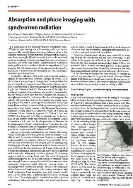
X-ray absorption changes abruptly with the energyor wavelength<br />
ofthe photons atthe absorption edges, correspond<strong>in</strong>gto the energy<br />
required to eject an electron from a deep atomic level. This<br />
variation has beenused for the selective observation ofblood vessels,<br />
<strong>in</strong> particular for the diagnosis ofheartproblems associated to<br />
the coronary arteries, via angiography. In the hospital version of<br />
this technique, a catheter is <strong>in</strong>troduced along an artery to br<strong>in</strong>g a<br />
contrast agent (an iod<strong>in</strong>e-conta<strong>in</strong><strong>in</strong>g solution) near the coronary<br />
arteryto be <strong>in</strong>spected. This approach <strong>in</strong>volves a fatal riskofabout<br />
0.1 %. SR makes it possible<br />
to obta<strong>in</strong> excellent images<br />
with reduced danger,<br />
RCA ___<br />
because an <strong>in</strong>jection of a<br />
contrast agent through a<br />
ve<strong>in</strong> is enough. The higher<br />
dilution is compensated by<br />
the logarithmic subtraction<br />
of two images.<br />
Because the heart moves,<br />
the approach consists <strong>in</strong><br />
simultaneously us<strong>in</strong>g two<br />
flat beams with slightly different<br />
energies, one above,<br />
the other below the K<br />
absorption edge of iod<strong>in</strong>e<br />
(33.17 keY). The beams<br />
cross at the location of the<br />
<strong>in</strong>vestigated organ, and<br />
they are measured by two<br />
detector l<strong>in</strong>es, while the<br />
patient is translated vertically.<br />
Fig. 1 shows the<br />
&"nl_<br />
Fig. 1: l<strong>in</strong>age of the heart and arteries 0<br />
the first human patient at ESRF's<br />
Medical Beaml<strong>in</strong>e: this differential<br />
angiogram results from logarithmic<br />
subtraction oftwo images obta<strong>in</strong>ed<br />
simultaneously on either side of the<br />
iod<strong>in</strong>e absorption edge. The right<br />
coronary artery (ReA) shows a stent anI<br />
the crux (courtesy H. Elleaume, ESRF [H.<br />
Elleaume et al., Phys. Med. BioI. 45, L39<br />
L43 (2000)]).<br />
46 europhysics news MARCHIAPRIL 2001