Whole issue in PDF - Europhysics News
Whole issue in PDF - Europhysics News
Whole issue in PDF - Europhysics News

You also want an ePaper? Increase the reach of your titles
YUMPU automatically turns print PDFs into web optimized ePapers that Google loves.
FEATURES<br />
Tomographic slices<br />
ab L4medulJa 50<br />
cortex ~<br />
&<br />
(.10')<br />
0.5 p=lglcm'<br />
O.lS<br />
Volume render<strong>in</strong>g<br />
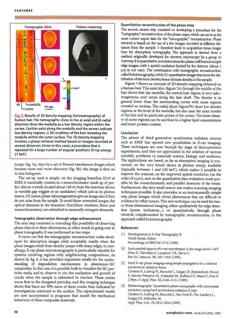
Fig. 7: Results of 3D density mapp<strong>in</strong>g (holotomography) of<br />
human hair. The tomographic slices <strong>in</strong> the a) axial and b) radial<br />
direction show the medulla as a low density region with<strong>in</strong> the<br />
cortex. Cavities exist along the medulla and the arrows <strong>in</strong>dicate<br />
low density regions. c) 3D rendition of the hair reveal<strong>in</strong>g the<br />
medulla with<strong>in</strong> the outer surface. The 3D density mapp<strong>in</strong>g<br />
<strong>in</strong>volves a phase retrieval method based on images recorded at<br />
several distances (three <strong>in</strong> this case), a procedure that is<br />
repeated for a large number of angular positions (X-ray energy<br />
21 keV).<br />
jumps (fig. Sa), then by a set ofFresnel <strong>in</strong>terference fr<strong>in</strong>ges which<br />
become more and more obtrusive (fig. 5b): the image is then an<br />
<strong>in</strong>-l<strong>in</strong>e hologram.<br />
The set-up used is simple: on the imag<strong>in</strong>g beaml<strong>in</strong>e ID19 at<br />
ESRF, it essentially consists <strong>in</strong> a monochromator made up of perfect<br />
silicon crystals, located about 140 m from the <strong>in</strong>sertion device<br />
(a variable gap wiggler or an undulator) which acts as its photon<br />
source. Ofcourse, phase <strong>in</strong>homogeneities give animage also ifthey<br />
do not arise from the sample. To avoid these unwanted images, the<br />
optical elements <strong>in</strong> the beaml<strong>in</strong>e (beryllium w<strong>in</strong>dows, filters and<br />
monochromators) are submitted to unusually str<strong>in</strong>gent demands.<br />
Tomographic observation through edge-enhancement<br />
The next step consisted <strong>in</strong> extend<strong>in</strong>g this possibility ofobserv<strong>in</strong>g<br />
phase objects to three dimensions, <strong>in</strong> other words <strong>in</strong> go<strong>in</strong>g over to<br />
phase tomography. Itwas performed <strong>in</strong> two steps.<br />
It turns out that the tomographic reconstruction codes developed<br />
for absorption images yield acceptable results when the<br />
phase images result from density jumps with sharp edges. In metallurgy,<br />
X-ray phase microtomographyis particularlyvaluable for<br />
systems <strong>in</strong>volv<strong>in</strong>g regions with neighbour<strong>in</strong>g compositions. As<br />
shown by fig. 6, it has provided important results for the understand<strong>in</strong>g<br />
of degradation mechanisms <strong>in</strong> alum<strong>in</strong>ium-SiC<br />
composites. In this case, it is possible bothto visualise the SiC particles<br />
easily, and to observe <strong>in</strong> situ the nucleation and growth of<br />
cracks when the sample is submitted to traction. These cracks<br />
occur first <strong>in</strong> the elongated particles, and this imag<strong>in</strong>g technique<br />
shows that there are 50% more of these cracks than <strong>in</strong>dicated by<br />
<strong>in</strong>vestigations restricted to the surface. The experimental results<br />
are now <strong>in</strong>corporated <strong>in</strong> programs that model the mechanical<br />
behaviour ofthese composite materials.<br />
so<br />
c<br />
Quantitative reconstruction of the phase map<br />
The second, recent step consisted <strong>in</strong> develop<strong>in</strong>g a procedure for the<br />
''holographic''reconstruction ofthe phase maps, which can serve as the<br />
more correct <strong>in</strong>put data for the ''tomographic'' reconstruction. Phase<br />
retrieval is based on the use ofa few images recorded at different distances<br />
from the sample. It therefore leads to acquisition times longer<br />
than for absorption tomography. The approach is derived from a<br />
method orig<strong>in</strong>ally developed for electron microscopy by a group <strong>in</strong><br />
Antwerp.Itis quantitative,anddeterm<strong>in</strong>es thephase,wellbeyondsimple<br />
edge images, with a spatial resolution limited by the detector (about 1<br />
J.U11 <strong>in</strong> our case). The comb<strong>in</strong>ation with tomographic reconstruction,<br />
called holotomography,yields 3D quantitativeimages that showthe distribution<br />
ofelectron density,hence ofmass density, <strong>in</strong>the sample.<br />
Figure 7 shows an example of3D density mapp<strong>in</strong>g obta<strong>in</strong>ed on<br />
a human hair. The axial slice (figure 7a) through the middle ofthe<br />
hair shows that the medulla, the central hair region, is very <strong>in</strong>homogeneous<br />
and varies along the hair shaft. The density is <strong>in</strong><br />
general lower than the surround<strong>in</strong>g cortex with some regions<br />
revealed as cavities. The radial slices (figure7b) show low density<br />
regions, at the level ofthe medulla, but also near the outer border<br />
ofthe hair and <strong>in</strong> particular po<strong>in</strong>ts ofthe cortex. The lower density<br />
ofsome regions can be ascribed to a higher lipid concentration<br />
and lower prote<strong>in</strong> content.<br />
Conclusion<br />
The advent of third generation synchrotron radiation sources<br />
such as ESRF has opened new possibilities <strong>in</strong> X-ray imag<strong>in</strong>g.<br />
These techniques are now beyond the stage of demonstration<br />
experiments, and they are appreciated <strong>in</strong> the solution of various<br />
scientific problems <strong>in</strong> materials science, biology and medic<strong>in</strong>e.<br />
The applications are based, as far as absorption imag<strong>in</strong>g is concerned,<br />
on the very broad choice <strong>in</strong> photon energy available<br />
(typically between 1 and 120 keY), which makes it possible to<br />
improve the contrast, on the improved spatial resolution (on the<br />
order ofa J.U11), andonthe quantitative data evaluation made possible<br />
by the monochromatic and parallel character of the beam.<br />
Furthermore, the very small source size makes scann<strong>in</strong>g imag<strong>in</strong>g<br />
techniques possible. It also provides, <strong>in</strong> an <strong>in</strong>strumentally simple<br />
way, phase images which reveal phenomena that are difficult to<br />
evidence byothermeans. This newtechnique canbe used for twoor<br />
three-dimensional imag<strong>in</strong>g, either qualitatively for edge detection<br />
(pores, <strong>in</strong>clusions...), or quantitatively, through phase<br />
retrieval complemented by tomographic reconstruction, <strong>in</strong> the<br />
approach called holotomography.<br />
References<br />
[I] Developments <strong>in</strong> X-Ray Tomography 11<br />
Ulrich Bonse, Editor<br />
Proceed<strong>in</strong>gs ofSPIEVoI.3772 (1999).<br />
[3] Instrumental aspects ofx-ray microbeams <strong>in</strong> the range above 1keV<br />
Dhez P., Chevallier P., Lucatorto LB., Tarrio C.<br />
Rev. Sci.Instrum. 70,1907-1920 (1999).<br />
[2] Hard X-ray phase imag<strong>in</strong>g us<strong>in</strong>g simple propagation ofa coherent<br />
synchrotron radiation beam<br />
Cloetens P., Ludwig w., Baruchel J., Guigay J.P., Rejmankova-Pernot<br />
P., Salome-Pateyron M., Schlenker M., Buffiere J.Y., Maire E., Peix G.<br />
J. Phys.D:Appl.Phys. 32,A145-A151 (1999).<br />
[4] Holotomography: Quantitative phase tomography with micrometer<br />
resolution us<strong>in</strong>g hard synchrotron radiation X-rays<br />
Cloetens P., Ludwig w., Baruchel J., Van Dyck D., Van Landuyt J. ,<br />
Guigay J.P., Schlenker M.<br />
.Appl. Phys. Lett., 75, 2912-2914 (1999).<br />
europhysics news MARCH/APRIL 2001