

The survival and rejection of epithelium in experimental corneal ...
The survival and rejection of epithelium in experimental corneal ...
The survival and rejection of epithelium in experimental corneal ...

Create successful ePaper yourself
Turn your PDF publications into a flip-book with our unique Google optimized e-Paper software.
Volume 8<br />
Number 2<br />
Epithelium <strong>survival</strong> <strong>and</strong> <strong>rejection</strong> 173<br />
lamellar autografts performed by the same technique<br />
as that employed previously for allografts.<br />
<strong>The</strong> response <strong>of</strong> the host to the auto graft was <strong>in</strong><br />
no way different from that described for the allograft,<br />
suggest<strong>in</strong>g that <strong>in</strong> the avascular rabbit cornea,<br />
the one is no more favored than the other.<br />
To establish the validity <strong>of</strong> the histologic observations<br />
described, it was necessary to ascerta<strong>in</strong><br />
the m<strong>in</strong>imum time required for an 8 mm. diameter<br />
<strong>corneal</strong> button to completely lose its <strong>epithelium</strong><br />
<strong>and</strong> rega<strong>in</strong> a normal thickness <strong>of</strong> recipient<br />
<strong>epithelium</strong>. To accomplish this, the donor <strong>epithelium</strong><br />
was <strong>in</strong>tentionally removed by scrap<strong>in</strong>g, prior<br />
to transplantation <strong>of</strong> 8 mm. lamellar buttons, <strong>and</strong><br />
the process <strong>of</strong> regeneration <strong>of</strong> the host <strong>epithelium</strong><br />
over the donor stroma was studied. Twenty-four<br />
hours after transplantation, the recipient <strong>epithelium</strong><br />
had just crossed over the graft marg<strong>in</strong> onto<br />
the donor stroma, as revealed by methylene blue<br />
sta<strong>in</strong><strong>in</strong>g. With<strong>in</strong> 2 to 3 days, the <strong>epithelium</strong> was<br />
found to cover the peripheral one third <strong>of</strong> the<br />
graft, <strong>and</strong> with<strong>in</strong> 4 to 5 days only the central<br />
area <strong>of</strong> the graft, some 2 to 3 mm. <strong>in</strong> diameter,<br />
was seen to take the sta<strong>in</strong>. Only at the sixth to<br />
seventh day after transplantation was the graft<br />
completely covered by host <strong>epithelium</strong>. Histologic<br />
sections <strong>of</strong> this process taken at regular <strong>in</strong>tervals<br />
showed a zone <strong>of</strong> flat <strong>and</strong> th<strong>in</strong> recipient epithelial<br />
cells slid<strong>in</strong>g over the graft until closure <strong>of</strong> the defect<br />
was complete. Restoration <strong>of</strong> the full thickness<br />
<strong>of</strong> the <strong>epithelium</strong> over the donor stroma required<br />
an additional 2 to 3 weeks. It would thus<br />
appear that even at its most rapid pace, complete<br />
replacement <strong>of</strong> the <strong>epithelium</strong> on an 8 mm. diameter<br />
graft would require no less than a month,<br />
<strong>and</strong> certa<strong>in</strong>ly much longer if the process were so<br />
gradual as to escape histologic detection.<br />
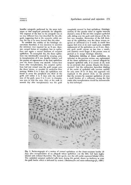
Radioactive label<strong>in</strong>g <strong>of</strong> donor <strong>epithelium</strong>. As<br />
<strong>in</strong>dicated <strong>in</strong> the previous section, any replacement<br />
<strong>of</strong> the donor <strong>epithelium</strong> on a <strong>corneal</strong> allograft by<br />
recipient epithelial cells, if it occurs at all, must<br />
be an extremely slow process. Despite the demonstration<br />
10 that the radioactive thymid<strong>in</strong>e label<strong>in</strong>g<br />
<strong>of</strong> the nuclei <strong>of</strong> epithelial cells survives usefully<br />
for only some 10 to 14 days, this approach was<br />
employed <strong>in</strong> the present study on the premise<br />
that the <strong>in</strong>vasion by recipient <strong>epithelium</strong> <strong>of</strong> even<br />
a small fraction <strong>of</strong> a millimeter dur<strong>in</strong>g the first<br />
weeks after transplantation should be demonstrable<br />
by this technique.<br />
Fig. 3. Radioautograph <strong>of</strong> a section <strong>of</strong> <strong>corneal</strong> <strong>epithelium</strong> at the donor-recipient border 10<br />
days after transplantation <strong>of</strong> an allograft whose <strong>epithelium</strong> (on the left) had been highly<br />
labeled with tritiated thymid<strong>in</strong>e. <strong>The</strong> dark specks over the cell nuclei represent labeled DNA.<br />
<strong>The</strong> unlabeled recipient epithelial cells have, dur<strong>in</strong>g this period, shown no tendency to cross<br />
the graft border which is <strong>in</strong>dicated by arrows. (Hematoxyl<strong>in</strong> <strong>and</strong> eos<strong>in</strong>-radioautograph, x500.)


