

The survival and rejection of epithelium in experimental corneal ...
The survival and rejection of epithelium in experimental corneal ...
The survival and rejection of epithelium in experimental corneal ...

Create successful ePaper yourself
Turn your PDF publications into a flip-book with our unique Google optimized e-Paper software.
Volume 8<br />
Number 2<br />
Epithelium <strong>survival</strong> <strong>and</strong> <strong>rejection</strong> 175<br />
have <strong>in</strong>vaded from the limbus to the graft border.<br />
<strong>The</strong> epithelial <strong>rejection</strong> l<strong>in</strong>e is characterized histologically<br />
by a dense <strong>in</strong>filtrate <strong>of</strong> <strong>in</strong>flammatory cells<br />
<strong>and</strong> dead <strong>and</strong> dy<strong>in</strong>g <strong>epithelium</strong>; <strong>in</strong> front <strong>of</strong> this<br />
advanc<strong>in</strong>g l<strong>in</strong>e lies a normal thickness <strong>of</strong> healthy<br />
donor <strong>epithelium</strong>, while beh<strong>in</strong>d it is found a th<strong>in</strong><br />
layer <strong>of</strong> recipient <strong>epithelium</strong> rapidly slid<strong>in</strong>g <strong>in</strong> to<br />
heal the advanc<strong>in</strong>g defect. It was felt that the<br />
demonstration <strong>of</strong> the specific nature <strong>of</strong> this <strong>rejection</strong><br />
process, <strong>and</strong> the ability to elicit it long after<br />
the graft had been put <strong>in</strong>to place, would constitute<br />
the best pro<strong>of</strong> that allogeneic donor <strong>epithelium</strong><br />
can survive <strong>in</strong>def<strong>in</strong>itely on the recipient eye.<br />
<strong>The</strong> most efficient way to achieve sensitization<br />
by <strong>and</strong> <strong>rejection</strong> <strong>of</strong> the lamellar allograft is to<br />
stimulate vascular <strong>in</strong>growth from the limbus to the<br />
graft marg<strong>in</strong>. This may readily be accomplished<br />
by plac<strong>in</strong>g the graft eccentrically <strong>in</strong> the recipient<br />
cornea, with<strong>in</strong> a millimeter or two <strong>of</strong> the limbus.<br />
In this circumstance, that quadrant <strong>of</strong> the graft<br />
nearest the limbus becomes vascularized. In a<br />
series <strong>of</strong> 50 such eccentrically placed 8 mm. lamellar<br />
allografts performed <strong>in</strong> connection with<br />
this <strong>and</strong> another study, spontaneous <strong>rejection</strong> <strong>of</strong><br />
the graft was observed to start with<strong>in</strong> 2 to 6<br />
weeks after transplantation <strong>in</strong> 50 per cent <strong>of</strong> the<br />
cases. In most <strong>in</strong>stances, a discrete epithelial <strong>rejection</strong><br />
l<strong>in</strong>e was observed. Whether early or late<br />
after transplantation, the l<strong>in</strong>e <strong>in</strong>variably was first<br />
seen precisely at the graft marg<strong>in</strong> <strong>in</strong> apposition<br />
to the zone <strong>of</strong> vascularization. As the sta<strong>in</strong>able<br />
<strong>rejection</strong> l<strong>in</strong>e proceeded across the cornea <strong>in</strong> the<br />
direction <strong>of</strong> the avascular area (Fig. 4), it always<br />
extended as an arcuate l<strong>in</strong>e from one marg<strong>in</strong> <strong>of</strong><br />
the graft to the other <strong>and</strong> never <strong>in</strong>vaded the recipient<br />
cornea.<br />
Another way to <strong>in</strong>duce <strong>rejection</strong> <strong>of</strong> the lamellar<br />
allograft is to <strong>in</strong>cite vascular <strong>in</strong>growth toward a<br />
centrally placed graft by postpon<strong>in</strong>g removal <strong>of</strong><br />
the irritat<strong>in</strong>g sutures employed to fix the graft <strong>in</strong><br />
place. Should only a s<strong>in</strong>gle suture be left <strong>in</strong><br />
place, then frequently only one or a few capillary<br />
loops will grow <strong>in</strong> toward the graft marg<strong>in</strong>. We<br />
have observed <strong>in</strong> a number <strong>of</strong> such <strong>in</strong>stances the<br />
onset <strong>of</strong> graft <strong>rejection</strong> only <strong>in</strong> the immediate<br />
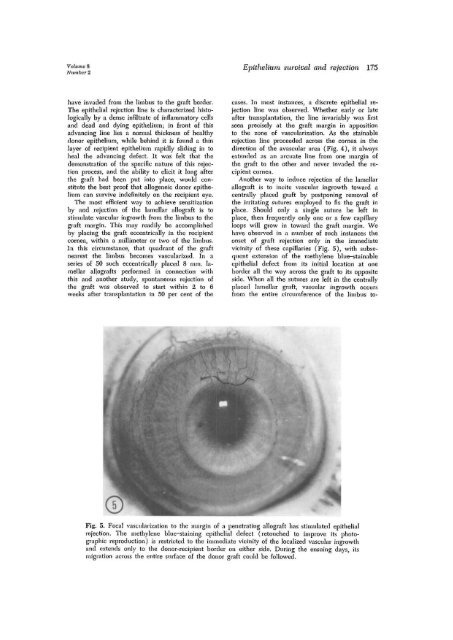
vic<strong>in</strong>ity <strong>of</strong> these capillaries (Fig. 5), with subsequent<br />
extension <strong>of</strong> the methylene blue-sta<strong>in</strong>able<br />
epithelial defect from its <strong>in</strong>itial location at one<br />
border all the way across the graft to its opposite<br />
side. When all the sutures are left <strong>in</strong> the centrally<br />
placed lamellar graft, vascular <strong>in</strong>growth occurs<br />
from the entire circumference <strong>of</strong> the limbus to-<br />
Fig. 5. Focal vascularization to the marg<strong>in</strong> <strong>of</strong> a penetrat<strong>in</strong>g allograft has stimulated epithelial<br />
<strong>rejection</strong>. <strong>The</strong> methylene blue-sta<strong>in</strong><strong>in</strong>g epithelial defect (retouched to improve its photographic<br />
reproduction) is restricted to the immediate vic<strong>in</strong>ity <strong>of</strong> the localized vascular <strong>in</strong>growth<br />
<strong>and</strong> extends only to the donor-recipient border on either side. Dur<strong>in</strong>g the ensu<strong>in</strong>g days, its<br />
migration across the entire surface <strong>of</strong> the donor graft could be followed.


