Expression of Collagen I, Smooth Muscle a-Actin, and Vimentin ...
Expression of Collagen I, Smooth Muscle a-Actin, and Vimentin ...
Expression of Collagen I, Smooth Muscle a-Actin, and Vimentin ...
You also want an ePaper? Increase the reach of your titles
YUMPU automatically turns print PDFs into web optimized ePapers that Google loves.
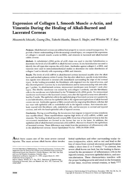
<strong>Expression</strong> <strong>of</strong> <strong>Collagen</strong> I, <strong>Smooth</strong> <strong>Muscle</strong> a-<strong>Actin</strong>, <strong>and</strong><br />
<strong>Vimentin</strong> During the Healing <strong>of</strong> Alkali-Burned <strong>and</strong><br />
Lacerated Corneas<br />
Masamichi Ishizaki, GuangZhu, Takeshi Haseba, ShaunS. Shafer, <strong>and</strong> Winston W.-Y. Kao<br />
Purposes. Alkali-burned corneas can seldom heal properly to restore corneal transparency. To<br />
provide a better underst<strong>and</strong>ing <strong>of</strong> this devastating corneal injury, we compared the expression<br />
<strong>of</strong> collagen I, smooth muscle a-actin (a-SMA), <strong>and</strong> vimentin in lacerated <strong>and</strong> alkali-burned<br />
rabbit corneas.<br />
Methods. A radiolabeled cDNA probe <strong>of</strong> a 1(1) chain was used in slot-blot hybridization to<br />
determine the levels <strong>of</strong> a 1(1) mRNA in alkali-burned corneas. In situ hybridization was used to<br />
identify the cell types that express the a 1(1) chain. Antibodies against collagen I, a-SMA, <strong>and</strong><br />
vimentin were used in immunohistochemical studies to determine the tissue distribution <strong>of</strong><br />
collagen I <strong>and</strong> to identify cells expressing a-SMA <strong>and</strong> vimentin.<br />
Results. The levels <strong>of</strong> al(I) mRNA in alkali-burned corneas increased steadily after the alkali<br />
burn <strong>and</strong> reached a plateau within 2 weeks. One day after alkali burn, specific in situ hybridization<br />
signals were detected in stromal cells immediately surrounding the edge <strong>of</strong> the corneal<br />
injury. As the healing proceeded, the fibroblastic cells migrated into the injured stroma, <strong>and</strong><br />
they showed positive reactions by in situ hybridization <strong>and</strong> by immunostaining with anti-collagen<br />
I probes. In alkali-burned corneas, retrocorneal membranes were formed 1 week after<br />
injury. This fibrillar membrane was stained by anti-collagen I antibody, <strong>and</strong> the fibroblastic<br />
cells in the membrane were hybridized by the 3 H-labeled al(I) cDNA probe. No retrocorneal<br />
membrane was formed in the lacerated corneas, even after the injured corneas were allowed to<br />
heal for 3 weeks. The epithelial cells in the epithelial plug <strong>of</strong> lacerated corneas were positive by<br />
in situ hybridization, whereas the epithelial cells in the regenerated epithelium <strong>of</strong> alkali-burned<br />
cornea was not. Antibodies against a-SMA reacted with the migrating fibroblastic cells but did<br />
not react with epithelial cells or endothelial cells in the injured corneas. Anti-vimentin antibody<br />
reacted with fibroblastic cells, endothelial cells, <strong>and</strong> keratocytes in normal <strong>and</strong> injured<br />
corneas, <strong>and</strong> with the basal epithelial cells <strong>of</strong> injured corneas.<br />
Conclusions. During wound healing, the keratocytes that migrate to injured stroma transform<br />
into my<strong>of</strong>ibroblasts. These my<strong>of</strong>ibroblasts express high levels <strong>of</strong> «1(I) mRNA, a-SMA, <strong>and</strong><br />
vimentin. The healing <strong>of</strong> alkali-burned corneas differ from that <strong>of</strong> lacerated corneas in that the<br />
retrocorneal membranes are formed in the former but not in the latter. In addition, the<br />
epithelial cells <strong>of</strong> alkali-burned corneas lack al(I) mRNA, whereas it is found in the epithelium<br />
<strong>of</strong> lacerated corneas. These differences may result from the persistence <strong>of</strong> inflammatory cells<br />
in the alkali-burned corneas. Invest Ophthalmol Vis Sci. 1993; 34:3320-3328.<br />
Alkali burn causes one <strong>of</strong> the most severe corneal<br />
injuries. The most serious alkali burns <strong>of</strong>ten involve<br />
From the Department <strong>of</strong> Ophthalmology, University <strong>of</strong> Cincinnati, Cincinnati, Ohio.<br />
Supported in part by National Eye Institute grant EY05629 <strong>and</strong> by unrestricted<br />
departmental grants from Research to Prevent Blindness, New York, New York,<br />
<strong>and</strong> Lions Eye Research Foundation, Columbus, Ohio.<br />
Submitted for publication September 21, 1992; accepted March 22, 1993.<br />
Proprietary interest category: N.<br />
Reprint requests: Winston W.-Y. Kao, Ph.D., Department <strong>of</strong> Ophthalmology,<br />
University <strong>of</strong> Cincinnati, Eden <strong>and</strong> Bethesda Ave, M.L. 527, Cincinnati, OH<br />
45267-0527.<br />
limbal epithelium <strong>and</strong> other surrounding ocular tissues<br />
(conjunctiva, sclera, <strong>and</strong> so on) <strong>and</strong> lead to the<br />
loss <strong>of</strong> vision from corneal ulceration <strong>and</strong> perforation<br />
within a short time (1 to 3 weeks). 1 " 5 In contrast, less<br />
severe alkali burns can heal, but the healing is characterized<br />
by the persistence <strong>of</strong> inflammatory cells in the<br />
injured corneas, recurrent epithelial defects, <strong>and</strong> neovascularization.<br />
These characteristics interfere with<br />
the proper healing process <strong>and</strong> result in the formation<br />
<strong>of</strong> scar tissue, recurrent corneal erosions, or both. 1 ' 2 ' 5 " 8<br />
3320<br />
Investigative Ophthalmology & Visual Science, November 1993, Vol. 34, No. 12<br />
Copyright © Association for Research in Vision <strong>and</strong> Ophthalmology
<strong>Expression</strong> <strong>of</strong> <strong>Collagen</strong> I in Injured Corneas 3321<br />
The cause or causes <strong>of</strong> this devastating process is not<br />
well understood, <strong>and</strong> the treatment <strong>of</strong> alkali-burned<br />
corneas is usually not satisfactory. Seldom do alkaliburned<br />
corneas heal properly to restore vision.<br />
<strong>Collagen</strong> constitutes about 80% <strong>of</strong> the organic<br />
constituents <strong>of</strong> the corneal stroma. 9 " 10 <strong>Collagen</strong> I is<br />
the major collagenous component found in stroma, 10<br />
with the well-organized collagen I fibrils contributing<br />
to corneal transparency. 11 However, after an alkali<br />
burn, polymorphonuclear leukocytes infiltrate the injured<br />
corneas, <strong>and</strong> the proteolytic enzymes, oxidative<br />
derivatives, or both, released by the inflammatory cells<br />
can cause severe loss <strong>of</strong> the extracellular matrix. 34<br />
Meanwhile, the stromal cells (keratocytes) that survive<br />
the alkali burn may proliferate <strong>and</strong> synthesize components<br />
<strong>of</strong> extracellular matrix for the repair <strong>of</strong> the injured<br />
corneas. Stromal ulceration takes place when<br />
the rate <strong>of</strong> degradation <strong>of</strong> extracellular matrix components<br />
(e.g., collagen, proteoglycans) exceeds the rate<br />
<strong>of</strong> synthesis. Many studies have focused on the degradation<br />
<strong>of</strong> the extracellular matrix after corneal alkali<br />
burns. 3412 " 14 However, there is little information available<br />
regarding the synthesis <strong>of</strong> the extracellular matrix<br />
during the healing <strong>of</strong> the alkali-burned corneas. In<br />
contrast, many investigators have examined the metabolism<br />
<strong>of</strong> fibrillar collagens during the healing <strong>of</strong> lacerated<br />
corneas. 15 " 17 For example, increases in the synthesis<br />
<strong>of</strong> collagen I, III, <strong>and</strong> V were reported by Cintron,<br />
et al. 1617 We previously reported that stromal cells<br />
play a major role in the healing <strong>of</strong> lacerated corneas.<br />
The stromal cells not only synthesize collagen I to repair<br />
the injured tissues but actively participate in the<br />
process <strong>of</strong> remodeling the extracellular matrix by phagocytizing<br />
collagen fibrils during'the healing <strong>of</strong> lacerated<br />
corneas. 15<br />
It has been suggested that my<strong>of</strong>ibroblasts participate<br />
in the healing <strong>of</strong> mechanical wounds. My<strong>of</strong>ibroblasts<br />
that synthesize <strong>and</strong> secrete collagen I during<br />
wound healing are characterized by the expression <strong>of</strong><br />
smooth muscle (a-SMA). 18 " 19 It has been suggested<br />
that my<strong>of</strong>ibroblasts contribute to wound contraction<br />
<strong>of</strong> mechanical injuries. 1920 Thus, it is <strong>of</strong> interest to<br />
examine whether my<strong>of</strong>ibroblasts may play a similar<br />
role in the healing <strong>of</strong> alkali burns.<br />
In the present study, we compared the expression<br />
<strong>of</strong> collagen I by alkali-burned corneas <strong>and</strong> lacerated<br />
corneas after allowing them to heal for various periods<br />
<strong>of</strong> time. Slot-blot hybridization <strong>and</strong> in situ hybridization<br />
with radiolabeled cDNA <strong>of</strong> al (I) mRNA was used<br />
to analyze the expression <strong>of</strong> collagen I. Antibodies<br />
against collagen I, smooth muscle a-actin (a-SMA),<br />
<strong>and</strong> vimentin were used in immunohistochemical studies<br />
<strong>of</strong> the injured corneas. Our results indicate that<br />
fibroblastic cells are the major cell type that synthesizes<br />
collagen I in the stroma <strong>of</strong> alkali-burned <strong>and</strong> lacerated<br />
corneas. The fibroblastic cells have the characteristics<br />
<strong>of</strong> my<strong>of</strong>ibroblasts because they express a-<br />
SMA in addition to vimentin. Alkali-burned corneas,<br />
formed a retrocorneal membrane consisting <strong>of</strong> collagen<br />
I, whereas the lacerated corneas did not form such<br />
a membrane.<br />
MATERIALS AND METHODS<br />
32 P-Labeled dNTP, 7-ATP, <strong>and</strong> [ 3 H]dCTP were purchased<br />
from DuPont-New Engl<strong>and</strong> Nuclear (Boston,<br />
MA). Restriction endonucleases were obtained from<br />
New Engl<strong>and</strong> Biolabs (Beverly, MA). BAS membranes<br />
were purchased from Schleicher <strong>and</strong> Schuell, Inc.<br />
(Keene, NH). Polyclonal anti-collagen I antibody was<br />
purchased from Southern Biotechnology Associates<br />
(Birmingham, AL). Monoclonal antibodies against a-<br />
SMA, vimentin, <strong>and</strong> desmin were obtained from Dakopatts,<br />
(Copenhagen, Denmark). Biotinylated second<br />
antibodies <strong>and</strong> ABC (avidin biotin peroxidase complex)<br />
reagents were purchased from Vector (Burlingame,<br />
CA). All other chemicals <strong>and</strong> reagents were obtained<br />
from either Sigma (St. Louis, MO) or Fisher<br />
Scientific (Pittsburgh, PA), unless otherwise specified.<br />
Animal Experiments<br />
Adult albino rabbits <strong>of</strong> either sex weighing 3 to 4 kg<br />
were purchased from Clerco Research Farm (Cincinnati,<br />
OH). Animal experiments were performed in<br />
compliance with the ARVO Resolution on the Use <strong>of</strong><br />
Animals in Research. Rabbits were anesthetized with a<br />
combined intramuscular administration <strong>of</strong> ketamine<br />
(30 mg/kg) <strong>and</strong> rompun (5 mg/kg). A drop <strong>of</strong> proparacaine-HCl<br />
(0.5%) was applied directly to the eye.<br />
Only one eye <strong>of</strong> each experimental rabbit was injured.<br />
The contralateral eyes were discarded because occasionally<br />
pathologic changes were detected in the uninjured<br />
eyes (our unpublished observation). For control<br />
experiments, corneas from untreated animals were<br />
used. To create the alkali burn, a filter paper 8 mm in<br />
diameter soaked in 1 M NaOH was applied to the<br />
center <strong>of</strong> the cornea for 1 minute <strong>and</strong> followed by a<br />
rinse with 20 ml <strong>of</strong> phosphate-buffered saline (PBS)<br />
containing 0.15 M NaCl <strong>and</strong> 0.01 M sodium phosphate,<br />
pH 7.5. 21 To create a laceration wound, an 8<br />
mm penetrating incision was made in the center <strong>of</strong> the<br />
cornea with a Micro-Sharp blade (Becton-Dickinson,<br />
Franklin Lakes, NJ). 15 Buprenorphine (0.3 mg/kg) was<br />
administered subcutaneously immediately after injury<br />
<strong>and</strong> 3 times daily for 3 days.<br />
The injured corneas were allowed to heal for<br />
various periods <strong>of</strong> time from 1 to 21 days. The rabbits<br />
were killed with an overdose <strong>of</strong> sodium pentobarbital<br />
intravenously (65 mg/kg). Corneal buttons (10 mm in<br />
diameter) were excised so that each excised cornea<br />
contained a portion <strong>of</strong> uninjured tissue. 21 The excised<br />
corneas were immediately frozen in liquid nitrogen
3322 Investigative Ophthalmology & Visual Science, November 1993, Vol. 34, No. 12<br />
<strong>and</strong> stored at — 70°C until use or were subjected to in<br />
situ hybridization as described below.<br />
Extraction <strong>of</strong> Total RNA<br />
All solutions were autoclaved in the presence <strong>of</strong> 0.1%<br />
diethylpyrocarbonate (DEPC), <strong>and</strong> all glassware <strong>and</strong><br />
appliances were either baked overnight or soaked in 1<br />
M NaOH for 2 hours. Total RNA was isolated from<br />
alkali-burned rabbit corneas <strong>and</strong> normal rabbit corneas<br />
with RNAzol (Molecular Research Center, Inc.,<br />
Cincinnati, OH), as previously described. 21 The RNAs<br />
were resuspended in a solution containing 0.5% SDS<br />
<strong>and</strong> 0.1 U/ml RNasin (Promega, Madison, WI) <strong>and</strong><br />
stored at —70°C until use.<br />
Slot-Blot Hybridization<br />
About 20 fig <strong>of</strong> total RNA from each sample were<br />
denatured in 50% formamide at 65°C for 5 minutes<br />
<strong>and</strong> were tw<strong>of</strong>old serially diluted. The RNA was then<br />
transblotted to BAS membranes with a slot-blotter<br />
(Schleicher & Schuell) 21 <strong>and</strong> hybridized with the 32 P-<br />
labeled cDNA <strong>of</strong> a 1(1) mRNA, as described previously.<br />
22<br />
In Situ Hybridization<br />
The normal, alkali-burned, <strong>and</strong> lacerated rabbit corneas<br />
were fixedin 4% methanol-free EM-grade formaldehyde<br />
(Polysciences, Inc., Warrington, PA) in 0.05 M<br />
sodium PBS, pH 7.4, at 4°C for 1 hour, <strong>and</strong> then<br />
immersed in 30% sucrose in DEPC-treated water for 2<br />
hours at 4°C. The specimens were embedded in OCT<br />
compound (Miles, Elkhart, IN), quick frozen in ethanol/dry<br />
ice, <strong>and</strong> stored at -70°C. Frozen sections <strong>of</strong><br />
5 fim thickness were cut by a cryostat <strong>and</strong> mounted on<br />
Superfrost/Plus microscope slides (Fisher Scientific,<br />
Pittsburgh, PA). The sections were then subjected to<br />
hybridization with 3 H-labeled cDNA probes <strong>of</strong> rabbit<br />
al(I) mRNA, as previously described. 21<br />
A 3 H-labeled DNA probe was prepared by [ 3 H]-<br />
dCTP incorporation using r<strong>and</strong>om primers as previously<br />
described. 21 The specificity <strong>of</strong> the probe was 2 X<br />
10 7 cpm/fxg. The hybridization reaction was carried<br />
out at 45°C overnight. The sections were washed<br />
under stringent conditions to reduce the background,<br />
as previously described. 21 The slides were then dehydrated<br />
in graded ethanol, air dried, <strong>and</strong> dipped in 50%<br />
Kodak NT-2 Nuclear emulsion (Eastman-Kodak, Manchester,<br />
NY); after exposure for 2 to 4 weeks, the<br />
slides were developed for 3 minutes in a Kodak D-19<br />
developer at 15°C, counterstained with hematoxylin<br />
<strong>and</strong> eosin, <strong>and</strong> mounted.<br />
Immunohistochemistry<br />
For detection <strong>of</strong> collagen I, frozen sections <strong>of</strong> 5 /um<br />
thickness were prepared from the OCT-embedded,<br />
unfixed rabbit corneas by a cryostat <strong>and</strong> mounted on<br />
Superfrost/Plus microscope slides (Fisher Scientific).<br />
Cryosections were subjected to immunostaining using<br />
the ABC method (avidin-biotin-peroxidase complex),<br />
described by Chida et al. 23 For detection <strong>of</strong> a-SMA,<br />
vimentin, <strong>and</strong> desmin, sections (3 /xm) <strong>of</strong> paraffin-embedded<br />
corneas were mounted on precleaned glass<br />
slides without adhesive. The sections were deparaffinized<br />
<strong>and</strong> rehydrated with PBS before incubation with<br />
antibodies.<br />
Briefly, the tissue sections were incubated with<br />
0.3% H 2 0 2 in methanol at room temperature for 30<br />
minutes to eliminate endogenous peroxidase activity<br />
in tissue. The sections were rinsed with PBS <strong>and</strong> incubated<br />
at room temperature for 120 minutes with<br />
preimmune serum <strong>of</strong> the source <strong>of</strong> biotinylated second<br />
antibody (10X diluted) to block nonspecific absorption<br />
<strong>of</strong> second antibodies to the tissue sections.<br />
The sections were incubated with goat polyclonal anticollagen<br />
I antibodies (1 jug/ml) or appropriately diluted<br />
monoclonal antibodies (0.1 to 1 jig/ml) at room<br />
temperature for 60 minutes or 4°C overnight. The<br />
sections were washed with PBS <strong>and</strong> further incubated<br />
with biotinylated second antibody for 60 minutes at<br />
room temperature, followed by incubation with streptavidin-biotin-peroxidase<br />
complex at room temperature<br />
for 60 minutes. The sections were soaked in a<br />
solution containing 0.2 mg/ml <strong>of</strong> 3-3'-diaminobenzidine<br />
hydrochloride, 0.005% <strong>of</strong> H 2 0 2 , <strong>and</strong> 50 mM Tris-<br />
HC1 buffer, pH 7.6, for 3 to 5 minutes <strong>and</strong> counterstained<br />
with Mayer's hematoxylin. Photomicrograms<br />
were prepared with a Nikon Diaphot microscope (Nikon,<br />
Garden City, NY).<br />
RESULTS<br />
Slot-Blot Hybridization With 32 P-Labeled<br />
cDNA<strong>of</strong>al(I)mRNA<br />
Total RNA was extracted from alkali-burned rabbit<br />
corneas that had healed for 1, 2, 3, 5, 7, 14, <strong>and</strong> 21<br />
days with RNAzol, <strong>and</strong> it was subjected to slot-blot<br />
hybridization with 32 P-labeled cDNA <strong>of</strong> a 1(1) mRNA<br />
as previously described. 21 Figure 1 demonstrates that<br />
levels <strong>of</strong> a 1(1) mRNA steadily increase <strong>and</strong> reach a<br />
plateau as the injured corneas are allowed to heal for<br />
more than 14 days. These results indicate that the alkali-burned<br />
corneas actively synthesize collagen I in<br />
attempts to repair the destroyed extracellular matrix<br />
induced by the alkali treatment.<br />
In Situ Hybridization With 3 H-Labeled cDNA<br />
Probe <strong>of</strong> al (I) mRNA<br />
To identify the cells synthesizing collagen I in alkaliburned<br />
corneas, the injured corneas that had healed<br />
for 1 day <strong>and</strong> for 1,2, <strong>and</strong> 3 weeks were subjected to in
<strong>Expression</strong> <strong>of</strong> <strong>Collagen</strong> I in Injured Corneas 3323<br />
E<br />
4.0<br />
3.0<br />
2.0<br />
1.0<br />
0<br />
-<br />
• • • • i • ' • • i • • • • i • • • • i • • • • i • • • •<br />
:<br />
I<br />
/<br />
N<br />
' 1 $ l/f J<br />
r<br />
0 5 10 15<br />
days after Injury<br />
FIGURE l. Slot-blot hybridization <strong>of</strong> «1 (I) mRNA from alkaliburned<br />
corneas by 32 P-labeled cDNA insert <strong>of</strong> ARC35. Total<br />
RNA was isolated from alkali-burned corneas healed for 1 to<br />
21 days as indicated. Twenty jug <strong>of</strong> total RNA from each<br />
sample were tw<strong>of</strong>old serially diluted <strong>and</strong> blotted to BASmembranes<br />
(10 Mg in the first well). The niters were then<br />
hybridized with 32 P-labelecl cDNA insert <strong>of</strong> ARC35, 22 <strong>and</strong><br />
autoradiograms were prepared. The relative intensities <strong>of</strong><br />
the samples were determined with a Helena Quick Scan densitometer<br />
(Helena Laboratories, Beaumont, TX). The figures<br />
are an average <strong>of</strong> four corneas; the bars indicate the<br />
st<strong>and</strong>ard deviations.<br />
situ hybridization with 3 H-labeled cDNA probe <strong>of</strong><br />
cd(I) mRNA as previously described. 18 Figure 2a<br />
shows that 1 day after alkali burn, the keratocytes adjacent<br />
to the injury express a low level <strong>of</strong> al(I) mRNA-<br />
As the injured corneas heal, the keratocytes migrate<br />
into the injured stroma <strong>and</strong> express high levels <strong>of</strong> al (I)<br />
mRNA. The observation is consistent with the results<br />
shown in Figure 1 in which the levels <strong>of</strong> a 1(1) mRNA<br />
increase as alkali-burned corneas heal for up to 21<br />
days. Results <strong>of</strong> the histologic examination <strong>and</strong> the<br />
bright field <strong>of</strong> in situ hybridization indicate that retrocorneal<br />
fibrillar membranes <strong>of</strong>ten form in the alkaliburned<br />
corneas healed for more than 1 week (Fig. 2f).<br />
The presence <strong>of</strong> the retrocorneal membrane in alkaliinjured<br />
corneas can also be seen in Figures 4 <strong>and</strong> 7.<br />
The formation <strong>of</strong> a retrocorneal membrane starts at<br />
the edge <strong>of</strong> injury <strong>and</strong> extends toward the center as the<br />
alkali-burned corneas healed progressively. The fibroblastic<br />
cells in the retrocorneal membranes in alkaliburned<br />
corneas that healed for 1 to 3 weeks also express<br />
high levels <strong>of</strong> al(l) mRNA (Figs. 2b, 2c, 2d, 3f).<br />
Figures 3b <strong>and</strong> 3c show that no specific hybridization<br />
signals can be readily identified in the regenerating<br />
epithelium <strong>of</strong> alkali-burned corneas that have healed<br />
for 1 day <strong>and</strong> 3 weeks. The results indicate that the<br />
regenerating epithelium either does not express or expresses<br />
a very low level <strong>of</strong> al(i) mRNA.<br />
20<br />
•<br />
In an attempt to examine whether the lacerated<br />
cornea may heal differently, a series <strong>of</strong> similar experiments<br />
<strong>of</strong> in situ hybridization was performed with lacerated<br />
corneas healed for 1, 2, <strong>and</strong> 3 weeks. Fibroblastic<br />
cells migrate into the wound <strong>of</strong> the lacerated cornea<br />
<strong>and</strong> express high levels <strong>of</strong> al(I) mRNA. Figure 2e<br />
shows a typical dark-field microgram from a lacerated<br />
cornea healed for 2 weeks. Similar results were obtained<br />
with lacerated corneas that had healed for 1<br />
<strong>and</strong> 3 weeks (data not shown). It is <strong>of</strong> interest to note<br />
that lacerated corneas do not form the retrocorneal<br />
membrane (Fig. 2e). The endothelial cells in the uninjured<br />
portion <strong>of</strong> the cornea either do not express or<br />
express a very low level <strong>of</strong> al(l) mRNA (Fig. 3d),<br />
whereas the epithelial cells in the regenerated epithelial<br />
plug have a low level but specific expression <strong>of</strong><br />
al(l) mRNA (Figs. 2e, 3a).<br />
Immunostaining With Antibody Against<br />
<strong>Collagen</strong> I<br />
To verify whether collagen I exists in the retrocorneal<br />
membrane, goat antibody against collagen I was used<br />
in immunohistochemical studies with alkali-burned<br />
<strong>and</strong> lacerated corneas. Figure 4a shows the reaction <strong>of</strong><br />
anti-collagen I antibody with the denatured collagenous<br />
components in the alkali-injured stroma healed<br />
for I day. Figure 4b demonstrates the presence <strong>of</strong> collagen<br />
I in the granulomatous tissue between the regenerated<br />
epithelium <strong>and</strong> stroma <strong>of</strong> alkali-burned corneas<br />
healed for 3 weeks. Figure 4c shows that the antibody<br />
reacts with the retrocorneal membrane but does<br />
not react with the Descemet's membrane. Similar immunohistochemical<br />
studies were performed with lacerated<br />
corneas healed for 1, 2, <strong>and</strong> 3 weeks. The results<br />
indicate that the lacerated corneas did not form retrocorneal<br />
membrane, <strong>and</strong> no fibrous structure can be<br />
detected underneath the endothelium <strong>of</strong> the uninjured<br />
portion <strong>of</strong> the lacerated corneas (data not<br />
shown).<br />
Immunostaining <strong>of</strong> Alkali-Burned Rabbit<br />
Corneas With Antibodies Against <strong>Smooth</strong><br />
<strong>Muscle</strong> a-<strong>Actin</strong>, <strong>Vimentin</strong>, <strong>and</strong> Desmin<br />
To characterize the fibroblastic cells in the alkaliburned<br />
corneas, antibodies against a-SMA, vimentin,<br />
<strong>and</strong> desmin were used in immunohistochemical studies<br />
with alkali-injured cornea healed for 3 weeks. Figure<br />
5a shows that anti-a-SMA does not react with either<br />
keratocytes or epithelial cells in the normal corneas.<br />
In alkali-burned cornea healed for 2 <strong>and</strong> 3<br />
weeks, the antibody reacts with the fibroblastic cells in<br />
stroma <strong>and</strong> retrocorneal membrane (Figs. 5b, 7a, 7c).<br />
The anti-a-SMA antibody does not react with the epithelial<br />
cells in alkali-burned <strong>and</strong> lacerated corneas<br />
(Figs. 5b, 7a, 7e). The endothelial cells in both normal
3324 Investigative Ophthalmology 8c Visual Science, November 1993, Vol. 34, No. 12<br />
1<br />
f<br />
RCM<br />
0.1 mm<br />
FIGURE 2 In situ hybridization <strong>of</strong> injured corneas with 3 H-labeled cDNA <strong>of</strong> al(I) mRNA. The<br />
alkali-injured corneas were allowed to heal for 1 day <strong>and</strong> for 1, 2, <strong>and</strong> 3 weeks. The corneas<br />
were then subjected to in situ hybridization with 3 H-labeled ARC35 cDNA probes encoding<br />
the 3'-end <strong>of</strong> the al(I) mRNA. Dark-field micrograms: Panel a, alkali-burned corneas healed<br />
for 24 hours; panel b, alkali-burned corneas 1 week after injury; panel c, alkali-burned<br />
corneas 2 weeks after injury; panel d, alkali-burned corneas 3 weeks after injury; panel e,<br />
lacerated cornea healed for 2 weeks; panel f, bright-field microgram <strong>of</strong> panel b. Large arrows<br />
indicate the sites <strong>of</strong> injury. Small arrows <strong>and</strong> stars indicate cells hybridized by the 3 H-labeled<br />
probe. D, Descemet's membrane; RCM, retrocorneal membrane.<br />
(data not shown) <strong>and</strong> lacerated corneas (Fig. 7g) are<br />
not stained by the anti-a-SMA antibody.<br />
In another experiment, antibody against vimentin<br />
was used to stain the normal, alkali-injured, <strong>and</strong> lacerated<br />
corneas. The anti-vimentin antibody reacts with<br />
keratocytes in normal corneas <strong>and</strong> fibroblastic cells in<br />
alkali-injured stroma (Figs. 6, 7b), fibroblastic cells in<br />
retrocorneal membranes (Fig. 7d), endothelial cells <strong>of</strong><br />
normal corneas (data not shown), <strong>and</strong> in lacerated corneas<br />
healed for 2 weeks (Fig. 7h). The epithelial cells in
<strong>Expression</strong> <strong>of</strong> <strong>Collagen</strong> I in Injured Corneas 3325<br />
FIGURE 3. In situ hybridization <strong>of</strong> injured corneas with 3 H-labeled cDNA <strong>of</strong> al(I) mRNA,<br />
Panels a <strong>and</strong> d, lacerated cornea healed for 2 weeks; panels b <strong>and</strong> e, alkali-burned cornea<br />
healed for 1 day; panels c <strong>and</strong> f, alkali-burned cornea healed for 3 weeks, epi, epithelium; D,<br />
Descemet's membrane. Magnification, bar = 0.2 mm.<br />
01 mm<br />
0.1mm<br />
FIGURE 4. Immunostaining <strong>of</strong> alkali-burned corneas with antibodies<br />
against collagen I. Injured corneas healed for 1 day<br />
<strong>and</strong> 3 weeks were subjected to immunostaining with specific<br />
anti-collagen I antibodies. Panel a, cornea 24 hours after<br />
injury. Degenerated stroma still reacts to the anti-collagen I<br />
antibodies (X270). Panels b <strong>and</strong> c, 3 weeks after injury;<br />
panel b, the newly formed granulation tissue between epithelium<br />
<strong>and</strong> stroma (*) (X270); panel c, retrocorneal fibriilar<br />
membrane. Magnification (X350), bar = 0.1 mm.<br />
FIGURE 5. Immunostaining <strong>of</strong> normal <strong>and</strong> alkali-burned corneas<br />
with antibody against smooth muscle a-actin. 0.1 fig/ml<br />
<strong>of</strong> anti-a-actin monoclonal antibody was used in the immunostaining.<br />
Panel a, normal cornea; panel b, alkali-burned corneas<br />
healed for 3 weeks. Magnification, bar = 0.1 mm.<br />
FIGURE 6. Immunostaining <strong>of</strong> normal <strong>and</strong> alkali-burned corneas<br />
with antibody against vimentin. 0.88 fig/ml <strong>of</strong> anti-vimentin<br />
monoclonal antibody was used. Panel a, normal cornea;<br />
panel b, alkali-burned cornea healed for 3 weeks. Magnification,<br />
bar = 0.1 mm.<br />
0.1 mm
3326 Investigative Ophthalmology & Visual Science, November 1993, Vol. 34, No. 12<br />
Viiiuillin<br />
e<br />
9<br />
0.1 mm<br />
FIGURE 7. Immunostaining <strong>of</strong> alkali-burned <strong>and</strong> lacerated corneas with antibodies against<br />
smooth muscle a-actin <strong>and</strong> vimentin. Alkali-burned <strong>and</strong> lacerated corneas healed for 2 weeks<br />
were subjected to immunostaining as described in Figures 5 <strong>and</strong> 6. Panels a, b, c, <strong>and</strong> d,<br />
alkali-burned cornea; panels e, f, g, <strong>and</strong> h, lacerated cornea; panels a, c, e, <strong>and</strong> g, anti-smooth<br />
muscle a-actin; panel b, d, f, <strong>and</strong> h, anti-vimentin. RCM, retrocorneal membrane; arrows<br />
indicate Descemet's membrane; *, granulation tissues in lacerated cornea. Magnification, bar<br />
= 0.1 mm.<br />
normal corneas are not stained by anti-vimentin antibody<br />
(Fig. 6a), but the basal epithelial cells in alkaliburned<br />
<strong>and</strong> lacerated corneas are stained by the antibody<br />
{Figs. 7b, 7f). The anti-desmin antibody does not<br />
react with any cell type in normal <strong>and</strong> injured corneas,<br />
but it does react with ocular muscle cells (data not<br />
shown).<br />
DISCUSSION<br />
In the present study, we measured the amounts <strong>of</strong><br />
al(I) mRNA in alkali-injured rabbit corneas. Our results<br />
indicate that the amount <strong>of</strong> al(I) mRNA steadily<br />
increases as the alkali-burned corneas heal, <strong>and</strong> it<br />
reaches a plateau after 14 days <strong>of</strong> injury (Fig. 1). We<br />
previously reported that the increase <strong>of</strong> the amount <strong>of</strong><br />
al(I) mRNA in lacerated rabbit corneas followed a<br />
biphasic kinetics. 15 The difference in the levels <strong>of</strong> a 1(1)<br />
mRNA can be explained by the fact that alkali burn<br />
causes severe cell death, whereas laceration does not.<br />
The increase <strong>of</strong> al(l) mRNA reflects the proliferation<br />
<strong>of</strong> fibroblastic cells <strong>and</strong> increased cellular activities<br />
when the alkali-burned corneas are allowed to heal<br />
(Fig. 2).<br />
The cell types that express a 1(1) mRNA are not<br />
exactly identical in alkali-burned <strong>and</strong> lacerated corneas,<br />
as judged by in situ hybridization (Figs. 2, 3). The<br />
alkali-burned corneas <strong>of</strong>ten form a retrocorneal membrane<br />
within a week after injury, <strong>and</strong> the fibroblastic<br />
cells in this membrane express high levels <strong>of</strong> a 1(1)
<strong>Expression</strong> <strong>of</strong> <strong>Collagen</strong> I in Injured Corneas 3327<br />
mRNA. However, the lacerated corneas do not form<br />
such retrocorneal membrane. Another interesting observation<br />
is that although the epithelial cells <strong>of</strong> lacerated<br />
corneas have a relatively low but specific expression<br />
<strong>of</strong> a 1(1) mRNA, the epithelial cells <strong>of</strong> alkaliburned<br />
corneas do not have detectable activity in<br />
expressing a 1(1) mRNA. The reasons for the differences<br />
in the expression <strong>of</strong> a 1(1) mRNA by the epithelial<br />
cells in alkali-burned <strong>and</strong> lacerated corneas remain<br />
unknown. It is possible, however, that the persistence<br />
<strong>of</strong> PMN or other inflammatory cells 1 " 5 in alkali-burned<br />
corneas may be responsible for the variation in the<br />
expression <strong>of</strong> a 1(1) mRNA by the epithelial cells. It is<br />
known that chemotactants derived from the proteolytic<br />
reaction <strong>and</strong> oxidative burst <strong>of</strong> PMN, as well as<br />
cytokines released by other inflammatory cells, can<br />
modulate the functions <strong>of</strong> cells residing in the injured<br />
tissues. 24 Kay et al have demonstrated that PMN secrete<br />
factors that stimulate production <strong>of</strong> basic fibroblast<br />
growth factor by cultured corneal endothelial<br />
cells. 25 ' 26 Basic fibroblast growth factor alters the synthesis<br />
<strong>of</strong> collagen I by the endothelial cells. The<br />
scheme is consistent with our observations <strong>of</strong> the persistence<br />
<strong>of</strong> inflammatory cells in alkali-burned corneas<br />
<strong>and</strong> the synthesis <strong>of</strong> collagen I by the fibroblastic cells<br />
in the retrocorneal membrane. Many investigators<br />
have also demonstrated that cytokines (interleukin 2<br />
<strong>and</strong> 7-interferon) modulate the expression <strong>of</strong> collagen<br />
genes in vivo <strong>and</strong> in vitro. 27 " 31<br />
Our immunohistochemical studies indicate that<br />
the fibroblastic cells in the injured stromas <strong>and</strong> in the<br />
retrocorneal membrane have characteristics <strong>of</strong> my<strong>of</strong>ibroblasts,<br />
in that these fibroblastic cells are stained by<br />
the antibodies against vimentin <strong>and</strong> a-SMA (Figs. 5, 6,<br />
7) but not by anti-desmin antibody. <strong>Vimentin</strong> has been<br />
shown to be expressed by most mesenchymal cells. 18<br />
However, the expression <strong>of</strong> a-SMA is restricted to the<br />
smooth muscle cells <strong>and</strong> some fibroblastic cells—the<br />
so-designated my<strong>of</strong>ibroblasts. 19 " 20 It should be mentioned<br />
that stromal cells (keratocytes) in the normal<br />
cornea do not react with the anti-a-actin antibody (Fig.<br />
5), indicating that there are phenotypic changes <strong>of</strong><br />
stromal fibroblastic cells in the injured stroma <strong>and</strong> in<br />
the newly formed retrocorneal membrane. Recently, it<br />
has been suggested that my<strong>of</strong>ibroblasts play a role in<br />
the contraction <strong>of</strong> incision cornea wounds. 20 It is likely<br />
that these fibroblastic cells contribute to wound contraction<br />
in our experimental model <strong>of</strong> alkali burns.<br />
However, further studies are needed to elucidate this<br />
hypothesis.<br />
The fibroblastic cells found in the injured stroma<br />
are most likely derived from the keratocytes in the injured<br />
tissue. However, the source <strong>of</strong> the fibroblastic<br />
cells in the retrocorneal membrane is not known. It is<br />
possible that the rabbit corneal endothelial cells may<br />
proliferate <strong>and</strong> transform to the fibroblastic cell type.<br />
This hypothesis is especially intriguing when one considers<br />
that corneal endothelial cells are derived from<br />
mesenchymal neurocrest during embryonic development,<br />
just as are the stromal keratocytes. 11 The results<br />
<strong>of</strong> immunohistochemical studies <strong>of</strong> anti-a-actin <strong>and</strong><br />
anti-vimentin are consistent with the notion that the<br />
fibroblastic cells in retrocorneal membrane are <strong>of</strong> endothelial<br />
cell origin. 18 It has been demonstrated recently<br />
that the endothelial cells can enter the cell cycle,<br />
possibly because <strong>of</strong> the presence <strong>of</strong> cytokines, growth<br />
factors, or both. 32 - 33 It is plausible to speculate that<br />
cytokines secreted by PMN or other inflammatory<br />
cells persisting in the alkali-burned corneas may induce<br />
the proliferation <strong>and</strong> transformation <strong>of</strong> endothelial<br />
cells to my<strong>of</strong>ibroblasts, which form the retrocorneal<br />
membranes. Although the alkali burns (8 mm in<br />
diameter) in our studies are relatively mild <strong>and</strong> do not<br />
usually involve other ocular tissues (e.g., trabecular<br />
meshwork or iris ciliary body), it is possible that the<br />
fibroblastic cells in the retrocorneal membrane may<br />
derive from these surrounding ocular tissues.<br />
The anti-vimentin antibody does not react with the<br />
epithelial cells <strong>of</strong> normal corneas. Sundar-Raj et al recently<br />
demonstrated that vimentin is transiently expressed<br />
by epithelial cells in nonpenetrating lacerated<br />
corneas that healed for fewer than 5 days. 34 However,<br />
in the present study we found that the basal epithelial<br />
cells still express vimentin 2 weeks after injury (Fig. 7b,<br />
7f). The reason for the differences between our observations<br />
<strong>and</strong> that <strong>of</strong> Sundar-Raj et al is not known. However,<br />
experimental designs (alkali burn, penetrating<br />
incision versus nonpenetrating incision) may account<br />
for our differing results.<br />
Key Words<br />
alkali-burned corneas, lacerated corneas, wound-healing,<br />
collagen I, my<strong>of</strong>ibroblast<br />
References<br />
1. Hughes FWF, Jr. Alkali burns <strong>of</strong> the eye: II: Clinical<br />
<strong>and</strong> pathological course. Arch Ophthalmol. 1946; 36:<br />
124-189.<br />
2. Matsuda H, Smelszer GK. Epithelium <strong>and</strong> stroma in<br />
alkali-burned corneas. Arch Ophthalmol. 1973; 89:296-<br />
401.<br />
3. Kao WWY, Ebert J, Kao CWC, Covington H, Cintron<br />
C. Development <strong>of</strong> monoclonal antibodies recognizing<br />
collagenase from rabbit PMN: The presence <strong>of</strong> the<br />
enzyme in ulcerating corneas. Curr Eye Res. 1986; 5:<br />
801-805.<br />
4. Burns FR, Gray RD, Paterson CA. Inhibition <strong>of</strong> alkaliinduced<br />
corneal ulceration <strong>and</strong> perforation <strong>of</strong> a thiol<br />
peptide. Invest Ophthalmol Vis Sci. 1990; 31:107-114.<br />
5. Omerod LD, Abelson MB, Kenyon KB. St<strong>and</strong>ard models<br />
<strong>of</strong> corneal injury using alkali-immersed filter discs.<br />
Invest Ophthalmol Vis Sci. 1989;30:2148-2153.<br />
6. Omerod LD, Garsd A, Reddy CV, et al. Dynamics <strong>of</strong>
3328 Investigative Ophthalmology 8c Visual Science, November 1993, Vol. 34, No. 12<br />
corneal epithelial healing after an alkali burn. Invest<br />
OpMhalmol Vis Sci. 1989; 30:1784-1793.<br />
7. Jughans BM, Collin HB. The limbal vascular response<br />
to corneal injury: Autoradiography study. Cornea.<br />
1989;67:685-693.<br />
8. Chung JH, Fagerholm P. Corneal alkali wound healing<br />
in the monkey. Ada Ophthalmol. 1989; 67:685-<br />
693.<br />
9. Kao WWY, Vergnes JP, Ebert J, Sundar-Raj CV,<br />
Brown SI. Increased collagenase <strong>and</strong> gelatinase activities<br />
in keratoconus. Biochem Biophys Res Comm. 1982;<br />
107:929-936.<br />
10. Kao WWY, Mai SH, Lee Chou KL. Biosynthesis <strong>of</strong><br />
procollagens <strong>and</strong> collagens by tissue explants <strong>and</strong> matrix<br />
free cells from embryonic chick cornea. Invest Ophthalmol<br />
Vis Sci. 1982;23:787-795.<br />
11. Hay ED. Development <strong>of</strong> the vertebrate cornea. In:<br />
Bourne GH, Danielli JF, eds. International Review <strong>of</strong><br />
Cytology. New York: Academic Press; 1980:263-322.<br />
12. Matsubara M, Zieski JD, Fini ME. Mechanism <strong>of</strong> basement<br />
membrane dissolution preceeding corneal ulceration.<br />
Invest Ophthalmol Vis Sci. 1991;32:3221-3237.<br />
13. Fini ME, Cui TY, Mouldovan A, et al. An inhibitor <strong>of</strong><br />
the matrix metalloproteinase synthesized by rabbit<br />
corneal epithelium. Invest Ophthalmol Vis Sci.<br />
1991;32:<br />
2997-3001.<br />
14. Hayashi K, Berman M, Smith D, et al. Pathogenesis <strong>of</strong><br />
corneal epithelial defects: Role <strong>of</strong> plasminogen activator.<br />
CurrEyeRes. 1991; 10:381-398.<br />
15. Sakai J, Hung J, Zhu G, et al. <strong>Collagen</strong> metabolism<br />
during healing <strong>of</strong> lacerated rabbit corneas. Exp Eye<br />
Res. 1991;52:237-244.<br />
16. Cintron C, Hong B-S, Kublin CL. Quantitative analysis<br />
<strong>of</strong> collagen from normal developing corneas <strong>and</strong><br />
corneal scars. Curr Eye Res. 1981:1-8.<br />
17. Cintron C, Hong B-S, Covington HI, Macarak EJ. Heterogeneity<br />
<strong>of</strong> collagens in rabbit cornea, type III collagen.<br />
Invest Ophthalmol Vis Sci. 1988; 29:767-775.<br />
18. Sappino AP, Schurch W, Gabbiani G. Biology <strong>of</strong> disease:<br />
Differentiation repertoire <strong>of</strong> fibroblastic cells:<br />
<strong>Expression</strong> <strong>of</strong> cytoskeletal proteins as marker <strong>of</strong> phenotypic<br />
modulations. Lab Invest. 1990;63:144-161.<br />
19. Majno G, Gabbiani G, Hirschel BJ, Rayan GB, Statkov<br />
PR. Contraction <strong>of</strong> granulation tissue in vitro-similarity<br />
to smooth muscle. Science. 1971; 173:548-550.<br />
20. Garana RMR, Petroll WM, Chen W-T, et al. Radial<br />
keratotomy: II: Role <strong>of</strong> the my<strong>of</strong>ibroblast in corneal<br />
wound contraction. Invest Ophthalmol Vis Sci. 1992;<br />
33:3271-3282.<br />
21. Zhu G, Ishizaki M, HasebaT, et al. <strong>Expression</strong> <strong>of</strong> K12<br />
keratin in alkali-burned rabbit corneas. Curr Eye Res.<br />
1992;11:<br />
22. Haseba T, Nakazawa M, Kao CWC, Murthy R, Kao<br />
WWY. Isolation <strong>of</strong> wound specific cDNA clones from<br />
a cDNA library prepared with mRNAs <strong>of</strong> alkaliburned<br />
rabbit corneas. Cornea. 1991; 10:322-329.<br />
23. Chida Y, Ishizaki M, Kao WWY. <strong>Expression</strong> <strong>and</strong> methylation<br />
<strong>of</strong> the /?-subunit gene <strong>of</strong> prolyl 4-hydroxylase:<br />
In erythrocytes, tendon <strong>and</strong> cornea <strong>of</strong> chick embryos.<br />
Connect Tiss Res. 1992;28:191-204.<br />
24. Gallin JI. Inflammation. In: Paul WE, ed. Fundamental<br />
Immunology. New York: Raven Press; 1989; 721-733.<br />
25. Kay EDP, Rivela L, He YG. Corneal endothelium modulation<br />
factors released by polymorphonuclear leukocytes.<br />
Invest Ophthalmol Vis Sci. 1990;31:313-322.<br />
26. Kay EDP, Gu X, Ninomiya Y, Smith RE. Corneal endothelial<br />
modulation: A factor released by leukocytes induces<br />
basic fibroblast growth factor that modulates<br />
cell shape <strong>and</strong> collagen. Invest Ophthalmol Vis Sci.<br />
1993;34:663-672.<br />
27. Lankat-Buttgereit B, Kulozik M, Hunzelmann N,<br />
Krieg T. Cytokines alter mRNA steady state levels <strong>of</strong><br />
basement membrane proteins in human skin fibrob\asts.<br />
J Dermatol Sci. 1991; 2:300-307.<br />
28. Kingnorth AN, Slavin J. Peptide growth factors <strong>and</strong><br />
wound healing. BrJSurg 1991;78:1286-1290.<br />
29. Dinarello CA. Inflammatory cytokines, interleukin-1<br />
<strong>and</strong> tumor necrosis factor as effector on molecules in<br />
autoimmune diseases. Curr Opin Immunol. 1991;3:<br />
941-948.<br />
30. Freundlich J, Bomalaski JS, Neilson E, Jimenez SA.<br />
Regulation <strong>of</strong> fibroblast proliferation <strong>and</strong> collagen<br />
synthesis by cytokines. Immunol Today. 1986; 7:303-<br />
307.<br />
31. Thomas KA. Fibroblast growth factors. FASEB J.<br />
1987; 1:434-440.<br />
32. Laing RA, Neubauer L, Oak SS, Kayne HL, Leibowitz<br />
HM. Evidence for mitosis in the adult corneal endothelium.<br />
Ophthalmology. 1984;91:1129-1134.<br />
33. Couch JM, Cullen P, Casey TA, Fabre JW.Mitotic activity<br />
<strong>of</strong> corneal endothelial cells in organ culture with<br />
recombinant human epidermal growth factor. Ophthalmology.<br />
1987; 94:1-6.<br />
34. Sundar Raj N, Rizzo JD, Anderson SC, Gesiotto JP.<br />
<strong>Expression</strong> <strong>of</strong> vimentin by rabbit corneal epithelial<br />
cells during wound repair. Cell Tissue Res. 1992;267:<br />
347-356.


