

Expression of Collagen I, Smooth Muscle a-Actin, and Vimentin ...
Expression of Collagen I, Smooth Muscle a-Actin, and Vimentin ...
Expression of Collagen I, Smooth Muscle a-Actin, and Vimentin ...

Create successful ePaper yourself
Turn your PDF publications into a flip-book with our unique Google optimized e-Paper software.
<strong>Expression</strong> <strong>of</strong> <strong>Collagen</strong> I in Injured Corneas 3325<br />
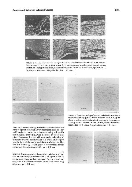
FIGURE 3. In situ hybridization <strong>of</strong> injured corneas with 3 H-labeled cDNA <strong>of</strong> al(I) mRNA,<br />
Panels a <strong>and</strong> d, lacerated cornea healed for 2 weeks; panels b <strong>and</strong> e, alkali-burned cornea<br />
healed for 1 day; panels c <strong>and</strong> f, alkali-burned cornea healed for 3 weeks, epi, epithelium; D,<br />
Descemet's membrane. Magnification, bar = 0.2 mm.<br />
01 mm<br />
0.1mm<br />
FIGURE 4. Immunostaining <strong>of</strong> alkali-burned corneas with antibodies<br />
against collagen I. Injured corneas healed for 1 day<br />
<strong>and</strong> 3 weeks were subjected to immunostaining with specific<br />
anti-collagen I antibodies. Panel a, cornea 24 hours after<br />
injury. Degenerated stroma still reacts to the anti-collagen I<br />
antibodies (X270). Panels b <strong>and</strong> c, 3 weeks after injury;<br />
panel b, the newly formed granulation tissue between epithelium<br />
<strong>and</strong> stroma (*) (X270); panel c, retrocorneal fibriilar<br />
membrane. Magnification (X350), bar = 0.1 mm.<br />
FIGURE 5. Immunostaining <strong>of</strong> normal <strong>and</strong> alkali-burned corneas<br />
with antibody against smooth muscle a-actin. 0.1 fig/ml<br />
<strong>of</strong> anti-a-actin monoclonal antibody was used in the immunostaining.<br />
Panel a, normal cornea; panel b, alkali-burned corneas<br />
healed for 3 weeks. Magnification, bar = 0.1 mm.<br />
FIGURE 6. Immunostaining <strong>of</strong> normal <strong>and</strong> alkali-burned corneas<br />
with antibody against vimentin. 0.88 fig/ml <strong>of</strong> anti-vimentin<br />
monoclonal antibody was used. Panel a, normal cornea;<br />
panel b, alkali-burned cornea healed for 3 weeks. Magnification,<br />
bar = 0.1 mm.<br />
0.1 mm


