

Expression of Collagen I, Smooth Muscle a-Actin, and Vimentin ...
Expression of Collagen I, Smooth Muscle a-Actin, and Vimentin ...
Expression of Collagen I, Smooth Muscle a-Actin, and Vimentin ...

Create successful ePaper yourself
Turn your PDF publications into a flip-book with our unique Google optimized e-Paper software.
3326 Investigative Ophthalmology & Visual Science, November 1993, Vol. 34, No. 12<br />
Viiiuillin<br />
e<br />
9<br />
0.1 mm<br />
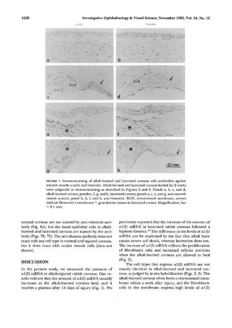
FIGURE 7. Immunostaining <strong>of</strong> alkali-burned <strong>and</strong> lacerated corneas with antibodies against<br />
smooth muscle a-actin <strong>and</strong> vimentin. Alkali-burned <strong>and</strong> lacerated corneas healed for 2 weeks<br />
were subjected to immunostaining as described in Figures 5 <strong>and</strong> 6. Panels a, b, c, <strong>and</strong> d,<br />
alkali-burned cornea; panels e, f, g, <strong>and</strong> h, lacerated cornea; panels a, c, e, <strong>and</strong> g, anti-smooth<br />
muscle a-actin; panel b, d, f, <strong>and</strong> h, anti-vimentin. RCM, retrocorneal membrane; arrows<br />
indicate Descemet's membrane; *, granulation tissues in lacerated cornea. Magnification, bar<br />
= 0.1 mm.<br />
normal corneas are not stained by anti-vimentin antibody<br />
(Fig. 6a), but the basal epithelial cells in alkaliburned<br />
<strong>and</strong> lacerated corneas are stained by the antibody<br />
{Figs. 7b, 7f). The anti-desmin antibody does not<br />
react with any cell type in normal <strong>and</strong> injured corneas,<br />
but it does react with ocular muscle cells (data not<br />
shown).<br />
DISCUSSION<br />
In the present study, we measured the amounts <strong>of</strong><br />
al(I) mRNA in alkali-injured rabbit corneas. Our results<br />
indicate that the amount <strong>of</strong> al(I) mRNA steadily<br />
increases as the alkali-burned corneas heal, <strong>and</strong> it<br />
reaches a plateau after 14 days <strong>of</strong> injury (Fig. 1). We<br />
previously reported that the increase <strong>of</strong> the amount <strong>of</strong><br />
al(I) mRNA in lacerated rabbit corneas followed a<br />
biphasic kinetics. 15 The difference in the levels <strong>of</strong> a 1(1)<br />
mRNA can be explained by the fact that alkali burn<br />
causes severe cell death, whereas laceration does not.<br />
The increase <strong>of</strong> al(l) mRNA reflects the proliferation<br />
<strong>of</strong> fibroblastic cells <strong>and</strong> increased cellular activities<br />
when the alkali-burned corneas are allowed to heal<br />
(Fig. 2).<br />
The cell types that express a 1(1) mRNA are not<br />
exactly identical in alkali-burned <strong>and</strong> lacerated corneas,<br />
as judged by in situ hybridization (Figs. 2, 3). The<br />
alkali-burned corneas <strong>of</strong>ten form a retrocorneal membrane<br />
within a week after injury, <strong>and</strong> the fibroblastic<br />
cells in this membrane express high levels <strong>of</strong> a 1(1)


