

Expression of Collagen I, Smooth Muscle a-Actin, and Vimentin ...
Expression of Collagen I, Smooth Muscle a-Actin, and Vimentin ...
Expression of Collagen I, Smooth Muscle a-Actin, and Vimentin ...

You also want an ePaper? Increase the reach of your titles
YUMPU automatically turns print PDFs into web optimized ePapers that Google loves.
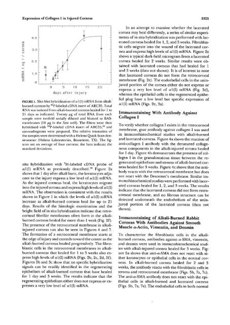
<strong>Expression</strong> <strong>of</strong> <strong>Collagen</strong> I in Injured Corneas 3323<br />
E<br />
4.0<br />
3.0<br />
2.0<br />
1.0<br />
0<br />
-<br />
• • • • i • ' • • i • • • • i • • • • i • • • • i • • • •<br />
:<br />
I<br />
/<br />
N<br />
' 1 $ l/f J<br />
r<br />
0 5 10 15<br />
days after Injury<br />
FIGURE l. Slot-blot hybridization <strong>of</strong> «1 (I) mRNA from alkaliburned<br />
corneas by 32 P-labeled cDNA insert <strong>of</strong> ARC35. Total<br />
RNA was isolated from alkali-burned corneas healed for 1 to<br />
21 days as indicated. Twenty jug <strong>of</strong> total RNA from each<br />
sample were tw<strong>of</strong>old serially diluted <strong>and</strong> blotted to BASmembranes<br />
(10 Mg in the first well). The niters were then<br />
hybridized with 32 P-labelecl cDNA insert <strong>of</strong> ARC35, 22 <strong>and</strong><br />
autoradiograms were prepared. The relative intensities <strong>of</strong><br />
the samples were determined with a Helena Quick Scan densitometer<br />
(Helena Laboratories, Beaumont, TX). The figures<br />
are an average <strong>of</strong> four corneas; the bars indicate the<br />
st<strong>and</strong>ard deviations.<br />
situ hybridization with 3 H-labeled cDNA probe <strong>of</strong><br />
cd(I) mRNA as previously described. 18 Figure 2a<br />
shows that 1 day after alkali burn, the keratocytes adjacent<br />
to the injury express a low level <strong>of</strong> al(I) mRNA-<br />
As the injured corneas heal, the keratocytes migrate<br />
into the injured stroma <strong>and</strong> express high levels <strong>of</strong> al (I)<br />
mRNA. The observation is consistent with the results<br />
shown in Figure 1 in which the levels <strong>of</strong> a 1(1) mRNA<br />
increase as alkali-burned corneas heal for up to 21<br />
days. Results <strong>of</strong> the histologic examination <strong>and</strong> the<br />
bright field <strong>of</strong> in situ hybridization indicate that retrocorneal<br />
fibrillar membranes <strong>of</strong>ten form in the alkaliburned<br />
corneas healed for more than 1 week (Fig. 2f).<br />
The presence <strong>of</strong> the retrocorneal membrane in alkaliinjured<br />
corneas can also be seen in Figures 4 <strong>and</strong> 7.<br />
The formation <strong>of</strong> a retrocorneal membrane starts at<br />
the edge <strong>of</strong> injury <strong>and</strong> extends toward the center as the<br />
alkali-burned corneas healed progressively. The fibroblastic<br />
cells in the retrocorneal membranes in alkaliburned<br />
corneas that healed for 1 to 3 weeks also express<br />
high levels <strong>of</strong> al(l) mRNA (Figs. 2b, 2c, 2d, 3f).<br />
Figures 3b <strong>and</strong> 3c show that no specific hybridization<br />
signals can be readily identified in the regenerating<br />
epithelium <strong>of</strong> alkali-burned corneas that have healed<br />
for 1 day <strong>and</strong> 3 weeks. The results indicate that the<br />
regenerating epithelium either does not express or expresses<br />
a very low level <strong>of</strong> al(i) mRNA.<br />
20<br />
•<br />
In an attempt to examine whether the lacerated<br />
cornea may heal differently, a series <strong>of</strong> similar experiments<br />
<strong>of</strong> in situ hybridization was performed with lacerated<br />
corneas healed for 1, 2, <strong>and</strong> 3 weeks. Fibroblastic<br />
cells migrate into the wound <strong>of</strong> the lacerated cornea<br />
<strong>and</strong> express high levels <strong>of</strong> al(I) mRNA. Figure 2e<br />
shows a typical dark-field microgram from a lacerated<br />
cornea healed for 2 weeks. Similar results were obtained<br />
with lacerated corneas that had healed for 1<br />
<strong>and</strong> 3 weeks (data not shown). It is <strong>of</strong> interest to note<br />
that lacerated corneas do not form the retrocorneal<br />
membrane (Fig. 2e). The endothelial cells in the uninjured<br />
portion <strong>of</strong> the cornea either do not express or<br />
express a very low level <strong>of</strong> al(l) mRNA (Fig. 3d),<br />
whereas the epithelial cells in the regenerated epithelial<br />
plug have a low level but specific expression <strong>of</strong><br />
al(l) mRNA (Figs. 2e, 3a).<br />
Immunostaining With Antibody Against<br />
<strong>Collagen</strong> I<br />
To verify whether collagen I exists in the retrocorneal<br />
membrane, goat antibody against collagen I was used<br />
in immunohistochemical studies with alkali-burned<br />
<strong>and</strong> lacerated corneas. Figure 4a shows the reaction <strong>of</strong><br />
anti-collagen I antibody with the denatured collagenous<br />
components in the alkali-injured stroma healed<br />
for I day. Figure 4b demonstrates the presence <strong>of</strong> collagen<br />
I in the granulomatous tissue between the regenerated<br />
epithelium <strong>and</strong> stroma <strong>of</strong> alkali-burned corneas<br />
healed for 3 weeks. Figure 4c shows that the antibody<br />
reacts with the retrocorneal membrane but does<br />
not react with the Descemet's membrane. Similar immunohistochemical<br />
studies were performed with lacerated<br />
corneas healed for 1, 2, <strong>and</strong> 3 weeks. The results<br />
indicate that the lacerated corneas did not form retrocorneal<br />
membrane, <strong>and</strong> no fibrous structure can be<br />
detected underneath the endothelium <strong>of</strong> the uninjured<br />
portion <strong>of</strong> the lacerated corneas (data not<br />
shown).<br />
Immunostaining <strong>of</strong> Alkali-Burned Rabbit<br />
Corneas With Antibodies Against <strong>Smooth</strong><br />
<strong>Muscle</strong> a-<strong>Actin</strong>, <strong>Vimentin</strong>, <strong>and</strong> Desmin<br />
To characterize the fibroblastic cells in the alkaliburned<br />
corneas, antibodies against a-SMA, vimentin,<br />
<strong>and</strong> desmin were used in immunohistochemical studies<br />
with alkali-injured cornea healed for 3 weeks. Figure<br />
5a shows that anti-a-SMA does not react with either<br />
keratocytes or epithelial cells in the normal corneas.<br />
In alkali-burned cornea healed for 2 <strong>and</strong> 3<br />
weeks, the antibody reacts with the fibroblastic cells in<br />
stroma <strong>and</strong> retrocorneal membrane (Figs. 5b, 7a, 7c).<br />
The anti-a-SMA antibody does not react with the epithelial<br />
cells in alkali-burned <strong>and</strong> lacerated corneas<br />
(Figs. 5b, 7a, 7e). The endothelial cells in both normal


