

Expression of Collagen I, Smooth Muscle a-Actin, and Vimentin ...
Expression of Collagen I, Smooth Muscle a-Actin, and Vimentin ...
Expression of Collagen I, Smooth Muscle a-Actin, and Vimentin ...

Create successful ePaper yourself
Turn your PDF publications into a flip-book with our unique Google optimized e-Paper software.
3324 Investigative Ophthalmology 8c Visual Science, November 1993, Vol. 34, No. 12<br />
1<br />
f<br />
RCM<br />
0.1 mm<br />
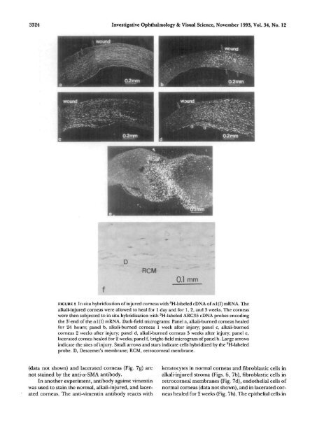
FIGURE 2 In situ hybridization <strong>of</strong> injured corneas with 3 H-labeled cDNA <strong>of</strong> al(I) mRNA. The<br />
alkali-injured corneas were allowed to heal for 1 day <strong>and</strong> for 1, 2, <strong>and</strong> 3 weeks. The corneas<br />
were then subjected to in situ hybridization with 3 H-labeled ARC35 cDNA probes encoding<br />
the 3'-end <strong>of</strong> the al(I) mRNA. Dark-field micrograms: Panel a, alkali-burned corneas healed<br />
for 24 hours; panel b, alkali-burned corneas 1 week after injury; panel c, alkali-burned<br />
corneas 2 weeks after injury; panel d, alkali-burned corneas 3 weeks after injury; panel e,<br />
lacerated cornea healed for 2 weeks; panel f, bright-field microgram <strong>of</strong> panel b. Large arrows<br />
indicate the sites <strong>of</strong> injury. Small arrows <strong>and</strong> stars indicate cells hybridized by the 3 H-labeled<br />
probe. D, Descemet's membrane; RCM, retrocorneal membrane.<br />
(data not shown) <strong>and</strong> lacerated corneas (Fig. 7g) are<br />
not stained by the anti-a-SMA antibody.<br />
In another experiment, antibody against vimentin<br />
was used to stain the normal, alkali-injured, <strong>and</strong> lacerated<br />
corneas. The anti-vimentin antibody reacts with<br />
keratocytes in normal corneas <strong>and</strong> fibroblastic cells in<br />
alkali-injured stroma (Figs. 6, 7b), fibroblastic cells in<br />
retrocorneal membranes (Fig. 7d), endothelial cells <strong>of</strong><br />
normal corneas (data not shown), <strong>and</strong> in lacerated corneas<br />
healed for 2 weeks (Fig. 7h). The epithelial cells in


