

The survival and rejection of epithelium in experimental corneal ...
The survival and rejection of epithelium in experimental corneal ...
The survival and rejection of epithelium in experimental corneal ...

Create successful ePaper yourself
Turn your PDF publications into a flip-book with our unique Google optimized e-Paper software.
174 Khodadoust <strong>and</strong> Silverste<strong>in</strong> 1nve.it'.igatioe Ophthalmology<br />
April 1969<br />
For this purpose, the <strong>epithelium</strong> <strong>of</strong> the prospective<br />
donor was <strong>in</strong>tensively labeled with tritiated<br />
thymid<strong>in</strong>e prior to transplantation. This was accomplished<br />
by first scrap<strong>in</strong>g the donor cornea centrally<br />
over an area <strong>of</strong> about 10 mm. <strong>in</strong> diameter.<br />
Several drops <strong>of</strong> a solution <strong>of</strong> tritiated thymid<strong>in</strong>e<br />
(100 MC per milliliter) were applied topically<br />
twice a day for 2 weeks, dur<strong>in</strong>g the process <strong>of</strong><br />
epithelial regeneration. Corneal transplantation was<br />
performed 3 days after discont<strong>in</strong>uation <strong>of</strong> the<br />
drops, <strong>and</strong> the grafts were removed 10 to 14 days<br />
after operation. <strong>The</strong> eyes were fixed, embedded,<br />
sectioned, <strong>and</strong> radioautographs prepared accord<strong>in</strong>g<br />
to well-established procedures 2 for the localization<br />
<strong>of</strong> isotopically labeled cells. <strong>The</strong>se radioautographs<br />
(Fig. 3) clearly demonstrate that not only does<br />
donor <strong>epithelium</strong> heal <strong>and</strong> survive dur<strong>in</strong>g this 2<br />
week period, but also that it shares <strong>in</strong> epithelial<br />
wound heal<strong>in</strong>g. Even after 2 weeks, one half <strong>of</strong><br />
the graft scar is covered by donor <strong>epithelium</strong> <strong>and</strong><br />
the other half by host cells which show no tendency<br />
to <strong>in</strong>vade the graft <strong>and</strong> replace the donor<br />
<strong>epithelium</strong> dur<strong>in</strong>g this period.<br />
Control experiment. In a parallel experiment,<br />
the cornea <strong>of</strong> the recipient was labeled by scrap<strong>in</strong>g<br />
its <strong>epithelium</strong> over a wide area, followed by<br />
topical application <strong>of</strong> tritiated thymid<strong>in</strong>e as described.<br />
Two weeks later, an 8 mm. lamellar graft<br />
with nonlabeled <strong>epithelium</strong> was placed centrally<br />
upon this cornea. <strong>The</strong> grafted corneas were removed<br />
10 days to 2 weeks after transplantation,<br />
<strong>and</strong> radioautographs prepared as described previously.<br />
In this <strong>in</strong>stance, labeled <strong>epithelium</strong> could<br />
be seen cover<strong>in</strong>g only the host stroma <strong>and</strong> a<br />
portion <strong>of</strong> the adjacent graft scar. Dur<strong>in</strong>g this 2<br />
week period, there was no tendency on the part<br />
<strong>of</strong> labeled recipient epithelial cells to <strong>in</strong>vade the<br />
surface <strong>of</strong> the donor button.<br />
Observations <strong>of</strong> <strong>epithelium</strong> <strong>in</strong> vascularized<br />
<strong>corneal</strong> allografts. In studies to be reported elsewhere<br />
7 ' s it was observed that the <strong>epithelium</strong><br />
cover<strong>in</strong>g a donor allograft is apparently able to<br />
participate <strong>in</strong> a specific manner <strong>in</strong> the <strong>rejection</strong><br />
process. To recapitulate these observations briefly,<br />
epithelial <strong>rejection</strong> <strong>in</strong>duced dur<strong>in</strong>g the early period<br />
after transplantation presents as a l<strong>in</strong>ear defect,<br />
sta<strong>in</strong>able by methylene blue, which slowly proceeds<br />
across the surface <strong>of</strong> the donor graft, until<br />
the entire donor surface has been traversed. <strong>The</strong><br />
"epithelial <strong>rejection</strong> l<strong>in</strong>e" <strong>in</strong>variably starts precisely<br />
at the donor-recipient graft junction, <strong>and</strong> always<br />
<strong>in</strong> close approximation to the blood vessels which<br />
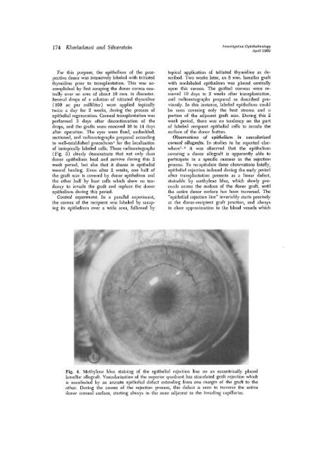
Fig. 4. Methylene blue sta<strong>in</strong><strong>in</strong>g <strong>of</strong> the epithelial <strong>rejection</strong> l<strong>in</strong>e on an eccentrically placed<br />
lamellar allograft. Vascularization <strong>of</strong> the superior quadrant has stimulated graft <strong>rejection</strong> which<br />
is manifested by an arcuate epithelial defect extend<strong>in</strong>g from one marg<strong>in</strong> <strong>of</strong> the graft to the<br />
other. Dur<strong>in</strong>g the course <strong>of</strong> the <strong>rejection</strong> process, this defect is seen to traverse the entire<br />
donor <strong>corneal</strong> surface, start<strong>in</strong>g always <strong>in</strong> the zone adjacent to the <strong>in</strong>vad<strong>in</strong>g capillaries.


