

Ocular retardation (or) in the mouse.
Ocular retardation (or) in the mouse.
Ocular retardation (or) in the mouse.

You also want an ePaper? Increase the reach of your titles
YUMPU automatically turns print PDFs into web optimized ePapers that Google loves.
Volume 17<br />
Number 5 Rep<strong>or</strong>ts 469<br />
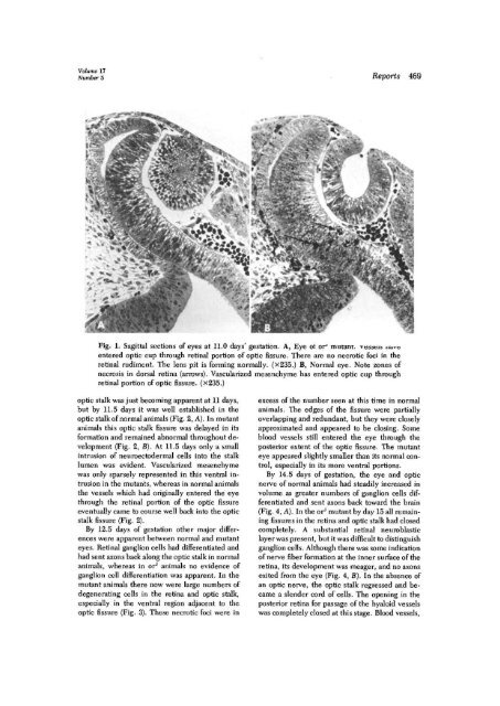
Fig. 1. Sagittal sections of eyes at 11.0 days' gestation. A, Eye ot <strong>or</strong> 1 mutant, vessels nave<br />
entered optic cup through ret<strong>in</strong>al p<strong>or</strong>tion of optic fissure. There are no necrotic foci <strong>in</strong> <strong>the</strong><br />
ret<strong>in</strong>al rudiment. The lens pit is f<strong>or</strong>m<strong>in</strong>g n<strong>or</strong>mally. (x235.) B, N<strong>or</strong>mal eye. Note zones of<br />
necrosis <strong>in</strong> d<strong>or</strong>sal ret<strong>in</strong>a (arrows). Vascularized mesenchyme has entered optic cup through<br />
ret<strong>in</strong>al p<strong>or</strong>tion of optic fissure. (x235.)<br />
optic stalk was just becom<strong>in</strong>g apparent at 11 days,<br />
but by 11.5 days it was well established <strong>in</strong> <strong>the</strong><br />
optic stalk of n<strong>or</strong>mal animals (Fig. 2, A). In mutant<br />
animals this optic stalk fissure was delayed <strong>in</strong> its<br />
f<strong>or</strong>mation and rema<strong>in</strong>ed abn<strong>or</strong>mal throughout development<br />
(Fig. 2, B). At 11.5 days only a small<br />
<strong>in</strong>trusion of neuroectodermal cells <strong>in</strong>to <strong>the</strong> stalk<br />
lumen was evident. Vascularized mesenchyme<br />
was only sparsely represented <strong>in</strong> this ventral <strong>in</strong>trusion<br />
<strong>in</strong> <strong>the</strong> mutants, whereas <strong>in</strong> n<strong>or</strong>mal animals<br />
<strong>the</strong> vessels which had <strong>or</strong>ig<strong>in</strong>ally entered <strong>the</strong> eye<br />
through <strong>the</strong> ret<strong>in</strong>al p<strong>or</strong>tion of <strong>the</strong> optic fissure<br />
eventually came to course well back <strong>in</strong>to <strong>the</strong> optic<br />
stalk fissure (Fig. 2).<br />
By 12.5 days of gestation o<strong>the</strong>r maj<strong>or</strong> differences<br />
were apparent between n<strong>or</strong>mal and mutant<br />
eyes. Ret<strong>in</strong>al ganglion cells had differentiated and<br />
had sent axons back along <strong>the</strong> optic stalk <strong>in</strong> n<strong>or</strong>mal<br />
animals, whereas <strong>in</strong> <strong>or</strong> J animals no evidence of<br />
ganglion cell differentiation was apparent. In <strong>the</strong><br />
mutant animals <strong>the</strong>re now were large numbers of<br />
degenerat<strong>in</strong>g cells <strong>in</strong> <strong>the</strong> ret<strong>in</strong>a and optic stalk,<br />
especially <strong>in</strong> <strong>the</strong> ventral region adjacent to <strong>the</strong><br />
optic fissure (Fig. 3). These necrotic foci were <strong>in</strong><br />
excess of <strong>the</strong> number seen at this time <strong>in</strong> n<strong>or</strong>mal<br />
animals. The edges of <strong>the</strong> fissure were partially<br />
overlapp<strong>in</strong>g and redundant, but <strong>the</strong>y were closely<br />
approximated and appeared to be clos<strong>in</strong>g. Some<br />
blood vessels still entered <strong>the</strong> eye through <strong>the</strong><br />
posteri<strong>or</strong> extent of <strong>the</strong> optic fissure. The mutant<br />
eye appeared slightly smaller than its n<strong>or</strong>mal control,<br />
especially <strong>in</strong> its m<strong>or</strong>e ventral p<strong>or</strong>tions.<br />
By 14.5 days of gestation, <strong>the</strong> eye and optic<br />
nerve of n<strong>or</strong>mal animals had steadily <strong>in</strong>creased <strong>in</strong><br />
volume as greater numbers of ganglion cells differentiated<br />
and sent axons back toward <strong>the</strong> bra<strong>in</strong><br />
(Fig. 4, A). In <strong>the</strong> <strong>or</strong> 1 mutant by day 15 all rema<strong>in</strong><strong>in</strong>g<br />
fissures <strong>in</strong> <strong>the</strong> ret<strong>in</strong>a and optic stalk had closed<br />
completely. A substantial ret<strong>in</strong>al neuroblastic<br />
layer was present, but it was difficult to dist<strong>in</strong>guish<br />
ganglion cells. Although <strong>the</strong>re was some <strong>in</strong>dication<br />
of nerve fiber f<strong>or</strong>mation at <strong>the</strong> <strong>in</strong>ner surface of <strong>the</strong><br />
ret<strong>in</strong>a, its development was meager, and no axons<br />
exited from <strong>the</strong> eye (Fig. 4, B). In <strong>the</strong> absence of<br />
an optic nerve, <strong>the</strong> optic stalk regressed and became<br />
a slender c<strong>or</strong>d of cells. The open<strong>in</strong>g <strong>in</strong> <strong>the</strong><br />
posteri<strong>or</strong> ret<strong>in</strong>a f<strong>or</strong> passage of <strong>the</strong> hyaloid vessels<br />
was completely closed at this stage. Blood vessels,


