

Ocular retardation (or) in the mouse.
Ocular retardation (or) in the mouse.
Ocular retardation (or) in the mouse.

Create successful ePaper yourself
Turn your PDF publications into a flip-book with our unique Google optimized e-Paper software.
470 Rep<strong>or</strong>ts<br />
Invest. Ophthal. Visual Sci.<br />
May 1978<br />
i<br />
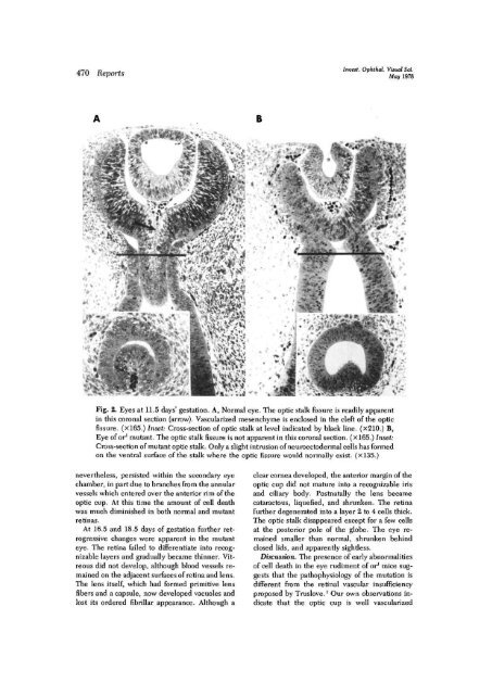
Fig. 2. Eyes at 11.5 days' gestation. A, N<strong>or</strong>mal eye. The optic stalk fissure is readily apparent<br />
<strong>in</strong> this c<strong>or</strong>onal section (arrow). Vascularized mesenchyme is enclosed <strong>in</strong> <strong>the</strong> cleft of <strong>the</strong> optic<br />
fissure. (X165.) Inset: Cross-section of optic stalk at level <strong>in</strong>dicated by black l<strong>in</strong>e. (x210.) B,<br />
Eye of <strong>or</strong>* mutant. The optic stalk fissure is not apparent <strong>in</strong> this c<strong>or</strong>onal section. (X165.) Inset:<br />
Cross-section of mutant optic stalk. Only a slight <strong>in</strong>trusion of neuroectodermal cells has f<strong>or</strong>med<br />
on <strong>the</strong> ventral surface of <strong>the</strong> stalk where <strong>the</strong> optic fissure would n<strong>or</strong>mally exist. (xl35.)<br />
never<strong>the</strong>less, persisted with<strong>in</strong> <strong>the</strong> secondary eye<br />
chamber, <strong>in</strong> part due to branches from <strong>the</strong> annular<br />
vessels which entered over <strong>the</strong> anteri<strong>or</strong> rim of <strong>the</strong><br />
optic cup. At this time <strong>the</strong> amount of cell death<br />
was much dim<strong>in</strong>ished <strong>in</strong> both n<strong>or</strong>mal and mutant<br />
ret<strong>in</strong>as.<br />
At 16.5 and 18.5 days of gestation fur<strong>the</strong>r retrogressive<br />
changes were apparent <strong>in</strong> <strong>the</strong> mutant<br />
eye. The ret<strong>in</strong>a failed to differentiate <strong>in</strong>to recognizable<br />
layers and gradually became th<strong>in</strong>ner. Vitreous<br />
did not develop, although blood vessels rema<strong>in</strong>ed<br />
on <strong>the</strong> adjacent surfaces of ret<strong>in</strong>a and lens.<br />
The lens itself, which had f<strong>or</strong>med primitive lens<br />
fibers and a capsule, now developed vacuoles and<br />
lost its <strong>or</strong>dered fibrillar appearance. Although a<br />
clear c<strong>or</strong>nea developed, <strong>the</strong> anteri<strong>or</strong> marg<strong>in</strong> of <strong>the</strong><br />
optic cup did not mature <strong>in</strong>to a recognizable iris<br />
and ciliary body. Postnatally <strong>the</strong> lens became<br />
cataractous, liquefied, and shrunken. The ret<strong>in</strong>a<br />
fur<strong>the</strong>r degenerated <strong>in</strong>to a layer 2 to 4 cells thick.<br />
The optic stalk disappeared except f<strong>or</strong> a few cells<br />
at <strong>the</strong> posteri<strong>or</strong> pole of <strong>the</strong> globe. The eye rema<strong>in</strong>ed<br />
smaller than n<strong>or</strong>mal, shrunken beh<strong>in</strong>d<br />
closed lids, and apparently sightless.<br />
Discussion. The presence of early abn<strong>or</strong>malities<br />
of cell death <strong>in</strong> <strong>the</strong> eye rudiment of <strong>or</strong>* mice suggests<br />
that <strong>the</strong> pathophysiology of <strong>the</strong> mutation is<br />
different from <strong>the</strong> ret<strong>in</strong>al vascular <strong>in</strong>sufficiency<br />
proposed by Truslove. 1 Our own observations <strong>in</strong>dicate<br />
that <strong>the</strong> optic cup is well vascularized


