

Ocular retardation (or) in the mouse.
Ocular retardation (or) in the mouse.
Ocular retardation (or) in the mouse.

You also want an ePaper? Increase the reach of your titles
YUMPU automatically turns print PDFs into web optimized ePapers that Google loves.
Volume 17<br />
Number 5 Rep<strong>or</strong>ts 471<br />
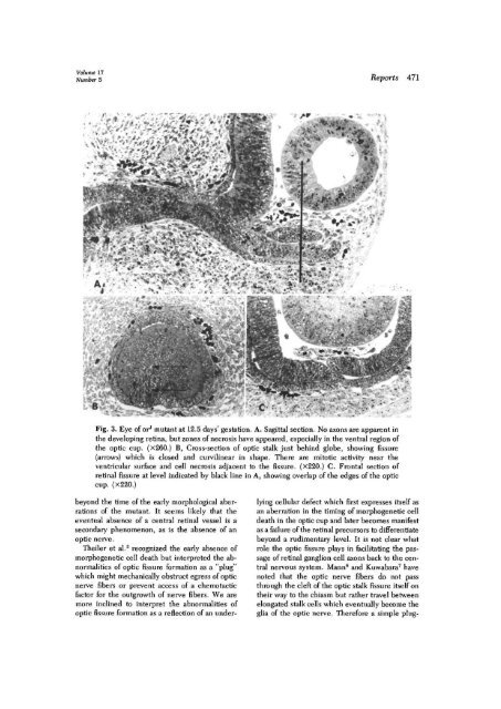
Fig. 3. Eye of <strong>or</strong> J mutant at 12.5 days' gestation. A. Sagittal section. No axons are apparent <strong>in</strong><br />
<strong>the</strong> develop<strong>in</strong>g ret<strong>in</strong>a, but zones of necrosis have appeared, especially <strong>in</strong> <strong>the</strong> ventral region of<br />
<strong>the</strong> optic cup. (x260.) B, Cross-section of optic stalk just beh<strong>in</strong>d globe, show<strong>in</strong>g fissure<br />
(arrows) which is closed and curvil<strong>in</strong>ear <strong>in</strong> shape. There are mitotic activity near <strong>the</strong><br />
ventricular surface and cell necrosis adjacent to <strong>the</strong> fissure. (x220.) C. Frontal section of<br />
ret<strong>in</strong>al fissure at level <strong>in</strong>dicated by black l<strong>in</strong>e <strong>in</strong> A, show<strong>in</strong>g overlap of <strong>the</strong> edges of <strong>the</strong> optic<br />
cup. (X220.)<br />
beyond <strong>the</strong> time of <strong>the</strong> early m<strong>or</strong>phological aberrations<br />
of <strong>the</strong> mutant. It seems likely that <strong>the</strong><br />
eventual absence of a central ret<strong>in</strong>al vessel is a<br />
secondary phenomenon, as is <strong>the</strong> absence of an<br />
optic nerve.<br />
Theiler et al. 3 recognized <strong>the</strong> early absence of<br />
m<strong>or</strong>phogenetic cell death but <strong>in</strong>terpreted <strong>the</strong> abn<strong>or</strong>malities<br />
of optic fissure f<strong>or</strong>mation as a "plug"<br />
which might mechanically obstruct egress of optic<br />
nerve fibers <strong>or</strong> prevent access of a chemotactic<br />
fact<strong>or</strong> f<strong>or</strong> <strong>the</strong> outgrowth of nerve fibers. We are<br />
m<strong>or</strong>e <strong>in</strong>cl<strong>in</strong>ed to <strong>in</strong>terpret <strong>the</strong> abn<strong>or</strong>malities of<br />
optic fissure f<strong>or</strong>mation as a reflection of an underly<strong>in</strong>g<br />
cellular defect which first expresses itself as<br />
an aberration <strong>in</strong> <strong>the</strong> tim<strong>in</strong>g of m<strong>or</strong>phogenetic cell<br />
death <strong>in</strong> <strong>the</strong> optic cup and later becomes manifest<br />
as a failure of <strong>the</strong> ret<strong>in</strong>al precurs<strong>or</strong>s to differentiate<br />
beyond a rudimentary level. It is not clear what<br />
role <strong>the</strong> optic fissure plays <strong>in</strong> facilitat<strong>in</strong>g <strong>the</strong> passage<br />
of ret<strong>in</strong>al ganglion cell axons back to <strong>the</strong> central<br />
nervous system. Mann 6 and Kuwabara 7 have<br />
noted that <strong>the</strong> optic nerve fibers do not pass<br />
through <strong>the</strong> cleft of <strong>the</strong> optic stalk fissure itself on<br />
<strong>the</strong>ir way to <strong>the</strong> chiasm but ra<strong>the</strong>r travel between<br />
elongated stalk cells which eventually become <strong>the</strong><br />
glia of <strong>the</strong> optic nerve. Theref<strong>or</strong>e a simple plug-


