Ultrasonography in Tarsal Tunnel Syndrome - Journal of Ultrasound ...
Ultrasonography in Tarsal Tunnel Syndrome - Journal of Ultrasound ...
Ultrasonography in Tarsal Tunnel Syndrome - Journal of Ultrasound ...

Create successful ePaper yourself
Turn your PDF publications into a flip-book with our unique Google optimized e-Paper software.
Article<br />
<strong>Ultrasonography</strong> <strong>in</strong><br />
<strong>Tarsal</strong> <strong>Tunnel</strong> <strong>Syndrome</strong><br />
Masahiro Nagaoka, MD, Hiromi Matsuzaki, MD<br />
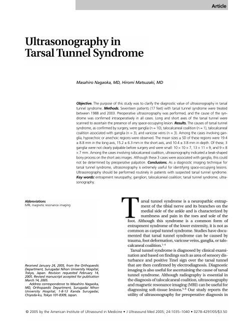
Objective. The purpose <strong>of</strong> this study was to clarify the diagnostic value <strong>of</strong> ultrasonography <strong>in</strong> tarsal<br />
tunnel syndrome. Methods. Seventeen patients (17 feet) with tarsal tunnel syndrome were treated<br />
between 1988 and 2003. Preoperative ultrasonography was performed, and the cause <strong>of</strong> the syndrome<br />
was confirmed <strong>in</strong>traoperatively <strong>in</strong> all cases. Long and short axes <strong>of</strong> the tarsal tunnel were<br />
scanned to ascerta<strong>in</strong> the presence <strong>of</strong> any space-occupy<strong>in</strong>g lesion. Results. The causes <strong>of</strong> tarsal tunnel<br />
syndrome, as confirmed by surgery, were ganglia (n = 10), talocalcaneal coalition (n = 1), talocalcaneal<br />
coalition associated with ganglia (n = 3), and varicose ve<strong>in</strong>s (n = 3). Among the cases <strong>in</strong>volv<strong>in</strong>g ganglia,<br />
hypoechoic or anechoic regions were observed. The mean sizes ± SD <strong>of</strong> these regions were 19.4<br />
± 8.8 mm <strong>in</strong> the long axis, 15.2 ± 6.3 mm <strong>in</strong> the short axis, and 10.4 ± 3.8 mm <strong>in</strong> depth. Of these, 3<br />
ganglia were not clearly palpable before surgery and were small: 10 × 10 × 7, 13 × 11 × 9, and 9 × 8<br />
× 7 mm. Among the cases <strong>in</strong>volv<strong>in</strong>g talocalcaneal coalition, ultrasonography <strong>in</strong>dicated a beak-shaped<br />
bony process on the short axis images. Although these 3 cases were associated with ganglia, this could<br />
not be determ<strong>in</strong>ed by preoperative palpation. Conclusions. As a diagnostic imag<strong>in</strong>g technique for<br />
tarsal tunnel syndrome, ultrasonography is extremely useful for identify<strong>in</strong>g space-occupy<strong>in</strong>g lesions.<br />
<strong>Ultrasonography</strong> should be performed rout<strong>in</strong>ely <strong>in</strong> patients with suspected tarsal tunnel syndrome.<br />
Key words: entrapment neuropathy; ganglion; talocalcaneal coalition; tarsal tunnel syndrome; ultrasonography.<br />
Abbreviations<br />
MRI, magnetic resonance imag<strong>in</strong>g<br />
Received January 24, 2005, from the Orthopaedic<br />
Department, Surugadai Nihon University Hospital,<br />
Tokyo, Japan. Revision requested February 14,<br />
2005. Revised manuscript accepted for publication<br />
March 14, 2005.<br />
Address correspondence to Masahiro Nagaoka,<br />
MD, Orthopaedic Department, Surugadai Nihon<br />
University Hospital, 1-8-13 Kanda Surugadai,<br />
Chiyoda-ku, Tokyo 101-8309, Japan.<br />
<strong>Tarsal</strong> tunnel syndrome is a neuropathic entrapment<br />
<strong>of</strong> the tibial nerve and its branches on the<br />
medial side <strong>of</strong> the ankle and is characterized by<br />
numbness and pa<strong>in</strong> <strong>in</strong> the toes and sole <strong>of</strong> the<br />
foot. Although this syndrome is a common form <strong>of</strong><br />
entrapment syndrome <strong>of</strong> the lower extremity, it is not as<br />
common as carpal tunnel syndrome. Studies have documented<br />
that tarsal tunnel syndrome can be caused by<br />
trauma, foot deformation, varicose ve<strong>in</strong>s, ganglia, or talocalcaneal<br />
coalition. 1–4<br />
<strong>Tarsal</strong> tunnel syndrome is diagnosed by cl<strong>in</strong>ical exam<strong>in</strong>ation<br />
and based on f<strong>in</strong>d<strong>in</strong>gs such as area <strong>of</strong> sensory disturbance<br />
and positive T<strong>in</strong>el sign over the tarsal tunnel<br />
that are then confirmed by electrodiagnosis. Diagnostic<br />
imag<strong>in</strong>g is also useful for ascerta<strong>in</strong><strong>in</strong>g the cause <strong>of</strong> tarsal<br />
tunnel syndrome. Although radiography is essential <strong>in</strong><br />
the diagnosis <strong>of</strong> talocalcaneal coalition, ultrasonography<br />
and magnetic resonance imag<strong>in</strong>g (MRI) can be useful for<br />
diagnos<strong>in</strong>g s<strong>of</strong>t-tissue lesions. 5–8 Our study reports the<br />
utility <strong>of</strong> ultrasonography for preoperative diagnosis <strong>in</strong><br />
© 2005 by the American Institute <strong>of</strong> <strong>Ultrasound</strong> <strong>in</strong> Medic<strong>in</strong>e • J <strong>Ultrasound</strong> Med 2005; 24:1035–1040 • 0278-4297/05/$3.50
<strong>Ultrasonography</strong> <strong>in</strong> <strong>Tarsal</strong> <strong>Tunnel</strong> <strong>Syndrome</strong><br />
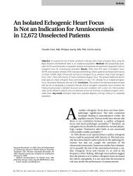
Figure 1. Malleolar-calcaneal axis.<br />
17 patients (17 feet) with tarsal tunnel syndrome<br />
and <strong>in</strong>vestigates the diagnostic value <strong>of</strong> the<br />
technique.<br />
Materials and Methods<br />
Between 1988 and 2003, ultrasonography was<br />
performed preoperatively and then surgery was<br />
performed on 17 patients (11 men and 6 women;<br />
17 feet) with tarsal tunnel syndrome. The mean<br />
age was 42.4 years (range, 12–72 years). <strong>Tarsal</strong><br />
tunnel syndrome affected the right foot <strong>in</strong> 8<br />
patients and the left foot <strong>in</strong> 9 patients. All patients<br />
showed a positive T<strong>in</strong>el sign with percussion <strong>of</strong><br />
the tibial nerve over the tarsal tunnel on the<br />
affected side. In all cases, electrophysiologic<br />
exam<strong>in</strong>ation confirmed abnormality <strong>in</strong> sensory<br />
nerve conduction velocity between the big toe<br />
and the tarsal tunnel or distal latency <strong>of</strong> the<br />
abductor hallucis muscle.<br />
Ultrasonographic exam<strong>in</strong>ations were performed<br />
with an SSD-1200 scanner (Aloka Co, Ltd,<br />
Tokyo, Japan) equipped with a probe (10-MHz<br />
mechanical sector transducer), and images were<br />
evaluated by an orthopedic surgeon (M.N.) with<br />
several years <strong>of</strong> experience <strong>in</strong> musculoskeletal<br />
ultrasonography. Ultrasonographic exam<strong>in</strong>ations<br />
were performed with<strong>in</strong> 1 week before<br />
surgery. The probe was positioned parallel<br />
(short axis) and perpendicular (long axis) to the<br />
malleolar calcaneal axis (Figure 1). 9 Preoperative<br />
ultrasonographic f<strong>in</strong>d<strong>in</strong>gs were compared with<br />
<strong>in</strong>traoperative f<strong>in</strong>d<strong>in</strong>gs. If a ganglion was suspected,<br />
the size, location, morphologic characteristics,<br />
and relationship between the posterior<br />
tibial artery and the ganglion were ascerta<strong>in</strong>ed.<br />
The maximum diameters <strong>of</strong> each mass were<br />
measured along the long axis and short axis and<br />
from a perspective <strong>of</strong> depth.<br />
Results<br />
The causes <strong>of</strong> tarsal tunnel syndrome, as determ<strong>in</strong>ed<br />
on the basis <strong>of</strong> <strong>in</strong>traoperative f<strong>in</strong>d<strong>in</strong>gs,<br />
were ganglia <strong>in</strong> 10 cases, talocalcaneal coalition<br />
<strong>in</strong> 1 case, talocalcaneal coalition associated with<br />
ganglia <strong>in</strong> 3 cases, and varicose ve<strong>in</strong>s <strong>in</strong> 3 cases.<br />
The f<strong>in</strong>d<strong>in</strong>gs based on preoperative ultrasonography<br />
were consistent with the <strong>in</strong>traoperative<br />
f<strong>in</strong>d<strong>in</strong>gs. There were no false-negative results <strong>in</strong><br />
our study.<br />
Ultrasonographic exam<strong>in</strong>ations <strong>in</strong> the 13<br />
cases <strong>in</strong>volv<strong>in</strong>g ganglia showed hypoechoic or<br />
anechoic regions. The mean sizes ± SD <strong>of</strong> these<br />
regions were 19.4 ± 8.8 mm (range, 8–31 mm) <strong>in</strong><br />
the long axis, 15.2 ± 6.3 mm (range, 8–25 mm) <strong>in</strong><br />
the short axis, and 10.4 ± 3.8 mm (range, 4–16<br />
mm) <strong>in</strong> depth. The size and morphologic characteristics<br />
<strong>of</strong> the ganglia as determ<strong>in</strong>ed <strong>in</strong>traoperatively<br />
were consistent with data from the<br />
preoperative ultrasonographic exam<strong>in</strong>ation.<br />
Regard<strong>in</strong>g the relationship between the posterior<br />
tibial artery and the ganglion <strong>in</strong> these cases,<br />
the artery was positioned under the ganglion <strong>in</strong><br />
7 cases, on top <strong>of</strong> the ganglion <strong>in</strong> 4 cases, and<br />
adjacent to the ganglion <strong>in</strong> 2 cases. A mass was<br />
clearly preoperatively palpable <strong>in</strong> the tarsal tunnel<br />
<strong>in</strong> 11 <strong>of</strong> the 17 cases. These cases <strong>in</strong>volved a<br />
ganglion (n = 8) or talocalcaneal coalition associated<br />
with a ganglion (n = 3).<br />
Of the 6 impalpable cases, a diffuse swell<strong>in</strong>g<br />
was seen <strong>in</strong> 3 patients but was not palpable, and<br />
the 3 ganglia were small: 10 × 10 × 7, 13 × 11 × 9,<br />
and 9 × 8 × 7 mm. The other 3 cases were due to<br />
varicose ve<strong>in</strong>s. The varicose ve<strong>in</strong>s appeared as<br />
bulbous <strong>in</strong>termittent hypoechoic shadows <strong>in</strong><br />
ultrasonographic exam<strong>in</strong>ation.<br />
The 4 cases <strong>of</strong> tarsal tunnel syndrome caused<br />
by talocalcaneal coalition were confirmed by<br />
radiography. <strong>Ultrasonography</strong> revealed a beakshaped<br />
bony process on short axis images.<br />
Although ganglia accompanied 3 cases, these<br />
were not detected by palpation preoperatively.<br />
1036 J <strong>Ultrasound</strong> Med 2005; 24:1035–1040
Nagaoka and Matsuzaki<br />
Case Presentation<br />
Case 1<br />
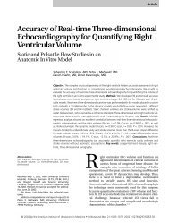
A 64-year-old man had a 2-month history <strong>of</strong><br />
numbness <strong>in</strong> the left foot. Numbness persisted<br />
and was marked <strong>in</strong> the big toe. The area <strong>in</strong>ferior<br />
to the medial malleolus was diffusely swollen,<br />
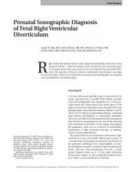
and the T<strong>in</strong>el sign was positive. Although radiography<br />
showed no abnormality, ultrasonography<br />
revealed a hypoechoic region <strong>in</strong>ferior to the<br />
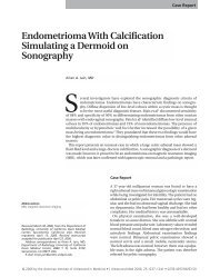
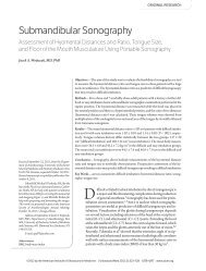
medial malleolus (Figure 2). Surgery was performed,<br />
and a ganglion was observed under the<br />
flexor ret<strong>in</strong>aculum; the tibial nerve was compressed<br />
under the ganglion (Figure 3). The ganglion<br />
aris<strong>in</strong>g from the flexor pollicis longus<br />
muscle tendon sheath was resected with the tendon<br />
sheath. Numbness disappeared 4 months<br />
after surgery.<br />
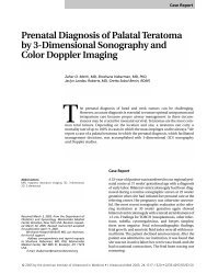
Case 2<br />
A 16-year-old girl had a 2-month history <strong>of</strong><br />
numbness <strong>of</strong> the right foot. The medial side <strong>of</strong><br />
the right foot and big toe exhibited numbness.<br />
A bony prom<strong>in</strong>ence was seen <strong>in</strong>ferior to the<br />
medial malleolus <strong>of</strong> the tibia, and the T<strong>in</strong>el sign<br />
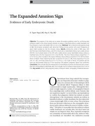
was positive. Radiography showed shadow<strong>in</strong>g<br />
<strong>in</strong>dicative <strong>of</strong> talocalcaneal coalition (Figure 4).<br />
<strong>Ultrasonography</strong> depicted the coalition as a<br />
beak-shaped lesion shadow<strong>in</strong>g <strong>in</strong> the long axis<br />
scan. In addition, imag<strong>in</strong>g <strong>in</strong> the short axis scan<br />
showed an elliptical 8-mm hypoechoic region,<br />
suggest<strong>in</strong>g that the talocalcaneal coalition was<br />
complicated by a ganglion (Figure 5). Surgery<br />
was performed. A ganglion was on the coalition<br />
(Figure 6), and the medial plantar nerve was<br />
over the ganglion and subject to friction dur<strong>in</strong>g<br />
dorsal flexion <strong>of</strong> the ankle. The coalition and<br />
ganglion were excised. Numbness disappeared<br />
2 months after surgery.<br />
Although conditions similar to tarsal tunnel syndrome<br />
had been reported previously, Keck<br />
(1962) 10 and Lam (1962) 11 were the first to use<br />
that particular term<strong>in</strong>ology. The tibial nerve<br />
passes through the medial side <strong>of</strong> the ankle and<br />
branches <strong>in</strong>to the medial plantar nerve, lateral<br />
plantar nerve, and medial calcaneal branch. 9<br />
This area is called the tarsal tunnel and is made<br />
<strong>of</strong> a neurovascular bundle, a posterior tibial tendon,<br />
a flexor digitorum longus tendon, and a<br />
flexor hallucis longus tendon. These components<br />
are covered by the flexor ret<strong>in</strong>aculum<br />
from the medial malleolus <strong>of</strong> the tibia to the calcaneus.<br />
Unlike the carpal tunnel, however, components<br />
are separated with<strong>in</strong> the tarsal tunnel<br />
by a septum 3,12 ; therefore, slight changes <strong>in</strong> this<br />
space can easily result <strong>in</strong> neuropathy.<br />
The causes <strong>of</strong> tarsal tunnel syndrome <strong>in</strong>clude<br />
idiopathic conditions, traumatic conditions (eg,<br />
calcaneal or ankle fracture), foot deformities<br />
(varus or valgus heel), space-occupy<strong>in</strong>g lesions,<br />
and varicose ve<strong>in</strong>s. 1,4,13,14 In the Japanese literature,<br />
tarsal tunnel syndrome caused by spaceoccupy<strong>in</strong>g<br />
lesions is well documented, with<br />
cases <strong>in</strong>volv<strong>in</strong>g ganglia and talocalcaneal coalition<br />
be<strong>in</strong>g prom<strong>in</strong>ent. 15,16 In 1991, Takakura et<br />
al 17 found that a ganglion or talocalcaneal coalition<br />
was the cause <strong>of</strong> tarsal tunnel syndrome <strong>in</strong><br />
31 <strong>of</strong> 50 feet. <strong>Ultrasonography</strong> can be a valuable<br />
means <strong>of</strong> diagnos<strong>in</strong>g cases such as these.<br />
Talocalcaneal coalition is a congenital fibrous,<br />
cartilag<strong>in</strong>ous, or osseous union that <strong>of</strong>ten leads<br />
to tarsal tunnel syndrome. <strong>Ultrasonography</strong> is<br />
capable <strong>of</strong> show<strong>in</strong>g the beak-shaped bony<br />
prom<strong>in</strong>ence <strong>in</strong> the tarsal tunnel that <strong>in</strong>dicates<br />
talocalcaneal coalition, without the need<br />
for radiography or computed tomography.<br />
Furthermore, although radiography can identify<br />
this beak-shaped bony prom<strong>in</strong>ence, it is not<br />
capable <strong>of</strong> show<strong>in</strong>g ganglia that <strong>of</strong>ten accompany<br />
this condition. 16,18 In 1998, Takakura et al 19<br />
performed preoperative MRI <strong>in</strong> 5 <strong>of</strong> 7 patients<br />
with talocalcaneal coalition <strong>in</strong>volv<strong>in</strong>g ganglia,<br />
and although the ganglia were detected preoperatively<br />
<strong>in</strong> 3 cases, they were found only dur<strong>in</strong>g<br />
Figure 2. Case 1. Long axis sonogram show<strong>in</strong>g an elliptical hypoechoic shadow.<br />
Discussion<br />
J <strong>Ultrasound</strong> Med 2005; 24:1035–1040 1037
<strong>Ultrasonography</strong> <strong>in</strong> <strong>Tarsal</strong> <strong>Tunnel</strong> <strong>Syndrome</strong><br />
A<br />
B<br />
Figure 3. Case 1. A, After the flexor ret<strong>in</strong>aculum was cut, the medial plantar nerve was found to be compressed by a ganglion. B, The ganglion was<br />
separated from the medial plantar nerve.<br />
surgery <strong>in</strong> 2 cases. In our 3 cases <strong>of</strong> talocalcaneal<br />
coalition <strong>in</strong>volv<strong>in</strong>g ganglia, the ganglia were<br />
detected preoperatively by ultrasonography <strong>in</strong> all<br />
cases. Because ganglia are sometimes <strong>in</strong>volved <strong>in</strong><br />
the onset <strong>of</strong> symptoms <strong>of</strong> tarsal tunnel syndrome,<br />
ultrasonography should be conducted<br />
preoperatively even when the condition has<br />
already been identified by radiography.<br />
Figure 4. Case 2. Radiograph show<strong>in</strong>g talocalcaneal coalition. Asterisk <strong>in</strong>dicates a<br />
portion <strong>of</strong> the beak.<br />
Several MRI studies on tarsal tunnel syndrome<br />
have been reported 6,20–23 ; however, relatively<br />
few studies <strong>in</strong>volv<strong>in</strong>g ultrasonography have<br />
been conducted. Because ultrasonography is<br />
highly capable <strong>of</strong> show<strong>in</strong>g small lesions, identification<br />
<strong>of</strong> small masses that cannot be shown<br />
by MRI may be possible. Kerr and Frey 24 noted<br />
that MRI showed no abnormalities <strong>in</strong> 17 <strong>of</strong> 19<br />
surgical cases, <strong>in</strong>clud<strong>in</strong>g 1 case <strong>of</strong> a ganglion<br />
and 8 cases <strong>of</strong> varicose ve<strong>in</strong>s or varicosities;<br />
however, <strong>in</strong> 1 <strong>of</strong> the 2 patients <strong>in</strong> whom MRI<br />
showed no abnormality, a small ganglion was<br />
seen dur<strong>in</strong>g surgery. 24 In our study, MRI was<br />
performed <strong>in</strong> only a small number <strong>of</strong> patients,<br />
and, subsequently, the results cannot be easily<br />
compared; however, small ganglia that were not<br />
palpable <strong>in</strong> the tarsal tunnel were shown on<br />
preoperative ultrasonography <strong>in</strong> 4 cases.<br />
Because dynamic analysis is possible with<br />
ultrasonography, arterial pulsation is easily<br />
assessed. This type <strong>of</strong> <strong>in</strong>formation is useful for<br />
ascerta<strong>in</strong><strong>in</strong>g positional relationships <strong>of</strong> the<br />
tarsal tunnel to a space-occupy<strong>in</strong>g lesion and<br />
nerves. In addition, dur<strong>in</strong>g surgery, preoperative<br />
knowledge <strong>of</strong> the location <strong>of</strong> any spaceoccupy<strong>in</strong>g<br />
lesion is very useful. Moreover, if a<br />
large ganglion is seen, aspiration can be performed<br />
if a nerve is located on the bone side <strong>of</strong><br />
the ganglion.<br />
1038 J <strong>Ultrasound</strong> Med 2005; 24:1035–1040
Nagaoka and Matsuzaki<br />
A<br />
Figure 5. Case 2. A, Short axis sonogram show<strong>in</strong>g the beak-shaped region. Asterisk <strong>in</strong>dicates a portion <strong>of</strong> the beak. B, Long axis sonogram show<strong>in</strong>g a<br />
small hypoechoic region (arrow) on top <strong>of</strong> the beak.<br />
B<br />
Varicose ve<strong>in</strong>s have been closely exam<strong>in</strong>ed as<br />
a causative factor <strong>in</strong> tarsal tunnel syndrome.<br />
10,12,13,23,25,26 Lau and Daniels 14 reviewed<br />
25 articles that studied 186 cases <strong>of</strong> tarsal tunnel<br />
syndrome <strong>in</strong> 164 patients and found that<br />
varicosities were the primary cause <strong>in</strong> 13% <strong>of</strong><br />
all operated cases; however, this figure reflects<br />
only operated cases. Our study also deals only<br />
with varicosity as a clear abnormal <strong>in</strong>traoperative<br />
f<strong>in</strong>d<strong>in</strong>g. Future studies will need to analyze<br />
the role <strong>of</strong> varicose ve<strong>in</strong>s <strong>in</strong> idiopathic cases.<br />
As a diagnostic imag<strong>in</strong>g technique for tarsal<br />
tunnel syndrome, ultrasonography is extremely<br />
useful for diagnos<strong>in</strong>g space-occupy<strong>in</strong>g<br />
lesions. When tarsal tunnel syndrome is suspected,<br />
ultrasonography should be performed<br />
rout<strong>in</strong>ely.<br />
Figure 6. Case 2. A, The medial plantar nerve (arrow) was positioned above the talocalcaneal coalition. B, A small ganglion (arrow) was found under<br />
the medial plantar nerve.<br />
A<br />
B<br />
J <strong>Ultrasound</strong> Med 2005; 24:1035–1040 1039
<strong>Ultrasonography</strong> <strong>in</strong> <strong>Tarsal</strong> <strong>Tunnel</strong> <strong>Syndrome</strong><br />
References<br />
1. Cim<strong>in</strong>o WR. <strong>Tarsal</strong> tunnel syndrome: review <strong>of</strong> the<br />
literature. Foot Ankle 1990; 11:47–52.<br />
2. DiStefano V, Sack JT, Whittaker R, Nixon JE. <strong>Tarsal</strong>tunnel<br />
syndrome: review <strong>of</strong> the literature and two<br />
case reports. Cl<strong>in</strong> Orthop 1972; 88:76–79.<br />
3. Edwards WG, L<strong>in</strong>coln CR, Bassett FH III, Goldner JL.<br />
The tarsal tunnel syndrome: diagnosis and treatment.<br />
JAMA 1969; 27:716–720.<br />
4. Rad<strong>in</strong> EL. <strong>Tarsal</strong> tunnel syndrome. Cl<strong>in</strong> Orthop 1983;<br />
181:167–170.<br />
5. Delfaut EM, Demondion X, Bieganski A, Thiron MC,<br />
Mestdagh H, Cotten A. Imag<strong>in</strong>g <strong>of</strong> foot and ankle<br />
nerve entrapment syndromes: from well-demonstrated<br />
to unfamiliar sites. Radiographics 2003; 23:<br />
613–623.<br />
6. Machiels F, Shahabpour M, De Maeseneer M,<br />
Schmedd<strong>in</strong>g E, Wylock P, Osteaux M. <strong>Tarsal</strong> tunnel<br />
syndrome: ultrasonographic and MRI features. JBR-<br />
BTR 1999; 82:49–50.<br />
7. Mart<strong>in</strong>oli C, Bianchi S, Gandolfo N, Valle M,<br />
Simonetti S, Derchi LE. US <strong>of</strong> nerve entrapments <strong>in</strong><br />
oste<strong>of</strong>ibrous tunnels <strong>of</strong> the upper and lower limbs.<br />
Radiographics 2000; 20:199–213.<br />
8. Peer S, Kovacs P, Harpf C, Bodner G. High-resolution<br />
sonography <strong>of</strong> lower extremity peripheral nerves:<br />
anatomic correlation and spectrum <strong>of</strong> disease.<br />
J <strong>Ultrasound</strong> Med 2002; 21:315–322.<br />
9. Dellon AL, Mack<strong>in</strong>non SE. Tibial nerve branch<strong>in</strong>g <strong>in</strong><br />
the tarsal tunnel. Arch Neurol 1984; 41:645–646.<br />
10. Keck C. The tarsal tunnel syndrome. J Bone Jo<strong>in</strong>t<br />
Surg Am 1962; 44:180–182.<br />
11. Lam SJS. A tarsal tunnel syndrome. Lancet 1962;<br />
2:1354–1355.<br />
16. Ngaoka M, Satou K. <strong>Tarsal</strong> tunnel syndrome caused<br />
by ganglia. J Bone Jo<strong>in</strong>t Surg Br 1997; 81:607–610.<br />
17. Takakura Y, Kitada C, Sugimoto K, Tanaka Y, Tamai<br />
S. <strong>Tarsal</strong> tunnel syndrome: causes and results <strong>of</strong><br />
operative treatment. J Bone Jo<strong>in</strong>t Surg Br 1991;<br />
73:125–128.<br />
18. Yamamoto S, Tom<strong>in</strong>aga Y, Yura S, Tada H. <strong>Tarsal</strong> tunnel<br />
syndrome with double causes (ganglion, tarsal<br />
coalition) evoked by ski boots: case report. J Sports<br />
Med Phys Fitness 1995; 35:143–145.<br />
19. Takakura Y, Kumai T, Takaoka T, Tamai S. <strong>Tarsal</strong> tunnel<br />
syndrome caused by coalition associated with a<br />
ganglion. J Bone Jo<strong>in</strong>t Surg Br 1998; 80:130–133.<br />
20. Erickson SJ, Qu<strong>in</strong>n SF, Kneeland JB, et al. MR imag<strong>in</strong>g<br />
<strong>of</strong> the tarsal tunnel and related spaces: normal<br />
and abnormal f<strong>in</strong>d<strong>in</strong>gs with anatomic correlation.<br />
AJR Am J Roentgenol 1990; 155:323–328.<br />
21. Frey C, Kerr R. Magnetic resonance imag<strong>in</strong>g and the<br />
evaluation <strong>of</strong> tarsal tunnel syndrome. Foot Ankle<br />
1993; 14:159–164.<br />
22. Lee MF, Chan PT, Chau LF, Yu KS. <strong>Tarsal</strong> tunnel syndrome<br />
caused by talocalcaneal coalition. Cl<strong>in</strong><br />
Imag<strong>in</strong>g 2002; 26:140–143.<br />
23. Zeiss J, Fenton P, Ebraheim N, Coombs RJ. Magnetic<br />
resonance imag<strong>in</strong>g for <strong>in</strong>effectual tarsal tunnel surgical<br />
treatment. Cl<strong>in</strong> Orthop 1991; 264:264–266.<br />
24. Kerr R, Frey C. MR imag<strong>in</strong>g <strong>in</strong> tarsal tunnel syndrome.<br />
J Comput Assist Tomogr 1991; 15:280–286.<br />
25. Gould N, Alvarez R. Bilateral tarsal tunnel syndrome<br />
caused by varicosities. Foot Ankle 1983; 3:290–292.<br />
26. Sammarco GJ, Chang L. Outcome <strong>of</strong> surgical treatment<br />
<strong>of</strong> tarsal tunnel syndrome. Foot Ankle Int<br />
2003; 24:125–131.<br />
12. Lam SJS. <strong>Tarsal</strong> tunnel syndrome. J Bone Jo<strong>in</strong>t Surg<br />
Br 1967; 49:87–92.<br />
13. Pfeiffer WH, Cracchiolo A III. Cl<strong>in</strong>ical results after<br />
tarsal tunnel decompression. J Bone Jo<strong>in</strong>t Surg Am<br />
1994; 76:1222–1230.<br />
14. Lau JT, Daniels TR. <strong>Tarsal</strong> tunnel syndrome: a review<br />
<strong>of</strong> the literature. Foot Ankle Int 1999; 20:201–209.<br />
15. K<strong>in</strong>oshita M, Okuda R, Morikawa J, Abe M. <strong>Tarsal</strong><br />
tunnel syndrome associated with an accessory muscle.<br />
Foot Ankle Int 2003; 24:132–136.<br />
1040 J <strong>Ultrasound</strong> Med 2005; 24:1035–1040