Elevated Renal Rind - Journal of Ultrasound in Medicine
Elevated Renal Rind - Journal of Ultrasound in Medicine
Elevated Renal Rind - Journal of Ultrasound in Medicine

Create successful ePaper yourself
Turn your PDF publications into a flip-book with our unique Google optimized e-Paper software.
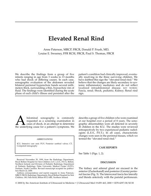
<strong>Elevated</strong> <strong>Renal</strong> <strong>R<strong>in</strong>d</strong><br />
Anne Paterson, MRCP, FRCR, Donald P. Frush, MD,<br />
Louise E. Sweeney, FFR RCSI, FRCR, Paul S. Thomas, FRCR<br />
We describe the f<strong>in</strong>d<strong>in</strong>gs from a group <strong>of</strong> five<br />
<strong>in</strong>fants rang<strong>in</strong>g <strong>in</strong> age from 3 weeks to 13 months<br />
who had shock <strong>of</strong> differ<strong>in</strong>g causes. In each case,<br />
sonographic evaluation <strong>of</strong> the abdomen revealed<br />
bilateral perirenal hyperechoic bands several millimeters<br />
thick, surround<strong>in</strong>g a th<strong>in</strong>, hypoechoic rim <strong>of</strong><br />
fluid. The f<strong>in</strong>d<strong>in</strong>gs were identified dur<strong>in</strong>g the acute<br />
phase <strong>of</strong> each child’s illness and persisted after the<br />
patient’s condition had cl<strong>in</strong>ically improved, eventually<br />
resolv<strong>in</strong>g <strong>in</strong> the three surviv<strong>in</strong>g children. We<br />
have dubbed this sign the “elevated renal r<strong>in</strong>d.” We<br />
believe that the changes are likely secondary to systemic<br />
<strong>in</strong>flammatory mediators and do not reflect<br />
localized <strong>in</strong>traabdom<strong>in</strong>al disease. KEY WORDS:<br />
Fascia, renal; Shock, pediatric; Kidney; <strong>Renal</strong> r<strong>in</strong>d<br />
sign.<br />
Abdom<strong>in</strong>al sonography is commonly<br />
requested as a screen<strong>in</strong>g exam<strong>in</strong>ation <strong>in</strong><br />
cases <strong>of</strong> shock, <strong>in</strong> an attempt to determ<strong>in</strong>e<br />
the underly<strong>in</strong>g cause for a patient’s symptoms. We<br />
ABBREVIATIONS<br />
ICU, Intensive care unit; PUV, Posterior urethral valves; CT,<br />
Computed tomography<br />
describe a group <strong>of</strong> five children who were exam<strong>in</strong>ed<br />
at our hospital over a period <strong>of</strong> 8 years. The sonographic<br />
abnormalities were all detected <strong>in</strong> severely<br />
ill children <strong>in</strong> the ICU. The studies were reviewed<br />
retrospectively by two experienced pediatric radiologists<br />
(L.E.S., P.S.T.). In all cases, characteristic<br />
changes were seen <strong>in</strong> the perirenal tissues, which we<br />
termed the “elevated renal r<strong>in</strong>d.”<br />
CASE REPORTS<br />
See Table 1 (Figs. 1, 2).<br />
Received November 29, 1999, from the Radiology Department,<br />
Royal Belfast Hospital for Sick Children (A.P., L.E.S., P.S.T.), Belfast,<br />
Northern Ireland; and Division <strong>of</strong> Pediatric Radiology, Department<br />
<strong>of</strong> Pediatric Radiology, Duke University Medical Center (D.P.F.),<br />
Durham, North Carol<strong>in</strong>a. Revised manuscript accepted for publication<br />
March 22, 2000.<br />
Address correspondence and repr<strong>in</strong>t requests to Anne Paterson,<br />
MRCP, FRCR, Radiology Department, Royal Belfast Hospital for Sick<br />
Children, 180, Falls Road, Belfast BT12 6BE, Northern Ireland.<br />
DISCUSSION<br />
The kidney and adrenal gland are encased <strong>in</strong> the<br />
anterior (Zuckerkandl) and posterior (Gerota) perirenal<br />
fasciae (Fig. 3). The lateroconal fascia lies laterally<br />
and blends anteriorly with the parietal peritoneum.<br />
© 2000 by the American Institute <strong>of</strong> <strong>Ultrasound</strong> <strong>in</strong> Medic<strong>in</strong>e • J <strong>Ultrasound</strong> Med 19:459–463, 2000 • 0278-4297/00/$3.50
460 ELEVATED RENAL RIND J <strong>Ultrasound</strong> Med 19:459–463, 2000<br />
Table 1: Data and Outcome <strong>of</strong> Five Cases <strong>of</strong> <strong>Elevated</strong> <strong>Renal</strong> <strong>R<strong>in</strong>d</strong><br />
Underly<strong>in</strong>g Maximum<br />
Sex/ Cause <strong>of</strong> Organisms Associated Creat<strong>in</strong><strong>in</strong>e Sonographic<br />
Age Shock Cultured Cl<strong>in</strong>ical Problems Level F<strong>in</strong>d<strong>in</strong>gs Outcome<br />
Case 1 M, 17 days Necrotiz<strong>in</strong>g Staphylococcus, Prematurity, symmetrical 152 m mol/l Bilateral pelviectasis, Death at 23 days due to<br />
(born at enterocolitis enterococcus <strong>in</strong>trauter<strong>in</strong>e growth elevated renal r<strong>in</strong>d cardiac failure<br />
33 weeks’ retardation, unbalanced<br />
gestation) atrioventricular canal defect<br />
Case 2 M, 28 days Men<strong>in</strong>gitis and Escherichia coli Posterior urethral valves 194 m mol/l Bilateral pelviectasis, Survival with chronic<br />
ur<strong>in</strong>ary tract elevated renal r<strong>in</strong>d renal impairment;<br />
<strong>in</strong>fection sonogram after 8 days<br />
was unchanged; long-term<br />
sonographic follow-up shows<br />
hyperechoic kidneys with<br />
normal perirenal tissues<br />
Case 3 M, 12 weeks Pneumonia None Apneic episode 98 m mol/l M<strong>in</strong>imal pelviectasis Survival with pr<strong>of</strong>ound<br />
(presumed with pelvic urothelial motor and developmental<br />
aspiration) thicken<strong>in</strong>g, elevated deficit; sonogram at 1 year<br />
renal r<strong>in</strong>d (Fig. 1) <strong>of</strong> age showed normal kidneys<br />
Case 4 F, 13 months Infection with Group A None 37 m mol/l M<strong>in</strong>imal pelviectasis, Survived; sonography at<br />
erythematous, streptococci elevated renal r<strong>in</strong>d follow-up evaluation showed<br />
maculopapular (Fig. 2) normal kidneys<br />
rash<br />
Case 5 M, 12 weeks Pneumonia Escherichia coli Prematurity, trisomy 21, 122 m mol/l Enlarged kidneys <strong>Renal</strong> function recovered<br />
(born at unbalanced atrioventricular with loss <strong>of</strong> cortico- after 4 weeks; sonography<br />
27 weeks’ canal defect, necrotiz<strong>in</strong>g medullary differentiation, unchanged at that time,<br />
gestation) enterocolitis with bowel elevated renal r<strong>in</strong>d but <strong>in</strong>fant died aged<br />
resection and ileostomy at 17 weeks due to cardiac<br />
4 weeks, bronchopulmonary failure and chronic lung<br />
dysplasia disease
J <strong>Ultrasound</strong> Med 19:459–463, 2000<br />
PATERSON ET AL<br />
461<br />
The perirenal space conta<strong>in</strong>s fat and is bounded by the<br />
perirenal fasciae. The anterior pararenal space separates<br />
the posterior aspect <strong>of</strong> the parietal peritoneum<br />
from the anterior perirenal fascia. It is cont<strong>in</strong>uous<br />
across the midl<strong>in</strong>e and conta<strong>in</strong>s the pancreas, duodenum,<br />
and ascend<strong>in</strong>g and descend<strong>in</strong>g colons. The posterior<br />
pararenal space is formed anteriorly by the<br />
posterior perirenal fascia and posteriorly by the transverse<br />
fascia; it conta<strong>in</strong>s no organs.<br />
Us<strong>in</strong>g CT, thicken<strong>in</strong>g <strong>of</strong> the perirenal fascia has<br />
been described <strong>in</strong> adults as a nonspecific <strong>in</strong>dicator <strong>of</strong><br />
<strong>in</strong>traabdom<strong>in</strong>al sepsis or neoplasia. 1,2 The normal<br />
perirenal fascia cannot be visualized at sonography,<br />
but two studies have measured the “anterior<br />
extrarenal space” (the sum <strong>of</strong> the anterior perirenal<br />
plus pararenal spaces) <strong>in</strong> adults, and the authors have<br />
concluded that this space <strong>in</strong>creases <strong>in</strong> width and<br />
echogenicity <strong>in</strong> association with acute, localized<br />
abdom<strong>in</strong>al <strong>in</strong>flammation, trauma, and malignancy. 3,4<br />
Belli and Joseph 3 co<strong>in</strong>ed the term “renal r<strong>in</strong>d sign”as<br />
a descriptor for this sonographic f<strong>in</strong>d<strong>in</strong>g. In addition,<br />
an <strong>in</strong>crease <strong>in</strong> the width but not the echogenicity <strong>of</strong><br />
the so-called anterior extrarenal space was noted with<br />
chronic, local <strong>in</strong>flammatory disease and <strong>in</strong> cush<strong>in</strong>goid<br />
patients. 4 In both <strong>of</strong> these studies, 3,4 the authors<br />
concluded that a careful search for <strong>in</strong>traabdom<strong>in</strong>al<br />
pathologic lesions should be conducted if the renal<br />
r<strong>in</strong>d sign was present.<br />
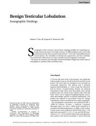
Figure 1 Case 3. A,B, Longitud<strong>in</strong>al and transverse sonograms <strong>of</strong> the right kidney dur<strong>in</strong>g the acute phase <strong>of</strong> the illness. A per<strong>in</strong>ephric<br />
hypoechoic rim with surround<strong>in</strong>g hyperechoic band is evident. C, Longitud<strong>in</strong>al sonogram <strong>of</strong> the left kidney dur<strong>in</strong>g the<br />
acute illness. D, Longitud<strong>in</strong>al sonogram <strong>of</strong> the right kidney at 10 months <strong>of</strong> age shows normal perirenal tissues.<br />
A<br />
B<br />
C<br />
D
462 ELEVATED RENAL RIND J <strong>Ultrasound</strong> Med 19:459–463, 2000<br />
A<br />
B<br />
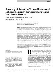
Figure 2 Case 4. Longitud<strong>in</strong>al (A) and transverse (B) sonograms <strong>of</strong> the right kidney dur<strong>in</strong>g the acute febrile illness. The perirenal<br />
hypoechoic rim and hyperechoic band are aga<strong>in</strong> present.<br />
Review <strong>of</strong> the literature reveals one additional case<br />
report describ<strong>in</strong>g an <strong>in</strong>crease <strong>in</strong> the thickness and<br />
echogenicity <strong>of</strong> the right-sided perirenal fascia at<br />
sonography. 5 The f<strong>in</strong>d<strong>in</strong>gs were observed <strong>in</strong> an<br />
elderly man with global <strong>in</strong>farction <strong>of</strong> the right kidney<br />
associated with aortic dissection. Aga<strong>in</strong>, the<br />
authors believed that such f<strong>in</strong>d<strong>in</strong>gs were specific and<br />
would serve as a useful <strong>in</strong>dicator <strong>of</strong> renal <strong>in</strong>farction<br />
<strong>in</strong> the absence <strong>of</strong> any other sonographic abnormalities.<br />
They documented resolution <strong>of</strong> the perirenal<br />
changes on a follow-up sonogram 5 months later.<br />
Sonographic descriptions <strong>of</strong> perirenal changes are<br />
scarce <strong>in</strong> the pediatric literature. A solitary case<br />
report by Swischuk and Hayden 6 described an<br />
<strong>in</strong>crease <strong>in</strong> the width and echogenicity <strong>of</strong> the right<br />
pararenal space <strong>in</strong> two children with pancreatitis. In<br />
this <strong>in</strong>stance, the authors concluded that the appearance<br />
should serve “as a strongly positive, ancillary<br />
f<strong>in</strong>d<strong>in</strong>g <strong>of</strong> pancreatitis.”<br />
The five patients described <strong>in</strong> this paper were <strong>in</strong><br />
shock and required ionotropic support. The underly<strong>in</strong>g<br />
cause for the shock was different <strong>in</strong> each <strong>of</strong> the<br />
children, although four <strong>of</strong> them had septicemia.<br />
Cl<strong>in</strong>ically, none <strong>of</strong> the patients had primary renal disease,<br />
but four <strong>of</strong> the children developed acute renal<br />
failure dur<strong>in</strong>g the course <strong>of</strong> their illness. Regardless<br />
<strong>of</strong> the underly<strong>in</strong>g cause <strong>of</strong> sepsis, the perirenal sonographic<br />
changes were identical. In all <strong>of</strong> the documented<br />
cases, a th<strong>in</strong> hypoechoic rim (presumed to be<br />
fluid) distended the perirenal space. The thickened,<br />
hyperechoic perirenal fascia was clearly demarcated<br />
adjacent to the hypoechoic perirenal fluid. The<br />
changes were bilateral <strong>in</strong> all cases. One patient with<br />
acute renal failure had associated swollen kidneys<br />
with loss <strong>of</strong> corticomedullary differentiation (case 5).<br />
The rema<strong>in</strong><strong>in</strong>g four <strong>in</strong>fants had mild pelviectasis but<br />
otherwise had a normal renal echotexture. No sonographic<br />
abnormalities were seen <strong>in</strong> the rema<strong>in</strong>der <strong>of</strong><br />
the abdomen or pelvis <strong>in</strong> any <strong>of</strong> the patients.<br />
Specifically, none <strong>of</strong> the <strong>in</strong>fants had associated ascites<br />
or focal signs <strong>of</strong> <strong>in</strong>traabdom<strong>in</strong>al sepsis. The changes<br />
<strong>in</strong> the perirenal tissues were documented to resolve<br />
<strong>in</strong> the three surviv<strong>in</strong>g children at follow-up exam<strong>in</strong>ations.<br />
Autopsy was refused by the parents <strong>of</strong> the<br />
two <strong>in</strong>fants who died.<br />
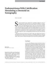
Figure 3 Cross-sectional diagram shows the retroperitoneal<br />
fascial planes. APaS, Anterior pararenal space; Ao, aorta; DC,<br />
descend<strong>in</strong>g colon; GF, Gerota fascia; IVC, <strong>in</strong>ferior vena cava;<br />
LF, lateroconal fascia; LK, left kidney; LV, lumbar vertebra;<br />
PeS, perirenal space; PP, parietal peritoneum; PPaS, posterior<br />
pararenal space; TF, transverse fascia; ZF, Zuckerkandl fascia.
J <strong>Ultrasound</strong> Med 19:459–463, 2000<br />
PATERSON ET AL<br />
463<br />
The elevated renal r<strong>in</strong>d differs therefore from the<br />
descriptions <strong>in</strong> prior reports <strong>of</strong> sonographic perirenal<br />
fascial changes. In the previously described cases, the<br />
thickened perirenal fascia (the renal r<strong>in</strong>d) could not<br />
be seen as separate from, and appeared adherent to,<br />
the renal capsule. In our patients, a hypoechoic<br />
perirenal fluid collection was observed, which elevated<br />
the hyperechoic perirenal fascia. In the children<br />
described here, the changes were bilateral and could<br />
not be attributed directly to localized <strong>in</strong>traabdom<strong>in</strong>al<br />
disease. In addition, resolution <strong>of</strong> the changes was<br />
documented <strong>in</strong> all <strong>of</strong> the surviv<strong>in</strong>g children.<br />
The cause <strong>of</strong> the visualized abnormalities is<br />
unknown. With regard to the renal r<strong>in</strong>d, the<br />
authors 3,4,6 postulated that changes <strong>in</strong> the perirenal<br />
fascia may be due to localized edema, fibrosis, hyperemia,<br />
or lymphatic obstruction. These papers<br />
described localized <strong>in</strong>traabdom<strong>in</strong>al pathologic<br />
changes associated with changes <strong>in</strong> the adjacent<br />
perirenal tissues, and their <strong>in</strong>dividual authors each<br />
believed they were describ<strong>in</strong>g a specific sonographic<br />
sign <strong>of</strong> the disease entity presented. The common<br />
denom<strong>in</strong>ator <strong>in</strong> the current patient population was<br />
shock. In these patients, systemic <strong>in</strong>flammatory<br />
mediators may well be responsible for the bilateral<br />
sonographic changes observed, <strong>in</strong>clud<strong>in</strong>g the transudation<br />
or exudation <strong>of</strong> fluid <strong>in</strong>to the perirenal space.<br />
A per<strong>in</strong>ephric hematoma would also appear acutely<br />
hypoechoic, but its echogenicity would <strong>in</strong>crease over<br />
time as the clot organized. Given that sonograms<br />
obta<strong>in</strong>ed 8 days (case 2) and 4 weeks (case 5) apart<br />
did not document a change <strong>in</strong> the appearance <strong>of</strong> the<br />
hypoechoic perirenal rim, this mechanism is believed<br />
to be unlikely to expla<strong>in</strong> the presented f<strong>in</strong>d<strong>in</strong>gs, especially<br />
given their bilaterality. In addition, none <strong>of</strong> the<br />
patients developed a coagulopathy that would predispose<br />
them to hematoma formation. Similarly, a<br />
per<strong>in</strong>ephric abscess would cause a hypoechoic rim<br />
surround<strong>in</strong>g the kidney, but this is usually secondary<br />
to focal, acute pyelonephritis. Bilateral abscesses<br />
would be extremely unusual. Primary renal disease<br />
was not documented as the underly<strong>in</strong>g cause <strong>of</strong> sepsis<br />
<strong>in</strong> any <strong>of</strong> the patients reported, although one<br />
patient (case 2) later developed a ur<strong>in</strong>ary tract <strong>in</strong>fection<br />
secondary to distal obstruction from PUV. Both<br />
per<strong>in</strong>ephric hematomas and abscesses might reasonably<br />
be expected to cause secondary, local <strong>in</strong>flammatory<br />
changes <strong>in</strong> the adjacent perirenal fascia, thus<br />
mak<strong>in</strong>g it appear thickened and hyperechoic at<br />
sonography.<br />
In summary, we present five children who demonstrate<br />
a sonographic sign we have called the elevated<br />
renal r<strong>in</strong>d. We believe that it may occur secondary to<br />
systemic <strong>in</strong>flammatory mediators with fluid leakage<br />
<strong>in</strong>to the perirenal tissues, although we have not been<br />
able to confirm this pathologically. Importantly, the<br />
elevated renal r<strong>in</strong>d is not an <strong>in</strong>dicator <strong>of</strong> <strong>in</strong>tr<strong>in</strong>sic<br />
renal disease or <strong>of</strong> a localized <strong>in</strong>traabdom<strong>in</strong>al pathologic<br />
condition. Rather, it is seen <strong>in</strong> constitutionally<br />
ill patients with shock, and further imag<strong>in</strong>g evaluation<br />
is not required when it is demonstrated. The<br />
patient’s prognosis depends ultimately on the treatment<br />
<strong>of</strong> the underly<strong>in</strong>g condition; the perirenal<br />
changes will resolve with time <strong>in</strong> those patients who<br />
survive.<br />
REFERENCES<br />
1. Parienty RA, Pradel J, Picard J-D, et al: Visibility and<br />
thicken<strong>in</strong>g <strong>of</strong> the renal fascia on computed tomograms.<br />
Radiology 139:119, 1981<br />
2. Nicholson RL: Abnormalities <strong>of</strong> the per<strong>in</strong>ephric fascia<br />
and fat <strong>in</strong> pancreatitis. Radiology 139:125, 1981<br />
3. Belli A-M, Joseph AEA: The renal r<strong>in</strong>d sign: A new ultrasound<br />
<strong>in</strong>dication <strong>of</strong> <strong>in</strong>flammatory disease <strong>in</strong> the<br />
abdomen. Br J Radiol 61:806, 1988<br />
4. Chen J-J, Changchien C-S, Kuo C-H: Causes <strong>of</strong> <strong>in</strong>creas<strong>in</strong>g<br />
width <strong>of</strong> right extrarenal space seen <strong>in</strong> ultrasonographic<br />
exam<strong>in</strong>ations. J Cl<strong>in</strong> <strong>Ultrasound</strong> 23:287, 1995<br />
5. Kodama K, Matsuura K, Yamashita R, et al: Case report:<br />
New sonographic f<strong>in</strong>d<strong>in</strong>g <strong>in</strong> renal <strong>in</strong>farction. Br J Radiol<br />
67:499, 1994<br />
6. Swischuk LE, Hayden CK: Pararenal space hyperechogenicity<br />
<strong>in</strong> childhood pancreatitis. AJR 145:1085,<br />
1985