Doppler Criteria for Identifying Proximal Vertebral Artery Stenosis of ...
Doppler Criteria for Identifying Proximal Vertebral Artery Stenosis of ...
Doppler Criteria for Identifying Proximal Vertebral Artery Stenosis of ...

Create successful ePaper yourself
Turn your PDF publications into a flip-book with our unique Google optimized e-Paper software.
ORIGINAL RESEARCH<br />
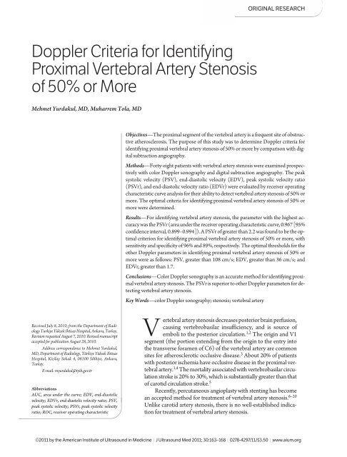
<strong>Doppler</strong> <strong>Criteria</strong> <strong>for</strong> <strong>Identifying</strong><br />
<strong>Proximal</strong> <strong>Vertebral</strong> <strong>Artery</strong> <strong>Stenosis</strong><br />
<strong>of</strong> 50% or More<br />
Mehmet Yurdakul, MD, Muharrem Tola, MD<br />
Objectives—The proximal segment <strong>of</strong> the vertebral artery is a frequent site <strong>of</strong> obstructive<br />
atherosclerosis. The purpose <strong>of</strong> this study was to determine <strong>Doppler</strong> criteria <strong>for</strong><br />
identifying proximal vertebral artery stenosis <strong>of</strong> 50% or more by comparison with digital<br />
subtraction angiography.<br />
Methods—Forty-eight patients with vertebral artery stenosis were examined prospectively<br />
with color <strong>Doppler</strong> sonography and digital subtraction angiography. The peak<br />
systolic velocity (PSV), end-diastolic velocity (EDV), peak systolic velocity ratio<br />
(PSVr), and end-diastolic velocity ratio (EDVr) were evaluated by receiver operating<br />
characteristic curve analysis <strong>for</strong> their ability to detect vertebral artery stenosis <strong>of</strong> 50% or<br />
more. The optimal criteria <strong>for</strong> identifying proximal vertebral artery stenosis <strong>of</strong> 50% or<br />
more were determined.<br />
Results—For identifying vertebral artery stenosis, the parameter with the highest accuracy<br />
was the PSVr (area under the receiver operating characteristic curve, 0.967 [95%<br />
confidence interval, 0.899–0.994]). A PSVr <strong>of</strong> greater than 2.2 was found to be the optimal<br />
criterion <strong>for</strong> identifying proximal vertebral artery stenosis <strong>of</strong> 50% or more, with<br />
sensitivity and specificity <strong>of</strong> 96% and 89%, respectively. The optimal thresholds <strong>for</strong> the<br />
other <strong>Doppler</strong> parameters in identifying proximal vertebral artery stenosis <strong>of</strong> 50% or<br />
more were as follows: PSV, greater than 108 cm/s; EDV, greater than 36 cm/s; and<br />
EDVr, greater than 1.7.<br />
Conclusions—Color <strong>Doppler</strong> sonography is an accurate method <strong>for</strong> identifying proximal<br />
vertebral artery stenosis. The PSVr is superior to other <strong>Doppler</strong> parameters <strong>for</strong> detecting<br />
vertebral artery stenosis.<br />
Key Words—color <strong>Doppler</strong> sonography; stenosis; vertebral artery<br />
Received July 8, 2010, from the Department <strong>of</strong> Radiology<br />
Turkiye Yüksek Ihtisas Hospital, Ankara, Turkey.<br />
Revision requested August 7, 2010. Revised manuscript<br />
accepted <strong>for</strong> publication August 28, 2010.<br />
Address correspondence to Mehmet Yurdakul,<br />
MD, Department <strong>of</strong> Radiology, Türkiye Yüksek Ihtisas<br />
Hospital, Kizilay Sokak 4, 06100 Sihhiye, Ankara,<br />
Turkey.<br />
E-mail: myurdakul@tyih.gov.tr<br />
Abbreviations<br />
AUC, area under the curve; EDV, end- diastolic<br />
velocity; EDVr, end-diastolic velocity ratio; PSV,<br />
peak systolic velocity; PSVr, peak systolic velocity<br />
ratio; ROC, receiver operating characteristic<br />
<strong>Vertebral</strong> artery stenosis decreases posterior brain perfusion,<br />
causing vertebrobasilar insufficiency, and is source <strong>of</strong><br />
emboli to the posterior circulation. 1,2 The origin and V1<br />
segment (the portion extending from the origin to the entry into<br />
the transverse <strong>for</strong>amen <strong>of</strong> C6) <strong>of</strong> the vertebral artery are common<br />
sites <strong>for</strong> atherosclerotic occlusive disease. 3 About 20% <strong>of</strong> patients<br />
with posterior ischemia have occlusive disease in the proximal vertebral<br />
artery. 1,4 The mortality associated with vertebrobasilar circulation<br />
stroke is 20% to 30%, which is substantially greater than that<br />
<strong>of</strong> carotid circulation stroke. 5<br />
Recently, percutaneous angioplasty with stenting has become<br />
an accepted method <strong>for</strong> treatment <strong>of</strong> vertebral artery stenosis. 6–20<br />
Unlike carotid artery stenosis, there is no well-established indication<br />
<strong>for</strong> treatment <strong>of</strong> vertebral artery stenosis.<br />
©2011 by the American Institute <strong>of</strong> Ultrasound in Medicine | J Ultrasound Med 2011; 30:163–168 | 0278-4297/11/$3.50 | www.aium.org
Yurdakul and Tola—<strong>Doppler</strong> <strong>Criteria</strong> <strong>for</strong> <strong>Proximal</strong> <strong>Vertebral</strong> <strong>Artery</strong> <strong>Stenosis</strong><br />
Color <strong>Doppler</strong> sonography, which is a noninvasive<br />
and cost-effective method, is a suitable screening test <strong>for</strong><br />
proximal vertebral artery stenosis. However, very few studies<br />
have been per<strong>for</strong>med to determine <strong>Doppler</strong> criteria <strong>for</strong><br />
identifying proximal vertebral artery stenosis. 21,22 The purpose<br />
<strong>of</strong> this study was to determine <strong>Doppler</strong> criteria <strong>for</strong><br />
identifying proximal vertebral artery stenosis <strong>of</strong> 50% or<br />
more by comparison with digital subtraction angiography.<br />
Materials and Methods<br />
From June 2008 to December 2009, 48 consecutive patients<br />
(28 men and 20 women; age range, 43–88 years;<br />
mean age, 64.7 years) were referred by the cardiovascular<br />
surgery clinic <strong>for</strong> digital subtraction angiography, which<br />
showed vertebral artery stenosis <strong>of</strong> 50% or more. These<br />
patients were examined by color <strong>Doppler</strong> sonography after<br />
digital subtraction angiography. Patients were excluded<br />
from this study if they had any <strong>of</strong> the following: (1) previous<br />
surgery or stenting, (2) calcification extensive enough<br />
to obscure the ultrasound signal intensity in the stenotic<br />
area, (3) an aortic origin <strong>of</strong> the vertebral artery, (4) a hypoplastic<br />
(diameter 50%). All patients gave their oral in<strong>for</strong>med consent,<br />
and the Institutional Review Board approved the<br />
study.<br />
Digital subtraction angiography was per<strong>for</strong>med with<br />
an Integris Allura system (Philips Healthcare, Best, the<br />
Netherlands). All catheterizations were per<strong>for</strong>med by a<br />
transfemoral approach with standard diagnostic catheters.<br />
After aortic arch injection, selective supra-aortic (carotid<br />
and subclavian) artery injections were per<strong>for</strong>med. Nonionic<br />
contrast media were used. <strong>Vertebral</strong> artery stenosis<br />
was calculated by comparison with the nearest normal distal<br />
segment.<br />
For color <strong>Doppler</strong> sonography, a LOGIQ 7 system<br />
(GE Healthcare, Tokyo, Japan) was used. All examinations<br />
were per<strong>for</strong>med while the patient was in a supine position<br />
with the head slightly turned to opposite side. First, the<br />
common carotid artery was located in the longitudinal<br />
plane with a 2.5- to 7-MHz linear transducer. The vertebral<br />
artery was then shown between the transverse<br />
processes by posterolateral movement <strong>of</strong> the transducer.<br />
The presence and direction <strong>of</strong> flow were determined, and<br />
a velocity wave<strong>for</strong>m was obtained. The peak systolic velocity<br />
(PSV) and end-diastolic velocity (EDV) <strong>of</strong> blood<br />
flow in the V2 segment <strong>of</strong> the vertebral artery (the portion<br />
extending from the transverse <strong>for</strong>amen <strong>of</strong> C6–C1)<br />
were recorded. The measurements in this segment were<br />
used only as reference measurements <strong>for</strong> velocity ratio<br />
calculations. The vertebral artery was followed downward<br />
to image the origin and V1 segment the vertebral<br />
artery with a 2.5- to 7-MHz linear or 1.5- to 4.5-MHz<br />
convex transducer. A velocity wave<strong>for</strong>m was obtained<br />
routinely from the origin <strong>of</strong> the vertebral artery. The V1<br />
segment was sampled proximally to distally, where color<br />
flow imaging showed areas <strong>of</strong> abnormal flow such as<br />
color aliasing and color bruits. The highest PSV and EDV<br />
<strong>of</strong> blood flow in the V1 segment were recorded. The peak<br />
systolic velocity ratio (PSVr) and end-diastolic velocity<br />
ratio (EDVr) were calculated as the maximum PSV and<br />
EDV <strong>of</strong> the V1 segment divided by the corresponding velocities<br />
<strong>of</strong> the V2 segment. The color <strong>Doppler</strong> sonographic<br />
examinations were per<strong>for</strong>med by the same<br />
experienced radiologist (M.T.), who was unaware <strong>of</strong> clinical<br />
data (digital subtraction angiographic results, any<br />
other imaging data, physical examination results, laboratory<br />
results, and patient history). The insonation angle<br />
was kept at 60° or less.<br />
For statistical analyses, the right and left sides <strong>of</strong> each<br />
patient were accepted as separate cases. The highest PSV<br />
and EDV at the V1 segment and the PSVr and EDVr<br />
were evaluated by receiver operating characteristic<br />
(ROC) curve analysis <strong>for</strong> their ability to detect vertebral<br />
artery stenosis <strong>of</strong> 50% or more. The ROC curves based on<br />
these <strong>Doppler</strong> data were compared by measuring the areas<br />
under the curves (AUCs). The AUCs were compared by<br />
a z test, as described by Hanley and McNeil. 23 For each<br />
<strong>Doppler</strong> parameter, the threshold at which sensitivity and<br />
specificity were optimum was determined. P < .05 was considered<br />
statistically significant.<br />
Results<br />
In our study, 19 vertebral arteries were excluded (1 with<br />
extensive calcification, 1 with an aortic origin, 4 hypoplastic<br />
arteries, 12 with occlusion, and 1 with subclavian artery<br />
occlusion). One right vertebral artery with extensive calcification<br />
and 1 left vertebral artery originating from the aorta<br />
could not be completely visualized by color <strong>Doppler</strong><br />
sonography. In no patient were both vertebral arteries excluded.<br />
All <strong>of</strong> the occlusions were correctly diagnosed by<br />
color <strong>Doppler</strong> sonography. The remaining 77 vertebral arteries<br />
(36 right and 41 left) were included in the statistical<br />
evaluation. Thirty-two patients had single-sided and 16<br />
had double-sided stenosis <strong>of</strong> 50% or more or occlusion.<br />
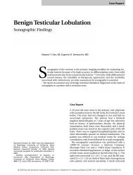
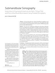
The overall distribution <strong>of</strong> vertebral arteries with respect<br />
to the degree <strong>of</strong> stenosis is shown in Figure 1. Images from<br />
a patient are shown in Figure 2.<br />
164<br />
J Ultrasound Med 2011; 30:163–168
Yurdakul and Tola—<strong>Doppler</strong> <strong>Criteria</strong> <strong>for</strong> <strong>Proximal</strong> <strong>Vertebral</strong> <strong>Artery</strong> <strong>Stenosis</strong><br />
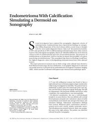
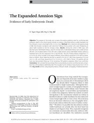
An ROC analysis was per<strong>for</strong>med <strong>for</strong> proximal vertebral<br />
artery stenosis <strong>of</strong> 50% or more based on the <strong>Doppler</strong><br />
velocity parameters, and AUCs <strong>for</strong> the parameters were<br />
calculated and compared (Figure 3 and Tables 1 and 2).<br />
The PSVr was found to be the most accurate parameter<br />
(AUC, 0.967 [95% confidence interval, 0.889–0.994]).<br />
For each parameter, the optimal threshold was determined<br />
by ROC curve analysis (Table 1). The optimal thresholds<br />
<strong>for</strong> identifying stenosis <strong>of</strong> 50% or more were as follows:<br />
PSV, greater than 108 cm/s; EDV, greater than 36 cm/s;<br />
PSVr, greater than 2.2; and EDVr, greater than 1.7. Accuracy<br />
ratios <strong>for</strong> these threshold values are shown in Table 1.<br />
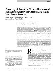
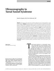
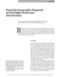
Figure 2. Severe right vertebral artery stenosis in a 70-year-old woman.<br />
A, Angiogram showing 60% stenosis at the origin <strong>of</strong> the right vertebral<br />
artery. B, Color <strong>Doppler</strong> sonogram showing high-velocity flow at the origin<br />
<strong>of</strong> the right vertebral artery with a peak systolic velocity <strong>of</strong> 203 cm/s<br />
and an end-diastolic velocity <strong>of</strong> 60 cm/s. C, Color <strong>Doppler</strong> sonogram<br />
showing a peak systolic velocity <strong>of</strong> 56 cm/s and an end-diastolic velocity<br />
<strong>of</strong> 26 cm/s in the V2 segment <strong>of</strong> the right vertebral artery.<br />
A<br />
Discussion<br />
The vertebral arteries typically arise from the superoposterior<br />
aspect <strong>of</strong> the first part <strong>of</strong> the subclavian artery and are<br />
usually the first branches <strong>of</strong> this vessel. In 6% <strong>of</strong> patients,<br />
they arise directly from the aortic arch between the origin <strong>of</strong><br />
the left common carotid and left subclavian arteries. 24 The<br />
vertebral arteries supply a low-resistance system to the<br />
brainstem and posterior cerebral circulation vessels. They<br />
also make an important contribution to the anterior circulation<br />
in certain circumstances. Both vertebral arteries deliver<br />
approximately 20% <strong>of</strong> the total cerebral blood flow. 25<br />
Atherosclerosis is the dominant nontraumatic condition<br />
seen in the vertebral artery. 26 The most frequent site<br />
<strong>of</strong> involvement is the vertebral artery origin from the subclavian<br />
artery. 3,27 The reference standard <strong>for</strong> detection <strong>of</strong><br />
stenosis <strong>of</strong> craniocervical vessels is still conventional<br />
catheter angiography; however, it is an invasive procedure<br />
associated with a low but definite incidence <strong>of</strong> complications.<br />
28–30 Noninvasive tests, such as color <strong>Doppler</strong><br />
sonography, magnetic resonance angiography, and, most<br />
B<br />
Figure 1. Distribution <strong>of</strong> vertebral artery stenosis according to angiographic<br />
measurements.<br />
C<br />
J Ultrasound Med 2011; 30:163–168<br />
165
Yurdakul and Tola—<strong>Doppler</strong> <strong>Criteria</strong> <strong>for</strong> <strong>Proximal</strong> <strong>Vertebral</strong> <strong>Artery</strong> <strong>Stenosis</strong><br />
Figure 3. Receiver operating characteristic curves <strong>for</strong> detection vertebral<br />
artery stenosis <strong>of</strong> 50% or more based on <strong>Doppler</strong> velocity parameters.<br />
EDV indicates end-diastolic velocity; EDVr, end-diastolic velocity<br />
ratio; PSV, peak systolic velocity; and PSVr, peak systolic velocity ratio.<br />
recently, computed tomographic angiography, have been<br />
used without digital subtraction angiography in screening<br />
<strong>for</strong> craniocervical vessel disease. Although magnetic resonance<br />
angiography and computed tomographic angiography<br />
are valuable imaging methods, these techniques are<br />
expensive and need additional contrast medium administration.<br />
Color <strong>Doppler</strong> sonography is a noninvasive and<br />
inexpensive method used to evaluate craniocervical vessels<br />
that provides anatomic and hemodynamic in<strong>for</strong>mation<br />
about those vessels.<br />
Color <strong>Doppler</strong> sonography is the most appropriate<br />
method <strong>for</strong> diagnosis <strong>of</strong> carotid artery stenosis, but it is not<br />
as appropriate <strong>for</strong> vertebral artery stenosis. It is difficult to<br />
show the proximal vertebral artery because <strong>of</strong> its many<br />
anatomic characteristics, such as a small diameter, a deep<br />
location, tortuosity, and a perpendicular origin from the<br />
subclavian artery. There<strong>for</strong>e, most vascular laboratories<br />
simply determine the presence and direction <strong>of</strong> blood flow<br />
in the midcervical vertebral artery without extensive exploration<br />
<strong>of</strong> the origin and proximal portion. For identifying<br />
proximal vertebral artery stenosis, indirect criteria are<br />
used, such as turbulent flow and spectral broadening at the<br />
origin or markedly reduced pulsatility beyond the area <strong>of</strong><br />
stenosis compared to the contralateral vessel.<br />
The origin and proximal segment <strong>of</strong> the vertebral artery<br />
can be easily identified by using the color mode. 31 In<br />
this study, the V2 segment <strong>of</strong> the vertebral artery was followed<br />
downward with the help <strong>of</strong> the color mode to identify<br />
the V1 segment and origin <strong>of</strong> the vertebral artery.<br />
Our study showed that the PSVr and EDVr had better<br />
diagnostic accuracy than the PSV and EDV, respectively.<br />
The PSV was found to be superior to the EDV, but<br />
the PSVr was the most accurate. The optimal PSVr threshold<br />
<strong>for</strong> identifying vertebral artery stenosis was 2.2, and its<br />
sensitivity and specificity were 96% and 89%, respectively.<br />
Two recently published retrospective studies aimed<br />
to develop <strong>Doppler</strong> criteria <strong>for</strong> identifying vertebral artery<br />
stenosis 21,22 ; however, the absolute velocity criteria obtained<br />
in those studies were different from those <strong>of</strong> our<br />
study. Although the PSVr values were similar, the PSV was<br />
the most accurate parameter in those studies, whereas the<br />
PSVr was the most accurate in our study. The reason <strong>for</strong><br />
this discrepancy may have been that the patient populations<br />
were different: whereas patients with carotid and contralateral<br />
vertebral artery disease were excluded in 1 <strong>of</strong><br />
those studies, our patient population included patients<br />
with occlusive carotid artery disease, and patients with<br />
contralateral vertebral artery disease were not excluded<br />
from our study. As is known, in occlusive carotid artery<br />
and contralateral vertebral artery disease, the collateral<br />
flow that develops from the ipsilateral vertebral artery<br />
by way <strong>of</strong> the circle <strong>of</strong> Willis may cause an increase in<br />
absolute flow velocities.<br />
In addition to occlusive disease <strong>of</strong> the carotid artery<br />
and contralateral vertebral artery, other conditions also<br />
cause vertebral artery flow variation. Asymmetry is the rule<br />
in the vertebral arteries, unlike the carotid arteries. In a<br />
dominant vertebral artery, the flow velocity is relatively<br />
high. In a vertebral artery ending at a posteroinferior cerebellar<br />
artery, flow is low. Tandem lesions in vertebral or<br />
Table 1. Diagnostic Accuracy <strong>of</strong> Color <strong>Doppler</strong> Sonography <strong>for</strong> <strong>Identifying</strong> <strong>Vertebral</strong> <strong>Artery</strong> <strong>Stenosis</strong> <strong>of</strong> 50% or More<br />
Parameter AUC (95% CI) <strong>Criteria</strong> Sensitivity, % Specificity, % PPV, % NPV, %<br />
PSV 0.884 (0.790–0.945) >108 cm/s 88 71 84 77<br />
EDV 0.774 (0.664–0.862) >36 cm/s 61 86 88 56<br />
PSVr 0.967 (0.899–0.994) >2.2 96 89 94 93<br />
EDVr 0.871 (0.775–0.936) >1.7 80 86 91 71<br />
AUC indicates area under the curve; CI, confidence interval; EDV, end-diastolic velocity; EDVr, end-diastolic velocity ratio; NPV, negative predictive<br />
value; PPV, positive predictive value; PSV peak systolic velocity; and PSVr, peak systolic velocity ratio.<br />
166 J Ultrasound Med 2011; 30:163–168
Yurdakul and Tola—<strong>Doppler</strong> <strong>Criteria</strong> <strong>for</strong> <strong>Proximal</strong> <strong>Vertebral</strong> <strong>Artery</strong> <strong>Stenosis</strong><br />
Table 2. Comparison <strong>of</strong> Areas Under the Curves <strong>of</strong> <strong>Doppler</strong> Velocity<br />
Parameters <strong>for</strong> <strong>Identifying</strong> <strong>Vertebral</strong> <strong>Artery</strong> <strong>Stenosis</strong><br />
Parameters Difference Between AUCs (95% CI) P<br />
PSV and EDV 0.110 (0.034–0.185) .005<br />
PSV and PSVr 0.083 (0.022–0.145) .007<br />
PSV and EDVr 0.013 (–0.060–0.086) .724<br />
EDV and PSVr 0.193 (0.095–0.291) .001<br />
EDV and EDVr 0.097 (0.006–0.187) .037<br />
PSVr and EDVr 0.097 (0.035–0.158) .002<br />
AUC indicates area under the curve; CI, confidence interval; EDV, enddiastolic<br />
velocity; EDVr, end-diastolic velocity ratio; PSV peak systolic<br />
velocity; and PSVr, peak systolic velocity ratio.<br />
basilar arteries also cause low flow. For these reasons, use<br />
<strong>of</strong> the PSVr seems to be appropriate <strong>for</strong> identifying vertebral<br />
artery stenosis.<br />
A limitation <strong>of</strong> our study was that the patients were<br />
not selected from a general population. All patients in our<br />
study had occlusive carotid artery disease. Because <strong>of</strong> this,<br />
the results obtained from this study do not represent the<br />
general population. The use <strong>of</strong> a single view on digital subtraction<br />
angiography, which is the reference standard <strong>for</strong><br />
identifying stenosis, was another limitation.<br />
In conclusion, color <strong>Doppler</strong> sonography is an accurate<br />
method <strong>for</strong> identifying proximal vertebral artery stenosis.<br />
The PSVr is superior to other <strong>Doppler</strong> parameters <strong>for</strong><br />
detecting vertebral artery stenosis.<br />
References<br />
1. Wityk RJ, Chang HM, Rosengart A, et al. <strong>Proximal</strong> extracranial vertebral<br />
artery disease in the New England Medical Center Posterior Circulation<br />
Registry. Arch Neurol 1998; 55:470–478.<br />
2. Caplan LR, Amarenco P, Rosengart A, et al. Embolism from vertebral artery<br />
origin occlusive disease. Neurology 1992; 42:1505–1512.<br />
3. Castaigne P, Lhermitte F, Gautier JC, et al. Arterial occlusions in the<br />
vertebro-basilar system: a study <strong>of</strong> 44 patients with post-mortem data.<br />
Brain 1973; 96:133–154.<br />
4. Caplan LR, Wityk RJ, Glass TA, et al. New England Medical Center Posterior<br />
Circulation Registry. Ann Neurol 2004; 56:389–398.<br />
5. Moufarrij NA, Little JR, Furlan AJ, Williams G, Marzewski DJ. <strong>Vertebral</strong><br />
artery stenosis: long-term follow-up. Stroke 1984; 15:260–263.<br />
6. Storey GS, Marks MP, Dake M, Norbash AM, Steinberg GK. <strong>Vertebral</strong><br />
artery stenting following percutaneous transluminal angioplasty. J Neurosurg<br />
1996; 84:883–887.<br />
7. Malek AM, Higashida RT, Phatouros CC, et al. Treatment <strong>of</strong> posterior<br />
circulation ischemia with extracranial percutaneous balloon angioplasty<br />
and stent placement. Stroke 1999; 30:2073–2085.<br />
8. Jenkins JS, White CJ, Ramee SR, et al. <strong>Vertebral</strong> artery stenting. Cathet<br />
Cardiovasc Interv 2001; 54:1–5.<br />
9. Mukherjee D, R<strong>of</strong>fi M, Kapadia SR, et al. Percutaneous intervention <strong>for</strong><br />
symptomatic vertebral artery stenosis using coronary stents. J Invasive Cardiol<br />
2001; 13:363–366.<br />
10. Piotin M, Spelle L, Martin JB, et al. Percutaneous transluminal angioplasty<br />
and stenting <strong>of</strong> the proximal vertebral artery <strong>for</strong> symptomatic stenosis.<br />
AJNR Am J Neuroradiol 2000; 21:727–731.<br />
11. Albuquerque FC, Fiorella D, Han P, Spetzler RF, McDougall CG. A reappraisal<br />
<strong>of</strong> angioplasty and stenting <strong>for</strong> the treatment <strong>of</strong> vertebral origin<br />
stenosis. Neurosurgery 2003; 53:607–616.<br />
12. SSYLVIA Study Investigators. Stenting <strong>of</strong> Symptomatic Atherosclerotic<br />
Lesions in the <strong>Vertebral</strong> or Intracranial Arteries (SSYLVIA): study results.<br />
Stroke 2004; 35:1388–1392.<br />
13. Lin YH, Juang JM, Jeng JS, Yip PK, Kao HL. Symptomatic ostial vertebral<br />
artery stenosis treated with tubular coronary stents: clinical results<br />
and restenosis analysis. J Endovasc Ther 2004; 11:719–726.<br />
14. Wehman JC, Hanel RA, Guidot CA, Guterman LR, Hopkins LN. Atherosclerotic<br />
occlusive extracranial vertebral artery disease: indications <strong>for</strong><br />
intervention, endovascular techniques, short-term and long-term results.<br />
J Interv Cardiol 2004; 17:219–232.<br />
15. Coward LJ, McCabe DJ, Ederle J, Featherstone RL, Clifton A, Brown<br />
MM. Long-term outcome after angioplasty and stenting <strong>for</strong> symptomatic<br />
vertebral artery stenosis compared with medical treatment in the Carotid<br />
and <strong>Vertebral</strong> <strong>Artery</strong> Transluminal Angioplasty Study (CAVATAS):<br />
a randomized trial. Stroke 2007; 38:1526–1530.<br />
16. Qureshi AI, Kirmani JF, Harris-Lane P, et al. <strong>Vertebral</strong> artery origin stent<br />
placement with distal protection: technical and clinical results. AJNR Am<br />
J Neuroradiol 2006; 27:1140–1145.<br />
17. Vajda Z, Miloslavski E, Güthe T, et al. Treatment <strong>of</strong> stenoses <strong>of</strong> vertebral<br />
artery origin using short drug-eluting coronary stents: improved followup<br />
results. AJNR Am J Neuroradiol 2009; 30:1653–1656.<br />
18. Taylor RA, Siddiq F, Memon MZ, et al. <strong>Vertebral</strong> artery ostial stent placement<br />
<strong>for</strong> atherosclerotic stenosis in 72 consecutive patients: clinical outcomes<br />
and follow-up results. Neuroradiology 2009; 51:531–539.<br />
19. Yu SC, Leung TW, Lam JS, Lam WW, Wong LK. Symptomatic ostial<br />
vertebral artery stenosis: treatment with drug-eluting stents—clinical and<br />
angiographic results at 1-year follow-up. Radiology 2009; 251:224–232.<br />
20. Jenkins JS, Patel SN, White CJ, et al. Endovascular stenting <strong>for</strong> vertebral artery<br />
stenosis. J Am Coll Cardiol 2010; 55:538–542.<br />
21. Koch S, Romano JG, Park H, Amir M, Forteza AM. Ultrasound velocity<br />
criteria <strong>for</strong> vertebral origin stenosis. J Neuroimaging 2009; 19:242–245.<br />
22. Hua Y, Meng XF, Jia LY, et al. Color <strong>Doppler</strong> imaging evaluation <strong>of</strong> proximal<br />
vertebral artery stenosis. AJR Am J Roentgenol2009; 193:1434–1438.<br />
23. Hanley JA, McNeil BJ. A method <strong>of</strong> comparing the areas under receiver<br />
operating characteristic curves derived from the same cases. Radiology<br />
1983; 148:839–843.<br />
24. Kadir S. Regional anatomy <strong>of</strong> the thoracic aorta. In: Kadir S (ed). Atlas <strong>of</strong><br />
Normal and Variant Angiographic Anatomy. Philadelphia, PA: WB Saunders<br />
Co; 1991:19–54.<br />
J Ultrasound Med 2011; 30:163–168<br />
167
Yurdakul and Tola—<strong>Doppler</strong> <strong>Criteria</strong> <strong>for</strong> <strong>Proximal</strong> <strong>Vertebral</strong> <strong>Artery</strong> <strong>Stenosis</strong><br />
25. Savitz SI, Caplan LR. Vertebrobasilar disease. N Engl J Med 2005;<br />
352:2618–2626.<br />
26. Hutchison E, Yates P. Carotico-vertebral stenosis. Lancet 1957; 2:2–11.<br />
27. Schwartz CJ, Mitchell JR. Atheroma <strong>of</strong> the carotid and vertebral arterial<br />
systems. Br Med J 1961; 2:1057–1063.<br />
28. Berteloot D, Leclerc X, Leys D, Krivosic R, Pruvo JP. Cerebral angiography:<br />
a study <strong>of</strong> complications in 450 consecutive procedures [in French].<br />
J Radiol 1999; 80:843–848.<br />
29. Hankey GJ, Warlow CP, Molyneux AJ. Complications <strong>of</strong> cerebral angiography<br />
<strong>for</strong> patients with mild carotid territory ischaemia being considered<br />
<strong>for</strong> carotid endarterectomy. J Neurol Neurosurg Psychiatry 1990;<br />
53:542–548.<br />
30. Hankey GJ, Warlow CP, Sellar RJ. Cerebral angiographic risk in mild cerebrovascular<br />
disease. Stroke 1990; 21:209–222.<br />
31. Kuhl V, Tettenborn B, Eicke BM, Visbeck A, Meckes S. Color-coded duplex<br />
ultrasonography <strong>of</strong> the origin <strong>of</strong> the vertebral artery: normal values <strong>of</strong><br />
flow velocities. J Neuroimaging 2000; 10:17–21.<br />
168<br />
J Ultrasound Med 2011; 30:163–168