Endometrioma With Calcification Simulating a Dermoid - Journal of ...
Endometrioma With Calcification Simulating a Dermoid - Journal of ...
Endometrioma With Calcification Simulating a Dermoid - Journal of ...

Create successful ePaper yourself
Turn your PDF publications into a flip-book with our unique Google optimized e-Paper software.
<strong>Endometrioma</strong> <strong>With</strong> <strong>Calcification</strong> <strong>Simulating</strong> a <strong>Dermoid</strong><br />
Pelvic sonography depicted a normal uterus<br />
and normal left ovary. In the right adnexa, there<br />
was a 4 × 3 × 3.5-cm mass, which showed<br />
enhanced through-transmission, and a fluidfluid<br />
level consisting <strong>of</strong> echogenic fluid layering<br />
with hypoechoic fluid. This was thought to be a<br />
fat-fluid level (Figure 1A). It also showed an 11-<br />
mm discrete calcification (Figure 1B). The mass<br />
was surrounded by normal-appearing ovarian<br />
parenchyma. These findings were thought to be<br />
diagnostic <strong>of</strong> a cystic teratoma. Magnetic resonance<br />
imaging was recommended for confirmation<br />
<strong>of</strong> the sonographic diagnosis because MRI is<br />
recommended to characterize complex masses<br />
for definitive diagnosis at our institution.<br />
Magnetic resonance imaging showed a normal<br />
uterus and normal left ovary containing physiologic<br />
follicles. There was a 4 × 3 × 3.5-cm mass in<br />
the right ovary. This mass showed high signal<br />
intensity on conventional and fat-suppressed<br />
T1-weighted images (Figure 2, A and B). High signal<br />
intensity was also seen on fat-suppressed T2-<br />
weighted images (Figure 2C). Bright signal<br />
intensity not suppressed by fat is indicative <strong>of</strong><br />
blood products. Normal ovarian parenchyma<br />
was seen surrounding the mass on fat-suppressed<br />
T2-weighted images (Figure 2C). The calcification<br />
within the mass that was well<br />
visualized on sonography (Figure 1B), was seen<br />
as a focus with low signal intensity on all the MRI<br />
sequences (Figure 2, A–D). A fluid-fluid level was<br />
seen in the mass, with partial shading indicative<br />
<strong>of</strong> old blood, on T2-weighted images (Figure 2, C<br />
and D). This fluid-fluid level did not show fat signal<br />
intensity on MRI and was determined to be<br />
related to old and new blood products. On MRI,<br />
this was not thought to be fat-fluid level. Thus,<br />
with analysis <strong>of</strong> the signal intensities on all the<br />
sequences, the mass was thought to be an<br />
endometrioma containing an unusual feature <strong>of</strong><br />
a discrete calcification.<br />
The patient then had a laparoscopic removal <strong>of</strong><br />
the mass. Histopathologic findings <strong>of</strong> the resected<br />
endometrioma revealed extensive fresh and<br />
old blood with hemosiderin-laden macrophages.<br />
The calcification was located in the fluid portion<br />
<strong>of</strong> the mass. There was no solid component in<br />
this cystic mass at histopathologic examination.<br />
A marblelike calcification fell out when the mass<br />
was cut open after removal.<br />
Discussion<br />
Although endometriomas can have variable<br />
sonographic appearances, certain sonographic<br />
criteria are useful in making a definitive diagnosis.<br />
Most experts 1,3–5 have agreed on one common<br />
sonographic feature <strong>of</strong> diffuse dispersion <strong>of</strong><br />
low-level echoes within a cystic mass. Patel et al 2<br />
addressed a set <strong>of</strong> features that would increase or<br />
decrease the likelihood ratio for the diagnosis <strong>of</strong><br />
an endometrioma. Their findings confirm that<br />
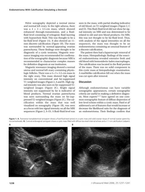
Figure 1. A, Transverse transabdominal sonogram shows a fluid-fluid level (arrow) in a cystic mass and solid ovarian tissue <strong>of</strong> normal ovarian parenchyma<br />
(arrowheads). B, Coronal endovaginal sonogram shows a cystic mass filled with diffuse low-level internal echoes and a 1.1-cm calcification (calipers).<br />
A<br />
B<br />
1238 J Ultrasound Med 2006; 25:1237–1241