Accustrip® URS 10 - Accutest
Accustrip® URS 10 - Accutest
Accustrip® URS 10 - Accutest

Create successful ePaper yourself
Turn your PDF publications into a flip-book with our unique Google optimized e-Paper software.
Accustrip ® <strong>URS</strong> <strong>10</strong><br />
CAT. NO: UA870<br />
Reagent Test Strips for Blood, Urobilinogen, Bilirubin, Protein,<br />
Nitrite, Ketones, Glucose, pH, Specific Gravity and Leukocytes in<br />
Urine by the Dip and Read Technique.<br />
Indications for Use<br />
The Accustrip ® <strong>URS</strong> <strong>10</strong> Reagent Strip for Urinalysis is a dip-and-read test strip. The<br />
product is intended for use as an in vitro diagnostic aid using urine specimens for<br />
screening for diabetes, metabolic abnormalities, liver diseases, biliary and hepatic<br />
obstructions and diseases of the kidneys and urinary tract. The strip provides qualitative<br />
and semi-quantitative tests for blood, urobilinogen, bilirubin, protein, nitrite, ketones,<br />
glucose, pH, specific gravity and leukocytes by visual comparison with a color chart<br />
for each concentration range. The strips may be read visually, requiring no additional<br />
laboratory equipment. The test strip may also be read instrumentally using the Accustrip ®<br />
<strong>URS</strong> Reader.<br />
InFormation REGARDING CLIA WAIVER<br />
These tests are CLIA waived when read visually and when run on the Accustrip ® <strong>URS</strong><br />
Reader. A certificate of CLIA waiver is required to perform the testing in a waived<br />
setting. If the laboratory does not have a Certificate of Waiver, the Application for<br />
Certification (Form CMS-116), can be obtained at http://www.cms.hhs.gov/clia/.<br />
The form should be mailed to the address of the local State Agency of the State<br />
in which the laboratory resides (http://www.cms.hhs.gov/clia/ssa-map.asp).<br />
Laboratories with a certificate of waiver must follow the manufacturer‘s instructions for<br />
performing the test. If the laboratory modifies the instructions, the test no longer meets<br />
the requirements for waived categorization. A modified test is considered to be high<br />
complexity and subject to all CLIA requirements.<br />
Instructions for use<br />
Make sure to use urine that is not older than 2 hours. Mix well before testing.<br />
Dip all test pads of the reagent strip into the urine for approximately 1 second. Draw<br />
the strip across the rim of the container to remove excess urine.<br />
a) If reading visually, start timing. After 30-60 seconds (leukocyte test field after 60 - 120<br />
seconds) compare the reagent areas to corresponding color chart on the bottle label. Hold<br />
strip close to color blocks and match carefully. Make sure to read the pads in good light.<br />
Color changes that take place after more than 2 minutes are of no significance.<br />
b) If reading instrumentally, carefully follow the directions in the operator’s manual.<br />
Principle<br />
Blood: The detection is based on the pseudoperoxidative activity of hemoglobin and<br />
myoglobin, which catalyzes the oxidation of an indicator by an organic hydroperoxide<br />
producing a green color.<br />
Urobilinogen: The test paper contains a stable diazonium salt producing a reddish azo<br />
compound with urobilinogen.<br />
Bilirubin: A red azo compound is obtained in the presence of acid by the coupling of<br />
bilirubin with a diazonium salt.<br />
Protein: The test is based on the “protein error” principle of indicators. The test zone is<br />
buffered to a constant pH value and changes color from yellow to greenish blue in the<br />
presence of albumin. Other proteins are indicated with less sensitivity.<br />
Nitrite: Microorganisms, which are able to reduce nitrate to nitrite, are indicated indirectly<br />
with this test. The principle of Griess reagent is the basis of this test. The test paper<br />
contains an amine and a coupling component. A red colored azo compound is obtained by<br />
diazotisation and subsequent coupling.<br />
Ketones: The test is based on the principle of Legal’s test. Acetoacetic acid and acetone<br />
form with sodium nitroprusside in alkaline medium a violet colored complex.<br />
Glucose: The detection is based on the glucoseoxidase-peroxidase-chromogen reaction.<br />
Apart from glucose, no other compound in urine is known to give a positive reaction.<br />
pH: The test paper contains indicators which clearly change color between pH 5 and pH 9<br />
(from orange to green to turquoise).<br />
Specific Gravity: The test determines the concentration of ions in urine and shows a<br />
good correlation to the refractometrical method. The color of the test strip changes from<br />
deep blue in urine with low ionic concentration through green to yellow in urines with high<br />
ionic concentrations.<br />
Leukocytes: The test is based on the esterase activity of granulocytes. This enzyme splits<br />
carboxylate. The alcohol constituent released reacts with a diazo salt producing a violet<br />
color.<br />
Performance Characteristics & Evaluation<br />
Sources of Error<br />
Blood: The minimum sensitivity of the test strip is 5 to <strong>10</strong> erythrocytes/µl urine<br />
corresponding to approx. 0.015 mg hemoglobin/dl urine. Intact erythrocytes are indicated<br />
by flecky discolorations of the test field. The color fields correspond to the following values:<br />
0 (negative), ca. 5-<strong>10</strong>, ca. 50, ca. 250 Ery/µl resp.<br />
hemoglobin concentration out of ca. <strong>10</strong>, ca. 50, ca. 250 Ery/µl<br />
Normal concentrations of ascorbic acid (< 40 mg/dl) do not influence the test results.<br />
Falsely positive reactions can be produced by a residue of peroxide containing cleansing<br />
agents.<br />
Urobilinogen: Depending on urine color, 0.5 to 1 mg urobilinogen/dl can be indicated.<br />
1 mg/dl is considered to be the normal excretion rate. Higher values are pathological.<br />
A complete absence of urobilinogen in the urine, which is likewise pathological, cannot<br />
be demonstrated by the strips, The color fields correspond to the following urobilinogen<br />
concentrations;<br />
norm. (normal), 2, 4, 8,12 mg/dl or norm. (normal), 35, 70, 140, 200 µmol/l<br />
The test will be inhibited by higher concentrations of formaldehyde. Exposure of the urine<br />
to light for a longer period of time may lead to lowered or falsely negative results. Too high<br />
or falsely positive results can be caused by the presence of diagnostic or therapeutic dyes<br />
in the urine. Larger amounts of bilirubin produce a yellow coloration.<br />
Bilirubin: The minimum sensitivity of the test strip is 0.5 to 1 mg bilirubin/dl urine. The color<br />
fields correspond to the following values:<br />
0 (negative), 1(+), 2(++), 4(+++) mg/dl or 0 (negative), 17(+), 35(++), 70(+++) µmol/l<br />
Some urine contents can produce a yellow coloration of the test strip. Ascorbic acid and<br />
nitrite in higher concentrations inhibit the test. Exposure of the urine to light for a longer<br />
period of time may lead to lowered or falsely negative results. Too high or falsely positive<br />
results can be caused by the presence of diagnostic or therapeutic dyes in the urine.<br />
Protein: The minimum sensitivity of the test strip is <strong>10</strong> mg protein/dl urine. The color fields<br />
correspond to the following ranges of albumin concentrations:<br />
negative, 30,<strong>10</strong>0 and 500 mg/dl or negative, 0.3, 1.0 and 5.0 g/l<br />
Falsely positive results are possible in alkaline urine samples (pH > 9), after infusions with<br />
polyvinylpyrrolidone (blood substitute), after intake of medicaments containing quinine and<br />
also by disinfectant residues in the urine sampling vessel. The protein coloration may be<br />
masked by the presence of medical dyes (e.g. methylene blue) or beetroot pigments.<br />
Nitrite: The test detects concentrations from 0.05 to 0.1 mg nitrite/dl urine. Every pink color<br />
indicates a bacterial infection of the urinary tract. The color intensity depends only on the<br />
nitrate concentration, but does not provide information about the extent of the infection. A<br />
negative result does not preclude an infection of the urinary tract, if bacteria, which cannot<br />
produce nitrite, are present. Falsely negative results can be produced by high doses of<br />
ascorbic acid, by antibiotic therapy and by very low nitrate concentrations in urine as the<br />
result of low nitrate diet or strong dilution (diuresis). Falsely positive results can be caused<br />
by the presence of diagnostic or therapeutic dyes in the urine.<br />
Ketones: Acetoacetic acid reacts more sensitively than acetone. Values of <strong>10</strong> mg/dl<br />
acetoacetic acid or 50 mg/dl acetone are indicated. The color fields correspond to the<br />
following acetoacetic acid values:<br />
0 (negative), 25(+), <strong>10</strong>0(++) and 300(+++) mg/dl or<br />
0 (negative), 2.5(+), <strong>10</strong>(++) and 30(+++) mmol/l<br />
Phenylketones in higher concentrations interfere with the test, and will produce variable<br />
colors. ß-Hydroxybutyric acid is not detected. Phthalein compounds interfere by producing<br />
a red coloration.<br />
Glucose: Pathological glucose concentrations are indicated by a color change from green<br />
to bluish green. Yellow or greenish test fields should be considered negative or normal.<br />
The color fields correspond to the following ranges of glucose concentrations:<br />
neg. (yellow), neg. or normal (greenish), 50, 150, 500 and ≥ <strong>10</strong>00 mg/dl or<br />
neg. (yellow), neg. or normal (greenish), 2.8, 8.3, 27.8 and ≥ 55.5 mmol/l<br />
An inhibitory effect is produced by gentisic acid. Falsely positive reactions can also be<br />
produced by a residue of peroxide containing cleansing agents.<br />
pH: The pH value of fresh urine of healthy people varies between pH 5 and pH 6. The<br />
color scale gives a clear distinction of pH value between pH5 and pH 9.
Specific Gravity: The test permits the determination of urine specific gravity between<br />
1.000 and 1.030. Urine from adults with normal diets and fluid intake will have a density<br />
of 1.015 -1.025. The chemical nature of the test strip may cause slightly different<br />
results from those obtained with other methods when elevated amounts of certain urine<br />
constituents are present, e.g. the increase of urine specific gravity because of high glucose<br />
concentrations of > <strong>10</strong>00 mg/dl (> 56 mmol/l) cannot be detected by the specific gravity<br />
test field. Elevated specific gravity readings may be obtained in the presence of moderate<br />
quantities of protein. Highly buffered alkaline urines may cause low readings.<br />
Leukocytes: The test records values starting from approx. <strong>10</strong>-25 leukocytes/µl urine.<br />
Changes in color that can not be assigned to the negative reference field and faint<br />
violet colors after 120 seconds must be evaluated as positive. The color reference fields<br />
correspond to the following leukocyte concentrations:<br />
negative (normal), 25, 75, 500 leukocytes/µl<br />
A weakened reaction can be expected in the case of proteinuria at over 500 mg/dl and a<br />
glucose concentration of over 2 g/dl as well as in the case of patients taking preparations<br />
containing cephalexin and gentamycin. Bacteria, trichomonads and erythrocytes do not<br />
react with this test. Formaldehyde (as a preservative) can result in a false positive reaction.<br />
Excretion of bilirubin, nitrofurantoin or other strongly-colored compounds may disguise the<br />
color of the reaction. Tests with female patients have shown that vaginal discharge can<br />
cause a false positive reaction.<br />
Reactive ingredients<br />
(minimum quantity resp. activity/cm at time of expiry)<br />
2<br />
Blood: Nitrite: pH:<br />
tetramethylbenzidine 59µg sulfanilic acid 80µg methyl red 2.8µg<br />
cumene hydroperoxide 253µg quinoline derivative 25µg bromothymol blue <strong>10</strong>µg<br />
Urobilinogen: Ketones: Specific Gravity:<br />
diazonium salt 28µg sodium nitroprusside 116µg bromothymol blue 12µg<br />
copolymer 295µg<br />
Bilirubin: Glucose: Leukocytes:<br />
diazonium salt 26µg glucoseoxidase 3.2U carboxylic acid ester <strong>10</strong>.6µg<br />
peroxidase 0.2U diazonium salt 4.4µg<br />
Protein: o-tolidine 65µg<br />
tetrabromophenol blue 7.5µg<br />
Directions<br />
In any case, in order to establish a final diagnosis and prescribe an appropriate therapy,<br />
the results obtained with test strips should be verified with other medical results.<br />
The effect of medications or their metabolic products on the test is not known in all cases.<br />
In case of doubt it is recommended not to take the medications and then repeat the test.<br />
Any change of medication should be approved by the patient’s physician.<br />
Only use well-washed and clean vessels for urine collection. The presence of usual urine<br />
preservatives will not affect the test results,<br />
Remove only as many test strips as are required, and reseal the container immediately<br />
after use. Do not touch the test paper. Avoid exposing the strips to sunlight and moisture.<br />
Store the container below +30 °C in a dry place. The test strips are stable, when stored<br />
properly up to the date of expiry indicated.<br />
SPECIMEN COLLECTION AND PREPARATION<br />
Collect urine in a clean container and test samples as soon as possible. If testing<br />
cannot be completed within one (1) hour after sample collection, REFRIGERATE THE<br />
SPECIMEN IMMEDIATELY AND LET IT RETURN TO ROOM TEMPERATURE BEFORE<br />
TESTING. Nitrite results are best optimized by using a first morning specimen or one<br />
which has incubated in the bladder for four (4) hours or more.<br />
Prolonged exposure of unpreserved urine to room temperature may result in microbial<br />
proliferation with resultant changes in pH and false positive nitrite test. A shift to alkaline<br />
pH may cause false positive results in the protein test area. Urine containing glucose may<br />
decrease in pH as organisms metabolize glucose. Bacterial growth from contaminating<br />
organisms may cause a positive blood reaction due to the peroxidases produced,<br />
Preservatives do not prevent deterioration of ketones in the sample. Some preservatives<br />
do not adequately protect glucose from being metabolized by contaminating or infecting<br />
organisms. Do not use formaldehyde as a urine preservative as it may produce unreliable<br />
results.<br />
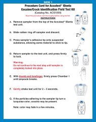
PROCEDURE - MUST BE FOLLOWED EXACTLY TO ACHIEVE<br />
RELIABLE TEST RESULTS<br />
1. Collect FRESH urine specimens in a clean, dry container.<br />
2. Remove one strip from aluminum container and replace cap. COMPLETELY immerse<br />
strip in FRESH urine and remove immediately to avoid the dissolving out of reagents.<br />
3. When removing, run the edge of the strip against the rim of the urine container to<br />
remove excess urine. Hold the strip in a horizontal position to prevent mixing of<br />
chemicals from adjacent reagent areas and/or soiling of hands with urine.<br />
4. Compare test areas to the corresponding color charts on the bottle label. HOLD STRIP<br />
CLOSE TO Color BLOCKS-MATCH CAREFULLY.<br />
If reading instrumentally, carefully follow the directions in the operator's manual.<br />
Some colors continue to become more intense for a short time and then fade. For<br />
this reason, the best time for comparison is AFTER 30 SECONDS AND BEFORE<br />
60 SECONDS. Color changes that take place after more than 2 minutes are of no<br />
significance.<br />
QUALITY CONTROL<br />
Test at least one known negative and one known positive specimen or control,<br />
whenever a new bottle of strips is first opened, for each new shipment,<br />
for each new lot or at least monthly. Do not use water as negative control.<br />
Positive and negative control solutions provide a convenient basis for a quality<br />
control program. Contact the service number below for ordering information.<br />
If proper results are not obtained, consult your local product representative or contact<br />
Customer Service by calling (800) 676-5565 for advise on testing techniques and results.<br />
RESULTS<br />
Results with test strips are obtained in clinically meaningful units directly from the color<br />
chart comparison.<br />
If reading instrumentally, carefully follow the directions in the operator's manual.<br />
STORAGE AND STABILITY HANDLING<br />
Accustrip ® <strong>URS</strong> <strong>10</strong> test strips should be stored between 39 – 86°F (4 - 30°C) in a COOL,<br />
DRY place. Do not freeze.<br />
Properly stored, the strips are stable until the date of expiration.<br />
RECOMMENDED PROCEDURES FOR HANDLING<br />
Unused test strips must remain in the original container. Desiccant material in the cap will<br />
keep dipsticks moisture free. Transfer to any other container may cause reagent strips to<br />
deteriorate and become non reactive. Replace cap immediately and tightly after removing<br />
dipstick. Do not touch reagent areas of test strip with your fingers. Do not allow dipsticks<br />
to come in contact with detergents which may be found in specimen containers and other<br />
contaminating substances found in work areas.<br />
WARNING AND PRECAUTIONS<br />
PROTECTION AGAINST MOISTURE, LIGHT AND HEAT IS ESSENTIAL. ALTERED<br />
REAGENT ACTIVITY MAY RESULT IF CARE IS NOT TAKEN. Discoloration or darkening<br />
of reagent areas may indicate deterioration. DO NOT USE STRIP IF THIS OCC<strong>URS</strong>.<br />
In this event, check to see that the unopened expiration date stamped on the vial has<br />
not been passed or examine vial for evidence of exposure to moisture, light or heat.<br />
Store strips out of reach of children!<br />
BIBLIOGRAPHY<br />
1. Fraser, J.; Fetter, M.C.; Mast, R.L. and Free, A.H.: Studies with a Simplified<br />
Nitroprusside Test for Ketone Bodies in Urine, Serum, Plasma and Milk, Clin. Chem.<br />
Acta II, (1965) 376-378.<br />
2. Free, A.H. and Free, H.M,: Urinalysis Critical Discipline of Clinical Science; CRC crit.<br />
Rev. Clin, Lab. Sci, 3 (4): 481-531, Dec. 1972.<br />
3. Kark R.M.; Lawrence, J.R.; Pollak V.E.; Pirani, C.L.; Muehrcke, R.C. and Silva, H.: A<br />
Primer of Urinalysis, 2nd ed., Harper & Row, New York, 1963.<br />
4. McGarry, J.D.; Lilly Lecture, 1978: New Perspectives in the Regulation of Ketogenesis,<br />
DIABETES 28 May, 1978, 517-523.<br />
5. Paterson, P.; Sheath, J.; Pincus, T. and Wood, C.: Maternal and Fetal Ketone<br />
Concentrations in Plasmas and Urine, the Lancet, April 22, 1967, 862-865.<br />
6. Williamson, D.H.: Physiological Ketosis, or Why Ketone Bodies?; Postgraduate<br />
Medical Journal (June Suppl., 1971) 371-375.<br />
7. Brumfitt: Urinary Cell Counts and their Value; J. Clin. Pathology 18, (1965) 550.<br />
8. Stansfeld and Webb: Observations on Pyuria in Children; Arch. Dis. Chil. 28, (1953)<br />
386.<br />
9. Stansfeld: The Measurement and the Meaning of Pyuria; Arch. Dis. Chil. 37, (1962)<br />
257,<br />
<strong>10</strong>. Gerber, Zbinden and Hoigne: Diagnose und klinische Bedeutung der Leukozyturie;<br />
Schweiz. Rundschau Med. (Praxis) 64 (1975) 35.<br />
11. Weiss: Diagnostische Bewertung von Laborbefunden; J.F. Lehmanns Verlag, München<br />
1969.<br />
Rev. 06/2008 / Axxxxxx / xxx/x<br />
<strong>URS</strong> <strong>10</strong><br />
Jant Pharmacal Corporation<br />
Encino, California 91436 USA