LabChip GXII for High Throughput Analysis of Protein ... - PerkinElmer
LabChip GXII for High Throughput Analysis of Protein ... - PerkinElmer
LabChip GXII for High Throughput Analysis of Protein ... - PerkinElmer

You also want an ePaper? Increase the reach of your titles
YUMPU automatically turns print PDFs into web optimized ePapers that Google loves.
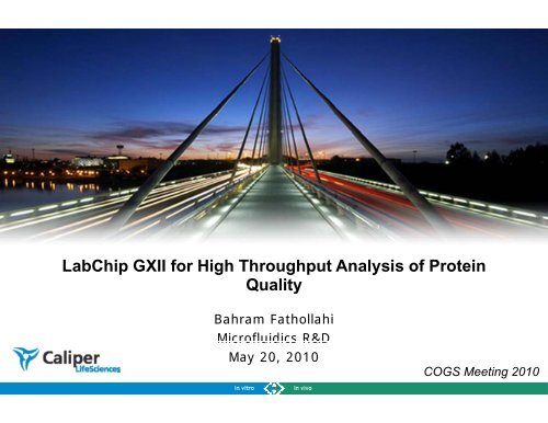
<strong>LabChip</strong> <strong>GXII</strong> <strong>for</strong> <strong>High</strong> <strong>Throughput</strong> <strong>Analysis</strong> <strong>of</strong> <strong>Protein</strong><br />
Quality<br />
Bahram Fathollahi<br />
Micr<strong>of</strong>luidics R&D<br />
May 20, 2010<br />
COGS Meeting 2010<br />
1
<strong>LabChip</strong> Plat<strong>for</strong>ms<br />
on-chip capillary electrophoresis<br />
Bioanalyzer 2100<br />
<strong>LabChip</strong> <strong>GXII</strong><br />
<strong>Protein</strong> analysis<br />
N-Glycan analysis<br />
DNA sizing & quantification<br />
RNA quality assessment<br />
<strong>High</strong>er <strong>Throughput</strong><br />
Experion<br />
Improved Data Quality<br />
Labor Savings vs. Gels<br />
Digital in<strong>for</strong>mation<br />
Easy-to-use<br />
2<br />
2
<strong>LabChip</strong> <strong>GXII</strong> Applications<br />
MicroChip-CE SDS<br />
• Crude lysate analysis<br />
• <strong>Protein</strong> expression<br />
• Antibody analysis<br />
• <strong>Protein</strong> purification<br />
• Fusion protein analysis<br />
• Formulation stability<br />
• Detection <strong>of</strong> covalent<br />
aggregates (up to 500 kDa)<br />
• Pre-labeled proteins <strong>for</strong> high<br />
sensitivity<br />
MicroChip-CE<br />
• N-Glycan pr<strong>of</strong>iling<br />
3
HT <strong>Protein</strong> Assay: Microchip-CE-SDS<br />
HT <strong>Protein</strong> Assay<br />
• Integrates the entire SDS-PAGE process onto a<br />
micr<strong>of</strong>luidic chip<br />
• Determine size, quantification, and purity<br />
• Automatic sampling from 96- or 384-well plate<br />
• Fast sample analysis - 41 seconds per sample<br />
• Can run crude samples<br />
• 96-well plate analyzed in < 1.25 h<br />
• Can do >400 samples per day per instrument<br />
Microchip CE<br />
• 2 μl <strong>of</strong> sample<br />
• Minimal sample preparation<br />
• 21 CFR Part 11 support<br />
4
<strong>Protein</strong> Assay Workflow<br />
Crude or purified<br />
protein sample<br />
Add denaturing<br />
sample buffer<br />
Heat<br />
Denature<br />
2μl<br />
96- or 384-well plate<br />
Add reagents to chip<br />
well<br />
Product Quality -<br />
Titer, % purity,<br />
degree <strong>of</strong><br />
glycosylation, ….<br />
5
Automated Sample <strong>Analysis</strong> Window<br />
Identified<br />
expected protein<br />
peaks<br />
Plate View<br />
Gel View<br />
Electropherogram<br />
Sizing, Conc.,<br />
and<br />
Purity results<br />
Well table view,<br />
Peak table view<br />
6
(1) (3) (2)<br />
Resolution Ladder Mix<br />
(1) Fermentas PAGE Ruler Unstained <strong>Protein</strong> Ladder<br />
(2) Fermentas Unstained Molecular Weight Marker<br />
(3) Mix<br />
Lowe er Marker<br />
10kDa<br />
14.4 and 15kDa 45 and 50kDa<br />
MIX<br />
7
Staccato <strong>Protein</strong> Workstation: Workflow Automation<br />
Features<br />
• Integrated sample<br />
processing & analysis<br />
• Walk away automation<br />
• Easy-to-use s<strong>of</strong>tware<br />
• Enables complex DOE<br />
experiment<br />
Scalable design: Twister plate loading, liquid handling<br />
sample prep, p and fully integrated system<br />
8
<strong>Protein</strong> Assay Specifications<br />
Type<br />
Sizing Range<br />
Specification<br />
<strong>Protein</strong> 100 Assay: 14 - 100 kDa<br />
<strong>Protein</strong> 200 Assay: 14 - 200 kDa ( can detect higher Mw proteins<br />
by increasing the separation and sample-load time)<br />
Sizing Accuracy* ± 20%<br />
Resolution*<br />
Linear Dynamic Range*<br />
Relative Concentration CV*<br />
Sensitivity*<br />
<strong>Analysis</strong> Time per Sample<br />
± 10% difference in MW across the sizing range, 50% valley<br />
Resolution is comparable to a 10 cm 4-20% SDS-PAGE gel<br />
5 - 2000 μg/mL<br />
30% up to 120 kDa relative to the ladder<br />
5 μg/mL (10 ng) Carbonic Anhydrase<br />
Sensitivity is equivalent to Coomassie stain<br />
<strong>Protein</strong> 100 Assay: ~34 seconds/sample<br />
<strong>Protein</strong> 200 Assay: ~41 seconds/sample<br />
Chip Lifetime<br />
<strong>Analysis</strong> Time Per Plate<br />
Maximum Salt Concentration<br />
4 96-well plates<br />
96-well plate
Conventional CE-SDS vs <strong>LabChip</strong>: Reduced mAb<br />
CE-SDS<br />
Microchip CE-SDS<br />
Chen X. et al. Microchip assays <strong>for</strong> screening monoclonal antibody protein quality. Electrophoresis, 29 (2008) 4993-5002<br />
10
CE-SDS vs. <strong>LabChip</strong>: Non-Reducing mAb<br />
cence<br />
Fluores<br />
550<br />
40 sec<br />
Microchip-CE-SDS<br />
6<br />
500<br />
Migration Corrected Migration Corrected<br />
450<br />
Time, Relative Time, Relative<br />
400<br />
350<br />
Peak Number seconds Area minutes Area Delta<br />
300<br />
1 18.6 0.4% 15.5 0.7% 0.3%<br />
250<br />
200<br />
150<br />
100<br />
50<br />
0<br />
-50<br />
5<br />
1 2 3 4<br />
6<br />
7<br />
16 18 20 22 24 26 28 30 32<br />
Time (seconds)<br />
8<br />
5<br />
9<br />
0<br />
1<br />
LC-90<br />
CE-SDS<br />
2 20.5 0.1% 18.7 0.2% 0.1%<br />
3 23.9 0.3% 22.2 0.3% 0.0%<br />
4 26.4 0.5% 25.2 0.5% 0.0%<br />
5 27.8 2.0% 26.7 2.4% 0.4%<br />
6 29.0 96.7% 27.8 95.9% 9% 08% 0.8%<br />
Total Impurities 3.3% 4.1% 0.8%<br />
35 minute separation<br />
Baseline<br />
variability<br />
Data is quite consistent, despite<br />
differences in:<br />
• Instrument<br />
• Sample preparation buffer<br />
• Detection method<br />
• Some impurities are known to<br />
be sensitive to sample<br />
preparation conditions<br />
Siemiatkoski et al. Use <strong>of</strong> <strong>LabChip</strong> 90 in Product Development <strong>of</strong> a<br />
Recombinant Fusion <strong>Protein</strong>. <strong>Protein</strong> Summit 2008, London UK<br />
11
Crude Lysate <strong>Analysis</strong><br />
Microchip-CE SDS vs SDS-PAGE<br />
Expressed protein 47.7 kDa<br />
Expressed protein 48kDa<br />
* Lysate sample and SDS-PAGE data courtesy <strong>of</strong> Structural GenomiX - San Diego, CA<br />
12
<strong>Analysis</strong> <strong>of</strong> Subunit Vaccine Expression in<br />
Pseudomonas<br />
<strong>High</strong> throughput<br />
expression<br />
monitoring with<br />
analysis on the<br />
<strong>LabChip</strong> <strong>GXII</strong> led<br />
to a 10-fold<br />
increase in yield<br />
13
<strong>Protein</strong> Expression Optimization using DOE<br />
Optimal expression condition<br />
is easily observed<br />
Swalley, S.E. et al., Quantitative <strong>Analysis</strong> <strong>of</strong> <strong>Protein</strong> Expression, Anal. Biochem, 351 (2006) 122-127<br />
14
<strong>LabChip</strong> GX Dependent Process Development <strong>for</strong> mAb Purification<br />
<strong>Analysis</strong> <strong>of</strong><br />
mAb yield<br />
and purity<br />
with varying<br />
purification<br />
parameters<br />
15
Purification Process Development<br />
Affinity resin capacity determination<br />
<strong>Protein</strong> are introduced continuously into the column and unbound fractions are<br />
monitored o by UV and Labchip90.<br />
20.0 the<br />
15.0<br />
10.0<br />
<strong>Protein</strong>s are detected in<br />
the unbound the<br />
dynamic binding<br />
capacity <strong>of</strong> the column<br />
has been reached<br />
5.0<br />
Slide courtesy <strong>of</strong> X.Le Saoût – COGS 2008 meeting<br />
16
<strong>High</strong> Sensitivity: Derivatization <strong>of</strong> <strong>Protein</strong>s<br />
with FL dye<br />
• Replaces on-chip staining with <strong>of</strong>f-chip labeling in order to increase sensitivity by<br />
10X or more<br />
• NHS esters reaction with amine groups <strong>of</strong> lysine residues<br />
O O<br />
N O<br />
Dye<br />
NH 3<br />
O NH<br />
Dye<br />
MAb<br />
Dye Succinimidyl Ester<br />
O<br />
Intact Antibody<br />
Reduction<br />
2 + 2<br />
O<br />
N<br />
O<br />
O<br />
O<br />
Dye<br />
NH<br />
NH<br />
O<br />
Dye<br />
Dye<br />
MAb<br />
Heavy Light<br />
Chain Chain<br />
Dye Succinimidyl Ester<br />
O<br />
HC and LC<br />
17
Linearity <strong>of</strong> intact mAb at constant dye<br />
concentration<br />
ti<br />
• linearity <strong>of</strong> 4 orders <strong>of</strong> magnitude <strong>for</strong> high sensitivity Pico protein assay<br />
100000<br />
10000<br />
HT <strong>Protein</strong> Express 200<br />
HT Pico <strong>Protein</strong> Express<br />
R 2 = 0.999<br />
R 2 = 0.9997<br />
Time Corrected<br />
Area<br />
1000<br />
100<br />
10<br />
1<br />
0.01 0.1 1 10 100 1000 10000<br />
MAb Concentration (μg/mL)<br />
18
Low Level Impurity Detection: Non-Covalent<br />
On-Chip Staining<br />
• Impurities <strong>of</strong> as low as 1% are clearly identified<br />
• Lysozyme was spiked into the sample at 1% <strong>of</strong> total protein and was<br />
readily identified, with a S:N <strong>of</strong> 9:1<br />
HC<br />
NGHC<br />
LC<br />
Ly<br />
19
Low Level Impurity Detection: Covalent<br />
Off-Chip Staining<br />
i<br />
• Carbonic anhydrase was spiked into mAb sample at 0.01%, 0.05% and 0.5% <strong>of</strong> the<br />
total protein at 1 mg/ml.<br />
• ~ 100X improvement in sensitivity!<br />
0.5%<br />
0.05%<br />
0.01%<br />
Background mAb trace<br />
Spiked CA<br />
20
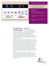
Antibody N-linked Glycans<br />
Complex<br />
Mannose<br />
Hybrid<br />
GlcNAc<br />
Fucose<br />
Galactose<br />
<strong>High</strong> Mannose<br />
• Glycosylation patterns <strong>of</strong> therapeutic proteins can influence<br />
- Efficacy and safety <strong>of</strong> therapeutic antibodies<br />
- <strong>Protein</strong>-protein interactions<br />
- Solubility<br />
- Intracellular trafficking<br />
• Important to characterize and monitor the glycosylation pattern during cell<br />
culture development<br />
Antibody image courtesy <strong>of</strong> Dr. Greg Flynn<br />
21
Neutral N-Glycan Types<br />
<strong>High</strong><br />
Mannose<br />
M5 M6* M7* M8 M9<br />
Hybrid<br />
M5G1F M5G0F M5G0 M5G1<br />
Complex<br />
G2F G1F G0F G2 G1 G0<br />
G0F‐GlcNac<br />
slide courtesy <strong>of</strong> Dr. Greg Flynn<br />
22
IgG Charged N-Glycan Types<br />
Sialylated<br />
23
Traditional N-Glycan Mapping (3-5 Days)<br />
Sample<br />
Digestion (PNGase-F)<br />
Removal <strong>of</strong> <strong>Protein</strong><br />
Carbohydrate labeling (2-AB, APTS)<br />
Removal <strong>of</strong> labeling<br />
Reagent<br />
CE-LIF<br />
Chromatography/MS<br />
• NP-HPLC<br />
• RP-HPLC<br />
24
Goals <strong>for</strong> HT N-Glycan Screening Assay in<br />
Early Development<br />
• Identification and determination <strong>of</strong> relative amounts <strong>of</strong><br />
major N-Glycans (Man 5, G0, G0F, G1F isomers, and<br />
G2F)<br />
– Sialylated glycans?<br />
– Low level glycans? gy<br />
• Ease-<strong>of</strong>-use<br />
• <strong>High</strong> throughput and fast turnaround<br />
– <strong>Analysis</strong> <strong>of</strong> 96-well plate sample in < 8 hours<br />
– Complete from digestion to results<br />
• Robust and Reproducible<br />
• Easy to transfer and establish in QC environment<br />
25
HT Glycan Pr<strong>of</strong>iling on <strong>LabChip</strong> System<br />
Denature and Reduce mAb in Denaturing Buffer<br />
<strong>LabChip</strong> <strong>GXII</strong><br />
Add Denatured Reduced mAb to Enzyme Plate<br />
Denaturing plate<br />
Heat (Denatured mAb + Enzyme) at 37 °C <strong>for</strong> ~1h<br />
microchip<br />
Transfer to Dye Plate<br />
PNGase‐F Plate<br />
Heat Assay Plate at 55 °C <strong>for</strong> ~2h<br />
G0F<br />
G1F<br />
G1F’<br />
G2F<br />
Dilute with separation Buffer<br />
Read 96-well plate on <strong>LabChip</strong> <strong>GXII</strong> ~1.5h<br />
Glycan Labeling Plate<br />
G0<br />
M5<br />
26
HT Glycan Assay Per<strong>for</strong>mance Specifications<br />
8ul with concentration range <strong>of</strong> 1.25mg/ml to<br />
Amount <strong>of</strong> sample required:<br />
7.5mg/ml (10ug to 60ug <strong>of</strong> MAb total).<br />
Reproducibility <strong>of</strong> %area:<br />
CV < 10% <strong>for</strong> a peak >= 2.5% <strong>of</strong> total glycan.<br />
1ng <strong>of</strong> G0F standard. (Smallest amount <strong>of</strong> labeled<br />
Limit <strong>of</strong> detection:<br />
G0f standard that can be detected.)<br />
>95% <strong>of</strong> all N-Linked glycans will be released from<br />
Deglycosylation:<br />
mAb.<br />
Appropriate <strong>for</strong> neutral glycans found on MAbs.<br />
Usable size range: Some charged glycans may run outside <strong>of</strong> our<br />
usable range.<br />
Sizing ing reproducibility: CV < 2.5%<br />
< 0.5% <strong>of</strong> previous sample can be detected in<br />
Carryover:<br />
following sample.<br />
Sample prep, chip prep, and < 8 hours <strong>for</strong> a 96-well plate.<br />
analysis time:<br />
Number <strong>of</strong> samples per chip Up to 192 samples be<strong>for</strong>e chip needs to be<br />
prep: reloaded with fresh gel.<br />
Number <strong>of</strong> samples per chip Up to 384 samples be<strong>for</strong>e chip needs to be<br />
lifetime: replaced.<br />
27
PNGase-F Digestion<br />
Observed complete deglycosylation<br />
• Digested IgG (red trace) shows shift towards NGHC<br />
when compared to undigested IgG (blue trace)<br />
HC<br />
NGHC<br />
28
Use <strong>of</strong> Internal Standards <strong>for</strong> Peak Identifications<br />
• Individual neutral glycan standards show good resolution <strong>of</strong> the main glycans<br />
• The two G1F isomers, differing only in the branch position <strong>of</strong> terminal galactose, are resolved<br />
Mixture <strong>of</strong> Glycan Standards<br />
G1<br />
29
Sialylated Glycan Standards<br />
•Mono- and di-sialylated glycan standards, with and without internal fucose (A1, A1F, A2, and<br />
A2F) were purchased from ProZyme (Hayward CA)<br />
30
Use <strong>of</strong> Glucose Ladder For Peak Identification<br />
31
Identification <strong>of</strong> Glycan Peaks<br />
Expected Peaks Function in <strong>Analysis</strong> Settings<br />
32
Intermediate-Assay Reproducibility<br />
•Labeled N-glycans released from three different mAbs at starting concentrations <strong>of</strong> 1.25 mg/mL, 2.5mg/mL, and 7.5<br />
mg/mL were analyzed.<br />
•The %RSD was calculated <strong>for</strong> 35 reactions (complete from deglycosylation to labeling and separation) per mAb, 2<br />
repeat injections, 4 different chips and instruments, and 2 users.<br />
•Total %RSD’s <strong>for</strong> the five main glycan peaks were all below 10% <strong>for</strong> peaks with relative amount greater than 2.5%.<br />
IgG_A AVG % Area CV_10ug CV_20ug CV_60ug Total CV<br />
G0 6.82 3.6% 3.4% 3.8% 6.3%<br />
G0F 53.39 3.4% 1.6% 2.5% 3.2%<br />
G1F 9.05 4.1% 1.9% 2.8% 4.1%<br />
G1F' 9.28 4.6% 1.9% 2.1% 4.9%<br />
G2F 2.05 12.2% 6.3% 2.2% 13.4%<br />
Man5 4.61 4.9% 3.6% 6.3% 8.7%<br />
IgG_B G0 10.55 5.8% 4.9% 4.2% 5.4%<br />
G0F 47.78 2.4% 1.6% 1.2% 2.1%<br />
G1F 5.70 5.1% 5.2% 4.3% 6.5%<br />
G1F' 17.92 2.9% 2.0% 1.4% 3.2%<br />
G2F 258 2.58 82% 8.2% 75% 7.5% 33% 3.3% 98% 9.8%<br />
Man5 1.79 7.2% 8.8% 6.8% 12.1%<br />
IgG_C G0 5.01 4.5% 3.2% 3.4% 6.8%<br />
G0F 57.33 3.1% 2.1% 3.3% 3.1%<br />
G1F 457 4.57 49% 4.9% 36% 3.6% 41% 4.1% 63% 6.3%<br />
G1F' 13.92 3.5% 2.3% 1.8% 3.1%<br />
G2F 1.25 14.4% 13.4% 6.7% 15.5%<br />
Man5 5.58 4.9% 3.5% 3.3% 6.8%<br />
33
NPLC (2-AB labeled)<br />
G0F Man5<br />
Comparison to NPLC and CE-LIF<br />
180 min<br />
G1FA G1FB G2F<br />
CE-LIF (APTS labeled)<br />
Int Std<br />
Man5<br />
G0F<br />
6min<br />
G1FA G1FB<br />
G2F<br />
Microchip-CE (FL labeled)<br />
G0F<br />
45 sec<br />
Int Std<br />
Man5<br />
G0<br />
G1FB<br />
G1FA<br />
G2F<br />
NPLC and CE pr<strong>of</strong>iles courtesy <strong>of</strong> Dr. Nate Lacher<br />
34
Relative Amounts Comparison<br />
Glycan NP‐HPLC Microchip‐CE<br />
% CPA %CPA<br />
G0 06 0.6<br />
Man5 2.00 3.4<br />
G0‐GlcNAc 3.00 4.8<br />
G0F 44.60 43.6<br />
G1F 14.80 16.3<br />
G1F' 19.40 20.3<br />
G2F 6.90 6.9<br />
35
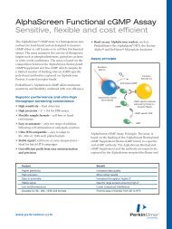
HT N-Glycan Assay: Summary <strong>of</strong> Key Features<br />
• Integrates the CE-LIF process onto a micr<strong>of</strong>luidic chip<br />
• Fast separation time – 45 sec per sample; 96-well plate in ~90 min<br />
• Reagents provided in 96-well plate <strong>for</strong>mat <strong>for</strong> convenience and ease <strong>of</strong> automation<br />
• Rapid digestion ~ 1hr<br />
• Labeling in presence <strong>of</strong> released antibody without requiring NaBH 3 CN<br />
• Able to identify major N-glycan peaks, including mono- and di-sialylated glycans<br />
• S<strong>of</strong>tware determines the relative amounts <strong>of</strong> major N-glycans<br />
• Up to 200 samples per chip with single chip preparation<br />
• Chip lifetime <strong>of</strong> 400 samples<br />
• Capable <strong>of</strong> interfacing with plate robotics <strong>for</strong> complete automation <strong>of</strong> sample prep and<br />
analysis<br />
• 21CFR Part 11 s<strong>of</strong>tware support<br />
36
Acknowledgements<br />
Collaborators<br />
– Greg Flynn - Amgen<br />
– Joe Semiatkoski – Biogen Idec<br />
– Xiaoyu Chen - Novartis<br />
– Nate Lacher – Pfizer<br />
Caliper Life Sciences<br />
• Assay Development<br />
– Mai Ho<br />
– Raj Singh<br />
• Micr<strong>of</strong>luidics R&D<br />
– Roger Dettl<strong>of</strong>f<br />
– Hui Xu<br />
– Melissa Takahashi<br />
• Chip Manufacturing<br />
• SW Engineering<br />
37
Select Publications<br />
38
<strong>LabChip</strong> System<br />
Sampling <strong>of</strong> <strong>LabChip</strong> Users Quantitative Screening Assays<br />
on One Plat<strong>for</strong>m<br />
labchip@caliperls.com<br />
• Antibody analysis<br />
– Titer<br />
– Purity <strong>for</strong> R and NR<br />
• Crude lysate analysis<br />
• <strong>Protein</strong> expression<br />
• Fusion protein analysis<br />
• Formulation stability<br />
• Detection <strong>of</strong> covalent aggregates<br />
• Pre-labeled proteins <strong>for</strong> high<br />
sensitivity<br />
• Purification process development<br />
– Purity and Yield<br />
• N-Glycan Pr<strong>of</strong>iling<br />
39