Oncogenic AKAP9-BRAF fusion is a novel mechanism of MAPK ...
Oncogenic AKAP9-BRAF fusion is a novel mechanism of MAPK ...
Oncogenic AKAP9-BRAF fusion is a novel mechanism of MAPK ...

Create successful ePaper yourself
Turn your PDF publications into a flip-book with our unique Google optimized e-Paper software.
esearch article<br />
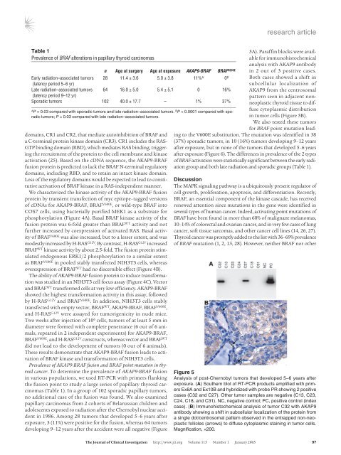
Table 1<br />
Prevalence <strong>of</strong> <strong>BRAF</strong> alterations in papillary thyroid carcinomas<br />
domains, CR1 and CR2, that mediate autoinhibition <strong>of</strong> <strong>BRAF</strong> and<br />
a C-terminal protein kinase domain (CR3). CR1 includes the RAS-<br />
GTP binding domain (RBD), which mediates RAS binding, triggering<br />
the recruitment <strong>of</strong> the protein to the cell membrane and kinase<br />
activation (25). Based on the cDNA sequence, the <strong>AKAP9</strong>-<strong>BRAF</strong><br />
<strong>fusion</strong> protein <strong>is</strong> predicted to lack the <strong>BRAF</strong> N-terminal regulatory<br />
domains, including RBD, and to retain an intact kinase domain.<br />
Loss <strong>of</strong> the regulatory domains would be expected to lead to constitutive<br />
activation <strong>of</strong> <strong>BRAF</strong> kinase in a RAS-independent manner.<br />
We characterized the kinase activity <strong>of</strong> the <strong>AKAP9</strong>-<strong>BRAF</strong> <strong>fusion</strong><br />
protein by transient transfection <strong>of</strong> myc epitope–tagged versions<br />
<strong>of</strong> cDNAs for <strong>AKAP9</strong>-<strong>BRAF</strong>, <strong>BRAF</strong> V600E , or wild-type <strong>BRAF</strong> into<br />
COS7 cells, using bacterially purified MEK1 as a substrate for<br />
phosphorylation (Figure 4A). Basal <strong>BRAF</strong> kinase activity <strong>of</strong> the<br />
<strong>fusion</strong> protein was 6-fold greater than <strong>BRAF</strong> WT activity and not<br />
further increased by coexpression <strong>of</strong> activated RAS. Basal activity<br />
<strong>of</strong> <strong>BRAF</strong> V600E was also increased, but to a lesser extent, and was<br />
modestly increased by H-RAS G12V . By contrast, H-RAS G12V increased<br />
<strong>BRAF</strong> WT kinase activity by about 2.5-fold. The <strong>fusion</strong> protein stimulated<br />
endogenous ERK1/2 phosphorylation to a similar extent<br />
as <strong>BRAF</strong> V600E in pooled stably transfected NIH3T3 cells, whereas<br />
overexpression <strong>of</strong> <strong>BRAF</strong> WT had no d<strong>is</strong>cernible effect (Figure 4B).<br />
The ability <strong>of</strong> <strong>AKAP9</strong>-<strong>BRAF</strong> <strong>fusion</strong> protein to induce transformation<br />
was studied in an NIH3T3 cell focus assay (Figure 4C). Vector<br />
and <strong>BRAF</strong> WT transformed cells at very low efficiency. <strong>AKAP9</strong>-<strong>BRAF</strong><br />
showed the highest transformation activity in th<strong>is</strong> assay, followed<br />
by H-RAS G12V and <strong>BRAF</strong> V600E . In addition, NIH3T3 cells stably<br />
transfected with empty vector, <strong>BRAF</strong> WT , <strong>AKAP9</strong>-<strong>BRAF</strong>, <strong>BRAF</strong> V600E ,<br />
and H-RAS G12V were assayed for tumorigenicity in nude mice.<br />
Two weeks after injection <strong>of</strong> 10 6 cells, tumors <strong>of</strong> at least 5 mm in<br />
diameter were formed with complete penetrance (6 out <strong>of</strong> 6 animals,<br />
repeated in 2 independent experiments) for <strong>AKAP9</strong>-<strong>BRAF</strong>,<br />
<strong>BRAF</strong> V600E , and H-RAS G12V constructs, whereas vector and <strong>BRAF</strong> WT<br />
did not lead to the development <strong>of</strong> tumors (0 our <strong>of</strong> 6 animals).<br />
These results demonstrate that <strong>AKAP9</strong>-<strong>BRAF</strong> <strong>fusion</strong> leads to activation<br />
<strong>of</strong> <strong>BRAF</strong> kinase and transformation <strong>of</strong> NIH3T3 cells.<br />
Prevalence <strong>of</strong> <strong>AKAP9</strong>-<strong>BRAF</strong> <strong>fusion</strong> and <strong>BRAF</strong> point mutation in thyroid<br />
cancer. To determine the prevalence <strong>of</strong> <strong>AKAP9</strong>-<strong>BRAF</strong> <strong>fusion</strong><br />
in various populations, we used RT-PCR with primers flanking<br />
the <strong>fusion</strong> point to study a large series <strong>of</strong> papillary thyroid carcinomas<br />
(Table 1). In a group <strong>of</strong> 102 sporadic papillary tumors,<br />
no additional case <strong>of</strong> the <strong>fusion</strong> was found. We also examined<br />
papillary carcinomas from 2 cohorts <strong>of</strong> Belarussian children and<br />
adolescents exposed to radiation after the Chernobyl nuclear accident<br />
in 1986. Among 28 tumors that developed 5–6 years after<br />
exposure, 3 (11%) were positive for the <strong>fusion</strong>, whereas 64 tumors<br />
developing 9–12 years after the accident were all negative (Figure<br />
n Age at surgery Age at exposure <strong>AKAP9</strong>-<strong>BRAF</strong> <strong>BRAF</strong> V600E<br />
Early radiation–associated tumors 28 11.4 ± 3.6 5.0 ± 3.8 11% A 0 B<br />
(latency period 5–6 yr)<br />
Late radiation–associated tumors 64 16.0 ± 5.0 5.4 ± 5.1 0 16%<br />
(latency period 9–12 yr)<br />
Sporadic tumors 102 40.0 ± 17.7 – 1% 37%<br />
A<br />
P = 0.03 compared with sporadic tumors and late radiation–associated tumors. B P < 0.0001 compared with sporadic<br />
tumors; P = 0.03 compared with late radiation–associated tumors.<br />
5A). Paraffin blocks were available<br />
for immunoh<strong>is</strong>tochemical<br />
analys<strong>is</strong> with <strong>AKAP9</strong> antibody<br />
in 2 out <strong>of</strong> 3 positive cases.<br />
Both cases showed a shift in<br />
subcellular localization <strong>of</strong><br />
<strong>AKAP9</strong> from the centrosomal<br />
pattern seen in adjacent nonneoplastic<br />
thyroid t<strong>is</strong>sue to diffuse<br />
cytoplasmic d<strong>is</strong>tribution<br />
in tumor cells (Figure 5B).<br />
We also tested these tumors<br />
for <strong>BRAF</strong> point mutation leading<br />
to the V600E substitution. The mutation was identified in 38<br />
(37%) sporadic tumors, in 10 (16%) tumors developing 9–12 years<br />
after exposure, but in none <strong>of</strong> the tumors that developed 5–6 years<br />
after exposure (Figure 6). The differences in prevalence <strong>of</strong> the 2 types<br />
<strong>of</strong> <strong>BRAF</strong> activation were stat<strong>is</strong>tically significant between the early radiation<br />
group and both late radiation and sporadic groups (Table 1).<br />
D<strong>is</strong>cussion<br />
The <strong>MAPK</strong> signaling pathway <strong>is</strong> a ubiquitously present regulator <strong>of</strong><br />
cell growth, proliferation, apoptos<strong>is</strong>, and differentiation. Recently,<br />
<strong>BRAF</strong>, an essential component <strong>of</strong> the kinase cascade, has received<br />
renewed attention since mutations in the gene were identified in<br />
several types <strong>of</strong> human cancer. Indeed, activating point mutations <strong>of</strong><br />
<strong>BRAF</strong> have been found in more than 60% <strong>of</strong> malignant melanomas,<br />
10–14% <strong>of</strong> colorectal and ovarian cancer, and in very few cases <strong>of</strong> lung<br />
cancer, s<strong>of</strong>t t<strong>is</strong>sue sarcomas, and other cancer cell lines (14, 26, 27).<br />
Thyroid cancer was promptly added to the l<strong>is</strong>t with 36–69% prevalence<br />
<strong>of</strong> <strong>BRAF</strong> mutation (1, 2, 13, 28). However, neither <strong>BRAF</strong> nor other<br />
Figure 5<br />
Analys<strong>is</strong> <strong>of</strong> post-Chernobyl tumors that developed 5–6 years after<br />
exposure. (A) Southern blot <strong>of</strong> RT-PCR products amplified with primers<br />
Ex8A and Ex10B and hybridized with probe PR showing 2 positive<br />
cases (C32 and C27). Other tumor samples are negative (C13, C23,<br />
C24, C18, and C31). NC, negative control; PC, positive control (index<br />
case). (B) Immunoh<strong>is</strong>tochemical analys<strong>is</strong> <strong>of</strong> tumor C32 with <strong>AKAP9</strong><br />
antibody showing a shift in subcellular localization <strong>of</strong> the protein from<br />
a single dot/centrosomal pattern observed in the entrapped non-neoplastic<br />
follicles (arrows) to diffuse cytoplasmic staining in tumor cells.<br />
Magnification, ×200.<br />
The Journal <strong>of</strong> Clinical Investigation http://www.jci.org Volume 115 Number 1 January 2005 97