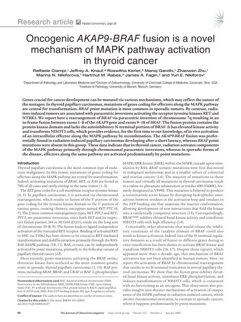
Oncogenic AKAP9-BRAF fusion is a novel mechanism of MAPK ...
Oncogenic AKAP9-BRAF fusion is a novel mechanism of MAPK ...
Oncogenic AKAP9-BRAF fusion is a novel mechanism of MAPK ...
Create successful ePaper yourself
Turn your PDF publications into a flip-book with our unique Google optimized e-Paper software.
Research article<br />
Related Commentary, page 20<br />
<strong>Oncogenic</strong> <strong>AKAP9</strong>-<strong>BRAF</strong> <strong>fusion</strong> <strong>is</strong> a <strong>novel</strong><br />
mechan<strong>is</strong>m <strong>of</strong> <strong>MAPK</strong> pathway activation<br />
in thyroid cancer<br />
Raffaele Ciampi, 1 Jeffrey A. Knauf, 2 Roswitha Kerler, 3 Manoj Gandhi, 1 Zhaowen Zhu, 1<br />
Marina N. Nikiforova, 1 Hartmut M. Rabes, 3 James A. Fagin, 2 and Yuri E. Nikiforov 1<br />
1<br />
Department <strong>of</strong> Pathology and Laboratory Medicine and 2 Div<strong>is</strong>ion <strong>of</strong> Endocrinology, University <strong>of</strong> Cincinnati College <strong>of</strong> Medicine, Cincinnati, Ohio, USA.<br />
3<br />
Institute <strong>of</strong> Pathology, University <strong>of</strong> Munich, Munich, Germany.<br />
Genes crucial for cancer development can be mutated via various mechan<strong>is</strong>ms, which may reflect the nature <strong>of</strong><br />
the mutagen. In thyroid papillary carcinomas, mutations <strong>of</strong> genes coding for effectors along the <strong>MAPK</strong> pathway<br />
are central for transformation. <strong>BRAF</strong> point mutation <strong>is</strong> most common in sporadic tumors. By contrast, radiation-induced<br />
tumors are associated with paracentric inversions activating the receptor tyrosine kinases RET and<br />
NTRK1. We report here a rearrangement <strong>of</strong> <strong>BRAF</strong> via paracentric inversion <strong>of</strong> chromosome 7q resulting in an<br />
in-frame <strong>fusion</strong> between exons 1–8 <strong>of</strong> the <strong>AKAP9</strong> gene and exons 9–18 <strong>of</strong> <strong>BRAF</strong>. The <strong>fusion</strong> protein contains the<br />
protein kinase domain and lacks the autoinhibitory N-terminal portion <strong>of</strong> <strong>BRAF</strong>. It has elevated kinase activity<br />
and transforms NIH3T3 cells, which provides evidence, for the first time to our knowledge, <strong>of</strong> in vivo activation<br />
<strong>of</strong> an intracellular effector along the <strong>MAPK</strong> pathway by recombination. The <strong>AKAP9</strong>-<strong>BRAF</strong> <strong>fusion</strong> was preferentially<br />
found in radiation-induced papillary carcinomas developing after a short latency, whereas <strong>BRAF</strong> point<br />
mutations were absent in th<strong>is</strong> group. These data indicate that in thyroid cancer, radiation activates components<br />
<strong>of</strong> the <strong>MAPK</strong> pathway primarily through chromosomal paracentric inversions, whereas in sporadic forms <strong>of</strong><br />
the d<strong>is</strong>ease, effectors along the same pathway are activated predominantly by point mutations.<br />
Introduction<br />
Thyroid papillary carcinoma <strong>is</strong> the most common type <strong>of</strong> endocrine<br />
malignancy. In th<strong>is</strong> tumor, mutations <strong>of</strong> genes coding for<br />
effectors along the <strong>MAPK</strong> pathway are central for transformation.<br />
Indeed, activating mutations <strong>of</strong> <strong>BRAF</strong>, RET, or RAS are found in<br />
70% <strong>of</strong> all cases and rarely overlap in the same tumor (1–3).<br />
The RET gene codes for a cell membrane receptor tyrosine kinase<br />
(4, 5). In papillary carcinomas, it <strong>is</strong> activated via chromosomal<br />
rearrangement, which results in <strong>fusion</strong> <strong>of</strong> the 3′ portion <strong>of</strong> the<br />
gene coding for the tyrosine kinase domain to the 5′ portion <strong>of</strong><br />
various genes, creating chimeric oncogenes named RET/PTC (6,<br />
7). The 2 most common rearrangement types, RET/PTC1 and RET/<br />
PTC3, are paracentric inversions, since both RET and its respective<br />
<strong>fusion</strong> partner, H4 or NCOA4 (ELE1), reside on the long arm<br />
<strong>of</strong> chromosome 10 (8, 9). The <strong>fusion</strong> leads to ligand-independent<br />
activation <strong>of</strong> the truncated RET receptor. Binding <strong>of</strong> activated RET<br />
to SHC via Y1062 has been shown to be critical to RET-mediated<br />
transformation and dedifferentiation, primarily through the RAS-<br />
RAF-<strong>MAPK</strong> pathway (10, 11). RAS, in turn, can be independently<br />
activated by point mutations, primarily in the follicular variant <strong>of</strong><br />
papillary thyroid cancer (12).<br />
Most recently, point mutations activating the <strong>BRAF</strong> serine/<br />
threonine kinase have surfaced as the most common genetic<br />
event in sporadic thyroid papillary carcinomas (1, 13). RAF proteins,<br />
including ARAF, <strong>BRAF</strong>, and CRAF or RAF-1, phosphorylate<br />
Nonstandard abbreviations used: BAC, bacterial artificial chromosome; FISH,<br />
fluorescence in situ hybridization; MEK, <strong>MAPK</strong>/ERK kinase; ORF, open reading<br />
frame; PAC, P1 artificial chromosome; PKA, protein kinase A; RACE, rapid amplification<br />
<strong>of</strong> cDNA ends; RBD, RAS-GTP binding domain; RII, type II regulatory subunit.<br />
Conflict <strong>of</strong> interest: The authors have declared that no conflict <strong>of</strong> interest ex<strong>is</strong>ts.<br />
Citation for th<strong>is</strong> article: J. Clin. Invest. 115:94–101 (2005).<br />
doi:10.1172/JCI200523237.<br />
<strong>MAPK</strong>/ERK kinase (MEK) within the <strong>MAPK</strong> cascade upon stimulation<br />
by RAS. <strong>BRAF</strong> somatic mutations were first d<strong>is</strong>covered<br />
in malignant melanomas and in a smaller subset <strong>of</strong> colorectal<br />
and ovarian cancers (14). The majority <strong>of</strong> mutations in those<br />
tumors and virtually all mutations in thyroid carcinomas result<br />
in a valine-to-glutamate substitution at residue 600 (V600E), formerly<br />
designated as V599E. Th<strong>is</strong> mutation <strong>is</strong> believed to produce<br />
a constitutively active kinase by d<strong>is</strong>rupting hydrophobic interactions<br />
between residues in the activation loop and residues in<br />
the ATP binding site that maintain the inactive conformation,<br />
allowing development <strong>of</strong> new interactions that fold the kinase<br />
into a catalytically competent structure (15). Correspondingly,<br />
<strong>BRAF</strong> V600E exhibits elevated basal kinase activity and transforms<br />
NIH3T3 cells with high efficiency (14).<br />
Conceivably, other alterations that would release the inhibitory<br />
constrains <strong>of</strong> the catalytic domain <strong>of</strong> <strong>BRAF</strong> could also<br />
result in kinase activation. Indeed, loss <strong>of</strong> the N-terminal regulatory<br />
domains as a result <strong>of</strong> <strong>fusion</strong> to different genes during in<br />
vitro transfection has been shown to activate <strong>BRAF</strong> kinase and<br />
transform NIH3T3 cells (16, 17). Despite those reports, which<br />
appeared more than a decade ago, th<strong>is</strong> mechan<strong>is</strong>m <strong>of</strong> <strong>BRAF</strong><br />
activation has not been identified in human tumors. Here, we<br />
report the activation <strong>of</strong> <strong>BRAF</strong> by chromosomal rearrangement<br />
that results in its N-terminal truncation in several papillary thyroid<br />
carcinomas. We show that the <strong>fusion</strong> gene exhibits elevated<br />
basal kinase activity, stimulates ERK phosphorylation, and<br />
induces transformation <strong>of</strong> NIH3T3 cells, which <strong>is</strong> cons<strong>is</strong>tent<br />
with its functioning as an oncogene. Th<strong>is</strong> observation also provides<br />
insights into d<strong>is</strong>tinct mechan<strong>is</strong>ms <strong>of</strong> activation <strong>of</strong> components<br />
<strong>of</strong> the <strong>MAPK</strong> pathway in radiation-induced cancers, which<br />
involve chromosomal inversions, in contrast to sporadic cancers,<br />
where it happens predominantly by point mutations.<br />
94 The Journal <strong>of</strong> Clinical Investigation http://www.jci.org Volume 115 Number 1 January 2005
esearch article<br />
Figure 1<br />
Identification <strong>of</strong> the <strong>BRAF</strong> gene rearrangement. (A) Genomic region<br />
on 7q containing the <strong>BRAF</strong> gene and position <strong>of</strong> PAC clones used as<br />
a probe for FISH. (B) Interphase nucleus from the index tumor showing<br />
split <strong>of</strong> 1 <strong>BRAF</strong> signal (red) and preservation <strong>of</strong> 2 chromosome<br />
7 centromeric signals (green), which indicates the rearrangement <strong>of</strong><br />
the <strong>BRAF</strong> gene.<br />
Results<br />
Identification <strong>of</strong> <strong>BRAF</strong> rearrangement. The possibility <strong>of</strong> <strong>BRAF</strong> involvement<br />
in chromosomal rearrangement was studied by fluorescence<br />
in situ hybridization (FISH). A contig <strong>of</strong> 3 P1 artificial chromosome<br />
(PAC) clones <strong>of</strong> approximately 330 kb, spanning the entire<br />
<strong>BRAF</strong> gene, was assembled to use as a probe (Figure 1A). Benign<br />
thyroid cells showed, as expected, 2 signals. Analys<strong>is</strong> <strong>of</strong> 32 papillary<br />
thyroid carcinomas revealed 1 tumor containing 3 signals, whereas<br />
subsequent hybridization with a chromosome 7 centromeric probe<br />
demonstrated the presence <strong>of</strong> 2 copies <strong>of</strong> the chromosome (Figure<br />
1B). Since the split <strong>of</strong> one <strong>BRAF</strong> signal resulted in a pair <strong>of</strong> signals<br />
located at a relatively constant d<strong>is</strong>tance from each other, the likely<br />
mechan<strong>is</strong>m for th<strong>is</strong> event was intrachromosomal inversion.<br />
Identification and characterization <strong>of</strong> the <strong>fusion</strong> partner. The identification<br />
<strong>of</strong> the <strong>fusion</strong> partner was achieved by 5′ rapid amplification<br />
<strong>of</strong> cDNA ends (RACE) using tumor poly(A) + RNA and<br />
a pool <strong>of</strong> primers designed along the 3′ end <strong>of</strong> the <strong>BRAF</strong> gene.<br />
RACE products were sequenced, and several <strong>of</strong> them revealed a<br />
<strong>fusion</strong> between exon 9 <strong>of</strong> <strong>BRAF</strong> and exon 8 <strong>of</strong> the A-kinase anchor<br />
protein 9 (<strong>AKAP9</strong>) gene (Figure 2A). Confirmation <strong>of</strong> the <strong>fusion</strong><br />
was obtained by RT-PCR with primers located in exon 8 <strong>of</strong> <strong>AKAP9</strong><br />
and exon 10 <strong>of</strong> <strong>BRAF</strong>, which yielded the expected product <strong>of</strong> 181<br />
bp. Genomic DNA was used for PCR to identify the <strong>fusion</strong> at the<br />
genomic level, which was found to reside between nucleotide 364<br />
<strong>of</strong> <strong>AKAP9</strong> intron 8 and nucleotide 4,930 <strong>of</strong> <strong>BRAF</strong> intron 8. The<br />
reciprocal <strong>fusion</strong> gene product was subsequently identified by<br />
RT-PCR using tumor cDNA, indicating the reciprocal nature <strong>of</strong><br />
the rearrangement. Further confirmation was obtained by doublecolor<br />
FISH with probes corresponding to the <strong>BRAF</strong> and <strong>AKAP9</strong><br />
genes (Figure 2B). Since the <strong>BRAF</strong> gene <strong>is</strong> located on chromosome<br />
7q34 (18) and <strong>AKAP9</strong> on 7q21–q22 (19–21), the <strong>AKAP9</strong>-<strong>BRAF</strong><br />
<strong>fusion</strong> results from inv(7)(q21–22q34) (Figure 2C). Using primers<br />
located within the 5′ and 3′ untranslated region <strong>of</strong> <strong>AKAP9</strong> and<br />
<strong>BRAF</strong>, respectively, the <strong>AKAP9</strong>-<strong>BRAF</strong> coding cDNA sequence was<br />
amplified from the tumor, cloned into the pCR-XL-TOPO vector,<br />
and sequenced (GenBank accession number AY803272). Analys<strong>is</strong><br />
<strong>of</strong> the sequence (4,567 bp total size) revealed a 4,476-bp open<br />
reading frame (ORF) containing exons 1–8 <strong>of</strong> <strong>AKAP9</strong> fused inframe<br />
with exons 9–18 <strong>of</strong> <strong>BRAF</strong> (Figure 2D). Based on the known<br />
Figure 2<br />
<strong>BRAF</strong> <strong>is</strong> recombined with the <strong>AKAP9</strong> gene and results in expression<br />
<strong>of</strong> a <strong>fusion</strong> protein. (A) Sequence <strong>of</strong> the 5′ RACE product showing<br />
a <strong>fusion</strong> point between <strong>AKAP9</strong> and <strong>BRAF</strong>. (B) Confirmation <strong>of</strong> the<br />
reciprocal <strong>fusion</strong> by FISH with probes corresponding to the <strong>BRAF</strong><br />
(red) and <strong>AKAP9</strong> (green) genes. (C) The <strong>fusion</strong> <strong>is</strong> a result <strong>of</strong> paracentric<br />
chromosomal inversion inv(7)(q21–22q34). Chr., chromosome.<br />
(D) Genomic structure <strong>of</strong> the <strong>BRAF</strong> and <strong>AKAP9</strong> genes showing the<br />
location <strong>of</strong> breakpoints (arrows) and the organization <strong>of</strong> the chimeric<br />
cDNA. Exons are represented by boxes and introns by lines. Numbers<br />
above indicate exon numbers. (E) Western blot analys<strong>is</strong> using <strong>BRAF</strong><br />
antibody (left) and <strong>AKAP9</strong> antibody (right), showing an approximately<br />
170-kDa protein, corresponding to the predicted molecular weight <strong>of</strong><br />
the <strong>fusion</strong> protein in the index case (number 02-28). Three other papillary<br />
carcinomas (T1–T3) are shown for compar<strong>is</strong>on. Wild-type <strong>AKAP9</strong><br />
<strong>is</strong> 453 kDa in size and was not detected in th<strong>is</strong> Western blot.<br />
The Journal <strong>of</strong> Clinical Investigation http://www.jci.org Volume 115 Number 1 January 2005 95
esearch article<br />
Figure 3<br />
Subcellular localization <strong>of</strong> <strong>AKAP9</strong> and <strong>AKAP9</strong>-<strong>BRAF</strong> detected by<br />
immunoh<strong>is</strong>tochem<strong>is</strong>try with an <strong>AKAP9</strong> antibody. (A) In a papillary thyroid<br />
carcinoma negative for the <strong>fusion</strong>, the protein <strong>is</strong> predominantly targeted<br />
to centrosomes, seen as a single perinuclear dot. There <strong>is</strong> also weak<br />
diffuse cytoplasmic staining. (B) The tumor expressing <strong>AKAP9</strong>-<strong>BRAF</strong><br />
reveals diffuse cytoplasmic d<strong>is</strong>tribution <strong>of</strong> the chimeric protein and the<br />
perinuclear dot <strong>is</strong> no longer v<strong>is</strong>ible. The shift in localization involves only<br />
tumor cells but not adjacent vascular endothelial cells, which preserve<br />
the dotted pattern <strong>of</strong> staining (arrow). Magnification, ×200.<br />
sequences <strong>of</strong> the untranslated regions <strong>of</strong> both genes, the predicted<br />
length <strong>of</strong> the complete <strong>AKAP9</strong>-<strong>BRAF</strong> cDNA <strong>is</strong> 4,849 bp. Four transcript<br />
variants <strong>of</strong> human <strong>AKAP9</strong> have been previously identified<br />
based on several coding region differences. The <strong>AKAP9</strong> sequence<br />
<strong>of</strong> the chimeric cDNA lacked 36 nucleotides at position 269–304<br />
<strong>of</strong> transcript version 1 (GenBank accession number NM_147171),<br />
which indicates that it corresponded to either transcript version<br />
2 or 3 <strong>of</strong> the gene. Multiple RT-PCR reactions revealed no splice<br />
variants <strong>of</strong> the <strong>fusion</strong> transcript in the tumor cDNA. Western<br />
blot analys<strong>is</strong> <strong>of</strong> the tumor using an antibody to the C-terminus <strong>of</strong><br />
<strong>BRAF</strong> showed a band <strong>of</strong> approximately 170 kDa, corresponding<br />
to the predicted molecular weight <strong>of</strong> 172 kDa for the <strong>fusion</strong> protein<br />
(Figure 2E). The same band was detected with an antibody<br />
against the N-terminus <strong>of</strong> <strong>AKAP9</strong>.<br />
The wild-type <strong>AKAP9</strong> gene encodes a 453-kDa protein capable<br />
<strong>of</strong> binding the type II regulatory subunit (RII) <strong>of</strong> protein kinase<br />
A (PKA) as well as other signaling proteins, anchoring them to<br />
the centrosome and Golgi apparatus (19–21). Several <strong>is</strong><strong>of</strong>orms<br />
ar<strong>is</strong>ing through alternative splicing are selectively expressed in<br />
most human t<strong>is</strong>sues at low abundance (19, 20). The <strong>AKAP9</strong> protein<br />
localizes by immun<strong>of</strong>luorescence as a single perinuclear dot<br />
and some surrounding d<strong>is</strong>persed fluorescence, corresponding to<br />
the centrosome and Golgi compartments, respectively, in interphase<br />
HeLa and SaOS2 osteosarcoma cells (19, 20). Based on the<br />
sequence <strong>of</strong> the chimeric cDNA, the <strong>fusion</strong> protein <strong>is</strong> predicted to<br />
lack 2 putative RII binding regions and the centrosome localization<br />
domain <strong>of</strong> <strong>AKAP9</strong> (20, 22, 23).<br />
In papillary thyroid carcinomas negative for the <strong>fusion</strong> and<br />
in non-neoplastic thyroid t<strong>is</strong>sue, <strong>AKAP9</strong> expression was readily<br />
detectable by RT-PCR (data not shown), and the protein was seen<br />
as a single perinuclear dot with faint diffuse cytoplasmic staining<br />
(Figure 3A). In the tumor expressing the <strong>AKAP9</strong>-<strong>BRAF</strong> protein,<br />
the perinuclear dot was no longer identifiable, whereas diffuse<br />
staining was observed in the cytoplasm (Figure 3B). However, the<br />
presence <strong>of</strong> intact centrosomes in the tumor cells was confirmed<br />
by immunoh<strong>is</strong>tochem<strong>is</strong>try with an antibody against the centrosomal<br />
protein γ–tubulin (data not shown). Th<strong>is</strong> <strong>is</strong> cons<strong>is</strong>tent with<br />
the absence <strong>of</strong> the centrosomal localization domain <strong>of</strong> <strong>AKAP9</strong> in<br />
the <strong>fusion</strong> protein, which prevents its preferential targeting to the<br />
centrosomal compartment.<br />
Functional characterization <strong>of</strong> the <strong>AKAP9</strong>-<strong>BRAF</strong> <strong>fusion</strong>. The wild-type<br />
human <strong>BRAF</strong> gene codes for an 84-kDa serine/threonine kinase in<br />
the <strong>MAPK</strong> pathway (16, 24). Structurally, <strong>BRAF</strong>, similar to other<br />
RAF kinases, contains 2 highly conserved N-terminal regulatory<br />
Figure 4<br />
The <strong>AKAP9</strong>-<strong>BRAF</strong> chimeric protein has increased <strong>BRAF</strong> kinase activity and results in NIH3T3 cell transformation. (A) In vitro kinase activity <strong>of</strong><br />
myc-tagged <strong>BRAF</strong> WT , <strong>AKAP9</strong>-<strong>BRAF</strong>, and <strong>BRAF</strong> V600E in the absence or presence <strong>of</strong> H-RAS G12V in COS7 cells. <strong>BRAF</strong> kinase activity was measured<br />
in myc-IgG immunoprecipitates using MEK1 substrate phosphorylation as a read-out. Each sample was assayed in triplicate, and bars<br />
represent the standard deviations from the mean. Similar results were obtained in 2 independent transfections. (B) Western blots <strong>of</strong> lysate from<br />
cells stably transfected with pEFP, pEFP-<strong>BRAF</strong> WT , pEFP–<strong>AKAP9</strong>-<strong>BRAF</strong>, pEFP-<strong>BRAF</strong> V600E , or pBabe puro–H-RAS G12V probed with antibodies<br />
to phospho-ERK1/2 (pERK), total ERK (tERK), or C-terminus <strong>of</strong> <strong>BRAF</strong>. Similar results were obtained in 3 additional independent experiments.<br />
(C) Foci formation <strong>of</strong> NIH3T3 cells transfected with pEFP-<strong>BRAF</strong> WT , pEFP–<strong>AKAP9</strong>-<strong>BRAF</strong>, pEFP-<strong>BRAF</strong> V600E , or pBabe puro–H-RAS G12V . Bars<br />
represent the average number <strong>of</strong> foci per plate from 6 plates. Similar results were obtained in 2 independent experiments.<br />
96 The Journal <strong>of</strong> Clinical Investigation http://www.jci.org Volume 115 Number 1 January 2005
esearch article<br />
Table 1<br />
Prevalence <strong>of</strong> <strong>BRAF</strong> alterations in papillary thyroid carcinomas<br />
domains, CR1 and CR2, that mediate autoinhibition <strong>of</strong> <strong>BRAF</strong> and<br />
a C-terminal protein kinase domain (CR3). CR1 includes the RAS-<br />
GTP binding domain (RBD), which mediates RAS binding, triggering<br />
the recruitment <strong>of</strong> the protein to the cell membrane and kinase<br />
activation (25). Based on the cDNA sequence, the <strong>AKAP9</strong>-<strong>BRAF</strong><br />
<strong>fusion</strong> protein <strong>is</strong> predicted to lack the <strong>BRAF</strong> N-terminal regulatory<br />
domains, including RBD, and to retain an intact kinase domain.<br />
Loss <strong>of</strong> the regulatory domains would be expected to lead to constitutive<br />
activation <strong>of</strong> <strong>BRAF</strong> kinase in a RAS-independent manner.<br />
We characterized the kinase activity <strong>of</strong> the <strong>AKAP9</strong>-<strong>BRAF</strong> <strong>fusion</strong><br />
protein by transient transfection <strong>of</strong> myc epitope–tagged versions<br />
<strong>of</strong> cDNAs for <strong>AKAP9</strong>-<strong>BRAF</strong>, <strong>BRAF</strong> V600E , or wild-type <strong>BRAF</strong> into<br />
COS7 cells, using bacterially purified MEK1 as a substrate for<br />
phosphorylation (Figure 4A). Basal <strong>BRAF</strong> kinase activity <strong>of</strong> the<br />
<strong>fusion</strong> protein was 6-fold greater than <strong>BRAF</strong> WT activity and not<br />
further increased by coexpression <strong>of</strong> activated RAS. Basal activity<br />
<strong>of</strong> <strong>BRAF</strong> V600E was also increased, but to a lesser extent, and was<br />
modestly increased by H-RAS G12V . By contrast, H-RAS G12V increased<br />
<strong>BRAF</strong> WT kinase activity by about 2.5-fold. The <strong>fusion</strong> protein stimulated<br />
endogenous ERK1/2 phosphorylation to a similar extent<br />
as <strong>BRAF</strong> V600E in pooled stably transfected NIH3T3 cells, whereas<br />
overexpression <strong>of</strong> <strong>BRAF</strong> WT had no d<strong>is</strong>cernible effect (Figure 4B).<br />
The ability <strong>of</strong> <strong>AKAP9</strong>-<strong>BRAF</strong> <strong>fusion</strong> protein to induce transformation<br />
was studied in an NIH3T3 cell focus assay (Figure 4C). Vector<br />
and <strong>BRAF</strong> WT transformed cells at very low efficiency. <strong>AKAP9</strong>-<strong>BRAF</strong><br />
showed the highest transformation activity in th<strong>is</strong> assay, followed<br />
by H-RAS G12V and <strong>BRAF</strong> V600E . In addition, NIH3T3 cells stably<br />
transfected with empty vector, <strong>BRAF</strong> WT , <strong>AKAP9</strong>-<strong>BRAF</strong>, <strong>BRAF</strong> V600E ,<br />
and H-RAS G12V were assayed for tumorigenicity in nude mice.<br />
Two weeks after injection <strong>of</strong> 10 6 cells, tumors <strong>of</strong> at least 5 mm in<br />
diameter were formed with complete penetrance (6 out <strong>of</strong> 6 animals,<br />
repeated in 2 independent experiments) for <strong>AKAP9</strong>-<strong>BRAF</strong>,<br />
<strong>BRAF</strong> V600E , and H-RAS G12V constructs, whereas vector and <strong>BRAF</strong> WT<br />
did not lead to the development <strong>of</strong> tumors (0 our <strong>of</strong> 6 animals).<br />
These results demonstrate that <strong>AKAP9</strong>-<strong>BRAF</strong> <strong>fusion</strong> leads to activation<br />
<strong>of</strong> <strong>BRAF</strong> kinase and transformation <strong>of</strong> NIH3T3 cells.<br />
Prevalence <strong>of</strong> <strong>AKAP9</strong>-<strong>BRAF</strong> <strong>fusion</strong> and <strong>BRAF</strong> point mutation in thyroid<br />
cancer. To determine the prevalence <strong>of</strong> <strong>AKAP9</strong>-<strong>BRAF</strong> <strong>fusion</strong><br />
in various populations, we used RT-PCR with primers flanking<br />
the <strong>fusion</strong> point to study a large series <strong>of</strong> papillary thyroid carcinomas<br />
(Table 1). In a group <strong>of</strong> 102 sporadic papillary tumors,<br />
no additional case <strong>of</strong> the <strong>fusion</strong> was found. We also examined<br />
papillary carcinomas from 2 cohorts <strong>of</strong> Belarussian children and<br />
adolescents exposed to radiation after the Chernobyl nuclear accident<br />
in 1986. Among 28 tumors that developed 5–6 years after<br />
exposure, 3 (11%) were positive for the <strong>fusion</strong>, whereas 64 tumors<br />
developing 9–12 years after the accident were all negative (Figure<br />
n Age at surgery Age at exposure <strong>AKAP9</strong>-<strong>BRAF</strong> <strong>BRAF</strong> V600E<br />
Early radiation–associated tumors 28 11.4 ± 3.6 5.0 ± 3.8 11% A 0 B<br />
(latency period 5–6 yr)<br />
Late radiation–associated tumors 64 16.0 ± 5.0 5.4 ± 5.1 0 16%<br />
(latency period 9–12 yr)<br />
Sporadic tumors 102 40.0 ± 17.7 – 1% 37%<br />
A<br />
P = 0.03 compared with sporadic tumors and late radiation–associated tumors. B P < 0.0001 compared with sporadic<br />
tumors; P = 0.03 compared with late radiation–associated tumors.<br />
5A). Paraffin blocks were available<br />
for immunoh<strong>is</strong>tochemical<br />
analys<strong>is</strong> with <strong>AKAP9</strong> antibody<br />
in 2 out <strong>of</strong> 3 positive cases.<br />
Both cases showed a shift in<br />
subcellular localization <strong>of</strong><br />
<strong>AKAP9</strong> from the centrosomal<br />
pattern seen in adjacent nonneoplastic<br />
thyroid t<strong>is</strong>sue to diffuse<br />
cytoplasmic d<strong>is</strong>tribution<br />
in tumor cells (Figure 5B).<br />
We also tested these tumors<br />
for <strong>BRAF</strong> point mutation leading<br />
to the V600E substitution. The mutation was identified in 38<br />
(37%) sporadic tumors, in 10 (16%) tumors developing 9–12 years<br />
after exposure, but in none <strong>of</strong> the tumors that developed 5–6 years<br />
after exposure (Figure 6). The differences in prevalence <strong>of</strong> the 2 types<br />
<strong>of</strong> <strong>BRAF</strong> activation were stat<strong>is</strong>tically significant between the early radiation<br />
group and both late radiation and sporadic groups (Table 1).<br />
D<strong>is</strong>cussion<br />
The <strong>MAPK</strong> signaling pathway <strong>is</strong> a ubiquitously present regulator <strong>of</strong><br />
cell growth, proliferation, apoptos<strong>is</strong>, and differentiation. Recently,<br />
<strong>BRAF</strong>, an essential component <strong>of</strong> the kinase cascade, has received<br />
renewed attention since mutations in the gene were identified in<br />
several types <strong>of</strong> human cancer. Indeed, activating point mutations <strong>of</strong><br />
<strong>BRAF</strong> have been found in more than 60% <strong>of</strong> malignant melanomas,<br />
10–14% <strong>of</strong> colorectal and ovarian cancer, and in very few cases <strong>of</strong> lung<br />
cancer, s<strong>of</strong>t t<strong>is</strong>sue sarcomas, and other cancer cell lines (14, 26, 27).<br />
Thyroid cancer was promptly added to the l<strong>is</strong>t with 36–69% prevalence<br />
<strong>of</strong> <strong>BRAF</strong> mutation (1, 2, 13, 28). However, neither <strong>BRAF</strong> nor other<br />
Figure 5<br />
Analys<strong>is</strong> <strong>of</strong> post-Chernobyl tumors that developed 5–6 years after<br />
exposure. (A) Southern blot <strong>of</strong> RT-PCR products amplified with primers<br />
Ex8A and Ex10B and hybridized with probe PR showing 2 positive<br />
cases (C32 and C27). Other tumor samples are negative (C13, C23,<br />
C24, C18, and C31). NC, negative control; PC, positive control (index<br />
case). (B) Immunoh<strong>is</strong>tochemical analys<strong>is</strong> <strong>of</strong> tumor C32 with <strong>AKAP9</strong><br />
antibody showing a shift in subcellular localization <strong>of</strong> the protein from<br />
a single dot/centrosomal pattern observed in the entrapped non-neoplastic<br />
follicles (arrows) to diffuse cytoplasmic staining in tumor cells.<br />
Magnification, ×200.<br />
The Journal <strong>of</strong> Clinical Investigation http://www.jci.org Volume 115 Number 1 January 2005 97
esearch article<br />
Figure 6<br />
Detection <strong>of</strong> V600E <strong>BRAF</strong> mutation cDNA from post-Chernobyl tumors<br />
that developed 9–12 years after exposure using LightCycler real-time<br />
PCR followed by fluorescence melting curve analys<strong>is</strong>. (A) Real-time<br />
amplification <strong>of</strong> cDNA samples. (B) Detection <strong>of</strong> point mutations by<br />
the postamplification fluorescence melting curve analys<strong>is</strong> based on<br />
a d<strong>is</strong>tinct melting temperature <strong>of</strong> duplexes formed between the WT<br />
probe and either WT (62.8°C) or mutant (57.0°C) sequences. The<br />
mutations in both samples are heterozygous, since both mutant and<br />
WT peaks are detectable.<br />
intracellular effectors along the <strong>MAPK</strong> pathway have been previously<br />
implicated in human carcinogenes<strong>is</strong> through mechan<strong>is</strong>ms other than<br />
point mutation. In the present study, we demonstrate <strong>BRAF</strong> activation<br />
by chromosomal rearrangement that results in <strong>BRAF</strong> <strong>fusion</strong> to<br />
the <strong>AKAP9</strong> gene in thyroid papillary carcinomas. The <strong>fusion</strong> protein<br />
lacks the regulatory domains <strong>of</strong> <strong>BRAF</strong>, which results in constitutive<br />
kinase activation in a RAS-independent manner, and exhibits transforming<br />
activity similar to that <strong>of</strong> the most common <strong>BRAF</strong> V600E .<br />
The <strong>fusion</strong> partner <strong>of</strong> <strong>BRAF</strong>, the <strong>AKAP9</strong> gene, belongs to the<br />
group <strong>of</strong> A-kinase anchor proteins that have the common function<br />
<strong>of</strong> binding to the regulatory subunit <strong>of</strong> PKA and targeting<br />
it to d<strong>is</strong>crete locations within the cell (29). <strong>AKAP9</strong>, in particular,<br />
has predominantly centrosomal and Golgi compartmentalization<br />
(19–21). The <strong>fusion</strong> protein lacks the C-terminal centrosomal<br />
domain and, as expected, loses its centrosomal localization<br />
in cancer cells. Moreover, immunoh<strong>is</strong>tochemical analys<strong>is</strong><br />
with <strong>AKAP9</strong> antibody revealed no evidence <strong>of</strong> wild-type <strong>AKAP9</strong><br />
within centrosomes <strong>of</strong> cancer cells in all cases positive for the<br />
<strong>fusion</strong> by RT-PCR. Th<strong>is</strong> may suggest the competitive inhibition<br />
<strong>of</strong> wild-type <strong>AKAP9</strong> by <strong>AKAP9</strong>-<strong>BRAF</strong>, although further studies<br />
are required to confirm th<strong>is</strong>. <strong>AKAP9</strong> <strong>is</strong> an important regulator<br />
<strong>of</strong> c-AMP–dependent PKA and interacts with many other<br />
signaling proteins, including serine/threonine kinase protein<br />
kinase N, protein phosphatase 1, protein phosphatase 2A, and<br />
PKC-ε (20, 30). Thus, deregulation <strong>of</strong> its function in cells harboring<br />
<strong>AKAP9</strong>-<strong>BRAF</strong> <strong>fusion</strong> may affect a variety <strong>of</strong> physiological<br />
functions. On the other hand, neither <strong>AKAP9</strong> nor other AKAP<br />
genes have been implicated in carcinogenes<strong>is</strong> so far. Therefore,<br />
it remains unclear whether or not the <strong>AKAP9</strong> portion <strong>of</strong> the<br />
<strong>fusion</strong> gene contributes independently to its transforming<br />
potential, besides providing an active promoter driving the<br />
expression <strong>of</strong> truncated <strong>BRAF</strong>.<br />
Over the last decade, it has become apparent that activation<br />
<strong>of</strong> components <strong>of</strong> the <strong>MAPK</strong> pathway in thyroid cancer can be<br />
achieved through several d<strong>is</strong>tinct genetic events, i.e., RET/PTC<br />
(or, less frequently, TRK) rearrangements, RAS point mutations,<br />
or <strong>BRAF</strong> point mutations. However, the relative frequency <strong>of</strong> each<br />
event varies significantly in different populations. <strong>BRAF</strong> point<br />
mutation <strong>is</strong> the most common change in sporadic adult papillary<br />
carcinomas. In contrast, tumors associated with accidental or<br />
therapeutic irradiation have exceedingly high frequency <strong>of</strong> RET/<br />
PTC rearrangements and absence or low prevalence <strong>of</strong> <strong>BRAF</strong> point<br />
mutations (31–38). The 2 most common RET/PTC types found in<br />
up to 80% <strong>of</strong> radiation-associated tumors, RET/PTC1 and RET/<br />
PTC3, are both paracentric inversions <strong>of</strong> chromosome 10q (39, 40).<br />
Another rearrangement, which contributes to about 7% <strong>of</strong> radiation-induced<br />
papillary carcinomas, involves the nerve growth factor<br />
gene NTRK1 and results from paracentric inversion <strong>of</strong> chromosome<br />
1q (41, 42). Here, we report the identification <strong>of</strong> another paracentric<br />
inversion, involving 7q, which <strong>is</strong> also more common in radiationassociated<br />
tumors, especially those appearing with a short latency.<br />
These tumors, however, revealed no <strong>BRAF</strong> point mutations. The<br />
sharp contrast in mechan<strong>is</strong>ms <strong>of</strong> activation <strong>of</strong> the same gene in<br />
radiation-induced and sporadic tumors points to the association<br />
between environmental factors and d<strong>is</strong>tinct genetic mechan<strong>is</strong>ms <strong>of</strong><br />
activation <strong>of</strong> <strong>MAPK</strong> pathway components: through chromosomal<br />
rearrangement in patients exposed to radiation or through point<br />
mutations in patients with no radiation h<strong>is</strong>tory (Figure 7).<br />
Figure 7<br />
Activation <strong>of</strong> <strong>MAPK</strong> pathway in papillary thyroid carcinogenes<strong>is</strong><br />
involves predominantly point mutations in sporadic tumors or chromosomal<br />
rearrangements in radiation-associated tumors.<br />
98 The Journal <strong>of</strong> Clinical Investigation http://www.jci.org Volume 115 Number 1 January 2005
esearch article<br />
The vast majority <strong>of</strong> RET/PTC and all TRK and <strong>AKAP9</strong>-<strong>BRAF</strong><br />
chromosomal rearrangements in radiation-induced tumors are<br />
paracentric inversions. In epithelial cancers as a whole, point<br />
mutations are more prevalent than chromosomal rearrangements,<br />
and the latter are usually translocations or interchromosomal<br />
exchanges. The reason for the striking pred<strong>is</strong>position <strong>of</strong><br />
radiation-induced thyroid cancers to paracentric inversions <strong>is</strong> not<br />
entirely clear, although some evidence suggests that th<strong>is</strong> may be<br />
related to the organization <strong>of</strong> chromosomal domains and arm<br />
domains and spatial interaction between pairs <strong>of</strong> various chromosomal<br />
regions. Indeed, there <strong>is</strong> a general consensus that free DNA<br />
ends produced in the nuclear volume after passage <strong>of</strong> radiation<br />
tracks have to be within a certain d<strong>is</strong>tance <strong>of</strong> each other in order<br />
to recombine (43, 44). Residing within the same chromosomal<br />
arm domain would limit their maximum separation and help to<br />
fulfill th<strong>is</strong> requirement. Indeed, the yields <strong>of</strong> intra-arm chromosomal<br />
exchanges after both low– and high–linear energy transfer<br />
radiation has been shown to exceed the expected levels, calculated<br />
based on spatial randomness, by up to 10-fold (45). The conditions<br />
would be even more favorable if potentially recombinogenic<br />
foci within the arm domain were frequently juxtaposed to each<br />
other, as was demonstrated in human thyroid cells for RET and<br />
H4, the RET/PTC1 partners (46).<br />
In th<strong>is</strong> study, we observed significant difference in mechan<strong>is</strong>ms<br />
<strong>of</strong> <strong>BRAF</strong> activation not only between early radiation–associated<br />
tumors and sporadic cancers, but also between early and<br />
late post-radiation tumors. Such a difference <strong>is</strong> not without<br />
precedent. Similar variation has been previously observed with<br />
respect to RET/PTC rearrangements between post-Chernobyl<br />
tumors developing 10 years or less after exposure and those<br />
manifesting after a longer latency (34, 35). Indeed, the overall<br />
prevalence <strong>of</strong> RET/PTC was significantly higher in the former<br />
group, and RET/PTC3 was the most common type. By contrast,<br />
tumors ar<strong>is</strong>ing after a longer latency had lower prevalence <strong>of</strong><br />
RET/PTC, and the predominant type was RET/PTC1, both <strong>of</strong><br />
which are features more typical <strong>of</strong> sporadic tumors. The present<br />
study demonstrates that late post-radiation tumors also occupy<br />
an intermediate position with respect to the predominant<br />
mechan<strong>is</strong>m <strong>of</strong> <strong>BRAF</strong> activation. Th<strong>is</strong> suggests that tumors ar<strong>is</strong>ing<br />
early after exposure are likely to be initiated through direct<br />
radiation damage–related genetic events, such as chromosomal<br />
rearrangements occurring after double-stranded DNA breaks.<br />
As for the tumors developing after a longer latency, their pathogenes<strong>is</strong><br />
may involve a combination <strong>of</strong> radiation-related and<br />
-unrelated mechan<strong>is</strong>ms and rely on molecular alterations which<br />
develop after repair <strong>of</strong> radiation-induced DNA damage that are<br />
stimulated by genomic instability that pers<strong>is</strong>ts in the progeny <strong>of</strong><br />
the exposed cells (47, 48).<br />
In summary, we report here a <strong>novel</strong> mechan<strong>is</strong>m <strong>of</strong> in vivo <strong>BRAF</strong><br />
activation via inversion <strong>of</strong> chromosome 7q, resulting in <strong>BRAF</strong><br />
<strong>fusion</strong> to the <strong>AKAP9</strong> gene. <strong>AKAP9</strong>-<strong>BRAF</strong> functions as an oncogene<br />
and <strong>is</strong> more prevalent in thyroid carcinomas with recent h<strong>is</strong>tory<br />
<strong>of</strong> radiation exposure. The significance <strong>of</strong> th<strong>is</strong> study <strong>is</strong> 2-fold. It<br />
demonstrates a <strong>novel</strong> mechan<strong>is</strong>m <strong>of</strong> <strong>BRAF</strong> activation in human<br />
tumors, showing for the first time that intracellular effectors<br />
along the <strong>MAPK</strong> pathway can be activated by recombination. In<br />
addition, it shows correlation between the nature <strong>of</strong> the mutagen<br />
and the mechan<strong>is</strong>m <strong>of</strong> mutation and suggests that paracentric<br />
chromosomal inversions represent a common genetic mechan<strong>is</strong>m<br />
<strong>of</strong> radiation-associated thyroid carcinogenes<strong>is</strong>.<br />
Methods<br />
Tumor samples. Frozen t<strong>is</strong>sues from sporadic thyroid carcinomas were<br />
obtained from the Department <strong>of</strong> Pathology, University <strong>of</strong> Cincinnati,<br />
following an institutionally approved t<strong>is</strong>sue procurement protocol<br />
and through the Cooperative Human T<strong>is</strong>sue Network. Post-Chernobyl<br />
tumors from Belarussian patients who underwent surgery in 1991–92<br />
were available as archival paraffin-embedded t<strong>is</strong>sue. Samples from<br />
Belarussian patients who underwent surgery in 1995–1998 were from<br />
the Experimental Div<strong>is</strong>ion <strong>of</strong> the Institute <strong>of</strong> Pathology, University <strong>of</strong><br />
Munich, and available as RNA extracted from frozen t<strong>is</strong>sue; they were<br />
negative for RET/PTC and TRK rearrangements (35). The study was<br />
approved by the University <strong>of</strong> Cincinnati Institutional Review Board.<br />
FISH analys<strong>is</strong>. Tumor touch preparations were made from snap-frozen<br />
t<strong>is</strong>sues. PAC clones for <strong>BRAF</strong> (RP4-726N20, RP5-839B19, RP4-813F11)<br />
were obtained from BAC/PAC Resources, Children’s Hospital Oakland,<br />
and bacterial artificial chromosome (BAC) clones for <strong>AKAP9</strong> (CTB-104F4,<br />
CTB-161K23) from Open Biosystems. The probes were labeled by nick<br />
translation using SpectrumOrange-dUTP and SpectrumGreen-dUTP<br />
(Vys<strong>is</strong> Inc.), respectively. An α-satellite DNA centromeric probe for chromosome<br />
7 was obtained from Vys<strong>is</strong> Inc. Nuclei were counterstained with<br />
DAPI. Microscopy was performed with a Leica Microsystems TCS 4D confocal<br />
laser scanning fluorescence microscope with digital image capture.<br />
One hundred nuclei were scored for each preparation.<br />
5′ RACE. Fifty micrograms <strong>of</strong> poly(A) + RNA from the index case were used<br />
for 5′ RACE using the 5′ RACE System for Rapid Amplification <strong>of</strong> cDNA<br />
Ends (Invitrogen Corp.). The <strong>BRAF</strong> gene–specific primers were designed<br />
for cDNA synthes<strong>is</strong> (5′-GATGACTTCTGGTGCCATCC-3′, 5′-AACTGCT-<br />
GAGGTGTAGGTGCTGTC-3′, and 5′-GTCTCGTTGCCCAAATTGAT-3′)<br />
and for subsequent PCR reactions (5′-TTTCACTGCCACATCACCAT-3′ for<br />
first PCR and 5′-TCACTCGAGTCCCGTCTACC-3′ for nested PCR). The 5′<br />
RACE products were exc<strong>is</strong>ed from the gel, purified, and sequenced using an<br />
automated ABI 377 sequencer (PerkinElmer).<br />
Cloning and sequencing analys<strong>is</strong>. Total RNA was used to amplify the ORF<br />
<strong>of</strong> the chimeric cDNA using primers located in the 5′-untranslated region<br />
<strong>of</strong> <strong>AKAP9</strong> (5′-ACCACCCCTCAACCCCTGTTTT-3′) and 3′-untranslated<br />
region <strong>of</strong> <strong>BRAF</strong> (5′-TTCCTTTTGTTGCTACTCTCCTGA-3′). The PCR<br />
product was electrophoresed in an agarose gel, purified from the gel, and<br />
cloned into the pCR-XL-TOPO vector (Invitrogen Corp.). Several clones<br />
were fully sequenced. The genomic <strong>AKAP9</strong>-<strong>BRAF</strong> <strong>fusion</strong> was amplified with<br />
primers 5′-GCAACTCAACCAAGTGAAAATG-3′ and 5′-GGTTGATCCTC-<br />
CATCACCAC-3′, yielding a product <strong>of</strong> approximately 2.4 kb, and the<br />
reciprocal <strong>fusion</strong> product cDNA was amplified with primers 5′-ACCC-<br />
GCCTCGGACTCTATT-3′ and 5′-GTTGGGCTTCCATCTGTAGC-3′,<br />
yielding a product <strong>of</strong> 242 bp.<br />
Western blot and immunoh<strong>is</strong>tochem<strong>is</strong>try. Frozen t<strong>is</strong>sues from papillary carcinomas<br />
were used for Western blotting, which was performed as previously<br />
described (49). We loaded 50 μg <strong>of</strong> protein on a 5% polyacrylamide<br />
gel. After transfer, the membranes were incubated with anti–C-terminus<br />
<strong>BRAF</strong> polyclonal (Raf-B, C-19; Santa Cruz Biotechnology Inc.) or anti–Nterminus<br />
<strong>AKAP9</strong> monoclonal (AKAP450; BD Biosciences) antibodies. The<br />
same <strong>AKAP9</strong> antibody in 1:50 dilution was used for immunoh<strong>is</strong>tochemical<br />
analys<strong>is</strong>, which was performed on paraffin sections after overnight pretreatment<br />
in borate buffer using avidin-streptavidin immunoperoxidase<br />
detection. Rabbit anti–γ-tubulin polyclonal antibody was provided by K.<br />
Fukasawa (University <strong>of</strong> Cincinnati College <strong>of</strong> Medicine, Cincinnati, Ohio,<br />
USA) (50) and was used in 1:800 dilution on paraffin sections after pretreatment<br />
in borate buffer.<br />
In vitro kinase assay. COS7 cells were transiently cotransfected with either<br />
pEFP vector or pEFP-containing myc-tagged <strong>BRAF</strong> WT , <strong>AKAP9</strong>-<strong>BRAF</strong>, or<br />
<strong>BRAF</strong> V600E and either pBabe puro or pBabe puro–HRAS G12V using Lipo-<br />
The Journal <strong>of</strong> Clinical Investigation http://www.jci.org Volume 115 Number 1 January 2005 99
esearch article<br />
fectamine 2000 (Invitrogen Corp.). After 48 hours, cell extracts were prepared,<br />
immunoprecipitated with mouse anti–myc-IgG (Upstate Cell Signaling<br />
Solutions), and kinase activity determined as directed by the manufacturer<br />
using bacterially purified MEK1 as substrate, and Western blotting with<br />
pMEK1 antibody as a read-out (B-RAF Kinase Assay Kit; Upstate Cell Signaling<br />
Solutions). To determine the effects <strong>of</strong> the <strong>BRAF</strong> mutants on endogenous<br />
ERK1/2 phosphorylation, we incubated pools <strong>of</strong> stably transfected<br />
cells for 12 hours without serum prior to lysate preparation. Lysates were<br />
centrifuged, the supernatant collected, and 100 μg <strong>of</strong> extract subjected to<br />
SDS-PAGE. Proteins were transferred to a nitrocellulose membrane and subject<br />
to Western blot analys<strong>is</strong> with anti–phospho-ERK1/2, anti–total ERK1/2,<br />
or anti-<strong>BRAF</strong> (Santa Cruz Biotechnology Inc.) antibodies.<br />
Transformation assays. For focus assays, NIH3T3 cells were transfected with<br />
empty vector or the indicated <strong>BRAF</strong> and H-RAS expression plasmids using<br />
lip<strong>of</strong>ectamine. After 2 days, cells were split into six 100-mm 3 d<strong>is</strong>hes and incubated<br />
with DMEM containing 5% calf serum for 21 days. Cells were stained<br />
with 0.4% crystal violet and foci counted. For the mouse xenograft experiments,<br />
NIH3T3 cells were cotransfected with pBabe puro and pEFP vector,<br />
pEFP–myc <strong>BRAF</strong> WT , pEFP–<strong>AKAP9</strong>-<strong>BRAF</strong>, pEFP–myc <strong>BRAF</strong> V600E , or pBabe<br />
puro–H-RAS G12V and mass selected by incubation with DMEM containing<br />
5% calf serum and 1 μg/ml puromycin for 3 weeks. A total <strong>of</strong> 10 6 pooled cells<br />
was injected into nude mice. Animals were killed after 2 weeks, and nodules<br />
larger than 5 mm in diameter were scored as positive.<br />
Mutation analys<strong>is</strong>. For the detection <strong>of</strong> <strong>AKAP9</strong>-<strong>BRAF</strong> <strong>fusion</strong>, cDNA amplification<br />
was achieved by PCR using primers located in exon 8 <strong>of</strong> <strong>AKAP9</strong><br />
(Ex8A, 5′-AGCAAGAACAGTTGATTTTGGA-3′) and exon 10 <strong>of</strong> <strong>BRAF</strong><br />
(Ex10B, 5′-GCAGACAAACCTGTGGTTGA-3′), with the expected product<br />
<strong>of</strong> 181 bp. The PCR products were electrophoresed on agarose gels and<br />
bands v<strong>is</strong>ualized by staining with ethidium bromide (frozen t<strong>is</strong>sues) or by<br />
transfer to a nylon filter and hybridization with an internal oligonucleotide<br />
probe (PR, 5′-AAACTTCAGAAAGAACTCAATGTACTT-3′) labeled<br />
with 50 μCi <strong>of</strong> [γ- 32 P]-ATP (paraffin-embedded t<strong>is</strong>sue). Detection <strong>of</strong> V600E<br />
<strong>BRAF</strong> mutation was performed from DNA using real-time PCR and fluorescence<br />
melting curve analys<strong>is</strong>, as previously reported (51), or from cDNA<br />
using real-time RT-PCR with primers 5′-CGACAGACTGCACAGG-3′ and<br />
5′-TGACTTCTGGTGCCAT-3′ and the same probes.<br />
Stat<strong>is</strong>tical analys<strong>is</strong>. The compar<strong>is</strong>on between groups was performed using<br />
a 2-tailed F<strong>is</strong>her exact test. Differences were considered significant when<br />
P was less than 0.05.<br />
Acknowledgments<br />
Th<strong>is</strong> study was supported by NIH grant R01 CA88041 and American<br />
Cancer Society grant RSG-03-027-01-CCE (to Y.E. Nikiforov),<br />
NIH grant R01 CA50706 (to J.A. Fagin), a grant from the American<br />
Cancer Society Ohio Chapter #06001011 (to J.A. Knauf), and a<br />
grant from Deutsche Krebshilfe, Bonn, Germany (to H.M. Rabes).<br />
Collection <strong>of</strong> t<strong>is</strong>sue samples was supported in part by a grant<br />
from the NIH (PHS M01 RR08084) and through the Cooperative<br />
Human T<strong>is</strong>sue Network funded by the National Cancer Institute.<br />
We thank Kenji Fukasawa for γ-tubulin antibody, Mickey Croyle<br />
for technical ass<strong>is</strong>tance, and Richard Mara<strong>is</strong> for the <strong>BRAF</strong> WT and<br />
<strong>BRAF</strong> V600E expression vectors.<br />
Received for publication September 1, 2004, and accepted in<br />
rev<strong>is</strong>ed form October 28, 2004.<br />
Address correspondence to: Yuri Nikiforov, Department <strong>of</strong> Pathology,<br />
University <strong>of</strong> Cincinnati, 231 Albert Sabin Way, PO Box<br />
670529, Cincinnati, Ohio 45267-0529, USA. Phone: (513) 558-<br />
5798; Fax: (513) 558-2289; E-mail: Yuri.Nikiforov@uc.edu.<br />
1. Kimura, E.T., et al. 2003. High prevalence <strong>of</strong> <strong>BRAF</strong><br />
mutations in thyroid cancer: genetic evidence for<br />
constitutive activation <strong>of</strong> the RET/PTC-RAS-<strong>BRAF</strong><br />
signaling pathway in papillary thyroid carcinoma.<br />
Cancer Res. 63:1454–1457.<br />
2. Soares, P., et al. 2003. <strong>BRAF</strong> mutations and RET/<br />
PTC rearrangements are alternative events in the<br />
etiopathogenes<strong>is</strong> <strong>of</strong> PTC. Oncogene. 22:4578–4580.<br />
3. Frattini, M., et al. 2004. Alternative mutations <strong>of</strong><br />
<strong>BRAF</strong>, RET and NTRK1 are associated with similar<br />
but d<strong>is</strong>tinct gene expression patterns in papillary<br />
thyroid cancer. Oncogene. 23:7436–7440.<br />
4. Takahashi, M., Ritz, J., and Cooper, G.M. 1985.<br />
Activation <strong>of</strong> a <strong>novel</strong> human transforming gene,<br />
ret, by DNA rearrangement. Cell. 42:581–588.<br />
5. Takahashi, M. 1988. Structure and expression<br />
<strong>of</strong> the ret transforming gene. IARC Sci. Publ.<br />
1988:189–197.<br />
6. Fusco, A., et al. 1987. A new oncogene in human<br />
thyroid papillary carcinomas and their lymphnodal<br />
metastases. Nature. 328:170–172.<br />
7. Grieco, M., et al. 1990. PTC <strong>is</strong> a <strong>novel</strong> rearranged<br />
form <strong>of</strong> the ret proto-oncogene and <strong>is</strong> frequently<br />
detected in vivo in human thyroid papillary carcinomas.<br />
Cell. 60:557–563.<br />
8. Pierotti, M.A., et al. 1992. Characterization <strong>of</strong> an<br />
inversion on the long arm <strong>of</strong> chromosome 10 juxtaposing<br />
D10S170 and RET and creating the oncogenic<br />
sequence RET/PTC. Proc. Natl. Acad. Sci. U. S. A.<br />
89:1616–1620.<br />
9. Minoletti, F., et al. 1994. The two genes generating<br />
RET/PTC3 are localized in chromosomal band<br />
10q11.2. Genes Chromosomes Cancer. 11:51–57.<br />
10. Asai, N., Murakami, H., Iwashita, T., and Takahashi,<br />
M. 1996. A mutation at tyrosine 1062 in<br />
MEN2A-Ret and MEN2B-Ret impairs their transforming<br />
activity and association with shc adaptor<br />
proteins. J. Biol. Chem. 271:17644–17649.<br />
11. Knauf, J.A., Kuroda, H., Basu, S., and Fagin, J.A.<br />
2003. RET/PTC-induced dedifferentiation <strong>of</strong><br />
thyroid cells <strong>is</strong> mediated through Y1062 signaling<br />
through SHC-RAS-MAP kinase. Oncogene.<br />
22:4406–4412.<br />
12. Zhu, Z., Gandhi, M., Nikiforova, M.N., F<strong>is</strong>cher,<br />
A.H., and Nikiforov, Y.E. 2003. Molecular pr<strong>of</strong>ile<br />
and clinical-pathologic features <strong>of</strong> the follicular<br />
variant <strong>of</strong> papillary thyroid carcinoma. An unusually<br />
high prevalence <strong>of</strong> ras mutations. Am. J. Clin.<br />
Pathol. 120:71–77.<br />
13. Cohen, Y., et al. 2003. <strong>BRAF</strong> mutation in papillary<br />
thyroid carcinoma. J. Natl. Cancer Inst. 95:625–627.<br />
14. Davies, H., et al. 2002. Mutations <strong>of</strong> the <strong>BRAF</strong> gene<br />
in human cancer. Nature. 417:949–954.<br />
15. Wan, P.T., et al. 2004. Mechan<strong>is</strong>m <strong>of</strong> activation<br />
<strong>of</strong> the RAF-ERK signaling pathway by oncogenic<br />
mutations <strong>of</strong> B-RAF. Cell. 116:855–867.<br />
16. Ikawa, S., et al. 1988. B-raf, a new member <strong>of</strong> the<br />
raf family, <strong>is</strong> activated by DNA rearrangement. Mol.<br />
Cell. Biol. 8:2651–2654.<br />
17. Miki, T., et al. 1991. Development <strong>of</strong> a highly efficient<br />
expression cDNA cloning system: application<br />
to oncogene <strong>is</strong>olation. Proc. Natl. Acad. Sci. U. S. A.<br />
88:5167–5171.<br />
18. Eychene, A., Barnier, J.V., Apiou, F., Dutrillaux, B.,<br />
and Calothy, G. 1992. Chromosomal assignment<br />
<strong>of</strong> two human B-raf(Rmil) proto-oncogene loci:<br />
B-raf-1 encoding the p94Braf/Rmil and B-raf-2, a<br />
processed pseudogene. Oncogene. 7:1657–1660.<br />
19. Witczak, O., et al. 1999. Cloning and characterization<br />
<strong>of</strong> a cDNA encoding an A-kinase anchoring<br />
protein located in the centrosome, AKAP450.<br />
EMBO J. 18:1858–1868.<br />
20. Takahashi, M., et al. 1999. Characterization <strong>of</strong> a <strong>novel</strong><br />
giant scaffolding protein, CG-NAP, that anchors<br />
multiple signaling enzymes to centrosome and the<br />
golgi apparatus. J. Biol. Chem. 274:17267–17274.<br />
21. Schmidt, P.H., et al. 1999. AKAP350, a multiply<br />
spliced protein kinase A-anchoring protein associated<br />
with centrosomes. J. Biol. Chem. 274:3055–3066.<br />
22. Gillingham, A.K., and Munro, S. 2000. The PACT<br />
domain, a conserved centrosomal targeting motif<br />
in the coiled-coil proteins AKAP450 and pericentrin.<br />
EMBO Rep. 1:524–529.<br />
23. Takahashi, M., Yamagiwa, A., N<strong>is</strong>himura, T.,<br />
Mukai, H., and Ono, Y. 2002. Centrosomal proteins<br />
CG-NAP and kendrin provide microtubule<br />
nucleation sites by anchoring gamma-tubulin ring<br />
complex. Mol. Biol. Cell. 13:3235–3245.<br />
24. Sithanandam, G., Kolch, W., Duh, F.M., and<br />
Rapp, U.R. 1990. Complete coding sequence<br />
<strong>of</strong> a human B-raf cDNA and detection <strong>of</strong> B-raf<br />
protein kinase with <strong>is</strong>ozyme specific antibodies.<br />
Oncogene. 5:1775–1780.<br />
25. Avruch, J., et al. 2001. Ras activation <strong>of</strong> the Raf<br />
kinase: tyrosine kinase recruitment <strong>of</strong> the MAP<br />
kinase cascade. Recent Prog. Horm. Res. 56:127–155.<br />
26. Brose, M.S., et al. 2002. <strong>BRAF</strong> and RAS mutations<br />
in human lung cancer and melanoma. Cancer Res.<br />
62:6997–7000.<br />
27. Rajagopalan, H., et al. 2002. Tumorigenes<strong>is</strong>:<br />
RAF/RAS oncogenes and m<strong>is</strong>match-repair status.<br />
Nature. 418:934.<br />
28. Xu, X., Quiros, R.M., Gattuso, P., Ain, K.B., and<br />
Prinz, R.A. 2003. High prevalence <strong>of</strong> <strong>BRAF</strong> gene<br />
mutation in papillary thyroid carcinomas and thyroid<br />
tumor cell lines. Cancer Res. 63:4561–4567.<br />
29. Edwards, A.S., and Scott, J.D. 2000. A-kinase<br />
anchoring proteins: protein kinase A and beyond.<br />
Curr. Opin. Cell Biol. 12:217–221.<br />
30. Takahashi, M., Mukai, H., O<strong>is</strong>hi, K., Isagawa,<br />
T., and Ono, Y. 2000. Association <strong>of</strong> immature<br />
hypophosphorylated protein kinase cepsilon<br />
with an anchoring protein CG-NAP. J. Biol. Chem.<br />
275:34592–34596.<br />
100 The Journal <strong>of</strong> Clinical Investigation http://www.jci.org Volume 115 Number 1 January 2005
esearch article<br />
31. Fugazzola, L., et al. 1995. <strong>Oncogenic</strong> rearrangements<br />
<strong>of</strong> the RET proto-oncogene in papillary thyroid carcinomas<br />
from children exposed to the Chernobyl<br />
nuclear accident. Cancer Res. 55:5617–5620.<br />
32. Klugbauer, S., Lengfelder, E., Demidchik, E.P., and<br />
Rabes, H.M. 1995. High prevalence <strong>of</strong> RET rearrangement<br />
in thyroid tumors <strong>of</strong> children from<br />
Belarus after the Chernobyl reactor accident. Oncogene.<br />
11:2459–2467.<br />
33. Nikiforov, Y.E., Rowland, J.M., Bove, K.E., Monforte-Munoz,<br />
H., and Fagin, J.A. 1997. D<strong>is</strong>tinct pattern<br />
<strong>of</strong> ret oncogene rearrangements in morphological<br />
variants <strong>of</strong> radiation-induced and sporadic<br />
thyroid papillary carcinomas in children. Cancer<br />
Res. 57:1690–1694.<br />
34. Smida, J., et al. 1999. D<strong>is</strong>tinct frequency <strong>of</strong> ret<br />
rearrangements in papillary thyroid carcinomas<br />
<strong>of</strong> children and adults from Belarus. Int. J. Cancer.<br />
80:32–38.<br />
35. Rabes, H.M., et al. 2000. Pattern <strong>of</strong> radiationinduced<br />
RET and NTRK1 rearrangements in 191<br />
post-Chernobyl papillary thyroid carcinomas:<br />
biological, phenotypic, and clinical implications.<br />
Clin. Cancer Res. 6:1093–1103.<br />
36. Bounacer, A., et al. 1997. High prevalence <strong>of</strong> activating<br />
ret proto-oncogene rearrangements, in thyroid<br />
tumors from patients who had received external<br />
radiation. Oncogene. 15:1263–1273.<br />
37. Collins, B.J., et al. 2002. RET expression in papillary<br />
thyroid cancer from patients irradiated in<br />
childhood for benign conditions. J. Clin. Endocrinol.<br />
Metab. 87:3941–3946.<br />
38. Nikiforova, M.N., et al. 2004. Low prevalence <strong>of</strong><br />
<strong>BRAF</strong> mutations in radiation-induced thyroid<br />
tumors in contrast to sporadic papillary carcinomas.<br />
Cancer Lett. 209:1–6.<br />
39. Pierotti, M.A., et al. 1992. Characterization <strong>of</strong> an<br />
inversion on the long arm <strong>of</strong> chromosome 10 juxtaposing<br />
D10S170 and RET and creating the oncogenic<br />
sequence RET/PTC. Proc. Natl. Acad. Sci. U. S. A.<br />
89:1616–1620.<br />
40. Minoletti, F., et al. 1994. The two genes generating<br />
RET/PTC3 are localized in chromosomal band<br />
10q11.2. Genes Chromosomes Cancer. 11:51–57.<br />
41. Greco, A., et al. 1997. Chromosome 1 rearrangements<br />
involving the genes TPR and NTRK1 produce<br />
structurally different thyroid-specific TRK<br />
oncogenes. Genes Chromosomes Cancer. 19:112–123.<br />
42. Beimfohr, C., Klugbauer, S., Demidchik, E.P.,<br />
Lengfelder, E., and Rabes, H.M. 1999. NTRK1<br />
re-arrangement in papillary thyroid carcinomas<br />
<strong>of</strong> children after the Chernobyl reactor accident.<br />
Int. J. Cancer. 80:842–847.<br />
43. Sachs, R.K., Chen, A.M., and Brenner, D.J. 1997.<br />
Review: proximity effects in the production <strong>of</strong><br />
chromosome aberrations by ionizing radiation.<br />
Int. J. Radiat. Biol. 71:1–19.<br />
44. Savage, J.R. 2000. Cancer. Proximity matters.<br />
Science. 290:62–63.<br />
45. Sachs, R.K., Brenner, D.J., Chen, A.M., Hahnfeldt,<br />
P., and Hlatky, L.R. 1997. Intra-arm and interarm<br />
chromosome intrachanges: tools for probing the<br />
geometry and dynamics <strong>of</strong> chromatin. Radiat. Res.<br />
148:330–340.<br />
46. Nikiforova, M.N., et al. 2000. Proximity <strong>of</strong> chromosomal<br />
loci that participate in radiation-induced rearrangements<br />
in human cells. Science. 290:138–141.<br />
47. Smith, L.E., Nagar, S., Kim, G.J., and Morgan, W.F.<br />
2003. Radiation-induced genomic instability:<br />
radiation quality and dose response. Health Phys.<br />
85:23–29.<br />
48. Little, J.B. 2003. Genomic instability and radiation.<br />
J. Radiol. Prot. 23:173–181.<br />
49. Kunzli, B.M., et al. 2002. Influences <strong>of</strong> the<br />
lysosomal associated membrane proteins (Lamp-1,<br />
Lamp-2) and Mac-2 binding protein (Mac-2-BP)<br />
on the prognos<strong>is</strong> <strong>of</strong> pancreatic carcinoma. Cancer.<br />
94:228–239.<br />
50. Kawamura, K., et al. 2004. Induction <strong>of</strong> centrosome<br />
amplification and chromosome instability in<br />
human bladder cancer cells by p53 mutation and<br />
cyclin E overexpression. Cancer Res. 64:4800–4809.<br />
51. Nikiforova, M.N., et al. 2003. <strong>BRAF</strong> mutations<br />
in thyroid tumors are restricted to papillary carcinomas<br />
and anaplastic or poorly differentiated<br />
carcinomas ar<strong>is</strong>ing from papillary carcinomas.<br />
J. Clin. Endocrinol. Metab. 88:5399–5404.<br />
The Journal <strong>of</strong> Clinical Investigation http://www.jci.org Volume 115 Number 1 January 2005 101


