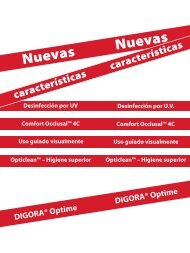
Brochure - Soredex
Brochure - Soredex
Brochure - Soredex

You also want an ePaper? Increase the reach of your titles
YUMPU automatically turns print PDFs into web optimized ePapers that Google loves.
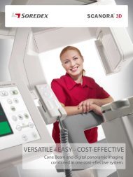
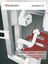
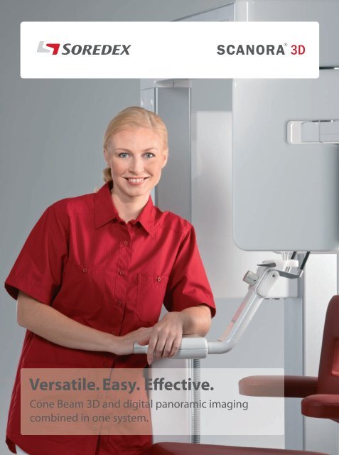
Versatile. Easy. Effective.<br />
Cone Beam 3D and digital panoramic imaging<br />
combined in one system.
More value for your money<br />
SOREDEX® has been designing and<br />
manufacturing high quality dental<br />
imaging systems for over thirty years.<br />
With SOREDEX® your investment is<br />
protected, because our products are<br />
renowned for their excellent quality,<br />
total reliability and extremely long<br />
service life.<br />
The SCANORA® 3D system makes<br />
advanced dental imaging fast and easy.<br />
We let you concentrate on your most<br />
important activity – treating your patient.
With SCANORA® 3D, the advanced dental imaging<br />
required for accurate diagnostics, implant treatment<br />
planning and oral surgery can now be done in your<br />
practice. Now, more demanding procedures can be<br />
performed efficiently and safely. Diagnostic information<br />
can be obtained without delay and with fewer referrals<br />
to outside facilities, such as medical CT examinations.<br />
The whole treatment planning process, from the<br />
first contact through radiological examinations, case<br />
planning, treatment acceptance, and follow-up, can be<br />
handled in one practice – yours.<br />
SCANORA® 3D<br />
System highlights<br />
• Stable seated patient<br />
• Fully motorized chair<br />
• 90 kV, 4 -12.5 mA, pulsed<br />
X-ray generation<br />
• Scan time 10 to 26 s<br />
• Reconstruction time less<br />
than 1 minute<br />
• Isotropic voxel size ranges<br />
from 0.133 to 0.35 mm<br />
• 12” HD ClearTouch Panel<br />
• Compatible with leading<br />
Navigation Systems<br />
• DICOM / PACS compatibility<br />
• Small footprint
Superior versatility<br />
3D imaging<br />
The SCANORA® 3D offers superior versatility by combining cone beam 3D imaging, with up to four easily selectable fields-ofview<br />
(FOV), plus optional dedicated panoramic imaging. At the touch of a key, the unit automatically switches between 3D and<br />
panoramic imaging modes, making it quick and efficient to use.<br />
The proper image volume can be selected for each specific diagnostic task.<br />
The field-of-view can be positioned anywhere within the maxillofacial area.<br />
SCANORA® 3D FOV´s<br />
The small FOV (6 cm x 6 cm) is<br />
ideal for single implant operations,<br />
localized dental examinations and<br />
temporomandibular joints.<br />
The medium FOV (7.5 cm x 10 cm)<br />
is suitable when the entire dental<br />
complex, including the wisdom<br />
teeth, need to be examined. This<br />
field-of-view can also provide<br />
information for drill guide<br />
planning.<br />
The large FOV (7.5 cm x 14.5 cm) is<br />
ideal when the complete dentition,<br />
both TM joints and upper cervical<br />
spine must be examined.<br />
The optional XL FOV<br />
(13 cm x 14.5 cm)<br />
can show the whole<br />
maxillofacial area<br />
with airways.<br />
Selectable 3D resolution<br />
The SCANORA® 3D combines low dose, fast imaging and high<br />
accuracy. Standard resolution offers fast imaging with low dose,<br />
suitable for most diagnostic tasks. High resolution improves<br />
accuracy with slightly higher imaging time and dose.<br />
The smallest attainable voxel (volume element) size is 0.133 mm.
Dedicated panoramic imaging<br />
In most examinations a panoramic image is<br />
the first step and provides an overview of the<br />
whole dentition. With the panoramic option,<br />
the SCANORA® 3D provides the speed and<br />
efficiency of traditional panoramic imaging.<br />
Panoramic imaging with AutoSwitch<br />
The SCANORA® 3D uses a dedicated CCD<br />
sensor for high-quality panoramic imaging.<br />
The unique patented AutoSwitch feature<br />
changes between panoramic and 3D<br />
modes at the touch of a key.<br />
Smooth workflow<br />
The SCANORA® 3D system has been<br />
designed to make your workflow as fast<br />
and efficient as possible. The AutoSwitch<br />
feature, easy patient positioning, short scan<br />
and image reconstruction times make it<br />
super fast.<br />
Optional<br />
• Superior quality, traditional full<br />
field panoramic images – not only<br />
low resolution synthesized<br />
panoramic images.<br />
• No need to overexpose the patient<br />
for a standard panoramic view.<br />
• No risk of dropping or damaging<br />
the integrated sensor.
Excellent diagnostic performance<br />
The SCANORA® 3D system offers a modern way of seeing dentomaxillofacial anatomy and solving diagnostic tasks. The highdefinition<br />
panoramic image shows the regions that need further investigation. The optimum 3D technique for a specific task<br />
can be easily selected, treatment planned and finally follow-up studies done, all with one efficient unit. The system contains<br />
user-selectable features that contribute to excellent diagnostic performance.<br />
Uncompromised quality<br />
The SCANORA® 3D system has been<br />
designed from the ground up using<br />
the very latest 3D imaging technology.<br />
The overall quality comes as a result<br />
of successful combination of the x-ray<br />
unit itself, ergonomic and rigid patient<br />
support devices, and the newest image<br />
handling procedures to enhance<br />
diagnostic information.<br />
High technology flat-panel detector<br />
The flat-panel detector is a<br />
masterpiece of modern CMOS<br />
technology. Compared to older<br />
generation of image intensifiers, flatpanel<br />
detectors offer superior image<br />
quality due to their large dynamic<br />
range, better contrast and lack of<br />
image distortion. Additionally they<br />
are insensitive to electromagnetic<br />
interference, are compact in size and<br />
have a very long service life.<br />
Full diagnostic information<br />
The FOV can be easily positioned<br />
anywhere in the maxillofacial area,<br />
thanks to the motorized patient<br />
seat. After scanning and image<br />
reconstruction, a full range of<br />
diagnostic options can be utilized.<br />
The diagnostic information can be<br />
thoroughly examined with the many<br />
powerful software tools and features.<br />
The small FOV is for localized problems.<br />
The optional XL FOV is suitable, for instance, in sinus examinations.
The medium FOV<br />
can show all the<br />
teeth in one image.<br />
An example of<br />
the large FOV.
Ideal for implant dentistry<br />
For proper implant site selection, accurate information is needed about the available bone,<br />
its quality, and the exact location of critical areas. The location of the mandibular nerve canal<br />
and maxillary sinus can be obtained accurately and easily. With the help of a multiplanar slice<br />
display, 3D rendering, measurement tools, and comprehensive implant symbol library,<br />
implant planning and surgery can be carried out efficiently and safely.<br />
An example of the planning tools.<br />
For third party drill guide systems the volume data can be exported in DICOM format.<br />
Through DICOM support, the SCANORA® 3D system integrates with other imaging software<br />
and modalities and is compatible with most specialty third-party software, drill and surgical<br />
guide applications.
Low dose 3D imaging<br />
X-ray imaging is optimization<br />
between image quality and x-ray<br />
dose. With the SCANORA® 3D this<br />
has been successfully resolved<br />
by combining high image quality<br />
with low dose. The key factors in<br />
achieving this are sophisticated<br />
x-ray generation, selectable imaging<br />
modes, a state-of-the-art flat-panel<br />
detector and innovative image<br />
reconstruction technology.<br />
The x-ray dose in all the fields-ofview<br />
of the SCANORA® 3D is low.<br />
The minimum effective dose can be<br />
compared to one digital panoramic<br />
exposure and, at maximum, to a few<br />
panoramic exposures for a larger<br />
field-of-view and higher resolution.<br />
The SCANORA® 3D gives you the<br />
ability to carefully minimize the<br />
dose according to the diagnostic<br />
task, whether it is a question of<br />
detailed primary diagnostics or<br />
a follow-up study. It is a safe and<br />
efficient diagnostic tool for your<br />
clinic.<br />
DOSE COMPARISON<br />
SCANORA® 3D<br />
PANORAMIC<br />
AVERAGE CBCT<br />
MEDICAL CT<br />
For more exact information, please refer for instance to research<br />
of the SEDENTEXCT: Pauwels et al. Effective dose range for dental<br />
cone beam computed tomography scanners. European Journal<br />
of Radiology. doi: 10.1016/j.ejrad.2010.11.028
Open software architecture<br />
The SCANORA® 3D produces image<br />
data in DICOM* format. With its open<br />
architecture it allows versatile and<br />
optimized software solutions to be<br />
tailored for your practice. The local<br />
area network (LAN) with several<br />
viewing stations is the solution for<br />
most practice applications allowing<br />
the system to be linked with the<br />
network and system server.<br />
The SCANORA® software is the main<br />
platform, including the local patient<br />
image database and panoramic image<br />
handling. 3D visualization software<br />
provides 3D image handling, diagnostic<br />
and implant planning.<br />
Freely distribute clinical cases on CD/DVD<br />
to referring clinicians. The referring clinician<br />
can utilize the free viewer without investing<br />
in special software or import the images in<br />
DICOM format into their own 3D software.<br />
* Digital Imaging and Communication<br />
in Medicine<br />
LAN (Local Area Network)<br />
Modality<br />
workstation PC<br />
PACS (Picture Archiving and<br />
Communication System)<br />
SCANORA®<br />
Image management<br />
Local database<br />
Panoramic image handling<br />
DICOM components<br />
3D visualization software<br />
3D image handling<br />
Implant planning<br />
Reporting<br />
Other 3rd<br />
party software<br />
- Implant planning<br />
- Image handling<br />
- Drill and surgical guides<br />
- Navigated surgery<br />
- 3D modelling
Technical data<br />
3D Imaging fields-of-view and specifications<br />
FOV<br />
[Height x<br />
diameter]<br />
Resolution<br />
Voxel size<br />
[mm]<br />
Scan/<br />
Exposure time [s]<br />
Total image<br />
processing<br />
time approx.<br />
[minutes]<br />
3D Small<br />
6x6 cm<br />
3D Medium<br />
7.5x10 cm<br />
3D Large<br />
7.5x14.5 cm<br />
3D XL<br />
13x14.5 cm<br />
Standard 0.20 13 / 3 1<br />
High 0.133 20 / 4.5 2<br />
Standard 0.30 11 / 2.5 1<br />
High 0.20 16 / 3.75 2<br />
Standard 0.35 10 / 2.25 1<br />
High 0.25 13/3 2<br />
Standard 0.35 20 / 4.5 2<br />
High 0.25 26 / 6 4<br />
3D image receptor<br />
Receptor type<br />
Receptor active area<br />
Pixel size<br />
CMOS Flat Panel<br />
124 mm x 124 mm<br />
200 μm<br />
Panoramic image receptor (Optional)<br />
Technology<br />
CCD<br />
Detector size (H x W)<br />
146 mm x 6 mm<br />
Detector pixel size<br />
48 μm<br />
(77.7")<br />
Panoramic imaging programs (Optional)<br />
Adult panoramic program<br />
Pediatric panoramic program<br />
TMJ programs<br />
X-ray generator<br />
Tube<br />
Fixed anode tube<br />
Focal spot 0.5 mm IEC 60336<br />
Target angle<br />
5 degrees<br />
kV 60-90<br />
Average mA 1.0-8.0<br />
1940 (76.4")<br />
1600 (63")<br />
General<br />
Weight<br />
Dimensions (HxWxD)<br />
310 kg (690 lbs)<br />
1973 mm x 1600 mm<br />
x 1400 mm (77.7” x 63”<br />
x 55.1”)<br />
1400 (55.1")<br />
1740 (68.5")<br />
Power requirements<br />
Line voltage 230-240 VAC (±10 %),<br />
50/60 Hz
Digital imaging made easy<br />
Head office and factory:<br />
SOREDEX<br />
Nahkelantie 160, Tuusula<br />
P.O. Box 148, FI-04301 Tuusula<br />
Finland<br />
Tel. +358 45 7882 2000<br />
Fax +358 9 701 5261<br />
info@soredex.com<br />
SOREDEX USA<br />
1245 W. Canal Street<br />
Milwaukee, WI 53233<br />
U.S.A.<br />
Tel. +1 800 558 6120<br />
Fax +1 414 481 8665<br />
info@soredexusa.com<br />
Pride. Passion. Performance.<br />
Since 1977 SOREDEX® has been a leader in providing innovative imaging solutions for<br />
demanding professionals. Through continuous evolution and refinement we have set<br />
the highest industry standards for Quality, Reliability and Efficiency.<br />
We are committed to follow in this path today and in the future.<br />
SCANORA® is a registered trademark of SOREDEX, PaloDEx Group Oy. Other product names and trademarks are<br />
the property of their respective owners. CE-marked, NB (CE) number 0537. Electrical safety meets the IEC 60601-1<br />
standard. Manufacturing complies with ISO 13485:2003, ISO 9001:2008, and ISO 14001:2004.<br />
SOREDEX® reserves the right to make changes in specifications and features shown herein at any time<br />
without notice or obligation. Contact your SOREDEX® representative for the most up-to-date information.<br />
© 2011 SOREDEX®<br />
207083-1 02/11 Printed in Finland<br />
SOREDEX Germany<br />
Schutterstrasse 12<br />
77746 Schutterwald<br />
Germany<br />
Tel: +49 (0) 781 28 41 98-0<br />
Fax: +49 (0) 781 28 41 98-30<br />
kontakt@soredex.de<br />
www.soredex.com • www.soredex.de • www.soredexusa.com