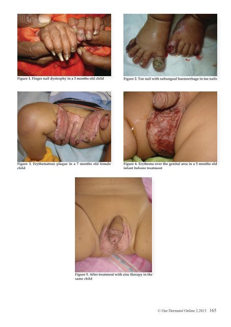
Figure 1. Finger nail dystrophy in a 3 months old child Figure 2. Toe nail with subungual haemorrhage in toe nails Figure 3. Erythematous plaque in a 7 months old female child Figure 4. Erythema over the genital area in a 5 months old infant befeore treatment Figure 5. After treatment with zinc therapy in the same child © <strong>Our</strong> Dermatol <strong>Online</strong> 2.2013 165
Treatment of acrodermatitis enteropathica requires lifelong zinc supplementation. Acrodermatitis enteropathica is treated with zinc supplements in the form of zinc sulfate. These supplements should be given as soon as diagnosis of the disorder is made and they have to be continued for life. The drug Diodoquin (iodoquinol) is another treatment that usually clears up symptoms within a week. If the disorder is caused by intravenous feeding, adding zinc supplements to the nutritional regimen can prevent and/ or clear up manifestations of AE.Typically, 3-5 mg/kg of zinc gluconate or sulfate is administered orally each day. Clinical improvement occurs prior to any significant change in the plasma zinc levels, usually within days to weeks of initiating treatment [14]. Monitor serum zinc levels and alkaline phosphatase values every 3-6 months [15,16]. Acrodermatitis enteropathica exacerbation during pregnancy or the stress of disease may require an increase in therapy. Warm compresses to remove the scale crust, followed by application of white petrolatum to eroded skin lesions, may enhance reepithelialization when used concurrently with zinc replacement [17]. Genetic counseling is recommended for families of patients with the congenital form of acrodermatitis enteropathica. Although no special diet is required for acrodermatitis enteropathica patients, as long as zinc supplementation is continued, certain foods contain increased levels of zinc, including oysters, crab, beef, pork, and fowl. Zinc content is directly related to protein [18,19]. REFERENCES 1. Perafan-Riveros C, Franca LF, Alves AC, Sanches JA Jr: Acrodermatitis enteropathica: case report and review of the literature. Pediatr Dermatol. 2002;19:426-31. 2. Kury S, Dreno B, Bezieau S: Identification of SLC39A4, a gene involved in acrodermatitis enteropathica. Nat Genet. 2002;31:239- 40. 3. Nakano A, Nakano H, Nomura K, Toyomaki Y, Hanada K: Novel SLC39A4 mutations in acrodermatitis enteropathica. J Invest Dermatol. 2003;120:963-6. 4. Wang K, Pugh EW, Griffen S, Doheny KF, Mostafa WZ, Al- Aboosi MM: Homozygosity mapping places the acrodermatitis enteropathica gene on chromosomal region 8q24.3. Am J Hum Genet. 2001;68:1055-60. 5. Maverakis E, Fung MA, Lynch PJ, Draznin M, Michael DJ, Ruben B: Acrodermatitis enteropathica and an overview of zinc metabolism. J Am Acad Dermatol. 2007;56:116-24. 6. Wang K, Zhou B, Kuo YM, Zemansky J, Gitschier J: A novel member of a zinc transporter family is defective in acrodermatitis enteropathica. Am J Hum Genet. 2002;71:66-73. 7. Evans GW, Johnson PE: Zinc-binding factor in acrodermatitis enteropathica. Lancet. 1976;2:131-3. 8. Connors TJ, Czarnecki DB, Haskett MI: Acquired zinc deficiency in a breast-fed premature infant. Arch Dermatol. 1983;119:319-21. 9. Perafán-Riveros C, França LF, Alves AC, Sanches JA Jr: Acrodermatitis enteropathica: case report and review of the literature. Pediatr Dermatol. 2002;19:426-31. 10. Sehgal VN, Jain S: Acrodermatitis enteropathica. Clin Dermatol. 2000;18:745-8. 11. Radja N, Charles-Holmes R: Acrodermatitis enteropathicalifelong follow-up and zinc monitoring. Clin Exp Dermatol. 2002;27:62-3. 12. Kiechl-Kohlendorfer U, Fink FM, Steichen-Gersdorf E: Transient symptomatic zinc deficiency in a breast-fed preterm infant. Pediatr Dermatol. 2007;24:536-40. 13. Gonzalez JR, Botet MV, Sanchez JL: The histopathology of acrodermatitis enteropathica. Am J Dermatopathol. 1982;4:303-11. 14. Sandstead HH: Understanding zinc: recent observations and interpretations. J Lab Clin Med. 1994;124:322-7. 15. Van Wouwe JP: Clinical and laboratory diagnosis of acrodermatitis enteropathica. Eur J Pediatr. 1989;149:2-8. 16. Prasad AS: Zinc: an overview. Nutrition. 1995;11:93-9. 17. Lombeck T, Schnippering HG, Ritzl F, Feinendegen LE, Bremer HJ: Letter: Absorption of zinc in acrodermatitis enteropathica. Lancet. 1975;1:85-6. 18. Barnes PM, Moynahan EJ: Zinc deficiency in acrodermatitis enteropathica: multiple dietary intolerance treated with synthetic diet. Proc R Soc Med. 1973;66:327-9. 19. Neldner KH, Hambidge KM: Zinc therapy of acrodermatitis enteropathica. N Engl J Med. 1975;24:879-82. Copyright by Neerja Puri. This is an open access article distributed under the terms of the Creative Commons Attribution License, which permits unrestricted use, distribution, and reproduction in any medium, provided the original author and source are credited. 166 © <strong>Our</strong> Dermatol <strong>Online</strong> 2.2013
- Page 1 and 2: Volume 4, Number 2, April 2013 ISSN
- Page 3: Editorial Board: Abdel-Naser Mohame
- Page 6 and 7: CONTENTS E d i t o r i a l O r i g
- Page 8 and 9: Original Article DOI: 10.7241/ourd.
- Page 10 and 11: Figure 1. Survival time of HIV/TB-
- Page 12 and 13: Original Article DOI: 10.7241/ourd.
- Page 14 and 15: Figure 3. A. melanocytic naevus on
- Page 16 and 17: Original Article DOI: 10.7241/ourd.
- Page 18 and 19: 82% of the children belonged to the
- Page 20 and 21: Comment to the article DOI: 10.7241
- Page 22 and 23: The skin around body openings such
- Page 26 and 27: Comment to the article DOI: 10.7241
- Page 28 and 29: Similar type of rash was also prese
- Page 30 and 31: 13. Mastaglia FL, Ojeda VJ: Inflamm
- Page 32 and 33: Figure 1. Atrophic areas on left up
- Page 34 and 35: 5. Brocard A, Quereux G, Moyse D, D
- Page 36 and 37: Figure 1. Macroglossia Figure 2. Ma
- Page 38 and 39: Case Report DOI: 10.7241/ourd.20132
- Page 40 and 41: Figure 4a, b. Skin biopsy shows hyp
- Page 42 and 43: Case Report DOI: 10.7241/ourd.20132
- Page 44 and 45: Case Report DOI: 10.7241/ourd.20132
- Page 46 and 47: Discussion Poikiloderma of Civatte
- Page 48 and 49: Case Report DOI: 10.7241/ourd.20132
- Page 50 and 51: Comment to the article DOI: 10.7241
- Page 52 and 53: Case Report A 56 year old Caucasian
- Page 54 and 55: Using a battery of neurotoxic agent
- Page 56 and 57: Figure 1. Multiple lesions on the u
- Page 58 and 59: Case Report DOI: 10.7241/ourd.20132
- Page 60 and 61: Figure 1. a PAS positive accentuati
- Page 62 and 63: The patient was put on melanocyl 0.
- Page 64 and 65: Case Report DOI: 10.7241/ourd.20132
- Page 66 and 67: The type of treatment prescribed by
- Page 68 and 69: On the basis of these clinical find
- Page 70 and 71: Surgery has been reported to be the
- Page 72 and 73: Figure 1. Histological section show
- Page 74 and 75:
Case Report DOI: 10.7241/ourd.20132
- Page 76 and 77:
Discussion The breast location of t
- Page 78 and 79:
Figure 2. The epithelial nests show
- Page 80 and 81:
Case Report DOI: 10.7241/ourd.20132
- Page 82 and 83:
Discussion SCACP is one of the cuta
- Page 84 and 85:
Figure 1. Tumor of the nasal colume
- Page 86 and 87:
Figure 2. Vesicles and crusts aroun
- Page 88 and 89:
Outbreaks have occurred recently in
- Page 90 and 91:
Clasificación En base al epitelio
- Page 92 and 93:
- Microcefalia, hipoplasia mediofac
- Page 94 and 95:
Caso Nº 3: · Niña, edad desconoc
- Page 96 and 97:
Clinical Images DOI: 10.7241/ourd.2
- Page 98 and 99:
Figure 7. Fixed cutaneous sporotric
- Page 100 and 101:
Letter to the Editor DOI: 10.7241/o
- Page 102 and 103:
Historical Article DOI: 10.7241/our
- Page 104 and 105:
Figure 6. Henri Mondor (1885-1962).
- Page 106 and 107:
Historical Article DOI: 10.7241/our
- Page 108 and 109:
Figure 5. Alfred Guido Miescher (18
- Page 110 and 111:
Historical Article DOI: 10.7241/our
- Page 112 and 113:
Names of ''Lines'', in dermatology
- Page 114 and 115:
Figure 1. Erik Adolf von Willebrand
- Page 116 and 117:
THOMAS INMAN English physician, 182
- Page 118 and 119:
There are three type of tinea capit
- Page 120:
Our Dermatology Online www.odermato