

download full issue - Our Dermatology Online Journal
download full issue - Our Dermatology Online Journal
download full issue - Our Dermatology Online Journal

You also want an ePaper? Increase the reach of your titles
YUMPU automatically turns print PDFs into web optimized ePapers that Google loves.
Figure 2. The epithelial nests showing lymphocytic<br />
infiltrates surrounded by desmoplastic stroma. H&E, 200X<br />
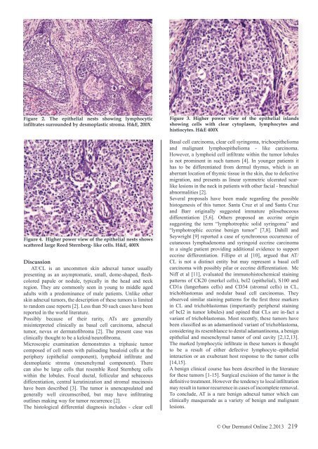
Figure 4. Higher power view of the epithelial nests shows<br />
scattered large Reed Sternberg- like cells. H&E, 400X<br />
Discussion<br />
AT/CL is an uncommon skin adnexal tumor usually<br />
presenting as an asymptomatic, small, dome-shaped, fleshcolored<br />
papule or nodule, typically in the head and neck<br />
region. They are commonly seen in young to middle aged<br />
adults with a predominance of male patients. Unlike other<br />
skin adnexal tumors, the description of these tumors is limited<br />
to random case reports [2]. Less than 50 such cases have been<br />
reported in the world literature.<br />
Possibly because of their rarity, ATs are generally<br />
misinterpreted clinically as basal cell carcinoma, adnexal<br />
tumor, nevus or dermatofibroma [2]. The present case was<br />
clinically thought to be a keloid/neurofibroma.<br />
Microscopic examination demonstrates a triphasic tumor<br />
composed of cell nests with palisading basaloid cells at the<br />
periphery (epithelial component), lymphoid infiltrate and<br />
desmoplastic stroma (mesenchymal component). There<br />
can also be large cells that resemble Reed Sternberg cells<br />
within the lobules. Focal ductal, follicular and sebaceous<br />
differentiation, central keratinization and stromal mucinosis<br />
have been described [3]. The tumor is unencapsulated and<br />
generally well circumscribed, but may have infiltrating<br />
outlines making way for tumor recurrence [2].<br />
The histological differential diagnosis includes - clear cell<br />
Figure 3. Higher power view of the epithelial islands<br />
showing cells with clear cytoplasm, lymphocytes and<br />
histiocytes. H&E 400X<br />
Basal cell carcinoma, clear cell syringoma, trichoepithelioma<br />
and malignant lymphoepithelioma - like carcinoma.<br />
However, a lymphoid cell infiltrate within the tumor lobules<br />
is not prominent in such tumors [4]. In younger patients it<br />
has to be differentiated from dermal thymus, which is an<br />
aberrant location of thymic t<strong>issue</strong> in the skin, due to defective<br />
migration, and presents as linear symmetric ulcerated scarlike<br />
lesions in the neck in patients with other facial - branchial<br />
abnormalities [2].<br />
Several proposals have been made regarding the possible<br />
histogenesis of this tumor. Santa Cruz et al and Santa Cruz<br />
and Barr originally suggested immature pilosebaceous<br />
differentiation [5,6]. Others proposed an eccrine origin<br />
suggesting the term “lymphotrophic solid syringoma” and<br />
“lymphotrophic eccrine benign tumor” [7,8]. Dahill and<br />
Seywright [9] reported a case of synchronous occurrence of<br />
cutaneous lymphadenoma and syringoid eccrine carcinoma<br />
in a single patient providing additional evidence to support<br />
eccrine differentiation. Fillipo et al [10], argued that AT/<br />
CL is not a distinct entity but may represent a basal cell<br />
carcinoma with possibly pilar or eccrine differentiation. Mc<br />
Niff et al [11], evaluated the immunohistochemical staining<br />
patterns of CK20 (merkel cells), bcl2 (epithelial), S100 and<br />
CD1a (langerhans cells) and CD34 (stromal cells) in CL,<br />
trichoblastomas and nodular basal cell carcinomas. They<br />
observed similar staining patterns for the first three markers<br />
in CL and trichoblastomas (importantly peripheral staining<br />
of bcl2 in tumor lobules) and opined that CLs are in-fact a<br />
variant of trichoblastomas. Most recently, these tumors have<br />
been classified as an adamantinoid variant of trichoblastoma,<br />
considering its resemblance to dental adamantinoma, a benign<br />
epithelial and mesenchymal tumor of oral cavity [2,12,13].<br />
The marked lymphocytic infiltrate in these tumors is thought<br />
to be a result of either defective lymphocyte–epithelial<br />
interaction or an exuberant host response to the tumor cells<br />
[14,15].<br />
A benign clinical course has been described in the literature<br />
for these tumors [1-15]. Surgical excision of the tumor is the<br />
definitive treatment. However the tendency to local infiltration<br />
may result in tumor recurrence in cases of incomplete removal.<br />
To conclude, AT is a rare benign adnexal tumor which can<br />
clinically masquerade as a variety of benign and malignant<br />
lesions.<br />
© <strong>Our</strong> Dermatol <strong>Online</strong> 2.2013 219


