

download full issue - Our Dermatology Online Journal
download full issue - Our Dermatology Online Journal
download full issue - Our Dermatology Online Journal

Create successful ePaper yourself
Turn your PDF publications into a flip-book with our unique Google optimized e-Paper software.
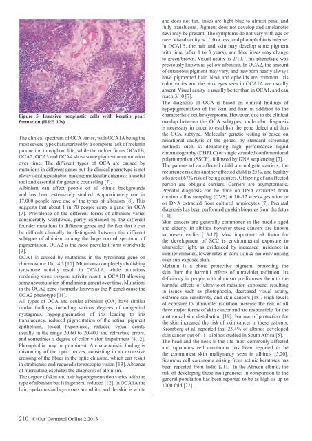
Figure 5. Invasive neoplastic cells with keratin pearl<br />
formation (H&E, 10x)<br />
The clinical spectrum of OCA varies, with OCA1A being the<br />
most severe type characterized by a complete lack of melanin<br />
production throughout life, while the milder forms OCA1B,<br />
OCA2, OCA3 and OCA4 show some pigment accumulation<br />
over time. The different types of OCA are caused by<br />
mutations in different genes but the clinical phenotype is not<br />
always distinguishable, making molecular diagnosis a useful<br />
tool and essential for genetic counseling [7].<br />
Albinism can affect people of all ethnic backgrounds<br />
and has been extensively studied. Approximately one in<br />
17,000 people have one of the types of albinism [8]. This<br />
suggests that about 1 in 70 people carry a gene for OCA<br />
[7]. Prevalence of the different forms of albinism varies<br />
considerably worldwide, partly explained by the different<br />
founder mutations in different genes and the fact that it can<br />
be difficult clinically to distinguish between the different<br />
subtypes of albinism among the large normal spectrum of<br />
pigmentation. OCA2 is the most prevalent form worldwide<br />
[9].<br />
OCA1 is caused by mutations in the tyrosinase gene on<br />
chromosome 11q14.3 [10]. Mutations completely abolishing<br />
tyrosinase activity result in OCA1A, while mutations<br />
rendering some enzyme activity result in OCA1B allowing<br />
some accumulation of melanin pigment over time. Mutations<br />
in the OCA2 gene (formerly known as the P-gene) cause the<br />
OCA2 phenotype [11].<br />
All types of OCA and ocular albinism (OA) have similar<br />
ocular findings, including various degrees of congenital<br />
nystagmus, hypopigmentation of iris leading to iris<br />
translucency, reduced pigmentation of the retinal pigment<br />
epithelium, foveal hypoplasia, reduced visual acuity<br />
usually in the range 20/60 to 20/400 and refractive errors,<br />
and sometimes a degree of color vision impairment [8,12].<br />
Photophobia may be prominent. A characteristic finding is<br />
misrouting of the optic nerves, consisting in an excessive<br />
crossing of the fibres in the optic chiasma, which can result<br />
in strabismus and reduced stereoscopic vision [13]. Absence<br />
of misrouting excludes the diagnosis of albinism.<br />
The degree of skin and hair hypopigmentation varies with the<br />
type of albinism but is in general reduced [12]. In OCA1A the<br />
hair, eyelashes and eyebrows are white, and the skin is white<br />
and does not tan. Irises are light blue to almost pink, and<br />
<strong>full</strong>y translucent. Pigment does not develop and amelanotic<br />
nevi may be present. The symptoms do not vary with age or<br />
race. Visual acuity is 1/10 or less, and photophobia is intense.<br />
In OCA1B, the hair and skin may develop some pigment<br />
with time (after 1 to 3 years), and blue irises may change<br />
to green/brown. Visual acuity is 2/10. This phenotype was<br />
previously known as yellow albinism. In OCA2, the amount<br />
of cutaneous pigment may vary, and newborn nearly always<br />
have pigmented hair. Nevi and ephelids are common. Iris<br />
color varies and the pink eyes seen in OCA1A are usually<br />
absent. Visual acuity is usually better than in OCA1, and can<br />
reach 3/10 [7].<br />
The diagnosis of OCA is based on clinical findings of<br />
hypopigmentation of the skin and hair, in addition to the<br />
characteristic ocular symptoms. However, due to the clinical<br />
overlap between the OCA subtypes, molecular diagnosis<br />
is necessary in order to establish the gene defect and thus<br />
the OCA subtype. Molecular genetic testing is based on<br />
mutational analysis of the genes, by standard screening<br />
methods such as denaturing high performance liquid<br />
chromatography (DHPLC) or single stranded conformational<br />
polymorphism (SSCP), followed by DNA sequencing [7].<br />
The parents of an affected child are obligate carriers, the<br />
recurrence risk for another affected child is 25%, and healthy<br />
sibs are at 67% risk of being carriers. Offspring of an affected<br />
person are obligate carriers. Carriers are asymptomatic.<br />
Prenatal diagnosis can be done on DNA extracted from<br />
chorion villus sampling (CVS) at 10–12 weeks gestation or<br />
on DNA extracted from cultured amniocytes [7]. Prenatal<br />
diagnosis has been performed on skin biopsies from the fetus<br />
[14].<br />
Skin cancers are generally commoner in the middle aged<br />
and elderly. In albinos however these cancers are known<br />
to present earlier [15-17]. Most important risk factor for<br />
the development of SCC is environmental exposure to<br />
ultraviolet light, as evidenced by increased incidence in<br />
sunnier climates, lower rates in dark skin & majority arising<br />
over sun-exposed skin.<br />
Melanin is a photo protective pigment, protecting the<br />
skin from the harmful effects of ultraviolet radiation. Its<br />
deficiency in people with albinism predisposes them to the<br />
harmful effects of ultraviolet radiation exposure, resulting<br />
in <strong>issue</strong>s such as photophobia, decreased visual acuity,<br />
extreme sun sensitivity, and skin cancers [18]. High levels<br />
of exposure to ultraviolet radiation increase the risk of all<br />
three major forms of skin cancer and are responsible for the<br />
anatomical site distribution [19]. No use of protection for<br />
the skin increased the risk of skin cancer in these patients.<br />
Kromberg et al. reported that 23.4% of albinos developed<br />
skin cancer out of 111 albinos studied in South Africa [5].<br />
The head and the neck is the site most commonly affected<br />
and squamous cell carcinoma has been reported to be<br />
the commonest skin malignancy seen in albinos [5,20].<br />
Sqamous cell carcinoma arising from actinic keratoses has<br />
been reported from India [21]. In the African albino, the<br />
risk of developing these malignancies in comparison to the<br />
general population has been reported to be as high as up to<br />
1000 fold [22].<br />
210 © <strong>Our</strong> Dermatol <strong>Online</strong> 2.2013


