

The Nature of the Antorbital Fossa of Archosaurs - Ohio University ...
The Nature of the Antorbital Fossa of Archosaurs - Ohio University ...
The Nature of the Antorbital Fossa of Archosaurs - Ohio University ...

You also want an ePaper? Increase the reach of your titles
YUMPU automatically turns print PDFs into web optimized ePapers that Google loves.
Figure 3. <strong>Antorbital</strong> sinus in a golden eagle<br />
(Aquila chrysaetos). Only <strong>the</strong> premaxillary and<br />
lacrimal diverticula are figured.<br />
Birds are not alone among archosaurs in exhibiting pneumatic features in <strong>the</strong> antorbital region.<br />
Although eusuchian crocodilians lack antorbital fenestrae, <strong>the</strong>y none<strong>the</strong>less resemble birds in<br />
possessing air-filled diverticula <strong>of</strong> <strong>the</strong> nasal cavity. But whereas birds have only a single diverticulum,<br />
<strong>the</strong> antorbital sinus, crocodilians have four (Parsons 1970); which <strong>of</strong> <strong>the</strong>se, if any, are homologous to<br />
<strong>the</strong> avian antorbital sinus is presently under study. <strong>The</strong>se diverticula produce vast pneumatic cavities in<br />
<strong>the</strong> maxillae and palatines <strong>of</strong> crocodilians (Fig. 4A; Wegner 1957).<br />
<strong>The</strong>ropod dinosaurs also exhibit unequivocal pneumatic features in <strong>the</strong> bones surrounding <strong>the</strong><br />
antorbital fossa. <strong>The</strong> most striking example is Oviraptor (Osmolska 1976, Barsbold 1983) in which all<br />
<strong>of</strong> <strong>the</strong> bones surrounding <strong>the</strong> fossa are produced into a lattice <strong>of</strong> pneumatic foramina that in some<br />
individuals result in an air-filled bony crest (Fig. 4B). Less extravagant manifestations are found in<br />
o<strong>the</strong>r <strong>the</strong>ropods. <strong>The</strong> most common pneumatic features are <strong>the</strong> distinctive chambering <strong>of</strong> <strong>the</strong> maxilla<br />
and lacrimal (Fig. 4C) in all but <strong>the</strong> more primitive ceratosaurians (Coelophysis and Syntarsus). <strong>The</strong><br />
nasal bones <strong>of</strong> Allosaurus (Fig. 4C) and Ceratosaurus also are pierced by pneumatic foramina<br />
communicating with chambers inside <strong>the</strong> bone. Additionally, some carnosaurs exhibit pneumatic<br />
formamina into <strong>the</strong> jugal bone. <strong>The</strong> muscular hypo<strong>the</strong>sis cannot accomodate <strong>the</strong>se observations as it is<br />
very unlikely that muscle fibers would enter bones such as <strong>the</strong> lacrimal, nasal, and jugal through small<br />
foramina and <strong>the</strong>n expand within <strong>the</strong> body <strong>of</strong> <strong>the</strong> bone. Likewise, <strong>the</strong> thin struts <strong>of</strong> bone forming <strong>the</strong><br />
maxillary sinuses (Madsen 1976) are too fragile to serve as area <strong>of</strong> muscle attachment.<br />
rla<br />
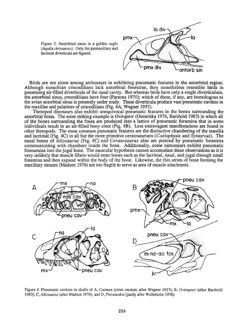
Figure 4. Pneumatic cavities in skulls <strong>of</strong> A, Caiman (cross section; after Wegner 1957); B, Oviraptor (after Barsbold<br />
1983); C, Allosaurus (after Madsen 1976); and D, Pteranodon (partly after Wellnh<strong>of</strong>er 1978).


