

The Nature of the Antorbital Fossa of Archosaurs - Ohio University ...
The Nature of the Antorbital Fossa of Archosaurs - Ohio University ...
The Nature of the Antorbital Fossa of Archosaurs - Ohio University ...

You also want an ePaper? Increase the reach of your titles
YUMPU automatically turns print PDFs into web optimized ePapers that Google loves.
Fourth Symposium on Mesozoic Terrestrial Ecosystems, Short Papers Ed. by P.J. Currie and EH. Koster.<br />
<strong>The</strong> <strong>Nature</strong> <strong>of</strong> <strong>the</strong> <strong>Antorbital</strong><br />
(Baltimore, Maryland).<br />
<strong>Fossa</strong> <strong>of</strong> <strong>Archosaurs</strong>: Shifting <strong>the</strong> Null Hypo<strong>the</strong>sis. L.M. Witrner<br />
<strong>The</strong> antorbital fenestra is a nearly ubiquitous feature <strong>of</strong> archosaurs and its presence remains one <strong>of</strong><br />
<strong>the</strong> few cranial synapomorphies <strong>of</strong> <strong>the</strong> group. <strong>The</strong> antorbital fenestra is an opening in <strong>the</strong> skull situated<br />
between <strong>the</strong> orbit and external naris, communicating with <strong>the</strong> nasal cavity, and surrounded primarily by<br />
<strong>the</strong> maxilla and lacrimal with varying contributions by <strong>the</strong> nasal and jugal. <strong>The</strong> lateral surfaces <strong>of</strong> <strong>the</strong><br />
bones surrounding <strong>the</strong> fenestra are <strong>of</strong>ten excavated into a basin-like depression; I will use <strong>the</strong> term<br />
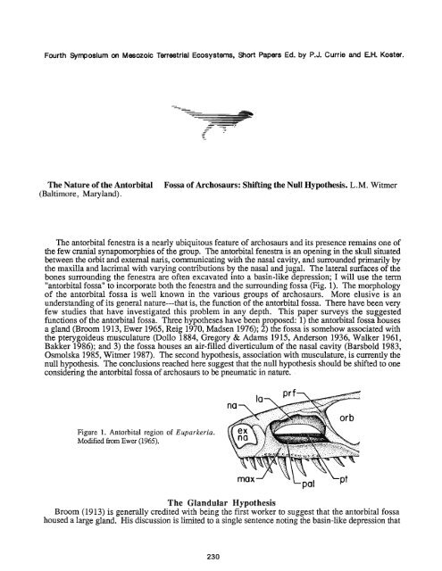
"antorbital fossa" to incorporate both <strong>the</strong> fenestra and <strong>the</strong> surrounding fossa (Fig. 1). <strong>The</strong> morphology<br />
<strong>of</strong> <strong>the</strong> antorbital fossa is well known in <strong>the</strong> various groups <strong>of</strong> archosaurs. More elusive is an<br />
understanding <strong>of</strong> its general nature---that is, <strong>the</strong> function <strong>of</strong> <strong>the</strong> antorbital fossa. <strong>The</strong>re have been very<br />
few studies that have investigated this problem in any depth. This paper surveys <strong>the</strong> suggested<br />
functions <strong>of</strong> <strong>the</strong> antorbital fossa. Three hypo<strong>the</strong>ses have been proposed: 1) <strong>the</strong> antorbital fossa houses<br />
a gland (Broom 1913, Ewer 1965, Reig 1970, Madsen 1976); 2) <strong>the</strong> fossa is somehow associated with<br />
<strong>the</strong> pterygoideus musculature (Dollo 1884, Gregory & Adams 19 15, Anderson 1936, Walker 1961,<br />
Bakker 1986); and 3) <strong>the</strong> fossa houses an air-filled diverticulum <strong>of</strong> <strong>the</strong> nasal cavity (Barsbold 1983,<br />
Osmolska 1985, Witrner 1987). <strong>The</strong> second hypo<strong>the</strong>sis, association with musculature, is currently <strong>the</strong><br />
null hypo<strong>the</strong>sis. <strong>The</strong> conclusions reached here suggest that <strong>the</strong> null hypo<strong>the</strong>sis should be shifted to one<br />
considering <strong>the</strong> antorbital fossa <strong>of</strong> archosaurs to be pneumatic in nature.<br />
Figure 1. <strong>Antorbital</strong> region <strong>of</strong> Euparkeria.<br />
Modified fiom Ewer (1965).<br />
<strong>The</strong> Glandular Hypo<strong>the</strong>sis<br />
Broom (1913) is generally credited with being <strong>the</strong> first worker to suggest that <strong>the</strong> antorbital fossa<br />
housed a large gland. His discussion is limited to a single sentence noting <strong>the</strong> basin-like depression that
constituted <strong>the</strong> antorbital fossa <strong>of</strong> Euparkeria (Fig. 1). Ewer (1965), responding to <strong>the</strong> same antorbital<br />
morphology <strong>of</strong> Euparkeria, agreed with Broom that it likely housed a gland and fur<strong>the</strong>r suggested that it<br />
was probably a nasal salt gland. Ewer considered <strong>the</strong> primitive function <strong>of</strong> <strong>the</strong> fossa to be <strong>the</strong> housing<br />
<strong>of</strong> this gland; she refuted <strong>the</strong> muscular hypo<strong>the</strong>sis---but only for <strong>the</strong> earliest archosaurs, arguing that<br />
later in archosaur phylogeny <strong>the</strong> "antorbital gland" was lost as <strong>the</strong> pterygoideus musculature moved<br />
anteriorly to occupy <strong>the</strong> fossa. Thus Ewer argued for both <strong>the</strong> glandular and <strong>the</strong> muscular hypo<strong>the</strong>ses.<br />
More recently, Madsen (1976) suggested that <strong>the</strong> large cavity in <strong>the</strong> body <strong>of</strong> <strong>the</strong> lacrimal <strong>of</strong> Allosm<br />
(Fig. 4C) may have housed a gland.<br />
<strong>The</strong> only worker to <strong>of</strong>fer evidence o<strong>the</strong>r than <strong>the</strong> general morphology <strong>of</strong> <strong>the</strong> antorbital fossa in<br />
support <strong>of</strong> <strong>the</strong> glandular hypo<strong>the</strong>sis was Reig (1970). His argument is based on <strong>the</strong> assumption that<br />
archosaurs were derived from varanopsid pelycosaurs. According to Reig, because mammals are<br />
ureotelic, <strong>the</strong>ir precursors---<strong>the</strong> pelycosaurs---were also ureotelic; thus, because wchosaurs were<br />
derived from pelycosaurs, ancestral archosaurs were also ureotelic. As archosaurs were considered<br />
primarily upland forms, Reig suggested that <strong>the</strong>y required some form <strong>of</strong> extrarenal salt excretion---a salt<br />
gland in <strong>the</strong> antorbital fossa. Through this circuitous and ra<strong>the</strong>r tenuous chain <strong>of</strong> reasoning, Reig<br />
concluded that <strong>the</strong> archosaurian antorbital fossa functioned as a housing for a nasal salt gland.<br />
While it is generally agreed that archosaurs are not related to pelycosaurs and instead are members <strong>of</strong><br />
a monophyletic group, <strong>the</strong> Diapsida (invalidating Reig's basic assumption), <strong>the</strong> glandular hypo<strong>the</strong>sis is<br />
a valid hypo<strong>the</strong>sis that deserves discussion. Most proponents <strong>of</strong> <strong>the</strong> hypo<strong>the</strong>sis have suggested that if a<br />
gland were present it was a site <strong>of</strong> extrarenal salt excretion. In extant diapsids such as squamates<br />
(Parsons 1970) and birds (Fange, et al. 1958) extrarenal salt excretion takes place via <strong>the</strong> lateral nasal<br />
gland. <strong>The</strong> question is whe<strong>the</strong>r or not <strong>the</strong> lateral nasal gland was housed in <strong>the</strong>,antorbital fossa <strong>of</strong> fossil<br />
archosaurs. Comparisons with extant diapsids suggest that it was not. In squamates <strong>the</strong> lateral nasal<br />
gland is situated in <strong>the</strong> anterior portion <strong>of</strong> <strong>the</strong> preorbital region <strong>of</strong> <strong>the</strong> head and lies dorsal or lateral to<br />
<strong>the</strong> nasal capsule (Parson 1970). Crocodilians exhibit a similar situation with <strong>the</strong> gland lying dorsal to<br />
<strong>the</strong> nasal capsule in <strong>the</strong> vicinity <strong>of</strong> <strong>the</strong> juncture <strong>of</strong> <strong>the</strong> premaxilla, maxilla, and nasal (Parsons 1970). In<br />
birds <strong>the</strong> lateral nasal gland is generally located in <strong>the</strong> orbital region (Marples 1932).<br />
Thus, although fossil archosaurs may have had lateral nasal glands, <strong>the</strong>y almost certainly did not<br />
reside in <strong>the</strong> antorbital fossa. Osmolska (1979) likewise rejected <strong>the</strong> antorbital fossa as a site <strong>of</strong> <strong>the</strong><br />
lateral nasal gland and suggested that <strong>the</strong> gland in some dinosaurs may have had a more primitive<br />
position in <strong>the</strong> vicinity <strong>of</strong> <strong>the</strong> external naris. <strong>The</strong> possibility will always remain that some glandular<br />
structure not known in modern organisms may have been housed in <strong>the</strong> antorbital fossa. But such an<br />
assertion will never be supported by much evidence and is not testable. Indeed, <strong>the</strong> morphology that<br />
originally led Broom to suggest <strong>the</strong> glandular hypo<strong>the</strong>sis---<strong>the</strong> basin-like morphology <strong>of</strong> <strong>the</strong> antorbital<br />
fossa---obtains in a group <strong>of</strong> archosaurs that show demonstrable pneumatic features in <strong>the</strong>ir antorbital<br />
fossae; many <strong>the</strong>ropods show very similar depressions in <strong>the</strong> maxilla and lacrimal (Fig. 2C). As is<br />
argued below, one <strong>of</strong> <strong>the</strong> best cases for a pneumatic antorbital fossa can be made for <strong>the</strong>ropod<br />
dinosaurs.<br />
<strong>The</strong> Muscular Hypo<strong>the</strong>sis<br />
<strong>The</strong> most widely accepted hypo<strong>the</strong>sis is that <strong>the</strong> antorbital fossa <strong>of</strong> archosaurs is closely associated<br />
with <strong>the</strong> pterygoideus musculature and I consider this <strong>the</strong> current "null hypo<strong>the</strong>sis." In a sense, <strong>the</strong><br />
fossa has been considered to be in <strong>the</strong> same general class as <strong>the</strong> temporal fenestrae (Gregory & Adams<br />
1915); that is, M. pterygoideus anterior originated from or bulged into <strong>the</strong> antorbital fossa. Evidence<br />
supporting this hypo<strong>the</strong>sis does not come from direct observation <strong>of</strong> <strong>the</strong> morphology <strong>of</strong> antorbital<br />
fossae. Ra<strong>the</strong>r, evidence derives from two sources: 1) biomechanical considerations "demanding" a<br />
large M. pterygoideus anterior, and 2) that extant crocodilians have a large M. pterygoideus anterior<br />
originating from <strong>the</strong> maxilla in <strong>the</strong> region <strong>of</strong> <strong>the</strong> skull where <strong>the</strong> antorbital fossa exists in o<strong>the</strong>r<br />
archosaurs.<br />
Walker's (1961) treatment <strong>of</strong> <strong>the</strong> jaw mechanics <strong>of</strong> early archosaurs represents <strong>the</strong> most cogent<br />
argument for <strong>the</strong> muscular hypo<strong>the</strong>sis. His biomechanical discussions are largely <strong>the</strong>oretical. Walker<br />
(1961) noted that in carnivorous archosaurs like Ornithosuchus and <strong>the</strong>ropods <strong>the</strong> gape must have been<br />
very large. Because <strong>the</strong> temporal muscles are <strong>the</strong> most powerful adductors when <strong>the</strong> jaws are almost<br />
closed (Anderson 1936), Walker (1961) suggested that a large M. pterygoideus anterior must have been<br />
present to act when <strong>the</strong> gape was wide. In his view, <strong>the</strong> large M. pterygoideus anterior originated from<br />
<strong>the</strong> antorbital fossa. This powerful adductor occupying much <strong>of</strong> <strong>the</strong> preorbital region would have
provided "an exceedingly rapid 'snap"' (Walker 1961). For <strong>the</strong> same functional reasons, Bakker<br />
(1986) postulated a huge M. pterygoideus anterior within <strong>the</strong> antorbital fossae <strong>of</strong> Coelophysis and<br />
Dimorphodon.<br />
Figure 2. Attachments <strong>of</strong> M. pterygoideus anterior in A, a<br />
crocodilian (Caiman); B, a bird (Gavia); and C, a <strong>the</strong>ropod<br />
(Coelophysis). Portions <strong>of</strong> <strong>the</strong> maxilla, jugal, and lower<br />
jaw have been cut away in A and B; <strong>the</strong>se bones are<br />
transparent in C.<br />
Support for this view was found in extant crocodilians which have a wide gape and a large M.<br />
pterygoideus anterior. In modem crocodilians M. pterygoideus anterior does indeed originate from <strong>the</strong><br />
maxilla (in addition to o<strong>the</strong>r bones; Schumacher 1973). <strong>The</strong> muscle passes over <strong>the</strong> dorsal surface <strong>of</strong><br />
<strong>the</strong> palate and inserts on <strong>the</strong> medial surface <strong>of</strong> <strong>the</strong> articulare and angular and also on <strong>the</strong> "intermuscular<br />
tendon" (Fig. 2A; Anderson 1936, Schumacher 1973). <strong>The</strong> paradox <strong>of</strong> using extant crocodilians as<br />
evidence for <strong>the</strong> muscular hypo<strong>the</strong>sis is that, despite <strong>the</strong> massive development <strong>of</strong> M. pterygoideus<br />
anterior, eusuchian crocodilians lack <strong>the</strong> antorbital fossa. Ewer (1965) and Osmolska (1985)<br />
recognized this paradox and Osmolska considered <strong>the</strong> muscular hypo<strong>the</strong>sis untenable.<br />
<strong>The</strong> o<strong>the</strong>r group <strong>of</strong> extant archosaurs, <strong>the</strong> birds, retain <strong>the</strong> antorbital fossa and also have a large<br />
anterior division <strong>of</strong> M. pterygoideus (M. pterygoideus lateralis or M. pterygoideus dorsalis <strong>of</strong> avian<br />
anatomy; this portion <strong>of</strong> M. pterygoideus is certainly homologous to M. pterygoideus anterior <strong>of</strong><br />
crocodilians). In birds, however, <strong>the</strong> pterygoideus musculature, although <strong>of</strong>ten <strong>the</strong> largest adductor,<br />
never arises from <strong>the</strong> bones forming <strong>the</strong> antorbital fossa. Instead, <strong>the</strong> muscle originates from <strong>the</strong><br />
dorsolateral surface <strong>of</strong> <strong>the</strong> palatine bone medial to <strong>the</strong> antorbital fossa (Fig. 2B). Thus, birds are like<br />
crocodilians in having a large anterior division <strong>of</strong> M. pterygoideus; but in birds <strong>the</strong> muscle nei<strong>the</strong>r<br />
originates from <strong>the</strong> antorbital fossa nor "bulges" into <strong>the</strong> fossa. Postulation <strong>of</strong> a large M. pterygoideus<br />
anterior on biomechanical grounds is insufficient evidence to support <strong>the</strong> association <strong>of</strong> this muscle<br />
with <strong>the</strong> antorbital fossa.<br />
But <strong>the</strong> functional arguments <strong>of</strong> Anderson (1936) and Walker (1961) suggesting a large M.<br />
pterygoideus anterior in fossil archosaurs are persuasive. I suggest, however, that <strong>the</strong> origin <strong>of</strong> this<br />
muscle was not <strong>the</strong> antorbital fossa itself but ra<strong>the</strong>r <strong>the</strong> bones medial to <strong>the</strong> fossa---<strong>the</strong> pterygoid and<br />
palatine. In many fossil archosaurs <strong>the</strong> pterygoids and especially <strong>the</strong> palatines were more or less<br />
vertically-oriented plates at <strong>the</strong>ir anterior extremities. It seems likely that in most fossil archosaurs, as<br />
in birds, M. pterygoideus anterior originated from <strong>the</strong> palatine and pterygoid and not <strong>the</strong> bones forming<br />
<strong>the</strong> antorbital fossa (Fig. 2C).<br />
<strong>The</strong> Pneumatic Hypo<strong>the</strong>sis<br />
In <strong>the</strong> course <strong>of</strong> my studies <strong>of</strong> <strong>the</strong> cranial air sac system <strong>of</strong> fossil and recent birds (Witmer 1987) it<br />
became apparent that <strong>the</strong> null hypo<strong>the</strong>sis <strong>of</strong> a muscular antorbital fossa had to be rejected for birds. In<br />
birds <strong>the</strong> antorbital fossa houses a large air-filled diverticulum <strong>of</strong> <strong>the</strong> nasal cavity---<strong>the</strong> antorbital sinus<br />
(Fig. 3). <strong>The</strong>re are several subsidiary diverticula <strong>of</strong> <strong>the</strong> antorbital sinus that may pneumatize <strong>the</strong><br />
maxilla, lacrimal, premaxilla, palatine, and o<strong>the</strong>r bones (Witmer 1987). Osmolska (1985) in a<br />
necessarily brief but highly insightful paper suggested that <strong>the</strong> antorbital fossae <strong>of</strong> some fossil<br />
archosaurs were also associated in some way with <strong>the</strong> airway.
Figure 3. <strong>Antorbital</strong> sinus in a golden eagle<br />
(Aquila chrysaetos). Only <strong>the</strong> premaxillary and<br />
lacrimal diverticula are figured.<br />
Birds are not alone among archosaurs in exhibiting pneumatic features in <strong>the</strong> antorbital region.<br />
Although eusuchian crocodilians lack antorbital fenestrae, <strong>the</strong>y none<strong>the</strong>less resemble birds in<br />
possessing air-filled diverticula <strong>of</strong> <strong>the</strong> nasal cavity. But whereas birds have only a single diverticulum,<br />
<strong>the</strong> antorbital sinus, crocodilians have four (Parsons 1970); which <strong>of</strong> <strong>the</strong>se, if any, are homologous to<br />
<strong>the</strong> avian antorbital sinus is presently under study. <strong>The</strong>se diverticula produce vast pneumatic cavities in<br />
<strong>the</strong> maxillae and palatines <strong>of</strong> crocodilians (Fig. 4A; Wegner 1957).<br />
<strong>The</strong>ropod dinosaurs also exhibit unequivocal pneumatic features in <strong>the</strong> bones surrounding <strong>the</strong><br />
antorbital fossa. <strong>The</strong> most striking example is Oviraptor (Osmolska 1976, Barsbold 1983) in which all<br />
<strong>of</strong> <strong>the</strong> bones surrounding <strong>the</strong> fossa are produced into a lattice <strong>of</strong> pneumatic foramina that in some<br />
individuals result in an air-filled bony crest (Fig. 4B). Less extravagant manifestations are found in<br />
o<strong>the</strong>r <strong>the</strong>ropods. <strong>The</strong> most common pneumatic features are <strong>the</strong> distinctive chambering <strong>of</strong> <strong>the</strong> maxilla<br />
and lacrimal (Fig. 4C) in all but <strong>the</strong> more primitive ceratosaurians (Coelophysis and Syntarsus). <strong>The</strong><br />
nasal bones <strong>of</strong> Allosaurus (Fig. 4C) and Ceratosaurus also are pierced by pneumatic foramina<br />
communicating with chambers inside <strong>the</strong> bone. Additionally, some carnosaurs exhibit pneumatic<br />
formamina into <strong>the</strong> jugal bone. <strong>The</strong> muscular hypo<strong>the</strong>sis cannot accomodate <strong>the</strong>se observations as it is<br />
very unlikely that muscle fibers would enter bones such as <strong>the</strong> lacrimal, nasal, and jugal through small<br />
foramina and <strong>the</strong>n expand within <strong>the</strong> body <strong>of</strong> <strong>the</strong> bone. Likewise, <strong>the</strong> thin struts <strong>of</strong> bone forming <strong>the</strong><br />
maxillary sinuses (Madsen 1976) are too fragile to serve as area <strong>of</strong> muscle attachment.<br />
rla<br />
Figure 4. Pneumatic cavities in skulls <strong>of</strong> A, Caiman (cross section; after Wegner 1957); B, Oviraptor (after Barsbold<br />
1983); C, Allosaurus (after Madsen 1976); and D, Pteranodon (partly after Wellnh<strong>of</strong>er 1978).
Ornithischian dinosaurs tend to close <strong>the</strong> external wall <strong>of</strong> <strong>the</strong> antorbital fossa and form a cavity within<br />
<strong>the</strong> maxilla (Osmolska 1985). Ankylosaurids have large pneumatic cavities in <strong>the</strong>ir maxillae formed by<br />
diverticula <strong>of</strong> <strong>the</strong> nasal cavity (Maryanska 1977). Thus, some ornithischians are like crocodilians in<br />
having closed <strong>the</strong> antorbital fenestra yet retaining pneurnaticity in <strong>the</strong> region.<br />
Pterosaurs are ano<strong>the</strong>r group in which diverticula <strong>of</strong> an antorbital air space can be inferred. Not<br />
having examined rhamphorhynchoids, I can only comment on pterydactyloids and in particular<br />
Pteranodon. In large pterydactyloids <strong>the</strong> external naris and antorbital fossa become confluent<br />
suggesting a relationship between <strong>the</strong> fossa and <strong>the</strong> nasal cavity. Fur<strong>the</strong>rmore, specimens <strong>of</strong><br />
Pteranodon exhibit a large pneumatic foramen at <strong>the</strong> posterodorsal comer <strong>of</strong> <strong>the</strong> antorbital fossa in <strong>the</strong><br />
same position as <strong>the</strong> pneumatic foramen <strong>of</strong> <strong>the</strong>ropods and birds (Fig. 4D); this foramen does not<br />
communicate with <strong>the</strong> orbit and cannot be for <strong>the</strong> nasolacximal duct.<br />
Thus, in recent and many fossil archosaurs <strong>the</strong> antorbital fossa shows many features indicative <strong>of</strong><br />
pneumaticity, and even if <strong>the</strong> fossa is closed externally, pneumaticity is retained. It should be noted that<br />
while <strong>the</strong> function <strong>of</strong> <strong>the</strong> fossa was to house an air sac, <strong>the</strong> function <strong>of</strong> <strong>the</strong> air sac itself is a separate<br />
matter and is not considered here.<br />
Discussion<br />
A relevant test <strong>of</strong> <strong>the</strong> hypo<strong>the</strong>sized function <strong>of</strong> a structure is <strong>the</strong> observation <strong>of</strong> <strong>the</strong> situation when <strong>the</strong><br />
structure is absent. Two different hypo<strong>the</strong>ses have been proposed for <strong>the</strong> loss <strong>of</strong> <strong>the</strong> antorbital fossa.<br />
Dollo (1884) noted that in dinosaurs that grind plant food, <strong>the</strong> temporal fossae are enlarged and <strong>the</strong><br />
antorbital fossa is reduced; he correlated this reduction <strong>of</strong> <strong>the</strong> antorbital fossa with reduction <strong>of</strong> <strong>the</strong><br />
pterygoideus musculature. Walker (1961), following Dollo, suggested that in herbivores <strong>the</strong> "speedy<br />
closure <strong>of</strong> <strong>the</strong> jaws" provided by M. pterygoideus anterior was "no longer essential." This correlation<br />
is real but <strong>the</strong> argument fails on consideration <strong>of</strong> modern crocodilians which have prominent<br />
pterygoideus muscles but lack <strong>the</strong> fossa.<br />
Osmolska (1985) advanced a perceptive alternative hypo<strong>the</strong>sis. Osmolska noted <strong>the</strong> "coincidence"<br />
between <strong>the</strong> positions <strong>of</strong> <strong>the</strong> antorbital fossa and <strong>the</strong> choana (that is, <strong>the</strong>y are in <strong>the</strong> same coronal plane)<br />
and cited this as evidence that <strong>the</strong> fossa was somehow associated with <strong>the</strong> nasal passage. Forms with<br />
large fossae retain this coincidence, but taxa that exhibit modifications <strong>of</strong> <strong>the</strong> nasal passage<br />
(ankylosaurids, hadrosaurids, ceratopsids) show reduction or loss <strong>of</strong> <strong>the</strong> antorbital fossa (Osmolska<br />
1985). An additional example are crocodilians. Primitive crocodilians retain <strong>the</strong> coincidence <strong>of</strong> fossa<br />
and choana, but with <strong>the</strong> modification <strong>of</strong> <strong>the</strong> nasal passage associated with <strong>the</strong>ir extensive secondary<br />
palates, derived crocodilians lost <strong>the</strong> antorbital fossa. Thus, despite <strong>the</strong>ir perceived biomechanical<br />
"need" <strong>of</strong> an antorbital fossa, crocodilians are like some ornithischians in loss <strong>of</strong> <strong>the</strong> fossa---not due to<br />
changes in muscle attachment but due to changes in <strong>the</strong> nasal cavity.<br />
A final consideration is whe<strong>the</strong>r or not <strong>the</strong> antorbital fossa originated in response to a diverticulum <strong>of</strong><br />
<strong>the</strong> nasal cavity. Unfortunately, this question may never have a satisfactory answer. It seems likely<br />
that <strong>the</strong> antorbital fossae <strong>of</strong> "<strong>the</strong>codonts" such as ornithosuchids, rauisuchids, stagonolepidids,<br />
phytosaurs, and proterochampsids housed an air-filled diverticulum; indeed, <strong>the</strong> antorbital morphology<br />
<strong>of</strong> some <strong>of</strong> <strong>the</strong>se taxa is strikingly similar to that <strong>of</strong> ceratosaurian <strong>the</strong>ropods such as Coelophysis. <strong>The</strong><br />
point is whe<strong>the</strong>r or not this diverticulum could have produced <strong>the</strong> fenestration. By way <strong>of</strong> analogy it<br />
may be noted that some mammals (many cervids, some fossil equids, lagomorphs) exhibit fenestrate<br />
preorbital regions due to <strong>the</strong> agency <strong>of</strong> air-filled diverticula; <strong>the</strong> morphology <strong>of</strong> <strong>the</strong> lagomorph<br />
Ochotona, in fact, is remarkably similar to <strong>the</strong> antorbital fossa <strong>of</strong> many archosaurs.<br />
In conclusion it may be stated that <strong>the</strong> pneumatic hypo<strong>the</strong>sis for <strong>the</strong> archosaurian antorbital fossa is<br />
<strong>the</strong> most strongly suphorted by evidenci. It involves direct comparisons with living forms, direct<br />
observation <strong>of</strong> morphology, explains all relevant data, and makes no recourse to <strong>the</strong> perceived "needs"<br />
<strong>of</strong> extinct organisms. As such, <strong>the</strong> pneumatic hypo<strong>the</strong>sis should replace <strong>the</strong> muscular hypo<strong>the</strong>sis as <strong>the</strong><br />
"null hypo<strong>the</strong>sis" for <strong>the</strong> nature <strong>of</strong> <strong>the</strong> antorbital fossa <strong>of</strong> archosaurs.<br />
Acknowledgements<br />
For help in various ways I would like to thank S. C. Bennett, M. D. Gottfried, J. A. McAllister, Dr. L. D. Martin,<br />
Dr. H.-P. Schultze, and Dr. D. B. Weishampel. Funding was provided by <strong>the</strong> Johns Hopkins <strong>University</strong> School <strong>of</strong><br />
Medicine.
Abbreviations<br />
antorb sin, antorbital sinus; ec, ectopterygoid; ex na, extemal naris; ex na - ao fos, confluent extemal naris and antorbital<br />
fossa; ju, jugal; la, lacrimal; la div, lacrimal diverticulum; mx, maxilla; na, nasal; nas cav, nasal cavity; orb, orbit; pal,<br />
palatine; pmx, premaxilla; pmx div, premaxillary diverticulum; pneu cav, pneumatic cavity; prf, prefkontal; pt, pterygoid.<br />
Literature Cited<br />
Anderson, H. T. 1936. <strong>The</strong> jaw musculature <strong>of</strong> <strong>the</strong> phytosaur Machaeroprosopus. Journal <strong>of</strong> Morphology, 59, pp.<br />
549-587.<br />
Bakker, R. T. 1986. <strong>The</strong> Dinosaur Heresies. William Morrow & Co., Inc., New York.<br />
Barsbold, R. 1983. Carnivorous dinosaurs from <strong>the</strong> Cretaceous <strong>of</strong> Mongolia. Joint Soviet-Mongolian Paleontological<br />
Expedition, Transactions, 19, pp. 5-120. (In Russian.)<br />
Broom, R. 1913. On <strong>the</strong> South African pseudosuchian Euparkeria and allied genera. Proceedings <strong>of</strong> <strong>the</strong> Zoological Society<br />
<strong>of</strong> London, pp. 619-633.<br />
Dollo, L. 1884. Cinquikme note sur les Dinosauriens de Bemissart. Bulletin du Musee Royal d'Histoire <strong>Nature</strong>lle de<br />
Belgique, 3, pp. 129-146.<br />
Ewer, R. F. 1965. <strong>The</strong> anatomy <strong>of</strong> <strong>the</strong> <strong>the</strong>codont reptile Euparkeria capensis Broom. Philosophical Transactions <strong>of</strong> <strong>the</strong><br />
Royal Society <strong>of</strong> London, Series B, 248, pp. 379-435.<br />
Fiinge, R., Schmidt-Nielsen, K., and Osaki, H. 1958. <strong>The</strong> salt gland <strong>of</strong> <strong>the</strong> herring gull (Larus argentatus). Biological<br />
Bulletin, 115, pp. 162-171.<br />
Gregory, W. K. and Adams, L. A. 1915. <strong>The</strong> temporal fossae <strong>of</strong> vertebrates in relation to <strong>the</strong> jaw muscles. Science, 41,<br />
pp. 763-765.<br />
Madsen, J. H., Jr. 1976. Allosaurus fragilis: a revised osteology. Utah Geological and Mineral Survey, Bulletin 109, pp.<br />
1-163.<br />
Marples, B. J. 1932. <strong>The</strong> structure and development <strong>of</strong> <strong>the</strong> nasal glands <strong>of</strong> birds. Proceedings <strong>of</strong> <strong>the</strong> Zoological Society <strong>of</strong><br />
London, pp. 829-844.<br />
Maryanska, T. 1977. Ankylosauridae (Dinosauria) from Mongolia. Palaeontologia Polonica, 37, pp. 85-151.<br />
Osmolska, H. 1976. New light on <strong>the</strong> skull anatomy and systematic position <strong>of</strong> Oviraptor. <strong>Nature</strong>, 262, pp. 683-684.<br />
--------- 1979. Nasal salt gland in dinosaurs. Acta Palaeontologia Polonica, 24, pp. 205-215.<br />
--------- 1985. <strong>Antorbital</strong> fenestra <strong>of</strong> archosaurs and its suggested function. In Vertebrate Morphology. Edited by H.-R.<br />
Dunclcer and G. Fleischer. Gustav Fischer Verlag, New York, pp. 159-162.<br />
Parsons, T. S. 1970. <strong>The</strong> nose and Jacobson's organ. In Biology <strong>of</strong> <strong>the</strong> Reptilia. Vol. 2. Morphology B. Edited by C.<br />
Gans. Academic Press, New York, pp. 99-191.<br />
Reig, 0. A. 1970. <strong>The</strong> Proterosuchia and <strong>the</strong> early evolution <strong>of</strong> <strong>the</strong> archosaurs; an essay about <strong>the</strong> origin <strong>of</strong> a major<br />
taxon. Bulletin <strong>of</strong> <strong>the</strong> Museum <strong>of</strong> Comparative Zoology, 139, pp. 229-292.<br />
Schumacher, G.-H. 1973. <strong>The</strong> head muscles and hyolaryngeal skeleton <strong>of</strong> turtles and crocodilians. In Biology <strong>of</strong> <strong>the</strong><br />
Reptilia. Vol. 4. Morphology D. Edited by C. Gans and T. S. Parsons. Academic Press, New York, pp. 101-199.<br />
Walker, A. D. 1961. Triassic reptiles from <strong>the</strong> Elgin area: Stagonolepis, Dasygnathus and <strong>the</strong>ir allies. Philosophical<br />
Transactions <strong>of</strong> <strong>the</strong> Royal Society <strong>of</strong> London, Series B, 244, pp. 103-204.<br />
Wegner, R. N. 1957. Die NebenhBhlen der Nase bei den Krokodilen. Wissenschaftliche Zeitschrift der Ernst<br />
Moritz-Universitilt Greifswald, 7, pp. 1-39.<br />
Wellnh<strong>of</strong>er, P. 1978. Pterosauria. In Handbuch der Palbherpetologie. Part 19. Edited by 0. Kuhn. Gustav Fischer Verlag,<br />
New York, pp. 1-82.<br />
Witmer, L. M. 1987. <strong>The</strong> cranial air sac system <strong>of</strong> Mesozoic birds. Unpublished M.A. <strong>the</strong>sis, <strong>University</strong> <strong>of</strong> Kansas.<br />
Museum <strong>of</strong> Natural History, <strong>The</strong> <strong>University</strong> <strong>of</strong> Kansas<br />
currently:<br />
Department <strong>of</strong> Cell Biology and Anatomy<br />
<strong>The</strong> Johns Hopkins <strong>University</strong> School <strong>of</strong> Medicine<br />
Baltimore, Maryland 21205 USA


