

Cranial osteology of a juvenile specimen of Tarbosaurus bataar from ...
Cranial osteology of a juvenile specimen of Tarbosaurus bataar from ...
Cranial osteology of a juvenile specimen of Tarbosaurus bataar from ...

Create successful ePaper yourself
Turn your PDF publications into a flip-book with our unique Google optimized e-Paper software.
Journal <strong>of</strong> Vertebrate Paleontology 31(3):497–517, May 2011<br />
© 2011 by the Society <strong>of</strong> Vertebrate Paleontology<br />
FEATURED ARTICLE<br />
CRANIAL OSTEOLOGY OF A JUVENILE SPECIMEN OF TARBOSAURUS BATAAR<br />
(THEROPODA, TYRANNOSAURIDAE) FROM THE NEMEGT FORMATION (UPPER<br />
CRETACEOUS) OF BUGIN TSAV, MONGOLIA<br />
Downloaded By: [Society <strong>of</strong> Vertebrate Paleontology] At: 14:24 10 May 2011<br />
TAKANOBU TSUIHIJI, *,1 MAHITO WATABE, 2 KHISHIGJAV TSOGTBAATAR, 3 TAKEHISA TSUBAMOTO, 2<br />
RINCHEN BARSBOLD, 3 SHIGERU SUZUKI, 2 ANDREW H. LEE, 4,† RYAN C. RIDGELY, 4<br />
YASUHIRO KAWAHARA, 2 and LAWRENCE M. WITMER 4<br />
1 Department <strong>of</strong> Geology and Paleontology, National Museum <strong>of</strong> Nature and Science, 3-23-1 Hyakunin-cho, Shinjuku-ku,<br />
Tokyo 169-0073, Japan, taka@kahaku.go.jp;<br />
2 Center for Paleobiological Research, Hayashibara Biochemical Laboratories, Inc., 1-2-3 Shimoishii, Okayama 700–0907, Japan;<br />
3 Mongolian Paleontological Center, Mongolian Academy <strong>of</strong> Sciences, Enkh Taivan Street-63, Ulaanbaatar 210351, Mongolia;<br />
4 Department <strong>of</strong> Biomedical Sciences, College <strong>of</strong> Osteopathic Medicine, Ohio University, Athens, Ohio 45701, U.S.A.<br />
ABSTRACT—A <strong>juvenile</strong> skull <strong>of</strong> the tyrannosaurid <strong>Tarbosaurus</strong> <strong>bataar</strong> found in the Bugin Tsav locality in the Mongolian<br />
Gobi Desert is described. With a total length <strong>of</strong> 290 mm, the present <strong>specimen</strong> represents one <strong>of</strong> the smallest skulls known for<br />
this species. Not surprisingly, it shows various characteristics common to <strong>juvenile</strong> tyrannosaurids, such as the rostral margin<br />
<strong>of</strong> the maxillary fenestra not reaching that <strong>of</strong> the external antorbital fenestra and the postorbital lacking the cornual process.<br />
The nasal bears a small lacrimal process, which disappears in adults. Lacking some <strong>of</strong> the morphological characteristics that<br />
are adapted for bearing great feeding forces in adult individuals, this <strong>juvenile</strong> <strong>specimen</strong> suggests that T. <strong>bataar</strong> would have<br />
changed its dietary niches during ontogeny. The numbers <strong>of</strong> alveoli in the maxilla (13) and dentary (14 and 15) are the same as<br />
those in adults, suggesting that they do not change ontogenetically in T. <strong>bataar</strong> and thus are not consistent with the hypothesis<br />
that the numbers <strong>of</strong> alveoli decreases ontogenetically in tyrannosaurids.<br />
INTRODUCTION<br />
The tyrannosaurid <strong>Tarbosaurus</strong> <strong>bataar</strong> (Maleev, 1955a) <strong>from</strong><br />
the Upper Cretaceous Nemegt Formation <strong>of</strong> the Gobi Desert<br />
is known <strong>from</strong> numerous <strong>specimen</strong>s (Hurum and Sabath, 2003),<br />
and its anatomy has been extensively studied (e.g., Maleev, 1965,<br />
1974; Hurum and Currie, 2000; Hurum and Sabath, 2003; Saveliev<br />
and Alifanov, 2007). These studies, however, were based mostly<br />
on adult or subadult <strong>specimen</strong>s, and <strong>juvenile</strong> individuals <strong>of</strong> this dinosaur<br />
have rarely been found or described, unlike North American<br />
tyrannosaurids such as Albertosaurus, Daspletosaurus, and<br />
Tyrannosaurus for which immature individuals and even growth<br />
series are known (Carr, 1999, 2010; Currie, 2003a; Carr and<br />
Williamson, 2004). One exception is the holotype <strong>specimen</strong> <strong>of</strong><br />
“Shanshanosaurus huoyanshanensis” <strong>from</strong> northwestern China<br />
(Dong, 1977), which Currie and Dong (2001) and Currie (2003b)<br />
suggested might pertain to a <strong>juvenile</strong> T. <strong>bataar</strong>. However, while<br />
preserving a fair number <strong>of</strong> postcranial bones, especially the presacral<br />
vertebral series, the <strong>specimen</strong> <strong>of</strong> “S. huoyanshanensis”<br />
lacks most <strong>of</strong> the cranial bones except for the right maxilla and<br />
lower jaw (Dong, 1977; Currie and Dong, 2001). Thus, ontogenetic<br />
changes in the cranial morphology in T. <strong>bataar</strong> remain<br />
largely unknown.<br />
During the Hayashibara Museum <strong>of</strong> Natural<br />
Sciences–Mongolian Paleontological Center Joint Expedition<br />
in the western Gobi Desert in 2006, an articulated, <strong>juvenile</strong><br />
skeleton <strong>of</strong> T. <strong>bataar</strong> was collected at the Bugin (Bügiin) Tsav<br />
locality, where larger, adult <strong>specimen</strong>s <strong>of</strong> this dinosaur have also<br />
been collected (e.g., Barsbold, 1974, 1983; Suzuki and Watabe,<br />
* Corresponding author. † Current address: Department <strong>of</strong> Anatomy,<br />
Midwestern University, 19555 N. 59th Ave., Glendale, Arizona 85308,<br />
U.S.A.<br />
2000; Watabe and Suzuki, 2000a, 2000b). This <strong>juvenile</strong> <strong>specimen</strong>,<br />
cataloged as MPC-D 107/7 in the Mongolian Paleontological<br />
Center, Mongolian Academy <strong>of</strong> Sciences, lacks parts <strong>of</strong> the<br />
vertebral column and associated ribs or chevrons (i.e., the entire<br />
cervical and cranial dorsal series and distal four fifths <strong>of</strong> the<br />
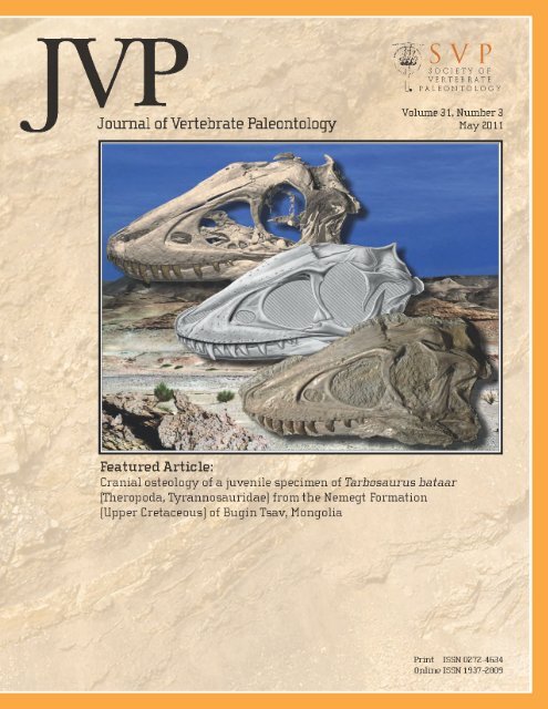
caudal series), but preserves almost all other bones (Fig. 1A, B).<br />
Most remarkably, MPC-D 107/7 includes an articulated skull<br />
(Figs. 1C, 2–4, 5A–E), which is especially well preserved on the<br />
left side where only the articular is missing. In the present paper,<br />
we describe the morphology <strong>of</strong> this skull with an emphasis on<br />
ontogenetically variable characteristics. An analysis <strong>of</strong> included<br />
s<strong>of</strong>t tissues (e.g., brain, inner ear, sinuses) along with a restoration<br />
<strong>of</strong> the skull and a description <strong>of</strong> the postcranial anatomy <strong>of</strong><br />
this <strong>specimen</strong> will be published elsewhere.<br />
MATERIALS AND METHODS<br />
Institutional Abbreviations—BMR, Burpee Museum <strong>of</strong><br />
Natural History, Rockford, Illinois, U.S.A.; CMNH, Cleveland<br />
Museum <strong>of</strong> Natural History, Cleveland, Ohio, U.S.A.; GIN,<br />
Institute <strong>of</strong> Geology, Ulaanbaatar, Mongolia; LACM, Natural<br />
History Museum <strong>of</strong> Los Angeles County, Los Angeles, California,<br />
U.S.A.; MPC, Mongolian Paleontological Center, Ulaanbaatar,<br />
Mongolia; ZPAL, Institute <strong>of</strong> Palaeobiology, Warszawa,<br />
Poland.<br />
Anatomical Abbreviations—?III, possible foramen for the<br />
oculomotor nerve; V 1 , foramen for the ophthalmic branch <strong>of</strong> the<br />
trigeminal nerve; V 2–3 , foramen for the maxillary and mandibular<br />
branches <strong>of</strong> the trigeminal nerve; VII, foramen for the facial<br />
nerve; A, angular; aj, aq,andas, articular surfaces <strong>of</strong> the quadratojugal<br />
for the jugal, quadrate, and squamosal, respectively;<br />
497
498 JOURNAL OF VERTEBRATE PALEONTOLOGY, VOL. 31, NO. 3, 2011<br />
Downloaded By: [Society <strong>of</strong> Vertebrate Paleontology] At: 14:24 10 May 2011<br />
FIGURE 1. Juvenile <strong>Tarbosaurus</strong> <strong>bataar</strong> (MPC-D 107/7). A, photograph <strong>of</strong> the <strong>specimen</strong> prior to complete preparation, and B, its interpretative<br />
drawing. C, skull (right) compared with that <strong>of</strong> a large adult (MPC-D 107/2, left, reversed) in the same scale; D, histological thin sections <strong>of</strong> the left<br />
tibia (above) and fibula (below) made at mid-diaphysis. Arrowheads indicate lines <strong>of</strong> arrested growth. (Figure appears in color online.).<br />
aoc, antotic crest; apo, articular surface <strong>of</strong> the jugal for the<br />
postorbital; BS, basisphenoid; bsr, basisphenoid recess; c and r,<br />
grooves marking courses <strong>of</strong> the caudal and rostral rami <strong>of</strong> the<br />
facial nerve, respectively; cap, capitate process <strong>of</strong> the laterosphenoid;<br />
cor, columellar recess; cp, cornual process; csf, caudal surangular<br />
foramen; ctr, caudal tympanic recess; cup, cultriform process;<br />
D,dentary;da, depression representing the jugal part <strong>of</strong> the<br />
antorbital fossa; dpaM and vpaM, dorsal and ventral prongs <strong>of</strong><br />
the ascending ramus <strong>of</strong> the maxilla, respectively; ECT, ectopterygoid;<br />
EPP, epipterygoid; F, frontal; ia<strong>of</strong>e, internal antorbital fenestra;<br />
J, jugal; L, bones on the left side; LA, lacrimal; llr and mlr,<br />
lateral and medial laminae <strong>of</strong> the rostral ramus <strong>of</strong> the lacrimal,<br />
respectively; lpr, lacrimal pneumatic recess; lr, aperture <strong>of</strong> the<br />
lacrimal recess; LS, laterosphenoid; M, maxilla; mc, medullary<br />
cavity; mf, maxillary fenestra; N, nasal; nlc, groove continuing<br />
rostrally <strong>from</strong> the nasolacrimal canal; OO, otoccipital; osc, otosphenoidal<br />
crest; P, parietal; PA, palatine; paroc, paroccipital<br />
process; pe, pneumatic excavation; PF, prefrontal; pf, pituitary<br />
fossa; pfp, pneumatic fossae on the palatine; PM, premaxilla; pmf,<br />
promaxillary fenestra; PO, postorbital; pqf, paraquadrate foramen;<br />
PRA, prearticular; PRO, prootic; prp, preotic pendant; PS,<br />
parasphenoid; PT, pterygoid; Q, quadrate; QJ, quadratojugal; R,<br />
bones on the right side; rtr, rostral tympanic recess; S, squamosal;<br />
SA, surangular; scf, subcutaneous flange; SCL, sclerotic ring;<br />
SE, sphenethmoid; snf, subnarial foramen; SO, supraoccipital;<br />
sop, suborbital process; SPL, splenial; stf, postorbital part <strong>of</strong> the<br />
supratemporal fossa; t, tubercle; V, vomer.<br />
Comparative Materials—Skulls <strong>of</strong> two cataloged <strong>specimen</strong>s <strong>of</strong><br />
<strong>Tarbosaurus</strong> <strong>bataar</strong>, MPC-D 107/2 and 107/14, were observed for<br />
comparison with MPC-D 107/7. MPC-D 107/2 is a large adult<br />
skeleton, <strong>of</strong> which the articulated skull is 122 cm in rostrocaudal<br />
length (Currie and Carpenter, 2000). This <strong>specimen</strong> was formerly<br />
known as GIN 107/2 in the scientific literature (e.g., Currie and<br />
Carpenter, 2000; Currie, 2003a; Hurum and Sabath, 2003). The<br />
acronym <strong>of</strong> this <strong>specimen</strong> (as well as <strong>of</strong> all other <strong>specimen</strong>s formerly<br />
housed in the GIN) has been changed to MPC after establishment<br />
<strong>of</strong> the Mongolian Paleontological Center in 1996. MPC-<br />
D 107/14 includes several disarticulated cranial and postcranial
Downloaded By: [Society <strong>of</strong> Vertebrate Paleontology] At: 14:24 10 May 2011<br />
TSUIHIJI ET AL.—SKULL OF A JUVENILE TARBOSAURUS 499<br />
FIGURE 2. Skull <strong>of</strong> a <strong>juvenile</strong> <strong>Tarbosaurus</strong> <strong>bataar</strong> (MPC-D 107/7) in left lateral view. A, photograph; B, drawing. In A, the caudal part <strong>of</strong> the left<br />
supratemporal fenestra is still covered by matrix (indicated by a white star).
Downloaded By: [Society <strong>of</strong> Vertebrate Paleontology] At: 14:24 10 May 2011<br />
500 JOURNAL OF VERTEBRATE PALEONTOLOGY, VOL. 31, NO. 3, 2011<br />
FIGURE 3. Skull <strong>of</strong> a <strong>juvenile</strong> <strong>Tarbosaurus</strong> <strong>bataar</strong> (MPC-D 107/7) in right lateral view. A, photograph; B, interpretative drawing showing bones on<br />
the right and left sides in white and grey in color, respectively.
Downloaded By: [Society <strong>of</strong> Vertebrate Paleontology] At: 14:24 10 May 2011<br />
TSUIHIJI ET AL.—SKULL OF A JUVENILE TARBOSAURUS 501<br />
FIGURE 4. Surface rendering images <strong>of</strong> the skull <strong>of</strong> a <strong>juvenile</strong> <strong>Tarbosaurus</strong> <strong>bataar</strong> (MPC-D 107/7) based on the CT data with the matrix digitally<br />
removed. A, left lateral view; B, right lateral view. Bones that are largely concealed within matrix in Figures 2 and 3 are identified in this figure. (Figure<br />
appears in color online.).
Downloaded By: [Society <strong>of</strong> Vertebrate Paleontology] At: 14:24 10 May 2011<br />
502 JOURNAL OF VERTEBRATE PALEONTOLOGY, VOL. 31, NO. 3, 2011<br />
FIGURE 5. Skull <strong>of</strong> a <strong>juvenile</strong> <strong>Tarbosaurus</strong> <strong>bataar</strong> (MPC-D 107/7). A and B, photograph and interpretative drawing, respectively, in dorsal view; C<br />
and D, photograph and volume rendering image based on the CT data, respectively, in caudal view; E and F, close-up <strong>of</strong> the frontal and parietal (E)<br />
in comparison with those <strong>of</strong> a larger individual (MPC-D 107/14, F) in dorsal view. The arrow in E indicates a mushroom-like rostral process <strong>of</strong> the<br />
parietal invading between the right and left frontals. Arrowheads in E and F indicate rostral margins <strong>of</strong> the supratemporal fossae.<br />
bones <strong>of</strong> at least three individuals <strong>of</strong> varying sizes. These individuals<br />
probably belong to large <strong>juvenile</strong>s or young adults and are<br />
much smaller than MPC-D 107/2.<br />
Computed Tomography Scan—In order to supplement observations<br />
on the external surface, the present <strong>specimen</strong> was subjected<br />
to X-ray computed tomographic (CT) imaging. It was<br />
scanned helically on a General Electric LightSpeed Ultra Multi-<br />
Slice CT scanner at O’Bleness Memorial Hospital, Athens, Ohio,<br />
with a slice thickness <strong>of</strong> 625 µm at 120 kV and 200 mA. Observations<br />
on image slices and three-dimensional (3D) visualization<br />
were done using the s<strong>of</strong>tware package Amira 4.1.2 (Visage Imaging<br />
Inc., Chelmsford, MA).
TSUIHIJI ET AL.—SKULL OF A JUVENILE TARBOSAURUS 503<br />
Downloaded By: [Society <strong>of</strong> Vertebrate Paleontology] At: 14:24 10 May 2011<br />
GEOLOGICAL SETTING<br />
The Bugin Tsav locality is situated in the western part <strong>of</strong> the<br />
Gobi Desert and has been known as one <strong>of</strong> the most fossiliferous<br />
dinosaur localities in Mongolia (e.g., Barsbold, 1983; Kurochkin<br />
and Barsbold, 2000). The Late Cretaceous Nemegt Formation<br />
crops out at this locality (e.g., Gradzínski et al., 1977; Shuvalov,<br />
2000), consisting mostly <strong>of</strong> sediments <strong>of</strong> a meandering fluvial system<br />
(e.g., Suzuki and Watabe, 2000; Weishampel et al., 2008).<br />
The age <strong>of</strong> the Nemegt Formation is not well constrained, with<br />
estimates ranging <strong>from</strong> late Campanian–early Maastrichtian to<br />
Maastrichtian (e.g., Gradzínski et al., 1977; Jerzykiewicz and Russell,<br />
1991; Jerzykiewicz, 2000), as reviewed in Weishampel et al.<br />
(2008).<br />
SYSTEMATIC PALEONTOLOGY<br />
DINOSAURIA Owen, 1842<br />
THEROPODA Marsh, 1881<br />
COELUROSAURIA Huene, 1914<br />
TYRANNOSAURIDAE Osborn, 1906<br />
TARBOSAURUS BATAAR (Maleev, 1955a)<br />
(Figs. 1–4, 5A–E, 6, 7, 8A, E, G, 9–11)<br />
Material—MPC-D 107/7, articulated, <strong>juvenile</strong> skeleton missing<br />
cervical and cranial dorsal vertebrae and ribs, as well as the distal<br />
four fifths <strong>of</strong> the caudal vertebral series.<br />
Locality—Bugin Tsav, western Gobi Desert, Mongolia.<br />
Formation/Age—Nemegt Formation (late Campanian–early<br />
Maastrichtian to Maastrichtian).<br />
DESCRIPTION<br />
Taxonomic Identification <strong>of</strong> MPC-D 107/7<br />
The tyrannosaurid affinity <strong>of</strong> MPC-D 107/7 is firmly established<br />
based on numerous cranial (e.g., fused nasals, infratemporal<br />
fenestra constricted by the squamosal-quadratojugal flange,<br />
‘D-shaped’ cross-section <strong>of</strong> the premaxillary teeth) and postcranial<br />
(e.g., third metacarpal bearing no phalanges, ilium bearing<br />
a prominent, ventral projection <strong>from</strong> the preacetabular process<br />
as well as a vertical crest dorsal to the acetabulum on the lateral<br />
surface) synapomorphies <strong>of</strong> Tyrannosauroidea and Tyrannosauridae<br />
observed in the <strong>specimen</strong> (Holtz, 2001, 2004; Currie<br />
et al., 2003). Following Rozhdestvensky (1965), Barsbold (1983),<br />
and Currie (2000) among others, we consider that the four tyrannosaurid<br />
taxa described by Maleev (1955a, 1955b) belong to a single<br />
species, <strong>Tarbosaurus</strong> <strong>bataar</strong>, which is by far the most common<br />
tyrannosaurid in the Nemegt Formation. Other tyrannosaurids<br />
known <strong>from</strong> the Nemegt Formation are Alioramus remotus described<br />
by Kurzanov (1976) and Alioramus altai recently described<br />
by Brusatte et al. (2009). Among these taxa, MPC-D<br />
107/7 is considered as belonging to T. <strong>bataar</strong> for the following<br />
reasons. Firstly, the Bugin Tsav locality has yielded adult <strong>specimen</strong>s<br />
<strong>of</strong> only T. <strong>bataar</strong> and no other tyrannosaurids. Secondly, the<br />
numbers <strong>of</strong> alveoli in the maxilla and dentary in MPC-D 107/7 are<br />
within the range <strong>of</strong> those <strong>of</strong> T. <strong>bataar</strong>, which has 12 or 13 teeth<br />
in the maxilla and 14 or 15 in the dentary (Maleev, 1974; Currie,<br />
2003a; Hurum and Sabath, 2003). The numbers <strong>of</strong> alveoli in<br />
MPC-D 107/7 (confirmed with the CT scan data) are 13 in the left<br />
maxilla and 15 and 14 in the dentaries (Fig. 6A). In contrast, both<br />
species <strong>of</strong> Alioramus have more teeth, 16 in the maxilla and 18 in<br />
the dentary in A. remotus (Kurzanov, 1976) and 17 in the maxilla<br />
and 20 in the dentary in A. altai (Brusatte et al., 2009), respectively.<br />
Thirdly, two features that are considered as characterizing<br />
T. <strong>bataar</strong> are observed in MPC-D 107/7. One is the caudal surangular<br />
foramen, which is relatively smaller than those in other<br />
tyrannosaurids (Holtz, 2004). The other is presence <strong>of</strong> an incipient<br />
subcutaneous flange, which is a ridge that extends along the<br />
ventral margin <strong>of</strong> the external antorbital fossa (Fig. 2) and was<br />
identified as a diagnostic character <strong>of</strong> T. <strong>bataar</strong> by Carr (2005).<br />
Fourthly, species <strong>of</strong> Alioramus, especially A. altai, are characterized<br />
by numerous autapomorphies. Such features include a series<br />
<strong>of</strong> osseous knobs or hornlets on the nasals observed in both A.<br />
remotus and A. altai, and an elongated maxillary fenestra, a laterally<br />
projecting horn on the jugal, and pneumatic foramina on<br />
dorsal ribs observed in A. altai (Brusatte et al., 2009). None <strong>of</strong><br />
these features are present in MPC-D 107/7, making the assignment<br />
<strong>of</strong> MPC-D 107/7 to T. <strong>bataar</strong> virtually certain.<br />
Estimation <strong>of</strong> the Age <strong>of</strong> Death<br />
To assess the age <strong>of</strong> death <strong>of</strong> MPC-D 107/7, transverse middiaphyseal<br />
sections were taken <strong>from</strong> the left fibula and tibia (Fig.<br />
1D). Both sections preserve similar numbers <strong>of</strong> lines <strong>of</strong> arrested<br />
growth (LAGs). Two LAGs occur in the section <strong>from</strong> the fibula.<br />
Because the fibular section lacks a medullary cavity and has only<br />
minor osteonal remodeling in the medial (ad-tibial) cortex, the<br />
sequence <strong>of</strong> LAGs appears complete with no loss <strong>of</strong> the early<br />
growth record. In contrast, a large medullary cavity occurs in<br />
the section <strong>from</strong> the tibia and suggests a need to retrocalculate<br />
the early portion <strong>of</strong> the growth record lost to the expansion <strong>of</strong> the<br />
medullary cavity (e.g., Horner and Padian, 2004). However, the<br />
tibial cortex preserves three LAGs, which is reasonably concordant<br />
with the two LAGs preserved in the fibular cortex. To estimate<br />
the growth rate <strong>of</strong> the tibia, the circumferences <strong>of</strong> the preserved<br />
LAGs were fitted to a linear difference equation, which<br />
takes into account the dependency between successive LAGs<br />
(Cooper et al., 2008:eq. 2.1). Least squares regression reveals<br />
that the mean circumferential growth rate <strong>of</strong> the tibia was 10.3<br />
mm/year and that the neonatal midshaft diameter and circumference<br />
<strong>of</strong> the tibia was about 10 mm and 35 mm, respectively.<br />
Together, the data suggest that MPC-D 107/7 was 2 to 3 years old<br />
at death and is comparable to the early, slow-growth phase before<br />
entering the exponential growth phase on the mass growth<br />
curves <strong>of</strong> North American tyrannosaurids (Erickson et al., 2004).<br />
Description <strong>of</strong> the Skull<br />
The skull is compressed mediolaterally and slightly sheared<br />
dorsoventrally such that the dorsal surface <strong>of</strong> the frontals and<br />
nasal are now visible on the left lateral aspect (Figs. 2, 4A). The<br />
<strong>specimen</strong> was found with the right side exposed at the locality.<br />
Accordingly, the left side <strong>of</strong> the skull is much better preserved<br />
than is the right side and is missing only the articular. On the right<br />
side, most bones in the orbital and temporal regions are missing<br />
or only partially preserved, exposing the lateral aspect <strong>of</strong> the<br />
braincase, <strong>of</strong> which the caudal and ventral parts are also badly<br />
crushed and weathered (Figs. 3, 4B, 10A). Post-dentary bones in<br />
the lower jaw are also mostly missing <strong>from</strong> the right side.<br />
The rostrocaudal length <strong>of</strong> the skull (measured <strong>from</strong> the rostral<br />
end <strong>of</strong> the left premaxilla to the caudoventral corner <strong>of</strong> the<br />
quadratojugal) is 290 mm. The skull length <strong>of</strong> the holotype <strong>specimen</strong><br />
<strong>of</strong> “Shanshanosaurus huoyanshanensis,” putatively representing<br />
a <strong>juvenile</strong> <strong>Tarbosaurus</strong> <strong>bataar</strong>, was estimated to be 288<br />
mm (Currie, 2003a). Therefore, the skull <strong>of</strong> the present <strong>specimen</strong><br />
is about the same size as the holotype <strong>of</strong> “S. huoyanshanensis.”<br />
Currie (2003b) suggested that in tyrannosaurids the length<br />
<strong>of</strong> the skull grows isometrically with that <strong>of</strong> the femur, with these<br />
two lengths being approximately the same in any individual. The<br />
length <strong>of</strong> the left femur <strong>of</strong> MPC-D 107/7 is 303 mm, only slightly<br />
greater than that <strong>of</strong> the skull, thus conforming to Currie’s (2003b)<br />
analysis.<br />
In the following description <strong>of</strong> cranial bones, we will mainly<br />
focus on features observable on the external surface <strong>of</strong> the <strong>specimen</strong>,<br />
supplemented by the CT data. In each bone, we will mainly<br />
concentrate on characteristics observed in MPC-D 107/7 that<br />
show ontogenetic differences <strong>from</strong> those in adult individuals. The<br />
morphology <strong>of</strong> cranial bones <strong>of</strong> T. <strong>bataar</strong> has been described in
Downloaded By: [Society <strong>of</strong> Vertebrate Paleontology] At: 14:24 10 May 2011<br />
504 JOURNAL OF VERTEBRATE PALEONTOLOGY, VOL. 31, NO. 3, 2011<br />
FIGURE 6. Dental morphology <strong>of</strong> a <strong>juvenile</strong> <strong>Tarbosaurus</strong> <strong>bataar</strong> (MPC-D 107/7). A, numbers <strong>of</strong> alveoli counted in frontal sections based on the<br />
CT data, with right and left premaxillae and left maxilla in ventral view (left) and right and left dentaries in dorsal view (right). B, rightandleft<br />
premaxillary teeth in labial (rostral) view; C and D, right and left premaxillary teeth in labiodistal view, respectively; E, select left maxillary teeth in<br />
ventral (above) and labial (below) views; F, right dentary teeth in labial view. The numbers indicate the position <strong>of</strong> each tooth counted <strong>from</strong> the first<br />
(most mesial) alveolus in each bone.
TSUIHIJI ET AL.—SKULL OF A JUVENILE TARBOSAURUS 505<br />
FIGURE 7. Premaxilla and maxilla <strong>of</strong> a <strong>juvenile</strong> <strong>Tarbosaurus</strong> <strong>bataar</strong> (MPC-D 107/7). A, left premaxilla in left lateral view, as well as the sutural<br />
surface <strong>of</strong> the right premaxilla in medial view; B, left maxilla <strong>of</strong> MPC-D 107/7 in lateral view; C, a transverse section <strong>of</strong> the rostral skull ro<strong>of</strong> based on<br />
the CT data showing the articulation mode between the ascending ramus <strong>of</strong> the maxilla and the rostral ramus <strong>of</strong> the lacrimal.<br />
Downloaded By: [Society <strong>of</strong> Vertebrate Paleontology] At: 14:24 10 May 2011<br />
detail by Maleev (1974) and Hurum and Sabath (2003), and these<br />
studies serve as a basis for comparison between MPC-D 107/7<br />
and adult conditions. Studies by Carr (e.g., 1999) on crani<strong>of</strong>acial<br />
ontogeny <strong>of</strong> North American tyrannosaurids serve as the basis<br />
for identifying <strong>juvenile</strong> characteristics in this <strong>specimen</strong>.<br />
Premaxilla—The body <strong>of</strong> the premaxilla is relatively narrow<br />
mediolaterally in MPC-D 107/7 compared to those in adult<br />
<strong>specimen</strong>s (e.g., MPC-D 107/2), which Carr (1999) regarded<br />
as a <strong>juvenile</strong> characteristic in North American tyrannosaurids.<br />
In lateral view, it is only slightly deeper dorsoventrally than<br />
long rostrocaudally (Fig. 7A). In adult <strong>specimen</strong>s (e.g., MPC-D<br />
107/2), in contrast, its dorsoventral depth is much greater than<br />
the rostrocaudal length. The external surface <strong>of</strong> this premaxillary<br />
body is pitted by approximately 10 neurovascular foramina, with<br />
the one at the base <strong>of</strong> the nasal (supranarial) process being the<br />
largest. The dorsolateral aspect <strong>of</strong> the premaxillary body is only<br />
shallowly depressed for flooring <strong>of</strong> the narial tissues, unlike in<br />
adults in which this depression (narial fossa) is more pronounced<br />
(Hurum and Sabath, 2003; MPC-D 107/2). The nasal process<br />
initially projects almost due dorsally <strong>from</strong> the premaxillary body,<br />
and then kinks and extends caudodorsally. Unlike the condition<br />
in a <strong>juvenile</strong> <strong>specimen</strong> <strong>of</strong> <strong>Tarbosaurus</strong> <strong>bataar</strong> mentioned by<br />
Currie (2003a), the distal ends <strong>of</strong> the nasal processes <strong>of</strong> the right<br />
and left premaxillae are appressed to each other and are not<br />
forked in MPC-D 107/7. The maxillary (subnarial) process is<br />
shaped like a rostrocaudally elongate triangle. It appears to be<br />
longer rostrocaudally than in the adult (MPC-D 107/2).<br />
Maxilla—The maxilla <strong>of</strong> MPC-D 107/7 is dorsoventrally much<br />
shallower than in adult <strong>specimen</strong>s (Fig. 7B). For example, the ratio<br />
<strong>of</strong> the maximum height to the maximum length <strong>of</strong> this bone<br />
is 0.39 in this <strong>specimen</strong>, whereas the same ratio <strong>of</strong> the <strong>specimen</strong><br />
described by Hurum and Sabath (2003; ZPAL MgD-I/4, skull<br />
length 1100 mm) is 0.57. The main body (alveolar process) <strong>of</strong><br />
this bone is especially shallow dorsoventrally, representing one<br />
<strong>of</strong> the most prominent <strong>juvenile</strong> characteristics apparent in this<br />
bone. The alveolar margin shows much weaker convexity than<br />
in adults, and is almost straight caudal to the fourth alveolus.<br />
Caudally, the maxillary body gradually tapers and its caudal end<br />
bifurcates into dorsal and ventral processes as in North American<br />
tyrannosaurids (Currie, 2003a). The dorsal process forms a<br />
part <strong>of</strong> the medial wall <strong>of</strong> the antorbital fossa, and extends between<br />
two processes at the rostral end <strong>of</strong> the jugal. The ventral<br />
process, the much longer <strong>of</strong> the two, extends caudally beneath<br />
the jugal. The part <strong>of</strong> the maxillary body rostral to the antorbital<br />
fossa appears as a triangle tapering rostrally in lateral view and<br />
is low dorsoventrally. At the rostral end, the alveolar and rostrodorsal<br />
margins <strong>of</strong> the bone meet at a more acute angle than<br />
in adults. At the premaxillary contact, the rostrodorsal margin is<br />
notched for the subnarial foramen, followed caudodorsally by a<br />
neurovascular foramen as in adults. The maxillary body is also<br />
not thickened laterally, representing an immature condition <strong>of</strong><br />
tyrannosaurids (Carr, 1999). Although the lateral surface <strong>of</strong> the<br />
maxillary body bears numerous neurovascular foramina and shallow<br />
grooves leading to them (for nerves and/or blood vessels),<br />
surface sculpturing is not nearly as pronounced as in adults. Four<br />
relatively large foramina lie along the rostrodorsal margin <strong>of</strong> this<br />
bone, arranged in a line about 1 cm <strong>from</strong> the margin. Another<br />
row <strong>of</strong> relatively large foramina extends along the ventral margin<br />
<strong>of</strong> the alveolar process within a few millimeters <strong>from</strong> the margin,<br />
comprising the alveolar row <strong>of</strong> the foramina <strong>of</strong> Brochu (2003).<br />
As described in Daspletosaurus by Carr (1999), a sulcus extends<br />
<strong>from</strong> the caudal-most foramen belonging to this row on the caudal,<br />
ventral process described above beneath the jugal in MPC-D<br />
107/7, as in MPC-D 107/2. Brochu’s (2003) circumfenestral row<br />
<strong>of</strong> neurovascular foramina, on the other hand, is apparent only<br />
in the caudal part <strong>of</strong> the alveolar process, where neurovascular<br />
foramina line up as a straight row along the ventral margin <strong>of</strong> the<br />
external antorbital fenestra.<br />
The margin <strong>of</strong> the antorbital fossa is well delimited. Unlike<br />
in adult <strong>specimen</strong>s such as the one illustrated in Hurum and<br />
Sabath (2003) and MPC-D 107/2, this fossa extends caudally beneath<br />
the internal antorbital fenestra to extend onto the jugal.<br />
That is, the medial wall <strong>of</strong> this fossa is visible beneath the internal<br />
antorbital fenestra in lateral view. The caudal one third <strong>of</strong><br />
the ventral margin <strong>of</strong> the antorbital fossa is marked by a sharp<br />
ridge representing an incipient subcutaneous flange (Carr, 2005),<br />
which becomes less prominent rostrally. At the rostroventral corner<br />
<strong>of</strong> the antorbital fossa, the medial wall <strong>of</strong> the fossa is slightly<br />
laterally convex, or ‘swollen’ in appearance due to underlying<br />
pneumatic sinuses, making the margin <strong>of</strong> this fossa further obscured.<br />
The internal antorbital fenestra, bounded by the maxilla,<br />
lacrimal, and jugal, is slightly longer than high (67 mm in length<br />
vs. 64 mm in height), whereas it is higher than long in adults (e.g.,<br />
MPC-D 107/2). The rostral margin <strong>of</strong> the maxillary fenestra is<br />
widely separated <strong>from</strong> that <strong>of</strong> the external antorbital fenestra as<br />
in the holotype <strong>specimen</strong> <strong>of</strong> “Shanshanosaurus huoyanshanensis”<br />
(Currie and Dong, 2001). This is a conspicuous, <strong>juvenile</strong> characteristic<br />
<strong>of</strong> tyrannosaurids described by Carr (1999). In adult<br />
<strong>Tarbosaurus</strong> <strong>bataar</strong> (Hurum and Sabath, 2003; MPC-D 107/2),<br />
in contrast, the rostral margins <strong>of</strong> the maxillary fenestra and
Downloaded By: [Society <strong>of</strong> Vertebrate Paleontology] At: 14:24 10 May 2011<br />
506 JOURNAL OF VERTEBRATE PALEONTOLOGY, VOL. 31, NO. 3, 2011<br />
FIGURE 8. Nasal (A–D), lacrimal (E, F), and postorbital (G, H)<strong>of</strong><strong>Tarbosaurus</strong> <strong>bataar</strong>, comparison between a <strong>juvenile</strong> (MPC-D 107/7) and a larger<br />
<strong>specimen</strong> (MPC-D 107/14). A, surface rendering image <strong>of</strong> the nasals <strong>of</strong> MPC-D 107/7 based on the CT data in left dorsolateral view, showing the<br />
presence <strong>of</strong> a small lacrimal process on the nasal (arrow), which is lost in larger individuals; B–D, nasals <strong>of</strong> MPC-D 107/14 (reversed) in right lateral<br />
(B), ventral (C), and ventrolateral (D) views. As shown in D, the maxillary contact in MPC-D 107/14 still consists <strong>of</strong> a longitudinal groove, lacking<br />
transverse ridges and grooves observed in fully adult individuals. E, left lacrimal <strong>of</strong> MPC-D 107/7 in lateral view; F, right lacrimal <strong>of</strong> MPC-D 107/14<br />
(reversed) in lateral view. Note that the antorbital fossa is fully exposed laterally on the rostral ramus <strong>of</strong> the lacrimal in E, whereas it is concealed by<br />
the lateral lamina <strong>of</strong> this ramus in F. G, left postorbital <strong>of</strong> MPC-D 107/7 in lateral view; H, right postorbital <strong>of</strong> MPC-D 107/14 (reversed) in lateral<br />
view.
TSUIHIJI ET AL.—SKULL OF A JUVENILE TARBOSAURUS 507<br />
Downloaded By: [Society <strong>of</strong> Vertebrate Paleontology] At: 14:24 10 May 2011<br />
FIGURE 9. Jugal and quadratojugal <strong>of</strong> a <strong>juvenile</strong> <strong>Tarbosaurus</strong> <strong>bataar</strong> (MPC-D 107/7). A, left jugal in lateral view; B and C, right quadratojugal in<br />
lateral and medial views, respectively.<br />
external antorbital fenestra approach each other. The maxillary<br />
fenestra is clearly longer rostrocaudally than high dorsoventrally<br />
(Figs. 2, 7B). Whereas this is still the case in MPC-D 107/2, Hurum<br />
and Sabath (2003) described that the height and depth <strong>of</strong> the<br />
maxillary fenestra are the same in the <strong>specimen</strong> <strong>of</strong> T. <strong>bataar</strong> that<br />
they described. In contrast, Carr (1999) argued that the length-toheight<br />
ratio <strong>of</strong> this fenestra increases ontogenetically in another<br />
tyrannosaurid, Gorgosaurus (Albertosaurus) libratus, thus showing<br />
an ontogenetic trend possibly opposite to that in T. <strong>bataar</strong>.<br />
Dorsal to the maxillary fenestra and along the rostrodorsal margin<br />
<strong>of</strong> the antorbital fossa is an obliquely elongate depression representing<br />
a pneumatic excavation (Figs. 2, 7B). The promaxillary<br />
fenestra lies at the rostroventral corner <strong>of</strong> the antorbital fossa,<br />
concealed laterally by the rostral rim <strong>of</strong> the external antorbital<br />
fenestra as in the holotype <strong>of</strong> “S. huoyanshanensis” (Currie and<br />
Dong, 2001) and most other tyrannosaurid <strong>specimen</strong>s, regardless<br />
<strong>of</strong> age (Witmer, 1997).<br />
The CT scan data revealed that the maxillary sinuses are well<br />
developed internally, even at this young age. They basically follow<br />
the same pattern as in adult T. <strong>bataar</strong>, G. libratus,andTyrannosaurus<br />
rex (Witmer, 1997; Hurum and Sabath, 2003; Witmer<br />
and Ridgely, 2008). The promaxillary recess is followed caudally<br />
by the maxillary antrum, separated <strong>from</strong> the latter by a vertical<br />
bony strut. From the promaxillary fenestra extends a pneumatic<br />
excavation forming three interconnected chambers within<br />
the ascending ramus (caudodorsal process) <strong>of</strong> the maxilla. Caudodorsal<br />
to the maxillary antrum, a well-developed epiantral recess<br />
is present at the junction between the interfenestral strut and<br />
pila postantralis. As in other tyrannosaurids (Witmer, 1997; Witmer<br />
and Ridgely, 2008), the pila postantralis is not only pneumatic<br />
but also fenestrate, allowing the sinus diverticulum within<br />
the maxilla to pass caudally to reach the palatine pneumatic<br />
chambers.<br />
The ascending ramus <strong>of</strong> the maxilla tapers caudally and is not<br />
particularly massive, unlike the more mature <strong>specimen</strong> described<br />
by Hurum and Sabath (2003). The CT scan data revealed that this<br />
process bifurcates into dorsal and ventral prongs at the caudal<br />
end and is medially covered by the medial lamina <strong>of</strong> the rostral<br />
ramus <strong>of</strong> the lacrimal (Fig. 7C). The dorsal prong terminates in a<br />
ventrally open trough formed by the medial and lateral laminae<br />
<strong>of</strong> the rostral ramus <strong>of</strong> the lacrimal. The ventral prong forms a<br />
dorsally open trough medially, to which the ventral end <strong>of</strong> the<br />
medial lamina <strong>of</strong> the lacrimal fits (Fig. 7C).<br />
Nasal—Even at this young age, the right and left nasals are already<br />
fused to each other at their mid-length (Figs. 2, 4A, 5A, B,<br />
8A). However, these bones are still separated <strong>from</strong> each other<br />
by a fissure for about one fourth <strong>of</strong> the length both <strong>from</strong> the<br />
rostral and caudal ends, as in large individuals <strong>of</strong> <strong>Tarbosaurus</strong><br />
<strong>bataar</strong> (Maleev, 1974; Hurum and Sabath, 2003). At the rostral<br />
end, the nasals are forked along the midline to receive the nasal<br />
processes <strong>of</strong> the premaxillae. The caudal end is apparently broken<br />
<strong>of</strong>f and missing <strong>from</strong> the <strong>specimen</strong>. The dorsal surface <strong>of</strong><br />
the fused nasals is still rather smooth, lacking prominent rugosities<br />
or papillae, unlike in adult individuals, and bears prominent<br />
neurovascular foramina along their lateral margins. Vaulting <strong>of</strong><br />
these bones is much less pronounced than those in adult individuals<br />
such as MPC-D 107/2. The rostral part <strong>of</strong> the contact surface<br />
with the maxilla exposed in right lateral view consists <strong>of</strong><br />
a shallow, longitudinal groove. The CT scan data revealed the<br />
rest <strong>of</strong> the maxillary articular surface is rather smooth, lacking<br />
prominent transverse ridges, or pegs and sockets, unlike the adult<br />
condition described by Hurum and Sabath (2003). The maxillary<br />
articular surface <strong>of</strong> the nasals <strong>of</strong> a larger but still young individual<br />
<strong>of</strong> T. <strong>bataar</strong> (MPC-D 107/14; preserved length <strong>of</strong> the nasals is<br />
293 mm) also consists <strong>of</strong> a longitudinal groove, still lacking transverse<br />
ridges and grooves (Fig. 8B–D). Such a maxillary articular
Downloaded By: [Society <strong>of</strong> Vertebrate Paleontology] At: 14:24 10 May 2011<br />
508 JOURNAL OF VERTEBRATE PALEONTOLOGY, VOL. 31, NO. 3, 2011<br />
FIGURE 10. Braincase region <strong>of</strong> a <strong>juvenile</strong> <strong>Tarbosaurus</strong> <strong>bataar</strong> (MPC-D 107/7). A, photo in right lateral view; B, right prootic in lateral view; C,<br />
surface rendering image based on the CT data in left lateral view.
TSUIHIJI ET AL.—SKULL OF A JUVENILE TARBOSAURUS 509<br />
Downloaded By: [Society <strong>of</strong> Vertebrate Paleontology] At: 14:24 10 May 2011<br />
surface consisting <strong>of</strong> a longitudinal groove is a general feature<br />
seen in young tyrannosaurids (Carr, 1999). The nasal is apparently<br />
excluded <strong>from</strong> the dorsal margin <strong>of</strong> the external antorbital<br />
fenestra in MPC-D 107/7, whereas it forms a part <strong>of</strong> this margin<br />
in adult <strong>specimen</strong>s (e.g., MPC-D 107/2).<br />
The nasal <strong>of</strong> MPC-D 107/7 has a small lacrimal process (Fig.<br />
8A). Whereas some other tyrannosaurids have this process (e.g.,<br />
Carr, 1999; Brochu, 2003; Currie, 2003a), Hurum and Sabath<br />
(2003) regarded it as lacking in adult T. <strong>bataar</strong>, and Currie et<br />
al. (2003) identified its absence (in adults) as a synapomorphy<br />
uniting T. <strong>bataar</strong>, Alioramus remotus, andDaspletosaurus. The<br />
presence <strong>of</strong> such a process, albeit being tiny, in MPC-D 107/7<br />
indicates that this is an ontogenetically variable character in T.<br />
<strong>bataar</strong>, as in Daspletosaurus in which the lacrimal process is<br />
present in <strong>juvenile</strong>s but is lost in mature individuals (Currie,<br />
2003a). Caudally, the nasals slightly expand mediolaterally between<br />
the lacrimals. The same condition was described for young<br />
stages <strong>of</strong> G. libratus by Carr (1999). The same parts become constricted<br />
between the lacrimals in adult T. <strong>bataar</strong> (Carr, 1999; also<br />
illustrated in Hurum and Sabath, 2003).<br />
Lacrimal—In lateral view, the lacrimal is T-shaped with the<br />
caudal ramus extending behind the ventral ramus (Fig. 8E), unlike<br />
in adults in which the caudal ramus is inflated and this bone<br />
is shaped like a ‘7’ (Carr et al., 2005) or an inverted ‘L’ (Fig. 8F).<br />
As Carr (1999) described in his youngest stage <strong>of</strong> Gorgosaurus libratus,<br />
the rostral ramus <strong>of</strong> the lacrimal is divided into the lateral<br />
and medial laminae, with the former situated dorsal to the latter<br />
(Fig. 8E). These two laminae are well separated <strong>from</strong> each other,<br />
and the caudodorsal part <strong>of</strong> the antorbital fossa expands onto the<br />
medial lamina. This part <strong>of</strong> the antorbital fossa is fully exposed<br />
in lateral view (i.e., not hidden by the lateral lamina <strong>of</strong> the rostral<br />
ramus extending ventrally). Such lateral exposure <strong>of</strong> the entire<br />
lacrimal antorbital fossa was identified as an immature feature in<br />
G. libratus by Carr (1999). Also as in immature, North American<br />
tyrannosaurids (Carr, 1999), the caudal end <strong>of</strong> this fossa is sharply<br />
bounded by the rostral edge <strong>of</strong> the ventral ramus <strong>of</strong> this bone.<br />
In larger <strong>specimen</strong>s <strong>of</strong> <strong>Tarbosaurus</strong> <strong>bataar</strong> (e.g., MPC-D 107/14;<br />
dorsoventral height <strong>of</strong> the lacrimal is 172 mm), the rostral part <strong>of</strong><br />
the antorbital fossa on the rostral ramus appears to be separated<br />
<strong>from</strong> its caudal part bearing the aperture <strong>of</strong> the lacrimal recess<br />
and is concealed in lateral view by the ventrally expanded lateral<br />
lamina (Fig. 8F). In an even larger individual (MPC-D 107/2), the<br />
lacrimal antorbital fossa almost completely disappears except for<br />
a deep lacrimal recess.<br />
The apertures <strong>of</strong> the lacrimal recess open at the caudodorsal<br />
corner <strong>of</strong> the antorbital fossa between the rostral and ventral<br />
rami <strong>of</strong> the lacrimal. The CT scan data show that the lacrimal recess<br />
hollows out the dorsal part <strong>of</strong> the bone as in Tyrannosaurus<br />
rex (Brochu, 2003; Witmer and Ridgely, 2008) and extends rostrally<br />
within the lateral lamina <strong>of</strong> the rostral ramus (Fig. 7C). The<br />
nasolacrimal canal extends rostrally ventral to the lacrimal recess.<br />
The canal opens via a single aperture in the orbit caudally, and<br />
then extends rostrally within the medial lamina <strong>of</strong> the rostral ramus<br />
<strong>of</strong> the lacrimal to open medially into the nasal cavity near the<br />
contact with the maxilla (Fig. 7C). This morphology conforms to<br />
the basic pattern observed in other theropods (Sampson and Witmer,<br />
2007).<br />
The dorsal margins <strong>of</strong> the rostral and caudal rami <strong>of</strong> the<br />
lacrimal lack pronounced rugosity (Fig. 8E), unlike in adults (Hurum<br />
and Sabath, 2003; MPC-D 107/2). Where the rostral, caudal,<br />
and ventral rami meet, the dorsal margin <strong>of</strong> the bone shows a sinuous<br />
curvature, with the rostral part <strong>of</strong> the caudal ramus plunging<br />
ventrally. The caudal ramus, articulating with the frontal, tapers<br />
caudally (Fig. 8E) and does not have an inflated appearance, unlike<br />
in larger <strong>specimen</strong>s (Hurum and Sabath, 2003; MPC-D 107/2<br />
and 107/14; Fig. 8F). It does not contact the postorbital caudally<br />
so that the frontal intervenes between the two to reach the orbital<br />
margin (Figs. 2, 5A, B, E). The ventral ramus is relatively thinner<br />
rostrocaudally than in larger <strong>specimen</strong>s, and bears a small tubercle<br />
near the dorsal end <strong>of</strong> the caudal margin (Fig. 8E). This tubercle<br />
appears to correspond to the one identified as the attachment<br />
<strong>of</strong> the suborbital ligament in Appalachiosaurus montgomeriensis<br />
by Carr et al. (2005), although its position in MPC-D 107/7 seems<br />
too dorsally placed for an attachment <strong>of</strong> such a ligament that<br />
marks the ventrolateral boundary <strong>of</strong> the orbit, particularly in such<br />
a young animal that would have had a relatively large eyeball.<br />
Postorbital—All three rami (rostral, caudal, and ventral rami)<br />
are much slenderer in MPC-D 107/7 than those in larger individuals<br />
(Fig. 8G, H). The most prominent ontogenetic change is observed<br />
in the ventral (jugal) ramus, which becomes much wider<br />
rostrocaudally as individuals grow. This rostrocaudal expansion<br />
<strong>of</strong> the ventral ramus makes the relative lengths <strong>of</strong> the rostral<br />
(frontal) and caudal (squamosal or intertemporal) rami appear<br />
shorter in larger individuals (Fig. 8H). Unlike in larger <strong>specimen</strong>s<br />
(Maleev, 1974; Hurum and Sabath, 2003; MPC-D 107/2 and<br />
107/14), the ventral ramus in MPC-D 107/7 does not bear a rostrally<br />
extending suborbital process (Fig. 8G). Instead, it only has a<br />
slight rostral expansion bearing a scar on its lateral aspect. Distal<br />
to this expansion, the ventral ramus tapers ventrally. The orbit,<br />
therefore, is not constricted at its mid-height, unlike in adults. In<br />
lateral view, the rostral ramus tapers quickly rostrally and has no<br />
cornual process on the caudodorsal margin <strong>of</strong> the orbit, bearing<br />
only weak rugosity (Fig. 8G). The cornual process appears and<br />
becomes progressively prominent in larger individual. In MPC-D<br />
107/2, for example, it is massively developed, bearing bony papillae.<br />
In dorsal view, the rostral ramus is narrow mediolaterally and<br />
its lateral margin meets that <strong>of</strong> the caudal ramus at an obtuse<br />
angle in MPC-D 107/7 (Fig. 5E). In other words, the rostral ramus<br />
extends rostromedially with very gentle curvature. In MPC-<br />
D 107/2, in contrast, these two rami meet at almost a right angle in<br />
dorsal view, with the rostral ramus appearing to extend due medially.<br />
In MPC-D 107/7, the lateral and rostrolateral boundaries<br />
<strong>of</strong> the supratemporal fossa are sharply delimited by a ridge along<br />
the dorsal margin <strong>of</strong> the postorbital. This part <strong>of</strong> the fossa on the<br />
rostral ramus <strong>of</strong> the postorbital is rather shallow, with the articular<br />
processes for the frontal and parietal forming the floor <strong>of</strong> this<br />
fossa (Fig. 8G). The CT data revealed that the frontal articular<br />
process is dorsoventrally much thinner than in larger individuals<br />
(e.g., MPC-D 107/14). The parietal articular process is tab-like,<br />
extends further medially than the frontal articular process (Fig.<br />
8G), and is followed ventrally by the articular surface for the laterosphenoid.<br />
Jugal—The rostral (maxillary) ramus and the suborbital<br />
part between the rostral and postorbital (ascending) rami are<br />
dorsoventrally shallower in MPC-D 107/7 (Fig. 9A) than in larger<br />
individuals (Maleev, 1974; Hurum and Sabath, 2003; MPC-D<br />
107/2). The latter part is also relatively longer rostrocaudally than<br />
in more mature <strong>specimen</strong>s. These features were identified as immature<br />
characteristics in Gorgosaurus libratus by Carr (1999).<br />
The lacrimal articulates with the dorsal aspect <strong>of</strong> the rostral ramus,<br />
and their articular surface appears as a straight, oblique<br />
line in lateral view. The rostrodorsal part <strong>of</strong> the rostral ramus<br />
has a shallow but well-demarcated depression, which, together<br />
with the rostroventral lamina <strong>of</strong> the ventral ramus <strong>of</strong> the lacrimal,<br />
forms the caudoventral corner <strong>of</strong> the antorbital fossa (Figs. 2,<br />
9A). Large individuals <strong>of</strong> <strong>Tarbosaurus</strong> <strong>bataar</strong> have a prominent<br />
foramen (called the secondary fossa <strong>of</strong> the jugal foramen in Carr,<br />
1999, pneumatopore in Currie, 2003a, and jugal foramen in Hurum<br />
and Sabath, 2003) in this depression (e.g., Maleev, 1974; Hurum<br />
and Sabath, 2003). In MPC-D 107/2, a very large adult, the<br />
ridge demarcating the antorbital fossa on the jugal is resorbed,<br />
making the margin <strong>of</strong> this deep foramen grade directly into the<br />
lateral surface <strong>of</strong> the rostal ramus. This is similar to the condition<br />
in Daspletosaurus torosus described by Carr (1999). In MPC-D<br />
107/7, in contrast, this foramen is rudimentary. However, the CT<br />
scan data reveal that much <strong>of</strong> the length <strong>of</strong> the jugal is pneumatic,
510 JOURNAL OF VERTEBRATE PALEONTOLOGY, VOL. 31, NO. 3, 2011<br />
Downloaded By: [Society <strong>of</strong> Vertebrate Paleontology] At: 14:24 10 May 2011<br />
extending into the base <strong>of</strong> the postorbital ramus and caudally to<br />
near the fork <strong>of</strong> the caudal (quadratojugal) ramus.<br />
The postorbital ramus is slender. The articular surface for the<br />
postorbital is apparent, but is much narrower than those in larger<br />
individuals (e.g., Hurum and Sabath, 2003). Hurum and Sabath<br />
(2003) described that the lateral surface <strong>of</strong> the main body <strong>of</strong> the<br />
jugal has a shallow depression at the base <strong>of</strong> the postorbital ramus<br />
in adult T. <strong>bataar</strong>. Such a depression is apparently absent in<br />
MPC-D 107/7, although the corresponding area is crushed and<br />
obscured in this <strong>specimen</strong>. The cornual process that is present on<br />
the ventral margin <strong>of</strong> this area in adults is very poorly developed,<br />
if present at all, in MPC-D 107/7.<br />
Quadratojugal—Both left and right quadratojugals are preserved<br />
in MPC-D 107/7. Whereas the left bone is in situ, articulating<br />
with adjacent bones, the right bone was found isolated<br />
<strong>from</strong> the rest <strong>of</strong> the skull, providing detailed information on its<br />
morphology (Fig. 9B, C). As in adults, the dorsal (squamosal)<br />
and ventral (jugal) rami extend rostrally. The rostral expansion<br />
or flaring <strong>of</strong> the dorsal ramus appears to start more dorsally in<br />
MPC-D 107/7 than in a large adult, MPC-D 107/2. In other words,<br />
the vertical shaft <strong>of</strong> this bone between the dorsal and ventral rami<br />
appears to be longer in the former than in the latter. The dorsal<br />
margin <strong>of</strong> the dorsal ramus appears to be inclined obliquely,<br />
<strong>from</strong> caudodorsal to rostroventral directions, unlike in adults in<br />
which the dorsal margin is almost horizontal (e.g., MPC-D 107/2).<br />
The lateral surface <strong>of</strong> the dorsal ramus is concave as in adults<br />
such as MPC-D 107/2. This concavity, however, lacks the pneumatic<br />
foramen present in the quadratojugals <strong>of</strong> two small, Maastrichtian<br />
tyrannosaurid <strong>specimen</strong>s <strong>from</strong> North America, CMNH<br />
7541 and BMR P2002.4.1 (Witmer and Ridgely, 2010). Absence<br />
<strong>of</strong> pneumatic foramina in the quadratojugal characterizes definitive<br />
adult <strong>specimen</strong>s <strong>of</strong> <strong>Tarbosaurus</strong> <strong>bataar</strong> (e.g., MPC-D 107/2)<br />
as well. The isolated, right quadratojugal shows that a notch is<br />
present in the caudal part <strong>of</strong> the dorsal end <strong>of</strong> the dorsal ramus<br />
(Fig. 9B, C), as illustrated in the same bone in a larger individual<br />
in Maleev (1974). Medially, this notch marks the rostral end<br />
<strong>of</strong> the articular surface for the quadrate, and the articular surface<br />
for the squamosal lies rostral to this notch (Fig. 9C). Another articular<br />
surface <strong>of</strong> the quadrate lies on the medial aspect <strong>of</strong> the<br />
caudoventral corner <strong>of</strong> this bone. Rostral to this quadrate articular<br />
surface, a rostrocaudally elongate facet occupies most <strong>of</strong> the<br />
length <strong>of</strong> the ventral ramus. This facet is for articulation with the<br />
lateral surface <strong>of</strong> the ventral prong <strong>of</strong> the forked, caudal ramus <strong>of</strong><br />
the jugal (Fig. 9C).<br />
Squamosal—In lateral view, the rostroventral (quadratojugal)<br />
ramus <strong>of</strong> the squamosal extends rostroventrally <strong>from</strong> the<br />
quadrate cotyle, rather than extending rostrally and almost due<br />
horizontally as in larger individuals (Hurum and Sabath, 2003;<br />
MPC-D 107/2), making an oblique contact line with the quadratojugal<br />
(Figs. 2, 4A). The CT data revealed that there is a single,<br />
large, presumably pneumatic fossa on the internal aspect<br />
<strong>of</strong> the body <strong>of</strong> this bone as in larger individuals <strong>of</strong> <strong>Tarbosaurus</strong><br />
<strong>bataar</strong> (e.g., Hurum and Sabath, 2003; MPC-D 107/14) and other<br />
tyrannosaurids in general, although it does not extend into the<br />
postquadratic process in MPC-D 107/7 unlike in North American<br />
tyrannosaurids (Witmer, 1997; Witmer and Ridgely, 2008). In a<br />
very large adult (MPC-D 107/2), a well-developed rugosity covers<br />
the dorsal to caudodorsal margins <strong>of</strong> the rostrodorsal (postorbital)<br />
ramus. In MPC-D 107/7, these margins are rather smooth<br />
with only weak striations present.<br />
Quadrate—The left quadrate is preserved in articulation with<br />
the quadratojugal and squamosal. As in adult <strong>Tarbosaurus</strong> <strong>bataar</strong><br />
(Hurum and Sabath, 2003) and other tyrannosaurids in general<br />
(e.g., Brochu, 2003; Currie, 2003a), a large paraquadrate foramen<br />
is formed between the quadrate and quadratojugal (Fig.<br />
5C, D). The pterygoid flange extends rostrally <strong>from</strong> the body <strong>of</strong><br />
the quadrate. This flange bears a prominent facet for articulation<br />
with the pterygoid on the medial aspect <strong>of</strong> its rostral part.<br />
The mandibular condyle is partially broken with the medial hemicondyle<br />
missing. A pneumatic fossa is located at the base <strong>of</strong> the<br />
pterygoid ramus and rostrodorsal to the mandibular condyle as<br />
in adult T. <strong>bataar</strong> (Hurum and Sabath, 2003) and other tyrannosaurids<br />
(e.g., Molnar, 1991; Brochu, 2003; Currie 2003a). In CT<br />
images, this fossa can be observed to open into internal pneumatic<br />
chambers in the quadrate body, mandibular condyle, and<br />
pterygoid flange.<br />
Prefrontal—A small prefrontal can be recognized as a separate<br />
bone on the skull ro<strong>of</strong>, exposed between the lacrimal and frontal<br />
(Figs. 2, 5A, B, E). The left prefrontal is crushed and fragmented<br />
between the lacrimal and frontal. Only the caudal part <strong>of</strong> the right<br />
prefrontal is exposed on the dorsal surface, overlain by the dislocated<br />
nasals rostrally. This bone, however, is well preserved,<br />
and the CT data show that it is rostrocaudally elongated and<br />
teardrop-shaped in dorsal view. This right prefrontal is slightly<br />
dislocated, and its triangular, lacrimal articular surface is exposed<br />
caudal to the lacrimal on the right lateral side <strong>of</strong> the <strong>specimen</strong><br />
(Fig. 3). This lacrimal articular surface is demarcated by a sharp<br />
ridge <strong>from</strong> a smooth and slightly concave orbital surface <strong>of</strong> the<br />
descending process. The latter process, which is broken halfway<br />
through, is wide mediolaterally but is very thin rostrocaudally.<br />
Frontal—The frontal is markedly longer rostrocaudally than<br />
wide mediolaterally in MPC-D 107/7, whereas the width-tolength<br />
ratio increases in larger individuals <strong>of</strong> <strong>Tarbosaurus</strong> <strong>bataar</strong><br />
(Fig. 5E, F). For example, the width-to-length ratio (the width<br />
is measured <strong>from</strong> the midline to the lateral margin <strong>of</strong> the bone<br />
between the articulations with the lacrimal and postorbital, and<br />
the length is measured <strong>from</strong> the most caudal part <strong>of</strong> the frontoparietal<br />
suture to the rostral end <strong>of</strong> the frontal bone) is 0.23 in<br />
MPC-D 107/7, in which the rostrocaudal length <strong>of</strong> this bone is 91<br />
mm (measured based on the CT data), whereas this ratio is 0.40<br />
in MPC-D 107/14, in which the length <strong>of</strong> the frontal is 145 mm.<br />
The same ontogenetic trend was found in tyrannosaurids in general<br />
by Currie (2003a) and was also described for Tyrannosaurus<br />
rex by Carr (1999) and Carr and Williamson (2004).<br />
The caudal part <strong>of</strong> the frontal in MPC-D 107/7 is rounded dorsally,<br />
and the rostral part <strong>of</strong> the supratemporal fossa extends onto<br />
it. Unlike in larger <strong>specimen</strong>s (e.g., MPC-D 107/2 and 107/14; Fig.<br />
5F), however, this frontal portion <strong>of</strong> the supratemporal fossa in<br />
MPC-D 107/7 is very shallow and is not markedly concave. A very<br />
low ridge extending rostrolaterally <strong>from</strong> the midline marks the<br />
rostral margin <strong>of</strong> this fossa (Fig. 5E). In adult T. <strong>bataar</strong>, the left<br />
and right supratemporal fossae meet along the midline to form<br />
the frontal sagittal crest (Hurum and Sabath, 2003; Holtz, 2004).<br />
MPC-D 107/7 lacks such a crest, and the area between these fossae<br />
is gently convex.<br />
In dorsal view, the suture line between the right and left<br />
frontals is clearly visible throughout the contact <strong>of</strong> these bones,<br />
unlike in more mature individuals in which this suture is obliterated<br />
caudally (Maleev, 1974; Hurum and Sabath, 2003). This<br />
suture line appears as a straight, smooth line in MPC-D 107/7<br />
(Fig. 5E), lacking interdigitation seen in larger individuals (e.g.,<br />
MPC-D 107/14; Fig. 5F) except for the most caudal part. The suture<br />
line with the parietal is almost a straight, transverse line, except<br />
on the midline, where a mushroom-like rostral process <strong>of</strong> the<br />
parietal invades between the right and left frontals (Fig. 5E). In<br />
more mature <strong>specimen</strong>s, in contrast, the median part <strong>of</strong> the parietals<br />
forms wedge-shaped articulation with the frontals as seen in<br />
MPC-D 107/14 (Fig. 5F) and MPC-D 107/2. Unlike in larger individuals<br />
(Maleev, 1974; Hurum and Sabath, 2003), a small part <strong>of</strong><br />
the frontal is exposed to the orbital margin between the lacrimal<br />
and postorbital in MPC-D107/7 (Figs. 2, 4A, 5A, B, E).<br />
Parietal—The left and right parietals are already fused together<br />
(Fig. 5A, B, E). The most prominent <strong>juvenile</strong> feature seen<br />
in the parietal is a very low nuchal crest, which does not extend<br />
much dorsally beyond the level <strong>of</strong> the frontal skull ro<strong>of</strong> (Figs. 2,<br />
4). The median, sagittal crest extends rostrally <strong>from</strong> the nuchal
TSUIHIJI ET AL.—SKULL OF A JUVENILE TARBOSAURUS 511<br />
Downloaded By: [Society <strong>of</strong> Vertebrate Paleontology] At: 14:24 10 May 2011<br />
crest almost horizontally, with its dorsal margin being slightly<br />
concave ventrally in lateral view. The sagittal crest does not extend<br />
onto the frontal (Figs. 2, 4A, 5A, E), unlike in large individuals<br />
such as the one illustrated in Hurum and Sabath (2003:fig. 17).<br />
The nuchal crest is higher than the sagittal crest in larger individuals<br />
<strong>of</strong> <strong>Tarbosaurus</strong> <strong>bataar</strong> (Hurum and Sabath, 2003; MPC-D<br />
107/2 and 107/14). In MPC-D 107/7, however, the height <strong>of</strong> the<br />
nuchal crest is almost the same as the maximum height <strong>of</strong> the<br />
sagittal crest at its caudal end (Figs. 2, 4).<br />
In larger individuals <strong>of</strong> T. <strong>bataar</strong>, both nuchal and sagittal crests<br />
are higher than those in MPC-D 107/7. This is especially the case<br />
with the nuchal crest. Measured <strong>from</strong> the dorsal margin <strong>of</strong> the<br />
foramen magnum in caudal view, the nuchal crest is approximately<br />
1.5 times as high as the supraoccipital in MPC-D 107/7<br />
(Fig. 5C, D), but is more than twice as high as the latter in MPC-D<br />
107/2, a large adult. An ontogenetic increase in the height <strong>of</strong> the<br />
nuchal crest was also reported in North American tyrannosaurids<br />
by Carr (1999).<br />
In caudal view, the dorsal part <strong>of</strong> the nuchal crest expands laterally<br />
only slightly in MPC-D 107/7 (Fig. 5C, D). In larger individuals,<br />
in contrast, the lateral expansion <strong>of</strong> this crest is pronounced<br />
(e.g., Hurum and Sabath, 2003; MPC-D 107/14). The dorsal margin<br />
<strong>of</strong> this crest, which is marked by strong rugosities in adults<br />
(e.g., MPC-D 107/2), is rather smooth with only faint scarring<br />
present. However, a pair <strong>of</strong> scars along the midline representing<br />
the fleshly insertion <strong>of</strong> m. spinalis capitis (Tsuihiji, 2010) is<br />
already apparent (Fig. 5C).<br />
Other Bones <strong>of</strong> the Braincase—Although the lateral surface <strong>of</strong><br />
the braincase is exposed on the right side <strong>of</strong> the <strong>specimen</strong> (Figs. 3,<br />
4B, 10A), the morphology <strong>of</strong> bones <strong>of</strong> the braincase on this side,<br />
except for the prootic (Fig. 10B), laterosphenoid, and part <strong>of</strong> the<br />
parasphenoid, has mostly been obscured due to weathering and<br />
crushing. The left side <strong>of</strong> the braincase, in contrast, is embedded<br />
in matrix, but is mostly preserved except for the ventral part <strong>of</strong><br />
the basisphenoid as revealed by the CT scan data (Fig. 10C). Notable<br />
features <strong>of</strong> these bones <strong>of</strong> the braincase are described here.<br />
The prootic is well preserved on both sides <strong>of</strong> the braincase,<br />
showing details <strong>of</strong> its morphology. A large fossa containing two<br />
foramina lies ventral and caudal to a curved otosphenoidal crest.<br />
These foramina are separated <strong>from</strong> each other by an oblique<br />
septum and represent the exits <strong>of</strong> the maxillary and mandibular<br />
branches <strong>of</strong> the trigeminal nerve (V 2–3 ) and facial nerve (VII; Fig.<br />
10). Two grooves come out <strong>of</strong> the foramen for the facial nerve,<br />
one extending rostroventrally and the other extending caudally<br />
(Fig. 10B), marking the courses <strong>of</strong> the rostral and caudal rami<br />
(palatine and hyomandibular nerves) <strong>of</strong> this nerve, respectively.<br />
Although the maxillomandibular and facial nerve foramina share<br />
a common fossa in the <strong>juvenile</strong> <strong>Tarbosaurus</strong> <strong>bataar</strong>, they are not<br />
united in a common external foramen in the braincase as they are<br />
in North American tyrannosaurids (Witmer and Ridgely, 2009).<br />
We regard the condition in MPC-D 107/7 as potentially representing<br />
the initial stage <strong>of</strong> an ontogenetic transformation that,<br />
with growth and thickening <strong>of</strong> the skull bones, results in the fossa<br />
transforming into more <strong>of</strong> a foramen. Our CT data on older <strong>specimen</strong>s<br />
<strong>of</strong> T. <strong>bataar</strong> (e.g., MPC-D 107/14) corroborate this hypothetical<br />
ontogenetic sequence in that the maxillomandibular and<br />
facial nerve foramina reside within a shared bony space that is<br />
closer to a foramen in degree <strong>of</strong> enclosure, suggesting that derived<br />
tyrannosaurids indeed exhibit a fossa-to-foramen ontogenetic<br />
continuum. Finally, unlike in the adult Tyrannosaurus rex<br />
(Brochu, 2003; Witmer and Ridgely, 2009), there is no apparent<br />
prootic pneumatic fossa caudal to the foramen for V 2–3 .<br />
On the lateral aspect <strong>of</strong> the laterosphenoid ventral to the antotic<br />
crest lies a shallow depression, to which the dorsal end<br />
<strong>of</strong> the ascending process <strong>of</strong> the epipterygoid is attached (Fig.<br />
10A). In this depression and medial to the epipterygoid lies a<br />
foramen through which the ophthalmic branch <strong>of</strong> the trigeminal<br />
nerve (V 1 ) would have exited the endocranial cavity, as described<br />
for T. rex by Molnar (1991), Brochu (2003), Holliday and Witmer<br />
(2008), and Witmer and Ridgely (2009). In addition, a foramen<br />
that may represent the exit <strong>of</strong> the oculomotor nerve (III)<br />
is present on the ventral part <strong>of</strong> the rostroventral surface <strong>of</strong> this<br />
bone (Fig. 10A).<br />
The preserved portion <strong>of</strong> the parasphenoid includes the cultriform<br />
process and pituitary fossa (Figs. 4, 10A, C). Although the<br />
cultriform process is mediolaterally compressed and fractured,<br />
the CT data show that it is hollow inside as in T. rex (Brochu,<br />
2003).<br />
Among pneumatic sinuses, the rostral and caudal tympanic recesses<br />
have apertures open on the lateral aspect <strong>of</strong> the braincase<br />
(Fig. 10) as in larger individuals <strong>of</strong> <strong>Tarbosaurus</strong> <strong>bataar</strong> (e.g.,<br />
MPC-D 107/14). The aperture <strong>of</strong> the rostral tympanic recess is<br />
dorsoventrally wide and opens caudal to the preotic pendant.<br />
The aperture <strong>of</strong> the caudal tympanic recess is large as in MPC-<br />
D 107/14, as well as in T. rex (Brochu, 2003; Witmer and Ridgely,<br />
2009), and opens on the rostral aspect <strong>of</strong> the otoccipital and caudal<br />
to the columellar recess. Although tyrannosaurids apparently<br />
lack the dorsal tympanic recess (Witmer and Ridgely, 2009), the<br />
prootic <strong>of</strong> the present <strong>specimen</strong> bears a slight depression dorsal<br />
to the otosphenoidal crest (Fig. 10C).<br />
Palatal Bones—The palate is mediolaterally compressed due<br />
to postmortem deformation, and its caudal part, especially on<br />
the right side, has mostly been weathered and is not preserved.<br />
The pterygoid is represented by the quadrate process, rostral part<br />
<strong>of</strong> the palatal plate, and rostrodorsal (vomerine) process <strong>of</strong> the<br />
left bone and rostrodorsal process <strong>of</strong> the right bone (Fig. 3). The<br />
rostrodorsal process expands dorsoventrally where it articulates<br />
with the palatine.<br />
As mentioned above, the dorsal part <strong>of</strong> the right epipterygoid<br />
is preserved as attached to the laterosphenoid (Fig. 10A). The<br />
left epipterygoid is completely preserved in the matrix and still<br />
articulates with the quadrate process <strong>of</strong> the pterygoid as revealed<br />
by the CT data.<br />
The preserved portion <strong>of</strong> the ectopterygoid includes the hooklike<br />
lateral process and adjacent part <strong>of</strong> the main body <strong>of</strong> the left<br />
bone, which is still mostly buried in the matrix (Figs. 2, 4A). The<br />
CT data revealed that these preserved parts are hollow inside.<br />
The preserved, rostral part <strong>of</strong> the right palatine is exposed in<br />
right lateral view (Fig. 3) whereas a complete left bone is preserved<br />
within the matrix. The CT data showed that this bone<br />
is pneumatic as in other tyrannosaurids (e.g., Witmer, 1997;<br />
Brochu, 2003; Carr, 2010). On the left palatine, a very shallow depression<br />
lies on the lateral aspect <strong>of</strong> the rostral part <strong>of</strong> the maxillary<br />
process, followed caudally by two large fossae. Such fossae<br />
(Fig. 4A) are observed in mature individuals <strong>of</strong> <strong>Tarbosaurus</strong><br />
<strong>bataar</strong> (e.g., Hurum and Sabath, 2003; MPC-D 107/2), as well as<br />
in most other large tyrannosaurids (Carr, 2010). The caudal fossa<br />
leads to a chamber that hollows out the vomeropterygoid (vomerine)<br />
process. The rostral projection <strong>of</strong> the vomeropterygoid process<br />
is relatively longer in the present <strong>specimen</strong> than in mature<br />
individuals (e.g., Hurum and Sabath, 2003; MPC-D 107/2). The<br />
maxillary process, which bears a deep groove for articulation with<br />
the maxilla on the lateral surface as exposed on the right side, is<br />
also apparently longer in the present <strong>specimen</strong> (Fig. 3) than in<br />
larger individuals.<br />
The CT data revealed that the left and right vomers are already<br />
fused rostrally, whereas they appear to be separate <strong>from</strong> each<br />
other in the caudal part as in adult individuals <strong>of</strong> T. <strong>bataar</strong> (Hurum<br />
and Sabath, 2003) and other tyrannosaurids (e.g., Molnar,<br />
1991; Brochu, 2003; Currie, 2003a). Significantly, the rostral end<br />
<strong>of</strong> the vomer is narrow, displaying the primitive, lanceolate shape<br />
typical <strong>of</strong> more basal tyrannosauroids (Holtz, 2001; Currie et al.,<br />
2003; Li et al., 2010). Given that adult T. <strong>bataar</strong> have the typically<br />
tyrannosaurine, transversely expanded, diamond-shaped vomer<br />
(Hurum and Sabath, 2003), it would seem that dramatic ontogenetic<br />
change is possible in this bone.
512 JOURNAL OF VERTEBRATE PALEONTOLOGY, VOL. 31, NO. 3, 2011<br />
Downloaded By: [Society <strong>of</strong> Vertebrate Paleontology] At: 14:24 10 May 2011<br />
Sclerotic Ring—The right sclerotic ring is exposed in right lateral<br />
view, whereas the CT data show that the left one is also preserved<br />
within the matrix (Figs. 3, 4). On the right side, 10 sclerotic<br />
ossicles are preserved in articulation, with the ventral part<br />
<strong>of</strong> the ring, which would probably have included three or four<br />
additional ossicles, missing (Fig. 11A). Hurum and Sabath (2003)<br />
reported that the sclerotic ring <strong>of</strong> a subadult <strong>specimen</strong> <strong>of</strong> <strong>Tarbosaurus</strong><br />
<strong>bataar</strong> consists <strong>of</strong> 15 ossicles. Each sclerotic ossicle is<br />
weakly convex laterally and has a low median ridge (Fig. 11B).<br />
As in birds and non-avian dinosaurs (e.g., Curtis and Miller, 1938;<br />
Ostrom, 1961), most ossicles have one end above and the other<br />
end beneath the adjacent ossicles. On several ossicles, a slightly<br />
concave facet on which the adjacent ossicle overlaps is visible<br />
laterally (Fig. 11B). Among the preserved ossicles, two <strong>of</strong> them<br />
overlap the adjacent ossicles on each end. These ossicles, referred<br />
to as ‘+’ or positive elements (Lemmrich, 1931), are located on<br />
the rostrodorsal and caudodorsal parts <strong>of</strong> the ring, respectively<br />
(Fig. 11A).<br />
On the left side, CT data revealed that the sclerotic ring occupies<br />
approximately the upper two thirds <strong>of</strong> the orbit, with its<br />
ventral end almost coinciding with that <strong>of</strong> the ventral ramus <strong>of</strong><br />
the postorbital (Fig. 4A). It follows that the eye <strong>of</strong> this <strong>juvenile</strong><br />
would have occupied a significant portion <strong>of</strong> the orbit, unlike in<br />
adult individuals in which the space for the eye is restricted to the<br />
dorsal part <strong>of</strong> the orbit by a rostrally extending suborbital process<br />
<strong>of</strong> the postorbital.<br />
Dentary—The dentary appears to be slender, relatively shallower<br />
dorsoventrally than that <strong>of</strong> adult individuals (Figs. 2–4),<br />
conforming to an ontogenetic trend <strong>of</strong> the increase in the relative<br />
depth <strong>of</strong> this bone in tyrannosaurids (Carr, 1999; Currie,<br />
2003b). The ventral margin <strong>of</strong> this bone is almost straight caudal<br />
to the symphyseal region. In ventral view, the dentary becomes<br />
the thickest mediolaterally at the point where the straight ventral<br />
margin angled rostrodorsally near the symphyseal region.<br />
At the rostral end <strong>of</strong> the dentary, numerous pits/neurovascular<br />
foramina with diameters up to a few millimeters cover the labial<br />
surface <strong>of</strong> the bone (Figs. 2, 3A). Caudal to this region, most such<br />
foramina are aligned in two, dorsal and ventral rows, with only a<br />
few foramina lying outside <strong>of</strong> these two rows. The dorsal row is<br />
located at about 7 to 8 mm <strong>from</strong> the dorsal margin <strong>of</strong> the bone. As<br />
described by Hurum and Sabath (2003), foramina in this row lie<br />
much closer to one another in the rostral part <strong>of</strong> the bone than in<br />
the more caudal part (Fig. 3A). The ventral row <strong>of</strong> neurovascular<br />
foramina lies at around 5 to 6 mm <strong>from</strong> the ventral margin <strong>of</strong> the<br />
dentary.<br />
Splenial—The splenial, especially the part rostral to the splenial<br />
foramen, is rostrocaudally more elongated (Fig. 3) than those<br />
in larger <strong>specimen</strong>s illustrated in Maleev (1974) and Hurum and<br />
Sabath (2003). The CT data revealed that the rostrodorsal margin<br />
<strong>of</strong> this bone has a pronounced ‘lip’ or step, as described in<br />
adult <strong>Tarbosaurus</strong> <strong>bataar</strong> by Hurum and Sabath (2003). The ventral<br />
border <strong>of</strong> the splenial foramen is barely closed (based on the<br />
CT data on the right splenial; this region on the left splenial is<br />
damaged). Currie and Dong (2001) suggested that the closure <strong>of</strong><br />
this ventral border <strong>of</strong> the splenial foramen may be an ontogenetic<br />
feature in tyrannosaurids based on the observations that this border<br />
is open in a putatively <strong>juvenile</strong> individual (the holotype <strong>specimen</strong><br />
<strong>of</strong> “Shanshanosaurus huoyanshanensis”) as well as in some<br />
larger <strong>specimen</strong>s <strong>of</strong> T. <strong>bataar</strong> (e.g., Maleev, 1974) but is closed in<br />
other <strong>specimen</strong>s <strong>of</strong> this species (Hurum and Currie, 2000; Hurum<br />
and Sabath, 2003). The present observation that this border is<br />
closed in MPC-D 107/7, which is an individual comparable to the<br />
holotype <strong>of</strong> “S. huoyanshanensis” in size, suggests that this characteristic<br />
may be simply individually variable rather than being<br />
controlled ontogenetically.<br />
Surangular—The preserved left surangular has a prominent<br />
caudal surangular foramen (Fig. 4A). The size <strong>of</strong> this foramen,<br />
however, is relatively smaller than those in other tyrannosaurids,<br />
FIGURE 11. Right sclerotic ring <strong>of</strong> a <strong>juvenile</strong> <strong>Tarbosaurus</strong> <strong>bataar</strong><br />
(MPC-D 107/7) in lateral view. A, entire preserved portion <strong>of</strong> the ring.<br />
Ossicles with ‘+’ are those that overlap the adjacent ossicles on each end.<br />
B, close-up view <strong>of</strong> two ossicles showing a well-developed facet on one<br />
element on which the other element overlaps.<br />
as in adult <strong>specimen</strong>s <strong>of</strong> <strong>Tarbosaurus</strong> <strong>bataar</strong> (Holtz, 2004). The<br />
surangular shelf is well developed dorsal to the caudal surangular<br />
foramen, extending horizontally without covering this foramen<br />
in lateral view, unlike in the <strong>specimen</strong> <strong>of</strong> Tyrannosaurus<br />
rex illustrated by Osborn (1912). The CT scan data revealed that<br />
the facet for the attachment <strong>of</strong> the jaw adductor muscle on the
TSUIHIJI ET AL.—SKULL OF A JUVENILE TARBOSAURUS 513<br />
Downloaded By: [Society <strong>of</strong> Vertebrate Paleontology] At: 14:24 10 May 2011<br />
dorsal surface <strong>of</strong> the surangular shelf is slightly concave and faces<br />
dorsally. Unlike in Alioramus altai (Brusatte et al., 2009), there<br />
is no evidence <strong>of</strong> pneumatization <strong>of</strong> the surangular caudal to the<br />
caudal surangular foramen.<br />
The articular is detached <strong>from</strong> the surangular and not preserved,<br />
suggesting that these two bones are not fused together,<br />
unlike in the adult condition <strong>of</strong> T. <strong>bataar</strong> described by Hurum<br />
and Currie (2000). With the caudal end <strong>of</strong> the prearticular broken<br />
<strong>of</strong>f, the rugose surface for articulation with the articular and<br />
the hook-like process in front for contact with the articular and<br />
caudal part <strong>of</strong> the prearticular were exposed on the medial aspect<br />
<strong>of</strong> the surangular (Fig. 3A).<br />
Other Bones <strong>of</strong> the Lower Jaw—Other mandibular bones are<br />
either only poorly preserved or morphologically very similar to<br />
adult conditions described in Hurum and Currie (2000) and Hurum<br />
and Sabath (2003). Based on the CT data, the supradentary<br />
and coronoid appear to be already fused to each other as in adult<br />
<strong>Tarbosaurus</strong> <strong>bataar</strong>. The left prearticular, partly exposed on the<br />
medial side <strong>of</strong> the left mandible (Fig. 3), shows that the bend between<br />
the caudal ramus and rostral ascending ramus is less acute<br />
than that in adults, which was described as “approximately perpendicular”<br />
by Hurum and Sabath (2003:186). The angular is preserved<br />
only on the left side. This bone is missing the rostral process<br />
that would have lied medial to the dentary. The preserved<br />
portion <strong>of</strong> this bone does not show much difference <strong>from</strong> those in<br />
larger individuals.<br />
Dentition—The numbers <strong>of</strong> alveoli were confirmed based on<br />
the CT scan data. There are four alveoli both in the right and<br />
left premaxillae, 13 in the left maxilla, and 15 and 14 in the right<br />
and left dentaries, respectively (Fig. 6A). These are all within<br />
the known range <strong>of</strong> the numbers <strong>of</strong> alveoli in <strong>Tarbosaurus</strong> <strong>bataar</strong><br />
(Maleev, 1974; Currie, 2003a, 2003b; Hurum and Sabath, 2003),<br />
suggesting that these are characteristics that do not change ontogenetically<br />
(Currie, 2003a, 2003b). The premaxillary teeth (Fig.<br />
6B–D) have ‘D-shaped’ cross-sections typical <strong>of</strong> tyrannosaurids<br />
(e.g., Holtz, 2004). The maxillary and dentary teeth (Fig. 6E, F)<br />
are blade-like, labiolingually more flattened than those in larger<br />
individuals (Maleev, 1974; MPC-D 107/2). Among the preserved<br />
teeth on the left maxilla, the tooth crown height measured <strong>from</strong><br />
the alveolar margin is the greatest on the third tooth (24.9 mm).<br />
All <strong>of</strong> the teeth in the left dentary and rostral five teeth in the<br />
right dentary are concealed by the maxillae. The CT images revealed<br />
that the fourth tooth is the longest among dentary teeth,<br />
both on the right and left sides.<br />
All exposed teeth (left and right premaxillary, left maxillary,<br />
and right dentary teeth) are serrated. Therefore, an ontogenetic<br />
change <strong>of</strong> the first maxillary tooth <strong>from</strong> the unserrated to serrated<br />
conditions suggested for Tyrannosaurus rex by Carr and<br />
Williamson (2004) does not occur in <strong>Tarbosaurus</strong> <strong>bataar</strong>. The<br />
number <strong>of</strong> denticles per 5 mm varies among teeth, as demonstrated<br />
in Tyrannosaurus rex by Smith (2005). In each <strong>of</strong> the maxillary<br />
and dentary teeth for which the numbers <strong>of</strong> denticles were<br />
counted both on the mesial and distal carinae, densities <strong>of</strong> denticles<br />
are larger on the mesial carina than on the distal carina. In<br />
the six left maxillary teeth examined, densities <strong>of</strong> denticles range<br />
<strong>from</strong> 15.6 to 18.8 denticles per 5 mm on the mesial carina and<br />
<strong>from</strong> 13.3 to 16.3 denticles per 5 mm on the distal carina. In seven<br />
right dentary teeth, densities <strong>of</strong> denticles range <strong>from</strong> 19.8 to 24.7<br />
denticles per 5 mm on the mesial carina and <strong>from</strong> 16.0 to 19.8 denticles<br />
per 5 mm on the distal carina. Because premaxillary teeth<br />
are packed tightly mesiodistally, the number <strong>of</strong> denticles could<br />
not be counted in most <strong>of</strong> these teeth, especially on the mesial<br />
carina. The density <strong>of</strong> denticles is 21.3 denticles per 5 mm on the<br />
mesial carina on the first tooth on the right side, the only premaxillary<br />
tooth for which this value could be examined, whereas such<br />
densities range <strong>from</strong> 16.2 to 17.4 denticles per 5 mm on the distal<br />
carinae in five premaxillary teeth (the third tooth on the left side<br />
and the second through fourth teeth on the right side).<br />
DISCUSSION<br />
Ontogenetic Changes in the <strong>Cranial</strong> Morphology<br />
The present <strong>specimen</strong>, MPC-D 107/7, provides important new<br />
information on the ontogenetic changes in the cranial morphology<br />
<strong>of</strong> <strong>Tarbosaurus</strong> <strong>bataar</strong> in particular and Tyrannosauridae in<br />
general. The skull <strong>of</strong> MPC-D 107/7 is only slightly larger than<br />
that <strong>of</strong> the holotype <strong>specimen</strong> <strong>of</strong> “Shanshanosaurus huoyanshanensis”<br />
considered as a <strong>juvenile</strong> and the smallest-known T. <strong>bataar</strong><br />
by Currie and Dong (2001) and is much more complete than the<br />
latter. MPC-D 107/7 thus increases the known growth range <strong>of</strong><br />
various cranial bones in this dinosaur.<br />
Most <strong>juvenile</strong> characteristics observed in MPC-D 107/7<br />
are shared with those <strong>of</strong> North American tyrannosaurids<br />
detailed by Carr (1999), thus indicating that the ontogenetic<br />
changes in the cranial morphology are very similar between<br />
T. <strong>bataar</strong> and North American tyrannosaurids. One notable<br />
exception, however, is the proportion <strong>of</strong> the antorbital<br />
fenestra. An elongated antorbital fenestra was identified as a <strong>juvenile</strong><br />
characteristic <strong>of</strong> tyrannosaurids by Carr (1999) and Carr<br />
and Williamson (2004). In MPC-D 107/7, however, the antorbital<br />
fenestra is only slightly longer than high (Fig. 2), and thus is not<br />
elongated rostrocaudally. This indicates that the shape <strong>of</strong> this fenestra<br />
does not change greatly in ontogeny <strong>of</strong> T. <strong>bataar</strong>, unlike that<br />
in North American tyrannosaurids.<br />
To evaluate the ontogenetic stage <strong>of</strong> MPC-D 107/7 quantitatively,<br />
we added this <strong>specimen</strong> to Carr and Williamson’s (2004)<br />
ontogenetic analysis on Tyrannosaurus rex (see Appendix 1 for<br />
coding <strong>of</strong> this <strong>specimen</strong>), as has previously been done for Alioramus<br />
altai by Brusatte et al. (2009). Obviously this procedure<br />
would work only if T. <strong>bataar</strong> followed an ontogenetic trajectory<br />
similar to that <strong>of</strong> T. rex. This assumption may be reasonable considering<br />
that T. <strong>bataar</strong> has been placed as the sister taxon <strong>of</strong> T. rex<br />
in past phylogenetic analyses (Holtz, 2001, 2004; Carr et al., 2005;<br />
Brusatte et al., 2009, 2010; Carr and Williamson, 2010; but see<br />
Currie et al., 2003, for a different view). In addition, T. <strong>bataar</strong> and<br />
T. rex are comparable in size (e.g., Holtz, 2004), and Bybee et al.<br />
(2006) demonstrated that theropods <strong>of</strong> similar maximum size followed<br />
nearly identical mass growth trajectories. We ran an analysis<br />
with the ‘artificial adult’ removed <strong>from</strong> the original data matrix<br />
in Carr and Williamson (2004) using PAUP ∗ 4.0b10 (Sw<strong>of</strong>ford,<br />
2002) under a branch-and-bound search. This analysis resulted<br />
in two most parsimonious trees, which have a tree length <strong>of</strong> 98<br />
and, excluding uninformative characters, have a consistency index<br />
<strong>of</strong> 0.94, homoplasy index <strong>of</strong> 0.06, retention index <strong>of</strong> 0.97, and<br />
rescaled consistency index <strong>of</strong> 0.91. These two trees are different<br />
only in the relative positions <strong>of</strong> MPC-D 107/7 and CMNH 7541,<br />
holotype <strong>specimen</strong> <strong>of</strong> Nanotyrannus lancensis. Thatis,MPC-D<br />
107/7 is placed at a younger growth stage than CMNH 7541 in<br />
one tree, and vice versa in the other (Fig. 12A). Incongruent distributions<br />
<strong>of</strong> certain character states (presence/absence and morphology<br />
<strong>of</strong> the frontal sagittal crest and shapes <strong>of</strong> the antorbital<br />
fenestra, orbital margin <strong>of</strong> the jugal, and dentary) contributed to<br />
this equivocal positioning <strong>of</strong> these two <strong>specimen</strong>s. That is, MPC-<br />
D107/7 lacks the frontal sagittal crest (state 0 <strong>of</strong> characters 57<br />
and 58) and retains a dorsoventrally shallow dentary (state 0 <strong>of</strong><br />
character 81) as in the hypothetical ‘embryo,’ but has a round antorbital<br />
fenestra and a concave orbital margin <strong>of</strong> the jugal (state<br />
1 <strong>of</strong> characters 28 and 43, respectively) as in individuals <strong>of</strong> more<br />
advanced growth stages. In contrast, CMNH 7541 has an elongated<br />
antorbital fenestra and a straight orbital margin <strong>of</strong> the jugal<br />
(state 0 <strong>of</strong> characters 28 and 43, respectively) as hypothesized<br />
to be present in the hypothetical ‘embryo,’ but has already acquired<br />
the frontal sagittal crest (state 1 <strong>of</strong> characters 57 and 58)<br />
and a deep dentary (state 1 <strong>of</strong> character 81) as in individuals <strong>of</strong><br />
more advanced growth stages. These incongruent character states<br />
may be due to difference in the ontogenetic trajectory between<br />
T. <strong>bataar</strong> and T. rex. For example, the shape <strong>of</strong> the antorbital
514 JOURNAL OF VERTEBRATE PALEONTOLOGY, VOL. 31, NO. 3, 2011<br />
in adult individuals. In addition, the suture between the nasal<br />
and maxilla in the <strong>juvenile</strong> skull consists <strong>of</strong> a longitudinal groove<br />
and lacks interdigitating transverse ridges and grooves that reinforce<br />
this suture in adult individuals as mentioned by Snively et<br />
al. (2006). Furthermore, lateral teeth <strong>of</strong> the <strong>juvenile</strong> T. <strong>bataar</strong> are<br />
relatively thinner labiolingually than those in adults (Fig. 6E, F)<br />
and thus have less labiolingual bending strength than the latter.<br />
These observations suggest that during ontogeny T. <strong>bataar</strong> would<br />
have changed its dietary niches, as has been suggested for tyrannosaurids<br />
in general (Molnar and Farlow 1990; Snively et al.,<br />
2006).<br />
Downloaded By: [Society <strong>of</strong> Vertebrate Paleontology] At: 14:24 10 May 2011<br />
FIGURE 12. Results <strong>of</strong> cladistic analyses based on published data matrices<br />
with addition <strong>of</strong> MPC-D 107/7. A, strict consensus <strong>of</strong> two most parsimonious<br />
trees resulting <strong>from</strong> the ontogenetic analysis based on the data<br />
matrix <strong>of</strong> Carr and Williamson (2004). B, strict consensus <strong>of</strong> 140 most<br />
parsimonious trees resulting <strong>from</strong> the phylogenetic analysis based on the<br />
data matrix <strong>of</strong> Sereno et al. (2009).<br />
fenestra may not change drastically during ontogeny in T. <strong>bataar</strong>,<br />
unlike in T. rex, as suggested above. Nonetheless, this result confirms<br />
a very young ontogenetic status <strong>of</strong> MPC-D107/7, emphasizing<br />
the fact that this <strong>specimen</strong> is among the youngest and most<br />
complete skulls known for tyrannosaurids.<br />
It has been suggested that adult tyrannosaurid skulls have high<br />
bending and torsional strength (e.g., Holtz, 2003; Snively et al.,<br />
2006). This is especially the case with adult T. <strong>bataar</strong> and T. rex,in<br />
which the articulation between the nasal and maxilla is reinforced<br />
by the interdigitating suture consisting <strong>of</strong> transverse ridges and<br />
grooves. In addition, Rayfield (2004) suggested that the rugose<br />
nasals and the postorbitals with thickened cornual processes <strong>of</strong><br />
tyrannosaurids are adapted for resisting compressive and shearing<br />
stresses that are concentrated on these bones. Together with<br />
mediolaterally thick lateral teeth (e.g., Farlow et al., 1991; Holtz,<br />
2003) and rigid lower jaws (Hurum and Currie, 2000), these aspects<br />
<strong>of</strong> morphology suggest that adult tyrannosaurids were well<br />
adapted for resisting high stress and strain resulting <strong>from</strong> engaging<br />
large prey (e.g., Snively et al., 2006). The skull <strong>of</strong> MPC-D<br />
107/7 demonstrates that the skull <strong>of</strong> the <strong>juvenile</strong> T. <strong>bataar</strong> lacks<br />
some <strong>of</strong> the morphological characteristics that are adapted for<br />
bearing great feeding forces. Firstly, the dorsal surfaces <strong>of</strong> the<br />
nasals are smooth and the postorbital lacks a cornual process in<br />
the <strong>juvenile</strong> T. <strong>bataar</strong>. In addition, bones such as the maxilla are<br />
relatively thinner mediolaterally in the <strong>juvenile</strong> skull than in adult<br />
individuals, suggesting that these bones in the former would have<br />
been weaker against stress and strain (especially transversely)<br />
than in the latter. Secondly, as exemplified by a dorsoventrally<br />
low maxilla (Fig. 7B), the dorsoventral height <strong>of</strong> the <strong>juvenile</strong> skull<br />
is relatively smaller that that <strong>of</strong> adult skulls, suggesting that the<br />
<strong>juvenile</strong> skull is weaker in dorsoventral bending than adult skulls<br />
(Snively et al., 2006). Thirdly, vaulting <strong>of</strong> the nasals, which contributes<br />
to increasing strength in bending and torsion (Snively et<br />
al., 2006), is much less pronounced in the <strong>juvenile</strong> T. <strong>bataar</strong> than<br />
Implications for Tyrannosauroid Taxonomy<br />
The present <strong>specimen</strong> sheds light on several issues in tyrannosauroid<br />
taxonomy, albeit indirectly. First, one contentious issue<br />
in this field is the status <strong>of</strong> the North American Nanotyrannus<br />
lancensis, whether it is a distinct taxon or represents a young<br />
individual <strong>of</strong> the coeval Tyrannosaurus rex. In his detailed analysis,<br />
Carr (1999) demonstrated that the holotype <strong>of</strong> N. lancensis<br />
(CMNH 7541) is an immature individual and further suggested<br />
that it pertains to a <strong>juvenile</strong> T. rex by proposing numerous cranial<br />
characteristics as being shared between CMNH 7541 and<br />
<strong>specimen</strong>s <strong>of</strong> T. rex. Currie (2003a), however, argued that almost<br />
all such characters proposed by Carr (1999) are not restricted<br />
to CMNH 7541 and T. rex but are more broadly distributed in<br />
Tyrannosauridae. This demonstrates that a consensus has not<br />
been reached on this issue, with some recent studies regarding<br />
CMNH 7541 as a <strong>juvenile</strong> T. rex (e.g., Holtz, 2001, 2004; Carr<br />
and Williamson, 2004; Brusatte et al., 2009, 2010), whereas others<br />
considering it as representing a distinct taxon (e.g., Currie et<br />
al., 2003; Larson, 2008).<br />
One focal point <strong>of</strong> this issue relates to tooth counts. The holotype<br />
<strong>of</strong> N. lancensis has 14 or 15 maxillary teeth (Gilmore, 1946;<br />
Bakker et al., 1988), whereas T. rex has 11 or 12 maxillary teeth<br />
(e.g., Currie, 2003a, 2003b; Smith, 2005). Likewise, Witmer and<br />
Ridgely (2010) reported 16 dentary tooth positions in CMNH<br />
7541 (based on the CT scan data), whereas definitive adult T. rex<br />
has 11–14 dentary teeth (Hurum and Sabath, 2003; Holtz, 2004;<br />
Smith, 2005). Carr (1999) suggested that the number <strong>of</strong> maxillary<br />
alveoli decreases ontogenetically in tyrannosaurids based on<br />
his observations on Gorgosaurus libratus, implying that the high<br />
count <strong>of</strong> the alveoli in CMNH 7541 is a <strong>juvenile</strong> characteristic that<br />
would have decreased later in ontogeny. Currie (2003a, 2003b),<br />
on the other hand, found no clear ontogenetic trends (neither decrease<br />
nor increase) in the number <strong>of</strong> the maxillary teeth in any<br />
tyrannosaurids he examined, including G. libratus.<br />
Data on MPC-D 107/7 may contribute to clarifying such ontogenetic<br />
trends. Currie (2003a, 2003b) examined <strong>specimen</strong>s <strong>of</strong> T.<br />
<strong>bataar</strong>, <strong>of</strong> which length <strong>of</strong> the maxillary tooth row ranges <strong>from</strong><br />
approximately 200 mm to approximately 600 mm, and found that<br />
those <strong>specimen</strong>s have either 12 or 13 maxillary teeth with no apparent<br />
trend in ontogenetic changes. In MPC-D 107/7, the left<br />
maxilla has the tooth row length <strong>of</strong> 137 mm and bears 13 alveoli.<br />
Likewise, the 14–15 dentary tooth positions we observe in<br />
MPC-D 107/7 agrees exactly with the range in adult T. <strong>bataar</strong><br />
as reported by Hurum and Sabath (2003). Therefore, numbers<br />
<strong>of</strong> alveoli in this <strong>specimen</strong>, being a much smaller (and presumably<br />
younger) individual than those examined in Currie (2003a,<br />
2003b), are consistent with the latter author’s finding that there<br />
is no tendency for reduction <strong>of</strong> numbers <strong>of</strong> maxillary alveoli in<br />
tyrannosaurids, and cast doubt on Carr’s (1999) hypothesis that<br />
the high numbers <strong>of</strong> the maxillary teeth observed in CMNH 7541<br />
would have decreased later in ontogeny to those seen in T. rex,<br />
especially considering that CMNH 7541 with a skull length <strong>of</strong><br />
572 mm is already a much larger and presumably considerably<br />
older individual than MPC-D 107/7. It is noteworthy, however,<br />
that data in both Carr (1999) and Currie (2003a, 2003b) show that
TSUIHIJI ET AL.—SKULL OF A JUVENILE TARBOSAURUS 515<br />
Downloaded By: [Society <strong>of</strong> Vertebrate Paleontology] At: 14:24 10 May 2011<br />
there is individual, non-ontogenetic variation in maxillary tooth<br />
counts in tyrannosaurids. Therefore, more <strong>specimen</strong>s comparable<br />
to MPC-D 107/7 in ontogenetic stage would be necessary to<br />
confirm apparently constant numbers <strong>of</strong> alveoli during ontogeny<br />
<strong>of</strong> T. <strong>bataar</strong> and other tyrannosaurids.<br />
The second issue relates to the significance <strong>of</strong> presence/absence<br />
<strong>of</strong> a pneumatic foramen within the concavity <strong>of</strong> the dorsal ramus<br />
<strong>of</strong> the quadratojugal. Both CMNH 7541 and another small tyrannosaurid<br />
<strong>specimen</strong> <strong>from</strong> the Maastrichtian <strong>of</strong> North America,<br />
BMR P2002.4.1, possess such a pneumatic foramen (Witmer and<br />
Ridgely, 2010), but it is absent in adult <strong>specimen</strong>s <strong>of</strong> definitive T.<br />
rex. Thus, if CMNH 7541 and BMR P2002.4.1 are regarded as <strong>juvenile</strong><br />
T. rex, one must argue for ontogenetic loss <strong>of</strong> the foramen.<br />
The absence <strong>of</strong> the foramen in MPC-D 107/7 and adult T. <strong>bataar</strong><br />
<strong>specimen</strong>s shows that this species underwent no such ontogenetic<br />
transformation. Tyrannosaurus rex may have gone through such<br />
a unique transformation or, again, the foramen may be an individual<br />
variant, but MPC-D 107/7 shows that this ontogeny was<br />
not typical for tyrannosaurids.<br />
The third issue is taxonomic assignment <strong>of</strong> the holotype <strong>specimen</strong><br />
<strong>of</strong> “Shanshanosaurus huoyanshanensis.” Between coeval<br />
<strong>Tarbosaurus</strong> and Alioramus, Currie and Dong (2001) concluded<br />
that this <strong>specimen</strong> more likely pertains to the former. Information<br />
on MPC-D 107/7 supports this conclusion. Firstly, the caudal<br />
surangular foramen <strong>of</strong> “S. huoyanshanensis” appears to be small,<br />
as is the one in MPC-D 107/7. A small size <strong>of</strong> this foramen is considered<br />
as a characteristic <strong>of</strong> T. <strong>bataar</strong> (Holtz, 2004). In contrast,<br />
this foramen in Alioramus remotus and Alioramus altai is large<br />
(Kurzanov, 1976; Brusatte et al., 2009). Secondly, the maxilla <strong>of</strong><br />
the “S. huoyanshanensis” is dorsoventrally deeper than that <strong>of</strong><br />
A. altai (this bone <strong>of</strong> A. remotus is not completely preserved).<br />
On the other hand, the height-to-length ratios <strong>of</strong> the maxillae<br />
are very similar between “S. huoyanshanensis” and MPC-D 107/7<br />
(75 mm in height vs. 180 mm in length in the former [Currie and<br />
Dong, 2001] and 78 mm in height vs. 187 mm in length in the latter).<br />
These similarities between “S. huoyanshanensis” andMPC-<br />
D 107/7 suggest that the former most likely belongs to T. <strong>bataar</strong><br />
among coeval, Asian tyrannosaurids.<br />
Finally, Sereno et al. (2009) recently described a new tyrannosauroid,<br />
Raptorex kriegsteini, reportedly <strong>from</strong> the Lower Cretaceous<br />
<strong>of</strong> China and regarded it as the sister taxon to Tyrannosauridae.<br />
Raptorex looks at first glance remarkably like MPC-<br />
D 107/7, and consequently we compare the two here. Excluding<br />
one character (character 61) that was miscoded for R. kriegsteini<br />
in Sereno et al.’s (2009) data matrix, plesiomorphic character<br />
states <strong>of</strong> 10 cranial and three postcranial characters unambiguously<br />
supported the phylogenetic position <strong>of</strong> this species outside<br />
<strong>of</strong> Tyrannosauridae in their phylogenetic analysis. Among these<br />
10 cranial, plesiomorphic character states, seven (state 0 for characters<br />
2, 4, 29, 30, 54, and 62 and state 1 for character 6) are shared<br />
with MPC-D 107/7, affirming the findings <strong>of</strong> others (e.g., Carr,<br />
1999; Brusatte et al., 2009) that discriminating <strong>juvenile</strong> characters<br />
<strong>from</strong> plesiomorphic characters can be challenging without<br />
additional criteria. For example, two such character states, the<br />
smooth dorsal surface and absence <strong>of</strong> the suborbital flange <strong>of</strong> the<br />
postorbital (coded as the plesiomorphic state 0 for characters 29<br />
and 30, respectively, by Sereno et al., 2009), were identified as <strong>juvenile</strong><br />
characteristics <strong>of</strong> tyrannosaurids by Carr (1999) and Carr<br />
and Williamson (2004). In addition, two <strong>of</strong> the characteristics that<br />
Sereno et al. (2009) proposed as diagnostic <strong>of</strong> R. kriegsteini, a<br />
shallow suborbital part <strong>of</strong> the jugal and presence <strong>of</strong> an accessory<br />
pneumatic fossa dorsal to the maxillary fenestra within the antorbital<br />
fossa, are also observed in MPC-D 107/7 (Fig. 2) as described<br />
above, again confounding the unambiguous assessment<br />
<strong>of</strong> characters as being primitive, derived, <strong>juvenile</strong>, or adult.<br />
To test the effects <strong>of</strong> potentially <strong>juvenile</strong> characteristics on the<br />
phylogenetic hypothesis, we added MPC-D 107/7 in Sereno et<br />
al.’s (2009) data matrix for tyrannosauroid phylogeny (see Appendix<br />
2 for coding <strong>of</strong> this <strong>specimen</strong>) and ran an analysis using<br />
PAUP ∗ 4.0b10 (Sw<strong>of</strong>ford, 2002) under a branch-and-bound<br />
search. This analysis resulted in 140 most parsimonious trees,<br />
which have a tree length <strong>of</strong> 127, consistency index <strong>of</strong> 0.83, homoplasy<br />
index <strong>of</strong> 0.17, retention index <strong>of</strong> 0.94, and rescaled consistency<br />
index <strong>of</strong> 0.79. The strict consensus <strong>of</strong> these 140 trees<br />
shows the same branching pattern within Tyrannosauroidea as<br />
the one obtained by Sereno et al. (2009:fig. 4) except for the addition<br />
<strong>of</strong> MPC-D 107/7 as the sister taxon to Tyrannosauridae (Fig.<br />
12B). This result therefore confirms that a <strong>juvenile</strong> tyrannosaurid<br />
would be placed at a much more basal position than an adult <strong>of</strong><br />
the same species in the phylogenetic analysis, and opens up the<br />
possibility that R. kriegsteini may be a <strong>juvenile</strong> <strong>of</strong> a more derived<br />
tyrannosaurid. Sereno et al. (2009) concluded that the holotype<br />
<strong>specimen</strong> <strong>of</strong> R. kriegsteini is a subadult or young adult nearing<br />
mature body size based on several skeletal features, despite the<br />
fact that its estimated age <strong>of</strong> death is 5 to 6 years old and thus is<br />
fairly young. If Sereno et al.’s (2009) conclusion is accepted, it follows<br />
that the holotype <strong>specimen</strong> <strong>of</strong> R. kriegsteini already shows<br />
matured cranial morphology <strong>of</strong> this species and that such morphology<br />
is recapitulated in <strong>juvenile</strong>s <strong>of</strong> derived tyrannosaurids as<br />
observed in MPC-D 107/7.<br />
Further scrutiny <strong>of</strong> R. kriegsteini is thus warranted to decide<br />
on whether it is a <strong>juvenile</strong> tyrannosaurid or a more basally positioned<br />
taxon. That said, comparison <strong>of</strong> MPC-D 107/7 and R.<br />
kriegsteini, which have similar skull lengths (290 mm and 300 mm,<br />
respectively), suggests that the latter may not pertain to a <strong>juvenile</strong><br />
T. <strong>bataar</strong>. Several details <strong>of</strong> skull morphology appear to be<br />
different between R. kriegsteini and MPC-D 107/7. For example,<br />
the rostroventral lamina <strong>of</strong> the ventral ramus <strong>of</strong> the lacrimal onto<br />
which the antorbital fossa extends is dorsoventrally much taller<br />
in R. kriegsteini than in MPC-D 107/7. In addition, the caudal<br />
surangular foramen is relatively large in R. kriegsteini compared<br />
to those in both <strong>juvenile</strong> and adult T. <strong>bataar</strong> (S. Brusatte, pers.<br />
comm.). Whereas a possibility that these differences in cranial<br />
features representing intraspecific variation cannot be completely<br />
excluded, there is also one postcranial feature that distinguishes<br />
R. kriegsteini <strong>from</strong> MPC-D 107/7, and in fact, <strong>from</strong> all other tyrannosauroids.<br />
That is, the ilium <strong>of</strong> R. kriegsteini is reported to lack<br />
a vertical crest dorsal to the acetabulum on the lateral surface<br />
(Sereno et al., 2009). The presence <strong>of</strong> such a crest is a tyrannosauroid<br />
synapomorphy (e.g., Holtz, 2001, 2004), and MPC-D<br />
107/7 does possess this characteristic. Accordingly, we tentatively<br />
conclude that, in accordance with its reportedly very different<br />
stratigraphic age and provenance, R. kriegsteini is not a <strong>juvenile</strong><br />
T. <strong>bataar</strong>. Perhaps <strong>of</strong> greater general significance, the impact <strong>of</strong><br />
MPC-D 107/7 on debates surrounding the status <strong>of</strong> R. kriegsteini,<br />
“S. huoyanshanensis,” and CMNH 7541 highlights the potential<br />
difficulties in discriminating plesiomorphic <strong>from</strong> <strong>juvenile</strong> characters<br />
and the importance <strong>of</strong> taxonomically unambiguous <strong>juvenile</strong><br />
<strong>specimen</strong>s such as MPC-D 107/7<br />
ACKNOWLEDGMENTS<br />
We are grateful to K. Hayashibara (former president <strong>of</strong> the<br />
Hayashibara Company Limited, Okayama, Japan) for his continuous<br />
financial support to the Japanese-Mongolian Joint Paleontological<br />
Expedition since 1993. Thanks are also due to<br />
the Japanese (Hayashibara Museum <strong>of</strong> Natural Sciences) and<br />
Mongolian (MPC) members <strong>of</strong> the joint expedition team for<br />
their help in the field and laboratories. We thank H. Rockhold,<br />
RT, and O’Bleness Memorial Hospital, in Athens, Ohio,<br />
for help with CT scanning and T. Hieronymus for his advice on<br />
preparation on histological thin sections. The drawing in Figure<br />
2 was skillfully executed by U. Kikutani. Olympus, Mitsubishi<br />
Motor Company, and Panasonic supported the expedition.<br />
T.T. thanks M. Manabe (National Museum <strong>of</strong> Nature<br />
and Science, Tokyo) for continuous support for his research.
516 JOURNAL OF VERTEBRATE PALEONTOLOGY, VOL. 31, NO. 3, 2011<br />
Downloaded By: [Society <strong>of</strong> Vertebrate Paleontology] At: 14:24 10 May 2011<br />
T.T. was supported by JSPS Postdoctoral Fellowships, JSPS<br />
Postdoctoral Fellowships for Research Abroad, and Grant-in-<br />
Aid for JSPS Fellows <strong>from</strong> the Japan Society for Promotion<br />
<strong>of</strong> Science. L.M.W.’s and R.C.R.’s work was supported by NSF<br />
IBN-0343744 and IOB-0517257. Editor H.-D. Sues, S. Brusatte,<br />
and an anonymous reviewer made constructive suggestions that<br />
improved the clarity <strong>of</strong> the manuscript. Translations <strong>of</strong> Russian<br />
papers were obtained courtesy <strong>of</strong> the Polygot Paleontologists<br />
website (http://www.paleoglot.org).<br />
LITERATURE CITED<br />
Bakker, R.T., M. Williams, and P. J. Currie. 1988. Nanotyrannus, anew<br />
genus <strong>of</strong> pygmy tyrannosaur, <strong>from</strong> the latest Cretaceous <strong>of</strong> Montana.<br />
Hunteria 1(5):1–30.<br />
Barsbold, R. 1974. Saurornithoididae, a new family <strong>of</strong> small theropod<br />
dinosaurs <strong>from</strong> central Asia and North America. Palaeontologia<br />
Polonica 30:5–22.<br />
Barsbold, R. 1983. [Carnivorous dinosaurs <strong>from</strong> the Cretaceous <strong>of</strong><br />
Mongolia]. Trudy, Sovmestnaya Sovietsko-Mongol’skaya Paleontologicheskaya<br />
Ekspeditsiya 19:1–117. [Russian]<br />
Brochu, C. A. 2003. Osteology <strong>of</strong> Tyrannosaurus rex: insights <strong>from</strong><br />
a nearly complete skeleton and high-resolution computed tomographic<br />
analysis <strong>of</strong> the skull. Society <strong>of</strong> Vertebrate Paleontology<br />
Memoir 7:1–138.<br />
Brusatte, S. L., T. D. Carr, G. M. Erickson, G. S. Bever, and M. A.<br />
Norell. 2009. A long-snouted, multihorned tyrannosaurid <strong>from</strong> the<br />
Late Cretaceous <strong>of</strong> Mongolia. Proceedings <strong>of</strong> the National Academy<br />
<strong>of</strong> Sciences <strong>of</strong> the United States <strong>of</strong> America 106:17261–17266.<br />
Brusatte, S. L., M. A. Norell, T. D. Carr, G. M. Erickson, J. R. Hutchinson,<br />
A. M. Balan<strong>of</strong>f, G. S. Bever, J. N. Choiniere, P. J. Makovicky,<br />
and X. Xu. 2010. Tyrannosaur paleobiology: new research on ancient<br />
exemplar organisms. Science 329:1481–1485.<br />
Bybee, P. J., A. H. Lee, and E.-T. Lamm. 2006. Sizing the Jurassic theropod<br />
dinosaur Allosaurus: assessing growth strategy and evolution <strong>of</strong><br />
ontogenetic scaling <strong>of</strong> limbs. Journal <strong>of</strong> Morphology 267:347–359.<br />
Carr, T. D. 1999. Crani<strong>of</strong>acial ontogeny in Tyrannosauridae (Dinosauria,<br />
Coelurosauria). Journal <strong>of</strong> Vertebrate Paleontology 19:497–520.<br />
Carr, T. D. 2005. Phylogeny <strong>of</strong> Tyrannosauroidea (Dinosauria:<br />
Coelurosauria) with special reference to North American forms.<br />
Ph.D. dissertation, University <strong>of</strong> Toronto, Toronto, Ontario,<br />
Canada, 1170 pp.<br />
Carr, T. D. 2010. A taxonomic assessment <strong>of</strong> the type series <strong>of</strong> Albertosaurus<br />
sarcophagus and the identity <strong>of</strong> Tyrannosauridae (Dinosauria,<br />
Coelurosauria) in the Albertosaurus bonebed <strong>from</strong> the<br />
Horseshoe Canyon Formation (Campanian–Maastrichtian, Late<br />
Cretaceous). Canadian Journal <strong>of</strong> Earth Sciences 47:1213–1226.<br />
Carr, T. D., and T. E. Williamson. 2004. Diversity <strong>of</strong> late Maastrichtian<br />
Tyrannosauridae (Dinosauria: Theropoda) <strong>from</strong> western North<br />
America. Zoological Journal <strong>of</strong> the Linnean Society 142:479–523.<br />
Carr, T. D., and T. E. Williamson. 2010. Bistahieversor sealeyi, gen. et<br />
sp. nov., a new tyrannosauroid <strong>from</strong> New Mexico and the origin <strong>of</strong><br />
deep snouts in Tyrannosauroidea. Journal <strong>of</strong> Vertebrate Paleontology<br />
30:1–16.<br />
Carr, T. D., T. E. Williamson, and D. R. Schwimmer. 2005. A new genus<br />
and species <strong>of</strong> tyrannosauroid <strong>from</strong> the Late Cretaceous (middle<br />
Campanian) Demopolis Formation <strong>of</strong> Alabama. Journal <strong>of</strong> Vertebrate<br />
Paleontology 25:119–143.<br />
Cooper, L. N., A. H. Lee, M. L. Taper, and J. R. Horner. 2008. Relative<br />
growth rates <strong>of</strong> predator and prey dinosaurs reflect effects <strong>of</strong> predation.<br />
Proceedings <strong>of</strong> the Royal Society B 275:2609–2615.<br />
Currie, P. J. 2000. Theropods <strong>from</strong> the Cretaceous <strong>of</strong> Mongolia; pp.<br />
434–455 in M. J. Benton, M. A. Shishkin, D. M. Unwin, and E. N.<br />
Kurochkin (eds.), The Age <strong>of</strong> Dinosaurs in Russia and Mongolia.<br />
Cambridge University Press, Cambridge, U.K.<br />
Currie, P. J. 2003a. <strong>Cranial</strong> anatomy <strong>of</strong> tyrannosaurid dinosaurs <strong>from</strong> the<br />
Late Cretaceous <strong>of</strong> Alberta, Canada. Acta Palaeontologica Polonica<br />
48:191–226.<br />
Currie, P. J. 2003b. Allometric growth in tyrannosaurids (Dinosauria:<br />
Theropoda) <strong>from</strong> the Upper Cretaceous <strong>of</strong> North America and<br />
Asia. Canadian Journal <strong>of</strong> Earth Sciences 40:651–665.<br />
Currie, P. J., and K. Carpenter. 2000. A new <strong>specimen</strong> <strong>of</strong> Acrocanthosaurus<br />
atokensis (Dinosauria: Theropoda) <strong>from</strong> the Lower Cretaceous<br />
Antlers Formation (Lower Cretaceous, Aptian) <strong>of</strong> Oklahoma,<br />
USA. Geodiversitas 22:207–246.<br />
Currie, P. J., and Z.-M. Dong. 2001. New information on Shanshanosaurus<br />
huoyanshanensis, a <strong>juvenile</strong> tyrannosaurid<br />
(Theropoda, Dinosauria) <strong>from</strong> the Late Cretaceous <strong>of</strong> China.<br />
Canadian Journal <strong>of</strong> Earth Sciences 38:1729–1737.<br />
Currie, P. J., J. H. Hurum, and K. Sabath. 2003. Skull structure and evolution<br />
in tyrannosaurid dinosaurs. Acta Palaeontologica Polonica<br />
48:227–234.<br />
Curtis, E. L., and R. C. Miller. 1938. The sclerotic ring in North American<br />
birds. Auk 55:225–243.<br />
Dong, Z.-M. 1977. [On the dinosaurian remains <strong>from</strong> Turpan, Xinjiang].<br />
Vertebrata PalAsiatica 15:59–66. [Chinese]<br />
Erickson, G. M., P. J. Makovicky, P. J. Currie, M. A. Norell, S. A.<br />
Yerby, and C. A. Brochu. 2004. Gigantism and comparative lifehistory<br />
parameters <strong>of</strong> tyrannosaurid dinosaurs. Nature 430:772–<br />
775.<br />
Farlow, J. M., D. L. Brinkma, W. L. Abler, and P. J. Currie. 1991. Size,<br />
shape, and serration density <strong>of</strong> theropod dinosaur lateral teeth.<br />
Modern Geology 16:161–198.<br />
Gilmore, C. W. 1946. A new carnivorous dinosaur <strong>from</strong> the Lance Formation<br />
<strong>of</strong> Montana. Smithsonian Miscellaneous Collections 106:1–<br />
19.<br />
Gradzínski, R., Z. Kielen-Jaworowska, and T. Maryańska. 1977. Upper<br />
Cretaceous Djadokhta, Barun Goyot and Nemegt formations <strong>of</strong><br />
Mongolia, including remarks on previous subdivisions. Acta Geologica<br />
Polonica 27:281–317.<br />
Holliday, C. M., and L. M. Witmer. 2008. <strong>Cranial</strong> kinesis in dinosaurs:<br />
intracranial joints, protractor muscles, and their significance for cranial<br />
evolution and function in diapsids. Journal <strong>of</strong> Vertebrate Paleontology<br />
28:1073–1088.<br />
Holtz, T. R., Jr. 2001. The phylogeny and taxonomy <strong>of</strong> the Tyrannosauridae;<br />
pp. 64–83 in D. H. Tanke and K. Carpenter (eds.), Mesozoic<br />
Vertebrate Life. Indiana University Press, Bloomington, Indiana.<br />
Holtz, T. R., Jr. 2003. Dinosaur predation: evidence and ecomorphology;<br />
pp. 325–340 in P. H. Kelly, M. Kowalewski, and T. A. Hansen (eds.),<br />
Predator-Prey Interactions in the Fossil Record. Topics in Geobiology,<br />
Volume 20. Kluwer Academic/Plenum Publishers, New York,<br />
New York.<br />
Holtz, T. R., Jr. 2004. Tyrannosauroidea; pp. 111–136 in D. B. Weishampel,<br />
P. Dodson, and H. Osmólska (eds), The Dinosauria, second edition.<br />
University <strong>of</strong> California Press, Berkeley, California.<br />
Horner, J. R., and K. Padian. 2004. Age and growth dynamics <strong>of</strong><br />
Tyrannosaurus rex. Proceedings <strong>of</strong> the Royal Society <strong>of</strong> London B<br />
271:1875–1880.<br />
Huene, F. von. 1914. Das natürliche System der Saurischia. Centralblatt<br />
für Mineralogie, Geologie und Palaöntologie Jahrgang Abteilung B<br />
1914:154–158.<br />
Hurum, J. H., and P. J. Currie. 2000. The crushing bite <strong>of</strong> tyrannosaurids.<br />
Journal <strong>of</strong> Vertebrate Paleontology 20:619–621.<br />
Hurum, J. H., and K. Sabath. 2003. Giant theropod dinosaurs <strong>from</strong><br />
Asia and North America: skulls <strong>of</strong> <strong>Tarbosaurus</strong> <strong>bataar</strong> and Tyrannosaurus<br />
rex compared. Acta Palaeontologica Polonica 48:161–190.<br />
Jerzykiewicz, T. 2000. Lithostratigraphy and sedimentary settings <strong>of</strong> the<br />
Cretaceous dinosaur beds <strong>of</strong> Mongolia; pp. 279–296 in M. J. Benton,<br />
M. A. Shishkin, D. M. Unwin, and E. N. Kurochkin (eds.), The Age<br />
<strong>of</strong> Dinosaurs in Russia and Mongolia. Cambridge University Press,<br />
Cambridge, U.K.<br />
Jerzykiewicz, T., and D. A. Russell. 1991. Late Mesozoic stratigraphy<br />
and vertebrates <strong>of</strong> the Gobi Basin. Cretaceous Research 12:345–<br />
377.<br />
Kurochkin, E. N., and R. Barsbold. 2000. The Russian-Mongolian expeditions<br />
and research in vertebrate paleontology; pp. 235–255 in M. J.<br />
Benton, M. A. Shishkin, D. M. Unwin, and E. N. Kurochkin (eds.),<br />
The Age <strong>of</strong> Dinosaurs in Russia and Mongolia. Cambridge University<br />
Press, Cambridge, U.K.<br />
Kurzanov, S. M. 1976. [A new late Cretaceous carnosaur <strong>from</strong> Nogon-<br />
Tsav, Mongolia.] Sovmestnaya Sovietsko-Mongol’skaya Paleontologicheskaya<br />
Ekspeditsiya 3:93–104. [Russian]<br />
Larson, P. 2008. Variation and sexual dimorphism in Tyrannosaurus rex;<br />
pp. 102–128 in P. Larson and K. Carpenter (eds.), Tyrannosaurus<br />
rex, The Tyrant King. Indiana University Press, Bloomington, Indiana.<br />
Lemmrich, W. 1931. Der Skleralring der Vögel. Jenaische Zeitschrift für<br />
Naturwissenschaften 65:513–586.<br />
Li, D., M. A. Norell, K.-Q. Gao, N. D. Smith, and P. J. Makovicky. 2010.<br />
A longirostrine tyrannosauroid <strong>from</strong> the Early Cretaceous <strong>of</strong> China.<br />
Proceedings <strong>of</strong> the Royal Society B 277:183–190.
TSUIHIJI ET AL.—SKULL OF A JUVENILE TARBOSAURUS 517<br />
Downloaded By: [Society <strong>of</strong> Vertebrate Paleontology] At: 14:24 10 May 2011<br />
Maleev, E. A. 1955a. [A gigantic carnivorous dinosaur <strong>of</strong> Mongolia].<br />
Doklady Akademii Nauk SSSR 104:634–637. [Russian]<br />
Maleev, E. A. 1955b. [New carnivorous dinosaurs <strong>from</strong> the Upper Cretaceous<br />
<strong>of</strong> Mongolia]. Doklady Akademii Nauk SSSR 104:779–783.<br />
[Russian]<br />
Maleev, E. A. 1965. [The carnosaur dinosaur brain.] Paleontologicheskii<br />
Zhurnal 1965:141–143. [Russian]<br />
Maleev, E. A. 1974. [Gigantic carnosaurs <strong>of</strong> the family Tyrannosauridae].<br />
Trudy, Sovmestnaya Sovietsko-Mongol’skaya Paleontologicheskaya<br />
Ekspeditsiya 1:132–191. [Russian]<br />
Marsh, O. C. 1881. Classification <strong>of</strong> the Dinosauria. American Journal <strong>of</strong><br />
Science (series 3) 23:81–86.<br />
Molnar, R. E. 1991. The cranial morphology <strong>of</strong> Tyrannosaurus rex.<br />
Palaeontographica A 217:137–176.<br />
Molnar, R. E., and J. O. Farlow. 1990. Carnosaur paleobiology; pp.<br />
210–224 in D. B. Weishampel, P. Dodson, and H. Osmólska (eds),<br />
The Dinosauria. University <strong>of</strong> California Press, Berkeley, California.<br />
Osborn, H. F. 1906. Tyrannosaurus, Upper Cretaceous carnivorous dinosaur<br />
(second communication). Bulletin <strong>of</strong> the American Museum<br />
<strong>of</strong> Natural History 22:281–296.<br />
Osborn, H. F. 1912. Crania <strong>of</strong> Tyrannosaurus and Allosaurus. Memoirs<br />
<strong>of</strong> the American Museum <strong>of</strong> Natural History (New series) 1:1–30.<br />
Ostrom, J. H. 1961. <strong>Cranial</strong> morphology <strong>of</strong> the hadrosaurian dinosaurs<br />
<strong>of</strong> North America. Bulletin <strong>of</strong> the American Museum <strong>of</strong> Natural<br />
History 122:33–186.<br />
Owen, R. 1842. Report on British fossil reptiles. Part II. Report <strong>of</strong> the<br />
British Association for the Advancement <strong>of</strong> Science 11:60–204.<br />
Rayfield, E. J. 2004. <strong>Cranial</strong> mechanics and feeding in Tyrannosaurus rex.<br />
Proceedings <strong>of</strong> the Royal Society <strong>of</strong> London B 271:1451–1459.<br />
Rozhdestvensky, A. K. 1965. [Growth changes in Asian dinosaurs and<br />
some problems <strong>of</strong> their taxonomy]. Paleontologicheskii Zhurnal<br />
1965:95–100. [Russian]<br />
Sampson, S. D., and L. M. Witmer. 2007. Crani<strong>of</strong>acial anatomy <strong>of</strong><br />
Majungasaurus crenatissimus (Theropoda: Abelisauridae) <strong>from</strong> the<br />
Late Cretaceous <strong>of</strong> Madagascar. Society <strong>of</strong> Vertebrate Paleontology<br />
Memoir 8:32–102.<br />
Saveliev, S., and V. Alifanov 2007. A new study <strong>of</strong> the brain <strong>of</strong> the predatory<br />
dinosaur <strong>Tarbosaurus</strong> <strong>bataar</strong> (Theropoda, Tyrannosauridae).<br />
Paleontological Journal 41:281–289.<br />
Sereno, P. C., L. Tan, S. L. Brusatte, H. J. Kriegstein, X. Zhao, and K.<br />
Cloward. 2009. Tyrannosaurid skeletal design first evolved at small<br />
body size. Science 326:418–422.<br />
Shuvalov, V. F. 2000. The Cretaceous stratigraphy and paleobiogeography<br />
<strong>of</strong> Mongolia; pp. 256–278 in M. J. Benton, M. A. Shishkin, D. M.<br />
Unwin, and E. N. Kurochkin (eds.), The Age <strong>of</strong> Dinosaurs in Russia<br />
and Mongolia. Cambridge University Press, Cambridge, U.K.<br />
Smith, J. B. 2005. Heterodonty in Tyrannosaurus rex: implications for the<br />
taxonomic and systematic utility <strong>of</strong> theropod dentitions. Journal <strong>of</strong><br />
Vertebrate Paleontology 25:865–887.<br />
Snively, E., D. M. Henderson, and D. S. Phillips. 2006. Fused and vaulted<br />
nasals <strong>of</strong> tyrannosaurid dinosaurs: implications for cranial strength<br />
and feeding mechanics. Acta Palaeontologica Polonica 51:435–<br />
454.<br />
Suzuki, S., and M. Watabe. 2000. Report on the Japan-Mongolia Joint<br />
Paleontological Expedition to the Gobi Desert, 1995. Hayashibara<br />
Museum <strong>of</strong> Natural Sciences Research Bulletin 1:45–57.<br />
Sw<strong>of</strong>ford, D. L. 2002. PAUP ∗ : Phylogenetic Analysis Using Parsimony<br />
( ∗ And Other Methods). Version 4. Sinauer Associates, Sunderland,<br />
Massachusetts.<br />
Tsuihiji, T. 2010. Reconstructions <strong>of</strong> the axial muscle insertions in the<br />
occipital region <strong>of</strong> dinosaurs: evaluations <strong>of</strong> past hypotheses on<br />
Marginocephalia and Tyrannosauridae using the Extant Phylogenetic<br />
Bracket approach. Anatomical Record 293:1360–1386.<br />
Watabe, M., and S. Suzuki. 2000a. Cretaceous fossil localities and a list<br />
<strong>of</strong> fossils collected by Hayashibara Museum <strong>of</strong> Natural Sciences and<br />
Mongolian Paleontological Center Joint Paleontological Expedition<br />
(JMJPE) <strong>from</strong> 1993 through 1998. Hayashibara Museum <strong>of</strong> Natural<br />
Sciences Research Bulletin 1:99–108.<br />
Watabe, M., and S. Suzuki. 2000b. Report on the Japan-Mongolia Joint<br />
Paleontological Expedition to the Gobi Desert, 1994. Hayashibara<br />
Museum <strong>of</strong> Natural Sciences Research Bulletin 1:30–44.<br />
Weishampel, D. B., D. E. Fastovsky, M. Watabe, D. Varricchio, F. Jackson,<br />
K. Tsogtbaatar, and R. Barsbold. 2008. New oviraptorid embryos<br />
<strong>from</strong> Bugin-Tsav, Nemegt Formation (Upper Cretaceous),<br />
Mongolia, with insights into their habitat and growth. Journal <strong>of</strong><br />
Vertebrate Paleontology 28:1110–1119.<br />
Witmer, L. M. 1997. The evolution <strong>of</strong> the antorbital cavity <strong>of</strong> archosaurs:<br />
a study in s<strong>of</strong>t-tissue reconstruction in the fossil record with an analysis<br />
<strong>of</strong> the function <strong>of</strong> pneumaticity. Society <strong>of</strong> Vertebrate Paleontology<br />
Memoir 3:1–73.<br />
Witmer, L. M., and R. C. Ridgely. 2008. The paranasal air sinuses<br />
<strong>of</strong> predatory and armored dinosaurs (Archosauria: Theropoda<br />
and Ankylosauria) and their contribution to cephalic architecture.<br />
Anatomical Record 291:1362–1388.<br />
Witmer, L. M., and R. C. Ridgely. 2009. New insights into the brain,<br />
braincase, and ear region <strong>of</strong> tyrannosaurs (Dinosauria, Theropoda),<br />
with implications for sensory organization and behavior. Anatomical<br />
Record 292:1266–1296.<br />
Witmer, L. M., and R. C. Ridgely. 2010. The Cleveland tyrannosaur skull<br />
(Nanotyrannus or Tyrannosaurus): new findings based on CT scanning,<br />
with special reference to the braincase. Kirtlandia 51:61–81.<br />
Submitted July 9, 2010; accepted January 12, 2011.<br />
Handling editor: Hans-Dieter Sues.<br />
APPENDIX 1. Character state values <strong>of</strong> MPC-D107/7 for the<br />
84 characters in the data matrix by Carr and Williamson (2004)<br />
coded in the present ontogenetic analysis.<br />
0000000000 00?011000? 00000001?? ?0000??000<br />
0010000000 00100?0000 00000?00?1 ?00000?00?<br />
0000<br />
APPENDIX 2. Character state values <strong>of</strong> MPC-D107/7 for the 101<br />
characters in the data matrix by Sereno et al. (2009) coded in the<br />
present phylogenetic analysis.<br />
10101101?1 00000100?1 000000?000 0111110012<br />
1?10001??? 1110?1111? 2010111011 1111111110<br />
11111111?1 1111111111 1


