

Cranial osteology of a juvenile specimen of Tarbosaurus bataar from ...
Cranial osteology of a juvenile specimen of Tarbosaurus bataar from ...
Cranial osteology of a juvenile specimen of Tarbosaurus bataar from ...

Create successful ePaper yourself
Turn your PDF publications into a flip-book with our unique Google optimized e-Paper software.
TSUIHIJI ET AL.—SKULL OF A JUVENILE TARBOSAURUS 505<br />
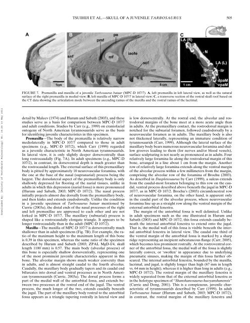
FIGURE 7. Premaxilla and maxilla <strong>of</strong> a <strong>juvenile</strong> <strong>Tarbosaurus</strong> <strong>bataar</strong> (MPC-D 107/7). A, left premaxilla in left lateral view, as well as the sutural<br />
surface <strong>of</strong> the right premaxilla in medial view; B, left maxilla <strong>of</strong> MPC-D 107/7 in lateral view; C, a transverse section <strong>of</strong> the rostral skull ro<strong>of</strong> based on<br />
the CT data showing the articulation mode between the ascending ramus <strong>of</strong> the maxilla and the rostral ramus <strong>of</strong> the lacrimal.<br />
Downloaded By: [Society <strong>of</strong> Vertebrate Paleontology] At: 14:24 10 May 2011<br />
detail by Maleev (1974) and Hurum and Sabath (2003), and these<br />
studies serve as a basis for comparison between MPC-D 107/7<br />
and adult conditions. Studies by Carr (e.g., 1999) on crani<strong>of</strong>acial<br />
ontogeny <strong>of</strong> North American tyrannosaurids serve as the basis<br />
for identifying <strong>juvenile</strong> characteristics in this <strong>specimen</strong>.<br />
Premaxilla—The body <strong>of</strong> the premaxilla is relatively narrow<br />
mediolaterally in MPC-D 107/7 compared to those in adult<br />
<strong>specimen</strong>s (e.g., MPC-D 107/2), which Carr (1999) regarded<br />
as a <strong>juvenile</strong> characteristic in North American tyrannosaurids.<br />
In lateral view, it is only slightly deeper dorsoventrally than<br />
long rostrocaudally (Fig. 7A). In adult <strong>specimen</strong>s (e.g., MPC-D<br />
107/2), in contrast, its dorsoventral depth is much greater than<br />
the rostrocaudal length. The external surface <strong>of</strong> this premaxillary<br />
body is pitted by approximately 10 neurovascular foramina, with<br />
the one at the base <strong>of</strong> the nasal (supranarial) process being the<br />
largest. The dorsolateral aspect <strong>of</strong> the premaxillary body is only<br />
shallowly depressed for flooring <strong>of</strong> the narial tissues, unlike in<br />
adults in which this depression (narial fossa) is more pronounced<br />
(Hurum and Sabath, 2003; MPC-D 107/2). The nasal process<br />
initially projects almost due dorsally <strong>from</strong> the premaxillary body,<br />
and then kinks and extends caudodorsally. Unlike the condition<br />
in a <strong>juvenile</strong> <strong>specimen</strong> <strong>of</strong> <strong>Tarbosaurus</strong> <strong>bataar</strong> mentioned by<br />
Currie (2003a), the distal ends <strong>of</strong> the nasal processes <strong>of</strong> the right<br />
and left premaxillae are appressed to each other and are not<br />
forked in MPC-D 107/7. The maxillary (subnarial) process is<br />
shaped like a rostrocaudally elongate triangle. It appears to be<br />
longer rostrocaudally than in the adult (MPC-D 107/2).<br />
Maxilla—The maxilla <strong>of</strong> MPC-D 107/7 is dorsoventrally much<br />
shallower than in adult <strong>specimen</strong>s (Fig. 7B). For example, the ratio<br />
<strong>of</strong> the maximum height to the maximum length <strong>of</strong> this bone<br />
is 0.39 in this <strong>specimen</strong>, whereas the same ratio <strong>of</strong> the <strong>specimen</strong><br />
described by Hurum and Sabath (2003; ZPAL MgD-I/4, skull<br />
length 1100 mm) is 0.57. The main body (alveolar process) <strong>of</strong><br />
this bone is especially shallow dorsoventrally, representing one<br />
<strong>of</strong> the most prominent <strong>juvenile</strong> characteristics apparent in this<br />
bone. The alveolar margin shows much weaker convexity than<br />
in adults, and is almost straight caudal to the fourth alveolus.<br />
Caudally, the maxillary body gradually tapers and its caudal end<br />
bifurcates into dorsal and ventral processes as in North American<br />
tyrannosaurids (Currie, 2003a). The dorsal process forms a<br />
part <strong>of</strong> the medial wall <strong>of</strong> the antorbital fossa, and extends between<br />
two processes at the rostral end <strong>of</strong> the jugal. The ventral<br />
process, the much longer <strong>of</strong> the two, extends caudally beneath<br />
the jugal. The part <strong>of</strong> the maxillary body rostral to the antorbital<br />
fossa appears as a triangle tapering rostrally in lateral view and<br />
is low dorsoventrally. At the rostral end, the alveolar and rostrodorsal<br />
margins <strong>of</strong> the bone meet at a more acute angle than<br />
in adults. At the premaxillary contact, the rostrodorsal margin is<br />
notched for the subnarial foramen, followed caudodorsally by a<br />
neurovascular foramen as in adults. The maxillary body is also<br />
not thickened laterally, representing an immature condition <strong>of</strong><br />
tyrannosaurids (Carr, 1999). Although the lateral surface <strong>of</strong> the<br />
maxillary body bears numerous neurovascular foramina and shallow<br />
grooves leading to them (for nerves and/or blood vessels),<br />
surface sculpturing is not nearly as pronounced as in adults. Four<br />
relatively large foramina lie along the rostrodorsal margin <strong>of</strong> this<br />
bone, arranged in a line about 1 cm <strong>from</strong> the margin. Another<br />
row <strong>of</strong> relatively large foramina extends along the ventral margin<br />
<strong>of</strong> the alveolar process within a few millimeters <strong>from</strong> the margin,<br />
comprising the alveolar row <strong>of</strong> the foramina <strong>of</strong> Brochu (2003).<br />
As described in Daspletosaurus by Carr (1999), a sulcus extends<br />
<strong>from</strong> the caudal-most foramen belonging to this row on the caudal,<br />
ventral process described above beneath the jugal in MPC-D<br />
107/7, as in MPC-D 107/2. Brochu’s (2003) circumfenestral row<br />
<strong>of</strong> neurovascular foramina, on the other hand, is apparent only<br />
in the caudal part <strong>of</strong> the alveolar process, where neurovascular<br />
foramina line up as a straight row along the ventral margin <strong>of</strong> the<br />
external antorbital fenestra.<br />
The margin <strong>of</strong> the antorbital fossa is well delimited. Unlike<br />
in adult <strong>specimen</strong>s such as the one illustrated in Hurum and<br />
Sabath (2003) and MPC-D 107/2, this fossa extends caudally beneath<br />
the internal antorbital fenestra to extend onto the jugal.<br />
That is, the medial wall <strong>of</strong> this fossa is visible beneath the internal<br />
antorbital fenestra in lateral view. The caudal one third <strong>of</strong><br />
the ventral margin <strong>of</strong> the antorbital fossa is marked by a sharp<br />
ridge representing an incipient subcutaneous flange (Carr, 2005),<br />
which becomes less prominent rostrally. At the rostroventral corner<br />
<strong>of</strong> the antorbital fossa, the medial wall <strong>of</strong> the fossa is slightly<br />
laterally convex, or ‘swollen’ in appearance due to underlying<br />
pneumatic sinuses, making the margin <strong>of</strong> this fossa further obscured.<br />
The internal antorbital fenestra, bounded by the maxilla,<br />
lacrimal, and jugal, is slightly longer than high (67 mm in length<br />
vs. 64 mm in height), whereas it is higher than long in adults (e.g.,<br />
MPC-D 107/2). The rostral margin <strong>of</strong> the maxillary fenestra is<br />
widely separated <strong>from</strong> that <strong>of</strong> the external antorbital fenestra as<br />
in the holotype <strong>specimen</strong> <strong>of</strong> “Shanshanosaurus huoyanshanensis”<br />
(Currie and Dong, 2001). This is a conspicuous, <strong>juvenile</strong> characteristic<br />
<strong>of</strong> tyrannosaurids described by Carr (1999). In adult<br />
<strong>Tarbosaurus</strong> <strong>bataar</strong> (Hurum and Sabath, 2003; MPC-D 107/2),<br />
in contrast, the rostral margins <strong>of</strong> the maxillary fenestra and


