

UW CT Protocol: FEMORAL ANTEVERSION
UW CT Protocol: FEMORAL ANTEVERSION
UW CT Protocol: FEMORAL ANTEVERSION

You also want an ePaper? Increase the reach of your titles
YUMPU automatically turns print PDFs into web optimized ePapers that Google loves.
<strong>UW</strong> <strong>CT</strong> <strong>Protocol</strong>: <strong>FEMORAL</strong> <strong>ANTEVERSION</strong> page 1 of 2<br />
Indications for Femoral Anteversion (aka “<strong>CT</strong> Scanogram”, “Tibial Torsion”)<br />
• THIS IS A VERY LIMITED STUDY.<br />
• The primary purpose of this scan is to allow the radiologist to measure the angle of rotation of<br />
the femoral necks relative to the femoral condyles, bilaterally.<br />
• A secondary measurement is femoral lengths, made by calculating the difference in table<br />
position at the ends of the bones.<br />
• (Similar measurements can also be made of the tibias, if specifically clinically requested.)<br />
Other Leg Length Imaging Studies<br />
Radiographic Scanogram<br />
• This is a radiographic study in which coned-down images of BOTH HIPS,KNEES,&ANKLES<br />
are shot on a single conventional-sized film (or CR Plate) with a radiopaque ruler in place.<br />
• The sole purpose of this study is to measure leg-lengths.<br />
• Currently at the <strong>UW</strong> this study is shot CR, and read soft-copy by the radiologists on the ALI.<br />
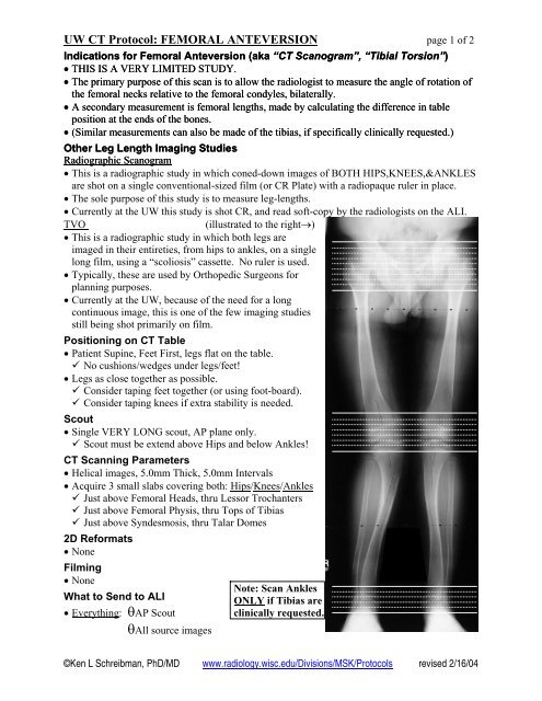
TVO<br />
(illustrated to the right→)<br />
• This is a radiographic study in which both legs are<br />
imaged in their entireties, from hips to ankles, on a single<br />
long film, using a “scoliosis” cassette. No ruler is used.<br />
• Typically, these are used by Orthopedic Surgeons for<br />
planning purposes.<br />
• Currently at the <strong>UW</strong>, because of the need for a long<br />
continuous image, this is one of the few imaging studies<br />
still being shot primarily on film.<br />
Positioning on <strong>CT</strong> Table<br />
• Patient Supine, Feet First, legs flat on the table.<br />
No cushions/wedges under legs/feet!<br />
• Legs as close together as possible.<br />
Consider taping feet together (or using foot-board).<br />
Consider taping knees if extra stability is needed.<br />
Scout<br />
• Single VERY LONG scout, AP plane only.<br />
Scout must be extend above Hips and below Ankles!<br />
<strong>CT</strong> Scanning Parameters<br />
• Helical images, 5.0mm Thick, 5.0mm Intervals<br />
• Acquire 3 small slabs covering both: Hips/Knees/Ankles<br />
Just above Femoral Heads, thru Lessor Trochanters<br />
Just above Femoral Physis, thru Tops of Tibias<br />
Just above Syndesmosis, thru Talar Domes<br />
2D Reformats<br />
• None<br />
Filming<br />
• None<br />
What to Send to ALI<br />
• Everything: θAP Scout<br />
θAll source images<br />
Note: Scan Ankles<br />
ONLY if Tibias are<br />
clinically requested.<br />
©Ken L Schreibman, PhD/MD www.radiology.wisc.edu/Divisions/MSK/<strong>Protocol</strong>s revised 2/16/04
<strong>UW</strong> <strong>CT</strong> <strong>Protocol</strong>: <strong>FEMORAL</strong> <strong>ANTEVERSION</strong> page 2 of 2<br />
Measurements To Be Made off of a <strong>CT</strong> Scanogram<br />
Femoral Anteversion<br />
• MEASURE RIGHT AND LEFT SIDES INDIVIDUALLY<br />
• Find the slice that best reveals the alignment of the femoral neck.<br />
• Measure the Neck-Horizontal Angle (NH).<br />
• Find the slice that best reveals the alignment of the femoral condyles.<br />
• Measure the Condyle-Horizontal Angle (CH).<br />
• Calculate the angle of the Neck relative to the Condyles (NC=NH-CH).<br />
NH<br />
CH<br />
NH<br />
¯CH<br />
= NC<br />
30°<br />
10°<br />
←For the example to the left,<br />
the Neck-Horizontal angle=30°,<br />
the Condyle-Horizontal=10°,<br />
thus the relative angle between<br />
the Femoral Neck and Condyles<br />
=20°.<br />
This is the Femoral Anteversion<br />
angle.<br />
• In cases when the femoral condyles are<br />
internally rotated (as shown right→),<br />
then the CH angle is ADDED to the NH<br />
angle. (In the example to the right,<br />
NC=NH+CH=30º+20º=50º).<br />
• The key to visualizing the Anteversion<br />
angle is to imagine rotating the femoral<br />
shaft such that the Condyles are straight<br />
horizontal (CH=0). The Anteversion<br />
angle is now equal to the angle between<br />
the femoral neck and horizontal (NH).<br />
20°<br />
30°<br />
Femoral Length<br />
• MEASURE RIGHT AND LEFT SIDES INDIVIDUALLY<br />
• Determine Table Positions at tops of femoral heads and bottom of femoral condyles.<br />
• Report difference to the nearest 0.5cm.<br />
©Ken L Schreibman, PhD/MD<br />
www.radiology.wisc.edu/Divisions/MSK/<strong>Protocol</strong>s<br />
revised 2/16/04


