Download - Straumann
Download - Straumann
Download - Straumann

You also want an ePaper? Increase the reach of your titles
YUMPU automatically turns print PDFs into web optimized ePapers that Google loves.
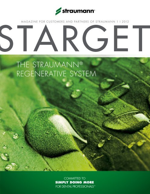
MAGAzINE fOR CUSTOMERS AND PARTNERS Of STRAUMANN 1 I 2012<br />
the straumann ®<br />
regenerative system
Imprint starget – magazine for Customers and Partners of straumann i © straumann usa i 60 minuteman road i andover, ma 01810 i Phone 800/448 8168<br />
Fax 978/747 2490 i Editors roberto gonzález i mildred Loewen i E-Mail info.usa@straumann.com i Internet www.straumann.us/starget<br />
Legal Notice exclusion of liability for articles by external authors: articles by external authors published in starget have been systematically assessed and carefully<br />
selected by the publisher of starget (straumann usa). such articles in every case reflect the opinion of the author(s) concerned and therefore do not necessarily<br />
coincide with the publisher’s opinion. nor does the publisher guarantee the completeness or accuracy and correctness of articles by external authors published in<br />
starget. the information given in clinical case descriptions, in particular, cannot replace a dental assessment by an appropriately qualified dental specialist in an<br />
individual case. any orientation to articles published in starget is therefore on the dentist’s responsibility. articles published in starget are protected by copyright<br />
and may not be reused, in full or in part, without the express consent of the publisher and the author(s) concerned. straumann ® and all other trademarks and logos are<br />
registered trademarks of straumann usa and/or of its affiliates. third party corporate names and brand names that may be mentioned may be registered or otherwise<br />
protected marks even if this is not specially indicated. the absence of such an indication shall not therefore be interpreted as allowing such a name to be freely used.
editorial<br />
STARGET 1 I 12<br />
3<br />
A New Era in Oral Tissue<br />
Regeneration<br />
Dear Valued Customer,<br />
It is my pleasure to introduce myself as the new head of <strong>Straumann</strong> North America,<br />
and also to introduce this first installment of STARGET for 2012. The focus of this issue,<br />
"The <strong>Straumann</strong> Regenerative System," is particularly significant for me as I come<br />
to <strong>Straumann</strong> USA as the former Head of <strong>Straumann</strong> Global Regenerative Sales.<br />
As you know, <strong>Straumann</strong> provides solutions for both tissue regeneration and GBR<br />
including Emdogain, BoneCeramic, <strong>Straumann</strong> AlloGraft, and most recently<br />
MembraGel ® , an innovative liquid membrane that can be precisely applied to the<br />
surgical site.<br />
Andy Molnar<br />
<strong>Straumann</strong> Executive Vice President<br />
North America<br />
You’ll find several interesting articles on tissue regeneration in this issue of<br />
STARGET: “Why Repair When You Can Regenerate?“ on page 6, “Redefining<br />
the Membrane“ on page 12, and an interview with Prof. Dr. Christoph Hämmerle<br />
on the innovation and distinctiveness of MembraGel on page 16. You’ll also see<br />
an article on our Esthetic Case Book, which documents 11 cases illustrating the<br />
dramatic results that can be achieved through Emdogain, on page 18.<br />
Restorative dentistry has seen breakthroughs over the past few years. This STARGET<br />
features articles on the latest development of <strong>Straumann</strong> CARES ® digital workflow.<br />
We hope you will enjoy the clinical case on <strong>Straumann</strong>'s screw-retained hybrid<br />
solution from your peers, and an article based on the interviews with three key<br />
opinion leaders on global trends in dentistry.<br />
Enjoy this issue of STARGET and please let us know how we can continue to<br />
improve upon it.<br />
Sincerely,<br />
Andy Molnar<br />
<strong>Straumann</strong> Executive Vice President, North America
4 STARGET 1 I 12<br />
overvieW<br />
Overview<br />
STARGET 1 I 2012<br />
6<br />
The <strong>Straumann</strong> ® Regenerative System<br />
offers solutions for oral tissue regeneration<br />
– ranging from conservative dentistry to<br />
dental restoration. New to the portfolio:<br />
<strong>Straumann</strong> ® regenerative S y S tem<br />
<strong>Straumann</strong> ® MembraGel ® .<br />
28<br />
<strong>Straumann</strong> ® CareS ® digital S olutionS<br />
<strong>Straumann</strong>, together with 3M ESPE,<br />
has introduced a streamlined digital<br />
workflow that connects the Lava C.O.S.<br />
Intra-Oral Scanner to the <strong>Straumann</strong> ®<br />
CARES ® Digital Solutions platform.<br />
Simply doing more<br />
54<br />
Global Trends – Where is the final<br />
destination? We asked three prominent<br />
dentists to give us their perspective<br />
on things: Lyndon Cooper, Kenneth<br />
Malament and Daniel Wismeijer.
C ontent<br />
STARGET 1 I 12<br />
5<br />
CONTENT<br />
FOCAL POINT 6 Why Repair When You Can Regenerate Instead?<br />
STRAUMANN ® REGENERATIVE SYSTEM 12 <strong>Straumann</strong> MembraGel ® – Re-Defining the Membrane<br />
16 Interview with Christoph Hämmerle<br />
18 An Esthetic Case Selection on <strong>Straumann</strong> Emdogain<br />
20 <strong>Straumann</strong> Allograft Portfolio Expands<br />
STRAUMANN ® CARES ® DIGITAL SOLUTIONS 22 Evaluating the Precision of <strong>Straumann</strong> ® CARES ® Guided Surgery<br />
Based on a Clinical Case<br />
28 Digital Workflow<br />
30 The Intraoral Workflow<br />
32 3M ESPE Lava Ultimate Restorative<br />
38 Interview with Mike Rynerson<br />
RESTORATIVE 43 <strong>Straumann</strong> ® Anatomic IPS e.max ® Abutment<br />
45 Immediate Full Mouth Restoration Using Implant-Supported Fixed<br />
Hybrid Prosthetics<br />
SURGICAL 50 Updated SLActive ® Scientific Evidence Brochure<br />
ITI / EDUCATION 52 <strong>Straumann</strong> & Baylor University Launch the First Interdisciplinary<br />
Digital Dentistry Course<br />
53 ITI Membership Tops 10,000<br />
SIMPLY DOING MORE 54 Global Trends<br />
60 <strong>Straumann</strong> AID: Access to Implant Dentistry<br />
64 Literature Alerts<br />
67 Upcoming 2012 Education Events
6 STARGET 1 I 12<br />
foC al point<br />
STrAUMANN ® reGeNerATive SYSTeM<br />
Why Repair When You Can Regenerate Instead?<br />
The More Complex an Organism is, the Lesser the Capacity of its Body to Perform Regenerative<br />
Processes<br />
The biological process, or self-sufficient capability to restore deficient tissue is referred to as the ability<br />
for regeneration. In contrast to repair, i.e., wound healing, whereby the original biological structure is<br />
not fully rebuilt, the goal of regeneration is to completely restore the structure and function of tissue that<br />
has been lost or injured. This ability has continuously diminished as creatures have evolved and the<br />
complexity of these organisms has increased. Compared to the “champions” of regeneration, present<br />
day cnidarians, which can re-grow severed extremities and internal organs that have been lost, this<br />
capability in humans, without additional support, is limited to a few types of external tissue.<br />
When the Body’s Own Healing Process “Overshoots its Target“<br />
When a person’s tissue is injured, the body’s repair process, rather than regenerative process, begins.<br />
Here, the body does not primarily attempt to restore the original state and function of the tissue,<br />
but rather to close the wound as quickly as possible. This spontaneous healing process involves the<br />
formation of connective tissues that penetrate deeply into the original tissue and also remove “good”<br />
tissue in the process. During such a “radical” healing process, it is possible that even important functions<br />
are permanently destroyed.<br />
Regeneration Can be Guided with Medical Treatment<br />
To prevent this type of overcompensation in the healing process wherever possible, strong antiinflammatory<br />
drugs are administered today, such as in cases of back injuries. This prevents this destructive<br />
process from occurring, in turn making it possible to preserve part of the nerve tissue and mitigating<br />
deficits such as paralyses. In medical terms, regeneration entails assisting desirable tissue formation<br />
processes, and guiding and limiting the repair mechanisms involved in wound healing as to not impede<br />
the regeneration of intact, functioning tissue.
foC al point<br />
STARGET 1 I 12<br />
7<br />
Fig. 1: The cnidarians (pictured here, a green anemone, anthopleura xanthogrammica) are equipped with astounding regenerative<br />
capabilities that far surpass those of humans.
8 STARGET 1 I 12<br />
<strong>Straumann</strong> ® regenerative S y S tem<br />
The Insertion of Implants Requires Healthy Bone Tissue<br />
Great advances in regeneration have been achieved in dentistry over that last 20 years. We can<br />
now regenerate the bone tissue of the jaw, opening the door for implants as a treatment for replacing<br />
teeth. This has required scientific evidence documenting the physiological processes involved in tissue<br />
formation and the necessary aids for controlling these processes. Today, for example, we know that<br />
lost or missing bone can only be regenerated by the body when the bone-forming cells can perform<br />
their work undisturbed. Without external intervention, this process would have to compete with the rapid<br />
natural wound repair mechanism, which would result in the growth of new connective tissue instead of<br />
the desired bone tissue.<br />
Fig. 2: The high porosity of <strong>Straumann</strong> ® BoneCeramic (90 %) allows blood vessels and vital bone to vascularize into the material.<br />
Regeneration Requires Matrices and Membranes<br />
Today, one means of guiding the regeneration process requires two aids: a matrix and a membrane.<br />
The matrix keeps the space available for the bone to grow while a membrane serves as a barrier to<br />
prevent the connective tissue infiltration of the gingiva.
<strong>Straumann</strong> ® regenerative S y S tem<br />
STARGET 1 I 12<br />
9<br />
Various types of these matrices and membranes are used today for regenerative purposes. Some of<br />
these “placeholders” remain in the body, integrated into the newly formed bone for the rest of the person’s<br />
life. Applying membranes for this purpose is also quite complex and time-consuming. <strong>Straumann</strong> ®<br />
MembraGel ® was developed to deal with the disadvantages of the types of membranes used in the<br />
past. The time-consuming cutting and fitting of the membrane is unnecessary since MembraGel is<br />
applied in liquid form and polymerizes to form a solid film in less than a minute. The resulting hydrogel<br />
is biocompatible and completely biodegradable.<br />
Periodontitis – The Greatest Threat to the Periodontium<br />
For implantology, the regeneration of the periodontium is equally as important as the new formation<br />
of bone, which is destroyed by periodontitis and can ultimately result in the loss of the tooth. Despite<br />
numerous attempts to regenerate the destroyed tissue, a breakthrough to achieve complete tissue<br />
reformation remains to be seen. <strong>Straumann</strong> ® Emdogain can be used in cases to undo some of this<br />
destruction, making it possible to prevent loss of the tooth. Emdogain contains the key components<br />
required for building up the cementum and periodontal ligament, important parts of the periodontium.<br />
These elements, or proteins, tap into the body’s own natural ability to rebuild the surrounding tooth<br />
structures, using nature’s help.<br />
“Simply Doing More“ – During the Regeneration Process<br />
<strong>Straumann</strong>’s successes in the field of regeneration are promising and we have yet to fully realize our<br />
dreams. We are already working on further innovations to guide regenerative processes in an even<br />
simpler and more effective manner. We are inspired by two great visions:<br />
» A matrix for bone augmentation that breaks down fully and can be used without requiring a<br />
membrane.<br />
» Guided tissue regeneration that allows for complete preservation of the tooth.<br />
Now making its first entrance into the dental market with <strong>Straumann</strong> MembraGel, we view PEG technology<br />
as the foundation for achieving these goals. Beyond MembraGel, there are numerous combinations and<br />
degrees of cross-linking these biocompatible hydrogels, which present an exciting range of possible<br />
options. This innovative technology is only in the early phases of trials and development and has<br />
enormous potential for the future.
10 STARGET 1 I 12<br />
<strong>Straumann</strong> ® regenerative S y S tem<br />
The <strong>Straumann</strong> ® Regenerative System: Everything from a Single Source<br />
The <strong>Straumann</strong> ® Regenerative System offers you a variety of solutions for oral tissue regeneration –<br />
ranging from conservative dentistry to dental restoration. Our goal is to offer you a variety of predictable<br />
and scientifically proven regenerative treatment solutions – all from a single source and in the tried and<br />
tested quality that is <strong>Straumann</strong>’s hallmark.<br />
Tissue Regeneration<br />
By mimicking the natural process of odontosis,<br />
<strong>Straumann</strong> ® emdogain forms an insoluble,<br />
three-dimensional extracellular matrix that<br />
remains on the root surface for 2 – 4 weeks<br />
and enables a selective cell population,<br />
proliferation and differentiation.<br />
Bone Formation<br />
<strong>Straumann</strong> ® allograft is a wide range of<br />
bone allograft solutions, allowing you the<br />
flexibility to choose the treatment that’s right for<br />
your case. Through a commercial partnership<br />
with LifeNet Health ® , <strong>Straumann</strong>’s allograft<br />
options provide confidence in the safety of the<br />
material you use to treat your patients.<br />
<strong>Straumann</strong> ® BoneCeramic is a fully synthetic<br />
bone substitute material with excellent<br />
morphology that promotes the new formation<br />
of vital bone. It can be used for a series of<br />
procedures used for dental bone regeneration.<br />
Bone Healing<br />
<strong>Straumann</strong> ® membragel ® is a membrane<br />
of the latest generation. It combines unique<br />
material properties that have been developed<br />
to promote undisturbed bone healing and to<br />
simplify the surgical procedure.
STARGET 1 I 12<br />
11<br />
straumann ® emdogain <br />
IS TRUE PERIODONTAL REGENERATION<br />
IMPORTANT TO YOU?<br />
Photos courtesy of Dr. G.<br />
Zuchelli, Bologna, Italy<br />
before<br />
after<br />
More than 100 clinical publications in peer-reviewed journals demonstrate<br />
<strong>Straumann</strong> ® Emdogain to be safe and effective in stimulating the formation of new<br />
periodontal soft and hard tissue. These clinical studies involve more than<br />
3000 defects in over 2500 patients.<br />
Contact <strong>Straumann</strong> Customer Service at 800/448 8168 to learn more<br />
about <strong>Straumann</strong> solutions or to locate a representative in your area.<br />
www.straumann.us<br />
excellent clinical results 1-4<br />
Long-term clinical benefit 5,6<br />
Less patient discomfort 3<br />
1<br />
Tonetti MS, et al. Enamel matrix proteins in the regenerative therapy of deep intrabony defects.J Periodontol.<br />
2002;29:317–325.<br />
2<br />
Froum SJ, et al. A comparative study utilizing open flap debridement with and without enamel matrix derivative in<br />
the treatment of periodontal intrabony defects: A 12-month re-entry.J Periodontol. 2001;72:25–34.<br />
3<br />
Jepsen S, et al. A randomized clinical trial comparing enamel matrix derivative and membrane treatment of<br />
buccal class II furcation involvement in mandibular molars. Part I: study design and results for primary outcomes.<br />
J Periodontol. 2004; 75:1150 –1160.<br />
4<br />
McGuire MK, et al. Evaluation of human recession defects treated with coronally advanced flaps and either enamel<br />
matrix derivative or connective tissue. Part 1: comparison of clinical parameters.J Periodontol. 2003;74:1110 –1125.<br />
5<br />
Sculean A, et al. Ten-year results following treatment of intra-bony defects with enamel matrix proteins and guided<br />
tissue regeneration. J Clin Periodontol. 2008; 35:817-824.<br />
6<br />
Data on file (McGuire 10 year)
12 STARGET 1 I 12<br />
<strong>Straumann</strong> ® regenerative S y S tem<br />
STrAUMANN ® MeMbraGel ®<br />
Re-Defining the Membrane<br />
In 2010, <strong>Straumann</strong> introduced <strong>Straumann</strong> MembraGel, an advanced technology membrane that<br />
is one of the most significant innovations in guided bone regeneration in recent history.<br />
Created with PEG (polyethylene glycol) hydrogel technology, <strong>Straumann</strong> MembraGel is applied in<br />
liquid form and molds to the defect. Shortly after application, the liquid components solidify, stabilizing<br />
the bone graft and providing an effective barrier to tissue infiltration. <strong>Straumann</strong> MembraGel then<br />
biodegrades over time. It is designed to achieve undisturbed bone regeneration, which is a prerequisite<br />
for achieving ideal clinical and esthetic results.<br />
“<strong>Straumann</strong> MembraGel is a key innovation in Guided Bone Regeneration. With the<br />
PEG technology we are on the verge of something new, entering a new era in oral<br />
tissue regeneration.“ Christoph Hämmerle, University of Zurich<br />
Designed for Improving GBR Procedures<br />
<strong>Straumann</strong> MembraGel provides effective support in bone formation for GBR (guided bone regeneration)<br />
cases due to its optimized barrier properties. It facilitates undisturbed bone healing as a basis for the<br />
optimal clinical outcome achieved by stabilization of the bone graft material. The precise and easy<br />
application simplifies the surgical procedure.
<strong>Straumann</strong> ® regenerative S y S tem<br />
STARGET 1 I 12<br />
13<br />
Stabilization of the Bone Graft<br />
The gel-like consistency of <strong>Straumann</strong> ® MembraGel<br />
® allows for precise application to the<br />
surgical site and adaptation to various types<br />
and sizes of bone defects.<br />
Solidification and Stabilization<br />
Once solidified in situ (20 – 50 seconds after<br />
application), <strong>Straumann</strong> ® MembraGel is<br />
designed to stabilize the underlying bone graft<br />
to facilitate undisturbed bone regeneration.<br />
Backed by Scientific Documentation<br />
<strong>Straumann</strong> MembraGel is backed by preclinical 1,2,3,4,5 and clinical 6 documentation including one- and<br />
three-year follow-up data 7 presented in 2010 and submitted for publication. The ongoing clinical<br />
program with <strong>Straumann</strong> MembraGel includes over 40 centers and more than 200 patients in Europe<br />
and North America.<br />
<strong>Straumann</strong> ® MembraGel Exclusive Education Program<br />
<strong>Straumann</strong> ® MembraGel was launched in conjunction with a well-received specialized education<br />
program that provides in-depth information on pre-clinical and clinical evidence, hands-on product<br />
training and the surgical techniques of this new application. Herbert Früh, Head of Business Unit<br />
Regenerative at <strong>Straumann</strong> explained, “MembraGel has allowed <strong>Straumann</strong> to bring a brilliant idea to
14 STARGET 1 I 12<br />
<strong>Straumann</strong> ® regenerative S y S tem<br />
fruition. It is an innovative technology that requires training. Customers’ initial experience is that it saves<br />
time during intraoral application but it is different to handle. For this reason, it was the right decision to<br />
combine the launch of the new product with an intensive education program.”<br />
Availability<br />
<strong>Straumann</strong> ® MembraGel ® was introduced in Europe and North America toward the end of 2010, with<br />
roll-outs to other markets planned in the future.<br />
Hands-on workshop at a <strong>Straumann</strong> MembraGel education event.<br />
1<br />
Humber CC, Sándor GK, Davis JM et al. Bone healing with an in situ formed bioresorbable polyethylene glycol hydrogel membrane<br />
in rabbit calvarial defects. Oral Surg Oral Med Oral Pathol Oral Radiol Endod 2010;109:372–384. Jung RE, Lecloux G,<br />
2<br />
Rompen E et al. A feasibility study evaluating an in situ formed synthetic biodegradable membrane for guided bone regeneration<br />
3<br />
in dogs. Clin Oral Implants Res 2009;20:151–161. Thoma DS, Halg G-A, Dard MM et al. Evaluation of a new biodegradable<br />
4<br />
membrane to prevent gingival ingrowth into mandibular bone defects in minipigs. Clin Oral Implants Res 2009;20:7–16. Herten<br />
M, Jung RE, Ferrari D et al. Biodegradation of different synthetic hydrogels made of polyethylene glycol hydrogel/RGD-peptide
<strong>Straumann</strong> ® regenerative S y S tem<br />
STARGET 1 I 12<br />
15<br />
PeG – The TechNOLOGY BehiNd STrAUMANN ® MeMbraGel ®<br />
<strong>Straumann</strong> MembraGel is a synthetic, biodegradable in situ-formed membrane made of polyethylene<br />
glycol (PEG) which forms a molecular network. The material is biocompatible, is applied in liquid form<br />
and sets quickly. Due to its gel-like consistency, it can be applied to cover the exact shape of the defect<br />
or augmented area and does not require cutting and trimming to shape before application – unlike<br />
traditional GBR membranes.<br />
What exactly is PEG, and what makes it suited to this purpose? PEG is a polymer (a large molecule<br />
comprised of repeating structural units). It is produced through the reaction between ethylene oxide and<br />
water or the organic compound ethylene glycol, which is often used as a precursor to polymer materials.<br />
PEG is available in a wide range of molecular weights. The molecular weight also determines the<br />
consistency of the material, which can vary greatly from waxy-solid to water-soluble liquid states. PEG<br />
has a number of properties that are particularly desirable in a number of biological, pharmaceutical<br />
and chemical applications: it is highly flexible, non-toxic and non-immunogenic. It is also hydrophilic,<br />
meaning that its attachment to biomolecules can increase solubility and decrease aggregation of the<br />
molecules.<br />
Thanks to these very advantageous properties, PEG has been extensively used in many medical,<br />
pharmaceutical and medical device applications. For instance, it is used as a spray-on adhesion barrier<br />
and as a main ingredient in laxatives; as an agent for bowel irrigation prior to surgery or colonoscopy;<br />
as an addition to protein medications to extend their effect and increase dosing intervals; and as a<br />
carrier in various medications, such as soft capsules, ointments, tablets, and lubricants. Preliminary<br />
research is also underway to investigate the potential of PEG as a component in many other exciting<br />
applications, such as gene therapy, spinal cord injuries and the suppression of carcinogenesis. The<br />
use of PEG as a customizable, liquid-applied membrane is therefore another milestone in a long line of<br />
applications for this useful, versatile and extensively researched material.<br />
5<br />
modifications: an immunohistochemical study in rats. Clin Oral Implants Res 2009;20:116–125. Jung RE, Zwahlen M, Weber FE<br />
et al. Evaluation of an in situ formed hydrogel as a biodegradable membrane for guided bone regeneration. Clin Oral Implants Res<br />
6<br />
2006;17:426–433. Jung RE, Hälg GA, Thoma DS, Hämmerle CHF. A randomized, controlled clinical trial to evaluate a new<br />
7<br />
membrane for guided bone regeneration around dental implants. Clin Oral Implants Res 2009;20:162–168. Ramel C, Halg G,<br />
Thoma D et al. A randomized clinical trial to evaluate a synthetic gel membrane for GBR around dental implants – 1- and 3-year<br />
results. European Association for Osseointegration 19 th Annual Scientific Meeting, Glasgow, UK, 6–9 October 2010; Abs 055.
16 STARGET 1 I 12<br />
<strong>Straumann</strong> ® regenerative S y S tem<br />
iNTerview<br />
“Being Part of this Network is Something Truly Exciting –<br />
Something We’ve Never Done in this Way Before.“<br />
Andy Molnar, <strong>Straumann</strong> Executive Vice President, North America, interviews<br />
Professor Dr. Christoph Hämmerle of the University of Zurich about<br />
<strong>Straumann</strong> ® MembraGel ® .<br />
professor Hämmerle, our company is introducing membragel by way of an<br />
international education-based program. this is a product which has been longawaited<br />
in the market; so many people would like to get their hands on it as<br />
Prof. Dr. Christoph Hämmerle<br />
Chairman of the Department of Fixed and<br />
Removable Prosthodontics and Dental Material<br />
Science, University of Zurich/Switzerland.<br />
soon as possible. What are your thoughts on this educational approach to the<br />
launch?<br />
I think it’s very important to use an educational approach. What we’re dealing<br />
with here is a new technology, a new kind of membrane, a new way to apply<br />
the membrane. There are a lot of differences from all the membranes we’ve had<br />
before. I think it’s very important to launch the product in a well-structured way so<br />
that we know what experience we can rely on. It’s also an opportunity to learn<br />
about new things we need to become familiar with before we can use a new<br />
technology like this successfully and predictably.<br />
“We treated over thirty defects and had minimal complications.<br />
Based on this experience and learning, we moved on to larger defects<br />
with a very good success rate once again.“ Christoph Hämmerle<br />
i know you’re excited about membragel and the progress the product is making.<br />
How would you describe your involvement from the start, from inception all the<br />
way through the development of the project?<br />
It has been very exciting for us. It’s really special to be there when the original<br />
idea is born and then to participate in the development of a new product, which<br />
is eventually able to solve problems and improve patient care. With all the sharing,<br />
teaching, and feedback we’re getting from different people all over the world,<br />
being part of this network is something really exciting, something we’ve never<br />
done this way before. For this reason, we’re looking forward to continuing with the
<strong>Straumann</strong> ® regenerative S y S tem<br />
STARGET 1 I 12<br />
17<br />
development of this scientific and educational network and<br />
really benefiting from the process. I think it’s great!<br />
from our learning so far, what are the key points that we<br />
need to pass on to our educators?<br />
There are three key points. One is that you have to change<br />
some of your surgical habits to adapt to the new technology,<br />
the new way to apply the membrane. The second is to adhere<br />
to published surgical guidelines. And the third is to begin with<br />
less complex cases until you have had around ten successful<br />
cases, and then move on to more difficult cases.<br />
When we conducted our initial study, in which we used a<br />
collagen membrane as a control, we did not see any more<br />
difficulties with the membrane using the new technology than<br />
with the membrane using the older technology. I think this was<br />
the case because we were careful to use small dehiscence<br />
defects in non-esthetic areas. We treated over thirty defects<br />
and had minimal complications. Based on that experience<br />
and learning, we began to expand to larger defects, again,<br />
with very good success.<br />
Professor Hämmerle, thank you very much for your time.<br />
It’s much appreciated.<br />
“I think it’s very important to launch the product<br />
in a well-structured way so that we know what<br />
experience we can rely on.“ Christoph Hämmerle<br />
Returning to the topic of changing habits, this is a new<br />
technology, and there is a great temptation to use it in all<br />
kinds of different ways because it is rather easy to use and<br />
very new and exciting. We need to exercise restraint and<br />
really adhere to new practices in aspects of our surgical<br />
work.<br />
The second point pertains to the surgical guidelines. We<br />
have seen that the surgical guidelines are not always<br />
followed accurately. Following the surgical guidelines could<br />
improve the practical outcomes when it comes to using this<br />
new product.<br />
The third aspect is the size of the defects we tackle first.
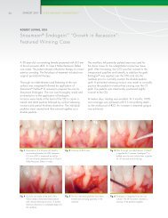
18 STARGET 1 I 12<br />
<strong>Straumann</strong> ® regenerative S y S tem<br />
GrOwTh iN receSSiON<br />
An Esthetic Case Selection on <strong>Straumann</strong> ® Emdogain<br />
Clinicians have been predictably treating gingival recession with <strong>Straumann</strong> ®<br />
Emdogain for years and talking about the beautiful smiles they gave back<br />
to their patients – we gave them the chance to prove it.<br />
The best cases from the 2009 “Growth in Recession” Esthetic Case Competition<br />
were chosen by a renowned international jury to appear in <strong>Straumann</strong>’s Growth in<br />
Recession Esthetic Case Book. This casebook, published in 2011, features eleven<br />
cases presented together with the patients’ histories and images of each step to<br />
facilitate a detailed understanding of the surgical procedures involved.<br />
This valuable tool is complimentary to you with your next order of <strong>Straumann</strong><br />
Emdogain. Please reference order code “RECESSION” when placing your order.<br />
Designed to help achieve excellent esthetic results<br />
Esthetic Casebook: <strong>Straumann</strong> ® Emdogain<br />
The following clinicians contributed their Case reports to this publication:<br />
p Brazil: Dr. Robert Carvalho da Silva, DDS, MS, PH.D. (co-authors: Dr. Julio<br />
Cesar Joly, DDS, MS, PH.D., and Dr. Paulo Fernando Mesquita de Carvalho,<br />
DDS, MS)<br />
p USA: Dr. Robert Levine, DDS – Dr. Ken Akimoto, DDS, MSD – Dr. Mark I. Gutt,<br />
DMD– Dr. Eunseok Eugene Oh, DDS (co-author: Dr. Vincent Iacono, DDS) – Dr.<br />
Paul G. Luepke, DDS, MS<br />
p Canada: Dr. Ira Paul Sy, DDS, MS, Dip. Periodontics<br />
p Germany: Dr. med. dent. Andreas Hofmann, M.Sc. – Dr. Bjørn Greven (coauthor:<br />
Dr. Bernd Heinz)<br />
p Spain: Dr. Ion Zabalegui, MD
<strong>Straumann</strong> ® regenerative S y S tem<br />
STARGET 1 I 12<br />
19<br />
“BOTH THE SCIENTIFIC EVIDENCE AND MY PERSONAL ExPERIENCE<br />
SUPPORT THAT WITH THE APPROPRIATE CASE STRAUMANN ® EMDOGAIN<br />
SIGNIFICANTLY IMPROVES ROOT COVERAGE COMPARED TO THE<br />
CORONALLY ADVANCED FLAP ALONE.”<br />
DR. MICHAEL K. MCGUIRE, DDS<br />
Treatment of recession defects with <strong>Straumann</strong> Emdogain<br />
Courtesy of Dr. Paul G. Luepke, DDS, MSD<br />
A referred 23-year-old female patient was presenting with multiple gingival recessions<br />
(teeth #8 to #3). The prominent canine showed a Miller Class III recession, the<br />
other teeth presented with Miller Class I recessions. The treatment procedure began<br />
with a thorough cleaning and scaling of the exposed root surfaces with hand<br />
and sonic instruments and was followed by a split thickness flap preparation of a<br />
Zucchelli-style flap (without vertical releasing incisions). A dissection into the vestibular<br />
mucosa allowed for further mobilization.<br />
Dr. Paul G. Luepke, DDS, MS<br />
1996 Master’s degree in Periodontics, University<br />
of Texas at San Antonio Health Science Center, Tx,<br />
USA • 1997 Diplomate of the American Board<br />
<strong>Straumann</strong> ® PrefGel ® (EDTA) was applied for two minutes on the root surface.<br />
Subsequently, the surgical area was rinsed with sterile saline and <strong>Straumann</strong> ®<br />
Emdogain was applied to the root surfaces. Connective tissue graft (CTG) was<br />
harvested from the palate with the single incision technique. The graft was then<br />
split. The CTG was fixed on the root surface and the flap coronally positioned and<br />
fixed with sling sutures.<br />
of Periodontology • 2008 Assistant Professor of<br />
Surgical Services Periodontics Division, Marquette<br />
University School of Dentistry in Milwaukee, WI,<br />
USA • 2009 Interim Department Chair of Surgical<br />
Sciences at Marquette University School of Dentistry<br />
in Milwaukee, WI, USA<br />
Mechanical tooth cleaning in the surgical area was avoided during the first 4<br />
weeks and a chlorhexidine solution was prescribed. Sutures were removed 10<br />
days after surgery.<br />
Initial situation<br />
6 week follow-up<br />
11 month follow-up
20 STARGET 1 I 12<br />
<strong>Straumann</strong> ® regenerative S y S tem<br />
ALLOGrAfT LiNe exPANSiON<br />
Building A Foundation for Success with More Options –<br />
<strong>Straumann</strong> AlloGraft Portfolio Expands<br />
<strong>Straumann</strong> is pleased to expand the options available for you through our commercial partnership with<br />
LifeNet Health, ® allowing you the flexibility of choice when treating your patient. Processed with LifeNet<br />
Health’s proprietary and patented Allowash xG ® technology, <strong>Straumann</strong> AlloGraft gives you confidence<br />
that your graft is safe and effective.<br />
<strong>Straumann</strong> AlloGraft C/C mix, Mineralized<br />
Ground Cortical/Cancellous mix<br />
A mix of the strength of cortical bone with the<br />
structure of cancellous bone to support bony<br />
ingrowth in one product<br />
<strong>Straumann</strong> AlloGraft OCAN (top) and <strong>Straumann</strong><br />
AlloGraft GC (bottom), electron microscope<br />
image, magnification 20x<br />
<strong>Straumann</strong> AlloGraft OCAN 0.25 cc, Mineralized Ground Cancellous<br />
<strong>Straumann</strong> AlloGraft GC 0.25 cc, Mineralized Ground Cortical<br />
<strong>Straumann</strong> AlloGraft DGC 0.25 cc, Demineralized Ground Cortical<br />
For smaller defects, when extensive grafting is not needed<br />
<strong>Straumann</strong> AlloGraft OCAN (left), <strong>Straumann</strong><br />
AlloGraft GC (middle), <strong>Straumann</strong> AlloGraft DGC<br />
(right), electron microscope image, magnification 20x<br />
SIMPLIFy WITH CONFIDENCE<br />
From crown to root, <strong>Straumann</strong> provides you with the<br />
convenience of ordering solutions from one provider – all<br />
from the scientifically driven company you know and trust.
STARGET 1 I 12<br />
21<br />
straumann ® Cares ® digitaL soLutions<br />
SEAMLESS CONNECTIONS<br />
Pave your way to success. Covering a full product range from temporary restorations to<br />
esthetic crown and bridge restorations, <strong>Straumann</strong> ® CARES ® Digital Solutions is now featuring:<br />
new generation scanner new cAd software<br />
new applications leading range of materials<br />
<strong>Straumann</strong> ® CARES ® Digital Solutions brings modern digital dentistry to dental professionals as a complete system –<br />
reliable, precise, and dedicated to your needs.<br />
intra-oraL sCan<br />
guided surgery<br />
CadCam<br />
Please contact us at 800/448 8168. More information on www.straumann-cares-digital-solutions.com
22 STARGET 1 I 12<br />
<strong>Straumann</strong> ® C areS ® digital S olutionS<br />
STefANO STOreLLi, LeONArdO AMOrfiNi, MAUriziO cAMANdONA ANd eUGeNiO rOMeO<br />
Evaluating the Precision of <strong>Straumann</strong> ® CARES ® Guided<br />
Surgery Based on a Clinical Case<br />
Introduction<br />
The use of guided surgery is paving the way for the future of implant surgery. Softwarebased<br />
pre-operative planning differs considerably from traditional planning with casts<br />
and x-ray printouts. The following case report involves a restoration for a partially<br />
edentulous woman with a fixed prosthesis preceded by pre-operative planning<br />
with the <strong>Straumann</strong> ® Guided Surgery System using coDiagnostix (software) and<br />
gonyx (scan and surgical template fabrication device).<br />
Dr. Stefano Storelli<br />
Graduation in Dentistry and Dental Prosthetics at the<br />
University of Milan/Italy. PhD in Implant Dentistry.<br />
Postgraduate in Oral Surgery (class of 2012).<br />
Lecturer at the Department of Implant Prosthetics at<br />
the University of Milan. Author of various national<br />
and international publications and collaboration on<br />
various transcriptions. ITI and SIO member. Private<br />
practice in Milan.<br />
Patient History<br />
An 87-year-old Caucasian woman referred to our clinic, asked for a solution for her<br />
faulty fixed bridge which was causing pain and difficulty in eating. She had never<br />
worn a removable prosthesis and was willing to do anything possible to keep her<br />
fixed dentition. The patient suffered from an unspecified choreia with symptoms of<br />
involuntary movements, and was on aspirin treatment for her high blood pressure.<br />
Despite these restrictions, she was in a good physical and mental health.<br />
Clinically, the following situation was diagnosed: (1) a faulty fixed bridge (7 to<br />
12), still anchored on the left side, where an old implant was still in use, (2) two<br />
Fig. 1 Fig. 2<br />
Fig. 3<br />
Fig. 4
<strong>Straumann</strong> ® C areS ® digital S olutionS<br />
STARGET 1 I 12<br />
23<br />
remaining teeth (8 and 11) which were fractured, and (3) a resorption on site 8<br />
and a fracture of 11 revealed by the OPG as well as the inclination of the distal<br />
implant placed in 12 (figs. 1 – 3). Under these circumstances the clinician decided<br />
to remove the bridge and to restore the patient with a removable prosthesis.<br />
After a couple of weeks the patient stated that it was impossible for her to wear<br />
the temporary denture and she returned to the dental practice several times due<br />
to fractures to the prosthesis. Therefore, it became apparent that a removable<br />
restoration was not suitable for this patient and that an implant solution had to<br />
be taken into consideration. Since minimally invasive surgery was intended, the<br />
clinician opted for a guided implant insertion in the post-extractive sites.<br />
Dr. Leonardo Amorfini<br />
Graduation in Dentistry and Dental Prosthetics at the<br />
University of Milan/Italy. Author of various national<br />
and international publications and collaboration<br />
on various transcriptions. ITI, AO, SICOI and SIO<br />
Treatment Planning<br />
member. Private practice in Gallarate (VA).<br />
A mock-up of the future teeth was evaluated, followed by the preparation of the<br />
diagnostic template with the <strong>Straumann</strong> ® gonyx table. The plate was attached<br />
to the barium teeth and the three titanium pins were placed according to the<br />
manufacturer’s instructions (figs. 4, 5). The diagnostic guide was tested for<br />
Fig. 5 Fig. 6<br />
Fig. 7 Fig. 8
24 STARGET 1 I 12<br />
<strong>Straumann</strong> ® C areS ® digital S olutionS<br />
stability in the mouth of the patient before performing the Cone Beam CT scan<br />
(fig. 6). The CT showed a remarkable resorption on site 8 and confirmed the<br />
inclination of the implant placed in 12. Therefore, the implant to be placed<br />
immediately in position 8 was moved to 7 and a miniflap was raised. The<br />
implant in position 11 had to deal with the position on the other implant.<br />
The final treatment decided upon was a fixed implant supported<br />
Dental Technician Maurizio Camandona<br />
Teacher of post-graduate courses in dental implant<br />
technologies at the University of Milan.<br />
Author and co-Author of professional articles and<br />
reference books. Private practice in Lomazzo<br />
(Como)/Italy, specialized in implant prosthetics,<br />
ceramics and state-of-the-art technologies (CADCAM<br />
prosthesis on 3 implants in positions 7-11-12. The <strong>Straumann</strong> ®<br />
coDiagnostix software was used to plan the treatment (figs. 7, 8). The<br />
implant positions were defined in the software and the resulting template plan<br />
was sent to the lab. The implant in position 7 was planned to be a Roxolid ®<br />
implant with small diameter (<strong>Straumann</strong> ® Bone Level implant, NC Ø 3.3 mm,<br />
SLActive ® 12.0 mm); for the implant in 11, a <strong>Straumann</strong> ® Bone Level implant<br />
(RC Ø 4.1 mm, SLActive 12.0 mm) was chosen.<br />
etc.). Speaker at national congresses on the topics<br />
listed above.<br />
Fig. 9<br />
Fig. 10<br />
Fig. 11 Fig. 12
<strong>Straumann</strong> ® C areS ® digital S olutionS<br />
STARGET 1 I 12<br />
25<br />
It was possible to identify the implant in 12 as a CoreVent1 Ø 4.0 mm<br />
implant placed about 15 years ago and, after some research, some prosthetic<br />
component of the respective implant manufacturer was found that was<br />
compatible with the internal connection to this implant. After drilling the holes<br />
into the scan template according to the template plan, the lab provided the<br />
surgical guide (figs. 9 – 11).<br />
Surgical Procedure<br />
The two teeth were removed together with the granulation tissue around the<br />
root of tooth 8 under local anesthesia. The considerable resorption needed to<br />
be treated with regenerative material (bovine bone substitute material covered<br />
by a resorbable collagen membrane) to avoid major alteration of the contour.<br />
The surgical guide was placed on the remaining teeth and on the healing<br />
cap of the distal implant (fig. 12). The surgical procedure was performed<br />
according to the surgical plan. The implant in position 7 was positioned after<br />
raising a mini-flap and by using the extra-long drill (Ø 2.8 mm) through the<br />
Prof. Eugenio Romeo<br />
Graduation in Medicine and Surgery in 1984 at the<br />
University of Milan/Italy. Director of the Department<br />
of Implant Prosthetics at the University of Milan since<br />
1992. Associate Professor since 2005. Author<br />
of various educational books and national and<br />
international publications. Chairman of the Advanced<br />
Oral Implantology course at the University of Milan.<br />
ITI fellow.<br />
Ø 2.8 mm sleeve (figs. 13, 14).<br />
Fig. 13 Fig. 14<br />
Fig. 15<br />
Fig. 16
26 STARGET 1 I 12<br />
<strong>Straumann</strong> ® C areS ® digital S olutionS<br />
The implant was positioned after the guide had been removed, since the implant, having a diameter of 3.3 mm, cannot<br />
be inserted through the Ø 2.8 mm sleeve. The implant in position 11 was placed after using the extra-long drill series<br />
(Ø 2.2/2.8/3.5 mm) with the 1 mm reduction handle through the Ø 5 mm diameter sleeve (figs. 15, 16). The implant<br />
was positioned through the surgical guide. Transmucosal healing caps were positioned and the bone substitute material<br />
was inserted into the extraction sockets. Both sites were covered with the resorbable collagen membrane.<br />
Final Prostheses and Follow-up<br />
After 6 weeks, the OPG showed correct healing with no radiolucencies (fig. 17). Clinically, the implants sounded<br />
correct and were stable (fig. 18). The healing abutments were removed and screw-retained transfer parts were placed<br />
Fig. 17 Fig. 18<br />
Fig. 19<br />
Fig. 20<br />
Fig. 21<br />
Fig. 22<br />
Fig. 23 Fig. 24<br />
Fig. 25
<strong>Straumann</strong> ® C areS ® digital S olutionS<br />
STARGET 1 I 12<br />
27<br />
in order to take the impression. The three impression copings were splinted<br />
with a bridge of acrylic resin that had been prepared a few days before (figs.<br />
21, 22). The three abutments were placed in position (fig. 22) and the metalceramic<br />
prosthesis (fig. 24) was cemented with a removable cement (fig. 23).<br />
After one month of loading, no complications were registered or stated by<br />
the patient. By comparing the values obtained by computer planning with the<br />
x-ray taken after implant placement (figs. 19, 20), the precision of the system<br />
became visible, with the postextractive implant being about 1 mm deeper than<br />
planned and without much variability in the other dimensions.<br />
Conclusion<br />
The use of computer-guided implant placement allowed expanded treatment<br />
options and a fast, minimal invasive surgery for a patient who was not able to<br />
withstand long procedures. Producing the guide in such a manner is efficient<br />
and it fits well on natural teeth because it has been customized on the patient's<br />
impressions. The implant placement in the case reported here demonstrates the<br />
reliability of the <strong>Straumann</strong> ® Guided Surgery system.<br />
With software-based treatment planning, the implants could be placed in<br />
the same practiced manner as with traditional techniques – but with higher<br />
precision and peace of mind. However, the learning curve for software-based<br />
planning should not be underestimated; in addition to familiarization with the<br />
software, new surgical techniques have to be applied and the surgeon needs<br />
to get accustomed to visualizing the surgical procedure through the software.
28 STARGET 1 I 12<br />
<strong>Straumann</strong> ® C areS ® digital S olutionS<br />
diGiTAL wOrkfLOw<br />
Seamlessly Connected with <strong>Straumann</strong> ® CARES ®<br />
Digital Solutions<br />
1. MULTiPLe dATA SOUrceS 2. STrAUMANN ® cAreS ® viSUAL deSiGN<br />
Surgical planning<br />
<strong>Straumann</strong> ® CARES ® Visual<br />
Digital impression taking<br />
Scan master model <strong>Straumann</strong> ® CARES ®<br />
Scan CS2<br />
A digital workflow to thousands of scanners<br />
<strong>Straumann</strong>, together with 3M ESPE, has introduced a<br />
streamlined digital workflow that connects the Lava C.O.S.<br />
Intra-Oral Scanner to the <strong>Straumann</strong> ® CARES ® Digital Solutions<br />
platform. Parallel to the Cadent iTero ® intraoral scanner,<br />
dentists using the Lava C.O.S. scanner are now able to<br />
transfer digital scan data of the patient’s oral geometry to<br />
the dental lab using the <strong>Straumann</strong> ® CARES ® system. The<br />
CARES ® platform offers seamless connectivity to thousands<br />
of scanners in dental practices worldwide.<br />
CARES ® 7.0: the open standard software platform<br />
<strong>Straumann</strong> ® CARES ® Visual 7.0 offers a wide range of<br />
benefits: the advantages of a flexible, open software<br />
standard – through the Dental Wings Open Software<br />
(DWOS ® ) software core – and the quality and predictability<br />
of the validated workflow of <strong>Straumann</strong> ® CARES ® – through<br />
specific <strong>Straumann</strong> software applications. Powered by the<br />
combined resources of the partners, the CARES ® platform<br />
strives for the leading role in dentistry and provides you<br />
with access to future high-class developments of the digital<br />
dental industry.
<strong>Straumann</strong> ® C areS ® digital S olutionS<br />
STARGET 1 I 12<br />
29<br />
3. vArieTY Of MANUfAcTUriNG OPTiONS 4. vArieTY Of PrOSTheTic OPTiONS<br />
VALIDATED STRAUMANN WORKFLOWS<br />
<strong>Straumann</strong> milling centers<br />
HIgH qUALITy RESTORATIONS<br />
For modern implant and restorative dentistry:<br />
Customized <strong>Straumann</strong> ® CARES ® Abutments (Ti, ZrO 2<br />
),<br />
<strong>Straumann</strong> ® CARES ® Screw-retained bars and bridges<br />
(CoCr, Ti)<br />
Copings, crowns and bridges, inlays, onlays and veneers<br />
Resin nano ceramic:<br />
3M ESPE Lava Ultimate Restorative Ceramics: zerion ®<br />
(ZrO 2<br />
), IPS e.max ® CAD, IPS Empress ® CAD, VITA Mark II,<br />
ExTERNAL WORKFLOWS<br />
Via open STL format<br />
VITA TriLuxe<br />
Metals: ticon ® (Ti), coron ® , (CoCr)<br />
Polymers: polyamide, polycon ® ae, polycon ® cast 2<br />
<strong>Straumann</strong> milling centers: specialists in prosthetics<br />
<strong>Straumann</strong> has a strong and long-time expertise in CADCAM<br />
manufacturing of prosthetic restorations in industrialgrade<br />
precision and quality. The high reliability of these<br />
restorations is based on <strong>Straumann</strong>’s strategy of validated<br />
design software and manufacturing that are compatible<br />
with each other. Via the <strong>Straumann</strong> milling centers, design<br />
expertise is offered as a service for complex restorations<br />
A leading material and application range<br />
The <strong>Straumann</strong> ® CARES ® Digital Solutions portfolio provides<br />
a leading range of CADCAM materials and applications –<br />
according to your needs of serving your customers’ requests<br />
and of working cost-effectively without compromising on<br />
quality: from single-tooth restorations to 16-element bridges<br />
and from well-known to innovative materials like the new<br />
3M ESPE Lava Ultimate Restorative.<br />
such as screw-retained bars and bridges, and as a scan<br />
service for customized abutments 1 .<br />
1 2<br />
Scan service available in Germany only burn out resin, not for clinical use<br />
IPS Empress ® and IPS e.max ® are registered trademarks of Ivoclar Vivadent AG, Liechtenstein. 3M, ESPE, Lava are trademarks of 3M or 3M ESPE AG.<br />
Used under license in Canada.
30 STARGET 1 I 12<br />
<strong>Straumann</strong> ® C areS ® digital S olutionS<br />
The iNTrAOrAL wOrkfLOw<br />
The <strong>Straumann</strong> Digital Workflow for Implant Restorations:<br />
Designed to be Simple, Accurate and Efficient<br />
The conventional prosthetic workflow using traditional impression taking, casting and waxing techniques<br />
can lead to inconsistent impression quality due to human errors. This can result in poor clinical and<br />
esthetic outcomes and time-consuming adjustments during seating. Digitalizing these processes can<br />
improve this situation from both a professional and business perspective.<br />
From the intra-oral scan to the final restoration<br />
<strong>Straumann</strong> offers a new and complete digital workflow for implant restorations. Starting with an intra-oral<br />
scan of the implant site, the customized <strong>Straumann</strong> ® CARES ® Abutment or full contour crown is designed<br />
to provide accuracy together with time and cost efficiency through the whole restorative procedure.<br />
This kind of digital workflow for implant restorations eliminates cumbersome and time-consuming manual<br />
steps in dental practice and in the laboratory. Digital impressions allow immediate quality control by<br />
the dentist, and result in an excellent impression being sent to the laboratory. The workflow therefore<br />
eliminates or reduces impression retakes and restoration remakes, ensuring that seating appointments<br />
are efficient due to the excellent occlusion and contact-points of the restoration.
<strong>Straumann</strong> ® C areS ® digital S olutionS<br />
STARGET 1 I 12<br />
31<br />
deNTiST<br />
Scan the scanbody directly on the implant<br />
with iTero intra-oral scanning and send<br />
the digital data to your partner laboratory.<br />
LABOrATOrY<br />
Design the customized <strong>Straumann</strong> ® CARES ®<br />
Abutment in <strong>Straumann</strong> CARES Visual and<br />
send data to the <strong>Straumann</strong> milling center<br />
for production.<br />
LABOrATOrY<br />
Finalize the restoration using the highprecision<br />
<strong>Straumann</strong> ® CARES ® Abutment,<br />
iTero model, <strong>Straumann</strong> Repositionable<br />
implant analog, and full contour crown.<br />
deNTiST<br />
Serve patient with high-quality customized<br />
restoration designed to provide optimal<br />
function and esthetics.
32 STARGET 1 I 12 <strong>Straumann</strong> ® C areS ® digital S olutionS<br />
3M eSPe LAvA ULTiMATe reSTOrATive<br />
A New Dimension for Dental Materials<br />
3M ESPE and <strong>Straumann</strong> have partnered up to offer a<br />
new CADCAM restorative material, 3M ESPE Lava<br />
Ultimate Restorative, through <strong>Straumann</strong> ® CARES ® Digital<br />
Solutions. The material is based on Resin Nano Ceramic<br />
(RNC) technology, defining a new material class that combines<br />
the benefits of ceramic based on true nano technology<br />
and highly cross-linked resin. 1<br />
Entering the field of nanotechnology<br />
The field of nanotechnology has expanded dramatically as<br />
nanostructured materials exhibit unique properties on the macroscale<br />
that offer high-potential technological benefits. Typically,<br />
the critical properties of nanomaterials are attributable<br />
to internal structures between 1 and 100 nanometers in dimension,<br />
defining the nano world. In comparison, a human<br />
hair is about 200,000 nanometers in diameter, and a typical<br />
virus is about 100 nanometers long, a size which is at the<br />
outer boundaries of nanotechnology. As size is decreased to<br />
nanoscale dimensions, physical properties, e.g., optical characteristics,<br />
get altered, especially when size nears the molecular<br />
scale, meaning < 5 nm. These unique properties are in<br />
the focus when research starts its innovative work to achieve<br />
materials with greatest efficiencies. In the dental field, 3M<br />
ESPE Lava Ultimate Restorative offers an advanced dental<br />
material designed and engineered to be tooth-like and to<br />
deliver workflow advances.<br />
Human hair: Ø 200,000 nm<br />
Virus: Ø 100 nm<br />
Nano particle: Ø 1-100 nm<br />
1<br />
3M, ESPE, Lava, Ultimate Restorative is available with release of <strong>Straumann</strong> ® CARES ® Visual 6.2
<strong>Straumann</strong> ® C areS ® digital S olutionS<br />
STARGET 1 I 12<br />
33<br />
<strong>Straumann</strong> ® CARES ® Restoration made of 3M ESPE Lava Ultimate Restorative. Courtesy of 3M ESPE Ag.
34 STARGET 1 I 12<br />
<strong>Straumann</strong> ® C areS ® digital S olutionS<br />
MATeriAL deScriPTiON<br />
3M ESPE Lava Ultimate Restorative is a Resin Nano Ceramic containing approximately 79 %<br />
surface-modified nanoceramic particles. The ceramic particles are made up of three different ceramic fillers<br />
(of silica and zirconia) ranging between 4 and 20 nm that reinforce a highly cross-linked polymeric matrix.<br />
The ceramic fillers are a combination of:<br />
Silica filler: non-agglomerated/non-aggregated, 20 nm<br />
Zirconia filler: non-agglomerated/non-aggregated 4 to 11 nm<br />
Zirconia/silica cluster filler: aggregated, comprised of 20 nm silica and 4 to 11 nm zirconia particles<br />
According to 3M ESPE<br />
Bonded zirconia/silica nano particles clustered and surface treated in a proprietary process. Courtesy of 3M ESPE AG.
<strong>Straumann</strong> ® C areS ® digital S olutionS<br />
STARGET 1 I 12<br />
35<br />
rNc – A New MATeriAL cLASS<br />
3M ESPE Lava Ultimate Restorative is a new CADCAM material based on Resin Nano Ceramic<br />
(RNC) technology, which is defined as a new material class. RNCs consist of nano ceramic components<br />
embedded in a highly cross-linked polymeric matrix. The true nanotechnology imparts excellent esthetics,<br />
strength and wear resistance. Using RNC technology 3M ESPE Lava Ultimate Restorative is designed<br />
to support a streamlined, flexible workflow.<br />
» Brilliant esthetics – Resin Nano Ceramic for lasting polish<br />
The physical properties of 3M ESPE Lava Ultimate Restorative make this material very similar to<br />
the natural translucency and fluorescence of the teeth in its behavior. The <strong>Straumann</strong> ® CARES ® Restorations<br />
made of it have a glossy appearance when delivered. An easy polishing step taking less than 4 minutes<br />
makes the restoration highly brilliant.<br />
» 10-year limited warranty* – Designed to be durable for reliable restorations<br />
The high flexural strength and fracture toughness of the 3M ESPE Lava Ultimate Restorative material<br />
make it a strong one-piece restoration. It is not brittle, allowing for chipping-free restorations. The material<br />
properties make it possible for the <strong>Straumann</strong> milling centers to produce thin and minimally invasive<br />
restorations, which also open up new treatment possibilities.<br />
» Maintains functional balance – Absorption of chewing forces and less wear to opposing enamel<br />
The nanoceramic technology makes it very kind to the opposing tooth regarding abrasion. 3M<br />
ESPE Lava Ultimate Restorative absorbs chewing forces and brings a new quality to all single tooth<br />
restorations.<br />
iMPrOved wOrkfLOw ThrOUGh AdvANTAGeS iN PrePArATiON ANd hANdLiNG<br />
The high efficiency of the workflow made possible with 3M ESPE Lava Ultimate Restorative is of<br />
special interest for both dental labs and dental practices.<br />
*If all the conditions of the <strong>Straumann</strong> Guarantee ® are fulfilled and if the material is used in strict compliance with approved<br />
indications and instructions for use of 3M ESPE.
36 STARGET 1 I 12<br />
<strong>Straumann</strong> ® C areS ® digital S olutionS<br />
» No further processing or firing<br />
Once the <strong>Straumann</strong> ® CARES ® Restorations made of 3M ESPE Lava Ultimate Restorative are milled<br />
and delivered to the dental professional – no firing is required before being seated. The restoration can<br />
be polished, characterized with light-cured restoratives, and the anatomy can be changed by addingon<br />
or build-up. The abolition of the firing step specially emphasizes the new material category RNC of<br />
3M ESPE Lava Restorative.<br />
» Easy adjustment. Adjustments and customizations can be carried out extra-orally or intra-orally with<br />
light-cured composite, such as 3M Filtek Supreme xTE Universal Restorative / 3M Filtek Ultimate<br />
Universal Restorative, for excellent esthetic match.<br />
» Benefits for dentists, dental labs and patients. <strong>Straumann</strong> ® CARES ® restorations made of 3M<br />
ESPE Lava Ultimate Restorative deliver esthetics without the requirement of further processing steps.<br />
They offer advantages across the dental workflow which are of true benefit to dentists, dental labs and<br />
patients. 3M ESPE Lava Ultimate Restorative offers high control and efficiency.<br />
STrAUMANN – SPeciALiST iN cAdcAM PrOSTheTicS<br />
The new 3M ESPE Lava Ultimate Restorative is indicated for single tooth restorations and can only<br />
be processed by using modern CADCAM technology. 3M ESPE and <strong>Straumann</strong> have partnered up<br />
to offer 3M ESPE Lava Ultimate Restorative, through <strong>Straumann</strong> ® CARES ® Digital Solutions. The<br />
backing of <strong>Straumann</strong> as a reliable provider supports the opportunities this material offers through highlevel<br />
milling expertise and qualitative prosthetic outcomes. New treatment options are possible using<br />
3M ESPE Lava Ultimate Restorative via the <strong>Straumann</strong> milling centers – specialists in CADCAM<br />
prosthetics.<br />
For more detailed information on 3M ESPE LAVA Ultimate Restorative, please go to website http://solutions.3m.com/wps.<br />
portal/3M/en_US/3M-ESPE-NA/dental-professionals/products/category/digital-materials/lava-ultimate/.<br />
3M, ESPE, Lava, Filtek are trademarks of 3M or 3M ESPE AG.
<strong>Straumann</strong> ® C areS ® digital S olutionS<br />
STARGET 1 I 12<br />
37<br />
BeNefiTS fOr deNTiSTS, deNTAL LABS ANd PATieNTS<br />
1. DATA DIgITALIzATION 4. PROCESSINg / POLISHINg<br />
Multiple data sources with <strong>Straumann</strong> ® CARES ®<br />
Digitalization of patient situation with various scanners, the<br />
intraoral scanners iTero ® or Lava C.O.S. and the <strong>Straumann</strong> ®<br />
CARES ® Scan CS2 desktop scanner.<br />
No firing required after milling<br />
An additional polish makes the restoration highly brilliant.<br />
2. DESIgN 5. SEATINg<br />
Validated workflow through <strong>Straumann</strong> CARES Visual<br />
Quality and predictability of the validated workflow via the<br />
specific <strong>Straumann</strong> software applications.<br />
Easy adjustment<br />
Adjustments and customizations can be carried out with lightcured<br />
composite.<br />
3. PRODUCTION 6. PATIENT<br />
<strong>Straumann</strong> CARES restorations in high <strong>Straumann</strong> quality<br />
Thin and minimally invasive restorations made of 3M<br />
ESPE Lava Ultimate Restorative via the <strong>Straumann</strong><br />
milling centers.<br />
Less wear to opposing enamel<br />
3M ESPE Lava Ultimate is not brittle, allowing for chipping<br />
free restorations. It shows no abrasion or opposite tooth<br />
damage.<br />
ShAdeS AvAiLABLe<br />
Eight shades available, four of which include high translucency<br />
HIGH<br />
Transluscency<br />
LOW<br />
Transluscency<br />
Courtesy of 3M ESPE AG<br />
A1 A2 A3 A3.5 B1 C2 D2 Bleach
38 STARGET 1 I 12<br />
<strong>Straumann</strong> ® C areS ® digital S olutionS<br />
iNTerview<br />
Scanning 2.0: Digital Impression-Taking<br />
with iTero
<strong>Straumann</strong> ® C areS ® digital S olutionS<br />
STARGET 1 I 12<br />
39<br />
An interview with Michael Rynerson, <strong>Straumann</strong> Global Head<br />
of iTero Sales, on the iTero intraoral scanning system.<br />
How do you view the market acceptance for a system such<br />
as itero?<br />
Interest in digital impression-taking is truly tremendous. In<br />
particular, we have experienced this at congresses where the<br />
system is demonstrated live. Commercially, the iTero system<br />
with its highly developed 3D technology has gained a leading<br />
position in the USA. We also enjoy widespread interest from<br />
dentists and dental laboratories across Europe, where we<br />
have achieved a leading position in intraoral scanning in<br />
some markets. There are a number of good reasons for<br />
deciding in favor of iTero: user-friendliness, precision, and<br />
efficiency – also in terms of time and costs. Of course there<br />
is an emotional element too – many of our customers are<br />
passionate about adopting great new technologies for the<br />
benefit of their patients.<br />
What role does itero play in the <strong>Straumann</strong> portfolio?<br />
Intraoral scanning is an integral part of our digital portfolio<br />
because it is the most direct link between the restorative<br />
dentist, the laboratory, and our CARES ® production centers.<br />
Our focus is on solutions which enable our customers to offer<br />
their patients the best possible treatment available on the<br />
market. The advantages of iTero make intraoral scanning a<br />
key technology in dentistry and, thus, of interest to all dental<br />
professionals.<br />
<strong>Straumann</strong> implants. True to the motto of our digital portfolio<br />
“Seamless Connections,” <strong>Straumann</strong> will now take the next<br />
step together with our customers. The new digital workflow<br />
is the basis for increasing efficiency and quality, and<br />
facilitates the cooperation between surgeons, dentists, and<br />
dental laboratories significantly. We view digitalization as<br />
a pronounced added value for customers at every stage of<br />
the workflow.<br />
“The role as a center of digital competence makes<br />
the laboratory an important partner for the dentist.”<br />
Michael Rynerson<br />
Can you explain digital workflow for implants in more detail?<br />
Following the recovery period, a scanbody is screwed onto<br />
the implant. The dentist scans the scanbody and the adjacent<br />
teeth in sequence and immediately sees on the screen the<br />
completed digital impression. The scanbody provides the<br />
necessary data on the position of the implant in the scan.<br />
Then the data is forwarded to the laboratory for the design of<br />
the customized <strong>Straumann</strong> CARES abutment in CARES Visual.<br />
The laboratory sends the production data to <strong>Straumann</strong><br />
for fabrication of a CARES custom abutment, and orders a<br />
precision-milled iTero model from an iTero Regional Milling<br />
Center. The laboratory orders a <strong>Straumann</strong> Repositional<br />
Implant Analog that fits into the iTero model.<br />
are there any innovations especially for implantologists?<br />
As an innovative company, <strong>Straumann</strong> has developed a<br />
complete digital workflow, from intraoral scans to <strong>Straumann</strong> ®<br />
CARES ® copings and crowns, to customized abutments for<br />
are dental laboratories also a target group for itero?<br />
In most cases modern dental laboratories are already centers<br />
of competence for digital technology – with CADCAM<br />
being a top subject for some time now. This role as a center
40 STARGET 1 I 12<br />
<strong>Straumann</strong> ® C areS ® digital S olutionS<br />
of digital competence makes the laboratory an important<br />
partner for the dentist. Thus, the iTero system also offers the<br />
dental laboratory new service opportunities. This is of special<br />
value because cooperation is closely linked via the intraoral<br />
scan workflow.<br />
are there other possibilities for using itero as part of the<br />
digital workflow?<br />
We are presently uniting the various workflows to provide<br />
optimum compatibility. For example, our surgical planning<br />
software, the <strong>Straumann</strong> ® Guided Surgery System, combines<br />
the DVT and CT information on the bone situation to a 3D<br />
image for implant planning. Even today, we can already<br />
combine such data sets with intraoral scanning information<br />
to provide better documentation on the bone, soft tissue and<br />
tooth situation. This allows improved surgical and prosthetic<br />
planning.<br />
Cadent, the manufacturer of itero, is part of align<br />
technology as of april 2011. What are the implications?<br />
Align Technology is a heavyweight in the orthodontic industry<br />
and includes invisalign in its portfolio, a well-known and<br />
widely used digital service for orthodontic treatments.<br />
Therefore Align truly understands digital dentistry, as well<br />
as knowing the needs of dental professionals, making them<br />
an ideal new home for the iTero technology. Furthermore,<br />
because <strong>Straumann</strong> is the exclusive distribution partner for<br />
iTero in Europe, and, since February of this year, also official<br />
distribution partner for iTero in North America, <strong>Straumann</strong> is<br />
one of the closest and most important partners of Align. Our<br />
cooperation to date with Align has been superb and we are<br />
enthusiastic about continuing our partnership.<br />
What does the future hold in your opinion?<br />
For me it is not a question of will the majority of dentists<br />
work predominantly with intraoral scanners, but when will<br />
this become reality. We have all seen a similar revolution<br />
in photography. Who still uses film cameras these days?<br />
Popular smartphones are another good example of how<br />
digital impression technology will evolve. iTero is comparable<br />
to smartphones in so far as it is a platform for a range of<br />
dental services that can be expanded through the installation<br />
of new software applications. If you will, iTero is an<br />
extremely precise digital 3D camera with which dentists can<br />
already cover a wide range of restorative and orthodontic<br />
indications today. Future developments of the software and<br />
connected services will generate added value. In short,<br />
if somebody purchases an iTero scanner today, they<br />
should have confidence that it will continue to gain in value<br />
through software innovations. The system already generates<br />
considerable added value, we are only really at the beginning<br />
of this technology – the development potential for the next<br />
few years is enormous. For example, in future one could well<br />
imagine taking 3D “snapshots” of patients at their first visit<br />
and after subsequent treatments. Thus, dentists could not only<br />
document tooth situations in a digital manner, but would be<br />
better able to assess future developments in the patient’s oral<br />
condition. Today, dentistry is still very reactive in many cases<br />
– using digital tools dental professionals will be able to be<br />
much more proactive.
<strong>Straumann</strong> ® C areS ® digital S olutionS<br />
STARGET 1 I 12<br />
41<br />
iNTrA-OrAL ScAN viA iTero<br />
The highly advanced intraoral scanner iTero<br />
utilizes parallel-confocal 3D imaging and is<br />
thus 100 % powder-free and autofocus. Both<br />
features allow the dentist to place the scanning<br />
head directly on the teeth to take a series of<br />
3D images which are combined into a precise<br />
3D representation of the patient’s teeth. This<br />
provides stable handling as well as high precision<br />
and is often more pleasant for the patients<br />
than conventional methods. iTero guides the<br />
treating professional from tooth to tooth, similar<br />
to an automobile navigation system. The<br />
scanner is not only easy to operate, but also<br />
inspires patients as they can follow every step<br />
on the screen.
DID yOU KNOW?<br />
<strong>Straumann</strong> is the #1 dental implant system worldwide
eS torative<br />
STARGET 1 I 12<br />
43<br />
ALL-cerAMic reSTOrATiONS<br />
<strong>Straumann</strong> ® Anatomic IPS e.max ® Abutment<br />
by <strong>Straumann</strong> ® and Ivoclar Vivadent ®<br />
Combining Outstanding Properties – Now Available for NC<br />
<strong>Straumann</strong> is the partner of choice for innovations in implant<br />
and restorative dentistry. Ivoclar Vivadent is the established<br />
specialist in ceramic materials and final restorations. The<br />
synergy of technologies has resulted in a premium esthetic<br />
solution: the <strong>Straumann</strong> Anatomic IPS e.max Abutment, now<br />
also available for Narrow CrossFit ® (NC).<br />
Esthetics by Design<br />
Designed by <strong>Straumann</strong> and produced by Ivoclar Vivadent<br />
to fit a range of patient needs, this abutment is specifically<br />
intended for reliable use and esthetic results and offers great<br />
flexibility in its application. It gives you the high-end esthetics<br />
of a custom restoration in combination with the flexibility and<br />
predictability of a stock abutment.<br />
High-end esthetics: all-ceramic zirconium dioxide (ZrO 2 )<br />
material in two shades for natural-looking restorations.<br />
Effectiveness: pre-shaped abutment, straight and angled<br />
design in two gingival heights for cost-efficient restoration.<br />
Predictability: <strong>Straumann</strong> Anatomic IPS e.max Abutment –<br />
strong connection between the implant and the restoration.<br />
Flexibility: modifiable abutment (chair-side and lab-side) for<br />
screw-retained and cemented all-ceramic restorations.<br />
Now also available for <strong>Straumann</strong> ® Narrow CrossFit ® (NC) Bone Level implants<br />
Color: white (MO 0)<br />
Article No. 022.2812 022.2814 022.2822 022.2824<br />
Color: Shaded (MO 1)<br />
Article No. 022.2832 022.2834 022.2842 022.2844<br />
GH 2 mm GH 3.5 mm GH 2 mm GH 3.5 mm
»<br />
»<br />
44 STARGET 1 I 12<br />
reS torative<br />
Screw-retained full ceramic restorations<br />
Cemented full ceramic restorations<br />
»<br />
directly veneered<br />
individualized emergence profile<br />
»<br />
overpressed<br />
cemented<br />
The abutment serves as an excellent basis for cost-efficient<br />
restorations for those clinicians looking to transition into the<br />
all-ceramic world of dental restorations.<br />
» Different shades<br />
» Different gingival heights<br />
» Straight and angled<br />
» RC and NC<br />
shaded (MO 1) white (MO 0)<br />
IPS e.max ® is a registered trademark of Ivoclar Vivadent AG, Liechtenstein.
eS torative<br />
STARGET 1 I 12<br />
45<br />
cOrBiN PArTridGe ANd BreNT GArriSON<br />
Immediate Full Mouth Restoration Using Implant-Supported<br />
Fixed Hybrid Prosthetics<br />
Initial Situation<br />
A 49-year-old woman with an unremarkable medical history<br />
presented for a full mouth extraction. Severe periodontal<br />
disease was present in addition to mobile teeth and noted<br />
bone loss (figs. 1, 2). She indicated that she wanted to<br />
have an implant-supported fixed prosthesis in order to avoid<br />
having to wear traditional dentures long term.<br />
maxilla using the implant planning software. Four <strong>Straumann</strong> ®<br />
Bone Level implants 1 were planned for the maxilla and four<br />
<strong>Straumann</strong> ® Soft Tissue Level implants 2 were planned for the<br />
mandible (figs. 3, 4). The posterior implants in the areas<br />
of #14 and #25 were angled to avoid the sinus and still<br />
provide for first molar occlusion in the final prosthesis. A<br />
guided surgical stent was then ordered through the software<br />
for the maxilla. The referring office supplied the immediate<br />
Treatment Plan<br />
A CT scan was performed and converted into implant<br />
planning software. Upon examination of the CT scan and<br />
denture prior to surgery. The patient was scheduled for surgery<br />
approximately one month after the second consultation to<br />
allow for creation of the stent and immediate dentures.<br />
reconsultation with the patient, it was determined that four<br />
implants in each arch would be placed to support a fixed<br />
prosthesis. Using the converted scan, the implant sizes and<br />
locations were planned for both the mandible and the<br />
Surgical Procedure<br />
The initial phase of the surgery involved removing all of the<br />
existing teeth with the exception of the #38 and #48, due<br />
Fig. 1 Fig. 2 Fig. 3<br />
Fig. 4 Fig. 5 Fig. 6
46 STARGET 1 I 12<br />
reS torative<br />
to nerve involvement. The patient was sedated and the teeth were extracted as<br />
atraumatically as possible. Once the teeth were removed, the maxillary arch was<br />
exposed and the surgical stent was secured to the maxilla. The osteotomies were<br />
performed through the guide using a <strong>Straumann</strong> ® Guided Surgical Kit with a<br />
final drill diameter of 3.5 mm (fig. 5). Stabilization pins were used to secure the<br />
stent while other osteotomy sites were prepared. The four <strong>Straumann</strong> ® Bone Level<br />
implants were then placed with primary stability using a hand piece at 35 Ncm<br />
(fig. 6). Sutures were used for ridge closure in a continuous and interrupted fashion.<br />
Corbin G. Partridge, DMD<br />
Oral and maxillofacial surgeon. Full-time private<br />
practice at Northeast Oral and Maxillofacial<br />
Surgery in Indianapolis, Indiana. He has published<br />
several papers in both the Journal of Oral and<br />
Maxillofacial Surgery and the Journal of the Indiana<br />
Dental Association. He served in the U.S. Army as<br />
Attention was then directed to the mandible, where osteotomies were performed<br />
in the areas of #36, #33, #43 and #46. The Standard Plus implants were placed<br />
in the anterior sites #33 and #43. The Regular Neck Tapered Effect implant<br />
was placed in the area of #36 and the Wide Neck Tapered Effect implant was<br />
placed in the area of #46 (fig. 7). All implants were placed with primary stability<br />
using a hand piece at 35 Ncm. No sutures were required as the mandibular ridge<br />
was not exposed.<br />
the Executive Officer of the Head and Neck Surgery<br />
Team with the 47 th Combat Support Hospital in<br />
Mosul, Iraq during Operation “Iraqi Freedom“<br />
2005 – 2007. He is an ITI member.<br />
cpartridge@neomsindy.com<br />
Fig. 7 Fig. 8<br />
Fig. 9<br />
Fig. 10
eS torative<br />
STARGET 1 I 12<br />
47<br />
Prosthetic Procedure<br />
Immediately after the implants were placed, impression posts were attached and<br />
impressions of both arches were taken. After the impressions were taken, healing<br />
caps were placed on all implants, the impressions and immediate dentures were<br />
sent to the lab, and the patient left the office. Using the impressions, the lab<br />
converted the immediate dentures into screw retained immediate prostheses, which<br />
were heat cured overnight (figs. 8 – 10). The patient returned to the office the next<br />
day for placement of the provisional prostheses. The healing caps were removed<br />
and the appropriate abutments were placed. The maxillary prosthesis was placed<br />
over the abutments and attached using four screws, with the mandibular prosthesis<br />
fixed in a similar fashion (figs. 11, 12). The patient’s bite was adjusted using a<br />
handpiece with a denture bur. Once the adjustments were finished and the patient<br />
was satisfied with her bite, a temporary filling material was placed in the screw<br />
holes of the prostheses and final x-rays were taken (figs. 13 – 17). The patient was<br />
given instructions for post-op hygiene and told not to chew for eight weeks to allow<br />
Brent T. Garrison, DDS, MSD<br />
Oral and maxillofacial surgeon. Full-time private<br />
practice at Northeast Oral and Maxillofacial Surgery<br />
in Indianapolis, Indiana. He has published several<br />
articles and given numerous presentations on all<br />
aspects of oral and maxillofacial surgery. He has<br />
served terms as President of the Great Lakes Society<br />
of Oral and Maxillofacial Surgery, the Indiana<br />
Society of Oral and Maxillofacial Surgeons and the<br />
Indianapolis District Dental Society. He is Assistant<br />
Clinical Professor of Oral and Maxillofacial Surgery<br />
at the Indiana University Medical Center. He is an<br />
ITI member.<br />
bgarrison@neomsindy.com<br />
Fig. 11 Fig. 12<br />
Fig. 13 Fig. 14
48 STARGET 1 I 12<br />
reS torative<br />
for proper integration, after which a limited soft-chew diet was recommended. This<br />
is recommended due to the limited strength of the provisional prostheses, which<br />
serve a more esthetic rather than functional purpose.<br />
Outcome<br />
The patient returned to the office for her one week check, and was healing<br />
well. She will wear the provisional fixed prostheses for approximately six months,<br />
allowing the ridges to form fully and heal. At this time, she will return to the office<br />
for final impressions, which will be used by the laboratory to create the permanent<br />
bar retained prostheses. Combining the milled bar-retained prostheses with the<br />
splinted <strong>Straumann</strong> ® SLActive ® implants will result in a strong and permanent<br />
alternative to traditional dentures.<br />
1<br />
4x <strong>Straumann</strong> ® Bone Level Implant RC Ø 4.1, 12 mm SLActive. 2 2x Standard Plus RN Ø 3.3, 12 mm<br />
SLActive/1 x Tapered Effect WN Ø 4.8, 12 mm WN/1 x Tapered Effect RN Ø 4.1 x 10 mm)<br />
Fig. 15<br />
Fig. 16<br />
Fig. 17
50 STARGET 1 I 12<br />
S urgiC al<br />
STrAUMANN ® SLactive ®<br />
Updated SLActive Scientific Evidence Brochure<br />
The <strong>Straumann</strong> SLActive implant surface offers confidence in all treatments thanks to its unique properties of hydrophilicity<br />
and chemical activity, which help to accelerate the osseointegration process. 1 The excellent osseoconductive properties of<br />
the SLActive surface have been supported by numerous preclinical and clinical studies.<br />
Now clinicians can access the updated SLActive Scientific Evidence Brochure to review some of the key scientific studies<br />
supporting the SLActive implant surface. The newly available and updated version of the SLActive Scientific Evidence Brochure<br />
features summaries of 14 preclinical and 7 clinical studies on SLActive. A few new clinical studies from this information-rich<br />
brochure are highlighted below. If you wish to have an SLActive Scientific Evidence Brochure sent to you, please contact your<br />
local <strong>Straumann</strong> territory manager or contact the <strong>Straumann</strong> Customer Service team at 800/448 8168.<br />
A prospective study on 3 weeks loading of chemically modified<br />
titanium implants in the maxillary molar region: 1-year results<br />
M. Roccuzzo, T.G. Wilson Int J Oral Maxillofac Implants 2009;24:65–72.<br />
Abstract: SLActive ® implants were placed in the posterior<br />
maxilla, which tends to have lower bone density, and loaded<br />
after 3 weeks. Preliminary results suggest no complications<br />
and no early implant failures in this challenging indication.<br />
Conclusions<br />
• Successful functional loading is possible in the maxillary<br />
molar region after 3 weeks with SLActive implants<br />
• Implant survival was 100 % after 12 months in low<br />
density bone<br />
• The procedure represents an important step toward faster<br />
healing and increased treatment predictability<br />
Early loading of nonsubmerged titanium implants with a<br />
chemically modified sand-blasted and acid-etched surface:<br />
6-month results of a prospective case series study in the<br />
posterior mandible focusing on peri-implant crestal bone<br />
changes and implant stability quotient (ISQ) values<br />
Bornstein MM, Hart CN, Halbritter SA, Morton D, Buser D.<br />
Clin Implant Dent Relat Res 2009;11(4):338-347.<br />
Abstract: Forty patients received 56 SLActive ® implants,<br />
which were functionally loaded after 3 weeks. Implant<br />
stability (ISQ) was measured at various time points for up<br />
to 26 weeks and showed a steady increase from implant<br />
placement to week 26.<br />
Conclusions<br />
• Early loading with SLActive implants 3 weeks after placement<br />
in the posterior mandible has a low risk for early<br />
failures<br />
• Definitive functional restoration after 3 weeks is possible<br />
1<br />
Compared to SLA in an animal model
S urgiC al<br />
STARGET 1 I 12<br />
51<br />
Early loading at 21 days of non-submerged titanium<br />
implants with a chemically modified sandblasted and<br />
acid-etched surface: 3-year results of a prospective stu dy<br />
in the posterior mandible<br />
Bornstein MM, Wittneben J-G, Brägger U, Buser D. J Periodontol<br />
2010;81(6):809-818.<br />
A multicenter prospective ‘non-interventional’ study to<br />
document the use of and success of <strong>Straumann</strong> ® SLActive<br />
implants in daily dental practice<br />
Luongo G, Oteri G. 24th Annual Meeting of the Academy of Osseointegration,<br />
February 26-28, San Diego, CA, USA; poster P220. J Oral Implantol 2010;<br />
36(4):305-314.<br />
Abstract: SLActive ® implants were placed in patients and<br />
functionally loaded after 21 days; clinical and radiographic<br />
parameters were evaluated for up to 36 months. No implants<br />
were lost and clinical attachment levels and probing depths<br />
were improved versus historical SLA ® controls.<br />
Conclusions<br />
• Early loaded SLActive implants can achieve and maintain<br />
successful tissue integration over 3 years<br />
• The procedure offers rehabilitation with a definitive restoration<br />
after 3 weeks, increasing cost-effectiveness for the<br />
patient<br />
• Loading after 3 weeks can be recommended in defined<br />
clinical situations for standard sites without bone defects<br />
Abstract: A multicenter non-interventional study was<br />
conducted, in which 276 SLActive implants were placed and<br />
documented in 218 patients according to situations where<br />
implants would normally be placed. After 1 year the survival<br />
and success rate was 98.2%, similar to that observed in<br />
strictly controlled clinical trials.<br />
Conclusions<br />
• The 1-year cumulative survival and success rate was 98.2%<br />
• All failed implants were associated with a simultaneous<br />
sinus floor augmentation procedure<br />
• The success rate of SLActive implants in daily practice is<br />
similar to that observed in formal clinical trials with strictly<br />
controlled patient populations
52 STARGET 1 I 12<br />
international team for implantology<br />
<strong>Straumann</strong> & Baylor University Launch the<br />
First Interdisciplinary Digital Dentistry Course<br />
On July 29-30, 2011, twenty-four dental professionals braved the Dallas, Texas drought and 100+<br />
degree temperatures to learn about the digital dentistry workflow at Baylor University’s College<br />
of Dentistry.<br />
Over the course of two days, the participants—comprised of clinicians and lab technicians—learned<br />
about where the dental world is headed and how interdisciplinary digital dentistry will continue to play<br />
a larger part in their practices and labs. The course, cosponsored by <strong>Straumann</strong>, was designed for<br />
the interdisciplinary team (surgeon, restorative dentist and lab technician) to attend and learn about<br />
the different digital technologies available to each member of the team, and to understand how each<br />
team member and their technology plays a role in achieving optimal patient care and esthetic results.<br />
Participants were exposed to all of the links in the digital dentistry chain, including <strong>Straumann</strong> Guided<br />
Surgery Planning Software, the gonyx table, the iTero scanner, and the CS2 scanner.<br />
It was a jam-packed two days, filled with lectures, case presentations and hands-on sessions.<br />
Participants learned how to treatment plan implant cases with the <strong>Straumann</strong> Guided Surgery treatment<br />
planning software and how a radiographic template and surgical guide can be made with precise<br />
accuracy on a gonyx table. They also experienced firsthand how to take an intra-oral impression with<br />
an iTero system and how to open an iTero file into the CS2 CADCAM scanner and design a coping,<br />
abutment or crown.<br />
It was an excellent course, and many of the attendees left excited to start incorporating digital dentistry<br />
into their practices.<br />
The advice from previous attendees is to register as a team- many who came alone said they wished<br />
they had brought their interdisciplinary team members.<br />
Baylor will host this course again July 27-28, 2012. If you are interested in learning more about<br />
the Interdisciplinary Digital Dentistry Courses, please contact the <strong>Straumann</strong> Education Department at<br />
800/448 8168 and press 5, or register on the Baylor CE website at: http://www.bcdce.com/courses/<br />
course/category.php?id=2
international team for implantology<br />
STARGET 1 I 12<br />
53<br />
ITI Membership Tops 10,000<br />
Basel, Switzerland – The International Team for Implantology<br />
(ITI), a leading academic organization dedicated to the<br />
promotion of evidence-based education and research in the<br />
field of implant dentistry, announced on October 17, 2011<br />
that it had welcomed its 10,000th member. This achievement<br />
marks another milestone in the 31-year history of the ITI.<br />
This growth is due in large part to the recently launched<br />
ITI Study Club concept, which has evoked an overwhelming<br />
response around the globe. Exceeding its own expectations,<br />
the ITI to date has established more than 500 Study Clubs on<br />
every continent. Free participation in ITI Study Clubs as part<br />
of the annual benefits package has proven to be a significant<br />
membership value. ITI Study Clubs are characterized by an<br />
approach to learning through presentations and interactive<br />
discussion in relatively small groups at a local level. They<br />
represent an efficient platform from which to disseminate and<br />
exchange knowledge on the latest treatment approaches in<br />
implant dentistry.<br />
The 10,000th member of the ITI is Dr. Jocelyn Shand, a<br />
well respected Oral & Maxillofacial Surgeon from the Royal<br />
Children’s Hospital, University of Melbourne and President<br />
of the Australian & New Zealand Association of Oral &<br />
Maxillofacial Surgeons. She was welcomed personally by<br />
the ITI President Prof. Dr. Daniel Buser and the leadership<br />
team of the ITI Section Australasia.<br />
“We are very pleased with the current membership<br />
development and the opportunity to welcome our 10,000th<br />
member,“ said Prof. Buser. “The continued growth of the<br />
ITI, which is a result of the systematic implementation of our<br />
strategic Vision 2017 developed in 2007, is a clear sign of the<br />
strong demand by colleagues for evidence-based education<br />
and treatment guidelines that are delivered independently of<br />
commercial interests.“<br />
“I heard about the ITI several years ago and since then traced<br />
its activities,“ said Dr. Shand. “After attending several ITI<br />
education courses in Melbourne and working with a number<br />
of esteemed ITI Fellows in Australia including Dr. Anthony<br />
Dickinson and Dr. Stephen Chen, I was immediately drawn<br />
to becoming a member of this global organization with its<br />
wealth of benefits that will significantly enhance both my<br />
professional activities and international perspectives.“<br />
Established in 1980 by a small group of visionary pioneers<br />
led by Prof. Dr. André Schroeder and Dr. Fritz <strong>Straumann</strong>, the<br />
ITI has championed the cause of implant dentistry since the<br />
early days, playing a continuous role in its development to<br />
the present day. Today, the organization has a true global<br />
presence through its 27 national or regional Sections. The<br />
ITI takes a leading role all over the world in continuing<br />
dental education and the development of standard treatment<br />
methods to ensure reliable outcomes. In addition, the ITI is the<br />
largest non-governmental organization worldwide to award<br />
research grants in implant dentistry.<br />
The ITI welcomes all appropriately qualified professionals with<br />
an interest in implant dentistry as members of the organization.<br />
Membership of the ITI offers a wealth of benefits and the<br />
highest quality educational support as well as the opportunity<br />
to meet likeminded professionals through the extensive event<br />
schedule. Dental practitioners who are interested in finding<br />
out more or joining the ITI should go to www.iti.org.
54 STARGET 1 I 12<br />
Simply doing more<br />
deNTiSTrY ON The MOve<br />
Global Trends – Setting the Pace of Innovation<br />
and Progress in Dentistry
Simply doing more<br />
STARGET 1 I 12<br />
55<br />
Where are We Headed?<br />
How will the world’s demographics, technologies and economies develop in the future? Will these<br />
developments impact implantology, dentistry and the other related medical disciplines? The answers<br />
to these questions determine how <strong>Straumann</strong> designs our products and services, and how we drive<br />
innovations. Perhaps the most globally influential trend is the aging population and the attendant need<br />
for health services, including restorative dentistry. Practices of the future will look different than those<br />
of today.<br />
In the Beginning, There Was Titanium<br />
In the 1970s, the discovery of titanium’s osseointegrative properties and high compatibility with the<br />
human body led to the development of dental implants for the replacement of lost teeth and anchoring<br />
of prostheses. Key advantages of implants over conventional denture techniques allowed manufacturers<br />
and specialized dentists to develop a new discipline of dentistry, which has since evolved into today’s<br />
standard treatments with restorative dentistry.<br />
The Excitement Continues<br />
Nearly forty years after discovering the osseointegration of titanium, we are entering the next quantum<br />
leap in dentistry: the digitalization of restorative dentistry. This will have tangible effects on the way<br />
we look at various occupations and, in turn, how dental experts work together. New products and<br />
innovations are constantly developed, potentially leading to new treatment opportunities.<br />
STARGET interviewed three prestigious dental specialists on trends and new technologies in restorative<br />
dentistry: lyndon Cooper, Kenneth malament and daniel Wismeijer.
56 STARGET 1 I 12<br />
Simply doing more<br />
restorations on a metal frame? Zirconia? What if the patient<br />
only has enough bone for 6 mm implants? Should we graft<br />
LYNdON cOOPer<br />
Stallings Distinguished Professor,<br />
Professor & Chair, Dept. of<br />
Prosthodontics; University<br />
of North Carolina School of Dentistry<br />
bone and place 8 mm or 10 mm implants, or is 6 mm enough?<br />
I’m not suggesting there is only one answer, but together the<br />
profession and industry can use evidence to determine which<br />
therapies provide maximum return on patient investment with<br />
a minimal number of interventions and components.<br />
What about standards for products?<br />
How can the implant industry serve its customers better?<br />
Implant therapies are evolving rapidly, yet dentists with<br />
mature practices learned little about implants in dental school<br />
10 years ago. The industry must continue to help educate<br />
dentists. There needs to be an ethical discussion between<br />
the industry and the profession about standards in product<br />
testing, documentation and training. Finally, the industry truly<br />
serves both patient and dentist by providing clinical data<br />
about the products. How many implants failed? We clinicians<br />
care and need to know.<br />
“The industry must continue to help educate dentists.<br />
There needs to be an ethical discussion between<br />
the industry and the profession about standards<br />
in product testing, documentation and training.“<br />
Lyndon Cooper<br />
One implant system rarely covers everything, so you may<br />
need at least two different systems. There may be some value<br />
in diversity. However, I also see value in having evidence<br />
based standards and shared criteria that reflect clinical<br />
outcomes. It would be good to have efficacy data for implants<br />
tested six to 12 months before a product is sold. The drug<br />
industry gathers data after the product has been introduced.<br />
That model might work for our very fast product evolution.<br />
Have we reached the limits in osseointegration?<br />
We continue to improve the osseointegration potential of<br />
implants, but we still must learn to identify high risk patients<br />
and develop successful strategies for them. From a broader<br />
perspective, our knowledge of how soft tissues integrate<br />
with the abutment is very immature. We need new scientific<br />
understanding so we can get healthy soft tissue integration<br />
that protects the underlying bone, stays free of inflammation<br />
and looks natural.<br />
Will dentistry develop standards of care?<br />
Simplification and standardization can improve the quality of<br />
care. For example, what possibilities exist for an edentulous<br />
maxilla – four, six or eight implants? Ceramic or acrylic<br />
is peri-implantitis a latent epidemic?<br />
We don’t know; we don’t fully appreciate its incidence or<br />
prevalence. We must acknowledge its presence and dentists<br />
must evaluate implants regularly for inflammation and act<br />
appropriately. Does the inflammation begin at the implant
Simply doing more<br />
STARGET 1 I 12<br />
57<br />
surface, at the implant-abutment interface, or at the abutment<br />
itself? The solutions could be very different.<br />
How has the economic crisis affected dentistry?<br />
US laboratories are making fewer crowns and veneers, while<br />
removable and complete denture volume is less affected,<br />
suggesting that people still need teeth but they have adopted<br />
a cost-conscious attitude. The growth of implant sales has<br />
been slower since 2008. The growing debate about<br />
nationalized healthcare in the US is fueling new discussion<br />
about access to dental care.<br />
complex restoration of defects. These three developments<br />
really open the door for tremendous growth in implant<br />
dentistry.<br />
What will the relevance of prosthodontics be in the future?<br />
As we move to smarter, more capable technologies, we need<br />
better understanding of how to create occlusion, form, color,<br />
and natural esthetic appearance within the whole mouth.<br />
These are the concerns of the specialty of prosthodontics,<br />
and I am very confident in its future.<br />
“Preventive dentistry is more successful than ever<br />
before, but many people don’t go to a dentist regularly,<br />
and their mouths show the usual levels of<br />
decay and periodontal problems.“ Kenneth Malament<br />
keNNeTh MALAMeNT<br />
Clinical Professor of Postdoctoral<br />
Prosthodontics, Tufts School of Dental<br />
Medicine, Boston<br />
What have been the most significant developments in<br />
prosthodontics in the past two to three years?<br />
Digital scanning equipment and software continues to improve.<br />
Then, for the very first time a material, lithium disilicate, has<br />
shown no signs of breakage over 2½ years. This monolithic<br />
material can be CADCAM’d to full contour in one step,<br />
unlike the bilayer materials. Finally, bone grafting materials<br />
and techniques have significantly improved, allowing more<br />
Will better prevention reduce the need for prosthodontics?<br />
I don’t think so. Preventive dentistry is more successful than ever<br />
before, but many people don’t go to a dentist regularly, and<br />
their mouths show the usual levels of decay and periodontal<br />
problems. Also, people are still affected by accidents<br />
and disease. Dental restorations break and cause serious<br />
problems. In my practice, I haven't seen any diminution of<br />
required care.<br />
are the products coming onto the market adequately tested?<br />
No. Look at the whole cosmetic dentistry concept, where people<br />
were putting plastics on everything and considering it conventional<br />
and thorough and long-term successful dentistry. Bacteria grow<br />
on and within resinous materials, and can cause serious<br />
breakdown when they attack tooth structure. Recent research<br />
on how ceramic materials flex and fracture has completely
58 STARGET 1 I 12<br />
Simply doing more<br />
changed the way we understand the mechanics and strength<br />
of ceramic materials. As we develop new materials, we must<br />
understand and test them better.<br />
are high-end esthetics out of reach for the average patient?<br />
Right now, access to great esthetics is limited by the supply of<br />
great dentist/technician teams. As we move to smart systems,<br />
the expertise of the best practitioners will be built into the<br />
software. I think CADCAM dentistry will eventually drop the<br />
price of dentistry, but I’m not sure when.<br />
“As we move to smart systems, the expertise of the<br />
best practitioners will be built into the software.“<br />
Kenneth Malament<br />
What major advances do you expect in the near to mid<br />
term?<br />
Low-radiation, noninvasive CT scanning for all dental<br />
impressions while the patient sits in a chair. Spectrophotometric<br />
color analysis will help design the whole mouth on a giant<br />
computer monitor.<br />
Has the economic crisis had an effect on prosthodontics?<br />
No. The companies that will win in the next 10 years are<br />
those that best integrate the technologies and educate<br />
dentists and technicians. The real explosion will come from<br />
well-funded industrial giants who are very active players<br />
within the story.<br />
dANieL wiSMeijer<br />
Professor & Chairman, Dept. Oral<br />
Function & Restorative Dentistry;<br />
Head, Oral Implantology & Prosthetic<br />
Dentistry Section, Academic Center<br />
for Dentistry, Amsterdam (ACTA)<br />
do you think all of dentistry is going to become digital?<br />
A dentist’s work is two-fold: handicraft and oral medicine. It<br />
will be a while before we digitize interaction with the patient,<br />
looking at the medical and psychological background,<br />
diagnosis, and individual treatment design. On the handicraft<br />
side, digital radiography improves diagnostics and reduces<br />
chemical waste. Starting with oral scanning, digital<br />
articulators and planners will replace physical models,<br />
and the whole workflow will become digital, ending in<br />
CADCAM production. Once we get good light images of<br />
the teeth, a machine will also produce the prosthesis in the<br />
right color.<br />
is digitalization the most important trend in dentistry now?<br />
It’s where things are happening. Virtual implant planning<br />
is getting more precise, and computer manufacturing of<br />
prosthetics is getting simpler than the casting method. We<br />
can design the treatment digitally (site, position, implant type,<br />
superstructure) and insert the implants plus superstructure<br />
in one treatment sequence with a fault margin of 40 μm<br />
– the width of the thinnest human hair. Everything is more<br />
controllable, and if something doesn’t fit during the digital<br />
design phase, we can correct it before it reaches the patient.<br />
So the digital environment is going to be very important.
Simply doing more<br />
STARGET 1 I 12<br />
59<br />
What do you see as the biggest risks in digital dentistry?<br />
We will become more dependent on commercial software.<br />
Some jobs will be lost. In order to rely on machines and<br />
software, the safety standard must be very high. And in order to<br />
rely on internet communication, the transmission, security, format<br />
and integrity of private medical information must be ensured.<br />
Finally, the machines are not cheap, and it may be a long time<br />
before we can deliver digital dentistry at a lower price.<br />
What sort of innovations can we expect in the next years?<br />
Improved navigation, so the dentist can monitor inside the<br />
bone and teeth as he or she works. Apparatuses that convert<br />
hand movements into very small precise machine movements<br />
will make micro treatment possible. Finally, I hope companies<br />
agree on data standards, so all the machines and software<br />
can communicate with each other.<br />
How will technological changes affect current dentists?<br />
Young graduates will want to practice digital dentistry. Mature<br />
dentists close to retirement face the choice of investing in<br />
new technologies to attract young dentists who will then buy<br />
their practices, or continuing the old way and losing some<br />
of their original investment. There is no choice in between. If<br />
they want to stay in dentistry they will have to invest in new<br />
technologies.<br />
does digitalization get ample attention in dental education?<br />
Students leaving ACTA in a few years will be used to working<br />
with intra-oral scanning, cone beam CT scans, digital<br />
planning, drill guides, and CADCAM. As they join practices,<br />
they will show other dentists the new techniques. We are<br />
lucky that our school is new and so are all the fittings. Many<br />
other schools are looking at what we are doing, but it will<br />
take them time to convert.
60 STARGET 1 I 12<br />
Simply doing more<br />
STrAUMANN Aid: AcceSS TO iMPLANT deNTiSTrY<br />
A Meaningful Contribution to a Different Quality of Life<br />
effecTive ANd PrAcTicAL cONTriBUTiONS<br />
TO SOcieTY<br />
Providing high quality, safe, effective, and lasting solutions that<br />
restore dental function and enhance the quality of life is perhaps<br />
the greatest contribution we can make to society. In 2009,<br />
around a million patients spread across 70 countries invested<br />
in a <strong>Straumann</strong> dental solution, and we have simply done<br />
more to make certain that it will meet their expectations for<br />
many years to come.<br />
At the same time, many people around the world are unable to<br />
benefit from dental care due to geographic and economic<br />
limitations. As a leader in our industry and as a responsible<br />
corporate citizen, we believe we have a duty to help in<br />
a practical, meaningful way. Our aim is to ensure that our<br />
contributions benefit not only the recipients but also, indirectly,<br />
our other stakeholders.<br />
AcceSS TO deNTAL SOLUTiONS<br />
‘<strong>Straumann</strong> AID’ (Access to Implant Dentistry) is our structured<br />
global program to assist patients who need implant treatment<br />
but are unable to afford it. Through this program, we donate<br />
all the <strong>Straumann</strong> products necessary, relying on dentists in<br />
the ITI network to provide the clinical services without charge.<br />
SPONSOriNG deNTAL cAre ArOUNd The GLOBe<br />
Our strategy for corporate sponsorship is to concentrate<br />
primarily on charitable activities where we can have the<br />
greatest impact, based on our own competencies and the<br />
collaboration of our partners. For this reason, we focus on<br />
dentistry-related programs that help people who do not have<br />
access to dental care or cannot afford implant dentistry.<br />
Basic dental care and oral hygiene programs<br />
We continued our tradition of sponsoring charitable missions<br />
and activities that bring basic dental care and education<br />
to the underprivileged. Our contributions in 2009 included<br />
support for the following:<br />
p SDI Secours Dentaire International, treating patients<br />
in Tanzania<br />
p A team of young dentists from the University of Connecticut<br />
Health Center, School of Dental Medicine to provide dental<br />
treatment and oral hygiene to underprivileged children<br />
in Chile<br />
p The ‘Hope for All’ project to sponsor the education of two<br />
dental students in Cambodia<br />
p A team of young dentists from the Dental Institute of<br />
Basel University, to provide dental treatment to orphaned<br />
children in Cambodia<br />
Providing dental treatment and oral hygiene to underprivileged children.
Simply doing more<br />
STARGET 1 I 12<br />
61
62 STARGET 1 I 12<br />
p Three teams of young dentists from the Dental Institute of<br />
Zurich University, to provide dental treatment to children<br />
in the Dominican Republic, Guatemala and the Ukraine<br />
p The Smile Charity Foundation/Korean Universities and<br />
Schools of Dentistry, to improve the oral health of disabled<br />
persons and to provide scholarships in Korea.<br />
In each case the sponsorship goals were met. Thanks to the<br />
assistance of our dental partners, these projects have been<br />
an extremely efficient use of funding and have made a real<br />
difference to hundreds of people, most of whom are children.<br />
We intend to continue our global charitable activities with the<br />
goal of providing access to dental treatment to disadvantaged<br />
people. Although economic downturns frequently result in<br />
reduced charitable giving, we plan to continue our <strong>Straumann</strong><br />
The joy of dentistry! One of several hundred children treated through dental<br />
projects sponsored by <strong>Straumann</strong>.<br />
AID program and to support the National Foundation for<br />
Ectodermal Dysplasia (NFED) and other relief projects. We<br />
will continue to focus our charitable sponsoring on fields<br />
related to dentistry, where we feel we have most to offer and<br />
where our involvement is appreciated by our stakeholders.<br />
Ectodermal dysplasia<br />
Ectodermal Dysplasia (ED) refers to a group of genetic<br />
disorders characterized by abnormal development of the skin<br />
Ectodermal dysplasia patients are typically stigmatized by severely malformed<br />
or missing teeth in addition to other debilitating symptoms.<br />
and associated structures. Patients typically have few teeth and<br />
the teeth they do develop are usually severely malformed. On<br />
average, each ED patient in the US faces dental charges of<br />
$28,000. This is a considerable burden because more than<br />
50% of patients are not covered by insurance. The NFED,<br />
based in Mascoutah, Illinois, is committed to providing help<br />
and hope to families around the globe who are affected by<br />
this condition.<br />
This picture demonstrates the tremendous difference that treatment can make.
STARGET 1 I 12<br />
63<br />
WE CONTINUE OUR TRADITION OF SPONSORING CHARITABLE<br />
MISSIONS AND ACTIVITIES THAT BRING BASIC DENTAL CARE AND<br />
EDUCATION TO THE UNDERPRIVILEGED.<br />
Today, several thousand individuals affected by ED in more<br />
than 65 countries receive services from the Foundation,<br />
which has provided financial assistance and information, in<br />
addition to connecting affected families with each other and<br />
with practitioners.<br />
Once patients reach adulthood, they can be treated with dental<br />
implants, which make a significant difference. <strong>Straumann</strong><br />
provides free implants to ED patients, but these represent a<br />
small portion of the overall treatment costs. We work with<br />
the NFED to expand its network of dental professionals who<br />
provide free services to ED patients; we also support the<br />
foundation with financial aid.<br />
Cleft lips and palates (Clefts)<br />
Cleft lips and palates (clefts) are one of the most common<br />
We contribute to dental relief projects to provide treatment and instruction on<br />
oral hygiene to orphans.<br />
major birth defects in developing countries and occur in<br />
1 in 600 to 1000 births. Despite the fact that surgery is<br />
straightforward, it is not widely available, with the result that<br />
millions of children suffer from unoperated clefts. Although<br />
the condition is not usually life-threatening, patients are<br />
stigmatized by their appearance and have great difficulty<br />
in eating and speaking. <strong>Straumann</strong> contributes to non-profit<br />
organizations providing free surgery to children with clefts in<br />
developing countries.<br />
On-the-spot treatment in a <strong>Straumann</strong>-supported dental relief project in Gambia.
64 STARGET 1 I 12<br />
Simply doing more<br />
LiTerATUre ALerTS<br />
Selected Literature of Potential Interest from Recently<br />
Published Journals<br />
STRAUMANN ® EMDOGAIN TM<br />
Casarin RCV, Ribeiro Edel P, Nociti FH Jr, Sallum AW, Ambrosano<br />
GMB, Sallum EA, Casati MZ. Enamel matrix derivative<br />
proteins for the treatment of proximal class II furcation<br />
involvements: a prospective 24-month randomized<br />
clinical trial. J Clin Periodontol 2010;37(12):1100-1109.<br />
A total of 12 patients with bilateral proximal furcation defects<br />
≥ 5 mm and bleeding on probing were treated with open<br />
flap debridement and EDTA alone (control) or in combination<br />
with EMD (test). Clinical parameters were evaluated prior<br />
to treatment and after 6, 12 and 24 months. No significant<br />
differences were observed after 24 months. Probing depth<br />
reduction was 1.9 ± 1.6 mm and 1.0 ± 1.3 mm in the test<br />
and control groups, respectively, and the gain in relative<br />
horizontal clinical attachment level was 1.4 ± 0.9 mm and<br />
0.7 ± 1.3 mm in the test and control groups, respectively.<br />
After 24 months, only five class II furcations remained in the<br />
test group, compared to 10 in the control group (p < 0.05).<br />
STRAUMANN ® DENTAL IMPLANT SySTEM<br />
Bilhan H, Kutay O, Arat S, Çekici A, Cehreli MC. Astra<br />
Tech, Branemark, and ITI implants in the rehabilitation<br />
of partial edentulism: two-year results. Implant Dent<br />
2010;19(5):437-446.<br />
In 26 patients, Astra Tech implants (42), Brånemark implants<br />
(36) and <strong>Straumann</strong> ® implants (29) were placed; abutment<br />
connection was performed for the Astra Tech and Brånemark<br />
implants after 3 months and all implants received fixed prostheses<br />
after 4 months. Clinical parameters were recorded<br />
after 6, 12 and 24 months and radiographic bone level was<br />
recorded after 2 years. Implant survival was 100 % for all systems.<br />
Plaque index and peri-implant inflammation scores were<br />
higher for the Brånemark implants in the first year. Marginal<br />
bone loss around ITI and Astra Tech implants was similar at 2<br />
years (p > 0.05). The marginal bone loss around Branemark<br />
implants was higher than Astra Tech implants (p < 0.05) but<br />
similar to ITI implants at 2-year recall appointment (p > 0.05).<br />
Nedir R, Nurdin N, Vazquez L, Szmukler-Moncler S,<br />
Bischof M, Bernard J-P. Osteotome sinus floor elevation<br />
technique without grafting: a 5-year prospective study. J<br />
Clin Periodontol 2010;37(11):1023-1028.<br />
Osteotome sinus floor elevation was performed without grafting<br />
in 17 patients (mean residual bone height 5.4 ± 2.3 mm)<br />
and a total of 25 <strong>Straumann</strong> SLA ® implants (10 mm in length)<br />
were placed. Implant survival after 5 years was 100 % and<br />
the mean increase in peri-implant bone was 3.2 ± 1.3 mm,<br />
while implant protrusion into the sinus decreased from 4.9 ±<br />
1.9 mm at baseline to 1.5 ± 0.9 mm. Mean crestal bone loss<br />
was 0.8 ± 0.8 mm, which stabilized over 5 years; bone gain<br />
after 1 year was noted at 20 implants. Grafting is therefore<br />
not required for bone gain of at least 3 mm.<br />
Bosshardt DD, Salvi GE, Huynh-Ba G, Ivanovski S, Donos<br />
N, Lang NP. The role of bone debris in early healing adjacent<br />
to hydrophilic and hydrophobic implant surfaces in<br />
man. Clin Oral Implants Res 2011;22(4):357-364.<br />
<strong>Straumann</strong> experimental implants (4.0 mm long and 2.8 mm<br />
diameter) with either an SLA or SLActive surface were placed<br />
in the retromolar region in 28 volunteers and retrieved by
Simply doing more<br />
STARGET 1 I 12<br />
65<br />
trephine after 7, 14, 28 and 42 days. All surfaces were partially<br />
coated with bone debris and new bone formation was<br />
observed after 7 days. New bone gradually increased over<br />
time while fractions of old bone, soft tissue and bone debris<br />
gradually decreased. New bone was higher with SLActive<br />
after 2 and 4 weeks. The change in bone debris:soft tissue<br />
ratio changed significantly from 7 to 42 days for both SLActive<br />
and SLA. The bone debris:soft tissue ratio suggested that<br />
bone debris had a significant influence on the initiation of<br />
bone deposition.<br />
Buser D, Wittneben J, Bornstein MM, Grütter L, Chappuis<br />
V, Belser UC. Stability of contour augmentation and esthetic<br />
outcomes of implant-supported single crowns in the<br />
esthetic zone: 3-year results of a prospective study with<br />
early implant placement postextraction. J Periodontol<br />
2011;82(3):342-349.<br />
A total of 20 patients received implant-supported crowns on<br />
<strong>Straumann</strong> Bone Level implants in the esthetic zone, and clinical,<br />
radiologic, and esthetic parameters were recorded for<br />
3 years follow-up. All implants were successfully osseointegrated<br />
and stable, with good peri-implant soft tissues after<br />
3 years. Good results were obtained for the pink and white<br />
esthetic scores, with white esthetic scores being slightly superior.<br />
Mean crestal bone loss was 0.18 mm after 3 years;<br />
bone loss from 0.5 to 1.0 mm was observed at only two<br />
implants, one of which showed minor recession of the facial<br />
mucosa (< 1 mm).<br />
Zupnik J, Kim SW, Ravens D, Karimbux N, Guze K. Factors<br />
associated with dental implant survival: a 4-year retrospective<br />
analysis. J Periodontol 2011;82:1390-1395.<br />
A retrospective chart review of patients at the Harvard School<br />
of Dental Medicine (HSDM) who had one of two types of<br />
rough-surface implants (group A: <strong>Straumann</strong> SLA ® , group B:<br />
Nobel TiUnite ® ) placed by periodontology residents from<br />
2003 to 2006 was performed. Demographic, health, and<br />
implant data were collected and analyzed by multi-model<br />
analyses to determine failure rates and any factors that may<br />
have increased the likelihood of an implant failure. The study<br />
cohort included 341 dental implants. The odds ratio for an<br />
implant failure was most clearly elevated for diabetes (2.59)<br />
and implant surface group B (7.84), and male groups (4.01).<br />
There was no significant difference regarding the resident<br />
experience. The success rate for HSDM periodontology residents<br />
was 96.48% during the 4-year study period.
66 STARGET 1 I 12<br />
Simply doing more<br />
Cochran DL, Jackson JM, Bernard J-P, ten Bruggenkate<br />
CM, Buser D, Taylor TD, Weingart D, Schoolfield JD, Jones<br />
AA, Oates TW Jr. A 5-year prospective multicenter study<br />
of early loaded titanium implants with a sandblasted<br />
and acid-etched surface. Int J Oral Maxillofac Implants<br />
2011;26(6):1324-1332.<br />
A total of 439 implants were placed in 135 partially and fully<br />
edentulous patients, with abutments connected after 6 weeks<br />
in type II and III bone and after 12 weeks in type IV bone.<br />
Evaluations were performed for up to 5 years, after which<br />
the cumulative implant survival and success rates were 99.1%<br />
and 98.8%, respectively; all implant failures were between<br />
surgery and 1 year. SLA-surfaced implants can therefore be<br />
loaded after 6 weeks in type II and III bone and maintain<br />
high survival and success over 5 years.
Simply doing more<br />
STARGET 1 I 12<br />
67<br />
STrAUMANN NOrTh AMericA edUcATiON cOUrSeS<br />
Upcoming 2012 Education Events<br />
For more information on programs in the US contact the<br />
<strong>Straumann</strong> US Education Department at 978/747 2553<br />
or education.us@straumann.com or visit us on the website:<br />
www.straumann.us and click on the courses tab.<br />
March 30-21, 2012<br />
The Art, Science and Business of Clinical Implant Practice<br />
las vegas, nv<br />
Lecturers: Dr. Paul Fugazzotto, Dr. Kanyon Keeney<br />
For more information on programs in Canada contact the<br />
<strong>Straumann</strong> Canada Department at 905/319 2900 or<br />
education.ca@straumann.com or visit us on the website:<br />
www.straumann.ca.com and click on the courses tab.<br />
April 20-21, 2012<br />
The Art, Science and Business of Clinical Implant Practice<br />
Washington, dC<br />
Lecturers: Dr. Paul Fugazzotto, Dr. Kanyon Keeney<br />
March 23, 2012<br />
Dental Implant Complications Symposium: Etiology,<br />
Prevention & Treatment<br />
new york, ny<br />
Lecturers: Dr. Stuart Froum et. al.<br />
Target Audience: Dental Clinicians<br />
Products: Implants<br />
Registration: <strong>Straumann</strong>.cvent.com/event/march23<br />
April 20-21, 2012<br />
Digital Interdisciplinary Dentistry<br />
louisville, Ky – university of louisville<br />
Lecturers: Dr. Dean Morton, Dr. Jay Beagle, Travis Roy<br />
Target Audience: Interdisciplinary Team consisting of<br />
Surgeon, Restorative Dentist and Lab Technician<br />
*must attend in a team<br />
Product: SGS, CADCAM, Implants, Restorative Components<br />
March 30-31, 2012<br />
Digital Interdisciplinary Dentistry<br />
farmington, Ct – university of Connecticut<br />
Lecturers: Dr. Don Somerville, Dr. David Shafer<br />
and Charlie Meneguzzo<br />
Target Audience: Interdisciplinary Team consisting of<br />
Surgeon, Restorative Dentist and Lab Technician<br />
*must attend in a team<br />
Product: SGS, CADCAM, Implants, Restorative Components<br />
For more information on this program or to register:<br />
http://smile.uthscsa.edu<br />
April 23, 2012<br />
"Are you Referring to Me?" Strategies for Marketing the<br />
Oral and Maxillofacial Surgery Practice<br />
JaWS Society annual meeting<br />
Dr. David Schwab
68 STARGET 1 I 12<br />
Simply doing more<br />
April 27-28, 2012<br />
Understanding the Basics of Surgical and Restorative<br />
Dental Implant Therapy<br />
detroit, mi – university of detroit mercy<br />
Lecturers: Ahmad M. Fard DDS, MS and Anthony Neely,<br />
DDS, PhD<br />
Target Audience: Clinicians<br />
Products: Implants<br />
For more information on this program or to register:<br />
http://dental.udmercy.edu/ce<br />
May 18, 2012<br />
Advanced Implant Surgery & Tissue Grafting<br />
dallas, tX at Baylor university<br />
Lecturer: Dr. Thomas Wilson<br />
May 19, 2012<br />
Advanced Implant Surgery & Tissue Grafting –<br />
Cadaver Course<br />
dallas, tX at Baylor university<br />
Lecturers: Dr. Thomas Wilson and Dr. Jim Ruskin<br />
June 4-8, 2011<br />
ITI Education Week<br />
Boston, ma<br />
July 27-28, 2012<br />
Digital Interdisciplinary Dentistry<br />
Baylor university, dallas, tX<br />
Lecturers: Dr. Frank Higginbottom and Dr. Thomas Wilson<br />
Target Audience: Interdisciplinary Team consisting of<br />
Surgeon, Restorative Dentist and Lab Technician<br />
*must attend in a team<br />
Product: SGS, CADCAM, Implants, Restorative Components<br />
For more information on this program or to register:<br />
http://www.bcdce.com/courses/course/category.php?id=2<br />
August 5-12, 2012<br />
Simplification and Predictability of Implant Dentistry<br />
alaska Cruise<br />
Lecturer: Dr. Robert Vogel<br />
Target Audience: Entire Implant Team<br />
Products: Implants<br />
To register please go through www.kennedyseminars.com<br />
and click on “REGISTER NOW“<br />
November 2-3, 2012<br />
Temple University 2nd Annual <strong>Straumann</strong> Lecture –<br />
Current Trends and Techniques in Planning and Restoring<br />
Implants in the Esthetically Demanding Patient<br />
philadelphia, pa<br />
Lecturer: Dr. Will Martin<br />
December 14-15, 2012<br />
Digital Interdisciplinary Dentistry<br />
Baylor university, dallas, tX<br />
Lecturers: Dr. Frank Higginbottom and Dr. Thomas Wilson<br />
Target Audience: Interdisciplinary Team consisting of<br />
Surgeon, Restorative Dentist and Lab Technician<br />
*must attend in a team<br />
Product: SGS, CADCAM, Implants, Restorative Components<br />
For more information on this program or to register:<br />
http://www.bcdce.com/courses/course/category.php?id=2
Simply doing more<br />
STARGET 1 I 12<br />
69<br />
ITI Education Week Boston – June 4-8, 2012<br />
Comprehensive Implant Dentistry: From Treatment Plan to Clinical Implementation
70 STARGET 1 I 12
WORLD-WIDE NEAR TO CUSTOMERS<br />
Subsidiary companies<br />
Distributors<br />
HQ Switzerland<br />
institut straumann ag<br />
Peter merian-Weg 12<br />
4002 Basel<br />
tel. +41/61 965 11 11<br />
Fax +41/61 965 11 01<br />
Subsidiary companies:<br />
Australia/New Zealand<br />
straumann Pty. Ltd.<br />
7 gateway Court<br />
Port melbourne 3207<br />
victoria<br />
tel. +61/39 64 67 060<br />
Fax +61/39 64 67 232<br />
Austria/Hungary<br />
straumann gmbh austria<br />
Florido tower<br />
Floridsdorfer hauptstr. 1<br />
1210 Wien<br />
tel. +43/12 94 06 60<br />
Fax +43/12 94 06 66<br />
Belgium<br />
straumann<br />
Belgicastraat 3<br />
1930 Zaventem<br />
tel. +32/27 90 10 00<br />
Fax +32/27 90 10 20<br />
Brazil<br />
straumann Brasil Ltda<br />
rua Funchal 263<br />
04551-060 são Paulo<br />
tel. +55/11 30 89 66 83<br />
Fax +55/11 30 89 66 84<br />
Canada<br />
straumann Canada Ltd.<br />
3115 harvester road<br />
suite 100<br />
Burlington/On-L7n 3n8<br />
tel. +1/905 319 29 00<br />
Fax +1/905 319 29 11<br />
Czech Republic<br />
straumann s.r.o.<br />
na Žertvách 2196<br />
180 00 Prague 8<br />
tel. +420/284 094 650<br />
Fax +420/284 094 659<br />
Denmark<br />
straumann Danmark aps<br />
hundige strandvej 178<br />
2670 greve<br />
tel. +45/46 16 06 66<br />
Fax +45/43 61 25 81<br />
Finland<br />
straumann Oy<br />
Fredrikinkatu 48a 7 krs.<br />
00100 helsinki<br />
tel. +358/96 94 28 77<br />
Fax +358/96 94 06 95<br />
France<br />
straumann France<br />
3, rue de la galmy – Chessy<br />
77701 marne-la-vallée cedex 4<br />
tel. +33/164 17 30 00<br />
Fax +33/164 17 30 10<br />
Germany<br />
straumann gmbh<br />
Jechtinger straße 9<br />
79111 Freiburg<br />
tel. +49/76 14 50 10<br />
Fax +49/76 14 50 11 49<br />
Great Britain<br />
straumann Ltd.<br />
3 Pegasus Place, gatwick road<br />
Crawley rh109ay,<br />
West sussex<br />
tel. +44/12 93 65 12 30<br />
Fax +44/12 93 65 12 39<br />
Italy<br />
straumann italia s.r.l.<br />
viale Bodio 37a<br />
20158 milano<br />
tel. +39/02 39 32 831<br />
Fax +39/02 39 32 8365<br />
Japan<br />
straumann Japan K.K.<br />
sapia tower 16F, 1-7-12<br />
marunouchi, Chiyoda-ku,<br />
tokyo, 100-0005 Japan<br />
tel. +81/352 18 26 00<br />
Fax +81/352 18 26 01<br />
Mexico<br />
straumann méxico sa de Cv<br />
rubén Darío # 281 int. 1702<br />
Piso 17<br />
Col. Bosque de Chapultepec<br />
11580 méxico DF.<br />
tel. +52/55 5282 6262<br />
Fax +52/55 5282 6289<br />
Netherlands<br />
straumann B.v.<br />
Postbus 338<br />
3400 ah iJsselstein<br />
tel. +31/30 60 46 611<br />
Fax +31/30 60 46 728<br />
Norway<br />
straumann as<br />
P.O.Box 1751 vika<br />
0122 Oslo<br />
tel. +47/23 35 44 88<br />
Fax +47/23 35 44 80<br />
South Korea<br />
straumann south Korea<br />
(formerly: B.i. trading Co. Ltd.)<br />
1467-75, seocho3 -Dong,<br />
seocho-gu, seoul<br />
tel. +82/72 265 8777<br />
Fax +82/72 265 8797<br />
Spain/Portugal<br />
straumann s.a.<br />
edificio arroyo - a<br />
avda. de Bruselas, 38<br />
Planta 1<br />
28108 alcobendas (madrid)<br />
tel. +34/902 400 979<br />
Fax +34/913 449 517<br />
Sweden<br />
straumann aB<br />
Fabriksgatan 13<br />
41250 göteborg<br />
tel. +46/31 708 75 00<br />
Fax +46/31 708 75 29<br />
USA<br />
straumann usa, LLC<br />
60 minuteman road<br />
andover, ma 01810<br />
tel. +1/800 448 8168<br />
+1/978 747 2500<br />
Fax +1/978 747 2490
GROWTH<br />
IN RECESSION<br />
AN ESTHETIC CASE SELECTION ON STRAUMANN ® EMDOGAIN<br />
This book, which presents 11 prominent clinical cases, is the product<br />
of an international competition organized by <strong>Straumann</strong>. The objective<br />
was to illustrate the esthetic results that can be achieved when using<br />
<strong>Straumann</strong> ® Emdogain in combination with conventional surgical<br />
methods in the treatment of gingival recession. See page 18 of this<br />
issue of STARGET for more information.<br />
Please contact your local <strong>Straumann</strong> sales team member if you would<br />
like to order a copy of this case book.<br />
03/12 USLIT 395