Download - Straumann
Download - Straumann
Download - Straumann

Create successful ePaper yourself
Turn your PDF publications into a flip-book with our unique Google optimized e-Paper software.
26 STARGET 1 I 12<br />
<strong>Straumann</strong> ® C areS ® digital S olutionS<br />
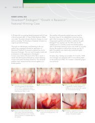
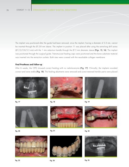
The implant was positioned after the guide had been removed, since the implant, having a diameter of 3.3 mm, cannot<br />
be inserted through the Ø 2.8 mm sleeve. The implant in position 11 was placed after using the extra-long drill series<br />
(Ø 2.2/2.8/3.5 mm) with the 1 mm reduction handle through the Ø 5 mm diameter sleeve (figs. 15, 16). The implant<br />
was positioned through the surgical guide. Transmucosal healing caps were positioned and the bone substitute material<br />
was inserted into the extraction sockets. Both sites were covered with the resorbable collagen membrane.<br />
Final Prostheses and Follow-up<br />
After 6 weeks, the OPG showed correct healing with no radiolucencies (fig. 17). Clinically, the implants sounded<br />
correct and were stable (fig. 18). The healing abutments were removed and screw-retained transfer parts were placed<br />
Fig. 17 Fig. 18<br />
Fig. 19<br />
Fig. 20<br />
Fig. 21<br />
Fig. 22<br />
Fig. 23 Fig. 24<br />
Fig. 25