Download - Straumann
Download - Straumann
Download - Straumann

You also want an ePaper? Increase the reach of your titles
YUMPU automatically turns print PDFs into web optimized ePapers that Google loves.
<strong>Straumann</strong> ® C areS ® digital S olutionS<br />
STARGET 1 I 12<br />
25<br />
It was possible to identify the implant in 12 as a CoreVent1 Ø 4.0 mm<br />
implant placed about 15 years ago and, after some research, some prosthetic<br />
component of the respective implant manufacturer was found that was<br />
compatible with the internal connection to this implant. After drilling the holes<br />
into the scan template according to the template plan, the lab provided the<br />
surgical guide (figs. 9 – 11).<br />
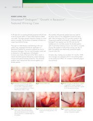
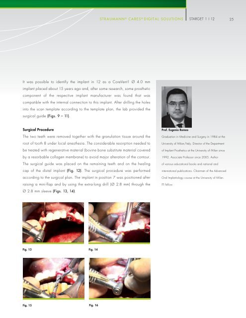
Surgical Procedure<br />
The two teeth were removed together with the granulation tissue around the<br />
root of tooth 8 under local anesthesia. The considerable resorption needed to<br />
be treated with regenerative material (bovine bone substitute material covered<br />
by a resorbable collagen membrane) to avoid major alteration of the contour.<br />
The surgical guide was placed on the remaining teeth and on the healing<br />
cap of the distal implant (fig. 12). The surgical procedure was performed<br />
according to the surgical plan. The implant in position 7 was positioned after<br />
raising a mini-flap and by using the extra-long drill (Ø 2.8 mm) through the<br />
Prof. Eugenio Romeo<br />
Graduation in Medicine and Surgery in 1984 at the<br />
University of Milan/Italy. Director of the Department<br />
of Implant Prosthetics at the University of Milan since<br />
1992. Associate Professor since 2005. Author<br />
of various educational books and national and<br />
international publications. Chairman of the Advanced<br />
Oral Implantology course at the University of Milan.<br />
ITI fellow.<br />
Ø 2.8 mm sleeve (figs. 13, 14).<br />
Fig. 13 Fig. 14<br />
Fig. 15<br />
Fig. 16