
Create successful ePaper yourself
Turn your PDF publications into a flip-book with our unique Google optimized e-Paper software.
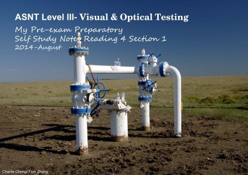
<strong>ASNT</strong> <strong>Level</strong> <strong>III</strong>- <strong>Visual</strong> & <strong>Optical</strong> <strong>Testing</strong><br />
My Pre-exam Preparatory<br />
Self Study Notes Reading 4 Section 1<br />
2014-August<br />
Charlie Chong/ Fion Zhang
Reading 4<br />
<strong>ASNT</strong> Nondestructive Handbook Volume 8<br />
<strong>Visual</strong> & <strong>Optical</strong> testing- Section 1<br />
For my coming <strong>ASNT</strong> <strong>Level</strong> <strong>III</strong> VT Examination<br />
2014-August<br />
Charlie Chong/ Fion Zhang
Charlie Chong/ Fion Zhang<br />
Fion Zhang<br />
2014/August/15
SECTION 1<br />
FUNDAMENTALS OF VISUAL AND OPTICAL<br />
TESTING<br />
Charlie Chong/ Fion Zhang
SECTION 1: FUNDAMENTALS OF VISUAL AND OPTICAL TESTING<br />
PART 1: Description of visual and optical tests<br />
1.1 Luminous Energy Tests<br />
1.2 Geometrical Optics<br />
PART 2: History of the borescope<br />
2.1 Development of the Borescope<br />
2.2 Certification of <strong>Visual</strong> Inspectors<br />
PART 3: Vision and light<br />
3.1 The Physiology of Sight<br />
3.2 Weber's Law<br />
3.3 Vision Acuity<br />
3.4 Vision Acuity Examinations<br />
3.5 <strong>Visual</strong> Angle<br />
3.6 Color Vision<br />
3.7 Fluorescent Materials<br />
Charlie Chong/ Fion Zhang
PART 4: Safety for visual and optical tests<br />
4.1 Need for Safety<br />
4.2 Laser Hazards<br />
4.3 Infrared Hazards<br />
4.4 Ultraviolet Hazards<br />
4.5 Photosensitizers<br />
4.6 Damage to the Retina<br />
4.7 Thermal Factor<br />
4.8 Blue Hazard<br />
4.9 <strong>Visual</strong> Safety Recommendations<br />
4.10 Eye Protection Filters<br />
Charlie Chong/ Fion Zhang
Part 1: DESCRIPTION OF VISUAL AND OPTICAL TESTS<br />
1.1.0 General:<br />
Nondestructive tests typically are done by applying a probing medium (such<br />
as acoustic or electromagnetic energy) to a material. After contact with the<br />
test material, certain properties of the probing medium are changed and can<br />
be used to determine changes in the characteristics of the test material.<br />
Density differences in a radiograph or location and peak of an oscilloscope<br />
trace are examples of means used to indicate probing media changes. In a<br />
practical sense, most nondestructive tests ultimately involve visual tests- a<br />
properly exposed radiograph is useful only when the radiographic interpreter<br />
has the vision acuity required to interpret the image.<br />
Likewise, the accumulation of magnetic particles over a crack indicates to the<br />
inspector an otherwise invisible discontinuity. The interface of visual testing<br />
with other nondestructive testing methods is discussed in more detail in a<br />
later section of this volume.<br />
Charlie Chong/ Fion Zhang
For the purposes of this book, visual and optical tests are those that use<br />
probing energy from the visible portion of the electromagnetic spectrum.<br />
Changes in the light's properties after contact with the test object may be<br />
detected by human or machine vision. Detection may be enhanced or made<br />
possible by mirrors, magnifiers, borescopes or other vision enhancing<br />
accessories.<br />
Keywords;<br />
■<br />
■<br />
■<br />
Visible Spectrum (380nm ~ 770nm),<br />
Human or machine vision,<br />
Vision enhancing tools- Borescope, mirror and other enhancing<br />
accessories.<br />
Charlie Chong/ Fion Zhang
1.2.0 Luminous Energy Tests<br />
<strong>Visual</strong> testing was probably the first method of nondestructive testing. It has<br />
developed from its ancient origins into many complex and elaborate optical<br />
investigation techniques. Some visual tests are based on the simple laws of<br />
geometrical optics. Others depend on properties of light, such as its wave<br />
nature. A unique advantage of many visual tests is that they can yield<br />
quantitative data more readily than other nondestructive tests.<br />
Luminous energy tests are used primarily for two purposes:<br />
1. testing of exposed or accessible surfaces of opaque test objects (including<br />
a majority of partially assembled or finished products) and<br />
2. testing of the interior of transparent test objects (such as glass, quartz,<br />
some plastics, liquids and gases). For many types of objects, visual testing<br />
can be used to determine quantity, size, shape, surface finish, reflectivity,<br />
color characteristics, fit, functional characteristics and the presence of<br />
surface discontinuities.<br />
Charlie Chong/ Fion Zhang
Keywords:<br />
Objects:<br />
• <strong>Testing</strong> of opaque objects<br />
• <strong>Testing</strong> of transparent objects<br />
VT is used to determined:<br />
• quantity,<br />
• size,<br />
• shape,<br />
• surface finish,<br />
• reflectivity,<br />
• color characteristics,<br />
• fit,<br />
• functional characteristics and the<br />
• presence of surface discontinuities.<br />
Question:<br />
Does VT covers Translucent object?<br />
Charlie Chong/ Fion Zhang
1.3.0 Geometrical Optics<br />
1.3.1 Image Formation<br />
Most optical instruments are designed primarily to form images. In many<br />
cases, the manner of image formation and the proportion of the image can be<br />
determined by geometry and trigonometry without detailed consideration of<br />
the physics of light rays.<br />
This practical technique is called geometrical optics and it includes the<br />
formation of images by lenses and mirrors. The operation of microscopes,<br />
telescopes and borescopes also can be partially explained with geometrical<br />
optics. In addition, the most common limitations of optical instruments can<br />
be similarly evaluated with this technique.<br />
Keyword:<br />
Geometrical optics<br />
Charlie Chong/ Fion Zhang
1.3.2 Light Sources<br />
The light source for visual tests typically emits radiation of a continuous or<br />
noncontinuous (line) spectrum. Monochromatic light is produced by use of a<br />
device known as a monochromator, which separates or disperses the<br />
wavelengths of the spectrum by means of prisms or gratings.<br />
Less costly and almost equally effective for routine tests are light sources<br />
emitting distinct spectral lines, These include mercury, sodium and other<br />
vapor discharge lamps. Such light sources may he used in combination with<br />
glass, liquid or gaseous filters or with highly efficient interference filters, for<br />
transmitting only radiation of a specific wavelength.<br />
Keywords:<br />
Continuous spectrum<br />
Non-continuous spectrum-Monochromatic light<br />
Monochromator used prisms or grating<br />
Charlie Chong/ Fion Zhang
Keywords:<br />
Monochromatic light produces by vapor discharged lamp (Mercury/sodium<br />
etc.) with glass/ liquid & gaseous filter to produces only radition with specific<br />
wavelength<br />
Charlie Chong/ Fion Zhang
Gas-discharge lamps<br />
are a family of artificial light sources that generate light by sending an<br />
electrical discharge through an ionized gas, a plasma. The character of the<br />
gas discharge depends on the pressure of the gas as well as the frequency of<br />
the current. Typically, such lamps use a noble gas (argon, neon, krypton and<br />
xenon) or a mixture of these gases. Most lamps are filled with additional<br />
materials, like mercury, sodium, and metal halides. In operation the gas is<br />
ionized, and free electrons, accelerated by the electrical field in the tube,<br />
collide with gas and metal atoms. Some electrons in the atomic orbitals of<br />
these atoms are excited by these collisions to a higher energy state. When<br />
the excited atom falls back to a lower energy state, it emits a photon of a<br />
characteristic energy, resulting in infrared, visible light, or ultraviolet radiation.<br />
Some lamps convert the ultraviolet radiation to visible light with a fluorescent<br />
coating on the inside of the lamp's glass surface. The fluorescent lamp is<br />
perhaps the best known gas-discharge lamp.<br />
http://en.wikipedia.org/wiki/Gas-discharge_lamp<br />
Charlie Chong/ Fion Zhang
Compared to incandescent lamps, gas-discharge lamps offer higher<br />
efficiency, but are more complicated to manufacture, and require auxiliary<br />
electronic equipment such as ballasts to control current flow through the gas.<br />
Some gas-discharge lamps also have a perceivable start-up time to achieve<br />
their full light output. Still, due to their greater efficiency, gas-discharge lamps<br />
are replacing incandescent lights in many lighting applications.<br />
Charlie Chong/ Fion Zhang
Vapor Discharged Lamp<br />
Charlie Chong/ Fion Zhang
Vapor Discharged Lamp<br />
Charlie Chong/ Fion Zhang
Vapor Discharged Lamp<br />
Charlie Chong/ Fion Zhang
A monochromator is an optical device<br />
that transmits a mechanically selectable<br />
narrow band of wavelengths of light or<br />
other radiation chosen from a wider<br />
range of wavelengths available at the<br />
input. The name is from the Greek roots<br />
mono-, single, and chroma, colour, and<br />
the Latin suffix -ator, denoting an agent.<br />
http://en.wikipedia.org/wiki/Monochromator<br />
Charlie Chong/ Fion Zhang
Monochromator used prisms or grating<br />
Charlie Chong/ Fion Zhang
Monochromator used prisms or grating<br />
Charlie Chong/ Fion Zhang
Monochromator used prisms or grating<br />
Charlie Chong/ Fion Zhang
Monochromator<br />
used prisms or<br />
grating<br />
Charlie Chong/ Fion Zhang
Monochromator used prisms or grating<br />
Charlie Chong/ Fion Zhang
Monochromator used prisms or grating<br />
Charlie Chong/ Fion Zhang
Monochromator used prisms or grating<br />
Charlie Chong/ Fion Zhang
1.3.3 Stroboscopic Sources<br />
The stroboscope is a device that uses synchronized pulses of high intensity<br />
light to permit viewing of objects moving with a rapid, periodic motion. A<br />
stroboscope can be used for direct viewing of the apparently stilled test object<br />
or for exposure of photographs. The timing of the stroboscope also can be<br />
adjusted so that the moving test object is seen to move but at a much slower<br />
apparent motion. The stroboscopic effect requires an accurately controlled,<br />
intermittent source of light or may be achieved with periodically interrupted<br />
vision.<br />
Charlie Chong/ Fion Zhang
1Stroboscopic Movement<br />
Charlie Chong/ Fion Zhang
Stroboscopic Movement<br />
Charlie Chong/ Fion Zhang
Stroboscopic Movement<br />
Charlie Chong/ Fion Zhang
Stroboscopic Sources<br />
Charlie Chong/ Fion Zhang
Stroboscopic Sources<br />
Charlie Chong/ Fion Zhang
Stroboscopic Glasses<br />
Charlie Chong/ Fion Zhang
千 手 观 音<br />
Charlie Chong/ Fion Zhang
千 手 观 音<br />
Charlie Chong/ Fion Zhang
千 手 观 音<br />
Charlie Chong/ Fion Zhang
1.3.4 Light Detection and Recording<br />
Once light has interacted with a test object (been absorbed, reflected or<br />
refracted), the resulting light waves are considered test signals that may be<br />
recorded visually or photoelectrically. Such signals may be detected by<br />
means of photoelectric cells, bolometers or thermopiles, photomultipliers<br />
or closed circuit television systems. Electronic image conversion devices<br />
often are used for the invisible ranges of the electromagnetic spectrum<br />
(infrared, ultraviolet or X-rays) but they also may he used to transmit<br />
visual data from hazardous locations or around obstructions. Occasionally,<br />
intermediary photographic recordings are made.<br />
The processed photographic plate can subsequently be evaluated either<br />
visually or photoelectrically. Some applications take advantage of the ability<br />
of photographic film to integrate low energy signals over long periods of time.<br />
Photographic film emulsions can be selected to meet specific test conditions,<br />
sensitivities and speeds.<br />
Charlie Chong/ Fion Zhang
Keywords:<br />
Photoelectricity detection<br />
• photoelectric cells,<br />
• bolometers or<br />
• thermopiles,<br />
• photomultipliers<br />
• or closed circuit television systems.<br />
Charlie Chong/ Fion Zhang
Bolometer consists of an absorptive element, such as a thin layer of metal,<br />
connected to a thermal reservoir (a body of constant temperature) through a<br />
thermal link. The result is that any radiation impinging on the absorptive<br />
element raises its temperature above that of the reservoir — the greater the<br />
absorbed power, the higher the temperature.<br />
The intrinsic thermal time constant, which sets the speed of the detector, is<br />
equal to the ratio of the heat capacity of the absorptive element to the thermal<br />
conductance between the absorptive element and the reservoir. The<br />
temperature change can be measured directly with an attached resistive<br />
thermometer, or the resistance of the absorptive element itself can be used<br />
as a thermometer. Metal bolometers usually work without cooling. They are<br />
produced from thin foils or metal films. Today, most bolometers use<br />
semiconductor or superconductor absorptive elements rather than metals.<br />
These devices can be operated at cryogenic temperatures, enabling<br />
significantly greater sensitivity.<br />
Charlie Chong/ Fion Zhang
Bolometer<br />
Charlie Chong/ Fion Zhang
Bolometer<br />
Charlie Chong/ Fion Zhang
Bolometer<br />
Charlie Chong/ Fion Zhang
Thermopiles<br />
Charlie Chong/ Fion Zhang
Thermopiles<br />
Charlie Chong/ Fion Zhang
Thermopiles<br />
Charlie Chong/ Fion Zhang
Multi-Junction Thermopiles<br />
The thermopile is a heat sensitive device that measures radiated heat. The<br />
sensor is usually sealed in a vacuum to prevent heat transfer except by<br />
radiation. A thermopile consists of a number of thermocouple junctions in<br />
series which convert energy into a voltage using the Peltier effect.<br />
Thermopiles are convenient sensor for measuring the infrared, because they<br />
offer adequate sensitivity and a flat spectral response in a small package.<br />
More sophisticated bolometers and pyroelectric detectors need to be chopped<br />
and are generally used only in calibration labs.<br />
Charlie Chong/ Fion Zhang
Photo Detector Comparisons<br />
http://homepages.inf.ed.ac.uk/rbf/CVonline/LOCAL_COPIES/RYER/ch10.html<br />
Charlie Chong/ Fion Zhang
Photomultiplier<br />
Photomultiplier tubes (photomultipliers or PMTs for short), members of the<br />
class of vacuum tubes, and more specifically vacuum phototubes, are<br />
extremely sensitive detectors of light in the ultraviolet, visible, and nearinfrared<br />
ranges of the electromagnetic spectrum. These detectors multiply the<br />
current produced by incident light by as much as 100 million times (i.e., 160<br />
dB), in multiple dynode stages, enabling (for example) individual photons to<br />
be detected when the incident flux of light is very low. Unlike most vacuum<br />
tubes, they are not obsolete.<br />
The combination of high gain, low noise, high frequency response or,<br />
equivalently, ultra-fast response, and large area of collection has earned<br />
photomultipliers an essential place in nuclear and particle physics, astronomy,<br />
medical diagnostics including blood tests, medical imaging, motion picture<br />
film scanning (telecine), radar jamming, and high-end image scanners known<br />
as drum scanners. Elements of photomultiplier technology, when integrated<br />
differently, are the basis of night vision devices.<br />
Charlie Chong/ Fion Zhang
Semiconductor devices, particularly avalanche photodiodes, are alternatives<br />
to photomultipliers; however, photomultipliers are uniquely well-suited for<br />
applications requiring low-noise, high-sensitivity detection of light that is<br />
imperfectly collimated.<br />
http://en.wikipedia.org/wiki/Photomultiplier<br />
Charlie Chong/ Fion Zhang
Photomultiplier<br />
Secondary emission<br />
Photoelectric effect<br />
Photon<br />
Electrons multiplying<br />
Charlie Chong/ Fion Zhang
Photomultiplier<br />
Charlie Chong/ Fion Zhang
Photomultiplier<br />
Charlie Chong/ Fion Zhang
Photoelectric Cell<br />
• Photovoltaic Cell<br />
• Photo emissivity<br />
Photovoltaic's (PV) is a method of generating electrical power by converting<br />
solar radiation into direct current electricity using semiconductors that exhibit<br />
the photovoltaic effect. Photovoltaic power generation employs solar panels<br />
composed of a number of solar cells containing a photovoltaic material. Solar<br />
photovoltaics power generation has long been seen as a clean sustainable[1]<br />
energy technology which draws upon the planet’s most plentiful and widely<br />
distributed renewable energy source – the sun. The direct conversion of<br />
sunlight to electricity occurs without any moving parts or environmental<br />
emissions during operation. It is well proven, as photovoltaic systems have<br />
now been used for fifty years in specialized applications, and grid-connected<br />
systems have been in use for over twenty years<br />
Charlie Chong/ Fion Zhang
Charlie Chong/ Fion Zhang
Photovoltaic Cell<br />
Charlie Chong/ Fion Zhang
Photovoltaic Cell<br />
Charlie Chong/ Fion Zhang
Photoemissive Cell- Photoemissive cell (electronics)<br />
A device which detects or measures radiant energy by measurement of the<br />
resulting emission of electrons from the surface of a photocathode.<br />
Charlie Chong/ Fion Zhang
Photoemissive Cell<br />
Charlie Chong/ Fion Zhang
Photoemissive Cell: Analysis of sodium levels in junk food by flame<br />
photometer<br />
http://www.pharmatutor.org/articles/analysis-sodium-levels-junk-food-flame-photometer?page=0,2<br />
Charlie Chong/ Fion Zhang
1.3.5 Fluorescence Detection<br />
A material is said to fluoresce when exposure to radiation causes the material<br />
to produce a secondary emission of longer wavelength than the primary,<br />
exciting light. <strong>Visual</strong> tests based on fluorescence play a part in qualitative and<br />
quantitative inorganic and organic chemistry, as a means of quality control of<br />
chemical compounds, for identifying counterfeit currency, tracing hidden<br />
water flow and for detecting discontinuities in metals and pavement.<br />
Charlie Chong/ Fion Zhang
Fluorescence Detection<br />
Charlie Chong/ Fion Zhang
More Reading on Light<br />
Light Measurement Handbook<br />
http://homepages.inf.ed.ac.uk/rbf/CVonline/LOCAL_COPIES/RYER/index.html<br />
http://homepages.inf.ed.ac.uk/rbf/CVonline/<br />
Charlie Chong/ Fion Zhang
Part 2: History of Borescope<br />
2.1.0 Introduction<br />
Development of the Borescope The development of self illuminated<br />
telescopic devices can be traced back to early interest in exploring the interior<br />
human anatomy without operative procedures. Devices for viewing the<br />
interior of objects are called endoscopes, from the Creek words for "inside<br />
view." Today the term endoscope in the United States is applied primarily to<br />
medical instruments. Nearly all of the medical endoscopes have an integral<br />
light source; some incorporate surgical tweezers or other devices. Industrial<br />
endoscopes are called horescopes because they were 'originally used in<br />
machined apertures and holes such as gun bores. There are both flexible<br />
and rigid, fiber optic and geometric light borescopes.<br />
Keywords:<br />
■<br />
■<br />
Endoscopes<br />
Horescopes<br />
Charlie Chong/ Fion Zhang
2.1.1 Cystoscopes and Borescopes<br />
In 1806 Philipp Bozzini of Frankfurt announced the invention of his Lichtleiter<br />
(German for "light guide"). Having served as a surgeon in the Napoleonic<br />
wars, Bozzini envisioned using his device for medical research. It is<br />
considered the first endoscope. In 1876, Dr. Max Nitze, a urologist, developed<br />
the first practical cystoscope to view the human bladder.' A platinum loop in<br />
its tip furnished a bright light when heated with galvanic current. Two years<br />
later, Thomas Edison introduced an incandescent light in the United States.<br />
Within a short time, scientists in Austria made and used a minute electric bulb<br />
in Nitze's cystoscope, even before the electric light was in use in America.<br />
The early cystoscopes contained simple lenses but these were soon replaced<br />
by achromatic combinations. In 1900, Reinhold Wappler revolutionized the<br />
optical system of the cystoscope and produced the first American models.<br />
The forward oblique viewing system was later introduced and has proved<br />
very useful in both medical and industrial applications. Direct vision and<br />
retrospective systems were also first developed for cystoscopic use.<br />
Charlie Chong/ Fion Zhang
Borescopes and related instruments for nondestructive testing have followed<br />
the same basic design used in cystoscopic devices. The range of borescope<br />
sizes has increased, sectionalized instruments have been introduced and<br />
other special devices have been developed for industrial applications.<br />
Charlie Chong/ Fion Zhang
2.1.2 Gastroscopes and Flexible Borescopes<br />
A flexible gastroscope, originally intended for observing the interior of the<br />
stomach wall, was first developed by Rudolph Schindler' and produced by<br />
Georg Wolf in 1932. The instrument consisted of a rigid section and a flexible<br />
section. Many lenses of small focal distance were used to allow bending of<br />
the instrument to an angle of 34 degrees in several planes. The tip of the<br />
device contained the objective and the prism causing the necessary axial<br />
deviation of the bundle of rays coming from the illuminated gastric wall. The<br />
size of the image depended on the distance of the objective from the<br />
observed surface. It could be magnified, reduced or normal size but the<br />
image was sharp and erect with correct sides. Flexible gastroscopes are now<br />
available, with rubber tubes over the flexible portion, in diameters of<br />
approximately 14 mm (0.55 in.) and 8 mm (0.31 in.).<br />
Charlie Chong/ Fion Zhang
Flexible borescopes for industrial use are more ruggedly constructed than<br />
gastroscopes, having flexible steel tubes instead of rubber for the outer tube<br />
of the flexible portion. A typical flexible borescope is 13 mm (0,5 in.) in<br />
diameter and has a 1 m (3 ft) working length, with flexibility in about 500 mm<br />
(20 in.) of the length. Extension sections are available in 1, 2 or 3 m (3, 6 or 9<br />
ft) lengths, permitting assembly of borescopes up to 10 m (30 ft) in length. In<br />
such flexible instruments the image remains round and sharp when the tube<br />
is bent to an angle of about 34 degrees. Beyond that limit, the image<br />
becomes elliptical but remains clear until obliterated at about 45 degrees of<br />
total bending.<br />
Charlie Chong/ Fion Zhang
Keywords: Conventional Borescope Bend angles & Images<br />
• 34 Degree- Round and Clear<br />
• 34 ~ 45 Degree- Elliptical but Clear<br />
• > 45 Degree- Obliterated<br />
Charlie Chong/ Fion Zhang
Digitized Borescope<br />
Charlie Chong/ Fion Zhang
2.1.3 American Development of Borescopes<br />
After the early medical developments, certain segments of American industry<br />
needed visual testing equipment for special inspection applications. One of<br />
the first individuals to help fill this need was George Sumner Crampton.<br />
George Crampton (Fig. 1) was born in Rock Island, Illinois in 1874. He was<br />
said to have set up a small machine shop by the age of 10 and his first<br />
ambition was to become an electrical engineer. He chose instead to study<br />
medicine and received his M.D. from the University of Pennsylvania in<br />
1898. While he was interning at Pennsylvania Hospital, Crampton's<br />
mechanical and engineering abilities were recognized and he was advised to<br />
become an oculist.' He returned to the university, took a degree in<br />
ophthalmology and later practiced in Philadelphia, Pennsylvania and<br />
Princeton, New Jersey‘ In 1921, the Westinghouse Company asked<br />
Crampton to make a device that could be used to check for discontinuities<br />
inside the rotor of a steam turbine (Fig. 2). Crampton developed the<br />
instrument in his Philadelphia shop and delivered the prototype within a<br />
week- it was the first borescope produced by his company.<br />
Charlie Chong/ Fion Zhang
Crampton continued to supply custom borescopes for testing inaccessible<br />
and often dark areas on power turbines, oil refinery piping, gas mains, soft<br />
drink tanks and other components (Fig. 3). Crampton soon was recognized<br />
for his ability to design and manufacture borescopes, periscopes and other<br />
optical equipment for specific testing applications. After retiring as emeritus<br />
professor of ophthalmology at the university Crampton continued private<br />
practice in downtown Philadelphia. At the same time, he worked on<br />
borescopes and other instruments in a small shop he had established in a<br />
remodeled nineteenth century coach house (Fig. 4).<br />
Charlie Chong/ Fion Zhang
FIGURE 1. George Crampton, developer of the borescope<br />
Charlie Chong/ Fion Zhang
FIGURE 2. Tests of forgings for a steam turbine generator shaft manufactured in the 1920s<br />
FIGURE 3. Inspectors use early borescopes to visually inspect piping at an Ohio oil refinery<br />
Charlie Chong/ Fion Zhang
FIGURE 4. Periscope built in the 1940s is checked before shipment to a Texas chemical plant<br />
Charlie Chong/ Fion Zhang
2.1.4 Wartime Borescope Developments<br />
After World War II began, Crampton devoted much of his energy to the war<br />
effort, filling defense orders for borescopes (Fig. 5). Crampton practiced<br />
medicine until noon, then went to the nearby workshop where he visually<br />
tested the bores of 37 mm antiaircraft guns and other weapons. During the<br />
war, borescopes were widely used for testing warship steam turbines<br />
(particularly their rotating shafts). The United States Army also used<br />
borescopes for inspecting the barrels of tank and antiaircraft weapons<br />
produced in Philadelphia. An even more challenging assignment lay<br />
ahead.<br />
The scientists working to develop a successful nuclear chain reaction in the<br />
top secret Manhattan Project asked Crampton to provide a borescope for<br />
inspecting tubes near the radioactive pile at its guarded location beneath the<br />
stadium seats at the University of Chicago's Stagg Field. Crampton devised<br />
an aluminum borescope tube 35 mm (1.4 in.) in diameter and 10 m (33 ft)<br />
long. The device consisted of 2 m (6 ft) sections of dual tubing joined by<br />
bronze couplings which also carried an 8 V lighting circuit.<br />
Charlie Chong/ Fion Zhang
FIGURE 5. Using a borescope, an inspector at an automobile plant during World War H checks<br />
the interiors of gun tubes for 90 mm antiaircraft guns<br />
Charlie Chong/ Fion Zhang
The inspector standing directly in front of the bore was subject to radioactive<br />
emissions from the pile, so Crampton mounted the borescope outside of a<br />
heavy concrete barrier. The operator stood at a right angle to the borescope,<br />
looking through an eyepiece and revolving the instrument manually. The<br />
borescope contained a prism viewing head and had to be rotated constantly.<br />
It was supported in a steel V trough resting on supports whose height could<br />
be varied. Crampton also mounted a special photographic camera on the<br />
eyepiece.<br />
The original Manhattan Project borescope was later improved with<br />
nondarkening optics and a swivel-joint eyepiece that permitted the operator to<br />
work from any angle (this newer instrument did not require the V trough). It<br />
also was capable of considerable bending to snake through the tubes in the<br />
reactor. A total of three borescopes were supplied fbr this epochal project and<br />
they are believed to be the first optical instruments to use glass resistant to<br />
radioactivity.<br />
Charlie Chong/ Fion Zhang
Manhattan Project<br />
Charlie Chong/ Fion Zhang
Manhattan Project<br />
Charlie Chong/ Fion Zhang
Manhattan Project<br />
Charlie Chong/ Fion Zhang
2.1.5 Borescopes and Aircraft Tests<br />
Aircraft inspection soon became one of the most important uses of borescope<br />
technology. In 1946, an ultraviolet light borescope was developed for<br />
fluorescent testing of the interior of hollow steel propeller blades. The 100 W<br />
viewing instniment revealed interior surface discontinuities as glowing green<br />
lines. Later, in 1958, the entire United States' B-47 bomber fleet was<br />
grounded because of metal fatigue cracks resulting from low level simulated<br />
bombing missions. <strong>Visual</strong> testing with borescopes proved to be the first step<br />
toward resolving the problem. The program became known as Project<br />
Milkbottle, a reference to the bottle shaped pin that was a primary connection<br />
between the fuselage and wing (Fig. 6).<br />
In the late 1950s, a system was developed for automatic testing of helicopter<br />
blades. The borescope, supported by a long bench, could test the blades<br />
while the operator viewed results on a television screen (Fig. 7). The system<br />
was used extensively during the Vietnam conflict and helicopter<br />
manufacturers continue to use borescopes for such critical tests.<br />
Charlie Chong/ Fion Zhang
FIGURE 6. Inspector using a borescope to check for metal fatigue cracks in a B-47 bomber<br />
during grounding of the bomber fleet in 1958<br />
FIGURE 7. <strong>Visual</strong> testing of the frame of a 10 m (32 ft) long helicopter blade using a 10 m (32 ftj<br />
borescope; the inspector could view magnified results on the television screen at bottom left<br />
Charlie Chong/ Fion Zhang
After a half century of pioneering work, George Crampton sold his borescope<br />
business to John Lang of Cheltenham, Pennsylvania, in 1962.6•7 Lang had<br />
developed the radiation resistant optics used in the Manhattan Project<br />
borescope, as well as a system for keeping it functional in high temperature<br />
environments. He also helped pioneer the use of closed circuit television with<br />
borescopes for testing the inner surfaces of jet engines and wings, hollow<br />
helicopter blades and nuclear reactors. In 1965, the company received a<br />
patent on a borescope whose mirror could he very precisely controlled.<br />
This borescope could zoom to high magnification and could intensely<br />
illuminate the walls of a chamber by means of a quartz incandescent lamp<br />
containing iodine vapor. The basic design of the borescope has been in use<br />
for many decades and it continues to develop, accommodating advances in<br />
video, illumination, robotic and computer technologies.<br />
Charlie Chong/ Fion Zhang
2.2.0 Certification of <strong>Visual</strong> Inspectors<br />
2.2.1 Introduction<br />
The recognition of the visual testing technique and the development of formal<br />
procedures for educating and qualifying visual inspectors were important<br />
milestones in the history of visual inspection. Because visual testing can be<br />
performed without any intervening apparatus, it was certainly one of the first<br />
forms of nondestructive testing. In its early industrial applications, visual tests<br />
were used simply to verify compliance to a drawing or specification. This was<br />
basically a dimensional check. The soundness of the object was determined<br />
by liquid penetrant, magnetic particle, radiography or ultrasonic testing.<br />
Following World War II, few inspection standards included visual testing. By<br />
the early 1960s, visual tests were an accepted addition to the American<br />
Welding Society's code hooks. In NAV SHIPS 250-1500-1, the US Navy<br />
included visual tests with its specifications for other nondestructive testing<br />
techniques for welds.<br />
Charlie Chong/ Fion Zhang
By 1965, there were standards for testing, and criteria for certifying the<br />
inspector had been established in five test methods: liquid penetrant,<br />
magnetic particle, eddy current, radiographic and ultrasonic testing. These<br />
five were cited in <strong>ASNT</strong> Recommended Practice No. SNT-TC-1A, introduced<br />
in the late 1960s. The broad use of visual testing hindered its addition to this<br />
group as a specific method- there were too many different applications on too<br />
many test objects to permit the use of specific acceptance criteria. It also was<br />
reasoned that visual testing would occur as a natural result of applying any<br />
other nondestructive test method.<br />
Charlie Chong/ Fion Zhang
2.2.2 Expanded Need for <strong>Visual</strong> Certification<br />
In the early 1970s, the need for certified visual inspectors began to increase.<br />
Nuclear power construction was at a peak, visual certification was becoming<br />
mandatory and nondestructive testing was being required. In 1976, the<br />
American Society for Nondestructive <strong>Testing</strong> began considering the need for<br />
certified visual inspectors. <strong>ASNT</strong> had become a leading force in<br />
nondestructive testing and American industry had accepted its <strong>ASNT</strong><br />
Recommended Practice No. SNT-TC-IA as a guide for certifying other NDT<br />
inspectors. In the spring of 1976, <strong>ASNT</strong> began surveying industry about their<br />
inspection needs and their position on visual testing. Because of the many<br />
and varied responses to the survey, a society task force was established to<br />
analyze the survey data. In 1977, the task force recommended that visual<br />
inspectors be certified and that visual testing be made a supplement to <strong>ASNT</strong><br />
Recommended Practice No. SNT-TC-IA (1975). At this time, the American<br />
Welding Society implemented a program that, following the US Navy, was the<br />
first to certify inspectors whose sole function was visual weld testing.<br />
Charlie Chong/ Fion Zhang
During 1978, <strong>ASNT</strong> subcommittees were formed for the eastern and western<br />
halves of the United States. These groups verified the need for both visual<br />
standards and trained, qualified and certified inspectors. In 1980, a <strong>Visual</strong><br />
Methods Committee was formed in <strong>ASNT</strong>'s Technical Council and the early<br />
meetings defined the scope and purpose of visual testing (dimensional testing<br />
was excluded). In 1984, the <strong>Visual</strong> Personnel Qualification Committee was<br />
formed in <strong>ASNT</strong>'s Education and Qualification Council. In 1986, a training<br />
outline and a recommended reference list was finalized and the Board of<br />
Directors approved incorporation of visual testing into <strong>ASNT</strong> Recommended<br />
Practice No. SN T-TC -1 A.<br />
Charlie Chong/ Fion Zhang
Part 3: VISION AND LIGHT<br />
3.1 The Physiology of Sight<br />
3.1.1 <strong>Visual</strong> Data Collection<br />
Human visual processing occurs in two steps. First the entire field of vision is<br />
processed. This is typically an automatic function of the brain, sometimes<br />
called pre-attentive processing. Secondly, focus is localized to a specific<br />
object in the processed field. Studies at the University of Pennsylvania<br />
indicate that segregating specific items from the general field is the foundation<br />
of the identification process. Based on this concept, it is now theorized that<br />
various light patterns reaching the eyes are simplified and encoded, as lines,<br />
spots, edges, shadows, colors, orientations and referenced locations within<br />
the entire field of view. The first step in the subsequent identification process<br />
is the comparison of visual data with the long-term memory of previously<br />
collected data. Some researchers have suggested that this comparison<br />
procedure is a physiological cause of deja vu, the uncanny feeling of having<br />
seen something before.<br />
Charlie Chong/ Fion Zhang
The accumulated data are then processed through a series of specific<br />
systems. Certain of our light sensors receive and respond only to certain<br />
stimuli and transmit their data to particular areas of the brain for translation.<br />
One kind of sensor accepts data on lines and edges; other sensors process<br />
only directions of movement or color. Processing of these data discriminates<br />
between different complex views by analyzing their various components.<br />
By experiment it has been shown that these areas of sensitivity have a kind of<br />
persistence. This can be illustrated by staring at a lit candle, then diverting the<br />
eyes toward a blank wall. For a short time, the image of the candle is retained.<br />
The same persistence occurs with motion detection and can he illustrated by<br />
staring at a moving object, such as a waterfall, then at a stationary object like<br />
the river bank. The bank will seem to flow because the visual memory of<br />
motion is still present.<br />
Charlie Chong/ Fion Zhang
3.1.2 Differentiation in the Field of View<br />
Boundary and edge detection can be illustrated by the pattern changes in Fig.<br />
8. When scanning the figure from left to right, the block of reversed Ls is<br />
difficult to separate from the upright Ts in the center but the boundary<br />
between the normal Ts and the tilted Ts is easily apparent. The difficulty in<br />
differentiation occurs because horizontal and vertical lines comprise the L and<br />
upright T groups, creating a similarity that the brain momentarily retains as<br />
the eye moves from one group to the other.<br />
On the other hand, the tilted Ts share no edge orientations with the upright Ts,<br />
making them stand out in the figure. Differentiation of colors is more difficult<br />
when the different colors are in similarly shaped objects in a pattern. The<br />
recognition of geometric similarities tends to overpower the difference in<br />
colors, even when colors are the object of interest. Additionally, in a grouping<br />
of different shapes of unlike colors, where no one form is dominant, a<br />
particular form may hide within the varied field of view. However, if the<br />
particular form contains a major color variance, it is very apparent.<br />
Experiments have shown that such an object may be detected with as much<br />
ease from a field of thirty as it is from a field of three.<br />
Charlie Chong/ Fion Zhang
FIGURE 8. Pattern changes illustrating boundary and edge detection<br />
Charlie Chong/ Fion Zhang
3.1.3 Searching the Field of View<br />
The obstacles to differentiation discussed above indicate that similar objects<br />
are difficult to identify individually. During pre-attentive processing, particular<br />
objects that share common properties such as length, width, thickness or<br />
orientation are not different enough to stand out. If the differences between a<br />
target object and the general field is dramatic, then a visual inspector requires<br />
little knowledge of what is to be identified. When the target object is similar to<br />
the general field, the inspector needs more specific detail about the target. In<br />
addition, the time required to detect a target increases linearly with the<br />
number of similar objects in its general field. When an unspecified target is<br />
being sought, the entire field must be scrutinized. If the target is known, it has<br />
been shown statistically that only about half of the field must be searched.<br />
Charlie Chong/ Fion Zhang
The differences between a search for simple features and a search for<br />
conjunctions or combinations of features can also have implications in<br />
nondestructive testing environments. For example, visual inspectors may be<br />
required to take more time to check a manufactured component when<br />
the possible errors in manufacturing are characterized by combinations of<br />
undesired properties. Less time could be taken for a visual test if the<br />
manufacturing errors always produced a change in a single property.<br />
Another aspect of searching the field of view addresses the absence of<br />
features. The presence of a feature is easier to locate than its absence. For<br />
example, if a single letter 0 is introduced to a field of many Qs, it is more<br />
difficult to detect than a single Q in a field of Os. The same difficulty is<br />
apparent when searching for an open 0 in a field of closed Os. In this case<br />
statistics show that the apparent similarity in the target objects is greater and<br />
even more search time is necessary<br />
Charlie Chong/ Fion Zhang
Experimentation in the area of visual search tasks encompasses several<br />
tests of many 'individuals. Such experiments start with studies of those<br />
features that should stand out readily, displaying the basic elements of early<br />
vision recognition. The experiments cover several categories, including<br />
quantitative properties such as length or number. Also included are search<br />
tasks concentrating on single lines, orientation, curves, simple forms and<br />
ratios of sizes. All these tests verify that visual systems respond more<br />
favorably to targets that have something added (Q versus 0) rather than<br />
something missing.<br />
In addition, it has been determined that the ability to distinguish differences in<br />
intensity becomes more acute with a decreasing field intensity. This is the<br />
basis of Weber's law. The features it addresses are those involved in the<br />
early visual processes: color, size, contrast, orientation, curvature, lines,<br />
borders, movement and stereoscopic depth.<br />
Charlie Chong/ Fion Zhang
3.2 Weber's Law<br />
3.2.1 General<br />
Weber's law is widely used by psychophysicists and entails the following<br />
tenets: (1) individual elements such as points or lines are more important<br />
singly than their relation to each other and (2) closed forms appear to stand<br />
out more readily than open forms. To view a complete picture, the visual<br />
system begins by encoding the basic properties that are processed within the<br />
brain, including their spatial relationships.<br />
Each item in a field of view is stored in a specific zone and is withdrawn when<br />
required to form a complete picture. Occasionally, these items are withdrawn<br />
and positioned in error. This malfunction in the reassembly process allows the<br />
creation of optical illusions, allowing a picture to be misinterpreted.<br />
Charlie Chong/ Fion Zhang
The diagram in Fig. 9 represents a model of the early stages of visual<br />
perception. The encoded properties are maintained in their respective spatial<br />
relationships and compared to the general area of vision. The focused<br />
attention selects and integrates these properties, forming a specific area of<br />
observation. In some cases, as the area changes, the various elements<br />
comprising the observance are modified or updated to represent present<br />
conditions. During this step, new data are compared to the stored information.<br />
Charlie Chong/ Fion Zhang
FIGURE 9. Stages of visual perception<br />
Charlie Chong/ Fion Zhang
http://art.nmu.edu/cognates/ad175/background.html<br />
Charlie Chong/ Fion Zhang
3.3. Vision Acuity<br />
3.3.1 General<br />
Vision acuity encompasses the ability to see and identify, what is seen. Two<br />
forms of vision acuity are recognized and must be considered when<br />
attempting to qualify visual ability. These are known as near vision and far<br />
vision (acuity).<br />
Charlie Chong/ Fion Zhang
3.3.2 Components of the Human Eye<br />
The components of the human eye (Fig. 10) are often compared to those of a<br />
camera. The lens is used to focus light rays reflected by an object in the field<br />
of view. This results in the convergence of the rays on the retina (film),<br />
located at the rear of the eyeball. The cornea covers the eye and protects the<br />
lens. The quantity of light admitted to the lens is controlled by the contraction<br />
of the iris (aperture). The lens has the ability to become thicker or thinner,<br />
which alters the magnification and the point of impingement of the light rays,<br />
changing the focus.<br />
Eye muscles aid in the altering of the lens shape as well as controlling the<br />
point of aim. This configuration achieves the best and sharpest image for the<br />
entire system. The retina consists of rod and cone nerve endings that lie<br />
beneath the surface. They are in groups that represent specific color<br />
sensitivities and pattern recognition sections. These areas may be further<br />
subdivided into areas that collect data from lines, edges, spots, positions or<br />
orientations.<br />
Charlie Chong/ Fion Zhang
The light energy is received and converted to electrical signals that are<br />
moved by way of the optic nerve system to the brain where the data are<br />
processed. Because the light is being reflected from an object in a particular<br />
color or combination of colors, the individual wavelengths representing each<br />
hue also vary. Each wavelength is focused at different depths within the retina,<br />
stimulating specific groups of rods and cones (see Figs. 10 and 11). The color<br />
sensors are grouped in specific recognition patterns as discussed above.<br />
Charlie Chong/ Fion Zhang
FIGURE 10. Components of the human eye in cross section<br />
Charlie Chong/ Fion Zhang
FIGURE 11. Magnified cross section showing the blind spot of the human eye<br />
Charlie Chong/ Fion Zhang
To ensure reliable observation, the eye must have all the rays of light in focus<br />
on the retina. When the point of focus is short or primarily near the inner<br />
surface of the retina closest to the lens, a condition known as<br />
nearsightedness exists. If the focal spot is deeper into the retina,<br />
farsightedness occurs. These conditions are primarily the result of the eyeball<br />
changing from nearly orb shaped to an elliptical or egg shape. In the case of<br />
the nearsighted person, the long elliptical diameter is horizontal, If the long<br />
diameter is in a vertical direction, farsightedness occurs. These clinical<br />
conditions result from a very small shift of the focal spot, on the order of<br />
micrometers (ten-thousandths of an inch).<br />
Charlie Chong/ Fion Zhang
3.3.3 Determining Vision Acuity<br />
The method normally used to determine what the eye can see is based on the<br />
average of many measurements. The average eye views a sharp image when<br />
the object subtends an arc of five minutes, regardless of the distance the<br />
object is from the eye. The variables in this feature are the diameter of the<br />
eye lens at the time of observation and the distance from the lens to the retina.<br />
When vision cannot he normally varied to create sharp clear images, then<br />
corrective lenses are required to make the adjustment. While the eye lens is<br />
about 17 mm (0.7 in.) from the retina, the ideal eyeglass plane is about 21<br />
mm (0.8 in.) from the retina. Differences in facial features must therefore be<br />
considered when fitting for eyeglasses. Under various working conditions, the<br />
glass lenses may not stay at their ideal location. This can cause slight<br />
variations when evaluating minute details and such situations must be<br />
individually corrected.<br />
For the majority of visual testing applications, near vision acuity is required.<br />
Most visual inspections are performed within arm's length and the inspector's<br />
vision should be examined at 400 mm (15.5 in.) distance. Examinations for<br />
far vision are done at distances of 6 m (20 ft).<br />
Charlie Chong/ Fion Zhang
Keywords:<br />
• The average eye views a sharp image when the object subtends an arc of<br />
five minutes, regardless of the distance the object is from the eye.<br />
• For the majority of visual testing applications, near vision acuity is required.<br />
• Near vision should be examined at 400 mm (15.5 in.) distance.<br />
• Far vision are done at distances of 6 m (20 ft).<br />
Charlie Chong/ Fion Zhang
3.4 Vision Acuity Examinations<br />
3.4.1 General<br />
<strong>Visual</strong> testing may occur once or more during the fabrication or manufacturing<br />
cycle to ensure product reliability. For critical products, visual testing may<br />
require qualified and certified personnel. Certification of the visual test itself<br />
may also be required to document the condition of the material at the time of<br />
testing. In such cases, testing personnel are required to successfully<br />
complete vision acuity examinations covering specific areas necessary to<br />
ensure product acceptability. For certain critical inspections, it may be<br />
required for the eyes of the inspector to be examined as often as twice per<br />
year.<br />
Charlie Chong/ Fion Zhang
3.4.2 Near Vision Examinations<br />
The examination distance should be 400 mm (16 in.) from the eyeglasses or<br />
from the eye plane, for tests without glasses. When reading charts are used,<br />
they should he in the vertical plane at a height where the eye is on the<br />
horizontal plane of the center of the chart. Each eye should be tested<br />
independently while the unexamined eye is shielded from reading the chart<br />
but not shut off from ambient light. The Jaeger" eye chart is widely used in the<br />
United States for near vision acuity examinations. The chart is a 125 X 200<br />
mm (5 x 8 in.) off-white or grayish card with an English language text<br />
arranged into groups of gradually increasing size. Each group is a few lines<br />
long and the lettering is black. In a vision examination using this chart, visual<br />
testing personnel may be required to read, for example, the smallest letters<br />
at a distance of 300 mm (12 in.). Near vision acuity examinations that are<br />
more clinically precise are described below.<br />
Charlie Chong/ Fion Zhang
3.4.3 Far Vision Examinations<br />
Conditions are the same as those for near vision examinations, except that<br />
the chart is placed 6 m (20 ft) from the eye plane. Again, each eye is tested<br />
independently.<br />
Charlie Chong/ Fion Zhang
3.4.4 Grading Vision Acuity<br />
The criterion for grading vision acuity is the ability to see and correctly identify<br />
7 of 10 optotypes of a specific size at a specific distance. The average<br />
individual should be able to read six words in four to five seconds, regardless<br />
of the letter size being viewed.<br />
The administration of a vision acuity examination does not necessarily require<br />
medical personnel, provided the administrator has been trained and qualified<br />
to standard and approved methods. In some instances specifications may<br />
require the use of medically approved personnel. In these cases, the<br />
administrator of the examination may be trained by medically approved<br />
personnel for this application. In no instance should any of these<br />
administrators try to evaluate the examinations.<br />
Charlie Chong/ Fion Zhang
If an applicant does not pass the examination (fails to give the minimum<br />
number of correct answers required by specification), the administrator should<br />
advise the applicant to seek a professional examination. If the professional<br />
responds with corrective lenses or a written evaluation stating the applicant<br />
can and does meet the minimum standards, the applicant may be considered<br />
acceptable for performance of the job.<br />
Charlie Chong/ Fion Zhang
An eye chart<br />
is a chart used to measure visual acuity. Types of eye charts include the<br />
logMAR chart, Snellen chart, Landolt C, Lea test and the Jaeger chart.<br />
Procedure<br />
Charts usually display several rows of optotypes (test symbols), each row in a<br />
different size. An optotype is a standardized symbol for testing vision.<br />
Optotypes can be specially shaped letters, numbers, or geometric symbols.<br />
The person is asked to identify the optotype on the chart, usually starting with<br />
large rows and continuing to smaller rows until the optotypes cannot be<br />
reliably identified anymore. Technically speaking, testing visual acuity with an<br />
eye chart is a psychophysical measurement that attempts to determine a<br />
sensory threshold (see also psychometric function).<br />
Charlie Chong/ Fion Zhang
Ototype<br />
Charlie Chong/ Fion Zhang
Snellen Chart- Far Vision Acuity<br />
Charlie Chong/ Fion Zhang
Golovin-Sivtsev Table<br />
Charlie Chong/ Fion Zhang
Jaeger chart<br />
Charlie Chong/ Fion Zhang
3.4.5 Vision Acuity Examination Requirements<br />
There are some basic requirements to be followed when setting up a vision<br />
acuity examination system. The distances mentioned above are examples but<br />
there are also detailed requirements for the vision chart. The chart should<br />
consist of a white matte finish with black characters or letters. The<br />
background should extend at least the width of one character beyond any line<br />
of characters. Sloan letters as shown in Fig. 12 were designed to be used<br />
where letters must be easily recognizable. Each character occupies a five<br />
stroke by five stroke space.<br />
Charlie Chong/ Fion Zhang
FIGURE 12. Letters used for acuity examination charts (measurements in stroke units)<br />
Charlie Chong/ Fion Zhang
The background luminance of the chart should be 85 ± 5 cd•m -2 . The<br />
luminance is a reading of the light reflected from the white matte finish toward<br />
the reader. When projected images are used, the parameters for the size of<br />
the characters, the background luminance and the contrast ratio are the same<br />
as those specified for charts. In no case should the contrast or illumination of<br />
the projected image be changed. A projection lamp of appropriate wattage<br />
should be used. When projecting the image, room lighting is subdued. This<br />
should not cause any change in the luminance of the projected background<br />
contrast ratio to that of the characters.<br />
The room lighting for examinations using charts should be 800 lx (75 ftc).<br />
Incandescent lighting of the chart is recommended to bring the background<br />
luminance up to 85 ±5 cd•m -2 . Fluorescent lighting should not be used for<br />
vision acuity examinations. Incandescent lamps emit more light in the yellow<br />
portion of the visible spectrum. This makes reading more comfortable for the<br />
examinee. Fluorescent lamps, especially those listed as full spectrum, are<br />
good for color vision examinations.<br />
Charlie Chong/ Fion Zhang
Many of the lighting conditions for vision acuity examinations can be met by<br />
using professional examination units. With one such piece of equipment, the<br />
examinee views slides under controlled, ideal light conditions. Another<br />
common design is used both in industrial and medical examinations. With this<br />
unit, the individual looks into an ocular system and attempts to identify<br />
numbers, letters or geometric differences noted in illuminated slides. The<br />
examinee is isolated from ambient light. The slides and their respective data<br />
were developed by the Occupational Research Center at Purdue University,<br />
based on many individuals tested in many different occupations. Categories<br />
were developed for different vocations and are provided as guides for<br />
examinations required by various industries. Such equipment is expensive<br />
and accordingly eye charts are still very popular. Table 1 compares the<br />
results of these three vision acuity examination systems.<br />
Charlie Chong/ Fion Zhang
TABLE 1. Eye examination system conversion chart<br />
Charlie Chong/ Fion Zhang
There are slight differences between the reading charts and the slides. The<br />
reading chart distance for one popular letter card is 400 mm (16 in.). The<br />
simple slide viewer is set for near vision testing at 330 min (13 in.). There also<br />
are some differences between individual examination charts. Most of the<br />
differences are the result of variances in typeface, ink and the paper's ink<br />
absorption rate. Regardless of the examination system that is used, the<br />
requirements for the lighting and contrast remain the same.<br />
Charlie Chong/ Fion Zhang
3.5 <strong>Visual</strong> Angle<br />
3.5.1 Posture<br />
Posture affects the manner in which an object is observed—appropriate<br />
posture and viewing angle are needed to minimize fatigue, eyestrain and<br />
distraction. The viewer should maintain a posture that makes it easy to<br />
maintain the optimum view on the axis of the lens.<br />
3.5.2 Peripheral Vision<br />
Eye muscles may manipulate the eye to align the image on the lens axis. The<br />
image is not the same unless it impinges on the same set of sensors in the<br />
retina (see Fig. 13). As noted above, different banks of sensors basically<br />
require different stimuli to perform their functions with optimum results. Also,<br />
light rays entering the lens at angles not parallel to the lens axis are refracted<br />
to a greater degree. This changes the quality and quantity of the light energy<br />
reaching the retina. Even the color and contrast ratios vary and depth<br />
perception is altered<br />
Charlie Chong/ Fion Zhang
FIGURE 13. Vision acuity of peripheral vision<br />
Charlie Chong/ Fion Zhang
The commonly quoted optimum, included angle of five (5) minutes of arc is<br />
the average in which an individual encloses a sharp image. There are other<br />
angles to be considered when discussing visual testing. The angle of<br />
peripheral vision is not a primary consideration when performing detailed<br />
visual tests. It is of value under certain inspection conditions:<br />
(1) when surveying large areas for a discontinuity indication that<br />
(2) has a high contrast ratio with the background and<br />
(3) is observed to one side of the normal lens axis.<br />
The inspector's attention is drawn to this area and it can then he scrutinized<br />
by focusing the eyes on the normal plane of the lens axis.<br />
Charlie Chong/ Fion Zhang
3.5.3 <strong>Visual</strong> <strong>Testing</strong> Viewing Angle<br />
The angle of view is very important during visual testing. The viewer should in<br />
all cases attempt to observe the target on the center axis of the eye. The<br />
angle of view should not vary more than 45 degrees from normal. Figure 14<br />
shows how the eye perceives an object from several angles and how the<br />
object appears to change or move with a change in viewing angle.<br />
Charlie Chong/ Fion Zhang
FIGURE 14. Shifting eye positions change apparent object size and location<br />
Charlie Chong/ Fion Zhang
The same principle applies to objects being viewed through accessories such<br />
as mirrors or borescopes. The field of view should be maintained much in the<br />
same way that it is when viewed directly.<br />
On reflective backgrounds, the viewing angle should be off normal but not<br />
beyond 45 degrees. This is done so that the light reflected off the surface is<br />
not directed toward the eyes, reducing the contrast image of the surface itself.<br />
It also allows the evaluation of discontinuities without distorting their size,<br />
color or location. This is very important when using optical devices to view<br />
areas not available to direct line of sight.<br />
Charlie Chong/ Fion Zhang
3.6 Color Vision<br />
3.6.1 General<br />
There are specific industries where accuracy of color vision is important: paint,<br />
fabrics and photographic film are examples. Surface inspections such as<br />
those made during metal finishing and in rolling mills are to determine<br />
manufacturing discontinuities. Color changes are not indicative of<br />
such discontinuities and therefore, for practical purposes, color is not as<br />
significant in these applications. However, heat tints are sometimes important<br />
and colors may be crucial in metallography and failure analysis. When white<br />
light testing is performed, it must be remembered that white light is composed<br />
of all the colors (wavelengths) in the spectrum. If the inspector has color<br />
vision deficiencies, then the test object is being viewed differently than when<br />
viewed by an inspector with normal color vision. Color deficiency may be as<br />
critical as the test itself. During visual testing of a white or near white object,<br />
slight deficiencies in color vision may be unimportant. During visual testing of<br />
black or near black objects, color vision deficiencies make the test object<br />
appear darker<br />
Charlie Chong/ Fion Zhang
3.6.2 Color Vision Examinations<br />
Ten percent of the male population have some form of color vision deficiency.<br />
The so-called color blind condition affects even fewer people truly color blind<br />
individuals are unable to distinguish red and green. But, there are many<br />
variations and levels of sensitivity between individuals with normal vision and<br />
those with color deficiencies. There are two causes of color deficiency:<br />
inherited and acquired. And each of these may be subdivided into specific<br />
medical problems. Most such subdivisions are typically discovered during the<br />
first vision examination.<br />
The most common color deficiencies are hereditary and occur in the redgreen<br />
range. About 0.5 percent of the affected individuals are female, in the<br />
red-green range. Women constitute about 50 percent of those affected in the<br />
blue-yellow range. Most such deficiencies occur in both eyes and in rare<br />
instances in only one eye. About 0.001 percent of the affected groups in the<br />
hereditary portion have their deficiency in the blue-green range. Individuals in<br />
the red green group may make misinterpretations of discontinuities in shades<br />
of red, browli, olive and gold.<br />
Charlie Chong/ Fion Zhang
Color Vision Examinations-Ishihara Plates<br />
Charlie Chong/ Fion Zhang
Color Vision Examinations-Ishihara Plates<br />
http://www.nature.com/nmeth/journal/v8/n6/full/nmeth.1618.html<br />
Charlie Chong/ Fion Zhang
Color Vision Examinations-Ishihara Plates<br />
Charlie Chong/ Fion Zhang<br />
http://www.today.com/health/surprise-side-effect-new-specs-may-fix-color-blindness-1C8487550
Acquired color deficiency is a greater problem to good color vision testing.<br />
The acquired deficiencies may affect only one eye and a change from<br />
acceptable color vision to a recognizable problem may he very gradual.<br />
Various medical conditions can cause such a change to occur (Table 2 lists<br />
conditions that produce color vision deficiencies in particular color ranges).<br />
Most acquired color vision problems vary in severity and may be associated<br />
with ocular pathology. If the disease continues for an extended period of time<br />
without treatment, the deficiencies may become erratic in intensity and may<br />
vary from the red-green or blue-yellow ranges. Aging can also affect color<br />
vision.<br />
Charlie Chong/ Fion Zhang
TABLE 2. Causes of acquired color vision deficiencies Color Vision Deficiency Cause of<br />
Deficiency<br />
Blue-yellow deficiency<br />
• Glaucoma<br />
• Myopic retinal degeneration<br />
• Retinal detachment<br />
• Pigmentary degeneration of the retina (including retinitis pigmentosa)<br />
• Senile macular degeneration<br />
• Chorioretinitis<br />
• Retinal vascular occlusion<br />
• Diabetic retinopathy<br />
• Hypertensive retinopathy<br />
• Papilledema<br />
• Methyl alcohol poisoning<br />
• Central serous retinopathy (accompanied by luminosity loss in red)<br />
Charlie Chong/ Fion Zhang
TABLE 2. Causes of acquired color vision deficiencies Color Vision Deficiency Cause of<br />
Deficiency<br />
Red-green deficiency<br />
• Optic neuritis (including retrobulbar<br />
• neuritis)<br />
• Tobacco or toxic amblyopia<br />
• Leber's optic atrophy<br />
• Lesions of the optic nerve and pathway<br />
• Papillitis<br />
• Hereditary juvenile macular degeneration<br />
• {Stargardt's and Best's disease)<br />
• Blue-yellow deficiency<br />
• Dominant hereditary optic atrophy<br />
• Red-green or blue-yellow deficiency<br />
• Juvenile macular degeneration<br />
Charlie Chong/ Fion Zhang
3.6.3 Color Vision Classifications<br />
Two functions that determine an individual's sensation range are their color<br />
perception and color discrimination. When a primary color is mistaken for<br />
another primary color, this is an error in perception. An error in discrimination<br />
is an error of lesser magnitude involving a mistake in hue selection. During a<br />
vision examination, these two functions are tested independently.<br />
A color vision examination performed with an anomaloscope allows the<br />
mixing of red and green lights to match a yellow light standard. Yellow and<br />
blue lights may be mixed to match a white light. An individual with normal<br />
vision requires red, blue and green light to mix and match colors of the entire<br />
color spectrum. A color deficient person may require fewer than the three<br />
lights to satisfy the color sensation. Table 3 indicates the type of deficiencies<br />
and the percent of the male population known to be affected<br />
Charlie Chong/ Fion Zhang
Anomaloscope<br />
Charlie Chong/ Fion Zhang
Anomaloscope Test<br />
Charlie Chong/ Fion Zhang
TABLE 3. Classification of color vision deficiencies and percent of affected males<br />
Color Vision<br />
Hereditary deficiencies<br />
trichromatism<br />
three colors: red, green. blue)<br />
normal vision<br />
anomalous (defective)<br />
dichromatism (two colors)*<br />
protanopia (red lacking)<br />
deureranopia (green lacking)<br />
trianopia {blue lacking)<br />
tetratanopia (yellow lacking)<br />
Acquired deficiencies<br />
tritan (blue yellow)<br />
protan-deutan (red-yellow)<br />
Percent Males Affected<br />
92<br />
6 or 7<br />
1<br />
1<br />
rare<br />
very rare<br />
data not available<br />
data not available<br />
*Deficiency most often referenced when discussing color blindness<br />
Charlie Chong/ Fion Zhang
TABLE 4. Naval Submarine Medical Research Laboratory color vision classification system<br />
Class<br />
Description<br />
0 Normal<br />
I<br />
Mild anomalous trichromat<br />
ll<br />
Unclassified anomalous trichromat<br />
(includes mild and moderate classes)<br />
<strong>III</strong><br />
Moderate anomalous trichromat<br />
IV<br />
Severely color deficient {includes severe anomalous<br />
trichromats, dichromats andmonochromats)<br />
Charlie Chong/ Fion Zhang
For the practical purpose of classifying personnel affected by hereditary color<br />
deficiencies, the Naval Submarine Medical Research Laboratory has<br />
developed the classifications shown in Table 4. about 50 percent of color<br />
deficient people can be categorized in accordance with this table. Class I<br />
covers 30 percent of the color deficient population and Class <strong>III</strong> accounts for<br />
20 percent. Individuals in Class I can judge colors used as standards for<br />
signaling, communication and identification as fast and as accurately as zero<br />
class persons can. The limitation of Class I people is when good color<br />
discrimination is necessary. Persons in Class <strong>III</strong> may be used in other areas<br />
such as radio repair, chemistry, medicine and surgery, electrical<br />
manufacturing or general painting. Class II encompasses staff members,<br />
managers or clerical help, whose need for color resolution is not critical.<br />
Individuals in Class IV must be restricted from occupations where color<br />
differentiation of any magnitude is required.<br />
Charlie Chong/ Fion Zhang
As with vision acuity examinations, there are many different examinations for<br />
color vision. Color vision is often tested with pseudoisochromatic plates or<br />
cards on which the detection of certain figures depends on red-green<br />
discrimination. Unfortunately, most common vision acuity examinations were<br />
designed to identify hereditary red-green deficiencies and ignore blue-yellow<br />
deficiencies.<br />
A good, discriminating examination technique is illustrated in color Plates 1 to<br />
7. The diagrams show the sequence in which the colors are arranged in each<br />
photograph for each deficiency, differing from the sequence according to<br />
normal vision illustrated in Plate 1. 21 (Caution: These plates are provided for<br />
educational purposes only. Photography, print reproduction and chemical<br />
changes all cause colors to vary from the original and fade with time. Under<br />
no circumstances should illustrations in this book be used for vision<br />
examinations.)<br />
Charlie Chong/ Fion Zhang
Pseudoisochromatic plates<br />
http://www.healthytimesblog.com/2011/04/facts-about-color-blindness/<br />
Charlie Chong/ Fion Zhang
Charlie Chong/ Fion Zhang<br />
http://www.healthytimesblog.com/2011/04/facts-about-color-blindness/
The exam consists of the examinee's arranging fifteen colored caps into a<br />
circle according to changes in hue progressing from a reference cap. To help<br />
evaluate the outcome, each cap is numbered on the back. A perfect score<br />
has the caps in numerical sequence. This test is used for those known to<br />
have a color vision deficiency. The test allows for the evaluation of the<br />
individual's ability and determines the specific area of the deficiency. The<br />
arrangement of colors allows confusion to exist across the quadrants of the<br />
circle.<br />
For instance, reds can be confused with blue-greens. One authority has<br />
stated that anyone who can pass this test should have no problem in any<br />
work requiring color vision acuity. Two types of red-green deficient patterns<br />
can be noted.<br />
Charlie Chong/ Fion Zhang
Individuals in these categories confuse green (4) with redpurple (13) and<br />
blue-green (3) with red (12). The sequence then appears as 4, 13, 3 and 12.<br />
Persons with the blue-yellow deficiency confuse yellow-green (7) with purple<br />
(15), creating a sequence of 7, 15, 8, 14 and 9.<br />
As in the normal vision acuity examinations, lighting requirements and time<br />
must be controlled for color vision examinations. The illumination intensity of<br />
full spectrum fluorescent lighting should be no less than 200 lx (20 ftc). The<br />
rating of the light source is known as the color temperature. A low color<br />
temperature lamp such as an incandescent lamp makes it easier for persons<br />
with borderline color deficiencies to guess the colors correctly. A color<br />
temperature of 6,700 K is preferred. Too high a color temperature increases<br />
the number of reading errors. To eliminate glare, the light source should be<br />
45 degrees to the surface while the patient is perpendicular to it. The reading<br />
distance should be about 400 to 600 mm (15 to 24 in.) or arm's length.<br />
Charlie Chong/ Fion Zhang
To perform such an examination, two minutes should be allotted to arrange all<br />
fifteen caps in their appropriate positions. In summary, color deficiency can be<br />
acquired or inherited. Some color deficiencies may be treated, alleviated or<br />
minimized. Pseudoisochromatic plates in conjunction with the progressive<br />
hue color caps provide an adequate test for most industrial visual inspectors.<br />
Full spectrum lighting (6,700 K) is necessary for accurate test results.<br />
It should be added that, because the visible spectrum is made up of colors of<br />
varying wavelengths and the black and white colors consist of various<br />
combinations of colors, deficiencies in any part of the color spectrum has an<br />
impact on certain black and white inspection methods, including X-ray film<br />
review It is recommended that all nondestructive testing personnel have their<br />
color vision tested annually, while taking their vision acuity examination.<br />
Charlie Chong/ Fion Zhang
Caps for Color Vision Examinations<br />
The exam consists of the examinee's arranging fifteen colored caps into a<br />
circle by a change in hue progressing from a reference cap. To help evaluate<br />
the outcome, each cap is numbered on the back. A perfect score has the<br />
caps in numerical sequence. The diagrams show the sequence in which the<br />
colors are arranged in each photograph for each deficiency, differing from the<br />
sequence according to normal vision illustrated in Plate 1.<br />
(Caution: These plates are provided for instructional purposes only.<br />
Photography, print reproduction and chemical changes all cause colors to<br />
vary from the Original and fade with time. Under no circumstances should<br />
illustrations in this book be used for vision examinations.)<br />
Charlie Chong/ Fion Zhang
PLATE 1. Colored caps for normal color vision examination<br />
Charlie Chong/ Fion Zhang
PLATE 2. Colored caps for normal color vision with minor errors<br />
Charlie Chong/ Fion Zhang
PLATE 3. Colored caps for normal color vision with one error<br />
Charlie Chong/ Fion Zhang
PLATE 4. Colored caps for red blindness<br />
Charlie Chong/ Fion Zhang
PLATE 5. Colored caps for green blindness<br />
Charlie Chong/ Fion Zhang
PLATE 6. Colored caps for blue blindness<br />
Charlie Chong/ Fion Zhang
PLATE 7. Colored caps for anomalous trichromatic vision<br />
Charlie Chong/ Fion Zhang
3.7 Fluorescent Materials<br />
3.7-1 General<br />
Fluorescence is a complex phenomenon that occurs in gases, liquids and<br />
solids. It has also proved to be the greatest and most efficient source of the<br />
so-called cold light. For the purpose of visual nondestructive testing,<br />
fluorescence is used in conjunction with long wave ultraviolet radiation as an<br />
excitation source (see Fig. 15). Visible light rays are made up of billions of<br />
photons, packets of particle-like energy. Photons are so small they have no<br />
mass. They do however carry energy and this is what we see when a light<br />
bulb is energized—the photons have carried energy from the bulb to the eye.<br />
Photons have different energies or wavelengths which we distinguish as<br />
different colors. Red light photons are less energetic than blue light photons.<br />
Invisible ultraviolet photons are more energetic than the most energetic violet<br />
light that our eyes can see.<br />
Studies show that the intensity of fluorescence in most situations is directly<br />
proportional to the intensity of the ultraviolet radiation that excites it.<br />
Charlie Chong/ Fion Zhang
Fluorescence is the absorption of light at one wavelength and reemission of<br />
this light at another wavelength. The whole absorption and emission process<br />
occurs in about a nanosecond and because it keeps happening as long as<br />
there are ultraviolet radiation photons to absorb, a glow is observed to begin<br />
and end with the turning on and off of the ultraviolet radiation. Care must be<br />
taken when using short wave or wide bandwidth ultraviolet sources. A safe,<br />
general operating principle is to always hold the lamp so the light is directed<br />
away from you.<br />
Long wave ultraviolet is generally considered safe. However, individuals<br />
should use adequate protection if they are photosensitive or subjected to long<br />
exposure times. Commercially available fluorescent dyes span the visible<br />
spectrum. Because the human eye is still the most commonly used sensing<br />
device, most nondestructive testing applications are designed to fluoresce as<br />
close as possible to the eye's peak response. Figure 16 shows the spectral<br />
response of the human eye, with the colors at the ends of the spectrum (red,<br />
blue and violet) appearing much dimmer than those in the center (orange,<br />
yellow and green).<br />
Charlie Chong/ Fion Zhang
While the fundamental aspects of fluorescence are still incompletely<br />
understood, there is enough known to ensure that nondestructive testing<br />
methods using fluorescence will continue to improve with the development of<br />
new dyes or new solvents to increase brightness or eye response matching.<br />
Charlie Chong/ Fion Zhang
Keywords:<br />
Studies show that the intensity of fluorescence in most situations is directly<br />
proportional to the intensity of the ultraviolet radiation that excites it.<br />
Charlie Chong/ Fion Zhang
FIGURE 15. Electromagnetic spectrum and an enlargement of the ultraviolet region<br />
Charlie Chong/ Fion Zhang
FIGURE 15. Electromagnetic spectrum and an enlargement of the ultraviolet region<br />
Charlie Chong/ Fion Zhang
FIGURE 15. Electromagnetic spectrum and an enlargement of the ultraviolet region<br />
Charlie Chong/ Fion Zhang
FIGURE 15. Electromagnetic spectrum and an enlargement of the ultraviolet region<br />
Charlie Chong/ Fion Zhang
FIGURE 15. Electromagnetic spectrum and an enlargement of the ultraviolet region<br />
Charlie Chong/ Fion Zhang
FIGURE 16. Human eye response at 1070 lx 1100 ftc)<br />
Charlie Chong/ Fion Zhang
FIGURE 16. Human eye response to light<br />
Digital ambient light sensor<br />
http://www.michaelhleonard.com/sensorcape-reference/<br />
Charlie Chong/ Fion Zhang
FIGURE 16. Human eye response to light<br />
Charlie Chong/ Fion Zhang
FIGURE 16. Human eye response at 1070 lx 1100 ftc)<br />
Charlie Chong/ Fion Zhang
Eye Spectra Responds: http://www.telescope-optics.net/eye_spectral_response.htm<br />
Charlie Chong/ Fion Zhang
Fluorescence <strong>Testing</strong><br />
Charlie Chong/ Fion Zhang
Fluorescence <strong>Testing</strong><br />
Charlie Chong/ Fion Zhang
Fluorescence <strong>Testing</strong><br />
Charlie Chong/ Fion Zhang
Fluorescence <strong>Testing</strong><br />
Charlie Chong/ Fion Zhang
Fluorescence <strong>Testing</strong><br />
Charlie Chong/ Fion Zhang
Fluorescence <strong>Testing</strong><br />
Charlie Chong/ Fion Zhang
Fluorescence <strong>Testing</strong><br />
Charlie Chong/ Fion Zhang
Part 4: SAFETY FOR VISUAL AND OPTICAL TESTS<br />
4.0 General:<br />
This information is presented solely for educational purposes and should not<br />
be consulted in place of current safety regulations. Note that units of measure<br />
have been converted to this book's format and are not those commonly used<br />
in all industries. Human vision can be disrupted or destroyed by improper use<br />
of any light source. Consult the most recent safety documents and the<br />
manufacturer's literature before working near any artificial light or radiation<br />
source.<br />
Charlie Chong/ Fion Zhang
4.1 Need for Safety<br />
Developments in optical testing technology have created a need for better<br />
understanding of the potential health hazards caused by high intensity 'light<br />
sources or by artificial light sources of any intensity in the work area. The<br />
human eye operates optimally in an environment illuminated directly or<br />
indirectly by sunlight, with characteristic spectral distribution and range of<br />
intensities that are very different from those of most artificial sources. The eye<br />
can handle only a limited range of night vision tasks.<br />
Over time, there has accumulated evidence that photochemical changes<br />
occur in eyes under the influence of normal daylight illumination- short term<br />
and long term visual impairment and exacerbation of retinal disease have<br />
been observed and it is important to understand why this occurs. Periodic<br />
fluctuations of visible and ultraviolet radiation occur with the regular diurnal<br />
light-dark cycles and with the lengthening and shortening of the cycle as a<br />
result of seasonal changes. These fluctuations are known to affect all<br />
biological systems critically.<br />
Charlie Chong/ Fion Zhang
The majority of such light-dark effects is based on circadian cycles and<br />
controlled by the pineal system, which can be affected directly by the<br />
transmission of light to the pineal gland or indirectly by effects on the optic<br />
nerve pathway. Also of concern are the results of work that has been done<br />
demonstrating that light affects immunological reactions in vitro and in vivo by<br />
influencing the antigenicity of molecules, antibody function and the reactivity<br />
of lymphocytes.<br />
Given the variety of visual tasks and illumination that confronts the visual<br />
inspector, it is important to consider whether failures in performance might be<br />
a result of excessive exposure to light or other radiation or even a result of<br />
insufficient light sources. A myth exists that 20/20 fovea vision, in the absence<br />
of color blindness, is all that is necessary for optimal vision. In fact, this is not<br />
so, there may be visual field loss in and beyond the fovea centralis for many<br />
reasons; the inspector may have poor stereoscopic vision; visual ability may<br />
be impaired by glare or reflection; or actual vision may be affected by medical<br />
or psychological conditions.<br />
Charlie Chong/ Fion Zhang
4.2 Laser Hazards<br />
4.2.1 General<br />
Loss of vision resulting from retinal burns following observation of the sun has<br />
been described throughout history. Now there is a common technological<br />
equivalent to this problem with laser light sources. In addition to the<br />
development of lasers, further improvement in other high radiance light<br />
sources (a result of smaller, more efficient reflectors and more compact,<br />
brighter sources) has presented the potential for chorioretinal injury. It is<br />
thought that chorioretinal burns from artificial sources in industrial situations<br />
have been very much less frequent than similar burns from the sun.<br />
Charlie Chong/ Fion Zhang
Because of the publicity of the health hazard caused by exposure to laser<br />
radiation, awareness of such hazards is probably much greater than the<br />
general awareness of the hazard from high intensity extended visible sources<br />
which may be as great or greater. Generally, lasers are used in specialized<br />
environments by technicians familiar with the hazards and trained to avoid<br />
exposure by the use of protective eyewear and clothing.<br />
Laser standards of manufacture and use have been well developed and<br />
probably have contributed more than anything else to a heightened<br />
awareness of safe laser operation.<br />
Charlie Chong/ Fion Zhang
Laser standards of manufacture and use have been well developed and<br />
probably have contributed more than anything else to a heightened<br />
awareness of safe laser operation.<br />
Laser hazard controls are common sense procedures designed to (1) restrict<br />
personnel from entering the beam path and (2) limit the primary and reflected<br />
beams from occupied areas. Should an individual be exposed to excessive<br />
laser light, the probability of damage to the retina is high because of the high<br />
energy pulse capabilities of some lasers.<br />
Charlie Chong/ Fion Zhang
However, the probability of visual impairment is relatively low because of the<br />
small area of damage on the retina. Once the initial flash blindness and pain<br />
have subsided, the resulting scotomas (damaged unresponsive areas) can<br />
sometimes be ignored by the accident victim. The tissue surrounding the<br />
absorption site can much more readily conduct away heat for small image<br />
sizes than it can for large image sizes. In fact, retinal injury thresholds (see<br />
Fig. 17) for less than 0.1 to 10 s exposure show a high dependence on the<br />
image size (0.01 to 0.1 W •mm - 2 for a 1,000 p.m image up to about 0.01<br />
KV•mm- 2 for a 20 μm image. To put the scale into perspective, the sun<br />
produces a 160 μm diameter image on the retina.<br />
Charlie Chong/ Fion Zhang
4.2.2 High Luminance Visible Light Sources<br />
The normal reaction to a high luminance light source is to blink and to direct<br />
the eyes away from the source. The probability of overexposure to noncoherent<br />
light sources is higher than the probability of exposure to lasers, yet<br />
extended (high luminance) sources are used in a more casual and possibly<br />
more hazardous way. In the nondestructive testing industry, extended<br />
sources are used as general illumination and in many specialized applications.<br />
Unfortunately, there are comparatively few guidelines for the safe use of<br />
extended sources of visible light.<br />
Charlie Chong/ Fion Zhang
FIGURE 1 7. Typical retinal burn thresholds<br />
Charlie Chong/ Fion Zhang
4.3 Infrared Hazards<br />
Infrared radiation comprises that invisible radiation beyond the red end of the<br />
visible spectrum up to about 1 mm wavelength. Infrared is absorbed by many<br />
substances and its principal biological effect is known as hyperthermia,<br />
heating that can be lethal to cells. Usually, the response to intense infrared<br />
radiation is pain and the natural reaction is to move away from the source so<br />
that burns do not develop.<br />
Keywords:<br />
Hyperthermia<br />
Charlie Chong/ Fion Zhang
4.4 Ultraviolet Hazards<br />
Before development of the laser, the principal hazard in the use of intense<br />
light sources was the potential eye and skin injury from ultraviolet radiation.<br />
Ultraviolet radiation is invisible radiation beyond the violet end of the visible<br />
spectrum with wavelengths down to about 185 nm. It is strongly absorbed by<br />
the cornea and the lens of the eye. Ultraviolet radiation at wavelengths<br />
shorter than 185 nm is absorbed by air, is often called vacuum ultraviolet and<br />
is rarely of concern to the visual inspector. Many useful high intensity arc<br />
sources and some lasers may emit associated, potentially hazardous, levels<br />
of ultraviolet radiation. With appropriate precautions, such sources can serve<br />
very useful visual testing functions.<br />
Keywords:<br />
380nm- 185nm Ultraviolet radiation damaging to eye<br />
< 185nm- Vacuum UV is absorbed by air and not a concern to the inspector<br />
Charlie Chong/ Fion Zhang
Studies have clarified the spectral radiant exposure doses and relative<br />
spectral effectiveness of ultraviolet radiation required to elicit an adverse<br />
biological response. These responses include kerato-conjunctivitis (known as<br />
welder's flash), possible generation of cataracts and erythema or reddening<br />
of the skin. Longer wavelength ultraviolet radiation can lead to fluorescence<br />
of the eye's lens and ocular media, eyestrain and headache. These<br />
conditions lead, in turn, to low task performance resulting from the fatigue<br />
associated with increased effort. Chronic exposure to ultraviolet radiation<br />
accelerates skin aging and possibly increases the risk of developing certain<br />
forms of skin cancer.<br />
Charlie Chong/ Fion Zhang
It should also be mentioned that some individuals are hypersensitive to<br />
ultraviolet radiation and may develop a reaction following, what would be for<br />
the average healthy human, suberythemal exposures. However, it is<br />
extremely unusual for these symptoms of exceptional photosensitivity to be<br />
elicited solely by the limited emission spectrum of an industrial light source.<br />
An inspector is typically aware of such sensitivity because of earlier<br />
exposures to sunlight. In industry, the visual inspector may encounter many<br />
sources of visible and invisible radiation: incandescent lamps, compact arc<br />
sources (solar simulators), quartz halogen lamps, metal vapor (sodium and<br />
mercury) and metal halide discharge lamps, fluorescent lamps and flash<br />
lamps among others. Because of the high ultraviolet attenuation afforded by<br />
many visually transparent materials, an empirical approach is sometimes<br />
taken for the problem of light sources associated with ultraviolet: the source is<br />
enclosed and provided with ultraviolet absorbing glass or plastic lenses.<br />
Charlie Chong/ Fion Zhang
If injurious effects continue to develop, the thickness of the protective lens is<br />
increased. The photochemical effects of ultraviolet radiation on the skin and<br />
eye are still not completely understood. Records of ultraviolet radiation's<br />
relative spectral effectiveness for eliciting a particular biological effect<br />
(referred to by photohiologists as action spectra) are generally available.<br />
Ultraviolet irradiance may be measured at a point of interest with a portable<br />
radiometer and compared with the ultraviolet radiation hazard criteria (Table<br />
5). For the near ultraviolet region (from 320 nm to the edge of the visible<br />
spectrum), the total irradiance incident on the unprotected skin or eye should<br />
not exceed 1 mW.cm -2 for periods greater than 1,000 s. For exposure times<br />
less than 1,000 s, incident irradiance on unprotected skin or eye should not<br />
exceed 1 J.cm -2 within an eight hour period. These values do not apply to<br />
exposures of photosensitive people or those simultaneously exposed to<br />
photosensitizing Agents.<br />
Charlie Chong/ Fion Zhang
For purposes of determining exposure levels, it is important to note that most<br />
inexpensive, portable radiometers are not equally responsive at all<br />
wavelengths throughout the ultraviolet spectrum and are usually only<br />
calibrated at one wavelength with no guarantees at any other wavelength.<br />
Such radiometers have been designed for a particular application using a<br />
particular lamp.<br />
A common example in the nondestructive testing industry is the so-called<br />
blacklight radiometer used in fluorescent liquid penetrant and magnetic<br />
particle applications. These meters are usually calibrated at 365 nm, the<br />
predominant ultraviolet output of the filtered 100 W medium pressure mercury<br />
vapor lamp commonly used in the industry. Use of the meter at any other<br />
wavelength in the ultraviolet spectrum may lead to significant errors. To<br />
minimize problems in assessing the hazard presented by industrial lighting, it<br />
is important to use a radiometer that has been calibrated with an ultraviolet<br />
spectral distribution as close as possible to the lamp of interest.<br />
Charlie Chong/ Fion Zhang
If the inspector is concerned about the safety of a given situation, ultraviolet<br />
absorbing eye protection and face wear is readily available from several<br />
sources. An additional benefit of such protection is that it prevents the<br />
annoyance of lens fluorescence and provides the wearer considerable<br />
protection from all ultraviolet radiation. In certain applications, tinted lenses<br />
can also provide enhanced visibility of the test object.<br />
Charlie Chong/ Fion Zhang
TABLE 5. Threshold limit values for ultraviolet radiation' within an eight hour<br />
period*<br />
Wavelength<br />
(nanometers)<br />
200<br />
210<br />
220<br />
230<br />
240<br />
250<br />
254<br />
260<br />
270<br />
280<br />
290<br />
300<br />
305<br />
310<br />
315<br />
Threshold Limit Values<br />
100<br />
40<br />
25<br />
16<br />
10<br />
7<br />
6<br />
4.6<br />
3<br />
3.4<br />
4.7<br />
10<br />
50<br />
200<br />
1,000<br />
*These values are presented for<br />
instructional purposes and not as<br />
guidelines. They do not apply to<br />
exposure of photosensitive people or<br />
those simultaneously exposed to<br />
photosensitizing agents. Consult<br />
current safety regulations,<br />
manufacturers data and inspection<br />
codes before any exposure to<br />
ultraviolet radiation.<br />
Charlie Chong/ Fion Zhang
4.5 Photosensitizers<br />
While ultraviolet radiation from most of the high intensity visible light sources<br />
may be the principal concern, the potential for chorioretinal injury from visible<br />
radiation should not be overlooked.<br />
Over the past few decades, a large number of commonly used drugs, food<br />
additives, soaps and cosmetics have been identified as phototoxic or<br />
photoallergenic agents even at the longer wavelengths of the visible spectrum.<br />
Colored drugs and food additives are possible photosensitizers for organs<br />
below the skin because longer wavelength visible radiations penetrate deeply<br />
into the body.<br />
Charlie Chong/ Fion Zhang
4.6 Damage to the Retina<br />
It is possible to multiply the spectral absorption data of the human retina by<br />
the spectral transmission data of the eye's optical media at all wavelengths to<br />
arrive at an estimate of the relative absorbed spectral dose in the retina and<br />
the underlying choroid for a given spectral radiant exposure of the cornea.<br />
The computation should provide a relative spectral effectiveness curve for<br />
chorioretinal burns. In practice, the evaluation of potential chorioretinal burn<br />
hazards may be complicated or straightforward, depending on the maximum<br />
luminance and spectral distribution of the source; possible retinal image sizes;<br />
the image quality; pupil size; spectral scattering and absorption by the cornea,<br />
aqueous humor, the lens and the vitreous humor; and absorption and<br />
scattering in the various retinal layers. For convenience, the product of total<br />
transmittance of the ocular media Tx and the total absorptance of the retinal<br />
pigment epithelium and choroid α λ , over all wavelengths may be defined as<br />
the relative retinal hazard factor R:<br />
Charlie Chong/ Fion Zhang
where L λ , could be any other spectral quantity. Qualitatively, the ocular media<br />
transmission rises steeply from somewhat less than 400 nm and does not fall<br />
off again until about 900 nm in the near infrared after which a peak at about<br />
1,100 nm is exhibited. These values finally fall off to virtually zero at about<br />
1,400 nm thus defining the potential hazardous wavelength range. For most<br />
extended visible sources, the retinal image size can be calculated by<br />
geometrical optics. As shown in Fig. 18, the angle subtended by an extended<br />
source defines the image size. Knowing the effective focal length f of the<br />
relaxed normal eye (17 mm), the approximate retinal image size d λ can be<br />
calculated if the viewing distance r and the dimensions of the light source D L ,<br />
are known.<br />
Charlie Chong/ Fion Zhang
This analysis strictly holds only for small angles—corrections must be made<br />
at angles exceeding about 20 degrees. Because the solid angles Ω<br />
subtended by the source and retinal image are clearly identical, the retinal<br />
illuminance area A L and source luminance area A r are likewise proportional.<br />
The source luminance L is related to the illuminance at the cornea E r as<br />
follows:<br />
FIGURE 18. The extended source of length D L imaged on the retina<br />
with length d r<br />
Charlie Chong/ Fion Zhang
Calculation of the permissible luminance from a permissible retinal<br />
illuminance for a source breaks down for very small retinal image sizes or for<br />
very small hot spots in an extended image caused by diffraction of light at the<br />
pupil, aberrations introduced by the cornea and lens and scattering from the<br />
cornea and the rest of the ocular media. Because the effects of aberration<br />
increase with increasing pupil size, greater blur and reduced peak retinal<br />
illuminance are noticed for larger pupil sizes and for a given corneal<br />
illumination.<br />
Charlie Chong/ Fion Zhang
4.7 Thermal Factor<br />
Visible and near infrared radiation up to about 1,400 nm (associated with<br />
most optical sources) is transmitted through the eye's ocular media and<br />
absorbed in significant doses principally in the retina. These radiations pass<br />
through the neural layers of the retina. A small amount is absorbed by the<br />
visual pigments in the rods and cones, to initiate the visual response, and the<br />
remaining energy is absorbed in the retinal pigment epithelium and choroid.<br />
The retinal pigment epithelium is optically the most dense absorbent layer<br />
(because of high concentrations of melanin granules) and the greatest<br />
temperature changes arise in this layer. For short (0.1 to 100 s) accidental<br />
exposures to the sun or artificial radiation sources, the mechanism of injury is<br />
generally thought to be hyperthermia resulting in protein denaturation and<br />
enzyme inactivation. Because the large, complex organic molecules<br />
absorbing the radiant energy have broad spectral absorption bands, the<br />
hazard potential for chorioretinal injury is not erected to depend on the<br />
coherence or monochromaticity of the source. Injury from a laser or a<br />
nonlaser radiation source should not differ if image size, exposure time and<br />
wavelength are the same.<br />
Charlie Chong/ Fion Zhang
Because different regions of the retina play different roles in vision, the<br />
functional loss of all or part of one of these regions varies in significance. The<br />
greatest vision acuity exists only for central (foveal) vision, so that the loss of<br />
this retinal area dramatically reduces visual capabilities. In comparison, the<br />
loss of an area of similar size located in the peripheral retina could be<br />
subjectively unnoticed. The human retina is normally subjected to irradiances<br />
below 1 1.1.W•mm -2 , except for occasional momentary exposures to the sun,<br />
arc lamps, quartz halogen lamps, normal incandescent lamps, flash lamps<br />
and similar radiant sources. The natural aversion or pain response to bright<br />
lights normally limits exposure to no more than 0.15 to 0.2 s. In some<br />
instances, individuals can suppress this response with little difficulty and stare<br />
at bright sources, as commonly occurs during solar eclipses.<br />
Charlie Chong/ Fion Zhang
Fortunately, few arc sources are sufficiently large and sufficiently bright<br />
enough to be a retinal burn hazard under normal viewing conditions. Only<br />
when an arc or hot filament is greatly magnified (in an optical projection<br />
system, for examlarge can hazardous irradiance be imaged on a sufficiently<br />
large area of the retina to cause a burn. <strong>Visual</strong> inspectors do not normally<br />
step into a projected beam at close range or view a welding arc with<br />
binoculars or a telescope. Nearly all conceivable accident situations require a<br />
hazardous exposure to be delivered within the period of a blink reflex. If an<br />
arc is stnick while an inspector is located at a very close viewing range, it is<br />
possible that a retinal burn could occur. At lower exposures, an inspector<br />
experiences a short term depression in photopic (daylight) sensitivity and a<br />
marked, longer term loss of scotopic (dark adapted) vision. That is why it is so<br />
important for visual inspectors in critical fluorescent penetrant and magnetic<br />
particle test environments to undergo dark adaptation before actually<br />
attempting to find discontinuities. Not only does the pupil have to adapt to the<br />
reduced visible level in a booth but the actual retinal receptors must attain<br />
maximum sensitivity. This effect may take half an hour or more, depending on<br />
the preceding state of the eye's adaptation.<br />
Charlie Chong/ Fion Zhang
4.8 Blue Hazard<br />
The so-called blue hazard function has been used in conjunction with the<br />
thermal factor to calculate exposure durations that do not damage the retina.<br />
The blue hazard is based on the demonstration that the retina can be<br />
damaged by blue light at intensities that do not elevate retinal temperatures<br />
sufficiently to cause a thermal hazard. It has been found that blue light can<br />
produce 10 to 100 times more retinal damage (permanent decrease in<br />
spectral sensitivity in this spectral range) than longer visible wavelengths.<br />
Note that there are some common situations in which both thermal and blue<br />
hazards may be present.<br />
Charlie Chong/ Fion Zhang
4.9 <strong>Visual</strong> Safety Recommendations<br />
The American Conference of Governmental Industrial Hygienists (ACGIH)<br />
has proposed two threshold limit values (TLVs) for noncoherent visible light,<br />
one covering damage to the retina by a thermal mechanism and one covering<br />
retinal damage by a photochemical mechanism. Threshold limit values for<br />
visible light, established by the American Conference of Governmental<br />
Industrial Hygienists, are intended only to prevent excessive occupational<br />
exposure and are limited to exposure durations of 8 h or less. They are not<br />
intended to cover photosensitive individual.<br />
Charlie Chong/ Fion Zhang
4.10 Eye Protection Filters<br />
Because continuous visible light sources elicit a normal aversion or pain<br />
response that can protect the eye and skin from injury, visual comfort has<br />
often been used as an approximate hazard index and eye protection and<br />
other hazard controls have been provided on this basis. Eye protection filters<br />
for various workers were developed empirically but now are standardized as<br />
shades and specified for particular applications.<br />
Other protective techniques include use of high ambient light levels and<br />
specialized filters to further attenuate intense spectral lines. Laser eye<br />
protection is designed to have an adequate optical density at the laser<br />
wavelengths along with the greatest visual transmission at all other<br />
wavelengths. Always bear in mind that hazard criteria must not be considered<br />
to represent fine lines between safe and hazardous exposure conditions. To<br />
be properly applied, interpretation of hazard criteria must he based on<br />
practical knowledge of potential exposure conditions and the user, whether a<br />
professional inspector or a general consumer. Accuracy of hazard criteria is<br />
limited by biological uncertainties including diet, genetic photosensitivity and<br />
the large safety factors required to be built into the recommendations.<br />
Charlie Chong/ Fion Zhang
Charlie Chong/ Fion Zhang