

PDF Version - Glidewell Dental Labs
PDF Version - Glidewell Dental Labs
PDF Version - Glidewell Dental Labs

Create successful ePaper yourself
Turn your PDF publications into a flip-book with our unique Google optimized e-Paper software.
When establishing a treatment plan (in this case, new<br />
crowns after appropriate periodontal surgery) it is imperative<br />
to establish the etiology of the pathology to assure<br />
that the outcome of proposed treatment would be different<br />
than the outcome of the previous treatment. Often, a<br />
bit of forensics is required to glimpse into the reason for<br />
clinical failures such as Rita’s case.<br />
From the patient’s history, we learned her crowns were<br />
five years old and were placed to address large fillings<br />
and fractures of the teeth. It was easy to ascertain how the<br />
current failure occurred after the old restorations were<br />
removed. Gross microleakage caused significant decay,<br />
cement dissolution, pulp pathology and tissue infection.<br />
The suspected cause was cement sepsis, a clinical scenario<br />
that plays out under all too many restorations with<br />
subgingival margins. Frequently, such clinical management<br />
results in missed impressions, poor marginal adaptation<br />
of provisionals and inflamed tissue at cementation.<br />
Cement contamination occurs when restorations are cemented<br />
into a pool of blood. The result is cement sepsis; a<br />
process where the compromised cement allows microbial<br />
growth in the cement zone under the restoration causing<br />
carious and periodontal pathology.<br />
Caries result from acids produced under the crown by<br />
microbial growth. Likewise, periodontal infection results<br />
from the microbial growth under the crown that cannot<br />
be controlled by preventive measures. Pulp pathology often<br />
is immediate, as was the case with Rita. She advised<br />
us that she had experienced extreme sensitivity after<br />
the preparation of her previous crowns and suffered a<br />
bad taste around the temporaries. This lead me to believe<br />
that the pulp was insulted both during the provisional<br />
phase and later when the crowns were cemented into the<br />
pool of blood that accompanies provisionals that do not<br />
fit properly.<br />
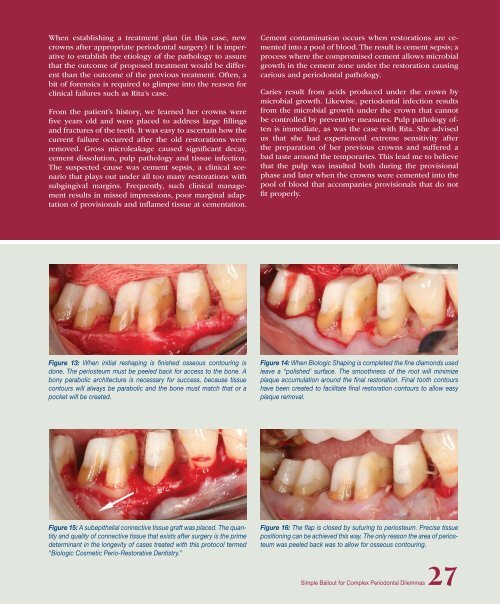
Figure 13: When initial reshaping is finished osseous contouring is<br />
done. The periosteum must be peeled back for access to the bone. A<br />
bony parabolic architecture is necessary for success, because tissue<br />
contours will always be parabolic and the bone must match that or a<br />
pocket will be created.<br />
Figure 14: When Biologic Shaping is completed the fine diamonds used<br />
leave a “polished’ surface. The smoothness of the root will minimize<br />
plaque accumulation around the final restoration. Final tooth contours<br />
have been created to facilitate final restoration contours to allow easy<br />
plaque removal.<br />
Figure 15: A subepithelial connective tissue graft was placed. The quantity<br />
and quality of connective tissue that exists after surgery is the prime<br />
determinant in the longevity of cases treated with this protocol termed<br />
“Biologic Cosmetic Perio-Restorative Dentistry.”<br />
Figure 16: The flap is closed by suturing to periosteum. Precise tissue<br />
positioning can be achieved this way. The only reason the area of periosteum<br />
was peeled back was to allow for osseous contouring.<br />
Simple Bailout for Complex Periodontal Dilemmas27


