

PDF Version - Glidewell Dental Labs
PDF Version - Glidewell Dental Labs
PDF Version - Glidewell Dental Labs

You also want an ePaper? Increase the reach of your titles
YUMPU automatically turns print PDFs into web optimized ePapers that Google loves.
Currently, the gingival<br />
complex is a vital<br />
aspect of any restorative<br />
treatment plan.<br />
Clinicians now are<br />
beginning to recognize<br />
the importance of the<br />
gingival complex in<br />
the treatment of short<br />
clinical crowns.<br />
tooth eruption to a level apical to the cervical convexity of the clinical crown.<br />
By contrast, passive eruption is a biologic process whereby tooth eruption occurs<br />
normally. During this normal tooth eruption the dentogingival junction<br />
shifts apically. 6 This process occurs when active eruption is complete and may<br />
continue until the early or mid-20s of adulthood. 7 At this time, the free gingival<br />
margin approximates the CEJ. Gottlieb and Orban classified passive eruption<br />
into four stages, believing this was a continuous physiologic process of tooth<br />
eruption (Figure 1). Although some debate currently exists when passive eruption<br />
becomes pathologic, it is generally accepted that cementum exposure or<br />
gingival recession (Stage 4) is a pathologic process.<br />
APE is one of the most commonly overlooked causes of short clinical crowns.<br />
Although literature provides limited information regarding the incidence of<br />
APE, Volchansky and Cleaton-Jones 7 found that 12 percent of patients studied<br />
had signs of APE. 8 Excessive gingival display has been estimated at 7 percent<br />
of men and 14 percent of women. 2 Thus any clinician must be cognizant of the<br />
dentogingival complex and be comfortable with the differential diagnoses of<br />
a gummy smile when striving for long-term optimal restorative esthetics and<br />
gingival health.<br />
Gargioulo and Ainomo 9,10 described the typical dentogingival relationship with<br />
the free gingival margin being located in close proximity to the CEJ. However,<br />
if APE exists, the gingival complex is situated in a more coronal position, making<br />
the CEJ difficult to detect clinically and thus displaying the pathognomonic<br />
signs of short clinical crowns and excessive gingival display.<br />
Figure 3A: Initial presentation: Teeth<br />
6-11 appear short and boxy.<br />
Coslet and others 11 classified APE into two case types, based on the gingival<br />
and osseous relationships. Type 1 presents with a noticeably wider band of<br />
keratinized tissue and Type 2 exhibits a smaller band of keratinized tissue falling<br />
within normal limits (Figure 2). Types 1 and 2 each have subcategories, A<br />
and B. In the A subgroup, the osseous crest is located 1.5 mm to 2 mm below<br />
the CEJ 9 (normal), while in the B subgroup, the osseous crest is found directly<br />
adjacent the CEJ.<br />
This article presents treatment for two common types of APE found clinically.<br />
To date, no scientific literature has investigated the incidence of Coslet’s four<br />
classifications of APE. However, it is believed that Type 1B is more prevalent. 1<br />
This article presents and discusses the more common case types and the treatments<br />
employed to achieve long-term esthetic results.<br />
CASE REPoRTS<br />
– Coslet Type 1A –<br />
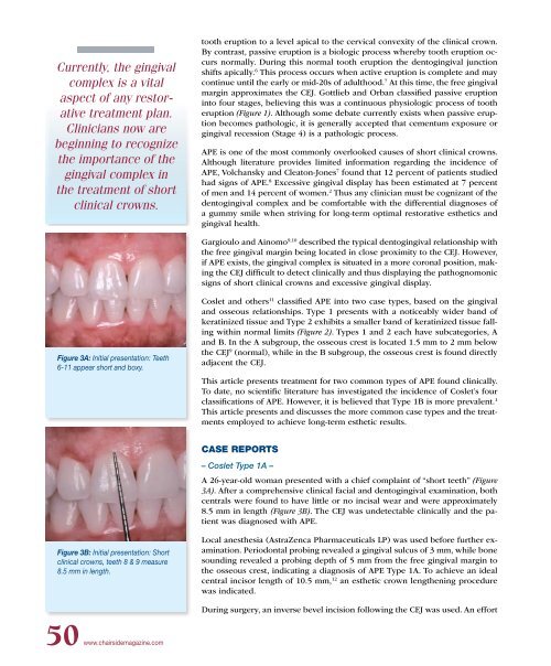
A 26-year-old woman presented with a chief complaint of “short teeth” (Figure<br />
3A). After a comprehensive clinical facial and dentogingival examination, both<br />
centrals were found to have little or no incisal wear and were approximately<br />
8.5 mm in length (Figure 3B). The CEJ was undetectable clinically and the patient<br />
was diagnosed with APE.<br />
Figure 3B: Initial presentation: Short<br />
clinical crowns, teeth 8 & 9 measure<br />
8.5 mm in length.<br />
Local anesthesia (AstraZenca Pharmaceuticals LP) was used before further examination.<br />
Periodontal probing revealed a gingival sulcus of 3 mm, while bone<br />
sounding revealed a probing depth of 5 mm from the free gingival margin to<br />
the osseous crest, indicating a diagnosis of APE Type 1A. To achieve an ideal<br />
central incisor length of 10.5 mm, 12 an esthetic crown lengthening procedure<br />
was indicated.<br />
During surgery, an inverse bevel incision following the CEJ was used. An effort<br />
50 www.chairsidemagazine.com


